what is your diagnosis - marshfield labs of first stage larvae are fecal examination by the baermann...
TRANSCRIPT

RFPT081201
What is Your Diagnosis?Anne L. KincAid, dVM, MArshfieLd LAbs, MArshfieLd WiheLene KeiLy, dVM, bArron VeterinAry cLinic, bArron Wi An approximately 6-year-old spayed female Dachshund mixed breed canine had a history of persistent cough since January 2012. The dog was treated twice with doxycycline and had initial slight improvement. CBC and chemistry profile were within normal limits. Radiographs revealed a generalized bronchial pattern. The dog was treated with Clavamox, prednisone and hydrocodone pulse therapy and did very well with resolution of the cough. The owner did not continue the pulse therapy with hydrocodone and two weeks after finishing the prednisone the owner reported the cough had returned. Clavamox and diphenhydramine were started. Five days later, the pet represented for coughing. The cough was moist and productive of mucus with progressive worsening especially at night. The respiratory rate was increased and increased bronchial tones were noted on auscultation of the chest. Radiographs had a diffuse bronchial pattern with an increase in peribronchial cuffing. A transtracheal wash revealed mild catarrhal eosinophilic and histiocytic inflammation with no etiologic agents. A Baermann fecal examination was performed and revealed nematode larvae in a C-shape averaging 275 um in length and 16-22 um in width. The larvae had an indistinct poorly defined nonrhabditiform esophagus and a slightly deflected tail (photos below). Answer on page 5.
s u M M E r 2 0 1 2
(Continued on page 5)
BEYOND numbers
What is Your Diagnosis? .....1
rEptilE hEMatologY ...............2
What is Your Diagnosis - ansWEr ....................................5
Inside This Issue
EsophagusDeflected Tail

2
rEptilE hEMatologYAnne L. KincAid, dVM Environmental conditions have a greater influence on the normal physiology and health of reptiles as compared with endothermic animals. Seasonal and gender factors affect reptilian hematology. These factors have to be taken into consideration when interpreting clinical data. Published reference ranges do not take into account environmental conditions and physiologic parameters.
If possible, a set of normal values should be obtained from patients housed under a given set of environmental and nutritional parameters for comparison if the patient later becomes ill. This blood should be obtained during the initial wellness exam.
Plastic tubes are recommended for collection and lithium heparin is the anticoagulant of choice. EDTA may cause lysis of reptile red cells, especially in turtles. Heparinized blood may be used for hematology and chemistry, but will give a bluish tinge to blood smears. Because of environmental conditions, age, gender and sampling techniques, total and differential leukocyte counts must have a two fold or greater increase or decrease over normal reference values to be considered clinically significant.
Reptile Hematology - Fast Facts
(Continued on page 3)
• May represent up to 80% of differential in some species
• Influenced by environmental and physiologic factors
• Lowest in winter• Highest in summer• Temperate reptiles-decreased or absent
during hibernation• Tropical reptiles-some decrease in winter• More lymphocytes in females• Lymphocytosis:
– Wound healing– Inflammatory disease– Parasitic infections– Viral infections– Ecdysis
• Lymphopenia-Malnutrition:– Secondary to diseases caused by stress– Immunosuppression
Monocytes:• 0-10% of differential• Azurophils in snakes:
– Function as bands– Increase as part of inflammatory
response• No seasonal variation• Monocytosis:
– Inflammatory disease, especially granulomatous disease
Platelets:• Normal range is 25-350/100 WBC• Polymorphic nuclei associated with
severe inflammatory disease
Heterophils:• % varies with species• Highest in summer• Lowest in hibernation• Phagocytes, no peroxidase activity• Lobation variation-increased lobation
suggestive of severe inflammation• Heterophilia:
– Inflammatory disease, especially microbial or parasitic infections– Tissue injury– Stress– Neoplasia including heterophilic
leukemia
Eosinophils:• % varies with species• Low in lizards; up to 20% in some turtles• Decreased in summer• Highest in hibernation in some species• Eosinophilia:
– Parasitic infections– Immune stimulation
Basophils:• Wide normal range: 0-40%• Higher range usually seen in turtles• Minimal seasonal variation• Basophilia:
– Parasitic infections – Viral infections
Lymphocytes:• Variable normal range
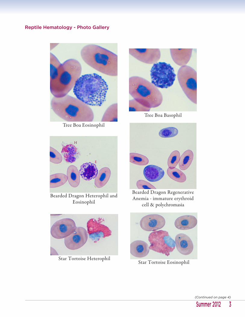
Reptile Hematology - Photo Gallery
3Summer 2012(Continued on page 4)
Tree Boa Eosinophil
Tree Boa Basophil
Bearded Dragon Heterophil and Eosinophil
Star Tortoise HeterophilStar Tortoise Eosinophil
Bearded Dragon Regenerative Anemia - immature erythroid
cell & polychromasia
E
H

4(Continued on page 5)
Ground Boa Azurophil
Tortoise Macrophage with Phagocytized Granules (not all
granulated cells are granulocytes)
Iguana Eosinophil vs. Degranulated Heterophil vs. Heterophil
Skink Basophil, Eosinophil and Heterophil
Iguana Lymphocytes vs. Platelets
L
L
P P P
H
E
DH
B
E
H

References:Mader DR. Reptile Medicine and Surgery, 2nd Ed. St. Louis: Elsevier, 2006: 453-460.
Campbell TW, Ellis CK. Avian and Exotic Animal Hematology and Cytology, 3rd Ed. Ames: Blackwell, 2007: 51-81
What is Your Diagnosis - ansWErAnne L. KincAid, dVM
Diagnosis: Crenosoma vulpis infection
Crenosoma vulpis is a metastrongylid nematode lungworm that is highly prevalent in the red fox population of Atlantic Canada and infects the bronchioles, bronchi and trachea of wild and domestic canids, as well as, various other carnivores including raccoons and skunks. Clinical signs in dogs consist predominantly of chronic cough, resembling allergic respiratory disease. The parasite is endemic in the northeastern region of North America (New York, Nova Scotia, New Brunswick, Newfoundland and Prince Edward Island). The red fox is the natural definitive host. Dogs acquire the infection by ingestion of gastropods (slugs and terrestrial snails) that serve as intermediate hosts for the parasite. The lifespan of C. vulpis is approximately 8-10 months with a prepatent period of 3 weeks. The most reliable methods for detection of first stage larvae are fecal examination by the Baermann technique (most reliable) or by zinc sulfate centrifugal fecal flotation (floatation is more reliable for lungworms Oslerus and Filaroides). This parasite has not been previously reported in Wisconsin, to our knowledge. Additional history for this dog indicated that the owner lived on a lake in western Wisconsin. The dog enjoyed riding on the owner’s boat and ate snails from the boat. Ingestion of terrestrial snails was not noted, but presumed to occur since the dog ate aquatic snails. The possibility that the larvae were an incidental finding from ingestion of raccoon or skunk feces was considered, but the presence of the eosinophilic inflammation in the transtracheal wash and the history of chronic cough support the diagnosis of true infection. Fenbendazole therapy was instituted at the approved dosage, but due to worsening coughing after the first dose, was concurrently treated with prednisone and the fenbendazole was given every second day for a total of three treatments. Severe coughing was noted after the second dose of fenbendazole but, the dog then continuously improved and the prednisone was tapered.
Infection may be treated by fenbendazole 50 mg/kg PO every 24 hours for 3 days; milbemycin oxime 0.5 mg/kg as a single dose PO or by a single dose of topical Advantage Multi (Bayer) at the approved dose. Per Dr. Gary Conboy, associate professor in the Dept. of Pathology and Microbiology at Atlantic Veterinary College, University of Prince Edward Island (PEI), milbemycin does not have any affect on immature stages of the parasite. Since the prepatent period is 21 days, monthly use of milbemycin won’t prevent patent infection. However, it will severely limit the length of any infection that might occur. The same may be true for Advantage Multi. Dr. Conboy also indicated that there is a species of Crenosoma in skunks, thought to be in the US that produces more severe signs of disease than he sees in PEI.
References:
Zajac AM, Conboy GA. Veterinary Clinical Parasitology, 8th Ed. Hoboken: Wiley-Blackwell, 2012: 26-27, 68-69.
Bihr T, Conboy GA. Lungworm (Crenosoma vulpis) infection in dogs on Prince Edward Island. Can Vet J 1999; 40: 555-559.
5Summer 2012(Continued on page 6)

6
Baermann Sedimentation (VBSED)
Specimen: At least 30 grams unpreserved fresh stool in clean, dry container.
Storage: Refrigerate
Available: Results available in 1-2 days
Fecal Floatation and Sediment (VFFR)
Specimen: 3-5 grams (1 cubic cm or cube approximately 1/2” per side) fresh stool in a sterile container.
Minimum: 1 gram
Cause for Rejection: Specimens submitted in formalin.
Storage: Room temperature. Refrigerate if kept for more than 6 hours.
Available: Set up daily with results available in 1-2 days.
Method: Fecal Floatation by Centrifugation and Sediment Examination
Oslerus/Filaroides Larva with Kinked Tail
Nematode Larva with Rhabditiform Esophagus
Crenosoma vulpis Larva
Crenosoma vulpis Larva
Tail
Esophagus
Deflected Tail
Esophagus
Isthmus
Bulb
Kinked Tail