what is evidence-based about myofascial chains? a ... · 56 myofascial meridians connecting distant...
TRANSCRIPT

Accepted Manuscript
What is evidence-based about myofascial chains? A systematic review
Jan Wilke, M.A., Frieder Krause, M.A., Lutz Vogt, PhD, Winfried Banzer, PhD
PII: S0003-9993(15)01064-3
DOI: 10.1016/j.apmr.2015.07.023
Reference: YAPMR 56278
To appear in: ARCHIVES OF PHYSICAL MEDICINE AND REHABILITATION
Received Date: 22 July 2015
Revised Date: 28 July 2015
Accepted Date: 29 July 2015
Please cite this article as: Wilke J, Krause F, Vogt L, Banzer W, What is evidence-based aboutmyofascial chains? A systematic review, ARCHIVES OF PHYSICAL MEDICINE AND REHABILITATION(2015), doi: 10.1016/j.apmr.2015.07.023.
This is a PDF file of an unedited manuscript that has been accepted for publication. As a service toour customers we are providing this early version of the manuscript. The manuscript will undergocopyediting, typesetting, and review of the resulting proof before it is published in its final form. Pleasenote that during the production process errors may be discovered which could affect the content, and alllegal disclaimers that apply to the journal pertain.
MANUSCRIP
T
ACCEPTED
ACCEPTED MANUSCRIPT1
Running Head: Evidence for Myofascial chains
What is evidence-based about myofascial
chains? A systematic review
Jan Wilke (M.A.), Frieder Krause (M.A.), Lutz Vogt (PhD), Winfried Banzer
(PhD)
Goethe University Frankfurt/Main, Department of Sports Medicine, Frankfurt
am Main, Germany
No conflict of interest and no external funding reported.
Corresponding author:
Jan Wilke
Department of Sports Medicine
Goethe University Frankfurt am Main Ginnheimer Landstraße 39 | 60487 Frankfurt am Main (Germany) Phone+49 (69) 798 24588 | Fax +49 (69) 798 24592 Email: [email protected]
MANUSCRIP
T
ACCEPTED
ACCEPTED MANUSCRIPT1
What is evidence-based about myofascial 1
chains? A systematic review 2
3
Objective: To provide evidence for the existence of six myofascial meridians 4
proposed by Myers (1997) based on anatomical dissection studies. 5
Data sources: Relevant articles published between 1900 and December 2014 were 6
searched in MEDLINE (Pubmed), ScienceDirect and Google Scholar. 7
Study selection: Peer-reviewed human anatomical dissection studies reporting 8
morphological continuity between the muscular constituents of the examined 9
meridians were included. If no study demonstrating a structural connection between 10
two muscles was found, papers on general anatomy of the corresponding body 11
region were targeted. 12
Data extraction: A continuity between two muscles was only documented if two 13
independent investigators agreed that it was reported clearly. Also, two independent 14
investigators rated methodological quality of included studies by means of a validated 15
assessment tool (QUACS). 16
Data synthesis: The literature search identified 6589 articles. Of these, 62 papers 17
met the inclusion criteria. The studies reviewed suggest strong evidence for the 18
existence of three myofascial meridians: the superficial back line (all three transitions 19
verified, based on 14 studies), the back functional line (all three transitions verified, 8 20
studies) and the front functional line (both transitions verified, 6 studies). Moderate to 21
strong evidence is available for parts of the spiral line (five of nine verified transitions, 22
21 studies) and the lateral line (two of five verified transitions, 10 studies). No 23
evidence exists for the superficial front line (no verified transition, 7 studies). 24

MANUSCRIP
T
ACCEPTED
ACCEPTED MANUSCRIPT2
Conclusions: The present systematic review suggests that most skeletal muscles of 25
the human body are directly linked by connective tissue. Examining the functional 26
relevance of these myofascial chains is the most urgent task of future research. 27
Strain transmission along meridians would both open a new frontier for the 28
understanding of referred pain and provide a rationale for the development of more 29
holistic treatment approaches. 30
31
Keywords: continuity, anatomy trains, meridians, fascia 32
33
Abbreviations: 34
QUACS: QUality Appraisal for Cadaveric Studies 35
PRISMA: Preferred Reporting Items for Systematic Reviews and Meta-Analyses 36
SBL: superficial back line 37
SFL: superficial front line 38
LL: lateral line 39
SL: spiral line 40
BFL: back functional line 41
FFL: front functional line 42
43
44

MANUSCRIP
T
ACCEPTED
ACCEPTED MANUSCRIPT3
Treatments of fascial tissues have become increasingly popular in musculoskeletal 45
disorders.1-5 This might be owing to recent histological findings. The discovery of 46
contractile cells, free nerve endings and mechanoreceptors suggests that fascia in 47
contrast to prior assumptions plays a proprioceptive and mechanically active role.6-13 48
Numerous therapists whoaddress fascia orientate themselves to concepts of 49
myofascial chains. Such approaches originate from the assumption that the muscles 50
of the human body do not function as independent units. Instead, they are regarded 51
as part of a tensegrity-like, bodywide network with fascial structures acting as linking 52
components. As fascia can transmit tension14,15 and in view of its proprioceptive and 53
nociceptive functions, existence of myofascial meridians could be responsive for 54
disorders and pain radiating to remote anatomical structures. Myers16 defined eleven 55
myofascial meridians connecting distant parts of the body by means of muscles and 56
fascial tissues (Fig. 1). The central rule for the selection of a meridian’s components 57
is a direct linear connection between two muscles. For instance, a part of the 58
superficial back line (Tab.1) is suggested to be formed by M. biceps femoris and the 59
erector spinae muscle both being linked by means of the sacrotuberous ligament and 60
the lumbar fascia. Even if the biceps femoris also displayed a structural continuity to 61
the gluteus maximus, this connection would not be considered part of the meridian 62
due to its curved, non-linear course. Though used and referred to in several 63
studies,17-22 the myofascial meridians are based on anecdotal evidence from practice 64
and have never been verified. Confirming body-wide direct morphological continuity 65
between muscle and fascial tissue would thus yield both an empirical background for 66
such trials and an argument for practitioners to take treatment of complete meridians 67
into consideration. This paper aimed to provide evidence for six of the myofascial 68
meridians based on anatomical dissection studies. 69
70
MANUSCRIP
T
ACCEPTED
ACCEPTED MANUSCRIPT4
Methods 71
72
73
The present systematic review adhered to the PRISMA (Preferred Reporting Items 74
for Systematic Reviews and Meta-Analyses) guidelines.23 Two independent 75
investigators performed a systematic literature research. The targeted myofascial 76
meridians were the superficial back line (SBL), superficial front line (SFL), lateral line 77
(LL), spiral line (SL), back functional line (BFL) and front functional line (FFL; Tab. 1, 78
Fig.1). For each transition of these meridians, peer reviewed anatomical dissection 79
studies (published 1900-2014) reporting myofascial continuity between the involved 80
muscles were searched. Animal studies and case reports were excluded; the same 81
applied to articles in languages other than German and English. 82
83
Relevant publications were identified using MEDLINE (Pubmed; searched with MeSH 84
terms), ScienceDirect and Google Scholar. While search algorithms for Pubmed and 85
ScienceDirect were of more general character (algorithms see Tab.2), a free-text 86
search including concrete names of the supposedly connected muscles was 87
performed in Google Scholar. Here, the first 100 hits for each transition of the 88
corresponding meridian were screened. If the initial literature research using the 89
above described procedure with three databases did not yield any results for a 90
specific transition, dissection studies describing the general anatomy of the 91
corresponding body region were targeted and screened for the myofascial 92
connection. Additionally, the reference lists of all detected studies were checked. 93
Data extraction was carried out by two independent investigators and a continuity 94
was only documented if both agreed that it was reported clearly. 95
96
MANUSCRIP
T
ACCEPTED
ACCEPTED MANUSCRIPT5
The methodological quality of the enclosed studies was evaluated by means of the 97
QUACS scale (QUality Appraisal for Cadaveric Studies) whose reliability and validity 98
have been demonstrated recently.24 The scale encompasses a checklist of 13 99
dichotomous items, each scored with either zero (no/not stated) or one (yes/clearly 100
present) point. The quality score is calculated in percent. Zero to 20 percent indicate 101
poor, 21 to 40 percent fair, 41 to 60 percent moderate, 61-80 percent substantial, and 102
81 to 100 percent excellent methodological quality. Ratings were done by two 103
independent researchers. In case of disagreement, a third reviewer casted the 104
decisive vote. For each meridians respectively its transitions, evidence was classified 105
as strong (consistent findings among multiple high quality studies), moderate 106
(consistent findings among multiple low quality studies and/or one high quality study), 107
limited (one low quality study), conflicting (inconsistent findings among multiple 108
studies), or not existent (no studies available) according to the recommendations of 109
the Cochrane Collaboration Back Review Group.25 110
111
112
Results 113
114
115
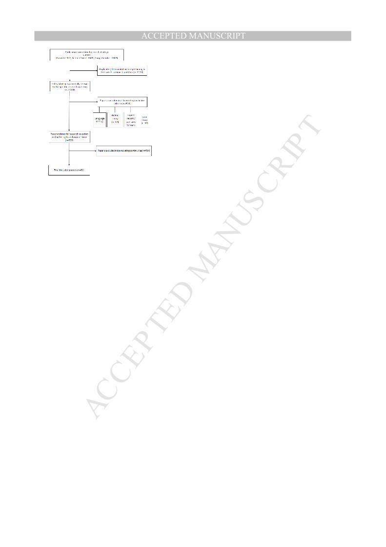
The initial literature research yielded 6584 publications. After removing duplicates 116
and papers not pertaining to the research question exclusion criteria were applied. 117
The resulting sample comprised 62 studies (see Fig. 2). The findings for each 118
meridian including detailed information about characteristics of the appendant 119
transitions are displayed in Tab.3, methodological quality of the included studies is 120
shown in Tab.4. 121
122

MANUSCRIP
T
ACCEPTED
ACCEPTED MANUSCRIPT6
Strong evidence exists for all transitions of the SBL (based on 14 studies), BFL (8 123
studies) and FFL (6 studies). With regard to the SL (moderate evidence for five of 124
nine transitions, 21 studies) and the LL (moderate to strong evidence for two of five 125
transitions, 10 studies) findings were ambivalent. No evidence from anatomical 126
dissection studies is available for the SFL (no verified transition, 7 studies). 127
128
129
Discussion 130
131
132
The present systematic review provides relevant information for movement therapists 133
and strength and conditioning coaches. It demonstrates that fascia, besides recently 134
discovered features such as pain perception and stiffness regulation connects the 135
skeletal muscles forming a body-wide web of myofascial chains. Extensive structural 136
continuity was clearly verified for three of the six examined meridians (SBL, BFL, 137
FFL). In addition, findings were at least ambivalent with respect to the LL and SL. 138
The fact that we could only confirm half of both lines’ transitions does not neglect 139
their existence. Most of the reviewed studies did not specifically search for 140
continuities mentioning them only as a subordinate finding. Since clinicians and 141
anatomists show increasing interest in fascia, it is well possible that future, more 142
focused research will verify the remaining myofascial links.88 In contrast to the solid 143
evidence for these five meridians, doubts have to be raised about the existence of 144
the SFL. There is no structural connection between the rectus femoris muscle and M. 145
rectus abdominis. Also, M. sternalis, which is suggested to be the cranial continuation 146
of rectus abdominis, exists only in a small percentage of the population.41-44 Even if 147
present, it does not fuse consistently with the rectus abdominis.41 148
MANUSCRIP
T
ACCEPTED
ACCEPTED MANUSCRIPT7
149
The practical relevance of the present research is twofold. First, the existence of 150
myofascial meridians might help to explain the phenomenon of referred pain which 151
often occurs in nonspecific disorders. For example, myofascial trigger points of the 152
calf have been shown to elicit pain that radiates to the sole of the foot and to the 153
dorsal thigh.89 As this projection pattern corresponds to the course of the SBL, it 154
might represent the morphological substrate. A second aspect relates to therapy and 155
training of the musculoskeletal system. Direct morphological continuity between 156
adjacent muscles provides the empirical background to extend diagnostic and 157
therapeutic focus beyond one single anatomical structure. Treatment according to 158
myofascial meridians could be effective in reducing low back pain. Several studies 159
have shown that low back pain patients display reduced hamstring flexibility.90-93 Due 160
to the direct morphological relationship of the hamstrings and the low back region 161
(both are part of the SBL), relieving tension of the posterior thigh muscles could be a 162
conceivable approach to alleviate pain. Overload injuries in competitive sports 163
represent another entity of pathologies which possibly occur due to the presence of 164
myofascial meridians. Recent studies indicate that tightness of the gastrocnemius 165
and the hamstrings are associated with plantar fasciitis.94-96 As both muscles and the 166
plantar aponeurosis belong to the SBL, they might represent a target of exercise 167
therapy. Finally, groin pain or athletic pubalgia is suggested to be provoked by a tight 168
adductor longus and a weak rectus abdominis97,98 which according to our results are 169
directly connected in the FFL. The tightening of the adductor may thus develop due 170
to communication with the rectus via the described linkage. 171
Study Limitations 172
Although our review yields solid evidence for the existence of myofascial chains, 173
several aspects call for further study. Future research should be dedicated to the 174

MANUSCRIP
T
ACCEPTED
ACCEPTED MANUSCRIPT8
presence of the meridians which could not be evidenced entirely in this work. Another 175
issue relates to the function of regional specializations which so far remains unclear. 176
Depending on its localization, fascia in general exhibits substantial differences 177
concerning thickness, amount of elastic fibers,11 and adherence to the underlying 178
muscle.8 Also, the number of connecting fibers is not uniform and shows 179
considerable variation for different transistions.27 This holds particular significance as 180
the structures linking the muscular stations of the meridians encompass tendinous, 181
aponeurotic and ligamentous tissue as well as the deep fascia. Finally, it is of utmost 182
importance to elucidate the functional significance of the myofascial chains as the 183
capability for strain transfer represents the decisive criterion to justify treatment of 184
meridians. Though the available evidence points towards existence of tensile 185
transmission via myofascial pathways, most experimental research was carried out in 186
vitro using cadavers.14,15,36 Randomized, controlled in-vivo studies are warranted in 187
order to draw more precise assumptions on the significance of myofasical chains for 188
the movement system. 189
190
191
Conclusions 192
193
194
Although the concept of myofascial meridians is widely used in exercise therapy and 195
osteopathic medicine, the scientific basis for the proposed connections is still a 196
matter of debate. The present review provides first systematical evidence based on 197
cadaveric dissection studies. While there is strong evidence for the existence of the 198
SBL, BFL and FFL, it is ambivalent with regard to the SL and LL respectively poor for 199
the SFL. Within its borders, the system of myofascial meridians represents a 200
MANUSCRIP
T
ACCEPTED
ACCEPTED MANUSCRIPT9
promising approach to transfer tensegrity principles into practice. Therapists may use 201
the myofascial chains as a conclusive orientation but should be aware that the 202
functional implications remain to be studied. 203
204
205
References 206
207
208
1 Engel RM, Brown BT, Swain MS, Lystad RP. The provision of chiropractic, 209
physiotherapy and osteopathic services within the Australian private health-care 210
system: a report of recent trends. Chiropr Man Therap 2014; 22: 3. 211
2. las Peñas CF de, Sohrbeck Campo M, Fernández Carnero J, Miangolarra Page 212
JC. Manual therapies in myofascial trigger point treatment: a systematic review. J 213
Body Mov Ther 2005; 9: 27-34. 214
3. Ndetan H, Evans MW, Hawk C, Walker C. Chiropractic or Osteopathic 215
Manipulation for Children in the United States: An Analysis of Data from the 2007 216
National Health Interview Survey. J Alt Complement Med 2012; 18: 347-353. 217
4. Ong C, Doll H, Bodeker G, Stewart-Brown S. Use of osteopathic or chiropractic 218
services among people with back pain: a UK population survey. Health Soc Care 219
Community 2004; 12: 265-273. 220
5. Wardle JL, Sibbritt DW, Adams J. Referrals to chiropractors and osteopaths: a 221
survey of general practitioners in rural and regional New South Wales, Australia. 222
Chiropr Man Therap 2013; 21: 5. 223
6. Bhattacharya V, Barooah P, Nag T, Chaudhuri G, Bhattacharya S. Detailed 224
microscopic analysis of deep fascia of lower limb and its surgical implication. 225
Indian J Plast Surg 2010; 43:135. 226

MANUSCRIP
T
ACCEPTED
ACCEPTED MANUSCRIPT10
7. Staubesand J, LiY. Zum Feinbau der Fascia cruris mit besonderer 227
Berücksichtigung epi- und intrafaszialer Nerven. Manuelle Medizin 1996; 34: 196-228
200. 229
8. Stecco A, Macchi V, Masiero S, et al. Pectoral and femoral fasciae: common 230
aspects and regional specializations. Surg Radiol Anat 2009; 31: 35-42. 231
9. Stecco C, Corradin M, Macchi V, et al. Plantar fascia anatomy and its relationship 232
with Achilles tendon and paratenon. J Anat 2013; 223: 665-676. 233
10. Stecco C, Gagey O, Belloni A, et al. Anatomy of the deep fascia of the upper limb. 234
Second part: study of innervation. Morphologie 2007; 91: 38-43. 235
11. Stecco C, Porzionato A, Lancerotto L, et al. Histological study of the deep fasciae 236
of the limbs. J Bodyw Mov Ther 2008; 12: 225-230. 237
12. Tesarz J, Hoheisel U, Wiedenhofer B, Mense S. Sensory innervation of the 238
thoracolumbar fascia in rats and humans. Neuroscience 2011; 194:302-308. 239
13. Yahia L, Rhalmi S, Newman N, Isler M. Sensory innervation of human 240
thoracolumbar fascia. An immunohistochemical study. Acta Orthop Scand 1992; 241
63: 195-197. 242
14. Barker PJ, Briggs CA, Bogeski G. Tensile transmission across the lumbar fasciae 243
in unembalmed cadavers: effects of tension to various muscular attachments. 244
Spine. 2004; 29: 129-138. 245
15. Norton-Old KJ, Schache AG, Barker PJ, Clark RA, Harrison SM, Briggs CA. 246
Anatomical and mechanical relationship between the proximal attachment of 247
adductor longus and the distal rectus sheath. Clin Anat 2013; 26: 522-530. 248
16. Myers TW. Anatomy trains: Myofascial meridians for manual and movement 249
therapists. 3rd ed. New York: Churchill Livingstone; 2014. 250
MANUSCRIP
T
ACCEPTED
ACCEPTED MANUSCRIPT11
17. Ajimsha M, Daniel B, Chithra S. Effectiveness of Myofascial release in the 251
management of chronic low back pain in nursing professionals. J Bodyw Mov 252
Ther 2014; 18: 273-281. 253
18. Behnam A, Mahyar S, Ezzati K, Rad SM. The Use of Dry Needling and Myofascial 254
Meridians in a Case of Plantar Fasciitis. J Chir Med 2014. 255
19. Hyouk HI, Kang JH. The Immediate Effects of Passive Hamstring Stretching 256
Exercises on the Cervical Spine Range of Motion and Balance. J Phys Ther Sci 257
2013; 25: 113-116. 258
20. Hyouk HI, Hyun KJ. The Effect of Forward Head on Ankle Joint Range of Motion 259
and Static Balance. J Phys Ther Sci. 2012;24(9):925-927. 260
21. Lumbroso D, Ziv E, Vered E, Kalichman L. The effect of kinesio tape application 261
on hamstring and gastrocnemius muscles in healthy young adults. J Bodyw Mov 262
Ther 2014; 18: 130-138. 263
22. Weisman MH, Haddad M, Lavi N, Vulfsons S. Surface electromyographic 264
recordings after passive and active motion along the posterior myofascial 265
kinematic chain in healthy male subjects. J Bodyw Mov Ther 2013; 18: 452- 461. 266
23. Liberati A. The PRISMA Statement for Reporting Systematic Reviews and Meta-267
Analyses of Studies That Evaluate Health Care Interventions: Explanation and 268
Elaboration. Ann Intern Med 2009; 151: W. 269
24. Wilke J, Krause F, Niederer D, Engeroff T, Vogt L, Banzer W. Appraising the 270
quality of cadaveric studies. Validation of the QUACS scale. J Anat. 2015; 271
226:440-446. 272
25. van Tulder M, Furlan A, Bombardier, C, Bouter, L. Updated method guidelines for 273
systematic reviews in the Cochrane collaboration back review group. Spine 2003; 274
28:1290-1299. 275
MANUSCRIP
T
ACCEPTED
ACCEPTED MANUSCRIPT12
26. Kamel R, Sakla FB. Anatomical compartments of the sole of the human foot. Anat 276
Rec 1961; 140: 57-60. 277
27. Snow SW, Bohne WH, DiCarlo E, Chang VK. Anatomy of the Achilles tendon and 278
plantar fascia in relation to the calcaneus in various age groups. Foot Ankle Int 279
1995; 16: 418-421. 280
28. Kim PJ, Richey J, Wissman LR, Steinberg JS. The Variability of the Achilles 281
Tendon Insertion: A Cadaveric Examination. J Foot Ankle Surg 2010; 49: 417-282
420. 283
29. Candal-Couto JJ, Deehan DJ. The accessory bands of Gracilis and 284
Semitendinosus: an anatomical study. Knee 2003; 10: 325-328. 285
30. Kolodychuk LB. Clinically significant anatomy of gracilis and semitendinosus 286
tendon insertions. Clinical J Sport Med 1992 ;(2). 287
31. Reina N, Abbo O, Gomez-Brouchet A, Chiron P, Moscovici J, Laffosse J. 288
Anatomy of the bands of the hamstring tendon: How can we improve harvest 289
quality? The Knee 2013; 20: 90-95. 290
32. Tuncay I, Kucuker H, Uzun I, Karalezli N. The fascial band from semitendinosus 291
to gastrocnemius: the critical point of hamstring harvesting: an anatomical study 292
of 23 cadavers. Acta Orthop 2007; 78: 361-363. 293
33. Mochizuki T, Akita K, Muneta T, Sato T. Pes anserinus: Layered supportive 294
structure on the medial side of the knee. Clin Anat 2004; 17: 50-54. 295
34. Martin BF. The origins of the hamstring muscles. J Anat 1968; 102: 345-352. 296
35. Sato K, Nimura A, Yamaguchi K, Akita K. Anatomical study of the proximal origin 297
of hamstring muscles. J Orthop Sci. 2012; 17: 614-618. 298
36. van Wingerden JP, Vleeming A, Snijders CJ, Stoeckart R. A functional-299
anatomical approach to the spine-pelvis mechanism: interaction between the 300

MANUSCRIP
T
ACCEPTED
ACCEPTED MANUSCRIPT13
biceps femoris muscle and the sacrotuberous ligament. Eur Spine J 1993; 2: 140-301
144. 302
37. Woodley SJ, Mercer SR. Hamstring muscles: architecture and innervation. Cells 303
Tissues Organs 2005; 179: 125-141. 304
38. Vleeming A, Stoeckart R, Snijders C. The sacrotuberous ligament: a conceptual 305
approach to its dynamic role in stabilizing the sacroiliac joint. Clin Biomech 306
1989;4: 201-203. 307
39. Andrikoula S, Tokis A, Vasiliadis HS, Georgoulis A. The extensor mechanism of 308
the knee joint: an anatomical study. Knee Surg Sports Traumatol Arthrosc 2006; 309
14:214-220. 310
40. Waligora AC, Johanson NA, Hirsch BE. Clinical anatomy of the quadriceps 311
femoris and extensor apparatus of the knee. Clin Orthop Relat Res 2009; 467: 312
3297-3306. 313
41. Barlow RN. The sternalis muscle in American whites and negroes. Anat Rec 314
1935; 61: 413-426. 315
42. Jelev L, Georgiev G, Surchev L. The sternalis muscle in the Bulgarian population: 316
classification of sternales. J Anat 2001; 199: 359-363. 317
43. Motabagani MA, Sonalla A, Abdel-Meguid E, Bakheit MA. Morphological study of 318
the uncommon rectus sterni muscle in German cadavers. East Afr Med J 2004; 319
81: 130-133. 320
44. Saeed M, Murshid KR, Rufai AA, Elsayed SE, Sadiq MS. Sternalis. An anatomic 321
variant of chest wall musculature. Neurosciences 2002; 7: 248-255. 322
45. Silveira D, Sousa KM, Siqueira SL, et al. Sternalis muscle: an anatomic variation 323
of the anterior chest wall. J Morphol Sci 2012; 29: 76-78. 324
46. Bogduk N, Macintosh JE. The applied anatomy of the thoracolumbar fascia. Spine 325
1984; 9: 164-170. 326
MANUSCRIP
T
ACCEPTED
ACCEPTED MANUSCRIPT14
47. Macintosh JE, Bogduk N. The morphology of the lumbar erector spinae. Spine 327
1987; 12: 658-668. 328
48. Vleeming A, Pool-Goudzwaard AL, Stoeckart R, van Wingerden JP, Snijders CJ. 329
The posterior layer of the thoracolumbar fascia. Its function in load transfer from 330
spine to legs. Spine 1995; 20: 753-758. 331
49. Stecco, A, Gilliar W, Hill R, Fullerton B, Stecco C. The anatomical and functional 332
relation between gluteus maximus and fascia lata. J Bodyw Mov Ther 2013; 17: 333
512-517. 334
50. Barker P, Hapuarachchi K, Ross J, Sambaiew E, Ranger T, Briggs C. Anatomy 335
and biomechanics of gluteus maximus and the thoracolumbar fascia at the 336
sacroiliac joint. Clin Anat 2014; 27: 234-240. 337
51. Vleeming A, Pool-Goudzwaard AL, Hammudoghlu D, Stoeckart R, Snijders CJ, 338
Mens JM. The function of the long dorsal sacroiliac ligament: its implication for 339
understanding low back pain. Spine 1996; 21: 556-562. 340
52. Stern JT. Anatomical and functional specializations of the human gluteus 341
maximus. Am. J. Phys. Anthropol 1972; 36: 315-339. 342
53. Jinde L, Jianliang S, Xiaoping C, et al. Anatomy and clinical significance of 343
pectoral fascia. Plast Reconstr Surg 2006; 118: 1557-1560. 344
54. Stecco A, Macchi V, Stecco C, et al. Anatomical study of myofascial continuity in 345
the anterior region of the upper limb. J Bodyw Mov Ther. 2009; 13: 53-62. 346
55. Stecco A, Masiero S, Macchi V, Stecco C, Porzionato A, Caro R de. The pectoral 347
fascia: Anatomical and histological study. J Bodyw Mov Ther 2009; 13: 255- 261. 348
56. Davis JA, Stringer MD, Woodley SJ. New insights into the proximal tendons of 349
adductor longus, adductor brevis and gracilis. Br J Sports Med 2012; 46: 871-876. 350
MANUSCRIP
T
ACCEPTED
ACCEPTED MANUSCRIPT15
57. Robinson P, Salehi F, Grainger A, et al. Cadaveric and MRI study of the 351
musculotendinous contributions to the capsule of the symphysis pubis. Am J 352
Roentgenol 2007; 188: W440-5. 353
58. Johnson GM, Zhang M, Jones DG. The fine connective tissue architecture of the 354
human ligamentum nuchae. Spine 2000; 25: 5-9. 355
59. Mercer SR, Bogduk N. Clinical anatomy of ligamentum nuchae. Clin Anat 2003; 356
16: 484-493. 357
60. Bharihoke V, Gupta M. Muscular attachments along the medial border of the 358
scapula. Surg Radiol Anat 1986; 8:71-73. 359
61. Nasu H, Yamaguchi K, Nimura A, Akita K. An anatomic study of structure and 360
innervation of the serratus anterior muscle. Surg Radiol Anat 2012; 34: 921- 928. 361
62. Vu P, Guedon C, Gehanno P, Andreassian B. Anatomic basis of serratus anterior 362
muscle flap transposition. Surg Radiol Anat 1988; 10: 173-185. 363
63. Williams GR, JR, Shakil M, Klimkiewicz J, Iannotti JP. Anatomy of the 364
scapulothoracic articulation. Clin Orthop Relat Res 1999; 359: 237-246. 365
64. Cuadros CL, Driscoll CL, Rothkopf DM. The anatomy of the lower serratus 366
anterior muscle: a fresh cadaver study. Plast Reconstr Surg 1995; 95: 93-99. 367
65. Peiper C, Junge K, Prescher A, Stumpf M, Schumpelick V. Abdominal 368
musculature and the transversalis fascia: an anatomical viewpoint. Hernia 2004; 369
8: 376-380. 370
66. Anson BJ, McVay CB. The anatomy of the inguinal and hypogastric regions of the 371
abdominal wall. Anat Rec 1938; 70: 211-225. 372
67. Askar OM. Surgical anatomy of the aponeurotic expansions of the anterior 373
abdominal wall. Ann R Coll Surg Engl 1977; 59: 313-321. 374
68. McVay CB, Anson BJ. Composition of the rectus sheath. Anat Rec 1940; 77: 375
213-225. 376

MANUSCRIP
T
ACCEPTED
ACCEPTED MANUSCRIPT16
69. Mwachaka P. Variations in the pattern of formation of the abdominis rectus 377
muscle sheath among kenyans. Int J Morphol 2009; 27: 1025-1029. 378
70. Rizk NN. A new description of the anterior abdominal wall in man and mammals. J 379
Anat 1980; 131: 373-385. 380
71. Ramasastry SS, Tucker JB, Swartz WM, Hurwitz D. The internal oblique muscle 381
flap. An anatomic and clinical study. Plast Reconstr Surg 1984; 73:721-723. 382
72. Ramasastry SS, Granick MS, Futrell JW. Clinical anatomy of the internal oblique 383
muscle. J Reconstr Microsurg 1986; 2:117-122. 384
73. Vieira ELC, Vieira EA, da Silva RT, Berlfein PAdS, Abdalla RJ, Cohen M. An 385
anatomic study of the iliotibial tract. Arthroscopy 2007; 23: 269-274. 386
74. Terry GC, LaPrade RF. The Posterolateral Aspect of the Knee: Anatomy and 387
Surgical Approach. Am J Sports Med. 1996;24(6):732-739. 388
75. Terry GC, Hughston JC, Norwood LA. The anatomy of the iliotibial band and 389
iliotibial tract. Am J Sports Med. 1986;14:39-45. 390
76. Patil V, Frisch NC, Ebraheim NA. Anatomical Variations in the Insertion of the 391
Peroneus (Fibularis) Longus Tendon. Foot Ankle Int 2007; 28: 1179-1182. 392
77. Shyamsundar S, Wazir A, Allen PE. Variations in the insertion of peroneus longus 393
tendon-a cadaver study. Foot Ankle Surg 2012; 18: 293-295. 394
78. Brenner E. Insertion of the tendon of the tibialis anterior muscle in feet with and 395
without hallux valgus. Clin Anat 2002; 15: 217-223. 396
79. El Gharbawy R.M. Relations of the common peroneal nerve to the insertion of 397
biceps femoris and origin of peroneus longus. Alex J Med 2006; 42: 817-828. 398
80. El Gharbawy RM, Skandalakis LJ, Skandalakis JE. Protective mechanisms of the 399
common fibular nerve in and around the fibular tunnel: A new concept. Clin Anat 400
2009; 22: 738-746. 401
MANUSCRIP
T
ACCEPTED
ACCEPTED MANUSCRIPT17
81. Marshall JL, Girgis FG, Zelko RR. The biceps femoris tendon and its functional 402
significance. J Bone Joint Surg Am 1972; 54:14444-1450. 403
82. Al-Hayani A. The functional anatomy of hip abductors. Folia Morphol. (Warsz) 404
2009; 68: 98-103. 405
83. Falvey EC, Clark RA, Franklyn-Miller A, Bryant AL, Briggs C, McCrory PR. 406
Iliotibial band syndrome: an examination of the evidence behind a number of 407
treatment options. Scand J Med Sci Sports 2010; 20: 580-587. 408
84. Doyle JF. The superficial inguinal arch. A re-assessment of what has been called 409
the inguinal ligament. J Anat 1971; 12: 297-304. 410
85. Flack N, Nicholson H, Woodley S. The anatomy of the hip abductor muscles. Clin 411
Anat 2014; 27: 241-253. 412
86. Naples V, Rothschild B. Do ribs actually have a bare area? A new analysis. J 413
Comp Hum Biol 2011; 62: 368-373. 414
87. Siddiqi MA, Mullick AN. On the Anatomy of Intercostal Spaces in Man and certain 415
other Mammals. J Anat 1935; 69: 350-355. 416
88. van der Waal J. The architecture of the connective tissue in the musculoskeletal 417
system- an often overlooked functional parameter as to proprioception in the 418
locomotor apparatus. Int J Ther Massage Bodywork 2009; 2: 9-23. 419
89. Travell JG, Simons DG. Myofascial pain and dysfunction: The trigger point 420
manual: the lower extremities. Baltimore: Williams & Wilkins, 1992. 421
90. Feldman DE. Risk Factors for the Development of Low Back Pain in Adolescence. 422
Am J Epidemiol 2001; 154: 30-36. 423
91. Hultman G, Saraste H, Ohlsen H. Anthropometry, spinal canal width, and flexibility 424
of the spine and hamstring muscles in 45-55-year-old men with and without low 425
back pain. J Spinal Disord 1992; 5: 245-253. 426

MANUSCRIP
T
ACCEPTED
ACCEPTED MANUSCRIPT18
92. Mierau D, Cassidy JD, Yong-Hing K. Low-back pain and straight leg raising in 427
children and adolescents. Spine 1989; 14: 526-528. 428
93. Salminen JJ, Maki P, Oksanen A, Pentti J. Spinal mobility and trunk muscle 429
strength in 15-year-old schoolchildren with and without low-back pain. Spine 430
1992; 17: 405-411. 431
94. Bolívar YA, Munuera PV, Padillo JP. Relationship between tightness of the 432
posterior muscles of the lower limb and plantar fasciitis. Foot Ankle Int 2013; 34: 433
42-48. 434
95. Harty J, Soffe K, O'Toole G, Stephens MM. The role of hamstring tightness in 435
plantar fasciitis. Foot Ankle Int 2005; 26: 1089-1092. 436
96. Labovitz JM, Yu J, Kim C. The Role of Hamstring Tightness in Plantar Fasciitis. 437
Foot & Ankle Specialist. 2011; 4: 141-144. 438
97. Anderson K, Strickland SM, Warren R. Hip and groin injuries in athletes. Am J 439
Sports Med 2001; 29: 521-533. 440
98. Morales-Conde S, Socas M, Barranco A. Sportsmen hernia: what do we know? 441
Hernia 2010; 14: 5-15. 442
99. Carvalhais VOdC, Ocarino JdM, Araújo VL, Souza TR, Silva PLP, Fonseca ST. 443
Myofascial force transmission between the latissimus dorsi and gluteus maximus 444
muscles: An in vivo experiment. J Biom 2013; 46: 1003-1007. 445
446
447
Figure captions 448
449
Fig.1. The six examined myofascial meridians (from left to right: SL, LL, FFL, BFL, 450
SBL, SFL/ illustration modified based on Myers, 2014). 451
452
MANUSCRIP
T
ACCEPTED
ACCEPTED MANUSCRIPT19
Fig.2. Flow chart displaying the literature research. 453
454
455
Table captions 456
457
Tab.1. Soft tissue components of the included myofascial meridians (modified based 458
on Myers, 2014) 459
460
Tab.2. Databases und algorithms for the literature research 461
462
Tab.3 Included studies and morphological details for the examined transitions (N= 463
number of studies reporting continuity, n=cumulative number of subjects, 464
N/GA=number of studies on general anatomy, C=finding observed in % of 465
subjects/cumulative calculation in studies stating consistency) 466
467
Tab.4 Numbers and quality of included studies reporting myofascial continuity 468
between the constituents of the meridians. Studies on general anatomy are depicted 469
in brackets. 470
471
472
MANUSCRIP
T
ACCEPTED
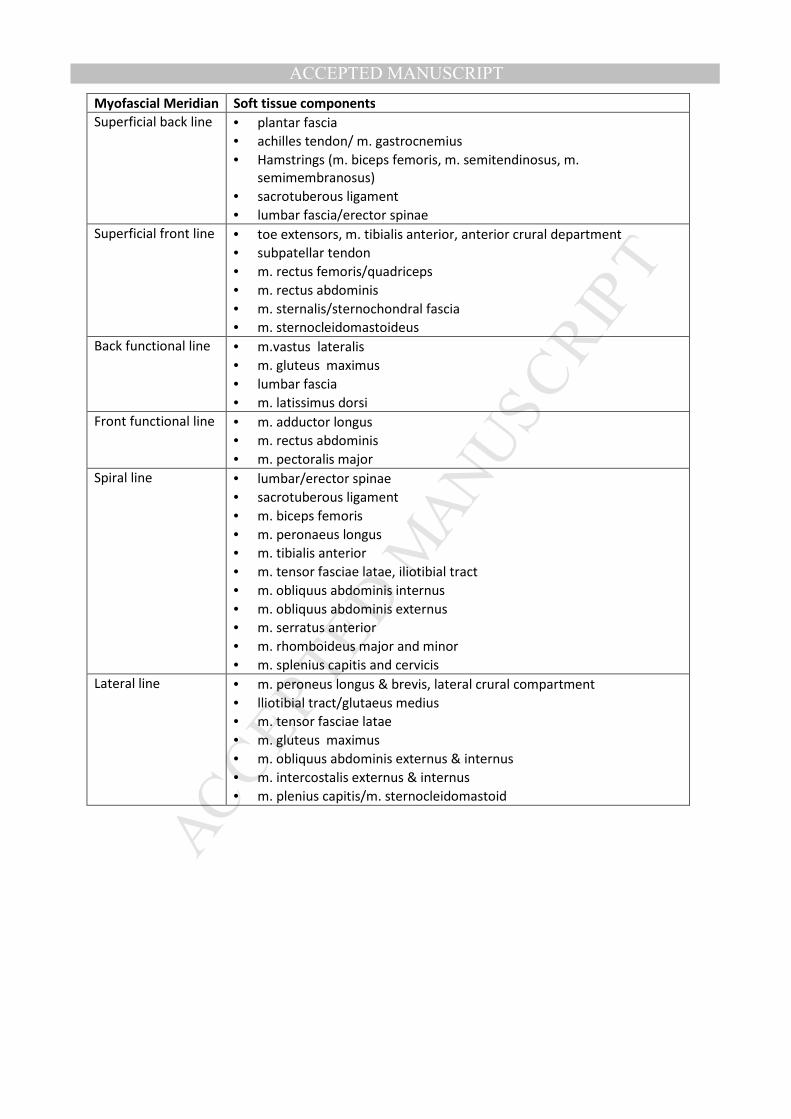
ACCEPTED MANUSCRIPTMyofascial Meridian Soft tissue components
Superficial back line • plantar fascia
• achilles tendon/ m. gastrocnemius
• Hamstrings (m. biceps femoris, m. semitendinosus, m.
semimembranosus)
• sacrotuberous ligament
• lumbar fascia/erector spinae
Superficial front line • toe extensors, m. tibialis anterior, anterior crural department
• subpatellar tendon
• m. rectus femoris/quadriceps
• m. rectus abdominis
• m. sternalis/sternochondral fascia
• m. sternocleidomastoideus
Back functional line • m.vastus lateralis
• m. gluteus maximus
• lumbar fascia
• m. latissimus dorsi
Front functional line • m. adductor longus
• m. rectus abdominis
• m. pectoralis major
Spiral line • lumbar/erector spinae
• sacrotuberous ligament
• m. biceps femoris
• m. peronaeus longus
• m. tibialis anterior
• m. tensor fasciae latae, iliotibial tract
• m. obliquus abdominis internus
• m. obliquus abdominis externus
• m. serratus anterior
• m. rhomboideus major and minor
• m. splenius capitis and cervicis
Lateral line • m. peroneus longus & brevis, lateral crural compartment
• lliotibial tract/glutaeus medius
• m. tensor fasciae latae
• m. gluteus maximus
• m. obliquus abdominis externus & internus
• m. intercostalis externus & internus
• m. plenius capitis/m. sternocleidomastoid

MANUSCRIP
T
ACCEPTED
ACCEPTED MANUSCRIPTDatabase Search algorithm
Pubmed (("cadaver"[Mesh]) AND ("anatomy"[Mesh])) AND ("Fascia"[Mesh]) OR
((myofascial OR aponeurotic OR fascial) AND (continuity OR decussation OR
interdigitation OR expansion OR extension))
Science Direct cadaver AND (fascia OR myofascial OR aponeurotic OR fascial) AND (continuity
OR decussation OR interdigitation OR expansion OR extension)
Google Scholar (dissection) AND ("obliquus internus" AND "obliquus externus") AND
(continuity OR expansion OR extension OR fuses OR merges OR blends)
MANUSCRIP
T
ACCEPTED
ACCEPTED MANUSCRIPT
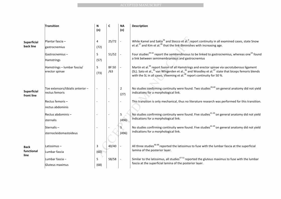
Transition N
(n)
C NA
(n)
Description
Superficial
back line
Plantar fascia –
gastrocnemius
4
(72)
25/72
-
While Kamel and Sakla26
and Stecco et al.9 report continuity in all examined cases, state Snow
et al.27
and Kim et al.28
that the link diminishes with increasing age.
Gastrocnemius –
Hamstrings
5
(57)
51/52 - Four studies29-32
report the semitendinosus to be linked to gastrocnemius, whereas one33
found
a link between semimembranosus and gastrocnemius
Hamstrings – lumbar fascia/
erector spinae
5
(73)
BF:50
/63
- Martin et al.34
report fusion of all Hamstrings and erector spinae via sacrotuberous ligament
(SL). Sato et al.,35
van Wingerden et al.,36
and Woodley et al.37
state that biceps femoris blends
with the SL in all cases, Vleeming et al.38
report continuity for 50 %.
Superficial
front line
Toe extensors/tibialis anterior –
rectus femoris
-
-
2
(27)
No studies confirming continuity were found. Two studies39,40
on general anatomy did not yield
indications for a morphological link.
Rectus femoris –
rectus abdominis
- - - This transition is only mechanical, thus no literature research was performed for this transition.
Rectus abdominis –
sternalis
- - 5
(496)
No studies confirming continuity were found. Five studies41-45
on general anatomy did not yield
indications for a morphological link.
Sternalis –
sternocleidomastoideus
- - 5
(496)
No studies confirming continuity were found. Five studies41-45
on general anatomy did not yield
indications for a morphological link.
Back
functional
line
Latissimus –
Lumbar fascia
3
(60)
40/40
-
All three studies46-48
reported the latissimus to fuse with the lumbar fascia at the superficial
lamina of the posterior layer.
Lumbar fascia –
Gluteus maximus
5
(68)
58/58 - Similar to the latissimus, all studies47-51
reported the gluteus maximus to fuse with the lumbar
fascia at the superficial lamina of the posterior layer.
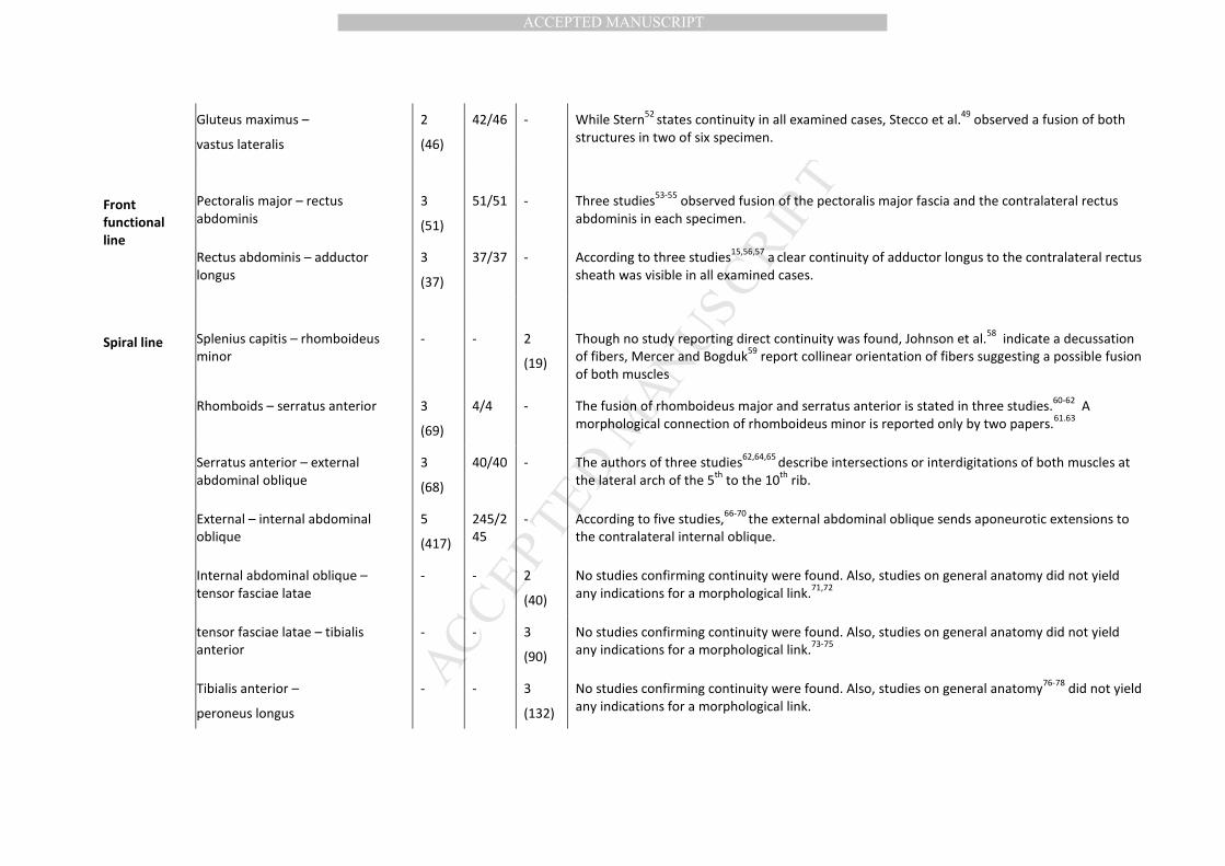
MANUSCRIP
T
ACCEPTED
ACCEPTED MANUSCRIPT
Gluteus maximus –
vastus lateralis
2
(46)
42/46 - While Stern52
states continuity in all examined cases, Stecco et al.49
observed a fusion of both
structures in two of six specimen.
Front
functional
line
Pectoralis major – rectus
abdominis
3
(51)
51/51
-
Three studies53-55
observed fusion of the pectoralis major fascia and the contralateral rectus
abdominis in each specimen.
Rectus abdominis – adductor
longus
3
(37)
37/37 - According to three studies15,56,57
a clear continuity of adductor longus to the contralateral rectus
sheath was visible in all examined cases.
Spiral line
Splenius capitis – rhomboideus
minor
-
-
2
(19)
Though no study reporting direct continuity was found, Johnson et al.58
indicate a decussation
of fibers, Mercer and Bogduk59
report collinear orientation of fibers suggesting a possible fusion
of both muscles
Rhomboids – serratus anterior 3
(69)
4/4 - The fusion of rhomboideus major and serratus anterior is stated in three studies.60-62
A
morphological connection of rhomboideus minor is reported only by two papers.61.63
Serratus anterior – external
abdominal oblique
3
(68)
40/40 - The authors of three studies62,64,65
describe intersections or interdigitations of both muscles at
the lateral arch of the 5th
to the 10th
rib.
External – internal abdominal
oblique
5
(417)
245/2
45
- According to five studies,66-70
the external abdominal oblique sends aponeurotic extensions to
the contralateral internal oblique.
Internal abdominal oblique –
tensor fasciae latae
- - 2
(40)
No studies confirming continuity were found. Also, studies on general anatomy did not yield
any indications for a morphological link.71,72
tensor fasciae latae – tibialis
anterior
- - 3
(90)
No studies confirming continuity were found. Also, studies on general anatomy did not yield
any indications for a morphological link.73-75
Tibialis anterior –
peroneus longus
- - 3
(132)
No studies confirming continuity were found. Also, studies on general anatomy76-78
did not yield
any indications for a morphological link.
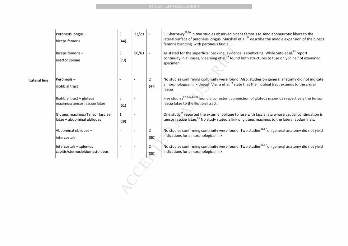
MANUSCRIP
T
ACCEPTED
ACCEPTED MANUSCRIPT
Peroneus longus –
biceps femoris
3
(44)
23/23 - El Gharbawy79,80
in two studies observed biceps femoris to send aponeurotic fibers to the
lateral surface of peroneus longus, Marshall et al.81
describe the middle expansion of the biceps
femoris blending with peroneus fascia
Biceps femoris –
erector spinae
5
(73)
50/63 - As stated for the superficial backline, evidence is conflicting. While Sato et al.35
report
continuity in all cases, Vleeming et al.38
found both structures to fuse only in half of examined
specimen.
Lateral line
Peroneals –
iliotibial tract
-
-
2
(47)
No studies confirming continuity were found. Also, studies on general anatomy did not indicate
a morphological link though Vieira et al.73
state that the iliotibial tract extends to the crural
fascia
Iliotibial tract – gluteus
maximus/tensor fasciae latae
5
(61)
- Five studies8,49,50,82,83
found a consistent connection of gluteus maximus respectively the tensor
fascia latae to the iliotibial tract.
Gluteus maximus/Tensor fasciae
latae – abdominal obliques
1
(29)
- One study84
reported the external oblique to fuse with fascia lata whose caudal continuation is
tensor fasciae latae.85
No study stated a link of gluteus maximus to the lateral abdominals.
Abdominal obliques –
intercostals
- - 2
(80)
No studies confirming continuity were found. Two studies86,87
on general anatomy did not yield
indications for a morphological link.
Intercostals – splenius
capitis/sternocleidomastoideus
- - 2
(80)
No studies confirming continuity were found. Two studies86,87
on general anatomy did not yield
indications for a morphological link.

MANUSCRIP
T
ACCEPTED
ACCEPTED MANUSCRIPT1
Total Excellent Substantial Moderate Fair Poor
Superficial back line 14 1 4 7 2 -
Superficial front line 0 (7) - (7) - - -
Back functional line 8 1 3 4 - -
Front functional line 7 3 3 - 1 -
Spiral line 13 (8) - 2 (3) 10 (5) 1 -
Lateral line 6 (4) - 6 1 (3) - (1)
1

MANUSCRIP
T
ACCEPTED
ACCEPTED MANUSCRIPT
MANUSCRIP
T
ACCEPTED
ACCEPTED MANUSCRIPT