what clinical chemists should know about monoclonal proteins
TRANSCRIPT

What Clinical Chemists Should Know About Monoclonal Proteins
Gary L. Horowitz, MDBeth Israel Deaconess Medical Center
Boston, MA

Objectives
• Explain the importance of evaluating both urine and serum in screening for monoclonal proteins
• Differentiate MGUS from multiple myeloma
• Explain why the free light chain ratio is (usually) more important than the absolute concentration of either free light chain

Topics for Today
• Techniques• Protein Electrophoresis• Immunofixation Electrophoresis• Quantitative Immunoglobulins
• Concepts• MGUS• Bence-Jones Proteins• Serum Free Light Chains

Protein ElectrophoresisSize Doesn’t Matter (much)
• separation is charge-dependent• net charge is (virtually) all that matters• secondary factors include
• size & shape of molecule• electric field strength• supporting medium• temperature

Resolution
• traditional SPEP (low resolution)– 5 bands:
• albumin, alpha1, alpha2, beta, gamma• multiple proteins in each “zone”
• now, high resolution– 10-16 bands!
• do we really need it?
• CAP recommendation: beta1/beta2 separation


Report of the Consensus Conference on Monoclonal Gammopathies. Arch Pathol Lab Med. 1999; 123:104-132.

Immunofixation Electrophoresis (IFE)
• run multiple PEPs of same sample• step 2
• precipitate all proteins in Lane 1• in Lanes 2-6, overlay antisera to G, A, M, k, l
• wash entire plate • only precipitated proteins remain
• stain entire plate• look for precipitates that line up


Antigen Excess (Hook Effect)
• nomenclature• antigen here is antibody
• in homogeneous immunoassay ([IgG]):• you may get falsely low results (with no error flag!)
• in IFE:• you get “donuts”• with Sebia, get “hourglass” effect

Hook Effect: What Is It?
Adapted from Burtis, CA & Ashwood, ER.Tietz Fundamentals of Clinical Chemistry (4th Edition). Philadelphia: W.B Saunders, 1996, p.136.
Y Y

M Protein Concentration
• two alternatives– densitometry (recommended)– quantitative immunoglobulin levels
• each has its place– if both polyclonal IgX and monoclonal IgX present,
[IgX] will OVERestimate– to assess suppression of other immunoglobulins,
need [IgG], [IgA], [IgM]


Recommended Reporting Language
• On protein electrophoresis, there is an abnormal band in the gamma region, representing 10% of the total protein, or 700 mg/dL (7 g/L)
• When subjected to immunofixation electrophoresis, this band is identified as monoclonal IgG lambda
• There is no suppression of IgA or IgM levels, indicating that this is probably an MGUS
• The total IgG concentration is 1200 mg/dL (12 g/L)

Clonality is Qualitative
• CAP recommendations
– do not use immunoglobulin levels for screening• high immunoglobulin levels may be polyclonal• normal immunoglobulin levels can include clonal populations
– screen with PEP (not immunoglobulin levels)

Clonality May Not Be Myeloma
• monoclonal gammopathy (M protein):• occurs in diseases other than multiple myeloma
– Waldenstrom’s, amyloidosis, …
• occurs in entities that may not even be “malignant”– monoclonal gammopathy of undetermined significance
(MGUS)
• when one reports M protein:• what do you think clinician’s next step should be?
Case HistoryA 70 year old man visits his primary care physician (PCP) with complaints of fatigue over the past three months. As part of his initial work-up, he’s found to be anemic, so his PCP orders a battery of follow-up tests, including a serum protein electrophoresis (SPEP) to rule out multiple myeloma.
The SPEP is reported as howing a trace monoclonal band, representing roughly 1% of serum protein (70 mg/dL). In addition, his immunoglobulin levels are reported as follows:
IgG 400 mg/dL (reference interval 700-1600)IgA 57 mg/dL (70-400)IgM 32 mg/dL (40-230)
The PCP infers that, with such a low concentration M-protein, it’s almost certainly an “MGUS”, that should be followed annually.

Diseases Associated with “M Proteins”
• Myeloma• Solitary• Asymptomatic• Multiple Myeloma• POEMS
• Waldenstrom’s• Amyloidosis• MGUS

Incidence/Survival in United States
disease cases/yearmedian survival(years)
Myeloma 13,000 3
Waldenstrom’s 3,000 5
Amyloidosis 2,000 1
MGUS 750,000 12

MGUS
• prevalence– 1% over age 50– 3% over age 70
• roughly 1.5% per year progress• majority die of unrelated disease
Features of MGUS
• asymptomatic, older individuals• [M protein] < 2500 mg/dL (<25 g/L)• marrow plasma cells < 10%• no bone lesions• BJP < 50 mg/day (<0.050 g/day)• other [immunoglobulin] preserved

Urine Proteins: Situations Where PEP Is Less Important
• “micro”albuminuria• glomerular pattern
• non-selective proteinuria• everything filtered, small proteins reabsorbed
• tubular pattern• failure to reabsorb low molecular proteins• e.g., beta2microglobulin

Urine Proteins:Situation Where PEP Is Critical
• Bence-Jones Protein (BJP)– 3-part definition:
– free– monoclonal– light chains
– precipitation characteristics are notdiagnostic
– precipitate @ 40-60oC– re-dissolve @ 100oC


Clinical & Diagnostic Significance of BJP
• false negative dipstick
• SPEP may show no “M protein”(probably shows hypogammaglobulinemia)
• think of Willie Sutton, famous bank robber

Willie Sutton
• one of the most famous bank robbers of the twentieth century
• a favorite of newspaper reporters, who could count on him for the kind of quote that makes a headline bounce
• spent most of his adult life in prison
• though he escaped more than once, his short bursts of freedom always ended with an arrest for bank robbery
• in an attempt to learn why he continued along such a futile course, one reporter asked, "Willie, why do you keep robbing banks?"
• "Because," Sutton said smoothly, "that's where the money is."

Clinical & Diagnostic Significance of BJP
• false negative dipstick
• SPEP may show no “M protein”(probably shows hypogammaglobulinemia)
• think of Willie Sutton, famous bank robber• look in the urine• not in the blood• BJP may be the ONLY evidence of disease

serum urine

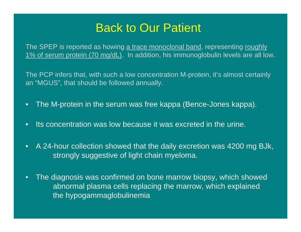
Back to Our PatientThe SPEP is reported as howing a trace monoclonal band, representing roughly 1% of serum protein (70 mg/dL). In addition, his immunoglobulin levels are all low.
The PCP infers that, with such a low concentration M-protein, it’s almost certainly an “MGUS”, that should be followed annually.
• The M-protein in the serum was free kappa (Bence-Jones kappa).
• Its concentration was low because it was excreted in the urine.
• A 24-hour collection showed that the daily excretion was 4200 mg BJk,strongly suggestive of light chain myeloma.
• The diagnosis was confirmed on bone marrow biopsy, which showedabnormal plasma cells replacing the marrow, which explainedthe hypogammaglobulinemia

Take Home Messages• Always submit urine
as well as blood for PEPs
• Role of PEPs– Screening– Quantitating
• Densitometry• [M-protein]=(PEP %) x [TP]
– Monitoring• Change in migration• Change in amount
• Role of IFE– PEP abnormality: initial ID– PEP change: re-ID– High clinical suspicion
• even with negative PEP• order IFE in addition to PEP
• Role of [IgG], [IgA], [IgM]– Can be misleading– More for suppression than
elevation– Except when M-protein overlies
normal proteins (e.g., beta region)


0.1
1
10
100
1000
10000
100000
0.1 1 10 100 1000 10000 100000
FLC (mg/L)
F
LC (m
g/L)
Normal seraκ LCMMλ LCMMIIMMHigh pIgGAL AmyloidosisRenal impairmentNSMM
SPE sensitivity
IFE sensitivity
FLC (mg/L)
FL
C (m
g/L)
Bradwell, AR. Serum Free Light Chain Analysis [4th Edition]. 2006.


Self-Assessment Question 1
The best combination of tests to screen for monoclonal proteins is:
A) Serum immunoglobulin concentrations (IgG, IgA, IgM)B) Serum protein electrophoresis and immunofixation
electrophoresisC) Serum and urine protein electrophoresisD) Urine protein electrophoresis and immunofixation
electrophoresis

Self-Assessment Question 2
Typical findings in Light Chain Myeloma include all of the following EXCEPT:
A) A discrete band in the serum protein electrophoresisB) (Serum) hypogammaglobulinemiaC) A discrete band in the urine protein electrophoresisD) A negative urine dipstick for protein

Self-Assessment Question 3
What is the most common diagnosis associated with monoclonal proteins?
A) AmyloidosisB) Multiple MyelomaC) Monoclonal Gammopathy of Undetermined
SignificanceD) Waldenstrom’s Macroglobulinemia