web viewretrograde root canal filling ... poor placement with lack of thickness and/or mechanical...
TRANSCRIPT

Department of Conservative Dentistry
Retrograde Filling Materials
Submitted by;
Usha Muraleedharan
Roll No: 60
Batch 2003-04

Retrograde Root Canal Filling Materials
Introduction
A retrograde root canal filling is
done when the canal is poorly sealed from
the surrounding tissues.The purpose of a
root-end filling is to establish, as well as
possible, a hermetic seal of all apical
avenues in the tooth from the oral
environment to the periradicular tissues.The
following literature is a discussion of the
various materials that have been used for
retrograde filling. The main consideration
regarding the materials being used as
retrograde fillings is the fact that these
materials come in very close contact with
the periapical tissues.
A plethora of materials have
been evaluated, with leakage,
histocompatibility, and toxicity studies, in an
attempt to identify the "perfect" rootend
filling material. To date none exists, and
recommendations can only be made on
what appears to be the best tolerated,
clinically successful material. Therefore, the
endodontic surgeon must be cognizant that

the success of the procedure does not lie in
the essence of the 'apical filling material
used, and that this material will not
compensate for lack of proper nonsurgical
management of the root canal system. In
addition, it must also be understood that
lack of understanding of the materials to be
used, coupled with their improper use, could
contribute to ultimate failure on either a
short- or longterm basis.

Ideal requirements of a root end filling material According to Gartner and Dorn, a
suitable root-end filling material should be
(l) Able to prevent leakage of bacteria and their by-products into the periradicular tissues(2) Nontoxic (3) Biocompatible with the host tissues(4) Non carcinogenic(5) Insoluble in tissue fluids(6) Dimensionally stable(7) Unaffected by moisture during setting(8) Easy to use(9) Radiopaque(10) One might add it should not stain tissue (tattoo).
Materials used – properties and manipulation
The following list of materials
have been identified as root-end filling
materials in either scientific evaluation or
clinical usage. The parameters of each
material are discussed as to their
acceptability and technique. A brief outlook
on the manipulation of these materials is
also given.
1) Gutta-percha.
2) Silver cones.
3) Amalgam.
4) Zinc oxide-eugenol.
5) Cavit.
6) Polycarboxylates.
7) Composites.
8) Zinc phosphate.
9) Gold foil.
10) Glass ionomers.
11) Miscellaneous materials.
Before going into the details of
the various materials used for retro-grade
filing, a small review of the root canal
sealers that are used along with these
materials inorder to provide an impervious
seal at the apex is needed.
Root canal sealers
Root canal sealers, used in
conjunction with a solid core obturating
material, are intended to cooperatively
effect a fluid-tight or hermetic seal
throughout the root canal system. To
achieve this goal, and promote
periradicular healing, the sealer must
possess certain characteristics:
1. It is non-irritating,
2. It has a hermetic sealing ability with
dimensional stability,
3. It is bacteriocidal or static,
4. It is insoluble in tissue fluids, and
adheres to the dentinal surface.

These factors will have a direct
bearing on the seal of the canal system
subsequent to root-end resection and on the
periradicular healing at the resected root
face, with or without a root-end filling.
Commercial sealers are generally grouped
as
Zinc oxide-eugenol based,
Noneugenol based, and
Therapeutic based.
Of importance in the first group is
that residual eugenol that remains after
sealer set can affect the sealer's properties
or the periradicular tissue response.
Noneugenol based sealers use
solvents, such as chloroform or eucalyptol,
which have demonstrated toxicity in the
initial stages of sealer set.
Therapeutic sealers contain
materials such as iodoform,
paraformaldehyde, or trioxymethylene,
which are claimed to have therapeutic
properties. However, this issue is highly
controversial and clinical reports of adverse
periradicular tissue response would tend to
question the efficacy of these sealers. This is
especially true when these sealers are used
as the sole root canal filling material.
Subsequent to resection in roots filled with
these sealers, there is significant contact of
these sealers with the periradicular tissues,
which may resuJt in extensive tissue
destruction, with or without a root-end
filling.
Recently, calcium hydroxide
based sealers have been developed. Thus
far, evaluations of these materials have
shown mixed results regarding both physical
properties and tissue compatibility.
Initially, all sealers can cause
tissue inflammation and cellular damage.
The severity of the damage and its
continuation appears to be related to the
nature of the material, its physical

properties, its setting time, and quantity or
surface area of the material in contact with
the tissues.
Gutta-Percha
Since gutta-percha is considered to
be relatively nonresorbable and impervious
to tissue fluid dissolution, its
biocompatibility and physical adaptability to
the root canal wall must be considered. The
more effective an obturating material is in
sealing the root canal system, and
maintaining that seal, the more compatible
it is likely to be.

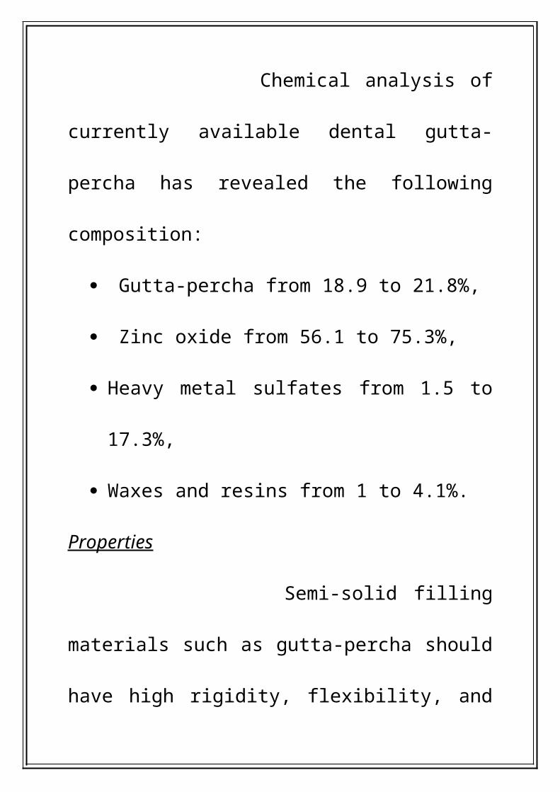
Composition of the gutta-percha
Chemical analysis of currently
available dental gutta-percha has revealed
the following composition:
Gutta-percha from 18.9 to 21.8%,
Zinc oxide from 56.1 to 75.3%,
Heavy metal sulfates from 1.5 to 17.3%,
Waxes and resins from 1 to 4.1%.
Properties
Semi-solid filling materials such as
gutta-percha should have high rigidity,
flexibility, and yield strength. Because of the
need to have a material which can be
readily condensed and adapted to the
irregularities of the canal system, gutta-
percha should also have a high percentage
elongation and low resilience.
Mechanically, polymers such as
gutta-percha are not perfectly elastic, but
have both elastic properties and properties
of viscous liquids. Therefore, these polymers
are referred to as viscoelastic. Clinically, the
importance of this viscoelastic property
manifests itself during use in the root canal.
Guttapercha requires a large, sustained
force of condensation over an adequate
period of time to deform plastically. The
more it deforms, the more it will flow and
adapt to the dentin wall, decreasing gaps in
the gutta-percha-dentin interface.

The quality of the root canal
treatment has a definite effect on the
marginal adaptation of gutta-percha
subsequent to resection only. However, this
result appears to also be dependent on the
type of gutta-percha, nature of the sealer
used, the condensation technique, type of
bur used for resection and operator skill.
Placement and condensation of gutta-
percha
As early as 1916 , the pulling of
the gutta-percha through the resected root-
end had been advocated to ensure
maximum adaptation to the dentin walls.
However, this technique has been shown to
result in voids in the gutta-percha – dentin
interface, as the gutta-percha tends to
retract from the walls creating significant
gaps at the interface. Most authors have
recommended coronal condensation of the
gutta-percha into the apical third of the
canal and through the foramen, prior to
removal of the excess material. This
approach would tend to ensure a better
gutta-percha-sealer adaptation to the dentin
walls.
Use of solvents
Various solvent techniques have
been advocated to enhance the adaptation
of the gutta-percha to the apical portion of
the canal prior to resection, to the root apex
if no resection is anticipated, or to the
resected root surface. Included in these
approach has been the use of eucalyptol or
chloroform and rosin to soften the gutta-
percha cone prior to placement into the
apical third of the canal or through the
resected root end, or chloroform to soften
and adapt the gutta-percha to the foraminal
margins at either the natural apex or the
resected root end. However, it has been
shown that the material loses its

dimensional stability as the solvent is
evaporated from the mixture.
Type of instrument used for adaptation of
gutta-percha
This includes burs, scalpels, spoon
excavators, plastic instruments, and
burnishers. No studies unequivocally
substantiate the best instrument for gutta-
percha removal at the apex and the quality
of the adaptation appears to be operator
dependent.
Temperature of instrument used to remove
gutta-percha
This has been a topic of
controversy for several years. For years the

use of an arm to hot instrument was
advocated to smooth or burnish the gutta-
percha filling material.
In 1980 this technique was
criticized by Tanzilli and coworkers in an
SEM study by. They compared the use of a
warm plastic instrument in a cutting or
searing motion and a cold ball burnisher to
adapt the gutta-percha at the resected root
end. Discrepancies in the adaptation of the
gutta-percha were identified under SEM
evaluation. These findings and their
subsequent interpretation, especially when
compared to cold burnished gutta-percha,
created consternation among endodontic

surgeons. This was especially true because
the cold-burnished gutta-percha appeared
to have superior adaptation to both
amalgam and gutta percha fills subsequent
to root-end resection only. The concept of
good marginal adaptation with heated
sealed gutta-percha had been challenged
and this study was cited as the bench mark
for future considerations in the management
of apically resected gutta-percha.
SILVER CONES
Silver cones have been used to
obturate root canals since the early 1930s.
However, their ability to seal the root canal
system three-dimensionally has been

justifiably challenged, as the circular,
tapered natured of the cone provides only a
central core material which is surrounded by
a sea of root canal sealer. This anatomic
problem is accelerated subsequent to
angled root-end resection, as large areas of
sealer are visible between the cone and
dentin wall. These gaps can be especially
wide in teeth with wide buccal-lingual canals
or which exhibit extensive fins or cul-de-
sacs along the facial or lingual anatomy of
the canal.
Placement of silver cone
Few reports exist in the dental
literature which address the use of a silver

cone as the apical filling material at the time
of periradicular surgery.
Contemporary studies using silver
cones at the time of root-end resection have
identified a low level of long-term success
and recommend a more bio-compatible
material.
In addition to the strong
potential for voids and leakage to exist
between a resected silver cone and dentin
wall, corrosion of the metal-worked cone
looms as a major factor for continued
periradicular tissue irritation and ultimate
failure of resected silver cone cases.

The silver content of the silver
cones range from 99.8 to 99.9%, silver salts
and sulfur sulfides formed by the contact of
the metal-worked or contaminated silver
cones have been demonstrated due to
extensive corrosion, with pitting and
cratering of the cones. However, the tissue
cytotoxicity of these corrosive silver salts
(silver chloride, silver carbonate, and silver
oxide) and sulfur sulfides has been
questioned.
The endodontic surgeon should
consider the following guidelines concerning
silver cones, root-end resection, and root-
end fills.

1) Silver cones cannot three-dimensionally
obdurate the root canal space,
especially in areas coronal to the apex
which are likely to be exposed during
resection.
2) Resection of a root end containing a
silver cone will open voids between the
cone and dentin wall.
3) Resection of a silver cone will cause the
material to be metal-worked,
accentuating its corrosive potential.
Over long periods of time, the corrosive
products that form may be highly
cytotoxic.
4) Silver cones cannot be burnished to
"perfect" the apical seal.
5) Ideally, teeth containing silver cones
and requiring surgery should be
nonsurgically retreated, if possible prior
to surgery, removing any silver
corrosion products from the root canal
system and replacing the silver cone
with a well condensed gutta-percha and
root canal sealer fill.
6) A root-end fill is indicated in all cases of
root-end resection when a silver cone is
present. When cutting a root-end
preparation into the resected root
containing a silver cone, establish a

good finger rest, use high-speed burs,
and frequently irrigate the surgical
area. Careful cutting is recommended to
prevent slipping off the silver cone and
gouging or perforating the root surface.
AMALGAM ALLOYS
One of the first reports of
placing a root-end amalgam filling
subsequent to resection is attributed to
Farrar.
Controversies and concerns
The main controversies and
concerns regarding the use of amalgam:
1) Type of amalgam (high copper versus
conventional; zinc versus nonzinc) and its
properties.
2) Leakage of amalgam root-end fills and
the use of cavity varnish, including setting
expansion and contraction;
3) Tissue compatibility;
4) Preparation and manipulation of the
amalgam;
5) Electric potentials - galvanic currents,
corrosion and degradation; and
6) Pigmentation or agyria of the
surrounding tissues.

It is essential that the endodontic
surgeon have a working knowledge of the
properties of amalgam alloys to enhance the
potential for successful treatment.
T ype of amalgam
Many of the early reports on the use
of amalgam for root-end fills did not specify
the nature of amalgam used, while some
authors specified the use of copper or silver
amalgam. At that time copper (>40% Cu)
amalgam was
identified as a tissue irritant and, as the
popularity of silver (<6% Cu) amalgam
increased, it was rapidly identified as the
root-end filling material of choice. Recently,
there has been a trend to use high copper-
content (>6% Cu) amalgams among mixed
reports of varied cellular and tissue
responses , and advanced mechanical
properties.
Root-end amalgams leak.
Key factors which interact with the
discrepancies cited include
The mean leakage observed and its
alteration with time ;

The standard deviation from the mean
leakage observed ;
The depth of the amalgam ,
The amount of amalgam corrosion and
Expansion anticipated (conventional
amalgam versus high copper amalgam;
zinc versus nonzinc);
Manipulation of the alloy during
preparation and placement ;
The placement of the alloy in the canal
prior to resection versus its use as a root-
end fill only ;
The cleanliness and seal of the root canal
system coronal to the root-end fill ; and
The use of cavity varnish

Endodontic research has identified a
significant improvement in the initial seal of
root-end amalgams when a cavity varnish is
used .The use of two coats of varnish to seal
not only the walls of the root-end
preparation but also the cut dentinal tubules
at the root surface has received substantial
support.
Material preparation and manipulation
The preparation and
manipulation of the amalgam alloy at the
time of placement is crucial in determining
amalgam strength, marginal adaptation,

degree of porosity, surface smoothness, and
the nature of surface constituents.
However, the ultimate
mechanical characteristics will be
dependent on the type of alloy chosen and
operator management. Some key points to
consider relative to alloys placed intraorally
are as follows:
1) Amalgams squeezed of their excess
mercury have a decrease in their final
strength. The Eames 1: 1 ratio technique
or that described by Jorgensen and Saito
are preferable.
2) Instructions supplied by the
manufacturer for trituration should be

closely followed. In addition,
amalgamators vary considerably in
function and performance due to warm-up
time, age, changes in line voltage, and
changes in capsule weight. Therefore, in
an attempt to minimize variations, mixes
of amalgam heavier than two spills should
be avoided, and single-speed
amalgamators or variable-speed
amalgamators of a newer design should
be used . Also, it has been noted that the
setting rate of high copper dispersed
phase alloys is more sensitive to varied
mixing speeds than spherical alloys.

3) Amalgams are more closely adapted to
the confines of the cavity during
mechanical rather than hand
condensation; however, the use of
mechanical condensers may be limited. In
addition, the condensation method has a
direct bearing on the ultimate leakage
demonstrated for both tin-mercury
containing and non tin-mercury-containing
alloys.
4) Alloys consisting of spherical, or mostly
spherical, particles are more fluid under
condensation ressures; and the use of a
large condenser in a lateral fashion may
be desirable because a small head

condenser tends to force the amalgam
mass away from the areas of
condensation. Also, less pressure is
required to properly condense these
alloys .
5) The high copper-content alloys have
been reported to be less susceptible to
the variances in operator manipulation .
6) The optimal structure for the amalgam
margins can be obtained by overfilling
and burnishing of the margins, and
removal of the excess by carving.
Burnishing of the alloy margins decreases
microporosity, improves the marginal
adaptation and seal, especially with
admixed alloys.
Carving is necessary after
burnishing, followed by burnishing to
render the surface smoother, thereby
discouraging formulation of small
corrosion cells on the surface.
The time frame for clinical
management of the alloy, including
marginal finish, may be reduced or
impaired when pressures; and the use of a
large condenser in a lateral fashion may
be desirable because a small head
condenser tends to force the amalgam
mass away from the areas of

condensation. Also, less pressure is
required to properly condense these
alloys. The high copper-content alloys
have been reported to be less susceptible
to the variances in operator manipulation .
Electric potentials - galvanic currents
The placement of a root-end amalgam
in a tooth which has a metallic post or crown
restoration could create a galvanic couple,
which has the potential to generate
significant amounts of electrical currents.
Tissue staining – argyria
The possibility of argyria
subsequent to root-end resection and/or

root-end amalgam fillings stems from
multiple sources .
1) Amalgam scattered in the surgical site.
During placement of the root-end filling.
2) Amalgam scattered in the surgical site
due to removal of a failing root-end
amalgam.
3) Fractured or loosened amalgam root-end
fills.
4) Chemical corrosion of the root-end
amalgam or silver cones at the resected
root surface.
5) Electrochemical corrosion - galvanism.

6) Silver scattered in the surgical site
during resection of roots containing silver
cones.
7) Deterioration of silver containing root
canal sealers.
During removal of previously
placed amalgams (surgical retreatrnent),
efficient irrigation and aspiration of the
surgical area are essential. Often, however,
small amounts of amalgam "dust" are
unavoidable. Fractured amalgams are often
due to lack of bulk in the thickness of the
root-end fill. Corrosion may also play a role,
as would the type of alloy and its clinical
manipulation. Loosened amalgams are due

to significant marginal corrosion, poor
placement with lack of thickness and/or
mechanical retention in the root-end, apical
resorption due to continued chronic
infIammation, and tooth fractures.
Guidelines for amalgam usage as a root-end
filling
The previous discussion on
amalgam alloys as rootend filling materials
has provided a cursory clinical and scientific
rationale for considering the use of this
material. Although amalgam is not the ideal
material, the endodontic surgeon should
be cognizant of the following concepts

when choosing amalgam as the root-end
filling material.
1. Control of moisture in the surgical site is
essential.
2. High copper alloys are the material of
choice at present.
3. Varnish must be used prior to alloy
placement. Dentin bonding agents can also
be considered in place of varnish.
4. Zinc alloys are the material of choice
when moisture is controlled.
5. When moisture cannot be controlled,
nonzinc alloys should be considered.
6. Carefully condense, burnish, carve, and
burnish the alloy using a minimal number

of firm strokes directed to the alloy dentin
interface
7. Create a smooth surface on the finished
alloy.
8. Prevent the dispersion of alloy particles
in the surgical site.
9. Keep the alloy as small as possible in
perimeter or diameter, although the bulk
of the alloy must be thick enough to resist
fracture and to obturate the entire canal
system at the resected root surface.
10. Radiographically, the amalgam will often
resemble the shape of both the canal
system and the external root anatomy.

ZINC OXIDE-EUGENOL
The mixture of clove oil, which is
eugenol in its unrefined state, and zinc
oxide to form a plastic mass was first
described by Chisolm during the Tennessee
State Dental Meeting (in 1873). Its use in

dentistry expanded rapidly to many areas,
including endodontics, periodontics, and
restorative dentistry.
Zinc oxide-eugenol (ZOE) has been
shown to prevent bacterial ingress along
cavity margins. It has also been shown to be
an irritant, primarily due to the eugenol
component. However, even the tissue
response to eugenol is varied and is
dependent on the short-term saturation of
the local environment with eugenol , at a
concentration level and time sufficient to
harm mammalian cells. The level of toxicity
incurred is due to the concentration of
eugenol released from the material and is
not always proportional to the content of the
source, or consistent within specific
preparations. Ultimately the extent of cell
death which occurs depends principally on
the efficiency of local clearance of eugenol.
At the root apex, local blood
flow and clearance are usually sufficient to
allow for healing. If the contact area at the
root apex of ZOE is minimal, then cell death
may-be minimal and, with the rate of
eugenol release declining over 1 to 2 weeks,
healing should occur. However, there is
ample evidence to show osseous tissue
toxicity to ZOE. The use of ZOE as a root-

end sealing agent in periradicular surgery
has had limited documention.
When ZOE is in contact with
water, it is hydrolyzed, breaking the CH3-O-
Zn coordinate bond to produce zinc
hydroxide and eugenol. The eugenol will
continue to be removed by the leaching of
water until all the original zinc eugenolate is
converted into zinc hydroxide. This
hydrolysis forms the basis for the
bioavailability of free eugenol and ultimately
determines whether the agent is therapeutic
or toxic. Depending on its concentration,
eugenol can:
- Competitively inhibit prostaglandin
synthetase by preventing the
biosynthesis of cyclo-oxygenase,
- Inhibit senory nerve activity,
- Reversibly inhibit repair in mammalian
cells,
- Kill cells with prolonged exposure,
- Kill a range of oral microorganisms
- be an allergen.
Also in this process of dissolution,
zinc has been identified leaching into the
dentinal tubules. Since it has been shown
that zinc, in low concentrations, may act as
a regulator ion in the process of
mineralization and wound healing, there
may be a beneficial effect to placement of a
ZOE root-end fill. However, extensive and
controlled evaluations of these concepts
within the parameters of periradicular
surgery are warranted.
Newer modifications of ZOE
compounds, such as IRM (LD Caulk Co.),
have also been evaluated as to their sealing
ability. Studies reveal that IRM sealed better
than nonzinc amalgam using
electrochemical longitudinal analysis (60
days) and dyes, and zinc amalgam using
pulp tissue response. At the same time, it
has been shown to allow leakage to a
bacterial species. Therefore,
additional evaluative parameters and
studies
are indicated.
In an attempt to alter the
setting time and increase the strength of
ZOE cements, the EBA (o-ethoxybenzoic
acid) cements were developed. EBA
cements have been shown to have much
better physica! properties than ZOE, with
compressive, tensile, and shear strengths
approaching those of zinc phosphate
cements, especially with the addition of
reinforcing fillers such as aluminum oxide,
silica, hydrogenated resin, and acrylic
resins. However, cement solubility was

increased with the addition of these
modifiers.
Further advances in the
formulation of EBA cements led to the
development of Stailine Super EBA cement
(Staines, UK) which consists of 60% zinc
oxide, 34% silicone dioxide, and 6% natural
resin
in powder form, which is mixed with eugenol
in a 62.5% : 37.5% ratio respectively. This
product has high compressive strength, high
tensional strength, neutral pH, and low
solubility. Even in moist conditions, this EBA
adheres to tooth structure. The use of EBA

cements to enhance root canal filling
materials has thus been recommended.
Studies conducted to evaluate the
biological aspect of this new modification
showed a good healing response to Super
EBA with minimal chronic inflammation at
the root apex. Scanning electron microscope
evaluation showed excellent material
adaptation to the dentin margins at the
resected root surface. Collagen fibers were
deposited on top of the Super EBA with
possible fiber ingrowth into the material.
Additional tissue compatibility
studies of EBA cements have confirmed
their low level of irritation, especially in the
set state. Leakage studies with EBA cements
have shown good adaptation and marginal
sealing.
Based on the various studies, the
prospects for future routine use of EBA
cements are promising.
CAVIT Cavit is a temporary filling
material which contains
zinc oxide,

calcium sulfate,
zinc sulfate,
Glycol acetate,
polyvinyl acetate,
polyvinyl chloride acetate,
triethanolamine, and
red pigment.
It is also available in forms without
the red pigment, such as Cavit-G and
Cavit-W. Cavit is soft when placed in the
tooth and subsequently undergoes a
hygroscopic set after permeation with
water, giving it a high linear expansion
(18%). This property has been cited as a

rationale for its use as a root-end filling
material.
The ability of Cavit to seal
cavities is controversial. Biocompatibility
studies with Cavit are in conflict, showing it
to be both toxic and nontoxic, which
emphasizes potential problems with
comparing diverse experimental conditions.
Tissue toxicity studies have shown that
Cavit is toxic to subcutaneous tissue and
bone. Longterm
radiographic and clinical evaluations of the
use of Cavit as a root-end filling material are
varied. Further studies or alteration in the
compound to enhance its tissue

biocompatibility and sealability are
warranted, if Cavit is to be considered as a
viable material to seal the root system.
POLYCARBOXYLATE CEMENTS
This group of dental cements
was introduced by Smith in the late

1960s.The zinc polycarboxylate cements
consist of
a powder - modified zinc oxide with fillers
such as magnesium oxide and stannous
fluoride) and
a liquid (aqueous solution of polyacrylic
acid)
When mixed this mass hardened,
and forms a cement of zinc oxide particles
dispersed in a crosslinked structureless
matrix of zinc polycarboxylate. This reaction
occurs between the zinc ions and the
carboxyl groups on the polyacrylic acid, with
the free carboxyl groups having the capacity
to chelate calcium. Therefore, adhesion to
tooth structure is a significant physical
property of the polycarboxylate.
Their solubility is similar to
that of zinc phosphate, provided the proper
powder-liquid ratio is used. Reducing the
powder content by a third has been shown
to amount to a threefold increase in cement
disintegration.
The pH of the cement is
approximately 1.7. However, the liquid is
rapidly neutralized by the powder during
material set. Despite the initial acidic nature
of the polycarboxylates, minimal irritation
has been reported to the dental pulp when
placed on adjacent dentin or used as a
direct pulp cap.
However, the osseous tissue
adjacent to the polycarboxylate implants
showed decalcification, which is probably
due to the chelating property of the cement.
If this were to occur to the dentin walls in a
root-end preparation filled with a
polycarboxylate, the possibility for leakage
might be enhanced at the cement-dentin
interface. This aspect of the cement requires
further evaluation. Polycarboxylates placed
in root canal systems or beyond the
confines of the root apex show a varied
periradicular tissue response.
Studies conducted when used as
root-end fillings, leak at levels significantly
greater than amalgam or gutta-percha.
Therefore, based on their poor sealing
ability and uncertain periradicular tissue
response,
the use of polycarboxylates as root-end
filling materials is highly questionable.
Further evaluation may be warranted.
COMPOSITE RESINS Composite resins have received
minimal attention as root-end filling
materials. This is due to their cytotoxic or
irritating effects on pulp tissue. To reduce

their irritation in the tooth, bases or liners
are recommended. As a root-end filling this
would be impossible with composites. In
addition, the release of formaldehydes from
composites has been identified and long-
term tissue irritation may be a factor when
placed in the rootend.
Recent evaluations have
challenged the cytotoxicity of composites
claiming the need for better material
adherence to dentin to eliminate marginal
leakage and the primary cause of their
claimed cytotoxicity, bacteria and their
products.

Good marginal adaptation was
observed with composite, while amalgam,
whether carved or burnished, consistently
exhibited marginal gaps.
Cell attachment to the surface of the
composite was remarkably less than that of
amalgam. In addition, cell attachment to the
root dentin was highly variable. Overall
composites exhibited a poorer
biocompatibility than amalgams.
Initial leakage studies with
composite resins as a root-end filling
materials have been favorable, with leakage
patterns less and marginal adaptation better
than amalgam, Cavit, polycarboxylate,
gutta-percha, and zinc phosphate. However,
when placed against clinically moist dentin,
composite materials do not produce seals
immediately after insertion and the final
adaptation is a function of polymerization
shrinkage and water sorption.
Due to the possibility for initial
leakage with all restorative materials,
various approaches to enhance marginal
adaptation and sealability have been
recommended.
Acid etchants and dentin bonding agents
have now improved levels of material
adaptation, prevention of bacterial

penetration, and decreased marginal
leakage with amalgam and composite.
ZINC PHOSPHATE CEMENTS Rhein, in 1897, used zinc
phosphate cement along with gutta-percha
to seal the root canal system prior to root-
end resection. In 1916 Provan
recommended the use of oxyphosphate of
copper to seal the canals of molar teeth
which had been resected.
Presently, few, if any, references
are
made in the current literature to the use of
zinc phosphate cements for root-end filling
materials for the following reasons:
1) They are soluble, especially in dilute
organic acids. This would be a significant
problem in rootend fills, particularly with the
presence of chronic inflammation in the
periradicular tissues.
2) They are irritating to tissues, especially in
the
presence of bacteria. However, many
studies
have provided varied and opposing findings
in this regard.
3) The cements are prone to leakage and
are affected by moisture during placement.
GOLD FOILFor years gold foil was acknowledged as the
premier restorative material. Some of the

first reports on its use as a root-end filling
material are attributed to Schuster in 1913
and Lyons in 1920. Reports in the 1960s,
1970S, and 1980s continued to recommend
its use because of ease of direct
manipulation, marginal adaptation, surface
smoothness, and tissue biocompatibility.
Cytotoxicity studies have indicated
variations in the inhibition of cell growth
based on the formulation of the gold. Fine
pellet gold did not inhibit cell growth,
whereas newer formulations (New Biofil and
Karat) inhibited up to 80% of the cellular
growth. Tissue biocompatibility studies have
indicated a mild response to

undercondensed, irregular pieces of gold
foil.
Marginal adaptation and
leakage studies in root-end preparations
have indicated minimal or no leakage. Key
to the claimed success in the use of gold foil
was the close adaptation to the dentinal
walls and the ability to highly polish the
metal filling.
Although it possesses favorable
material properties, the routine use of gold
foil as a root-end filling material does not
appear practical because of the need to
establish a moisture free environment, the

need for careful placement and finishing,
the possibility
of root fracture under excessive
condensation
pressures, and the need for surgeon
expertise in material management.
However, its use in isolated cases may be
justified, especially when a long cast gold
metal post is present in the root, or access
to the surgical site can be carefully
controlled.
GLASS IONOMERS Glass ionomers are a hybrid of
the silicate and polycarboxylate cements,
which bond physicochemically to dentin and
enamel, and possess anticariogenic activity.
They are formed by the reaction of calcium-
alumino silicate glass particles with aqueous
solutions of polyacrylic acid. The acid
extracts calcium and aluminum ions from
the glass
particles, initiating a prolonged two-phase
setting reaction. Calcium ions bind. to the
polyacrylic acid producing a firm gel that
provides initial adhesion to tooth structure.
The final set material consists of
unreacted glass particles coated with silica
gel embedded in a matrix of calcium
aluminum polysalts. During the initial
setting period the surface of glass ionomers
is highly sensitive to acidic environments
(4.8 pH) with the elution of AI, F, Si, and Ca
ionic species. With longer aging, less
dissolution of ions is observed. However, in
the periradicular environment the glass
ionomer would be immediately exposed to
inflammatory products during its primary
setting reaction, which may result in
significant ionic release.
Glass ionomers have been shown
to have antibacterial properties, due to their
acidity and fluoride release. Marginal
adaptation and adhesion of glass ionomer
cements to dentin have been shown to be
improved with the use of acid conditioners
and varnishes.
Although these properties would
enhance the use of glass ionomers as root-
end filling materials, their sensitivity to
moisture contamination would tend to
restrict their
use to cases in which thorough root-end
isolation is achieved.

MISCELLANEOUS ROOT-END FILLING MATERIALS
The following list of materials has
received brief mention in the dental
literature for use as root-end filling materials
in periradicular surgery. Little substantiation
exists for the use of some of these
materials,
while others require further evaluation to
determine the long-term efficacy of their
use. The endodontic surgeon should
consider using materials which have been
biologically and clinically evaluated and
which give evidence for favorable longterm
success.

1) Lead points, cited by Lyons, to seal the
canal followed by burnishing to seal the root
apex after resection.
2) Gold screws .
3) Ward's Wonderpack cement.
4) Poly-Hema, which was shown to leak to
bacterial products over short-term
evaluation.
5) Tin foil
6) Ivory and plastics.
7) Powdered dentin mixed with sulfathiazole

8) Rickert's root canal sealer
9) Titanium posts; titanium screws
10) Silver posts
11) Tin posts
12) Aluminum-oxide ceramic posts for both
apical seal and increased root length
subsequent to resection.
13) Resorcine-formalin resin (Foredent).
14) Diaket, a polyvinyl resin, used as a root
canal sealer, has been empirically
mentioned by clinicians for use as a root-
end filling. However, this material has been

shown to produce long-term chronic
inflammation in osseous tissue and
subcutaneous tissue ,and to be cytotoxic in
cell culture. On the other hand, it has been
that it was tolerated by tissues, was
impervious to methylene blue
penetration, and did not dissolve or absorb
in the presence of periradicular tissues and
fluids.
The Diaket is mixed to a thicker consistency
than when normally used as a sealer, and is
condensed into small voids identified in the
root canal fill at the resected root surface.
Leakage studies comparing Diaket with
nonzinc alloy and glass ionomers have

shown Diaket to display superior sealing
qualities.
15) Biobond, and EDH-adhesive, originally
used for the prevention of intracranial
aneurysm and reinforcement of vessel walls,
has been evaluated by Nordenram for use
as a root-end filling material. Clinical and
radiographic evaluation showed results
comparable to root-end resection only, and
only slightly better than teeth with root-end
gutta-percha fills.
16) Root-end capping with a metallic
retentive pin (Remanit G - metallic
properties of vitallium) - A thin metal cap

with a vertical loop (similar to an umbrella)
is cemented into the prepared apex.
Chloropercha NO is applied between the cap
and the cut surface. The perimeter of the
cap would encompass the entire resected
root face and be flush with the cemental
wall. Not only would the root canal system
be sealed with this technique, but also the
cut dentin tubules.
17) Cyano acrylate - Because of bonding
properties and soft tissue compatibility,
cyanoacrylate was evaluated as a root-end
filling material. Studies on extractedteeth
indicated that cyanoacrylate leaked less

than amalgam with or without varnish and
hot or cold-burnished gutta-percha foot-end
fills. Adverse tissue response to
cyanoacrylatehas been demonstrated as
compared to amalgam and composites with
bonding agents. However controversies over
the ultimate biocompatibility of
cyanoacrylates have minimized its extensive
and aggressive use in dental procedures.
18) Radiopaque bone cements, which are
polymethacrylate based and contain an
antibiotic (gentamicin sulphate) have been
recommended for rootend filling.Evaluation
has shown the cements to have distinct
bacteriocidal properties and to exhibit
favorable compatibility in tissue cultures,
compared to amalgam. However, when
placed in the root end, the sealability of
these cements was less than that of
amalgam.
Bibliography
1) Endodontics – Ingle, Drakeland
2) Endodontic Surgery - Gutman
3) Textbook of Endodontic - Weine
Contents
1) Introduction
2) Ideal requirements of retrograde
filling materials
3) Materials – Properties and
Manipulation