wayne state university dissertations 1-1-2018 design and
TRANSCRIPT
Wayne State University
Wayne State University Dissertations
1-1-2018
Design And Synthesis Of Oligo-(3,5-Dithio-Β-D-Glucopyranoside) As Β-(1→3)-Glucan MimeticsXiaoxiao LiaoWayne State University,
Follow this and additional works at: https://digitalcommons.wayne.edu/oa_dissertations
Part of the Organic Chemistry Commons
This Open Access Dissertation is brought to you for free and open access by DigitalCommons@WayneState. It has been accepted for inclusion inWayne State University Dissertations by an authorized administrator of DigitalCommons@WayneState.
Recommended CitationLiao, Xiaoxiao, "Design And Synthesis Of Oligo-(3,5-Dithio-Β-D-Glucopyranoside) As Β-(1→3)-Glucan Mimetics" (2018). WayneState University Dissertations. 2111.https://digitalcommons.wayne.edu/oa_dissertations/2111

DESIGN AND SYNTHESIS OF OLIGO-(3,5-DITHIO-β-D-
GLUCOPYRANOSIDE) AS β-(1→3)-GLUCAN MIMETICS
by
XIAOXIAO LIAO
DISSERTATION
Submitted to the Graduate School
of Wayne State University
Detroit, Michigan
in partial fulfillment of the requirements
for the degree of
DOCTOR OF PHILOSOPHY
2018
Major: Chemistry (Organic)
Approved By:
Advisor Date

ii
DEDICATION
I dedicate my PhD work to my parents Mr. Kaijun Liao and Mrs. Hong Li, my cousins
and friends for their endless love, encouragement and support.

iii
ACKNOWLEDGEMENT
First of all, I would like to express the gratitude to my Ph.D advisor Prof. David
Crich for giving me this opportunity to study in the field of carbohydrate chemistry. In
the 5 years Ph.D. life, teach me everything from the beginning. He taught me how to
do organic chemistry research. I remember it was an April afternoon in 2015 when we
sit down in front of a table to analyze the spectrum of rearrangement product and
figured out that I synthesized a compound with new structure! His education is not only
about chemistry but also about how to work. He also edited my dissertation and taught
me scientific writing skills. I appreciate him for his guidance and encouragement
throughout the Ph.D.
Thanks to our collaborator Prof. Václav Větvička lab for their help in the
biological testing of β-(1→3)-D-glucan mimetics. Thanks to Prof. Peter Andreana lab
for their help in the microwave deprotection. Many thanks to my committee member
Prof. Stockdill for her teaching since my entering this department at 2013 and
throughout the Ph.D. She gave me the knowledge of chemistry as well as scientific
writing and literature search. These knowledges helped me throughout the Ph.D and
will help me in the future as well. Many thanks to my committee members Prof.
SantaLucia and Prof. Dutta for their precious advice in the thesis dissertation. Thanks
to Prof. Kodanko and Prof. Pflum for their teaching in the first year and suggestions in
my thesis dissertation. Many thanks to Dr. Bashar who gave me NMR training. Many
thanks to Melissa who helped me throughout Ph.D. from orientation to the graduation.
Many thanks to Jackie who maintained the function of chemistry building. Many thanks

iv
to Nestor who maintained the computers and email system of the Chemistry
Department.
I would like to give great gratitude to my past and present lab mates. Frist, I
would like to thank the postdoc members: Dr. Takayuki Furukawa, Dr. Takayuki Kato,
Dr. Takahiko Matzushita. Dr. Szyman Buda, Dr. Suresh Dharuman, Dr. Oskar Popik,
Dr. Parasuraman, Dr. Vikram Sarpe and Dr. Govind. They taught me techniques in the
lab and much chemistry knowledge. Many thanks to senior lab members: Dr. Appi
Reddy Mandhapati, Dr. Amr Sonusi, Dr. Peng Wen, Dr. Girish Sati, Dr. Philip Adero
and Dr. Harsha Amarasekhara. Without their encouragement and support I would not
be able to finish this Ph.D. Many thanks to my lab mates: Sandeep Dhanju, Bibek
Dhakal, Guanyu Yang, Michael Pirrone, Jonny Quirke, Dean Jarios, Tim Mcmillan,
Philemon Ngoje, Mohammad Hawsawi, Nuwan Kondasinghe, Sameera Jayanath,
Onobun Emmanuel, Fathima Rukshana, Kondor Courtney and Samarbakhsh Amirreza.
They are amazing people and very responsible to maintain the operation of the Crich
lab. Especially thanks to Mike Pirrone who maintained the Mass Spec and the NMR
operation and serve as mechanics in the lab. Finally, I would like to thank A. Paul.
Schapp who sponsored renovating this Chemistry Building.

v
TABLE OF CONTENTS
DEDICATION............................................................................................................. ii
ACKNOWLEDGEMENT ......................................................................................... iii
TABLE OF CONTENTS ............................................................................................ v
LIST OF FIGURES .................................................................................................... ix
LIST OF SCHEMES ................................................................................................... x
LIST OF TABLES ................................................................................................... xiii
LIST OF ABBREVIATIONS .................................................................................. xiv
CHAPTER 1: INTRODUCTION ............................................................................... 1
1.00 Glucose .................................................................................................................. 1
1.10 β-D-Glucans ........................................................................................................... 2
1.11 Structure and origin................................................................................................ 2
1.20 Biological activity .................................................................................................. 2
1.21 Introduction of the immune system ................................................................... 2
1.22 Immunostimulating effect of β-(1→3)-D-glucans ............................................. 3
1.23 β-(1→3)-D-glucan receptors .............................................................................. 5
1.24 Saturation transfer difference NMR (STD-NMR) study ................................... 6
1.30 Synthesis of β-(1 → 3)-D-glucans .......................................................................... 7
1.31 Linear approach ................................................................................................. 8
1.32 Convergent approach ......................................................................................... 9

vi
1.33 Solid phase oligosaccharide synthesis (SPOS) ................................................ 10
2.00 Glycan mimetics .................................................................................................. 11
2.10 The challenge of oligosaccharides synthesis ....................................................... 11
2.20 Precedent β-(1→3)-D-glucan mimetics ............................................................... 13
2.21 Hydroxylamine based β-(1→3)-D-glucan mimetics ........................................ 13
2.22 β-(1→3)-D-Glucan with thiolinkage ................................................................ 19
3.00 Sulfur in medicinal chemistry .............................................................................. 22
4.00 Conclusion ........................................................................................................... 23
CHAPTER 2: DESIGN AND SYNTHESIS OF OLIGO-(3,5-DITHIO-β-D-
GLUCOPYRANOSIDES) AS β-(1 → 3)-D-GLUCAN MIMETICS ..................... 25
1.00 Introduction .......................................................................................................... 25
1.10 Example of base promoted S-glycosylation: S-analogue of Sialyl Lewis X
synthesis ....................................................................................................................... 25
1.20 Example of base promoted anomeric thiol alkylation: synthesis of 4-
thiomaltooligosaccharide ............................................................................................. 26
1.30 Examples of acid catalyzed S-glycosylation........................................................ 28
1.31 Tf2O / DTBMP promoted S-glycosylation using glucosyl sulfoxide donor .... 28
1.32 TESOTf promoted S-glycosylation using glucopyranosyl trichloroacetimidate
donor ........................................................................................................................ 28
2.00 Synthesis of 3,5-dithio-glucopyranose................................................................. 30
2.10 Synthesis of 3,5-dithio-α-D-glucofuranose .......................................................... 31
2.11 Synthesis of 3,5-dithio-α-D-glucofuranose by nucleophilic substitution on 3-
O-trifluoromethanesulfonyl group of 1,2-O-isopropylidene-5,6-dideoxy-5,6-
epithio-α-L-talofuranose........................................................................................... 31

vii
2.12 Synthesis of 3,5-dithio-glucofuranose by epoxide-episulfide transformation of
3-S-acetyl-1, 2-O-isopropylidene-5, 6-anhydro-α-D-glucofuranose. ....................... 33
2.13 Attempted synthesis of 3,5-dithio-glucofuranose by nucleophilic substitution
of the trifluoromethanesulfonyl group of 3-O-trifluoromethanesulfonyl-1,2-O-
isopropylidene-5,6-anhydro-α-D-allofuranose ......................................................... 35
2.2 Synthesis of the 3,5-dithio-glucopyranose by nucleophilic substitution of the C-3
triflate of 5-thio-allopyranose ...................................................................................... 36
3.00 S-Glycosylation study .......................................................................................... 39
3.10 Base promoted SN2 substitution of anomeric bromide by sodium
ethanethiolate ........................................................................................................... 39
3.20 Acid catalyzed S-glycosylation of 3,5-dithio-glucopyranosyl
trichloroacetimidate ................................................................................................. 40
4.00 Oligo-(3,5-dithio-β-D-glucopyranosides) synthesis ............................................. 41
4.10 Synthesis of 3-O-trifluoromethanesulfonyl-1,2-O-isopropylidene-5-S,6-O-
isopropylidene-5-thio-α-D-allofuranose and its application in the disaccharide
synthesis ................................................................................................................... 41
4.20 Large-scale synthesis of penta-O-acetyl-5-thio-α,β-D-glucopyranose ............ 42
4.30 Synthesis of the disaccharide mimetic ............................................................. 44
4.40 Synthesis of the trisaccharide and tetrasaccharide mimetic ............................. 45
5.00 Biological evaluation of the oligo-(3,5-dithio-β-D-glucopyranosides) ................ 47
6.00 Conclusion ........................................................................................................... 49
CHAPTER 3: DEVELOPMENT OF A MICROWAVE CLEAVABLE
PROTECTING GROUP AND ITS APPLICATION IN GLYCOSYLATION. .. 51
1.00 Introduction .......................................................................................................... 51
1.10 Chemical cleavable benzyl protecting groups ..................................................... 51
1.11 Benzyl protecting groups cleaved by hydrogenolysis ..................................... 51
1.12 Benzyl based protecting groups cleaved by oxidation ..................................... 51

viii
1.20 Microwave ........................................................................................................... 52
1.30 Microwave cleavable benzyl-based protecting groups ........................................ 52
1.31 Microwave cleavage of 4-O-siloxyl benzyl ether ............................................ 52
1.32 Microwave cleavage of PDMAB protecting group ......................................... 53
2.00 p-N,N-Dimethylamino benzyl group protection .................................................. 53
2.10 Installation of PDMAB group by nucleophilic substitution of PDMAB
chloride or PDMAB tosylate ................................................................................... 53
2.20 Buchwald amination of 4-halobenzyl ether. .................................................... 54
2.30 Application of PEMAB group in glycosylation................................................... 56
3.00 Conclusion ........................................................................................................... 58
CHAPTER 4: EXPERIMENTAL SECTION ......................................................... 60
REFERENCES ......................................................................................................... 105
ABSTRACT .............................................................................................................. 112
AUTOBIOGRAPHICAL STATEMENT .............................................................. 114

ix
LIST OF FIGURES
Figure 1. Structures of α,β-D-glucopyranose and α,β-D-glucofuranose ........................ 1
Figure 2. Structure variability of β-(1→3)-D-glucans according to their origin ............ 2
Figure 3. Schematic representation of STD effects between β-(1→3)-D-glucan and
Dectin-1.................................................................................................................. 7
Figure 4. Glycan mimetics with exocyclic and endocyclic oxygen modification ....... 13
Figure 5. Chemical structure of oligomeric hydroxylamine-linked β-(1 → 3)-D-glucan
mimetics ............................................................................................................... 13
Figure 6. Chemical structure of oligo-β-(1 → 3)-D-glucans with thiolinkage ............ 19
Figure 7. Percentage composition of sulfur-containg and fluorine-containing
pharmaceuticals that comprise each of the 12 representative disease categories 23
Figure 8. Chemical structure of oligomeric β-(3 → 5)-dithio-D-glucan mimetics ...... 23
Figure 9. Illustration of 3 methods to build up thio-linked oligosaccharide ................ 25
Figure 10. Retrosynthetic analysis of oligo-3,5-dithio-β-D-glucopyranoside. ............ 31
Figure 11. Proposed mechanism for for 6-S-acetyl-3,5-anhydro-1,2-O-isopropylidene-
3, 5-epithio-α-D-idofuranose formation ............................................................... 33
Figure 12. Proposed mechanism for the rearrangement product 93 formation ........... 35
Figure 13. Proposed synthesis of the 3,5-dithio-glucopyranose by nucleophilic
substitution of the C-3 triflate of 5-thio-pyranose ............................................... 37
Figure 14. The rearrangement from compound 131 to compound 133 ....................... 45
Figure 15. Installation of PDMAB group by nucleophilic substitution ....................... 53
Figure 16. Installation of PDMAB group by Buchwald amination of 4-halobenzyl
ether...................................................................................................................... 55

x
LIST OF SCHEMES
Scheme 1. β-(1 → 3)-D-glucan synthesis by iterative glycosylation process ................ 9
Scheme 2. Guo and co-workers’ convergent synthesis of linear β-(1→3)-D-glucans . 10
Scheme 3. Seeberger’s automated solid phase synthesis of oligo-β-(1→3)-D-glucan 11
Scheme 4. Dialdehyde synthesis by oxidative cleavage of cyclopentene derivatives . 15
Scheme 5. Monomer synthesis by oxidative cleavage and double ring closing reductive
amination.............................................................................................................. 15
Scheme 6. Synthesis of dimeric hydroxylamine based β-(1 → 3)-D-glucan mimetics16
Scheme 7. Synthesis of trimeric hydroxylamine based β-(1 → 3)-D-glucan mimetics
.............................................................................................................................. 17
Scheme 8. Synthesis of oligo-β-(1 → 3)-D-glucans with thiolinkage ......................... 21
Scheme 9. Synthesis of the S-analogue of the Sialyl Lewis X via base promoted S-
glycosylation ........................................................................................................ 26
Scheme 10. The convergent synthesis of 4-thiomaltooligosaccharide ........................ 27
Scheme 11. β-selective S-glycosylation of mannosyl sulfoxide 42 and thiol acceptor 43.
.............................................................................................................................. 28
Scheme 12. Oxidation of ethyl β-1,5-dithio-glucopyranosides with m-CPBA. .......... 28
Scheme 13. TESOTf catalyzed S-glycosylation of compound 67 and compound 68 . 29
Scheme 14. TESOTf promoted S-glycosylation with compound 74 as the donor and
glucopyranose with 4-OH, 4-SH and 4-SeH (70-73) as the acceptors. ............... 29
Scheme 15. Proposed synthesis of 3,5-dithio-glucopyranose ...................................... 32
Scheme 16. Attempted synthesis of 3,5-dithio-glucofuransoe from compound 87 ..... 33
Scheme 17. Proposed synthesis of 3,5-dithio-glucofuranose by epoxide-episulfide
transformation ...................................................................................................... 34
Scheme 18. The attempted epoxide formation of compound 92. ................................ 34

xi
Scheme 19. Proposed synthesis of compound 98 by nucleophilic substitution of 3-O-
trifluoromethanesulfonyl group of compound 97 ................................................ 35
Scheme 20. The attempted epoxide episulfide transformation of compound 98 ......... 36
Scheme 21. The attempted epoxide episulfide transformation of compound 99 ......... 36
Scheme 22. Synthesis of compound 105 ..................................................................... 38
Scheme 23. The 4,6-O-benzylidene protection of compound 105 and selective
deprotection of the NAP group. ........................................................................... 38
Scheme 24. The bromination reaction of compound 108 ............................................ 39
Scheme 25. Preparation of 3,5-dithio-glucospyranosyl bromide. ................................ 39
Scheme 26. Attempted sodium ethane thiolate substitution of 5-thio-glucopyranosyl
bromide. ............................................................................................................... 40
Scheme 27. Preparation of compound 113 as S-glycosylation acceptor ..................... 40
Scheme 28. Synthesis of donor 116 and attempted TMSOTf catalyzed S-glycosylation
.............................................................................................................................. 41
Scheme 29. Synthesis of compound 119 ..................................................................... 41
Scheme 30. The disaccharide synthesis by thiol-triflate coupling reaction. ................ 42
Scheme 31.The large-scale synthesis of penta-O-acetyl-5-thio-a,β-D-glucopyranose 42
Scheme 32. The Large-scale synthesis of compound 128 ........................................... 43
Scheme 33 The Large-scale synthesis of 3-O-trifluoromethanesulfonyl-1,2-O-
isopropylidene-5-S,6-O-isopropylidene-5-thio-α-D-allofuranose ........................ 44
Scheme 34. Synthesis of disaccharide mimetic 134 .................................................... 44
Scheme 35. Synthesis of the trisaccharide mimetic 140 .............................................. 46
Scheme 36. Synthesis of the tetrasaccharide mimetic 145 .......................................... 47
Scheme 37. Microwave cleavage of 4-(tert-Butyldiphenylsiloxy)-3-fluorobenzyl group
.............................................................................................................................. 53

xii
Scheme 38. Microwave cleavage of PDMAB group of compound 148...................... 53
Scheme 39. Attempted synthesis of PDMAB tosylate ................................................ 54
Scheme 40. Synthesis of PDMAB chloride hydrochloride and attempted PDMAB
protection ............................................................................................................. 54
Scheme 41. Installation of PDEAB group and microwave deprotection ..................... 56
Scheme 42. Installation of ODMAB group and attempted microwave deprotection .. 56
Scheme 43. Glycosylation with PEMAB protected donor 161 and deprotection of
PEMAB group after glycosylation reaction ......................................................... 58

xiii
LIST OF TABLES
Table 1. Linear β-(1→3)-D-glucans synthesized to date. .............................................. 7
Table 2. Percentage inhibition of anti-CR3 and anti-Dectin-1-FITC antibody staining
of neutrophils, macrophages by 0.1μg/mL substrate ........................................... 18
Table 3. Percentage stimulation of phagocytosis. ........................................................ 19
Table 4. Glycosylation reaction of 5-thio-glucopyranosyl trichloroacetimidate ......... 30
Table 5. Percentage inhibition of anti-CR3 and anti-Dectin-1-FITC antibody staining
of neutrophils, macrophages by 0.1μg/mL substrate ........................................... 48
Table 6. Percentage stimulation of phagocytosis. ........................................................ 48
Table 7. Percentage stimulation of pinocytosis ........................................................... 49

xiv
LIST OF ABBREVIATIONS
TBAI Tetrabutyl ammonium iodide
cm centimeter(s)
DDQ 2,3-Dichloro-5,6-dicyano-1,4-benzoquinone
DBU 1,8-Diazabicyclo[5.4.0]undec-7-ene
DTBMP 2,6-Di-(tert-butyl)-4-methylpyridine
DTT Dithiothreitol
DMAP 4-(N,N-Dimethylamino)pyridine
DMP Dess-Marin periodinane
NAP 2-Napthylmethyl
KLH keyhole limpet hemocyanin
UV-Vis Ultraviolet and visible
PRR Pattern recognition receptors
PAMP Pathogen-associated molecular patterns
APC Antigen-presenting cell
Ac2O Acetic anhydride
TLRs Toll like receptors
CRD Carbohydrate recognition domain
CR3 Complement receptor 3
Trp Tryptophan
His Histidine
STD-NMR Saturation transfer difference nuclear magnetic resonance

xv
SPOS Solid phase oligosaccharide synthesis
DMF N,N-Dimethyl formamide
NMO N-Methylmorpholine-N-Oxide
2,4-DNP 2,4-Dinitrophenol
Boc tert-Butyloxycarbonyl protecting group
BCl3 Boron trichloride
Bz Benzoyl
Bn Benzyl
THF Tetrahydrofuran
m-CPBA meta-Chloroperoxybenzoic acid
t-Bu tert-Butyl
Tf2O Trifluoromethanesulfonic anhydride
TMSOTf Trimethylsilyl trifluoromethanesulfonate
TESOTf Triethylsilyl trifluoromethanesulfonate
FITC Fluorescein isothiocyanate
RNA Ribonucleic acid
PMB para-Methoxybenzyl protecting group
DMB 3,4-Dimethoxybenzyl protecting group
SET Single electron transfer
IR Infrared
PDMAB para-N,N-Dimethylamino benzyl protecting group
PEMAB para-N,N-Diethylamino benzyl protecting group
NIS N-Iodo-succinimide
NK Cell Natural Killer cell
xvi
CSC Cancer stem cell
NaH Sodium hydride
DCM Dichloromethane
COSY Correlation spectroscopy
IL-2 Interleukin 2
IL-1β Interleukin 1β
TNF-α Tumor necrosis factor α
SO2Cl2 Sulfonyl chloride
Na2HPO4 Sodium phosphate dibasic

1
CHAPTER 1: INTRODUCTION
1.00 Glucose
Natural glucose has the D-configuration (derived from D-glyceraldehyde). With a
chemical formula C6H12O6, D-glucose is a six-carbon aldehyde attached to five hydroxyl
groups in the open chain form. The intramolecular nucleophilic addition of the C-5 hydroxyl
group and the C-1 aldehyde forms a 6-membered hemiacetal ring, which is called D-gluco-
pyranose. D-gluco-furanose is the 5-membered hemiacetal ring generated when the C-4
hydroxyl group attacks at C-1 aldehyde (Figure 1). The ring closure generates two
stereoisomers at C-1 known as the α and β anomers. When drawn as Fischer's projection, the
α isomer has anomeric hydroxyl group on the same side as the hydroxyl group of the C-5
stereogenic center, whereas the β anomer places the anomeric hydroxyl at the opposite side to
the hydroxyl at the C-5 stereogenic center (Figure 1).
Figure 1. Structures of α,β-D-glucopyranose and α,β-D-glucofuranose
O
OH OH
OH
OH
CH2OHO
OH
OH
OH
OH
CH2OH
CHO
OHH
HHO
OHH
OHH
CH2OH
OHOH
OHO OH
OH
OHO
HO CH2OHHO CH2OH
α-D-glucopyranose β-D-glucopyranose
Open chain form of D-glucose
α-D-glucofuranose β-D-glucofuranose
1
2
3
4
5
6
OHH
HHO
OHH
OH
CH2OH
HO
OHH
HHO
OHH
OH
CH2OH
H OH

2
1.10 β-D-Glucans
1.11 Structure and origin
Glucans are the polysaccharides consisting of multiple glucose units. Starch, the most
common carbohydrate in human diet, is a mixture of glucans consisting mainly of α (1 → 4)
linked D-glucose units. Cellulose, the most abundant organic polymer on earth,1 is a glucan
consisting of a large number of β (1 → 4) linked D-glucopyranose units. β-(1 → 3)-D-glucans
are natural polysaccharides consisting of β (1 → 3)-linked D-glucopyranose units and are the
major constituents of many fungal and yeast cell walls.2 β-D-Glucans are also abundant in
cereals and bacteria.3 Their structures vary according to their origin; β-(1 → 3)-D-glucans can
be linear, as in the case of curdlan (produced by bacteria Alcaligenes faecalis), laminarin
(polysaccharide found in brown algae). Others can be branched, as in the case of schyzophillan
(extracellular polysaccharide of the fungus Schizophyllum), and lentinan (a component of the
cell wall of the Japanese fungus Lentinula edodes). These glucans differ from each other in the
number and position of branches (positions 2, 4 or 6), which depends on their origin (Figure
2). 3
Figure 2. Structure variability of β-(1→3)-D-glucans according to their origin
1.20 Biological activity
1.21 Introduction of the immune system
When the body encounters an invading pathogen, the innate immune system is the first
line of defense.4 Phagocytic cells can kill invading pathogens nonspecifically. Monocytes and

3
macrophages together make up one of the three types of phagocytes in the immune system.
The others being the granulocytes (neutrophils, eosinophils and basophils) and dendritic cells.
Macrophages perform several different functions in the innate immune responses. An
important function is to engulf and kill invading microorganisms; This process is known as
phagocytosis. Pattern recognition receptors (PRR) on the surface of phagocytic cells recognize
pathogen-associated molecular patterns (PAMP) of microorganisms. After recognition, a
microorganism is trapped in a phagosome which then fuses with a lysosome to form a
phagolysosome, within which enzymes and toxic peroxides digest the microorganism. The
recognition and interaction of PAMPs by PRRs is a critical step in the immune response. It
allows the innate immune system to distinguish self (the body) and nonself (pathogen). After
phagocytosis, macrophage will activate T lymphocyte cell by presenting antigen derived from
pathogen. For this reason, macrophage is also known as antigen-presenting cell (APC).
Activated APC bearing pathogen antigens are delivered to the lymphoid tissues to activate the
adaptive immune response. For example, immature dendritic cells are stimulated by
recognition of the pathogen and migrate through the lymphatics to regional lymph nodes. They
arrive as fully mature non-phagocytic dendritic cells that express antigen and co-stimulatory
molecules to activate naive T cell thus initiating the adaptive immune response.
1.22 Immunostimulating effect of β-(1→3)-D-glucans
The immunostimulating properties of β-(1→3)-D-glucans were first discovered in the
1960’s and extensive studies on them have continued ever since.5 Binding of β-(1→3)-D-
glucans to the PRR of macrophages will activate phagocytosis and several other process
including increased chemokinesis, chemotaxis and migration of macrophages to pathogen.6

4
There are many medicinal applications of β-(1→3)-D-glucans and some of them have reached
Phase I/II in clinical trials. 7
1.221 Effect of β-(1→3)-D-glucans on cancer
Over the last 25 years, Japan has used several mushroom-derived β-glucans in cancer
patients. For example, lentinan8 is used in the treatment of colorectal and gastro-intestinal
cancers, whereas schizophyllan9 is used for the treatment of stomach and uterine cancers.
Commercially available β-glucans have been applied to patients receiving chemotherapy.
Clinical studies of β-glucans have shown that they prolong patients' lives and improve their
quality of life.7 Indeed, the administration of these glucans allows a better recovery of the
immune system, after damage from exposure to radiation. In addition, the stimulated
production of macrophages and therefore of phagocytosis by glucans, is an important factor in
oncology, since macrophage limit the growth of tumors.
1.222 Effect of β-(1→3)-D-glucans on infections
In 1994, Alpha-Beta Technologies conducted a series of trials, which showed that
surgical patients who received β-D-glucan had significantly reduced infections and a decrease
in the use of antibiotics.10 Many β-D-glucans are also effective against bacterial infections. The
lentinans reduce infections in rats caused by Mycobacterium tuberculosis by increasing the rate
and effectiveness macrophages in vivo.11 PGG-glucan, a homopolymer of glucose derived from
the cell wall of the yeast Saccharomyces cerevisiae has a β - (1 → 3) backbone and side chain
branching at C-6. It increases the anti-infectious activity of leukocytes in vitro and in vivo, and
effectively suppresses infections caused by Staphylococcus aureus,12 including cell lines
resistant to certain antibiotics such as β-lactams (including methicillin), improving patient

5
survival by 80%.13 Overall, there is abundant evidence to demonstrate that the immune system
can be stimulated by β - (1 → 3)-D-glucans.
1.23 β-(1→3)-D-glucan receptors
Several receptors of β-glucans have been identified: scavenger receptors,14
lactosylceramide,12 Toll-like receptors (TLRs),15 complement receptors 3 (CR3) 16-17and
Dectin-1.18 Among these receptors, CR3 and Dectin-1 are the most important receptors.
1.231 Complement receptor 3 (CR3)
In 1987, the Ross group identified Complement Receptor 3 (CR3) as a receptor for β-
D-glucans.19 Complement Receptor 3 is widely expressed on immune cells including
leukocytes, macrophages and NK cells. CR3 is also known as αMβ2-integrin because it is made
up of two protein subunits: the αM unit CD11b and the β2 unit CD18.20 β-glucans can bind with
high affinity to the lectin site and the overlapping I-domain of CD11b. However, β-glucan
binding alone cannot activate the immune response. A simultaneous binding of iC3b-opsonized
molecules and β-glucan on CR3 is required to trigger the immune system.21
1.232 Dectin-1
Dectin-1 is a C-type lectin widely expressed on macrophages, neutrophils and dendritic
cell surface membranes. It has been found to be the major receptor for β-(1 → 3)-D-glucans. 18,
22-23 The high affinity to β-(1 → 3)-D-glucan comes from the carbohydrate binding domain
(CRD) on Dectin-1.24 To understand the interaction between β-glucans and Dectin-1, Ohno
and co-workers prepared 32 point mutants with mutations in the CRD of Dectin-1 and analyzed
their binding with SPG (a 1,6-branched 1,3-β-glucan from S. commune). They found that
mutations at Trp 221 and His 223 resulted in a decrease in β-glucan binding.24 In 2007, Brown

6
and co-workers acquired the crystal structure of murine Dectin-1.25 The crystal structure
reveals a shallow surface groove between Trp 221 and His 223. Further analysis of the
electrostatic potential surface reveals the binding groove between Trp 221 and His 223 doesn’t
have any imbalance of charge. This result indicating that β-glucan binding is driven mainly by
vander waals interactions.
1.24 Saturation transfer difference NMR (STD-NMR) study
Saturation transfer difference NMR is a spectropic technique to study the interactions
between the large molecule (receptor) and small molecule (ligand). The protein was selectively
saturated and the saturation is transferred to the ligand via spin diffusion through the
intermolecular nuclear overhauser effect.26
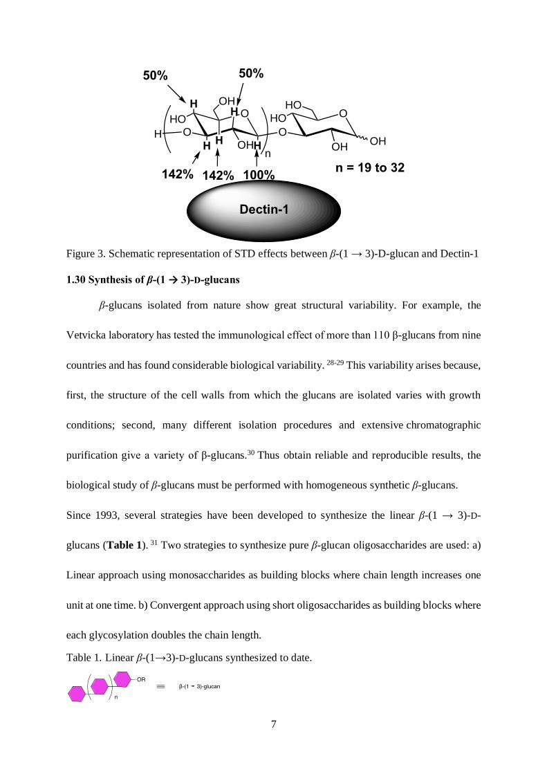
To precisely identify the binding epitope of β-(1 → 3)-D-glucan with its receptors, saturation
transfer difference (STD) NMR experiments were performed on laminarin (oligo- β-(1 → 3)-
D-glucan found in brown algae) in the presence of Dectin-1(Figure 3). In this study, the H-1
was selected as internal standard and the STD-effect was set as 100%. The α-face protons H-
3, H-5 display a 142% STD-effect, whereas H-2 and H-4 protons only display 50% STD-effect.
These results indicate that the binding affinity is mainly from the hydrophobic interactions
between the α face of glucan and the hydrophobic groove of Dectin-1.27
7
Figure 3. Schematic representation of STD effects between β-(1 → 3)-D-glucan and Dectin-1
1.30 Synthesis of β-(1 → 3)-D-glucans
β-glucans isolated from nature show great structural variability. For example, the
Vetvicka laboratory has tested the immunological effect of more than 110 β-glucans from nine
countries and has found considerable biological variability. 28-29 This variability arises because,
first, the structure of the cell walls from which the glucans are isolated varies with growth
conditions; second, many different isolation procedures and extensive chromatographic
purification give a variety of β-glucans.30 Thus obtain reliable and reproducible results, the
biological study of β-glucans must be performed with homogeneous synthetic β-glucans.
Since 1993, several strategies have been developed to synthesize the linear β-(1 → 3)-D-
glucans (Table 1). 31 Two strategies to synthesize pure β-glucan oligosaccharides are used: a)
Linear approach using monosaccharides as building blocks where chain length increases one
unit at one time. b) Convergent approach using short oligosaccharides as building blocks where
each glycosylation doubles the chain length.
Table 1. Linear β-(1→3)-D-glucans synthesized to date.
O
OHO
OH
HO
O
OHO
OH
OH
OHH
n
n = 19 to 32
Dectin-1
142% 142% 100%
50% 50%
H
HH
H
H

8
Reference
Reference
n = 1 1993, Takeo et al.32
2005, Jamois et
al.33
2011, Adamo et al.
34
2013, Jong et al.35
n = 6 1993, Takeo et
al.32
2010, Tanaka et
al.36
2015, Liao et
al.37
n = 2 1993, Takeo et al.32
2003, Zeng et al.38
2005, Jamois et
al.33
n = 8 2015, Liao et
al.37
n = 3 1993, Takeo et al.32
2005, Huang et
al.39
2005, Jamois et
al.33
n = 10 2010, Tanaka et
al.36
2013, Weishaupt
et al.40
2015, Liao et
al.37
n = 4 1993, Takeo et al.32
2009, Mo et al.41
2011, Adamo et
al.34
2015, Liao et al.37
2015, Elsaidi et
al.42
n = 12 2010, Tanaka et
al.36
n = 5 1993, Takeo et al.32 n = 14 2010, Tanaka et
al.36
1.31 Linear approach
In a typical example of the linear approach, Vetvicka and co-workers synthesized tri,
tetra and pentasaccharides using an iterative deprotection-glycosylation process (Scheme 1).33
In this process monosaccharide 1 acted as both donor and acceptor to elongate the chain. The

9
3-ONAP group of monosaccharide 1 was selectively deprotected by DDQ to serve as acceptor.
The donor 1 was activated by NIS / TESOTf for glycosylation to give the disaccharide. The
Benzoyl group at C-2 was installed for the neighboring group participation leading to β-
selectivity. After glycosylation, the oligosaccharides were deprotected using Zemplen
deacetylation and catalytic hydrogenolysis to afford the corresponding free oligosaccharides.
It was found that the synthesized laminaritetraose and laminaripentaose have similar
immunostimlatory effects to the natural β-(1→3)-glucan phycarine. 33
Scheme 1. β-(1 → 3)-D-glucan synthesis by iterative glycosylation process
1.32 Convergent approach
The convergent strategy improves the “n+1” elongation process into an “n+n” process
by employing short oligosaccharides as building blocks. In 2012, Takahashi and co-workers
synthesized linear hexadecasaccharides (16 units) employing a tetra-saccharide as key building
block.43 In 2015, the Guo lab accomplished the convergent synthesis of octa-, deca-, and
dodeca- β-(1→3)-D-glucans. The synthesis was accomplished with preactivation-based
iterative glycosylation using p-tolylthioglycosides as donors and disaccharide 4 as key building
OO
NAPOOBz
SEt
OPh 1) NIS, BnOH, TESOTf 89%
2) DDQ, CH2Cl2, MeOH 85%
3) 1, NIS, TESOTf, 83%1
OO
NAPOOBz
O
OPhOO
OBzOBn
OPh
OO
OOBz
O
OPhOO
OBzOBn
OPhOO
NAPOOBz
OPh
n = 1, 2, 3
2
OHO
OOH
O
HOOHO
OHOH
HOOHO
HOOH
HO
n
n = 1, 2, 3
deprotection
1) DDQ, CH2Cl2, MeOH 86%
2) 1, NIS, TESOTf, 78%
OO
OOBz
O
OPhOO
OBzOBn
OPhOO
NAPOOBz
OPh
n
iterative process
3

10
block (Scheme 2). The synthesized oligosaccharides were coupled with a carrier protein
keyhole limpet hemocyanin (KLH) to form a new glycoconjugate vaccine. These conjugates
successfully provoked protective immunity against Candida albicans infections. 37
Scheme 2. Guo and co-workers’ convergent synthesis of linear β-(1→3)-D-glucans
1.33 Solid phase oligosaccharide synthesis (SPOS)
Traditional solution phase oligosaccharide synthesis has several limitations for longer
saccharide synthesis. For example, the need for separation of side products after each
glycosylation, the poor solubility of longer oligosaccharides, low stepwise yields and time-
consuming protecting group manipulation. Solid phase oligosaccharide synthesis provides a
more efficient way of oligosaccharide synthesis. Automated solid phase synthesis of
oligopeptides and oligonucleotides have made great contributions to the progress in proteomics
and genomics research. 44-45 The automated oligosaccharide synthesis, however, still leaves
much to be accomplished.46 Many laboratories have realized the solid phase oligosaccharide
synthesis.47 In 2013, Seeberger’s group developed the automated solid phase synthesis of a
linear β-(1→3)-D-glucan with 12 glucose units.40 The glycosylation method employs a
NAPO
OO
OBz
OPh
O
OO
OBzSTol
OPh
4
HO
OO
OBz
OPh
O
OO
OBzO
OPh
i) 2-azidoethanol, AgOTf TTBP, p-TolSCl, CH2Cl2 -78 oC
ii) DDQ, CH2Cl2, H2O 91%
O
OO
OBz
OPh
O
OO
OBzO
OPh
2HO
OO
OBz
OPh
O
OO
OBz
OPh
O
OO
OBzO
OPh
nHO
OO
OBz
OPh
n = 4, 6, 8, 10
O
OHO
OH
HO
O
OHO
OHO
HO
nHO
OHO
OH
HO
n = 4, 6, 8, 10
i) 4, AgOTf, TTBP p-TolSCl, CH2Cl2, -78 oC
ii) DDQ, CH2Cl2, H2O 90%N3
N3N3
H2N
deprotection
iterative process
5
6

11
glucosyl phosphate as donor with a pivaloyl group in the C-2 hydroxyl group and an orthogonal
protecting group at C-3 (Fmoc) (7). The first glycosylation connected 7 to the Merrifield resin
photolabile linker 8. After glycosylation, the Fmoc group at C-3 was selectively cleaved by
piperidine in DMF and ready for the next glycosylation. This two-step process was repeated
12 times for elongation before the Merrisfield resin linkage was cleaved under UV irradiation
to give the protected dodecasaccharide 9. Global deprotection gave the final product 10 in 4.6%
overall yield after 25 steps, with an average yield of 88% per step (Scheme 3).
Scheme 3. Seeberger’s automated solid phase synthesis of oligo-β-(1→3)-D-glucan
2.00 Glycan mimetics
2.10 The challenge of oligosaccharide synthesis
The other two major biomolecules, oligopeptides and oligonucleotides, are linear
biopolymers connected with amide and phosphodiester bonds. Unlike templated peptide and
OBnO
FmocO OOPiv
BnO
P
O
BuOOBu
O2N
NCbzHO
O
5
+
Glycosylation
OBnO
O O
OPiv
BnO
O2N
NCbz
O
5
Fmoc
n
Glycosylation
OBnO
O O
OPiv
BnO
O2N
NCbz
O
5
H
n
Deprotection of O-Fmochν, CH2Cl2
OBnO
O O
OPiv
BnO
NHCbz
5
H
12
Global deprotection OHO
O O
OH
HO
NH2
510
OHO
OOH
HO
OHO
HOOH
HO
Merrifield Resin
Merrifield Resin
Merrifield Resin
7 8
9 10

12
nucleotide synthesis, oligosaccharide synthesis encounters two major challenges: A)
Regioselectivity of different hydroxyl groups. B) Stereoselectivity of glycosidic bond
formation, also branched structures. Furthermore, the complexity of oligosaccharides is
enormously greater than in the oligonucleotides and oligopeptides. For example, in the case of a
hexanucleotide there are a total of 46 (=4096) different structures possible and 206(=64 million) for
hexapeptide. In the case of hexasaccharides, based on the 10 mammalian monosaccharides,
regioisomers and two different stereoisomers, it was calculated that 192 billion different structures
are theoretically possible.48 On one hand, oligosaccharide synthesis is still challenging by
conventional organic synthesis, and only few laboratories in the world could accomplish long
oligo-β-(1→3)-D-glucans synthesis (Table 1). On the other hand, as the hydrophilic nature of
oligosaccharides make them poor ligands to receptors, and carbohydrate protein interactions
are dependent on multivalent interactions.49 In addition, many natural oligosaccharides are
easily degraded by glycosidase, which makes them poor drug candidates. Our projects are
designed to provide solutions to these problems by modifying the original β-(1→3)-D-glucan
structure to achieve 3 goals. A) Higher synthetic efficiency: Modification of the structure could
allow development of a simple and efficient methods for connecting the different units and
would allow access to long-chain polymers, ideally on solid support. B) Higher binding affinity
to receptors: Rather than working on synthesizing long oligosaccharide, we seek to redefine
the problem and utilize new approaches. Thus, by preparing glycan mimetics with short chain
lengths we hope to reproduce the biological activity of natural long oligosaccharides. For
example, natural glucans are hydrophilic and through modification we could increase the
13
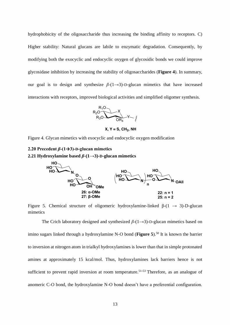
hydrophobicity of the oligosaccharide thus increasing the binding affinity to receptors. C)
Higher stability: Natural glucans are labile to enzymatic degradation. Consequently, by
modifying both the exocyclic and endocyclic oxygen of glycosidic bonds we could improve
glycosidase inhibition by increasing the stability of oligosaccharides (Figure 4). In summary,
our goal is to design and synthesize β-(1→3)-D-glucan mimetics that have increased
interactions with receptors, improved biological activities and simplified oligomer synthesis.
Figure 4. Glycan mimetics with exocyclic and endocyclic oxygen modification
2.20 Precedent β-(1→3)-D-glucan mimetics
2.21 Hydroxylamine based β-(1→3)-D-glucan mimetics
Figure 5. Chemical structure of oligomeric hydroxylamine-linked β-(1 → 3)-D-glucan
mimetics
The Crich laboratory designed and synthesized β-(1→3)-D-glucan mimetics based on
imino sugars linked through a hydroxylamine N-O bond (Figure 5).50 It is known the barrier
to inversion at nitrogen atom in trialkyl hydroxylamines is lower than that in simple protonated
amines at approximately 15 kcal/mol. Thus, hydroxylamines lack barriers hence is not
sufficient to prevent rapid inversion at room temperature.51-53 Therefore, as an analogue of
anomeric C-O bond, the hydroxylamine N-O bond doesn’t have a preferential configuration.
HO N
HOO
HO
N
HO
OAll
HO
n
22: n = 125: n = 2
OHO
HOOMeOH
OHO N
HOHO
26: α-OMe27: β-OMe

14
They developed a ring-closing double reductive amination method to prepare the
hydroxylamine mimetics.54
The enantiomerically pure cyclopentadiene-derived mesyloxy epoxide 11 was
subjected to ring opening with potassium hydroxide and acetophenone oxime in hot DMF to
give the desired o-cyclopentenyl oxime 12 in 34% yield. Subsequent benzylation and
elimination was completed in one pot to give cyclopentene derivative 13 in 99% yield.
Cleavage of the oxime with 2,4-DNP catalyzed by sulfuric acid gave hydroxylamine 14 in 74%
yield (Scheme 4). The hydroxylamine 14 was protected as its N, N-diBoc derivative 15 by a
standard carbamate forming reaction in 97% yield. Ring opening of N, N-diBoc cyclopentenyl
hydroxylamine 15 using catalytic osmium tetroxide and sodium metaperiodate to give a
dialdehyde 16 with a protected ONH2 in 49% yield. Treatment of compound 19 with catalytic
amount of osmium tetroxide and NMO gave a diol intermediate which was immediately
cleaved by sodium metaperiodate to give the dialdehyde 20 in 80% yield (Scheme 4).

15
Scheme 4. Dialdehyde synthesis by oxidative cleavage of cyclopentene derivatives
Dialdehyde 16 was subjected to ring-closing double reductive amination with allyl
hydroxylamine HCl salt to give N,N-diBoc protected hydroxylamine intermediate 17. N,N-
diBoc protecting group was removed under acidic condition to give free hydroxylamine 18 in
88% yield (Scheme 5).
Scheme 5. Monomer synthesis by oxidative cleavage and double ring closing reductive
amination
Hydroxylamine intermediate 18 was subjected to another reductive amination with
dialdehyde 20 to give benzyl protected dimeric mimetic 21 in 30% yield. The benzyl group
O
MsO
BnO N
Ph
OH
KOH, DMF, 90 oCOH
MsO
BnO
34%O
Ph
N
NaH, BnBr
DMF99%
OBn
BnO
O
Ph
N
2,4-DNP, H+
MeOH
74%
OBn
BnO
O
NH2
11 1213
14
Boc2O, DMAP
MeCN
97%
OBn
BnO
O
NBoc2
15
OsO4, NaIO4
Dioxane / H2OOBn
BnO
O
NBoc2
O
O
16
49%
OBn
BnO
OBn
19
OsO4, NaIO4
Dioxane / H2OOBn
BnO
OBn
O
O
20
80%
N
BnO
Boc2NO
OBn
O
i) HCl, MeOH
ii) NaHCO3
88%
N
BnO
H2NO
OBn
O
1817
OBn
BnO
O
NBoc2
O
O
i) AllONH3Cl NaOAc, MeOH / H2O
ii) NaBH3CN, AcOH MeOH / H2O 87%
16

16
was deprotected by BCl3 to give the hydroxylamine based disaccharide mimetic 22 (Scheme
6).
Scheme 6. Synthesis of dimeric hydroxylamine based β-(1 → 3)-D-glucan mimetics
The Trisaccharide mimetic 25 was also synthesized from compound 18. The N,N-diBoc
protecting group of 18 was removed under acidic condition and the intermediate was subjected
to double reductive amination with dialdehyde 16 to give the dimeric intermediate 23 in 53%
yield. The N,N-diBoc protecting group were deprotected under acidic condition to give free
hydroxylamine, which was subjected to double reductive amination with dialdehyde 20 to give
benzyl protected trimeric hydroxylamine 24 in 7% yield. The benzyl protecting group was
removed by BCl3 in DCM to give the hydroxylamine based trisaccharide mimetic 25 (Scheme
7).
N
BnOH2NO
OBn
O
20, NaBH3CN, AcOH MeOH / H2O 30%
N
BnOO
OBn
ON
BnOBnO
OBn
1821
22
BCl3, DCM
N
HOO
OH
O
N
HOHO
OH
100%

17
Scheme 7. Synthesis of trimeric hydroxylamine based β-(1 → 3)-D-glucan mimetics
To evaluate the binding affinity of the hydroxylamine to CR3 and Dectin-1, the
Vetvicka laboratory tested the mimetics’ ability to inhibit anti-CR3 and anti-Dectin-1
fluorescein isothiocyanate (FITC) conjugated antibody staining of human neutrophils and
mouse macrophages. For comparison purposes, monohydroxylamines 26 and 27 were also
screened. In terms of CR3 binding affinity, incubation of a 0.1μg / ml solution of β-(1→3)-
dimer mimetic 22 and β-(1→3)-trimer mimetic 25 caused 26% and 34% decreases in inhibition
of staining human neutrophils, while the anomeric β-(1→6)-dimer mimetic 26 and 27
decreases were 19% and 22% (Table 2). In terms of Dectin-1 binding affinity, incubation of a
0.1μg/ml solution of β-(1→3)-trimer mimetic 25 with mouse macrophage led to 43% decrease
in the inhibition of anti-dectin-1-FITC staining of mouse neutrophils. Under the same
i) 20, HCl, MeOH MeOH / H2O
N
BnOO
OBn
O
N
BnOO
OBn
N
BnOBnO
OBn
ii) NaBH3CN, AcOH MeOH / H2O 7%
N
BnOO
OBn
O
N
BnOBoc2NO
OBn
23
BCl3, DCM
N
BnOH2NO
OBn
O
16, NaBH3CN, AcOH MeOH / H2O 53%
18
24
N
HOO
OH
O
N
HOO
OH
N
HOHO
OH
25
100%

18
conditions, the β-(1→3)-dimeric mimetic 22 caused 28% decrease in inhibition of antibody
staining whereas the β-(1→6)-dimer mimetics 26 and 27 caused 29% and 21% decreases
respectively (Table 2). These results indicate that binding of the hydroxylamines to the lectin
domains of both CR3 and Dectin-1 is correlate to length and linkage.
Table 2. Percentage inhibition of anti-CR3 and anti-Dectin-1-FITC antibody staining of
neutrophils, macrophages by 0.1μg/mL substrate. a Mean ± SD
Compound
Linkage-
mimicked
Oligomer
NO.
% inhibition of
anti-CR3-FITC
staining
human
neutrophilsa
% inhibition of
anti-dectin-1-
FITC staining of
mouse
neutrophilsa
26 1→ 6 dimer 19.7±1.7 29.3±2.2
27 1→ 6 dimer 22.3±1.9 20.6±2.1
22 1→ 3 dimer 26.4±2.7 28.2±2.9
25 1→ 3 trimer 34.2±3.3 43.1±3.5
Hydroxylamine based β-glucan mimetics 26, 27, 22, 25 (10 μg / mL) were also tested
for their ability to stimulate phagocytosis of synthetic polymeric 2-hydroxyethyl methacrylate
microspheres by human macrophage-like RAW 264 cells.55 Commercial yeast derived
insoluble whole glucan particles WGP (hollow spheres of long polymers of primarily β-(1→
3)-D-glucan) were used as reference (Table 3). The result indicates that the β-(1→3)-trimer
mimetic 25 could stimulate 16% of phagocytosis, which is more effective than the β-(1→6)-
dimer mimetic 26, 27 and 22. It is notable that the level of phagocytosis stimulated by the β-
(1→3)-trimer mimetic 25 was more than 50% of that induced by WGP. These results indicate

19
that the hydroxylamine glucan mimetics have good immunostimulating ability even at short
length.
Table 3. Percentage stimulation of phagocytosis. a Mean ± SD
Compound Linkage
mimicked
Oligomer
NO.
% stimulation of phagocytosis
(RAW 264 macrophages, 10μg /
mL, 24h) a
26 1→ 6 dimer 6.7±0.9
27 1→ 6 dimer 4.1±0.5
22 1→ 3 dimer 7.8±1.1
25 1→ 3 trimer 16.6±2.0
WGP 1→ 3 Insol polymer 30.1±2.8
2.22 β-(1→3)-D-Glucan with thiolinkage
Figure 6. Chemical structure of oligo-β-(1 → 3)-D-glucans with thiolinkage
Thioglycoside, in which the glycosidic oxygen atom is replaced by sulfur, is known to
be more stable to acidic or enzymatic hydrolysis. Moreover, this modulation has only minor
impact on overall conformation. Based on these facts, the Vetvicka laboratory designed
thioglycosidic-linked oligo-β-(1→3)-glucans families (Figure 6).56-57 The synthesis started
from peracetylated laminaribiose 28 and laminaritriose 29. Treatment of the compound 28 and
29 with 33% HBr / HOAc afforded the anomeric bromide 30 and 31 in 86% and 80% yield

20
respectively. The anomeric bromide was substituted by thioacetate anion to give the β-
glucopyranosyl thioacetate 32 and 33 which were subjected to selective deacylation to give the
anomeric thiol 34 and 35 in 85% and 68% respectively. In the presence of the crown ether
Kryptofix 21 and sodium hydride, anomeric thiol was activated to replace the triflate group of
3-O-trifluoromethanesulfonyl-1,2;5,6-di-O-isopropylidene-α-D-allofuranose. Compound 34
and 35 were coupled with triflate 36 to give the disaccharide intermediates 37 and 38 in 86%
and 71% respectively. After acidic hydrolysis, acetylation and Zemplén deacetylation, the
intermediates 37 and 38 were deprotected and acetylated to give trisaccharide 39 and
tetrasaccharide 40 in 70% and 51% respectively. Final Zemplén deacetylation and Sephadex
G-25 gel purification gave the thio-linked β-(1 → 3)-D-glucan mimetics 41 and 42 (Scheme
8).

21
Scheme 8. Synthesis of oligo-β-(1 → 3)-D-glucans with thio-linkage
The same protocol was applied to synthesize trimeric mimetic 43 and tetrameric
mimetic 44. These four compounds were evaluated by Vetvicka laboratory. First, the
phagocytic abilities were tested, including their effect on stimulation of peritoneal
macrophages and peritoneal blood neutrophils and monocytes. A strong immunostimlating
effect was observed from compound 42, 43 and 44. To determine if the samples influence
cytokine production, they tested the production of IL-2 by splenocytes and the levels of IL-1β
and TNF-α in peripheral blood. The compounds 41, 43 and 44 stimulated the production of
tested cytokines and 44 is the most active mimetic. Finally, theses samples were assessed for
AcO
OAcO
OAc
AcO
O
OAcO
AcOOAc
AcOHBr / HOAc (33%) KSAc / DMF
n
28: n = 129: n = 2
AcO
OAcO
OAc
AcO
O
OAcO
AcOBr
AcO
n
30: n = 1, 86% 31: n = 2, 80%
AcO
OAcO
OAc
AcO
O
OAcO
AcO
SAc
AcO
n
32: n = 1, 85% 33: n = 2, 68%
H2NCH2CH2SH
MeCNAcO
OAcO
OAc
AcO
O
OAcO
AcO
SH
AcO
n
34: n = 1, 85% 35: n = 2, 68%
OTfO
O
O
OO
36
36, NaH, DMF
Kryptofix 21
AcO
OAcO
OAc
AcO
O
OAcO
AcO
S
AcO
n
OO
O
OO
37: n = 1, 86% 38: n = 2, 71%
i) TFA / H2O
ii) Ac2O, PyridineAcO
OAcO
OAc
AcO
O
OAcO
AcO
AcO
n
S
OAcO
AcO
AcO
OAc
39: n = 1, 70% 40: n = 2, 51%
NaOMe / MeOH
HO
OHO
OH
HO
O
OHO
OH
HO
n
S
OHO
OH
HO
OH
41: n = 1, 78% 42: n = 2, 77%
CH2Cl2
OO
NH
O
HN
Kryptofix 21

22
their potency in inhibiting colon CSC-mediated tumor formation and/or metastasis. It is
noteworthy that the β-(1 → 3)-thioglucan 43 which has two thioglycosidic linkages,
demonstrated good anti-cancer activity. Compound 43 significantly suppressed spheroid
formation and proliferation of colon cancer stem-like cells from human colon adenocarcinoma,
more effective than the natural laminarin. These promising results indicate the presence of
sulfur atom is beneficial for biological activity.
3.00 Sulfur in medicinal chemistry
Sulfur is the 5th most abundant element on earth. Our body consists 0.25% of sulfur,
which is crucial for many biological processes.58 Two amino acids, cysteine and methionine
and two vitamins (biotin and thiamine) are organosulfur compounds. The disulfide bond is a
common linkage in proteins that is crucial to the protein structures. Sulfur-aromatic interactions
were critical in chemical and biological process with many examples in the context of protein-
protein and protein-ligand interactions.59 The preferential conformation of a bridged
oxathiolane compound gave the evidence for sulfur-aromatic interactions.60 Sulfur’s history in
drugs dates to ancient Egypt, where people used sulfur as an antiseptic. Today, the organosulfur
compounds have extraordinary impact on medicinal chemistry. In the U.S. 40% and 25% of
the top 20 drugs by retail sales and prescription respectively contain sulfur.61 In addition, a
survey of the top 200 brand name drugs by total U.S. prescriptions in 2011 revealed that 24.8%
of drugs contain sulfur61. Accoridng to statistics (Figure 7), over the 12 major diseases
categories, sulfur containing drugs is 50% more than fluorine in the Anti-infectives category.
In Cardiovascular and Musculoskeletal categories, sulfur containing drugs comprises 60%
more than the fluorine containing drugs.61

23
Figure 7. Percentage composition of sulfur-containg and fluorine-containing pharmaceuticals
that comprise each of the 12 representative disease categories
4.00 Conclusion
Figure 8. Chemical structure of oligomeric β-(3 → 5)-dithio-D-glucan mimetics
Over the past few years, great effort was spent on understanding and improving the
immunostimulating effect of β-(1 → 3)-D-glucan.62 It was demonstrated that laminaripentaose,
a five-membered oligo-β-(1→3)-D-glucan, could also trigger immunostimulating activity
equivalent to that of laminarine.33 X-Ray crystallographic studies of recombinant Dectin-1
have revealed a hydrophobic pocket lined by the side chains of Trp 221 and His 223.25 STD-
NMR experiments revealed that laminarin binding through vander waals interaction of the α-
face of terminal pyranose rings (at both the reducing and nonreducing ends) with the Trp 221
36.4
27.2 25.222 20.4 20.3 18.8 17.5
13.9 13.4 12.9 11.8
15.9
9.9 12.3
5.513.9 12.2 14.1 17.5
10.8 12.418.6
5
0
10
20
30
40
50
60
Fluorine Sulfur
SHOHO
OHS
HOSHO
OHS
HOS
HO
OH OH
HO
n
n = 1, 2

24
and His 223 of the Dectin-1 binding domain. 27, 63 Crich and coworkers prepared hydroxylamine
analogues of β-glucan. Interestingly, although the oligomer is short, they have very good
biological activity. The good activity of these small analogues is possibly because of the
increased hydrophobicity of the synthetic 2-deoxy hydroxylamine mimetics. These studies and
preliminary results proved the concept of principle that the increased hydrophobicity of the β-
glucans increased their binding affinity to CR3 and Dectin-1. More importantly,
hydroxylamine analogues of β-glucan provide an unprecedented example using hydroxylamine
N-O bond mimic glycosidic C-O bond, the fact that low barrier to inversion of hydroxylamine
nitrogen lone pair helps it mimic both α and β glycosidic bond. Based on these studies, we
hypothesized that replacement of both the exocyclic oxygen and endocyclic oxygen atom by
more lipophilic sulfur can increase the noncovalent interactions between β-(1 → 3)-D-glucan
mimetics and the receptors. Second, the modification with endocyclic and exocyclic sulfur can
resist the acidic and enzymatic hydrolysis to the largest extent. It is very likely that these
modifications will give excellent β-(1 → 3)-D-glucan mimetics. Therefore, we designed the
oligo-3,5-dithio-β-D-glucopyranoside as β-(1 → 3)-D-glucan mimetics (Figure 8).

25
CHAPTER 2: DESIGN AND SYNTHESIS OF OLIGO-(3,5-DITHIO-β-D-
GLUCOPYRANOSIDES) AS β-(1 → 3)-D-GLUCAN MIMETICS
1.00 Introduction
R. Schmidt’s review64 about thioglycoside synthesis provide many examples of
thiooligoglycoside synthesis. There are three different methods to synthesize thioglycosides: 1)
Base promoted thioglycosylation; 2) Anomeric thiol alkylation; 3) Acid catalyzed
thioglycosylation.
Figure 9. Illustration of 3 methods to build up thio-linked oligosaccharide
1.10 Example of base promoted S-glycosylation: S-analogue of Sialyl Lewis X synthesis
Sialyl Lewis X is a tetrasaccharide carbohydrate that is usually attached to O-glycans
on the cell surface. Schmidt and co-workers65 performed base promoted S-glycosylation to

26
synthesize Sialyl Lewis X thio-analogue. In the presence of Kryptofix 21 in THF, the thiol of
46 substituted the anomeric chloride of compound 45 to give thioglycoside 47 in 75% yield.
The anomeric siloxyl group was deprotected and acetylated to give compound 49 in 85% yield
and the anomeric carbon of disaccharide 49 was brominated to give compound 50 in 95% yield.
The anomeric bromide of 50 was substituted by thiol of 51 to give tetrasaccharide 52 in 70%
yield.
Scheme 9. Synthesis of the S-analogue of Sialyl Lewis X via base promoted S-glycosylation
1.20 Example of base promoted anomeric thiol alkylation: synthesis of 4-
O
O
HSOBz
O
OTDS
Ph
O
Cl
CO2Me
AcOOAc
OAc
AcHNAcO
NaH, Kryptofix 21 O
O
SOBz
O
OTDSO
MeO2CAcOOAc
OAc
AcHNAcO
i) TBAF, THF, RT
ii) Ac2O, Pyridine 85%
HBr / HOAc, 0 oC, 95%
O
AcO
SBzO
OAc
OAcO
MeO2CAcOOAc
OAc
AcHNAcO
OHSS
OBz
BzO
S
O
OAcOAc
OAc
NaH, DMF
70%
O
AcO
SBzO
OAc
O
MeO2CAcOOAc
OAc
AcHNAcO
OSS
OBz
BzO
S
O
OAcOAc
OAc
45 46 47
49
51
52
Ph
i) TFA, H2O
ii) Ac2O, Pyridine 2 steps 89%
O
AcO
SOBz
OAc
OTDSO
MeO2CAcOOAc
OAc
AcHNAcO 48
THF, 75%
O
AcO
SBzO
OAc
Br
O
MeO2CAcOOAc
OAc
AcHNAcO
50

27
thiomaltooligosaccharide
Scheme 10. The convergent synthesis of 4-thiomaltooligosaccharide
Driguez and co-workers66synthesized the 4-thiomaltopentaoside by anomeric thiol
alkylation. The synthetic route is an iterative process and the α-(1→4)-S-linkage was built up
by SN2 substitution of C-4 triflate from anomeric thiol. Coupling of compound 53 with
compound 54 afforded the triphenylmethyl 1,4-dithio-α-maltoside 55 in 78% yield. The 1-S-
triphenylmethyl group was deprotected in a two steps process. First, S-trityl group was cleaved
by PhHgOAc in the presence of hydrogen sulfide. Then, the anomeric thiol was converted to
S-acetyl to give compound 56 in 59% yield. Compound 57 was coupled with triflate 54 to give
the trisaccharide 57 in 78% yield. The same two steps procedure to converted the S-
triphenylmethyl into S-acetyl group was applied to 57 afforded 58 in 96% yield. In a
convergent approach, coupling of 58 with disaccharide triflate 59 afforded thio-malto-
pentasaccharide 60 in 68% yield.
OAcOAcO
AcO
AcO
SAc
O
OTf
AcOAcO
OAc
SC(Ph)3
ii) PhHgOAc, H2S DCM / MeOH
iii) Ac2O / Pyridine 59%
OAcOAcO
AcO
AcO
SO
AcOAcO
OAc
SC(Ph)353
54
55
54, Et2NH, DTT
56
OAcOAcO
AcO
AcO
SO
AcOAcO
OAc
SO
AcOAcO
OAc
SAc
i) PhHgOAc, H2S DCM / MeOH
ii) Ac2O / Pyridine 96%
O
OTf
BzO
BzO
OBz
SO
BzOBzO
OBz
OMe
58
59
57
DMF, 0 oC, 78%
OAcOAcO
AcO
AcO
SO
AcOAcO
OAc
SAc
OAcOAcO
AcO
AcO
SO
AcOAcO
OAc
SO
AcOAcO
OAc
SC(Ph)3
54, Et2NH, DTT
DMF, 0 oC, 78%
59, Et2NH, DTT
DMF, 0 oC, 68%
AcOO
AcOAcO
OAc
SO
AcOAcO
OAc
S
60
O
BzO
BzO
OBz
SO
BzOBzO
OBz
OMe
2

28
1.30 Examples of acid catalyzed S-glycosylation
1.31 Tf2O / DTBMP promoted S-glycosylation using glucosyl sulfoxide donor
Crich lab67 developed the β-selective glycosylation in which anomeric S-phenyl
sulfoxide acts as glycosylation donor and Tf2O / DTBMP as the activator. They applied this
method in S-glycosylation. The sulfoxide donor 61 was activated by Tf2O / DTBMP at -78 °C
to 0 °C, the generated mannosyl triflate intermediate was substituted by thiol acceptor 62 to
give disaccharide 63 with β thio-glycosidic linkage (Scheme 11).
Scheme 11. β-selective S-glycosylation of mannosyl sulfoxide 61 and thiol acceptor 62.
Application of this method to 5-thio-glucose was not reported because of the lack of
selectivity in the oxidization of exocyclic and endocyclic sulfur (Scheme 12). 68
Scheme 12. Oxidation of ethyl β-1,5-dithioglucopyranosides with m-CPBA.
1.32 TESOTf promoted S-glycosylation using glucopyranosyl trichloroacetimidate
donor
S-glycosylation using acid catalyzed trichloroacetimidate donor and thiol acceptor was
studied by Pinto and co-workers.69 In the glycosylation of the thiol 67 with 2,3,4,6-tetra-O-
acetyl-α-D-glucopyranosyl trichloroacetimidate 68. When 0.14 equivalent of the catalyst
OO
BnO
S
BnOO
OHS
AcO
OMeOAc
BnO Tf2O/ DTBMP
DCM
-78 oC, 61%O
Ph
Ph
OO
BnO
BnOOPh
OS
AcO
OMeOAc
BnO
61 62 63

29
TESOTf were added, both glycosylation product 69 were formed in 59% yield with an α and
β ratio of 1: 2.3 (Scheme 13).
Scheme 13. TESOTf catalyzed S-glycosylation with 5-thio-glucopyranosyl
trichloroacetimidate 68 as the donor compound 67 as the acceptor.
They also studied the TESOTf promoted glycosylation of 5-thioglucopyranosyl
trichloroacetmidate 74 with 4-OH, 4-SH and 4-SeH groups of glucopyranoside acceptors 70-
73 (Scheme 14) . 70
Scheme 14. TESOTf promoted S-glycosylation using 5-thio-glucopyranosyl
trichloroacetimidate 74 as the donor with glucose with 4-OH, 4-SH and 4-SeH (70-73) as the
acceptors.
The glycosylation of acceptor 70 with glucopyranosyl trichloroacetimidate 74 activated
by triethylsilyl triflate (donor : acceptor : promotor = 1:2:0.1) afforded exclusively α-
disaccharide 75 in 87% yield (Table 4, Entry 1). When glycosylation was performed with the
same substrate but different ratio (donor : acceptor : promotor = 2:1:0.2) at -50 °C, the
glycosylation gave the orthoester in 88% yield (Table 4, Entry 2). The glycosylation with
more reactive benzylated acceptor 71 gave a 1:1 mixture of α and β disaccharide 76 and 77

30
(Table 4, Entry 3). The glycosylation with the acceptor 4-SH-glucopyranoside 72 afforded
mainly α-disaccharide 78 in 53% and minor β-disaccharide 79 was isolated in only 1.5% yield
(Table 4, Entry 4). The glycosylation with the acceptor 4-selenol-glucopyranoside 73 gave α-
disaccharide 80 in 46% and β-disaccharide 81 in 11% yield (Table 4, Entry 5).
Table 4. Glycosylation reaction of 5-thio-glucopyranosyl trichloroacetimidate
a Donor: Acceptor: TESOTf
2.00 Synthesis of 3,5-dithio-glucopyranose

31
Figure 10. Retrosynthetic analysis of oligo-3,5-dithio-β-D-glucopyranoside.
At first, we planned to synthesize the oligo-3,5-dithio-β-D-glucopyranoside by base
promoted S-glycosylation between the acceptor 3,5-dithioglucopyranoside with free 3-SH and
3,5-dithio-glucopyranosyl bromide as the donor (Figure 10). According to Whistler et al,71 5-
thio-glucopyranose can be prepared from acetolysis of 5-thio-glucofuranose. Therefore, we
decided to synthesize 3,5-dithio-1,2-O-isopropylidene-glucofuranose as the starting material.
The proposed synthesis started from 1,2-O-isopropylidene-5,6-didexoy-5,6-epithio-α-D-
allofuranose, which can be prepared in large scale from 1,2;5,6-O-diisopropylidene-α-D-
glucofuranose.72 S-acetyl group could be introduced at C-3 via nucleophlic substitution of 3-
O-trifluoromethanesulfonyl of compound 1,2-O-isopropylidene-5,6-didexoy-5,6-epithio-α-D-
allofuranose to give 3-S-acetyl-1,2-O-isopropylidene-5,6-dideoxy-5,6-dithio-α-D-
glucofuranose. Finally, after ring opening of episulfide by acetate, the acidic acetolysis would
give the 3,5-dithio-glucopyranose (Scheme 15).
2.10 Synthesis of 3,5-dithio-α-D-glucofuranose
2.11 Synthesis of 3,5-dithio-α-D-glucofuranose by nucleophilic substitution on 3-O-
trifluoromethanesulfonyl group of 1,2-O-isopropylidene-5,6-dideoxy-5,6-epithio-α-L-
talofuranose

32
Scheme 15. Proposed synthesis of 3,5-dithio-glucopyranose
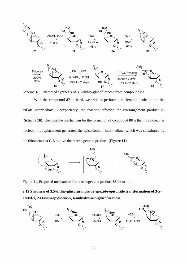
For the shorter synthetic route, we decided to synthesize 1,2-O-isopropylidene-5,6-
dideoxy-5,6-epithio-β-L-talofuranose 87 as a practice. Starting from 1,2;5,6-O-
diisopropylidene-α-D-glucofuranose 82. The 5,6-O-isopropylidene group was selectively
removed by acidic hydrolysis and the 6-OH was selectively tosylated to give 6-O-p-
toluenesulfonyl-1,2-O-isopropylidene-glucofuranose in 48% yield. Subsequent deprotonation
by sodium hydride initiated intramolecular substitution generated the 5,6-anhydro-1,2-O-
isopropylidene-α-D-glucofuranose 85 in 61% yield. The 5,6-epoxide of compound 85 was
converted to the 5,6-epithio compound 86 by treatment with thiourea. Conversion of the 3-
hydroxyl group by Dess-Martin oxidation and sodium borohydride reduction gave 1,2-O-
isopropylidene-5,6-epithio-α-L-talofuranose 88 in 95% yield.
33
Scheme 16. Attempted synthesis of 3,5-dithio-glucofuranose from compound 87
With the compound 87 in hand, we tried to perform a nucleophilic substitution the
triflate intermediate. Unexpectedly, the reaction afforded the rearrangement product 88
(Scheme 16). The possible mechanism for the formation of compound 88 is the intramolecular
nucleophilic replacement generated the episulfonium intermediate, which was substituted by
the thioacetate at C-6 to give the rearrangement product. (Figure 11).
Figure 11. Proposed mechanism for rearrangement product 88 formation.
2.12 Synthesis of 3,5-dithio-glucofuranose by epoxide-episulfide transformation of 3-S-
acetyl-1, 2-O-isopropylidene-5, 6-anhydro-α-D-glucofuranose.
OO
SAcO
S
OO
SAcO
O
OO
SAcO
HO
OO
SAcO
AcOAcS
Thiourea KOAc
Ac2O, AcOH
TsO
NaH
DMF MeOH

34
Scheme 17. Proposed synthesis of 3,5-dithio-glucofuranose by epoxide-episulfide
transformation
The first rearrangement product 88 taught us a lesson about the good nucleophilicity of
epislfide sulfur. To avoid intramolecular nucleophilic substitution, in another synthetic route,
we decided to install the first thioacetate at C-3 before the second thioacetate was introduced
at C-5 (Scheme 17).
Scheme 18. The attempted epoxide formation of compound 92.
The synthesis started from 1,2;5,6-di-O-isopropylidene-α-D-glucofuranose. After
Dess-Martin oxidation and sodium borohydride reduction, the 3-OH underwent inversion to
give 1,2;5,6-di-O-isopropylidene-α-D-allofuranose 89 in 80% yield. 3-OH of compound 89
was triflated followed by thioacetate displacement to give 3-S-acetyl-1,2;5,6-di-O-
isopropylidene-α-D-glucofuranose 90 in 84% yield. After the 5,6-isopropylidene group was
selectively removed with 50% acetic acid, 6-OH was tosylated to give compound 92 in 67%
yield. When we were treating compound 92 with DBU to prepare 5,6-epoxide, the
rearrangement product 93 was generated (Scheme 18). The structure was confirmed by
comparing the spectral data with that of the known compound.73 The possible mechanism is

35
that the acetyl migrated from S-acetyl to the neighboring hydroxyl and the thiolate cyclized to
give the rearrangment product 93 (Figure 12).
Figure 12. Proposed mechanism for the rearrangement product 93 formation
2.13 Attempted synthesis of 3,5-dithio-glucofuranose by nucleophilic substitution of the
trifluoromethanesulfonyl group of 3-O-trifluoromethanesulfonyl-1,2-O-isopropylidene-
5,6-anhydro-α-D-allofuranose
Scheme 19. Proposed synthesis of compound 98 by nucleophilic substitution of 3-O-
trifluoromethanesulfonyl group of compound 97.
To avoid the rearrangement, we proposed a new synthetic route in which the thioacetate
was introduced after the epoxide formation (Scheme 19). We started the synthesis from
1,2;5,6-di-O-isopropylidene-α-D-allofuranose. After acetylation of the 3-OH and selectively
acidic hydrolysis of 5,6-O-isopropylidene, we prepared compound 94 in 95% yield. Primary
alcohol benzoylationg and secondary alcohol sulfonylation gave the desired product 95 in 67%
yield. After treatment with sodium methoxide, the epoxide formation afforded the 5,6-anhydro-
1,2-O-isopropylidene-α-D-glucofuranose 96 in 49% yield. After the 3-OH was converted to
the triflate, thioacetate was introduced by slowly adding thioacetate / DMF solution at 0°C to
give the desired product 3-S-acetyl-5,6-anhydro-1,2-O-isopropylidene-α-D-glucofuranose 98
in 67% yield. When we performed the epoxide episulfide transformation on compound 98, the
OO
SAcO
HOTsO
DBU
OO
S
OO
TsO
O
OO
S
O
OAcTsO
O
OAc
S
OO

36
reaction gave complex mixtures from which no anticipated product was identified (Scheme
20).
Scheme 20. The attempted epoxide episulfide transformation of compound 98
We installed the S-pivalyl group to prevent the transesterification of the S-acetyl group.
The S-pivalyl group was installed by a SN2 substitution of triflate by potassium S-pivalate to
give 3-S-pivalyl-1,2-O-isopropylidene-5,6-anhydro-α-D-glucofuranose 99 in 66% yield. When
we performed the epoxide-episulfide transformation of the compound 99, the reaction gave the
product together with inseparable rearrangement prodcut. This result indicated that the S-
pivalyl was not able to completely prevent transesterification (Scheme 21).
Scheme 21. The attempted epoxide episulfide transformation of compound 99
2.2 Synthesis of the 3,5-dithio-glucopyranose by nucleophilic substitution of the C-3
triflate of 5-thio-allopyranose

37
Figure 13. Proposed synthesis of the 3,5-dithio-glucopyranose by nucleophilic substitution of
the C-3 triflate of 5-thio-pyranose
We decided to synthesize the 5-thio-allopyranose with an axial triflate at C-3 to install
the thioacetate (Figure 13). The synthesis started from 1,2;5,6-di-O-isopropylidene-α-D-
allofuranose. The installation of (2-naphthyl)methyl protecting group at 3-OH gave the product
100 in 91% yield. After acidic hydrolysis of the 5,6-O-isopropylidene group, the primary
hydroxyl was benzoylated and secondary hydroxyl was mesylated to give the product 102 in
70% yield. Treatment with sodium methoxide in methanol, neutralization with Amberlyst IR-
120 resin, and subsequent heating with thiourea afforded the 5,6-dideoxy-5,6-epithio product
103 in 70% yield. Compound 103 was refluxed with potassium acetate in the mixture of acetic
anhydride and acetic acid to give the ring opening product 104 in 56% yield. With compound
104, treatment with 60% acqueous trifluoroacetic acid gave a mixture of deprotection products
including small amount of cyclized product. We followed a two steps protocol of Driguez.74
First, compound 103 was refluxed in 50% aqueous acetic acid to remove the 1,2-O-
isopropylidene group. Then, the hemiacetal intermediate was treated with sodium methoxide
and cyclized to give 3-O-(2-naphthyl)methyl-5-thio-α,β-D-allopyranose. The crude mixture
was subjected to acetylation to give the desired product 105 in 45% yield (Scheme 22).

38
Scheme 22. Synthesis of compound 105.
With the compound 105, Zemplén deacetylation follwed by 4,6-O-benzylidene
protection and acetylation give the 4,6-O-benzylidene protected product 106 in 46% yield.
Removal of the 3-ONAP protecting group gave the product 107 as an anomeric mixture (α: β
= 1: 3) (Scheme 23).
Scheme 23. The 4,6-O-benzylidene protection of compound 105 and selective deprotection of
the NAP group.
With compound 107, the installation of triflate group at C-3 followed by SN2
substitution of the triflate using potassium thioacetate provided 3,5-dithio-glucopyranose
product 108 in 66% yield (Scheme 24). Reaction of compound 108 with 33% hydrogen
bromide in acetic acid gave the rearrangement product 109 in 39% (Scheme 24).

39
Scheme 24. The bromination reaction of compound 108
Alternatively, we decided to use acetyl protecting group to proceed the synthesis.
Oxidative deprotection of the 3-ONAP group of 105 afforded the product 110 as anomeric
mixture (α: β = 1: 1.3). The axial 3-OH of compound 110 was triflated and substituted by
thioacetate anion to give the acetyl protected 3,5-dithio-glucopyranose derivative 111 in 55%
yield. The bromination reaction provided the desired glucopyranosyl bromide 112 as an
anomeric mixture (α: β = 1:1) in 76% yield (Scheme 25).
Scheme 25. Preparation of 3,5-dithio-glucopyranosyl bromide.
3.00 S-Glycosylation study
3.10 Base promoted SN2 substitution of anomeric bromide by sodium ethanethiolate
With the 3,5-dithio-glucosyl bromide 112 in hand, we performed the SN2 substitution
of anomeric bromide by ethane thiolate in DMF. The reaction did not give the desired product.
(Scheme 26).
SAcO
AcSOAc
OAc
AcOi) Tf2O (3 equiv) Pyridine (7 equiv) DCM, 0 oC
ii) CsAc (1.5 equiv) DMF, 0 oC to RT
55%
112
111
SAcO
AcSOAc
Br
AcOHBr / HOAc
DCM, 0 oC
α: β = 1:176%
SAcO
NAPOOAc
OAc
AcODDQ (3 eq)
(20%) MeOH / DCM RT
SAcO
HOOAc
OAc
AcO
105 110 α : β = 1: 1
94%

40
Scheme 26. Attempted sodium ethane thiolate substitution of 5-thio-glucopyranosyl bromide
3.20 Acid catalyzed S-glycosylation of 3,5-dithio-glucopyranosyl trichloroacetimidate
Alternatively, we decided to try the acid catalyzed S-glycosylation to build up the thio-
linked disaccharide. Selective S-acetyl deprotection was performed to prepare the
glycosylation acceptor. However, when the compound 111 was treated with hydrazine
monohydrate, the reaction gave both 3-S-acetyl deprotection product 113 and anomeric O-
acetyl deprotection product 114 in 13% and 45% respectively. To improve the yield, the
sodium ethanethiolate was added in DMF at -40 °C and 3-thioacetate was selectively removed
to give 3-S-acetyl deprotected product 113 in 59% yield75 (Scheme 27).
Scheme 27. Preparation of compound 113 as S-glycosylation acceptor
To avoid 3-S-acetyl deprotection, a two steps protocol was applied to prepare the
glycosylation donor. First, the anomeric bromide of 112 was hydrolyzed in the presence of
TBAI to give compound 115 in 62% yield. With compound 115, the anomeric hydroxyl was
treated with trichloroacetonitrile and catalytic amount of DBU to give the glucopyranosyl
trichloroacetimidate 116 in 75% yield. With both donor and acceptor in hand, we performed
the TMSOTf promoted glycosylation at -78 °C and it was not successful (Scheme 28).
SAcO
AcSOAc
Br
AcOSAcO
AcSOAc
SEt
AcO
DMFX
112 α
EtSNa
SAcO
AcSOAc
OAc
AcONH2NH2.H2O (1.5 eq)
MeCN
SAcO
HSOAc
OH
AcOSAcO
HSOAc
OAc
AcO
45%13%
111 114113
+
EtSNa (1.5 eq)
DMF, -40 oC 59%
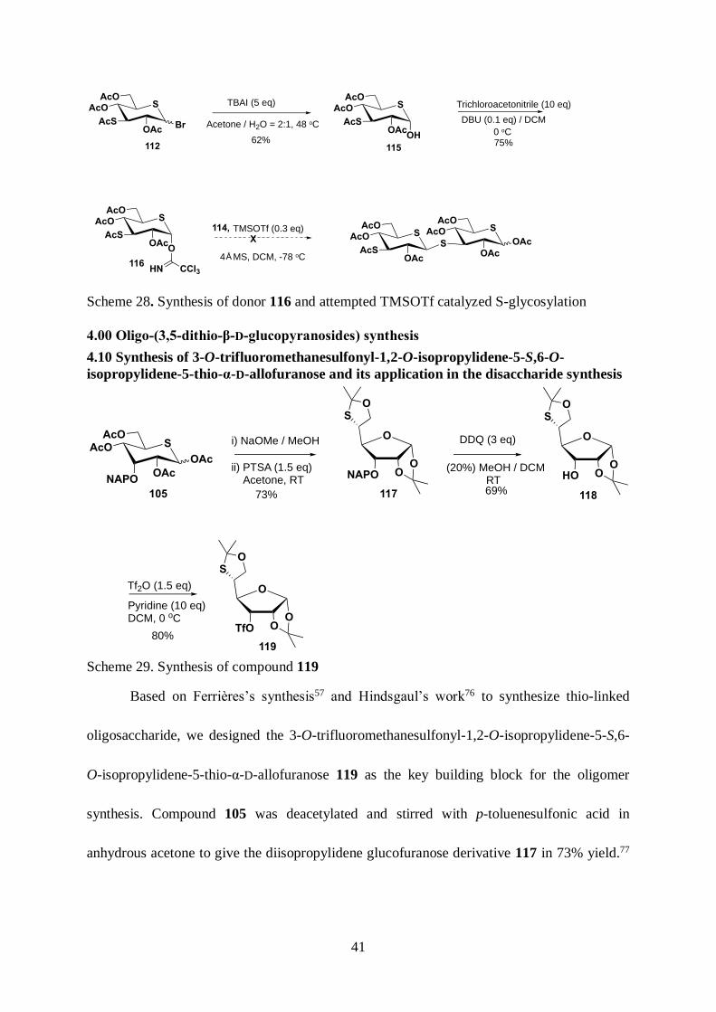
41
Scheme 28. Synthesis of donor 116 and attempted TMSOTf catalyzed S-glycosylation
4.00 Oligo-(3,5-dithio-β-D-glucopyranosides) synthesis
4.10 Synthesis of 3-O-trifluoromethanesulfonyl-1,2-O-isopropylidene-5-S,6-O-
isopropylidene-5-thio-α-D-allofuranose and its application in the disaccharide synthesis
Scheme 29. Synthesis of compound 119
Based on Ferrières’s synthesis57 and Hindsgaul’s work76 to synthesize thio-linked
oligosaccharide, we designed the 3-O-trifluoromethanesulfonyl-1,2-O-isopropylidene-5-S,6-
O-isopropylidene-5-thio-α-D-allofuranose 119 as the key building block for the oligomer
synthesis. Compound 105 was deacetylated and stirred with p-toluenesulfonic acid in
anhydrous acetone to give the diisopropylidene glucofuranose derivative 117 in 73% yield.77
SAcO
AcSOAc
Br
AcOSAcO
AcSOAc
OH
AcO TBAI (5 eq)
Acetone / H2O = 2:1, 48 oC
SAcO
AcSOAc
O
AcO
CCl3HN
62%
Trichloroacetonitrile (10 eq)
DBU (0.1 eq) / DCM
0 oC75%112 115
116
TMSOTf (0.3 eq)
4 MS, DCM, -78 oC
SAcO
AcSOAc
AcO SAcO
SOAc
OAc
AcO
Å
X
114,
SAcO
NAPOOAc
OAc
AcO
ii) PTSA (1.5 eq) Acetone, RT
105
OONAPO
O
SO
73%
DDQ (3 eq)
(20%) MeOH / DCM RT
OOHO
O
SO
69%
OOTfO
O
SO
Tf2O (1.5 eq)
Pyridine (10 eq)DCM, 0 oC
80%
117 118
119
i) NaOMe / MeOH

42
The 3-O-(2-naphthyl)methyl protecting group was removed by DDQ and the 3-OH was
converted to the triflate to give the building block 119 in 80% yield (Scheme 29).
Scheme 30. The disaccharide synthesis by coupling reaction.
With the compound 119, we performed the coupling reaction with the glucopyranosyl
thioacetate 120 to give the disaccharide mimetic 121 in 46% yield. After the silica gel
chromatography, extra triflate compound 119 were recycled without any rearrangement or
elimination reaction. This result demonstrated that the triflate compound 119 can serve as the
building block for oligomerization (Scheme 30).
4.20 Large-scale synthesis of penta-O-acetyl-5-thio-α,β-D-glucopyranose
Scheme 31.The large-scale synthesis of penta-O-acetyl-5-thio-a,β-D-glucopyranose
The success in thiol-triflate coupling reaction helps us to proceed the oligomer synthesis.
The linear synthetic route requires a large amount of monosaccharide 5-thio-glucopyranose as
SAcOAcS
OAc
SAc
AcO
OOTfO
O
SO
Et2NH
DMF / DTT0 oC to RT 46%
SAcOAcS
OAc
S
AcO
OO
O
SO
120 119 121
+
OO
OHO
O
124
12312282
i) Ac2O, Pyridine
O
ii) AcOH (50%) Pyridine (10 eq)
83%
OO
OAcO
HOHO
79% for 2 steps
i) NaOMe, MeOH
BzCl (1.1 eq)
OO
OHO
S
OO
OAcO
AcSAcO
125 126
OO
OAcO
MsOBzO
KOAc (5 eq)
(20%) AcOH / Ac2O
SAcO
AcOOAc
OAc
AcO
ii) Thiourea (2 eq)
i) AcOH (50%), 90 oC
ii) NaOMe / MeOH
iii) Ac2O / Pyridine
42%
65% for 2 steps
60%
MsCl (2 eq)
DCM MeOH, 80 oC
145 oC

43
starting material. We followed the synthetic route of Whistler78 to scale up 5-thio-
glucopyranose synthesis. 3-O-acetyl-1,2-O-isopropylidene-α-D-glucofuranose 122 was
synthesized after acetylation and the selective isopropylidene deprotection of 5,6-O-
isopropylidene.79 The primary alcohol was selectively benzoylated followed by sulfonylation
of secondary alcohol to give fully functionalized glucofuranose derivative 123 in 83% yield.
Treatment of sodium methoxide and neutralization by Amberlyst IR 120 resin gave 1,2-O-
isopropylidene-5,6-anhydro-α-D-glucofuranose, which was converted to 1,2-O-
isopropylidene-5,6-didexoy-5,6-epithio-α-D-glucofuranose 124 in 65% yield. The ring
opening of episulfide by potassium acetate gave the 5-S-acetyl-3,6-di-O-acetyl-1,2-O-
isopropylidene-α-D-glucofuranose 125 in 42% yield. Adapting the protocol of Driguez74, the
5-thio-glucofuranose was rearranged to 5-thio-glucopyranose penta-acetate 126 in 60% yield
(Scheme 31).
With large amount of penta-O-acetyl-5-thio-α,β-D-glucopyranose in hand. We
synthesized the building block 1-S-Acetyl-2,3,4,6-tetra-O-acetyl-1,5-dithio-β-D-
glucopyranose 128. The bromination of penta-O-acetyl-5-thio-glucopyranose gave a mixture
of anomeric bromide 127, which was substituted by potassium thioacetate to give 1-S-acetyl-
2,3,4,6-tetra-O-acetyl-1,5-dithio-β-D-glucopyranose 128 in 46% yield (Scheme 32).
Scheme 32. The Large-scale synthesis of compound 128
126
SAcO
AcOOAc
OAc
AcO
127
SAcO
AcOOAc
Br
AcO
128
SAcO
AcOOAc
SAc
AcO(33%) HBr / HOAc
DCM, 0 oC
KSAc (1.5 eq)
DMF, RT46% in 2 steps

44
Another building block 119 was also synthesized in gram scale from penta-O-acetyl-5-
thio-glucopyranose. First, Zemplén deacetylation of the penta-O-acetyl-5-thio-a,β-D-
glucopyranose gave a crude preparation of 5-thio-glucose which was stirred in anhydrous
acetone with p-toluenesulfonic acid to give the 1,2-O-5-S,6-O-di-isopropylidene glucofuranose
129 in 73% yield77. Swern Oxidation was performed on substrate 129 to give a ketone
intermediate which was immediately subjected to sodium borohydride reduction to give the
1,2-O-5-S,6-O-di-isopropylidene-5-thio-α-D-allofuranose 118 in 57% yield. Finally, the 3-OH
was converted to triflate to give the building block 119 in 85% yield (Scheme 33).
Scheme 33 The Large-scale synthesis of 3-O-trifluoromethanesulfonyl-1,2-O-isopropylidene-
5-S,6-O-isopropylidene-5-thio-α-D-allofuranose
4.30 Synthesis of the disaccharide mimetic
Scheme 34. Synthesis of disaccharide mimetic 133
With grams of the building blocks 128 and 119. The first coupling reaction was
performed under Von Itzstein condition to give the disaccharide intermediate 130 in 86% yield.
The deprotection was based on a 3 steps procedure. First, Zemplén deacetylation gave free-OH
OO
OHO
SO
126
i) DMSO (6.3 eq)Oxalyl Chloride (3.1 eq)
Et3N (7 eq), DCM
OOOH
O
SO
ii) NaBH4 (1.5 eq), EtOH
Tf2O (1.5 eq) Pyridine (10 eq)
DCM, 0 oCO
OOTf
O
SO
57% for 2 steps 85%
SAcO
AcOOAc
OAc
AcO
i) NaOMe / MeOH
ii) PTSA / Acetone
73% for 2 steps
129 118 119

45
disaccharide intermediate 131, which was soluble in water. In the aqueous solution of
intermediate 131 was added DOWEX-50 and heat up to 90 oC to remove the isopropylidene
groups. In the acidic hydrolysis procedure, the furanose ring opened to give the open chain
form with the protonated aldehyde. Cyclization of the aldehyde intermediate gave the
pyranose-form product as the thermodynamic product (Figure 14). The crude mixture was
acetylated to give the per-O-acetyl disaccharide 132 in 93% yield. Finally, Zemplén
deacetylation and Sephadex G-25 gel purification gave the free-OH disaccharide mimetic 133
in 95% yield (Scheme 34).
Figure 14. The rearrangement from compound 131 to compound 133
4.40 Synthesis of the trisaccharide and tetrasaccharide mimetic
The elongation of disaccharide was based on a 3 steps procedure. First, bromination
reaction of the anomeric carbon gave the anomeric α-bromide at the reducing end. Second, the
anomeric bromide was substituted by potassium thioacetate in a SN2 fashion to give the
anomeric thioacetate with β-configuration. Finally, under Von Itzstein condition, the terminal
β-thioacetate was deprotected and substituted the triflate of compound 119 to give the coupling
product. The synthesis started from 2,3,4,6-tetra-O-acetyl-5-thio-β-D-glucopyranosyl-(1 → 3)-
2,4,6-tri-O-acetyl-3,5-dideoxy-3,5-dithio-α,β-D-glucopyranose 132, bromination reaction
gave the glucopyranosyl bromide intermediate 134 in 73% yield. The anomeric bromide was
HOHO
OH
S
HOS
HO
OHOH
HOH
S
OH
OH
HO
OH
SH
S
OH
OH
HO
OH
Δ Δ, -H+
OH
OH
O+
HSHO
SHO
HOOH
S
HO S
131 133
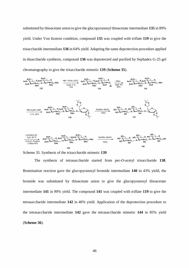
46
substituted by thioacetate anion to give the glucopyranosyl thioacetate intermediate 135 in 89%
yield. Under Von Itzstein condition, compound 135 was coupled with triflate 119 to give the
trisaccharide intermediate 136 in 64% yield. Adapting the same deprotection procedure applied
in disaccharide synthesis, compound 136 was deprotected and purified by Sephadex G-25 gel
chromatography to give the trisaccharide mimetic 139 (Scheme 35).
Scheme 35. Synthesis of the trisaccharide mimetic 139
The synthesis of tetrasaccharide started from per-O-acetyl trisaccharide 138.
Bromination reaction gave the glucopyranosyl bromide intermediate 140 in 43% yield, the
bromide was substituted by thioacetate anion to give the glucopyranosyl thioacetate
intermediate 141 in 90% yield. The compound 141 was coupled with triflate 119 to give the
tetrasaccharide intermediate 142 in 46% yield. Application of the deprotection procedure to
the tetrasaccharide intermediate 142 gave the tetrasaccharide mimetic 144 in 85% yield
(Scheme 36).

47
Scheme 36. Synthesis of the tetrasaccharide mimetic 144
5.00 Biological evaluation of the oligo-(3,5-dithio-β-D-glucopyranosides)
To evaluate the binding affinity of the oligo-(3,5-dithio-β-D-glucopyranosides) to CR3
and Dectin-1, the Vetvicka laboratory tested the mimetics’ ability to inhibit anti-CR3 and anti-
Dectin-1 fluorescein isothiocyanate (FITC) conjugated antibody staining of human neutrophils
and mouse macrophages. In terms of CR3 binding affinity, incubation of a 0.1μg / ml solution
of β-(1→3)-dimer mimetic 133 caused 20% decreases in inhibition of staining human
neutrophils and β-(1→3)-trimer mimetic 139 caused 37% decreases in inhibition of staining
human neutrophils, while the β-(1→3)-tetramer mimetic 144 caused 21% decreases (Table 5).
In terms of Dectin-1 binding affinity, incubation of a 0.1μg/ml solution of β-(1→3)-dimer
mimetic 133 caused 31% decreases in inhibition of staining human neutrophils and β-(1→3)-
trimer mimetic 139 caused 42% decreases in inhibition of staining human neutrophils, while
the β-(1→3)-tetramer mimetic 144 caused 33% decreases (Table 5).

48
Table 5. Percentage inhibition of anti-CR3 and anti-Dectin-1-FITC antibody staining of
neutrophils, macrophages by 0.1μg/mL substrate. a Mean ± SD
Compound
Oligomer
NO.
% inhibition of anti-
CR3-FITC staining
human neutrophilsa
% inhibition of anti-
dectin-1-FITC
staining of mouse
neutrophilsa
133 Dimer 20.2 ± 1.7 30.9 ± 3.4
139 Trimer 37.3 ± 3.2 42.2 ± 3.8
144 Tetramer 21.5 ± 1.4 33.1 ± 2.9
Oligo-(3,5-dithio-β-D-glucopyranosides) 133, 139, 144 (10 μg / mL) were also tested
for their ability to stimulate phagocytosis of human macrophage-like RAW 264 cells.55
Commercial available β-glucan Glucan #300 were used as reference (Table 6). The result
indicates that the β-(1→3)-trimer mimetic 139 could stimulate 16% of phagocytosis, which is
more effective than the β-(1→3)-dimer 133 and β-(1→3) tetramer mimetic 144. It is notable
that the level of phagocytosis stimulated by the β-(1→3)-trimer mimetic 139 was about 50%
of that induced by Glucan #300.
Table 6. Percentage stimulation of phagocytosis. a Mean ± SD
Compound Oligomer NO. % stimulation of phagocytosis (RAW 264
macrophages, 10μg / mL, 24h) a
133 Dimer 8.7 ± 0.7
139 Timer 16.3 ± 0.9
144 Tetramer 6.9 ± 0.3
Glucan #300 Polymer 37.5 ± 2.5
PBS 2.1 ± 0.2

49
Oligo-(3,5-dithio-β-D-glucopyranosides) 133, 139, 144 (10 μg / mL) were also tested
for their ability to stimulate pinocytosis of human macrophage-like RAW 264 cells.55
Commercial available Glucan #300 was used as reference. The result indicates that the β-
(1→3)-trimer mimetic 139 could stimulate 13% of pinocytosis, which is more effective than
the β-(1→3)-dimer mimetic 144 and β-(1→3)-tetramer mimetic 145 (Table 7).
Table 7. Percentage stimulation of pinocytosis. a Mean ± SD*
Compound Oligomer NO. % stimulation of pinoocytosis (RAW 264
macrophages, 10μg / mL, 24h) a
133 Dimer 7.77 ± 1.61
139 Timer 12.8 ± 1.72
144 Tetramer 3.37 ± 0.36
Glucan #300 Polymer 38.9 ± 2.81
PBS 1.77 ± 0.11
*Pinocytosis was assayed by spectrophotometric measurement of neutral red dye accumulation
by mouse macrophages after 2 hr incubation with 40 μg/ml of neutral red.
6.00 Conclusion
As already demonstrated with the di- and trimeric hydroxylamines 22 and 25,54 the
present study demonstrates that small molecule mimetics of β-(1→3)-glucans can be designed
that display significant activity in the inhibition of staining of human neutrophils and mouse
macrophages by fluorescent anti-CR3 and anti-Dectin-1 antibodies suggestive of binding to
the carbohydrate binding domains of the respective proteins. The affinity for the carbohydrate
binding domains of CR3 and Dectin-1 is reflected in the stimulation of phagocytosis and of
pinocytosis by compounds 133, 139 and 144. Unexpectedly, the trimer 139 is more active than

50
either the dimer 133 or the tetramer 144 in each of the three assays conducted suggesting that,
at least for the present series of glucan mimetics, there is little to be gained by preparing higher
oligomers. This maximization of activity in the trimer 144 might be accounted for by a
tradeoff between the greater affinity for the CR3 and Dectin-1 carbohydrate binding sites
arising from the presence of the multiple thioethers on the positive side, and the accumulation
of multiple long C-S bonds eventually causing a mismatch with the binding site on the negative
side.

51
CHAPTER 3: DEVELOPMENT OF A MICROWAVE CLEAVABLE PROTECTING
GROUP AND ITS APPLICATION IN GLYCOSYLATION.
1.00 Introduction
Protecting groups are integral for advancing the art of organic synthesis.80-81 They are
employed in the construction of complex natural products, oligosaccharides, polyketides, RNA,
and peptides. Despite reports on protecting group-free complex molecule synthesis.82 In
organic synthesis, especially carbohydrate synthesis, it is necessary to manipulate different
functional groups, which lead to the concept of orthogonal protecting groups. The benzyl
protecting group, as well as modified benzyl groups, have been significantly employed in
hydroxyl group protection. The electronic tuning of the benzyl group has generated different
orthogonal protecting groups: a) Chemical cleavable protecting groups; b) Photocleavable
protecting groups; c) Enzyme cleavable protecting groups. We have expanded this boundary
to a new type of protecting group: Microwave cleavable protecting groups.
1.10 Chemical cleavable benzyl protecting groups
1.11 Benzyl protecting groups cleaved by hydrogenolysis
Benzyl groups are most frequently used for hydroxyl group protection. They are stable
under acidic and basic conditions, and they are only hardly cleavable under mild oxidizing
agents (Dess-Martin Reagent, etc) or metal hydrides (Lithium Aluminum Hydride, etc). The
deprotection method is usually hydrogenolysis in the presence of Palladium catalyst on
charcoal, this process is mild and compatible with most functional groups.83
1.12 Benzyl based protecting groups cleaved by oxidation
p-Methoxy benzyl (PMB) as well as 3,4-dimethoxybenzyl (DMB) ethers were
developed by Yonemitsu group.84 They are more labile under acidic conditions than benzyl

52
ether. However, they undergo oxidative cleavage, a process where single electron transfer (SET)
to 2,3-dichloro-5,6-dicyano-1,4-benzoquinone (DDQ), generating an oxonium ion which can
be captured by water. This characteristic provides an orthogonal set which helps to selectively
remove PMB ethers in the presence of benzyl ether.
1.20 Microwave
Mirowave is a form of electromagnetic energy with wavelength range from one meter
to one millimeter. The development of microwave technology was initiated by World War II
as the RADAR devices. Accidentally, Percy LeBaron Spencer of the Raytheon Company
discovered that microwave energy could heat up food. This discovery gave birth to commercial
microwave oven in the 1950s. Microwave was applied in Organic synthesis in 1980s, the first
two papers on microwave-enhanced organic chemistry were published and many in 1986.
Organic chemists have since discovered the benefits of using microwave energy to drive
synthetic reactions.85-86 Microwave heating is much more quickly than conductive heating
since heat doesn’t need to pass through the walls of vessel to heat up the reactant. Microwave
heating has been proved to be significantly beneficial in accelerating many conventional heated
reactions. The ease of manipulation, milder reaction conditions and formation of cleaner
products are the benefits come with improved reaction rates.87
1.30 Microwave cleavable benzyl-based protecting groups
1.31 Microwave cleavage of 4-O-siloxyl benzyl ether
The Crich lab developed a microwave assisted cleavage of 4-(tert-Butyldiphenylsiloxy)-
3-fluorobenzyl group. 88 The deprotection was performed with TBAF under microwave
condition. Orthogonality was tested with PMB ether and it was proved that the 4-(tert-

53
Butyldiphenylsiloxy)-3-fluorobenzyl ether was deprotected in the presence of PMB ether
(Scheme 37).
Scheme 37. Microwave cleavage of 4-(tert-Butyldiphenylsiloxy)-3-fluorobenzyl group
1.32 Microwave cleavage of PDMAB protecting group
Andreana group discovered PDMAB protecting group in a microwave reaction of
compound 148. Under the cleavage of p-N,N-dimethylamine benzyl group in 98% yield. This
discovery led us to look for appropriate protection reagents to help to extent PDMAB group
into organic synthesis (Scheme 38).
Scheme 38. Microwave cleavage of PDMAB group of compound 148
2.00 p-N,N-Dimethylamino benzyl group protection
2.10 Installation of PDMAB group by nucleophilic substitution of PDMAB chloride or
PDMAB tosylate
Figure 15. Installation of PDMAB group by nucleophilic substitution
OO
OO
OPMB
TBDPSO
FO OBnO
BnOBnO OMe
PhO
OOHO
OPMB
O OBnOBnO
BnO OMe
Ph
TBAF (2 eq), THF
microwave, 90 oC 76%
146 147
N O
CO2Et
HN O
NMe2
NH
O
CO2Et
HN OMicrowave
H2O, 200 oC
300 W, 20 min 98%
148 149

54
In benzyl ether synthesis, the most common method is the alkylation of alkoxides with
benzyl bromide or chloride. We wanted to apply this method to PDMAB group installation
(Figure 15). With the commercially available p-N,N-dimethylamino benzaldehyde, the
reduction with sodium borohydride gave p-N,N-dimethylamino benzyl alcohol 150 in 85%
yield. Tosylation of the PDMAB alcohol in DMF gave the 4,4’-methylenebis-(N,N-
dimethylaniline) 151 as the major product (Scheme 39).
Scheme 39. Attempted synthesis of PDMAB tosylate
The PDMAB aocohol was refluxed in concentrated hydrogen chloride at 100 oC
overnight to give the p-N,N-dimethylamino benzyl chloride hydrochloride salt 152 in 96%
yield. When applying this reagent in hydroxyl group protection, no protection product was
isolated and the major product was 4,4’-methylenebis-(N,N-dimethylaniline) 151. This result
indicates that p-N,N-dimethylamino benzyl chloride hydrochloride salt decompose under basic
condition (Scheme 40).
Scheme 40. Synthesis of PDMAB chloride hydrochloride and attempted PDMAB protection
2.20 Buchwald amination of 4-halobenzyl ether.

55
Figure 16. Installation of PDMAB protecting group by Buchwald amination of 4-halobenzyl
ether
Obadiah et al89 reported that p-N,N-dimethylamino benzyl protecting group could be
installed by Buchwald amination of 4-halobenzyl ether. We successfully installed PEMAB
group on the 1,2;5,6-di-O-isopropylidene-α-D-glucofuranose by applying this protocol. First,
the 3-OH of compound 82 was protected with 4-chlorobenzyl group to give 3-O-(4-
Chlorobenzyl)-1,2;5,6-di-O-isopropylidene-α-D-glucofuranose 153 in 78% yield. Buchwald
amination was performed with compound 153 using diethyl amine to give PEMAB protected
1,2;5,6-di-O-isopropylidene-α-D-glucofuranose 154. We tested the deprotection of PEMAB
group under microwave condition and the PEMAB group was successfully cleaved under
microwave irradiation to give compound 82 in 53% yield together with the p-N,N-diethylamino
benzyl methyl ether 159 as the byproduct (Scheme 41).
Pd2(dba)3 / JonPhos
Et2NH / tBuONaToluene, 50 oC
72%
Cl
Br
NaH / DMF 78%
Microwave
160 oC, 300W30 min
53%
15382 154
OO
HO O
OO
OO
O
OO
Cl
O
OO
O
OO
NEt2
O
OO
HO O
OO
82
Et2N
OMe+
155
P
JonPhos

56
Scheme 41. The installation of p-N,N-diethylamino benzyl ether and microwave deprotection
Out of curiosity, we also tested microwave deprotection of the ortho-N,N-
dimethylamino benzyl group. First, the 3-OH of compound 82 was protected with ortho-N,N-
dimethylamino benzyl group using ortho-N,N-dimethylamino benzyl chloride. The reaction
gave the desired product 158 in 55% yield. When compound 158 was subjected to microwave
irradiation in methanol, the ortho-N,N-dimethylamino benzyl group was not cleaved (Scheme
42).
Scheme 42. Installation of ortho-N,N-dimethylamino benzyl ether and attempted microwave
deprotection
2.30 Application of PEMAB group in glycosylation
The task of manipulating different hydroxyl groups becomes particularly important in
oligosaccharide synthesis. To accomplish this task, orthogonal protection and deprotection is
required in the complex molecule synthesis. Different protecting groups are employed, for
example, 4-O-methoxy benzyl ethers, which is labile under oxidative condition and acidic
condition, can be selectively cleaved in the presence of benzyl groups. More importantly, the
compatibility of protecting groups with the glycosylation reaction will help to diversify the

57
oligosaccharide synthesis. To test the compatibility of PEMAB group in glycosylation, we
synthesized the phenyl 4-O-(p-N,N-diethylamino benzyl)-2,3-O-isopropylidene-1-thio-β-L-
rhamnopyranoside 161 and it was subjected to NIS / TMSOTf activated glycosylation. The
synthesis started from Phenyl 2,3-O-isopropylidene-1-thio-β-L-rhamnopyranoside 161, the 4-
OH was protected by 4-bromobenzyl ether give compound Phenyl 4-O-(4-bromobenzyl)-2,3-
O-isopropylidene-1-thio-β-L-rhamnopyranoside 162 in 85% yield. The Buchwald amination
converted the 4-bromobenzyl ether protected rhamnose 162 into p-N,N-diethylamino benzyl
protected rhamnose 163 in 82% yield. With glycosylation donor 163 in hand, the glycosylation
with methyl 2,3,4-tri-O-benzyl-α-D-glucopyranoside was undertaken. The reaction was
activated by NIS / TMSOTf at -78 oC and stirred at -78 oC for 12 h to give the glycosylation
product 162 in 22% yield (Scheme 43). With glycosylation product 162, we performed the
deprotection under microwave irradiation. The PEMAB group was cleaved to give the product
163 in 73% yield (Scheme 43).

58
Scheme 43. Glycosylation with PEMAB protected donor 161 and deprotection of PEMAB
group after glycosylation reaction
3.00 Conclusion
In conclusion, other than traditional orthogonal protecting groups: the chemical labile,
photo labile and enzymatic labile protecting groups. A new type of protecting group:
microwave labile protecting group, has been developed.) To the best of oue knowledge, this is
the first example where microwave irradiation has been used to deprotect benzyl ether
protecting groups. It was convinced that the PEMAB group is compatible with glycosylation
conditions. The PEMAB group can be selectively deprotected in the presence of benzyl ether,
isopropilydene, ester as well as benzylidene acetals under microwave irradiation at neutral
conditions. As for the protection, the effective procedure requires a two steps synthesis:

59
installation of p-halo-benzyl ether and Buchwald amination. The one step protection is limited
by the unstability of PEMAB chloride or tosylate.

60
CHAPTER 4: EXPERIMENTAL SECTION
General information
All reagents and solvents were purchased from commercial suppliers and were used
without further purification unless otherwise stated. All reactions were performed under Argon
atmosphere unless otherwise stated. Reactions were monitored by analytical thin-layer
chromatography with pre-coated glass backed plates (w/UV 254) and visualized by UV
absorption (254 nm) or by staining with a 5% solution of H2SO4 in MeOH or ceric ammonium
molybdate solution (4.0 g ceric sulfate; 10 g ammonium molybdate; 40 mL H2SO4; 360 mL
H2O) followed by heating. Optical rotations were measured with an Autopol III polarimeter
(Rudolph Research Analytical, Hackettstown, NJ) in the solvent specified at 589 nm at 23 °C,
the path length is 10 cm. 1H, 13C, HSQC, HMBC, COSY, TOCSY NMR spectra were recorded
with Agilent 600 MHz and Varian 400 MHz instruments. High resolution mass spectra were
recorded with a Walters LC/ MS with an electrospray source coupled to a time-of-flight mass
analyzer. Melting points were recorded with a Barnstead International electrothermal melting
point apparatus.
General Procedure A: Coupling Reactions
To a solution of 1-S-Acetyl-5-thio-β-D-glucopyranose (1.0 eq) and 1,2-O-5-S,6-O-di-
isopropylidene-5-thio-α-D-allofuranose (1.1 eq) in DMF (1.0 M) was added diethylamine (2.5
eq) dropwise at 0 °C. The reaction mixture was stirred at room temperature until completion.
The reaction mixture was diluted with ethyl acetate and washed with water, brine, dried over
MgSO4 and concentrated in vacuo. The residue was purified by silica gel chromatography.

61
General Procedure B: Deacetylation
At 0 °C, sodium methoxide (0.2 eq) was added to the substrate (1.0 eq) in anhydrous methanol
(0.1M) and stirred until the completion of the reaction (observed by mass spectrometry).
Amberlyst IR120 resin was added to neutralize the reaction. When the pH was neutral
(monitored by pH paper), the resin was filtered off and the solution was concentrated in vacuo.
Furanosyl systems were subjected to acidic hydrolysis according to general procedure C, and
the pyranose forms were purified by Sephadex G-25 gel eluting with water.
General procedure C: Hydrolysis and Acetylation
The substrate (1.0 eq) was dissolved in deionized water (0.05 M). DOWEX-50WX2
hydrogen form resin (400 mg per 100 mg substrate) was added to the reaction. The reaction
mixture was stirred at 90 °C until the completion of the reaction (monitored by mass
spectrometry). The resin was filtered off and the solution was concentrated in vacuo. The crude
mixture was dissolved in pyridine (0.5 M) and acetic anhydride (2 eq per OH) 4-
(dimethylamino)-pyridine (0.1 eq) was added, the reaction mixture was stirred at room
temperature until the completion of reaction (monitored by mass spectrometry). The solution
was concentrated in vacuo and purified by silica gel chromatography.
General Procedure D: Bromination
To a solution of penta-O-acetyl-5-thio-D-glucopyranose (1.0 eq) in anhydrous DCM
(0.5 M) was added 33% HBr in acetic acid (7.0 eq) at 0 °C. The reaction mixture was kept at
5 °C for 12 h. The reaction mixture was diluted by DCM and quenched with ice cold aqueous

62
NaHCO3. The organic layer was washed with water, brine, dried with MgSO4, and
concentrated in vacuo. The residue was purified by silica gel chromatography.
General procedure E: Thioacylation
At 0 °C, potassium thioacetate (1.5 eq) was added to a solution of penta-O-acetyl-5-
thio-D-glucopyranosyl bromide (1.0 eq) in N,N-dimethyl formamide (1.0 M). The reaction
mixture was stirred 12 h at room temperature. After completion, the reaction mixture was
diluted with ethyl acetate and washed by water, brine, and dried with MgSO4. The reaction
mixture was concentrated in vacuo and purified by silica gel chromatography.
5,6-Dideoxy-5,6-epithio-1,2-O-isopropylidene-β-L-talofuranose (87)
To a solution of compound 86 (100 mg, 0.46 mmol) in 10 mL DCM was added Dess
Martin reagent (390 mg, 0.92 mmol). The reaction was stirred at room temperature 24h and
saturated NaS2O3 aqueous solution was added to quench the reaction. The reaction mixture was
extracted into DCM and washed with NaHCO3 aqueous solution, water, brine and dried over
MgSO4. The crude mixture was concentrated in vacuo and re-dissolved in 20 mL EtOH. At
0 °C, sodium borohydride (26 mg, 0.69 mmol) was added into reaction mixture and it was
stirred at 0 °C for 1h. The reaction was quenched with aqueous NH4Cl and concentrated in
vacuo to remove EtOH. After the EtOH was removed, the crude mixture was diluted in ethyl
acetate, the organic layer was separated and washed with water, brine and dried over MgSO4.
The crude mixture was concentrated in vacuo and purified by silica gel chromatography to give
product 87 (85 mg, 85%) as a colorless oil. [α]23D = +57.2° (c 0.6, CHCl3). 1H NMR (400 MHz,
CDCl3) δ 5.81 (d, J = 3.9 Hz, 1H, H1), 4.60 (dd, J = 5.2, 3.9 Hz, 1H, H2), 3.91 – 3.79 (m, 1H,

63
H4), 3.58 (dd, J = 8.5, 6.4 Hz, 1H, H3), 3.06 (td, J = 6.5, 5.4 Hz, 1H, H5), 2.54 (dd, J = 6.6, 1.4
Hz, 1H, H6’), 2.48 (dd, J = 5.4, 1.4 Hz, 1H, H6), 2.43 (d, J = 10.6 Hz, 1H, OH), 1.54 (s, 3H),
1.36 (s, 3H). 13C NMR (101 MHz, CDCl3) δ 112.9 (O,O-isopropylidene), 103.9 (C1), 83.2 (C2),
78.7 (C4), 75.8 (C3), 34.4 (C5), 26.6, 26.5 (isopropylidene), 21.1 (C6). HRMS m/z [M+Na]+
calcd for C9H14O4SNa 241.0510, found 241.05010
6-S-Acetyl-3,5-dideoxy-3,5-epithio-1,2-O-isopropylidene-β-L-idofuranose (88)
At 0°C, to a solution of compound 87 (107 mg, 0.49 mmol) in pyridine (1.5 mL) was
added trifluoromethanesulfonic anhydride (0.1 mL, 0.6 mmol). The reaction was stirred at 0 °C
for 25 min before it was diluted with diethyl ether 20 mL. The reaction mixture was quenched
with aqueous NaHCO3 and the organic layer was separated and washed with water, brine and
dried over MgSO4. The reaction mixture was concentrated in vacuo and re-dissolved in 2 mL
DMF. At room temperature, potassium thioacetate (171 mg, 1.5 mmol) was added to the
solution. The reaction was stirred at room temperature for 1h before it was diluted in diethyl
ether 20 mL and washed with water, brine, dried over MgSO4 and concentrated in vacuo. The
crude mixture was purified on silica gel chromatography with eluent (hexane : ethyl acetate =
10 : 1) to give product 88 (50 mg, 37%) as a colorless oil. [α]23D = +81.9° (c 0.4, CHCl3). 1H
NMR (400 MHz, CDCl3) δ 6.35 (d, J = 3.4 Hz, 1H, H1), 5.24 (t, J = 5.0 Hz, 1H, H3), 4.63 (d,
J = 3.4 Hz, 1H, H2), 3.97 (d, J = 5.0 Hz, 1H, H4), 3.91 (td, J = 7.8, 5.0 Hz, 1H, H5), 3.13 (d, J
= 7.8 Hz, 2H, H6), 2.32 (s, 3H, S-Acetyl), 1.45 (s, 3H), 1.36 (s, 3H). 13C NMR (101 MHz,
CDCl3) δ 195.2 (S-Acetyl), 114.3 (Isopropylidene C), 108.7 (C1), 85.9 (C2), 83.4 (C3), 42.6

64
(C5), 42.0 (C4), 30.6 (S-Acetyl), 30.1 (C6), 27.8 (Isopropylidene), 27.3 (Isopropylidene).
HRMS m/z [M+Na]+ calcd for C11H16O4S2Na 299.0388, found 299.0384.
3-S-Acetyl-6-O-p-toluenesulfonyl-1,2-O-isopropylidene-α-D-glucofuranose (92).
To a solution of 3-S-acetyl-1,2-O-isopropylidene-α-D-glucofuranose in pyridine 1 mL
was added p-toluenesulfonyl chloride (56 mg, 0.30 mmol). The reaction mixture was kept in
5 °C for 24 h before it was diluted in 20 mL DCM and quenched with aqueous NaHCO3. The
organic layer was separated and washed with water, brine and dried over MgSO4. The crude
mixture was concentrated in vacuo and purified by silica gel chromatography to give product
92 (85 mg, 67%) as a colorless oil. [α]23D = -2.6° (c 0.4, CHCl3). 1H NMR (400 MHz, CDCl3)
δ 7.85 – 7.70 (m, 2H), 7.33 (d, J = 8.1 Hz, 2H), 5.72 (d, J = 3.5 Hz, 1H, H1), 4.56 (d, J = 3.5
Hz, 1H, H2), 4.30 (dd, J = 10.4, 2.2 Hz, 1H, H6), 4.25 (dd, J = 9.1, 3.6 Hz, 1H, H4), 4.17 (d, J
= 3.5 Hz, 1H, H3), 4.03 (dd, J = 10.4, 7.0 Hz, 1H, H6’), 3.93 (ddd, J = 9.2, 7.0, 2.2 Hz, 1H, H5),
2.44 (s, 6H), 2.39 (s, 3H), 1.48 (s, 3H), 1.28 (s, 3H). 13C NMR (101 MHz, CDCl3) δ 194.5,
145.0, 132.5, 129.9, 128.0, 112.4, 104.6 (C1), 85.6 (C2), 77.3 (C4), 71.9 (C6), 68.8 (C5), 50.3
(C3), 31.0, 26.4, 26.2, 21.7. HRMS m/z [M+Na]+ calcd for C18H24O8S2Na 455.0810, found
455.0814
3-O-Acetyl-5-O-methanesulfonyl-6-O-benzoyl-1,2-O-isopropyliene-α-D-allofuranose (96).
At 0 °C, to a solution of compound 95 (460 mg, 1.75 mmol) in DCM 18 mLwas added
pyridine (0.35 mL, 4.38 mmol). At -30 °C, benzoyl chloride (0.22 mL, 1.93 mmol) was added
into the reaction, after the reaction was stirred at -30 °C for 1h, methanesulfonyl chloride (0.27
mL, 3.5 mmol) was injected into the reaction mixture. The reaction was stirred at room

65
temperature overnight before aqueous NaHCO3 was added to quench the reaction. The DCM
layer was separated and washed with water, brine, dried over MgSO4 and concentrated in vacuo.
The crude mixture was purified by silica gel chromatography to give product 96 (660 mg, 85%)
as a colorless crystal. m.p. 128 - 130 °C. [α]23D = +73.6° (c 0.5, CHCl3). 1H NMR (400 MHz,
CDCl3) δ 8.06 (dt, J = 7.2, 1.4 Hz, 2H), 7.65 – 7.52 (m, 1H), 7.45 (dd, J = 8.4, 7.1 Hz, 2H),
5.83 (d, J = 3.3 Hz, 1H), 5.22 (dt, J = 7.5, 3.6 Hz, 1H), 4.97 – 4.80 (m, 2H), 4.65 (dd, J = 12.4,
3.5 Hz, 1H), 4.41 (ddd, J = 7.8, 6.8, 4.2 Hz, 2H), 3.05 (s, 3H), 2.11 (s, 3H), 1.55 (s, 3H), 1.34
(s, 3H). 13C NMR (101 MHz, CDCl3) δ 170.0, 165.9, 133.5, 129.8, 129.2, 128.6, 113.6 (O,O-
isopropylidene), 104.1 (C1), 78.1 (C2), 76.0 (C4), 72.4 (C3), 62.6 (C6), 38.8 (OMs), 26.7, 26.6,
20.6 (OAc). HRMS m/z [M+Na]+ calcd for C19H24O10SNa 467.0988, found 467.0993.
5,6-Anhydro-1,2-O-isopropylidene-β-L-talofuranose (97)
To a solution of comound 96 (288 mg, 0.65 mmol) in 11 mL MeOH was added sodium
methoxide (12 mg , 0.22 mmol). The reaction mixture was stirred at room temperature for 1h
before it was neutralized with Amberlyst IR 120. The crude mixture was concentrated and it
was purified by sílica gel chromatography to give product 97 (64 mg, 49%) as a colorless oil.
[α]23D = +30.2° (c 0.5, CHCl3). 1H NMR (400 MHz, CDCl3) δ 5.77 (d, J = 3.8 Hz, 1H, H1),
4.55 (dd, J = 5.1, 3.8 Hz, 1H, H2), 3.96 (ddd, J = 10.4, 8.8, 5.1 Hz, 1H, H3), 3.67 (dd, J = 8.8,
4.3 Hz, 1H, H4), 3.14 (td, J = 4.2, 2.8 Hz, 1H, H5), 2.90 – 2.74 (m, 2H, H6 & H6’), 2.57 (d, J =
10.4 Hz, 1H, OH), 1.53 (s, 3H), 1.34 (s, 3H). 13C NMR (101 MHz, CDCl3) δ 112.9 (O,O-
isopropylidene), 104.1 (C1), 79.7 (C2), 78.5 (C4), 72.9 (C3), 51.0 (C5), 43.9 (C6), 26.5 (O,O-
isopropylidene). HRMS m/z [2M+Na]+ calcd for C18H28O10Na 427.1580, found 427.1586.

66
3-S-Acetyl-5,6-anydro-1,2-O-isopropylidene-β-L-idofuranose (98).
To a solution of compound 97 (74 mg, 0.37 mmol) in DCM 7.5 mL was added pyridine
(0.12 mL, 1.42 mmol). At 0 °C, trifluoromethanesulfonic anhydride (0.12 mL, 0.71 mmol) was
added dropwise. The reaction mixture was stirred at 0 °C for 30 min before it was poured into
8 mL ice-cold water. The mixture was extracted by 3 x 4 mL DCM. The DCM layer was
washed by brine, dried over MgSO4 and concentrated in vacuo. The crude mixture was
dissolved in 4 mL DMF, at 0 °C, the solution of potassium thioacetate (84 mg, 0.73 mmol) in
4 mL DMF was slowly injected. After 1h, the reaction mixture was poured into 50 mL ice
water and extracted by 50 mL ethyl acetate. The organic layer was washed with brine, dried
over MgSO4 and concentrated in vacuo. The crude mixture was purified by silica gel
chromatography to give product 98 (67 mg, 67%) as a colorless oil. [α]23D = -5.7° (c 0.4,
CHCl3). 1H NMR (400 MHz, CDCl3) δ 5.85 (d, J = 3.6 Hz, 1H, H1), 4.56 (d, J = 3.5 Hz, 1H,
H2), 4.22 (t, J = 4.6 Hz, 1H, H4), 4.14 (dt, J = 4.1, 0.7 Hz, 1H, H3), 3.09 (ddd, J = 5.0, 4.2, 2.7
Hz, 1H, H5), 2.77 (dd, J = 5.0, 4.2 Hz, 1H, H6), 2.69 (dd, J = 4.9, 2.7 Hz, 1H, H6’), 2.40 (s, 3H,
S-acetyl), 1.51 (s, 3H), 1.31 (s, 3H) (isopropylidene). 13C NMR (101 MHz, CDCl3) δ 193.5 (S-
acetyl), 112.3 (O,O-isopropylidene), 104.7 (C1), 86.0 (C2), 79.1 (C4), 50.7 (C5), 50.3 (C3), 43.5
(C6), 30.8 (S-acetyl), 26.6, 26.3 (isopropylidene). HRMS m/z [M+Na]+ calcd for C11H16O5NaS
283.0616, found 283.0615.
3-S-Pivalyl-5,6-anydro-1,2-O-isopropylidene-β-L-idofuranose (99)
Compound 97 (110 mg, 0.54 mmol) and potassium thiopivalate (170 mg, 1.1 mmol)
were applied to same procedure as the preparation of 98 to give product 99 (108 mg, 67%) as

67
a colorless oil. [α]23D = -6.7° (c 0.6, CHCl3). 1H NMR (400 MHz, CDCl3) δ 5.85 (d, J = 3.6 Hz,
1H, H1), 4.50 (d, J = 3.6 Hz, 1H, H2), 4.17 (dd, J = 5.3, 4.3 Hz, 1H ,H4), 4.05 (dt, J = 4.2, 0.7
Hz, 1H, H3), 3.06 (dddd, J = 5.2, 4.3, 2.7, 0.8 Hz, 1H, H5), 2.72 (td, J = 4.6, 0.8 Hz, 1H, H6),
2.65 (ddd, J = 4.9, 2.7, 0.8 Hz, 1H, H6’), 1.49 (s, 3H), 1.29 (s, 3H), 1.23 (s, 9H). 13C NMR (101
MHz, CDCl3) δ 204.43 (S-pivalyl) 112.2 (O,O-isopropylidene), 104.8 (C1), 86.1 (C2), 79.5 (C4),
50.7 (C5), 49.7 (C3), 43.5 (C6), 27.2 (S-pivalyl), 26.6, 26.3 (isopropylidene). HRMS m/z
[M+Na]+ calcd for C14H22O5NaS 325.1086, found 325.1090.
3-O-(2-Napthyl)methyl-5-O-methanesulfonyl-6-O-benzoyl-1,2-O-isopropyliene-α-D-
allofuranose (102)
To a stirred solution of compound 101 (1.01 g, 2.8 mmol) in 20 mL DCM was added
pyridine (1.2 mL, 4.66 mol) and DMAP (0.017 g, 0.14 mmol). At -30 °C benzoyl chloride
(0.36 mL, 3.1 mmol) was added dropwise and the reaction mixture was stirred at -30 °C for 1
h. After completion (monitored by TLC), methanesulfonyl chloride (0.31 mL, 4.1 mmol) was
added to the reaction mixture at -30 °C. The reaction mixture was stirred at room temperature
for 10 min before it was concentrated at 40 °C to remove DCM. After DCM was removed, the
reaction mixture was stirred at room temperature until the completion (monitored by TLC).
The reaction was diluted with 40 mL DCM and quenched with aqueous NaHCO3. The solution
was washed with water, brine and dried over MgSO4. The reaction mixture concentrated in
vacuo and purified by silica gel chromatography to give product 102 (1.02 g, 70%) as a
colorless oil. [α]23D = +60.0° (c 0.8, CHCl3). 1H NMR (400 MHz, CDCl3) δ 8.14 – 7.99 (m,
2H), 7.93 – 7.79 (m, 4H), 7.62 – 7.36 (m, 6H), 5.74 (d, J = 3.5 Hz, 1H, H1), 5.28 (dt, J = 8.7,

68
3.1 Hz, 1H, H5), 4.94 (d, J = 11.6 Hz, 1H, NAP CH2), 4.78 (d, J = 11.7 Hz, 1H, NAP CH2),
4.65 – 4.57 (m, 2H, H2 & H6’), 4.52 (dd, J = 12.6, 8.6 Hz, 1H, H6), 4.37 (dd, J = 8.9, 2.8 Hz,
1H, H4), 4.05 (dd, J = 8.9, 4.2 Hz, 1H, H3), 2.95 (s, 3H), 1.62 (s, 3H), 1.37 (s, 3H). 13C NMR
(101 MHz, CDCl3) δ 170.0, 166.0, 134.3, 133.3, 133.2, 129.8, 128.5, 128.0, 127.7, 127.2, 126.3,
126.2, 125.9, 113.6 (O,O-isopropylidene), 104.1 (C1), 78.1 (C2), 76.0 (C3), 72.5 (NAP CH2),
62.7 (C6), 38.8 (C5), 26.7, 26.6 (isopropylidene), 20.6 (Acetyl). HRMS m/z [M+Na]+ calcd for
C28H30O9NaS 565.1508, found 565.1494.
3-O-(2-Napthyl)methyl-5,6-dideoxy-5,6-epithio-1,2-O-isopropyliene-α-D-allofuranose
(103)
At 0 °C, to a stirred solution of compound 102 (0.54 g, 1.0 mmol) in anhydrous
methanol (50 mL, 0.02 M) was added NaOMe (64 mg, 1.2 mmol). The reaction was stirred at
0 °C until the completion of the reaction mixture (monitored by TLC). Amberlyst IR 120 was
added portionwise to neutralize the reaction mixture (monitored by pH paper). The resin was
filtered off and thiourea (76 mg, 1 mmol) was added to the reaction mixture. The reaction was
stirred at 80 °C for 3 h before it was concentrated in vacuo. The residue was dissolved in DCM
and washed by water, brine and dried over MgSO4 and concentrated in vacuo. The crude
mixture was concentrated in vacuo and purified by silica gel chromatography to give product
103 (0.25 g, 70%) as a colorless oil. [α]23D = +74.2° (c 1.2, CHCl3). 1H NMR (400 MHz, CDCl3)
δ 7.94 – 7.78 (m, 4H), 7.60 – 7.42 (m, 3H), 5.72 (d, J = 3.7 Hz, 1H, H1), 4.95 (d, J = 12.1 Hz,
1H, NAP CH2), 4.76 (d, J = 12.1 Hz, 1H, NAP CH2), 4.59 (t, J = 4.1 Hz, 1H, H2), 4.01 (dd, J
= 8.6, 6.2 Hz, 1H, H4), 3.72 (dd, J = 8.6, 4.4 Hz, 1H, H3), 3.04 – 2.93 (m, 1H, H5), 2.47 (dd, J

69
= 6.5, 1.4 Hz, 1H, H6’), 2.39 (dd, J = 5.4, 1.4 Hz, 1H, H6), 1.60 (s, 3H), 1.36 (s, 3H). 13C NMR
(101 MHz, CDCl3) δ 134.8, 133.2, 133.1, 128.4, 127.9, 127.8, 126.9, 126.4, 126.3, 126.1, 125.9,
113.1 (O,O-isopropylidene), 103.8 (C1), 80.7 (C2), 77.6 (C3), 72.3 (NAP CH2), 43.6 (C4), 30.5
(C5), 26.8, 26.6 (isopropylidene), 20.7 (C6). HRMS m/z [M+Na]+ calcd for C20H22O4SNa
381.1137, found 381.1138.
3-O-(2-Napthyl)methyl-5-S-acetyl-6-O-acetyl-1,2-O-isopropyliene-α-D-allofuranose (104)
To a stirred solution of compound 103 (0.36 g, 1.0 mmol) in mixture of acetic anhydride
(20 mL) and glacial acetic acid (4 mL) was added anhydrous potassium acetate (0.49 g, 5.0
mmol) and refluxed at 145 °C for 12 h. After the reaction mixture was cooled, it was poured
into ice water and extracted with chloroform (3×10 mL). The choloroform was collected and
washed with aqueous NaHCO3, brine, dried over MgSO4, and concentrated in vacuo. The
residue was purified over silica gel chromatography to give 104 (0.26 g, 56%) as a colorless
oil. [α]23D = +45.5° (c 0.5, CHCl3). 1H NMR (400 MHz, CDCl3) δ 7.90 – 7.79 (m, 4H), 7.57 –
7.40 (m, 3H), 5.69 (d, J = 3.7 Hz, 1H), 4.91 (d, J = 11.9 Hz, 1H), 4.68 (d, J = 11.9 Hz, 1H),
4.57 (t, J = 4.1 Hz, 1H), 4.34 – 4.25 (m, 2H), 4.20 (dd, J = 11.4, 5.9 Hz, 1H), 4.08 (q, J = 5.6
Hz, 1H), 3.78 (dd, J = 8.8, 4.4 Hz, 1H), 2.25 (s, 3H), 1.96 (s, 3H), 1.59 (s, 3H), 1.35 (s, 3H).
13C NMR (101 MHz, CDCl3) δ 193.9, 170.5, 134.7, 133.2, 133.1, 128.3, 127.9, 127.7, 126.9,
126.2, 126.1, 125.9, 113.2 (O,O-isopropylidene), 103.8 (C1), 80.2 (C2), 77.9 (C4), 77.5 (C3),
72.4 (NAP CH2), 63.3 (C6), 43.6 (C5), 30.5 (S-acetyl), 26.9, 26.6 (isopropylidene), 20.8
(Acetyl). HRMS m/z [M+Na]+ calcd for C24H28O7SNa 483.1453, found 483.1460.
3-O-(2-Napthyl)methyl-1,2,4,6-tetra-O-acetyl-5-thio-α,β-D-allopyranose (105)

70
A solution of compound 104 (460 mg, 1.0 mmol) in 20 mL aqueous acetic acid (50%)
was stirred at 90 °C for 12 h. The reaction mixture was concentrated in vacuo and co-
evaporated with toluene (3×10 mL) to remove acetic acid. The crude mixture was dissolved in
anhydrous methanol (300 mL) and NaOMe (3.2 mg, 0.06 mmol) was added at 0 °C. The
reaction was stirred at 0 °C for 4 h before Amberlyst IR120 resin was added to quench the
reaction (monitored by pH paper). After reaction mixture was neutralized, the resin was filtered
off and reaction was concentrated in vacuo. The residue was dissolved in pyridine (10 mL) and
acetic anhydride (4.6 mL, 0.05 mol) was added at 0 °C. The reaction mixture was stirred at
room temperature for 12 h before it was concentrated in vacuo. The residue was purified by
silica gel chromatography to give 105 (227 mg, 45%) as a colorless oil. 1H NMR (400 MHz,
CDCl3) δ 7.90 – 7.77 (m, 5H), 7.48 (ddt, J = 8.9, 6.8, 1.7 Hz, 5H), 6.30 (d, J = 9.3 Hz, 1H,
H1β), 6.14 (d, J = 3.7 Hz, 1H, H1α), 5.30 – 5.21 (m, 2H, H4α &H4β), 5.16 (td, J = 10.8, 10.4, 2.3
Hz, 2H, Η2α &H2β), 4.90 – 4.75 (m, 4H, Nap CH2), 4.45 (dd, J = 12.1, 5.1 Hz, 1H, Η6α’), 4.32
(dd, J = 12.0, 5.5 Hz, 1H, Η6β), 4.21 – 4.09 (m, 2H, Η6α& Η6β), 3.99 (ddd, J = 10.9, 5.1, 2.7 Hz,
1H, Η5α), 3.85 (ddd, J = 10.2, 5.4, 3.3 Hz, 1H, Η5β), 2.08 (s, 3H), 2.06 (s, 3H), 2.05 (s, 3H),
2.03 (s, 3H), 2.01 (s, 3H). 13C NMR (101 MHz, CDCl3) δ 170.6, 169.9, 169.6, 169.4, 169.1,
135.8, 134.9, 133.1, 133.0, 128.2, 128.0, 127.9, 127.7, 126.8, 126.3, 126.2, 126.0, 125.6, 75.6
(C1α), 75.4 (C1β), 74.0 (C2), 72.6 (NAP CH2), 72.5 (NAP CH2), 71.9, 70.6 (C4α), 70.1 (C4β),
61.7 (C6α), 61.2 (C6β), 40.0 (C5α), 35.6 (C5β), 21.1, 20.7. HRMS m/z [M+Na]+ calcd for
C25H28O9SNa 527.1352, found 527.1354.

71
4,6-O-Benzylidene-3-O-(2-napthyl)methyl-1,2-di-O-acetyl-5-thio-α,β-D-allopyranose
(106)
To a solution of compound 105 (171 mg, 0.34 mmol) in 10 mL anhydrous MeOH was
added NaOMe (3.2 mg, 0.06 mmol). The reaction mixture was stirred at room temperature
until the completion and Amberlyst IR 120 was added to neutralize the reaction mixture. The
reaction mixture was filtered and concentrated in vacuo and was re-dissolved in 4 mL
anhydrous MeCN. To the reaction mixture was added camphorsulfonic acid (8.0 mg, 0.03
mmol) and benzaldehyde dimethyl acetal (258 mg, 1.70 mmol). The reaction mixture was
stirred at room temperature for 25 min before aqueous NaHCO3 was added. The reaction
mixture was extracted by 3 x 10 mL ethyl acetate, the organic layer was washed with brine,
dried over MgSO4 and concentrated in vacuo. The crude mixture was acetylated and purified
by silica gel chromatography to give product 106 (66 mg, 46%) as a colorless oil. 1H NMR
(400 MHz, CDCl3) δ 7.90 – 7.70 (m, 6H), 7.60 – 7.34 (m, 11H), 6.36 (d, J = 9.8 Hz, 1H, H1β),
6.12 (d, J = 3.6 Hz, 1H, H1α), 5.66 (s, 1H, α isomer benzylidene), 5.60 (s, 1H, β isomer
benzylidene), 5.22 – 5.15 (m, 2H, H2α & H2β), 5.12 (d, J = 12.2 Hz, 1H, α isomer NAP CH2),
5.02 (d, J = 12.0 Hz, 1H, β isomer NAP CH2), 4.83 (dd, J = 12.1, 6.3 Hz, 2H, α,β isomers NAP
CH2), 4.31 (ddd, J = 11.6, 7.3, 4.6 Hz, 1H, H6α), 4.26 – 3.99 (m, 4H, H3 &H4 &H6β), 3.94 –
3.87 (m, 1H, H5β), 3.76 (td, J = 11.1, 4.5 Hz, 1H, H5α), 2.13 (s, 3H), 2.07 (s, 3H, β isomer
acetate), 1.97 (s, 3H), 1.87 (s, 3H, β isomer acetate). 13C NMR (101 MHz, CDCl3) δ 170.1,
169.8, 169.0, 137.6, 136.5, 133.2, 132.8, 129.3, 128.4, 128.0, 127.9, 127.7, 127.0, 126.3, 126.2,
126.1, 126.0, 125.8, 125.7, 102.1 (Benzylidene), 82.4 (C1α), 82.3 (C1β), 75.1 (C4), 74.9(C2α),

72
74.5 (C2β), 72.1 (NAP CH2), 70.7 (C3α), 70.1 (C3β), 68.5 (C6), 35.2, 31.8(C5α), 31.6(C5β), 22.7,
21.2, 20.8. HRMS m/z [M+Na]+ calcd for C28H28O7SNa 531.1453, found 531.1454.
4,6-O-Benzylidene-1,2-di-O-acetyl-5-thio-α,β-D-allopyranose (107)
To a solution of compound 106 (68 mg, 0.13 mmol) in 10 mL DCM and 2.5 mL MeOH
was added DDQ (92 mg, 0.40 mmol). The reaction was stirred at room temperature for
overnight before aqueous NaHCO3 was added to quench the reaction. The reaction mixture was
extracted into DCM and washed by water, brine and dried over MgSO4. The crude mixture was
concentrated in vacuo and purified by silica gel chromatography to give product 107 (41 mg,
83%) as a colorless oil. α:1H NMR (400 MHz, CDCl3) δ 7.49 (dd, J = 6.8, 2.9 Hz, 2H), 7.38
(dd, J = 5.2, 2.0 Hz, 3H), 6.08 (d, J = 3.6 Hz, 1H, H1), 5.70 (s, 1H, benzylidene), 5.21 (t, J =
3.3 Hz, 1H, H2), 4.38 – 4.30 (m, 2H, H3 & H6), 4.02 (dd, J = 9.8, 2.3 Hz, 1H, H4), 3.86 (td, J =
10.5, 9.9, 4.3 Hz, 1H, H5), 3.77 (t, J = 11.0 Hz, 1H, H6), 2.51 (s, 1H, OH), 2.18 (s, 3H), 2.13
(s, 3H). β:1H NMR (400 MHz, CDCl3) δ 7.52 – 7.44 (m, 2H), 7.42 – 7.33 (m, 3H), 6.33 (d, J
= 9.8 Hz, 1H, H1), 5.65 (s, 1H, benzylidene), 5.26 (dd, J = 9.8, 2.6 Hz, 1H, H2), 4.43 – 4.36 (m,
1H, H3), 4.35 – 4.26 (m, 1H, H4), 3.98 (dd, J = 9.4, 2.1 Hz, 1H, H6’), 3.82 – 3.69 (m, 2H,
H5&H6), 2.30 (bs, 1H, OH), 2.12 (s, 3H), 2.08 (s, 3H). α:13C NMR (101 MHz, CDCl3) δ
169.7, 169.6, 137.1, 129.4, 128.4, 126.2, 102.0 (Benzylidene), 81.3 (C1), 71.3 (C2), 71.2 (C4),
69.4 (C3), 68.3 (C6), 30.8 (C5), 21.2, 20.9. β: 13C NMR (101 MHz, CDCl3) δ 129.3, 128.4,
126.0, 101.8 (Benzylidene), 81.2 (C1), 73.9 (C2), 70.3 (C4), 69.5 (C3), 68.4 (C6), 34.6 (C5), 20.9,
20.7. HRMS m/z [M+Na]+ calcd for C17H20NaO7S 391.0827 found 391.0823
4,6-O-Benzylidene-3-S-acetyl-1,2-di-O-acetyl-3,5-dithio-α,β-D-glucopyranose (108)

73
To a solution of compound 107 (73 mg, 0.20 mmol) in DCM 10 mL was added pyridine
(0.12 mL, 1.42 mmol). At 0 °C, trifluoromethanesulfonic anhydride (0.12 mL, 0.71 mmol) was
added dropwise. The reaction mixture was stirred at 0 °C for 30 min before it was poured into
8 mL ice-cold water. The mixture was extracted by 3 x 4 mL DCM. The DCM layer was
washed by brine, dried over MgSO4 and concentrated in vacuo. The crude mixture was
dissolved in 4 mL DMF, at 0 °C, the solution of potassium thioacetate (84 mg, 0.73 mmol) in
4 mL DMF was slowly injected. After 1h, the reaction mixture was poured into 50 mL ice
water and extracted by 50 mL ethyl acetate. The organic layer was washed with brine, dried
over MgSO4 and concentrated in vacuo. The crude mixture was purified by silica gel
chromatography to give product 108 (56 mg, 66%) as a colorless oil. β: 1H NMR (400 MHz,
CDCl3) δ 7.48 – 7.29 (m, 5H), 5.97 (d, J = 8.2 Hz, 1H, H1), 5.58 (s, 1H, Benzylidene), 5.36 (t,
J = 8.2 Hz, 1H, H2), 4.27 (dd, J = 11.1, 4.6 Hz, 1H, H6 equatorial), 3.94 (t, J = 10.1 Hz, 1H, H4),
3.84 (dd, J = 10.5, 8.3 Hz, 1H, H3), 3.73 (t, J = 11.0 Hz, 1H, H6 axial), 3.33 (ddd, J = 11.0, 9.7,
4.6 Hz, 1H, H5), 2.33 (s, 3H, S-acetyl), 2.09 (s, 3H, O-acetyl), 2.06 (s, 3H, O-acetyl). α:1H
NMR (400 MHz, CDCl3) δ 7.52 – 7.31 (m, 5H), 6.10 (d, J = 3.0 Hz, 1H, H1), 5.61 (s, 1H,
Benzylidene), 5.31 – 5.22 (m, 1H, H2), 4.25 (dd, J = 10.9, 4.5 Hz, 1H, H6 equatorial), 4.21 – 4.12
(m, 1H, H3), 3.93 (dd, J = 10.9, 9.6 Hz, 1H, H4), 3.73 (t, J = 11.2 Hz, 1H, H6 axial), 3.61 – 3.50
(m, 1H, H5), 2.33 (s, 3H, S-acetyl), 2.21 (s, 3H, O-acetyl), 2.01 (s, 3H, O-acetyl). β:13C NMR
(101 MHz, CDCl3) δ 193.1, 169.4, 168.7, 137.1, 129.1, 128.2, 126.0, 101.7 (Benzylidene), 80.3
(C1), 73.1 (C2), 73.0 (C4), 68.6 (C6), 48.4 (C3), 40.1 (C5), 30.5 (S-acetyl), 20.7, 20.6. α:13C
NMR (101 MHz, CDCl3) δ 193.5, 169.7, 169.2, 137.2, 129.0, 128.2, 125.9, 101.7

74
(Benzylidene), 81.0 (C1), 71.9 (C2), 70.8 (C4), 68.3 (C6), 46.0 (C3), 38.1 (C5), 30.8 (S-acetyl),
21.0, 20.6. HRMS m/z [M+Na]+ calcd for C19H22NaO7S2 449.0705, found 449.0710.
2,6-Di-O-acetyl-3-S,4-O-benzylidene-3,5-dithio-α-D-glucopyranosyl bromide (109)
At 0°C, to a stirred solution of compound 108 (28.5 mg, 0.07 mmol) in DCM (2 mL)
was added 33% HBr / AcOH (0.08 mL). The reaction was stirred at room temperature overnight
and concentrated in vacuo. The crude mixture was purified by silica gel chromatography to
give product 109 (11.4 mg, 39%) as a colorless oil. 1H NMR (400 MHz, CDCl3) δ 7.51 – 7.31
(m, 5H), 6.18 (s, 1H, O,S-benzylidene), 5.72 (d, J = 3.4 Hz, 1H, H1), 4.80 (dd, J = 10.7, 3.3
Hz, 1H, H2), 4.60 (dd, J = 12.0, 3.5 Hz, 1H, H6), 4.29 (dd, J = 12.0, 6.7 Hz, 1H, H6’), 4.18 –
3.91 (m, 2H, H3 & H4), 3.76 (ddd, J = 10.0, 6.7, 3.5 Hz, 1H, H5), 2.10 (S, 3H), 2.08 (s, 3H).
13C NMR (101 MHz, CDCl3) δ 170.4, 169.7, 138.8, 129.2, 128.6, 126.9, 85.5 (C4), 85.3 (S,O-
benzylidene), 76.2 (C2), 62.1 (C6), 54.5 (C1), 51.8 (C3), 42.7 (C5), 20.7 (O-Acetyl). HRMS m/z
[M+Na]+ calcd for C17H19BrNaO5S2 468.9755, found 468.9757.
1,2,4,6-Tetra-O-acetyl-5-thio-α,β-D-allopyranose (110)
To a solution of compound 105 (115 mg, 0.23 mmol) in 20 mL DCM and 5 mL MeOH
was added DDQ (92 mg, 0.40 mmol). The reaction was stirred at room temperature for
overnight before aqueous NaHCO3 was added to quench the reaction. The reaction mixture was
extracted into DCM and washed by water, brine and dried over MgSO4. The crude mixture was
concentrated in vacuo and purified by silica gel chromatography to give product 110 (69 mg,
83%) as a colorless oil.α: 1H NMR (400 MHz, CDCl3) δ 6.09 (d, J = 3.6 Hz, 1H), 5.23 – 5.09
(m, 2H), 4.43 (dd, J = 12.1, 5.0 Hz, 1H), 4.29 (s, 1H), 4.14 (dd, J = 12.1, 3.0 Hz, 1H), 3.83

75
(ddd, J = 11.0, 4.9, 2.9 Hz, 1H), 2.65 (s, 1H), 2.14 (s, 3H), 2.11 (s, 3H), 2.07 (s, 3H), 2.04 (s,
3H). β: 1H NMR (400 MHz, CDCl3) δ 6.23 (d, J = 9.0 Hz, 1H), 5.24 (dd, J = 9.0, 2.5 Hz, 1H),
5.17 (dd, J = 9.9, 2.4 Hz, 1H), 4.39 – 4.26 (m, 2H), 4.17 (dd, J = 11.9, 3.8 Hz, 1H), 3.78 (ddd,
J = 9.6, 5.5, 3.8 Hz, 1H), 2.45 (s, 1H), 2.11 (s, 3H), 2.09 (s, 3H), 2.07 – 2.05 (m, 6H). α:13C
NMR (101 MHz, CDCl3) δ 170.57, 169.5, 169.2, 168.9, 72.1, 71.6, 71.1, 69.9, 61.1, 34.6, 21.1,
20.8, 20.7, 20.6. β:13C NMR (101 MHz, CDCl3) δ 170.6, 169.6, 169.4, 169.0, 73.5, 71.5, 69.7,
69.2, 61.8, 39.5, 20.8, 20.8, 20.7, 20.6. HRMS m/z [M+Na]+ calcd for C14H20NaO9S 387.0726,
found 387.0728.
3-S-Acetyl-1,2,4,6-tetra-O-acetyl-3,5-dithio-α,β-D-glucopyranose (111)
To a solution of compound 110 (39 mg, 0.11 mmol) in 5 mL DCM was added
pyridine (0.06 mL, 0.75 mmol). At 0 °C, trifluoromethanesulfonic anhydride (0.05 mL, 0.32
mmol) was added dropwise. The reaction mixture was stirred at 0 °C for 30 min before it was
poured into 8 mL ice-cold water. The mixture was extracted by 3 x 4 mL DCM. The DCM
layer was washed by brine, dried over MgSO4 and concentrated in vacuo. The crude mixture
was dissolved in 3 mL DMF, at 0 °C, cesium thioacetate (34 mg, 0.17mmol) added and the
reaction mixture was stirred at room temperature. After 1h, the reaction mixture was poured
into 20 mL ice water and extracted by 50 mL ethyl acetate. The organic layer was washed
with brine, dried over MgSO4 and concentrated in vacuo. The crude mixture was purified by
silica gel chromatography to give product 111 (25 mg, 55%) as a colorless oil. β: 1H NMR
(400 MHz, CDCl3) δ 5.90 (d, J = 8.0 Hz, 1H, H1), 5.31 (dd, J = 9.3, 8.0 Hz, 1H, H2), 5.25 (t,
J = 10.0 Hz, 1H, H4), 4.27 (dd, J = 11.9, 5.8 Hz, 1H, H6’), 4.17 – 4.07 (m, 1H, H6), 3.81 (dd,

76
J = 10.3, 9.3 Hz, 1H, H3), 3.38 (ddd, J = 9.7, 5.7, 4.0 Hz, 1H, H5), 2.33 (s, 3H, S-acetyl), 2.08
(s, 3H, O-acetyl), 2.06 (s, 3H, O-acetyl), 2.02 (d, J = 1.2 Hz, 6H, O-acetyl). α: 1H NMR (400
MHz, CDCl3) δ 6.09 (d, J = 3.6 Hz, 1H, H1), 5.19 (dd, J = 3.7, 2.8 Hz, 1H, H2), 5.15 (dd, J =
11.0, 2.4 Hz, 1H, H4), 4.43 (dd, J = 12.1, 5.0 Hz, 1H, H6), 4.29 (s, 1H), 4.14 (dd, J = 12.1, 3.0
Hz, 1H, H6’), 3.83 (ddd, J = 11.0, 4.9, 2.9 Hz, 1H), 2.65 (s, 1H). 2.14 (s, 3H), 2.11 (s, 3H),
2.07 (s, 3H), 2.04 (s, 3H). β: 13C NMR (101 MHz, CDCl3) δ 192.8, 170.5, 169.4, 169.2,
168.7, 72.8 (C2 & C1), 70.7 (C4), 62.2 (C6), 48.6 (C3), 43.8 (C5), 30.5 (S-acetyl), 20.7 (O-
acetyl), 20.6 (O-acetyl). α:13C NMR (101 MHz, CDCl3) δ 170.5, 169.5, 169.4, 168.9, 72.1
(C1), 71.6 (C2), 70.0 (C4), 61.1 (C6), 47.6 (C3), 42.5 (C5), 30.6 (S-acetyl), 20.7 (O-acetyl),
20.6 (O-acetyl), 20.5 (O-acetyl). HRMS m/z [M+Na]+ calcd for C14H19NaO7S2Br 464.9653,
found 464.9653.
3-S-Acetyl-1,2,4,6-tetra-O-acetyl-3,5-dithio-α,β-D-glucopyranosyl Bromide (112)
Compound 111 (25 mg, 0.06 mmol) was subjected to bromination according to general
procedure D to give the product 112 (20 mg, 76%) as a colorless oil. α: 1H NMR (400 MHz,
Chloroform-d) δ 5.53 (d, J = 3.5 Hz, 1H, H1), 5.30 (t, J = 10.7 Hz, 1H, H4), 4.94 (dd, J = 11.2,
3.5 Hz, 1H, H2), 4.41 (dd, J = 12.2, 4.9 Hz, 1H, H6), 4.23 – 4.04 (m, 2H, H3 & H6’), 3.67 (dd,
J = 10.4, 1.9 Hz, 1H, H5), 2.34 (s, 3H, S-acetyl), 2.06 (s, 6H), 2.04 (s, 3H). β: [α]23D = -6.3°(c
1.1, CHCl3).1H NMR (400 MHz, CDCl3) δ 5.36 – 5.21 (m, 2H, H2 & H4), 4.75 (d, J = 9.7 Hz,
1H, H1), 4.22 (dd, J = 12.1, 5.7 Hz, 1H, H6), 4.17 – 4.08 (m, 1H, H6’), 3.71 (t, J = 10.9 Hz, 1H,
H3), 3.32 (ddd, J = 10.3, 5.7, 3.3 Hz, 1H, H5), 2.33 (s, 3H), 2.07 (s, 3H), 2.05 (s, 3H), 2.00 (s,
3H). α: 13C NMR (101 MHz, CDCl3) δ 192.8, 170.5, 169.5, 169.4, 73.3 (C2), 70.4 (C4), 61.1

77
(C6), 54.6 (C3), 47.6 (C1), 42.5 (C5), 30.6 (S-acetyl), 20.7, 20.6, 20.5. β:13C NMR (101 MHz,
CDCl3) δ 192.9, 170.5, 169.2, 169.0, 75.0 (C2), 70.7 (C4), 61.3 (C6), 51.0 (C3), 48.2 (C1), 45.5
(C5), 30.5 (S-acetyl), 20.6, 20.5, 20.5. HRMS m/z [M+Na]+ calcd for C14H19NaO7S2Br
464.9653, found 464.9649.
3-Deoxy-1,2,4,6-tetra-O-acetyl-3,5-dithio-α,β-D-glucopyranose (113)
At 0 °C, to a solution of 111 (52 mg, 0.13 mmol) in 5 mL MeCN was added hydrazine
monohydrate (8 mg, 0.25 mmol). The reaction mixture was stirred at 0 °C for 1h and quenched
with acetone. The crude mixture was concentrated in vacuo and purifided by silica gel
chromatography to give 113 (6.5 mg, 13%) and 114 (20 mg, 45%) as a colorless oil. Compound
113: 1H NMR (400 MHz, CDCl3) δ 5.82 (d, J = 8.7 Hz, 1H, H1), 5.23 (dd, J = 10.3, 8.7 Hz,
1H, H2), 5.12 (t, J = 10.2 Hz, 1H, H4), 4.26 (dd, J = 11.9, 5.8 Hz, 1H, H6), 4.13 (dd, J = 11.8,
3.7 Hz, 1H, H6’), 3.26 (ddd, J = 9.6, 5.7, 3.6 Hz, 1H, H5), 2.86 (q, J = 10.1 Hz, 1H, H3), 2.13
(s, 3H), 2.10 (s, 3H), 2.07 (d, J = 3.2 Hz, 6H). 13C NMR (101 MHz, CDCl3) δ 170.5, 169.7,
169.5, 168.9, 75.9 (C1), 73.8 (C2), 72.7 (C4), 62.1 (C6), 46.9 (C5), 44.3 (C3), 20.7, 20.7. HRMS
m/z [M+Na]+ calcd for C14H20NaO8S2 403.0497, found 403.0495.
3-Deoxy-2,4,6-tri-O-acetyl-3,5-dithio-α,β-D-glucopyranose (114)
Compound 114 was prepared in the previous experiment: 1H NMR (400 MHz, CDCl3)
δ 5.20 – 5.04 (m, 3H, H1 & H2 & H4), 4.32 (dd, J = 12.0, 5.1 Hz, 1H, H6), 4.07 (dd, J = 12.0,
3.3 Hz, 1H, H6’), 3.58 (ddd, J = 10.5, 5.2, 3.3 Hz, 1H, H5), 3.44 – 3.31 (m, 1H, H3), 2.16 (s,
3H), 2.13 (s, 3H), 2.07 (s, 3H). 13C NMR (101 MHz, CDCl3) δ 170.7, 170.0, 169.9, 74.4 (C2),

78
71.3 (C1 & C4), 61.9 (C6), 43.1 (C3), 39.5 (C5), 20.9, 20.7. HRMS m/z [M+Na]+ calcd for
C12H28NaO7S2 361.0392, found 361.0395.
3-S-Acetyl-2,4,6-tri-O-acetyl-3,5-dithio-α-D-glucopyranose (115)
To a solution of compound 112 (16 mg, 0.04 mmol) in 10 mL aqueous acetone (50%)
was added TBAI (66 mg, 0.18 mmol). After the reaction was stirred at room temperature
overnight, it was diluted with 10 mL ethyl acetate and washed with water, brine and dried over
MgSO4. The reaction mixture was concentrated in vacuo and purified by silica gel
chromatography to give product 115 (8.4 mg, 62%) as a colorless oil. [α]23D = +80.7°(c 0.3,
CHCl3). 1H NMR (400 MHz, CDCl3) δ 5.27 (t, J = 10.7 Hz, 1H, H2), 5.21 (dd, J = 11.6, 2.8
Hz, 1H, H4), 5.09 (d, J = 2.8 Hz, 1H, H1), 4.34 (dd, J = 12.0, 5.1 Hz, 1H, H6), 4.21 (t, J = 11.2
Hz, 1H, H3), 4.06 (dd, J = 12.0, 3.3 Hz, 1H, H6’), 3.69 (ddd, J = 10.6, 5.1, 3.3 Hz, 1H, H5), 2.49
(s, 1H, OH), 2.33 (s, 3H, S-acetyl), 2.06 (S, 6H), 2.02 (s, 3H). 13C NMR (101 MHz, CDCl3) δ
193.2, 170.6, 169.6, 169.5, 74.1 (C1 & C2), 71.3 (C4), 61.8 (C6), 46.2 (C3), 39.5 (C5), 30.6 (S-
acetyl), 20.8, 20.7, 20.5. HRMS m/z [M+Na]+ calcd for C14H20NaO8S2 403.0497, found
403.0497.
3-S-Acetyl-2,4,6-tri-O-acetyl-3,5-dithio-α-D-glucopyranosyl Trichloroacetimidate (116)
To a solution of compound 115 (16 mg, 0.04 mmol) in 2 mL DCM was added
trichloroacetonitrile (6 mg, 0.4 mmol). At 0 °C, DBU (6 mg, 0.04 mmol) was added into
reaction mixture and the reaction mixture was stirred for 30 min before it was concentrated in
vacuo. The crude mixture was purified by basified silica gel chromatography to give product
119 as a colorless oil. 1H NMR (400 MHz, CDCl3) δ 8.68 (s, 1H, trichloroacetimidate NH),

79
6.31 (d, J = 3.0 Hz, 1H, H1), 5.42 – 5.25 (m, 2H, H2 & H4), 4.35 (dd, J = 12.1, 4.9 Hz, 1H, H6’),
4.23 (t, J = 11.3 Hz, 1H, H3), 4.06 (dd, J = 12.1, 3.2 Hz, 1H, H6), 3.66 – 3.58 (m, 1H, H5), 2.33
(s, 3H, S-acetyl), 2.06 (s, 3H), 2.04 (s, 3H), 1.98 (s, 3H). 13C NMR (101 MHz, CDCl3) δ 192.8
(trichloroacetimidate), 170.5, 169.5, 169.4, 160.8, 75.5 (C1), 72.7 (C2), 70.6 (C4), 61.4 (C6),
46.8 (C3), 41.1 (C5), 30.6 (S-acetyl), 20.6, 20.5. HRMS m/z [M+Na]+ calcd for
C16H20NCl3NaO8S2 545.9594, found 545.9597.
3-O-(2-Naphthyl)methyl-1,2-O-isopropylidene-5-S,6-O-isopropylidene-α-D-allofuranose
(117)
To a solution of compound 105 (13 mg, 0.04 mmol) in anhydrous acetone was added
p-toluenesulfonic acid (9 mg, 0.05 mmol). The reaction was stirred at room temperature
overnight before NaHCO3 was added to quench the reaction. The reaction mixture was
extracted into ethyl acetate and washed with water, brine and dried over MgSO4. The crude
mixture was concentrated in vacuo and purified by silica gel chromatography to give product
117 (12 mg, 73%) as a colorless oil. 1H NMR (400 MHz, CDCl3) δ 7.88 – 7.74 (m, 4H), 7.53
– 7.40 (m, 3H), 5.94 (d, J = 3.8 Hz, 1H, H1), 4.78 (d, J = 11.5 Hz, 1H, NAP CH2), 4.69 (d, J =
11.5 Hz, 1H, NAP CH2), 4.63 (d, J = 3.8 Hz, 1H, H2), 4.41 (dd, J = 9.9, 2.5 Hz, 1H, H6), 4.27
(dd, J = 10.4, 3.2 Hz, 1H), 4.16 (dd, J = 9.9, 5.0 Hz, 1H, H6’), 4.02 (d, J = 3.2 Hz, 1H, H3), 3.87
(ddd, J = 10.4, 4.9, 2.4 Hz, 1H, H5), 1.68 (s, 3H), 1.62 (s, 3H), 1.52 (s, 3H), 1.32 (s, 3H). 13C
NMR (101 MHz, CDCl3) δ 155.2, 133.2, 128.7, 128.2, 127.9, 127.7, 127.0, 126.2, 113.1(O,O-
isopropylidene), 103.9 (C1), 92.6 (S,O-isopropylidene), 80.0 (C2), 79.4 (C3), 77.8 (C4), 72.6
(NAP CH2), 71.1 (C6), 52.7 (C5), 31.0, 30.6, 26.9, 26.6 (isopropylidene).

80
3-O-Trifluoromethanesulfonyl-1,2-O-5-S,6-O-di-isopropylidene-5-thio-α-D-allofuranose
(119)
At 0 °C, to a stirred solution of compound 118 (2.0 g, 7.2 mmol) and pyridine (5.8 mL,
72 mmol) was added trifluoromethanesulfonic anhydride (1.8 mL, 10.8 mmol) dropwise. The
reaction mixture was stirred at 0 °C for 30min before ice cold aqueous NaHCO3 was added.
The reaction mixture was diluted in DCM, washed by water, brine and dried over MgSO4. The
reaction mixture was concentrated in vacuo and purified by silica gel chromatography with
eluent (10 : 1 = Hexane : Ethyl Acetate) to give product 119 as a white solid (2.5 g, 85%). m.p.
79-80 °C. [α]23D = +15.0° (c 0.6, CHCl3). 1H NMR (600 MHz, CDCl3) δ 5.78 (d, J1, 2 = 3.9 Hz,
1H, H1), 4.87 (dd, J3,4 = 7.6 Hz, J3,2 = 5.3 Hz, 1H, H3), 4.73 (t, J = 4.6 Hz, 1H, H2), 4.32 (t, J =
7.2 Hz, 1H, H4), 4.25 (dd, J6, 6’ = 10.3 Hz, J6, 5 = 2.5 Hz, 1H, H6), 4.13 (dd, J6, 6’ = 10.3 Hz, J6,
5 = 5.8 Hz, 1H, H6’), 3.79 (m, 1H, H5), 1.69 (s, 3H), 1.60 (s, 3H), 1.56 (s, 3H), 1.36 (s, 3H). 13C
NMR (151 MHz, CDCl3) δ 118.3 (CF3), 114.2 (isopropylidene C), 103.8 (C1), 93.2
(isopropylidene C), 83.2 (C3), 79.0 (C4), 77.6 (C2), 70.6 (C6), 51.7 (C5), 30.7, 30.4, 26.8, 26.5
(isopropylidene CH3). HRMS m/z [M+Na]+ calcd for C13H19O7F3NaS2 431.0422, found
431.0424.
1,3-Di-S-acetyl-2,4,6-tri-O-acetyl-1,3,5-trithio-β-D-glucopyranose (120)
Compound 112 (161 mg, 0.37 mmol) was applied to general procedure E to give
product 124 (80 mg, 50%) as a colorless oil. [α]23D = +157.2°(c 0.9, CHCl3).1H NMR (400
MHz, CDCl3) δ 5.21 (dt, J = 14.7, 10.7 Hz, 2H, H1 & H2), 4.68 (d, J = 10.7 Hz, 1H, H4), 4.24
(dd, J = 12.0, 5.4 Hz, 1H, H6), 4.10 (dd, J = 12.1, 3.3 Hz, 1H, H6’), 3.76 (t, J = 10.9 Hz, 1H,

81
H3), 3.40 (ddd, J = 9.2, 5.4, 3.2 Hz, 1H, H5), 2.36 (s, 3H), 2.32 (s, 3H), 2.07 – 2.04 (m, 3H),
2.01 (s, 3H), 1.98 (s, 3H). 13C NMR (101 MHz, CDCl3) δ 192.8, 191.5, 170.5, 169.4, 169.3,
72.1, 71.9, 70.8, 61.5, 51.8, 47.1, 46.0, 30.5, 30.5, 20.6, 20.5, 20.4. HRMS m/z [M+Na]+ calcd
for C16H22NaO8S3 461.0375, found 461.0374.
3-S-Acetyl-2,4,6-tri-O-acetyl-3,5-dithio-β-D-glucopyranosyl-(1 → 3)-1,2-O-5-S,6-O-di-
isopropylidene-3,5-dideoxy-3,5-dithio-α-D-glucofuranose (121)
Compound 120 (4.0 mg, 9.0 μmol) and compound 119 (6.5 mg, 16 μmol) was subjected
to coupling reaction according to general procedure A to give product 121 (2.7 mg, 46%) as a
colorless oil. [α]23D = -22.8°(c 1.0, CHCl3). 1H NMR (400 MHz, CDCl3) δ 5.85 (d, J = 3.6 Hz,
1H), 5.22 (t, J = 10.7 Hz, 1H), 5.07 (t, J = 10.7 Hz, 1H), 4.70 (d, J = 3.6 Hz, 1H), 4.39 (dd, J
= 10.5, 3.9 Hz, 1H), 4.32 (dd, J = 10.0, 2.3 Hz, 1H), 4.25 (dd, J = 12.0, 5.1 Hz, 1H), 4.15 –
4.02 (m, 2H), 3.96 (d, J = 10.5 Hz, 1H), 3.74 (t, J = 10.9 Hz, 1H), 3.62 (ddd, J = 10.4, 4.9, 2.3
Hz, 1H), 3.58 (d, J = 4.0 Hz, 1H), 3.22 (ddd, J = 10.2, 5.1, 3.3 Hz, 1H), 2.32 (s, 3H), 2.06 (s,
3H), 2.03 (d, J = 2.0 Hz, 4H), 1.98 (s, 3H), 1.66 (s, 3H), 1.58 (s, 3H), 1.52 (s, 3H), 1.34 (s, 3H).
13C NMR (101 MHz, CDCl3) δ 193.2, 170.5, 169.3, 169.2, 112.3, 105.3, 92.6, 86.0, 81.4, 73.1,
72.0, 71.0, 61.6, 53.5, 51.8, 50.6, 50.0, 47.0, 31.6, 31.6, 30.7, 30.6, 26.7, 26.4, 20.7, 20.6, 20.5.
1-S-Acetyl-2,3,4,6-tetra-O-acetyl-1,5-dithio-β-D-glucopyranose (128)
Compound 126 (4.95 g, 12.2 mmol) was subjected to bromination according to general
procedure D to give an α,β mixture of anomeric bromides. The crude product was subjected to
thioacylation according to general procedure E to give the α,β mixture of anomeric thioacetate,
the β isomer is isolated by silica gel chromatography with eluent (Hexane : Ethyl Acetate =

82
3:1) to give 128 (2.36 g, 46%) as a colorless solid. m.p. 121-122 °C. [α]23D = +66.1° (c 0.6,
CHCl3). 1H NMR (600 MHz, CDCl3) δ 5.26 (dd, J4,5 = 10.7 Hz, J4,3 = 9.6 Hz, 1H, H4), 5.22
(dd, J2,1 = 11.0 Hz, J2,3 = 9.4 Hz, 1H, H2), 5.08 (t, J = 9.5 Hz, 1H, H3), 4.66 (d, J1,2 = 11.0 Hz,
1H, H1), 4.25 (dd, J6,6’ = 12.1 Hz, J6,5 = 5.4 Hz, 1H, H6), 4.09 (dd, J6’,6 = 12.1 Hz, J6’,5 = 3.2
Hz, 1H, H6’), 3.41 – 3.35 (m, 1H, H5), 2.35 (s, 3H, SCOCH3), 2.05 (s, 3H), 2.01 (s, 3H), 1.98
(s, 3H), 1.97 (s, 3H).13C NMR (151 MHz, CDCl3) δ 191.4 (SCOCH3), 170.5, 169.5, 169.3
(OCOCH3), 74.4 (C3), 72.4 (C2), 71.5 (C4), 61.0 (C6), 44.9 (C1), 44.0 (C5), 30.5 (SCOCH3),
20.6, 20.5, 20.4, 20.4 (OCOCH3). HRMS m/z [M+Na]+ calcd for C16H22O9NaS2 445.0603,
found 445.0605.
2,3,4,6-Tetra-O-acetyl-5-thio-β-D-glucopyranosyl-(1 → 3)-1,2-O-5-S,6-O-di-
isopropylidene-3,5-dideoxy-3,5-dithio-α-D-glucofuranose (130)
Compound 128 (0.66 g, 1.5 mmol) and 119 (0.73 g, 1.8 mmol) was subjected to
coupling according to general procedure A. The crude mixture was purified by silica gel
chromatography with eluent (1 : 4 = Ethyl Acetate : Hexane) to give product 130 (0.75 g, 78%)
as a colorless solid. m.p. 161-162 °C. [α]23D = +20.0° (c 0.6, CHCl3). 1H NMR (400 MHz,
CDCl3) δ 5.85 (d, J1b, 2b = 3.6 Hz, 1H, H1b), 5.29 (dd, J4a, 5a = 10.7 Hz, J4a, 3a = 9.3 Hz, 1H, H4a),
5.13 (dd, J2a, 1a = 10.6 Hz, J2a, 3a = 9.4 Hz, 1H, H2a), 5.05 (t, J = 9.4 Hz, 1H, H3a), 4.69 (d, J2b,
1b = 3.6 Hz, 1H, H2b), 4.39 (dd, J4b, 5b = 10.5 Hz, J4b, 3b = 4.0 Hz, 1H, H4b), 4.34 – 4.26 (m, 2H,
H6a&6b), 4.15 – 4.06 (m, 2H, H6’a&6’b), 3.98 (d, J1a, 2a = 10.6 Hz, 1H, H1a), 3.65– 3.60 (m, 1H,
H5b), 3.57 (d, J3b, 4b = 4.0 Hz, 1H, H3b), 3.23 – 3.18 (m, 1H, H5a), 2.07 (s, 3H), 2.06 (s, 3H),
2.01 (s, 3H), 1.99 (s, 3H), 1.66 (s, 3H), 1.59 (s, 3H), 1.53 (s, 3H), 1.34 (s, 3H). 13C NMR (101

83
MHz, CDCl3) δ 170.5, 169.7, 169.3, 169.2 (OCOCH3), 112.3 (isopropylidene C), 105.3 (C1b),
92.7 (isopropylidene C), 86.0 (C2b), 81.4 (C4b), 74.5 (C3a), 73.7 (C2a), 72.0 (C6b), 71.6 (C4a),
61.0 (C6a), 53.4 (C3b), 50.6 (C5b), 47.9 (C1a), 44.6 (C5a), 31.6, 30.7, 26.7, 26.4 (isopropylidene
CH3), 20.6, 20.6, 20.5, 20.4 (OCOCH3). HRMS m/z [M+Na]+ calcd for C26H38O12NaS3
661.1423, found 661.1423.
5-Thio-β-D-glucopyranosyl-(1 → 3)-1,2-O-5-S,6-O-di-isopropylidene-3,5-dideoxy-3,5-
dithio-α-D-glucofuranose (131)
Compound 130 (800 mg, 1.2 mmol) was subjected to deacylation according to general
procedure B to give the product 131 (590 mg, quantitative) as a colorless solid. [α]23D = +43.7°
(c 1.4, MeOH).1H NMR (600 MHz, CD3OD) δ 5.87 (d, J1b, 2b = 3.6 Hz, 1H, H1b), 4.89 (d, J2b,
1b = 3.6 Hz, 1H, H2b), 4.36 (dd, J4b, 5b = 10.5 Hz, J4b, 3b = 3.9 Hz, 1H, H4b), 4.25 (dd, J6b, 6’b =
9.9 Hz, J6b, 5b = 2.2 Hz, 1H, H6b), 4.09 (dd, J6’b, 6b = 9.9 Hz, J6’b, 5b = 4.8 Hz, 1H, H6’b), 3.89 (dd,
J6a,6’a = 11.4 Hz, J6a, 5a = 4.3 Hz, 1H, H6a), 3.82 (d, J1a, 2a = 10.2 Hz, 1H, H1a), 3.75 (dd, J6’a, 6a
= 11.4 Hz, J6’a, 5a = 6.0 Hz, 1H, H6’a), 3.63 (d, J3b, 2b = 3.9 Hz, 1H, H3b), 3.62 – 3.60 (m, 1H,
H5b) 3.53 (t, J = 9.5 Hz, 1H, H4a), 3.35 (dd, J2a, 1a = 10.3 Hz, J2a, 3a = 8.7 Hz, 1H, 3H, H2a), 3.16
(t, J = 8.8 Hz, 1H, H3a) 2.90 – 2.86 (m, 1H, H5a) 1.62 (s, 3H), 1.55 (s, 3H), 1.46 (s, 3H), 1.30
(s, 3H). 13C NMR (151 MHz, CD3OD) δ 111.6 (isopropylidene C), 105.4 (C1b), 91.9
(isopropylidene C), 85.6 (C2b), 81.1 (C4b), 79.4 (C3a), 76.3 (C2a), 74.8 (C4a), 71.6 (C6b), 61.5
(C6a), 52.2 (C3b), 50.5 (C5b), 49.6 (C5a), 49.2 (C1a), 30.7, 29.6, 25.5, 25.1 (isopropylidene CH3).
HRMS m/z [M+Na]+ calcd for C18H30O8S3Na 493.1000, found 493.0999.

84
2,3,4,6-Tetra-O-acetyl-5-thio-β-D-glucopyranosyl-(1 → 3)-2,4,6-tri-O-acetyl-3,5-dideoxy-
3,5-dithio-α,β-D-glucopyranose (132)
Compound 130 (590 mg, 1.2 mmol) was deprotected according to general procedure C
to give the product 132 (830 mg, 91%) as a colorless oil. [α]23D = +94.6° (c 0.1, CHCl3). α:β =
3:1 from NMR integration of H1b signals. 1H NMR (600 MHz, CDCl3) δ 6.02 (d, J = 3.1 Hz,
1H, H1bα), 5.84 (d, J = 8.4Hz, 1H, H1bβ), 5.37 (dd, J1,2 = 9.6 Hz, J2,3 = 8.4 Hz, 1H, H2bβ), 5.31
– 5.21 (m, 2H, H4a& H2bα), 5.15 – 4.94 (m, 3H, H2a& H3a& H4b), 4.36 – 4.01 (m, 5H, H6a,6’a&
H6b, 6’b& H1a), 3.54 – 3.49 (m, 1H, H5bα), 3.36 – 3.22 (m, 2H, H5a & H3bα), 3.20 – 3.17 (m, 1H,
H5bβ), 3.05 (t, J = 10.1 Hz, 1H, H3bβ), 2.20 (s, 3H), 2.15 (s, 3H), 2.12 (s, H), 2.08 (s, 3H), 2.05
(s, 3H), 2.00 (s, 3H), 1.98 (s, 3H). 1.97 (s, 3H). 13C NMR (151 MHz, CDCl3) δ 170.5, 169.7,
169.2, 169.1, 168.9 (Carbonyl C), 75.8 (C2b), 74.2 (C3a), 73.2 (C2a), 71.8 (C4a), 70.5 (C4b& C1b),
61.5 (C6b), 61.1 (C6a), 50.3 (C1a), 49.9 (C3b), 44.5 (C5a), 40.6 (C5b), 21.1, 20.8, 20.6, 20.5, 20.4,
20.3 (Acetyl CH3). HRMS m/z [M+Na]+ calcd for C28H38O16S3Na 749.1220, found 749.1223.
5-Thio-β-D-glucopyranosyl-(1 → 3)-3,5-dideoxy-3,5-dithio-α,β-D-glucopyranose (133)
Compound 132 (20 mg, 0.03 mmol) was subjected to deacetylation according to general
procedure B to give 133 (8 mg, 75%) as a white solid. [α]23D = +102.8° (c 1.3, H2O). α:β = 4:1
from NMR integration of C1b signals. 1H NMR (600 MHz, D2O) δ 4.86 (d, J = 3.0 Hz, 1H,
H1bα), 4.60 (d, J = 8.9Hz, 1H, H1bβ), 3.95 (d, J = 10.4 Hz, 1H, H1a), 3.82 – 3.62 (m, 5H, H2b&
H6a& H6b), 3.54 – 3.41 (m, 2H, H4a&H4b), 3.34 (dd, J2,1 = 11.8 Hz, J2,3 = 9.8 Hz, 1H, H2a), 3.17
(t, J = 9.8Hz, 1H, H3a), 3.12 – 3.09 (m, 1H, H5bα), 3.00 (t, J = 10.7 Hz, 1H, H3bα), 2.94–2.91
(m, 1H, H5bβ), 2.90–2.84 (m, 1H, H5a), 2.67 (t, J = 10.4 Hz, 1H, H3bβ). 13C NMR (151 MHz,

85
D2O) δ 77.8 (C3a), 77.2 (C2bβ) 76.2 (C2a), 75.0 (C1bβ) 74.6 (C2bα), 72.9 (C4a), 72.4 (C1bα), 70.8
(C4b), 60.7 (C6bβ), 60.5 (C6bα), 60.0 (C6a), 57.5 (C3bβ) 54.4 (C3bα), 48.59 (C1a), 48.56 (C5a), 48.1
(C5bβ), 43.4 (C5bα). HRMS m/z [M+Na]+ calcd for C12H22O8NaS3 413.0374, found 413.0374.
2,3,4,6-Tetra-O-acetyl-5-thio-β-D-glucopyranosyl-(1 → 3)-2,4,6-tri-O-acetyl-3,5-dideoxy-
3,5-dithio-α-D-glucopyranosyl bromide (134)
Compound 132 (246 mg, 0.34 mmol) was subjected to bromination according to
general procedure D to give product 134 (184 mg, 73%) as a colorless oil. [α]23D = +112.0° (c
1.0, CHCl3). 1H NMR (600 MHz, CDCl3) δ 5.44 (d, J1b,2b = 3.4 Hz, 1H, H1b), 5.26 (dd, J4a,3a =
10.7 Hz, J4a,5a = 9.6 Hz, 1H, H4a), 5.09 (t, J = 10.7 Hz, 1H, H3a), 5.05 (t, J = 10.9 Hz, 1H, H2a),
5.00 (t, J = 9.5 Hz, 1H, H4a), 4.92 (dd, J2b,3b = 10.9 Hz, J2b,1b = 3.5 Hz, 1H, H2b), 4.36 (dd, J6b,6’b
= 12.2 Hz, J6b,5b = 4.9 Hz, 1H, H6b), 4.25 (dd, J6a,6’a = 12.0 Hz, J6a,5a = 5.0 Hz, 1H, H6a), 4.13
(dd, J6a,6’a = 12.1 Hz, J6b,5b = 3.4 Hz, 1H, H6’a), 4.10 – 4.07 (m, 2H, H6’a & H1a), 3.62 – 3.59 (m,
1H, H5b), 3.37 (t, J = 10.9 Hz, 1H, H3b), 3.27– 3.34 (m, 1H, H5a), 2.20 (s, 3H), 2.15 (s, 3H),
2.06 (s, 4H), 2.03 (s, 3H), 1.99 (s, 3H), 1.97 (s, 3H), 1.96 (s, 3H) 13C NMR (151 MHz, CDCl3)
δ 170.5, 169.7, 169.3, 169.2, 169.0, 76.7 (C2b), 74.3(C3a), 73.3 (C2a), 71.6 (C4a), 70.1 (C4b),
61.1 (C6a), 61.0 (C6b), 54.8 (C3b), 50.7 (C1a), 49.9 (C1b), 44.5 (C5a), 42.4 (C5b), 20.9, 20.8, 20.6,
20.5, 20.4, 20.3 (OCOCH3). HRMS m/z [M+Na]+ calcd for C26H35O14NaS3Br 769.0270, found
769.0269.
2,3,4,6-Tetra-O-acetyl-5-thio-β-D-glucopyranosyl-(1 → 3)-1-S-acetyl-2,4,6-tri-O-acetyl-
1,3,5-trideoxy-1,3,5-trithio-β-D-glucopyranose (135)

86
Compound 134 (67 mg, 0.09 mmol) was used according to general procedure E to give
135 (60 mg, 89%) as a colorless solid. [α]23D = +43.5° (c 1.0, CHCl3). 1H NMR (600 MHz,
CDCl3) δ 5.25 (dd, J = 10.7, 9.5 Hz, 1H, H4a), 5.21 (t, J = 10.7 Hz, 1H, H2b), 5.06 (dd, J = 11.0,
9.5 Hz, 1H, H2a), 4.95 (t, J = 10.7 Hz, 1H, H4b), 4.93 (t, J = 9.5 Hz, 1H, H3a), 4.57 (d, J = 10.7
Hz, 1H, H1b), 4.23 (dd, J = 12.0, 5.3 Hz, 1H, H6a), 4.18 (dd, J = 12.0, 5.6 Hz, 1H, H6b), 4.13
(dd, J = 12.0, 3.3 Hz, 1H, H6’a), 4.09 (dd, J = 12.0, 3.2 Hz, 1H, H6’b), 3.99 (d, J = 11.1 Hz, 1H,
H1a), 3.34 (m, 1H, H5b), 3.16 (m, 1H, H5a), 3.01 (t, J = 10.8 Hz, 1H, H3b), 2.35 (s, 3H, SCOCH3),
2.12 (s, 3H), 2.10 (s, 3H), 2.06 (s, 3H), 2.02 (s, 3H), 1.99 (s, 3H), 1.98 (s, 3H), 1.95 (s, 3H).
13C NMR (151 MHz, CDCl3) δ 191.9 (SCOCH3), 170.5, 170.4, 169.7, 169.3, 169.2, 169.1,
168.9 (OCOCH3), 75.2 (C2b), 74.4 (C3a), 72.9 (C2a), 71.6 (C4a), 70.2 (C4b), 61.6 (C6a), 61.0
(C6b), 55.1 (C3b), 50.5 (C1a), 47.2 (C5b), 45.8 (C1b), 44.6 (C5a), 30.5 (SCOCH3), 20.8, 20.8, 20.6,
20.6, 20.4, 20.4, 20.2 (OCOCH3). HRMS m/z [M+Na]+ calcd for C28H38O15NaS4 765.0991,
found 765.0989.
2,3,4,6-Tetra-O-acetyl-5-thio-β-D-glucopyranosyl-(1 → 3)-2,4,6-tri-O-acetyl-3,5-dideoxy-
3,5-dithio-β-D-glucopyranosyl-(1 → 3)- 1,2-O-5-S,6-O-di-isopropylidene-3,5-dideoxy-3,5-
dithio-α-D-glucofuranose (136)
Compound 135 (615 mg, 0.83 mmol) and 119 (507 mg, 1.24 mmol) were subjected to
coupling according to general procedure A to give 136 (510 mg, 64%) as white crystals. m.p.
163-164 °C. [α]23D = +26.5° (c 0.4, CH2Cl2). 1H NMR (600 MHz, CDCl3) δ 5.87 (d, J1c,2c = 3.6
Hz, 1H, H1c), 5.26 (dd, J4a,5a = 10.7 Hz, J4a,3a = 9.6 Hz, 1H, H4a), 5.16 (t, J = 10.6 Hz, 1H, H2b),
5.08 (dd, J1a,2a = 11.0 Hz, J2a,3a = 9.5 Hz, 1H, H2a), 5.02 – 4.94 (m, 2H, H4b & H3a), 4.71 (d,

87
J1c,2c = 3.6 Hz, 1H, H2c), 4.39 (dd, J4c,5c = 10.5 Hz, J3c,4c = 4.0 Hz, 1H, H4c), 4.32 (dd, J6c,5c =
10.0 Hz, J6c,6’c = 2.5 Hz, 1H, H6c), 4.25 (dd, J6a,6’a = 12.0 Hz, J6a,5a = 5.1 Hz, 1H, H6a), 4.22 (dd,
J6b,6’b = 12.0 Hz, J6b,5b = 5.4 Hz, 1H, H6b), 4.15 – 4.11 (m, 2H, H6’a & H6’c), 4.09 (dd, J6b,6’b =
12.0 Hz, J6’b,5b = 3.3 Hz, 1H, H6’b), 4.00 (d, J1a,2a = 11.1 Hz, 1H, H1a), 3.83 (d, J1b,2b = 10.6 Hz,
1H, H1b), 3.64 (m, 1H, H5c), 3.55 (d, J3c, 4c = 4.0 Hz, 1H, H3c), 3.25 – 3.14 (m, 2H, H5a & H5b),
2.93 (t, J = 10.8 Hz, 1H, H3b), 2.20 (s, 3H), 2.16 (s, 3H), 2.13 (s, 3H), 2.08 (s, 3H), 2.05 (s, 3H),
2.00 (s, 3H), 1.97 (s, 3H), 1.66 (s, 3H), 1.60 (s, 3H), 1.53 (s, 3H), 1.35 (s, 3H). 13C NMR (151
MHz, CDCl3) δ 170.6, 170.5, 169.8, 169.3, 169.2, 169.1, 168.8, 112.3 (isopropylidene C),
105.4 (C-1c), 92.7 (isopropylidene C), 86.1 (C2c), 81.4 (C4c), 76.2 (C2b), 74.3 (C3a), 72.6 (C2a),
72.0 (C6c), 71.9 (C4a), 70.3 (C4b), 61.7 (C6b), 61.2 (C6a), 55.3 (C3b), 53.6 (C3c), 50.7 (C1a), 50.6
(C5c), 50.1 (C1b), 47.0 (C5b), 44.4 (C5a), 31.6, 30.7, 26.7, 26.5 (Isopropylidene CH3), 21.0, 20.9,
20.6, 20.5, 20.4, 20.2 (Acetyl CH3). HRMS m/z [M+Na]+ calcd for C38H54O18NaS5 981.1811,
found 981.1810.
5-Thio-β-D-glucopyranosyl-(1 → 3)-3,5-dideoxy-3,5-dithio-β-D-glucopyranosyl-(1 → 3)-
1,2-O-5-S,6-O-di-isopropylidene-3,5-dideoxy-3,5-dithio-α-D-glucofuranose (137)
Compound 136 (568 mg, 0.59 mmol) was deacetylated according to general procedure
B to give 137 (394 mg, quantitative) as a colorless solid. [α]23D = +17.2° (c 2.6, MeOH). 1H
NMR (600 MHz, CD3OD) δ 5.90 (d, J1c,2c = 3.5 Hz, 1H, H-1c), 4.92 (d, J2c,1c = 3.5 Hz, 1H,
H2c), 4.36 (dd, J4c,5c = 10.5 Hz, J4c,3c = 3.9 Hz, 1H, H4c), 4.25 (dd, J6c,6’c = 10.0 Hz, J6c,5c = 2.1
Hz, 1H, H6c), 4.10 (dd, J6c,6’c = 9.9 Hz, J6’c,5c = 4.8 Hz, 1H, H6’c), 3.92 (d, J = 10.1 Hz, 1H, H1a),
3.91 – 3.85 (m, 3H, H-1b, H-6a, H6b), 3.82 (dd, J6b,6’b = 11.5 Hz, J6’b,5b = 5.6 Hz, 1H, H6’b),
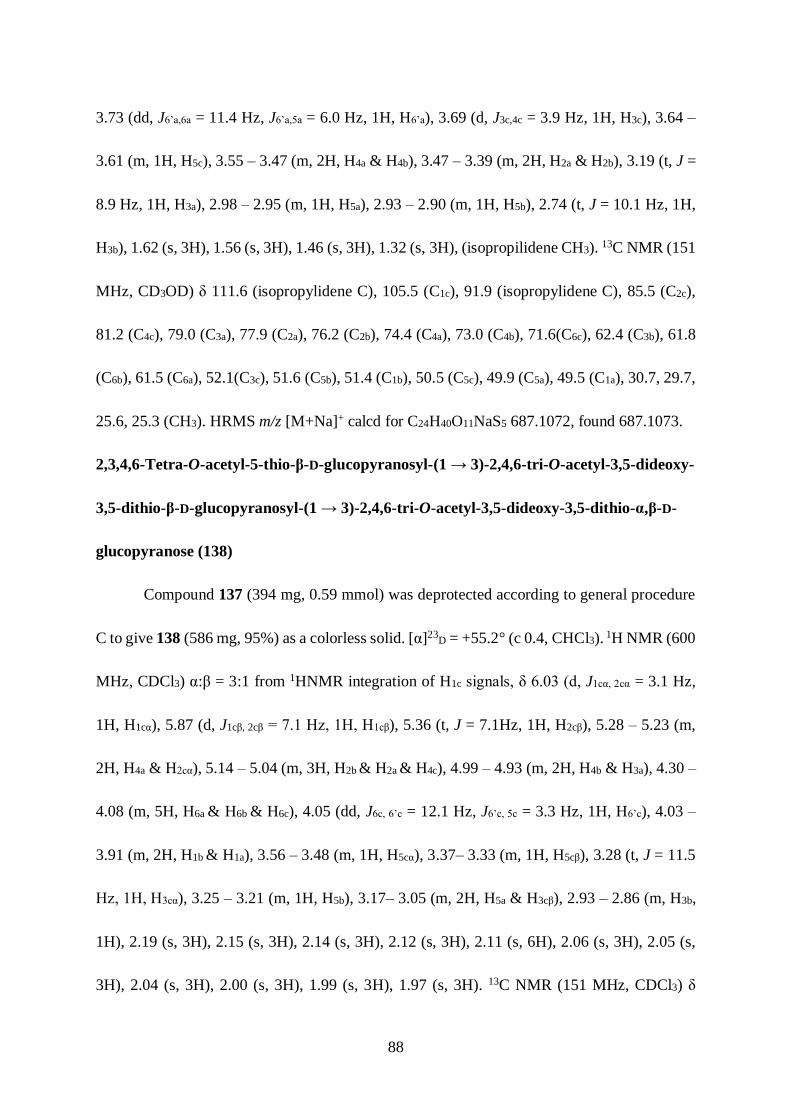
88
3.73 (dd, J6’a,6a = 11.4 Hz, J6’a,5a = 6.0 Hz, 1H, H6’a), 3.69 (d, J3c,4c = 3.9 Hz, 1H, H3c), 3.64 –
3.61 (m, 1H, H5c), 3.55 – 3.47 (m, 2H, H4a & H4b), 3.47 – 3.39 (m, 2H, H2a & H2b), 3.19 (t, J =
8.9 Hz, 1H, H3a), 2.98 – 2.95 (m, 1H, H5a), 2.93 – 2.90 (m, 1H, H5b), 2.74 (t, J = 10.1 Hz, 1H,
H3b), 1.62 (s, 3H), 1.56 (s, 3H), 1.46 (s, 3H), 1.32 (s, 3H), (isopropilidene CH3). 13C NMR (151
MHz, CD3OD) δ 111.6 (isopropylidene C), 105.5 (C1c), 91.9 (isopropylidene C), 85.5 (C2c),
81.2 (C4c), 79.0 (C3a), 77.9 (C2a), 76.2 (C2b), 74.4 (C4a), 73.0 (C4b), 71.6(C6c), 62.4 (C3b), 61.8
(C6b), 61.5 (C6a), 52.1(C3c), 51.6 (C5b), 51.4 (C1b), 50.5 (C5c), 49.9 (C5a), 49.5 (C1a), 30.7, 29.7,
25.6, 25.3 (CH3). HRMS m/z [M+Na]+ calcd for C24H40O11NaS5 687.1072, found 687.1073.
2,3,4,6-Tetra-O-acetyl-5-thio-β-D-glucopyranosyl-(1 → 3)-2,4,6-tri-O-acetyl-3,5-dideoxy-
3,5-dithio-β-D-glucopyranosyl-(1 → 3)-2,4,6-tri-O-acetyl-3,5-dideoxy-3,5-dithio-α,β-D-
glucopyranose (138)
Compound 137 (394 mg, 0.59 mmol) was deprotected according to general procedure
C to give 138 (586 mg, 95%) as a colorless solid. [α]23D = +55.2° (c 0.4, CHCl3). 1H NMR (600
MHz, CDCl3) α:β = 3:1 from 1HNMR integration of H1c signals, δ 6.03 (d, J1cα, 2cα = 3.1 Hz,
1H, H1cα), 5.87 (d, J1cβ, 2cβ = 7.1 Hz, 1H, Η1cβ), 5.36 (t, J = 7.1Hz, 1H, H2cβ), 5.28 – 5.23 (m,
2H, H4a & H2cα), 5.14 – 5.04 (m, 3H, H2b & H2a & H4c), 4.99 – 4.93 (m, 2H, H4b & H3a), 4.30 –
4.08 (m, 5H, H6a & H6b & H6c), 4.05 (dd, J6c, 6’c = 12.1 Hz, J6’c, 5c = 3.3 Hz, 1H, H6’c), 4.03 –
3.91 (m, 2H, H1b & H1a), 3.56 – 3.48 (m, 1H, H5cα), 3.37– 3.33 (m, 1H, H5cβ), 3.28 (t, J = 11.5
Hz, 1H, Η3cα), 3.25 – 3.21 (m, 1H, H5b), 3.17– 3.05 (m, 2H, H5a & H3cβ), 2.93 – 2.86 (m, H3b,
1H), 2.19 (s, 3H), 2.15 (s, 3H), 2.14 (s, 3H), 2.12 (s, 3H), 2.11 (s, 6H), 2.06 (s, 3H), 2.05 (s,
3H), 2.04 (s, 3H), 2.00 (s, 3H), 1.99 (s, 3H), 1.97 (s, 3H). 13C NMR (151 MHz, CDCl3) δ

89
170.57, 170.54, 170.42, 169.67, 169.23, 169.17, 169.14, 169.10, 168.99, 168.86, 168.72
(Carbonyl C), 76.2 (C2b), 75.5 (C2cα), 74.8 (C2cβ), 74.2 (C3a), 72.7 (C1cβ), 72.6 (C2a), 71.9 (C4a),
70.8 (C4cα), 70.5 (C1cα & C4b), 70.2 (C4cβ), 62.8 (C6cβ), 61.8 (C6cα), 61.5 (C6b), 61.1 (C6a), 51.6
(C1b), 50.8 (C1a), 50.0 (C3cα), 46.6 (C5b), 44.5 (C5a), 42.9 (C5cβ), 40.7 (C5cα), 21.1, 21.0, 20.9,
20.8, 20.7, 20.7, 20.6, 20.6, 20.5, 20.4, 20.2 (Acetyl CH3). HRMS m/z [M+Na]+ calcd for
C40H54O22NaS5 1069.1608, found 1069.1613.
5-Thio-β-D-glucopyranosyl-(1 → 3)-3,5-dideoxy-3,5-dithio-β-D-glucopyranosyl-(1 → 3)-
3,5-dideoxy-3,5-dithio-α,β-D-glucopyranose (139)
Compound 138 (20 mg, 0.02 mmol) was deacetylated according to general procedure
B to give 139 (11 mg, quantitative) as a colorless solid, [α]23D = +78.8° (c 2.6, MeOH). α:β =
4:1 from 13CNMR integration of C1c signals. 1H NMR (600 MHz, D2O) δ 4.85 (d, J = 2.9 Hz,
1H, H1cα), 4.59 (d, J = 12.0 Hz, 1H, H1cβ) 3.98 (t, J = 10.9 Hz, 2H, H1a&H1b), 3.83 – 3.64 (m,
6H, H6a,6’a&H6b,6’b&H6c,6’c), 3.58 – 3.42 (m, 4H, H2b&H4a&H4b&H4c), 3.35 (t, J = 9.0 Hz, 1H,
H2a), 3.17 (t, J = 9.0 Hz, 1H, H3a), 3.14 – 3.10 (m, 1H, H5c), 3.05 (t, J = 10.7 Hz, 1H, H3c),
2.95– 2.91 (m, 1H, H5b), 2.90–2.85 (m, 1H, H5a), 2.73 (t, J = 10.4 Hz, 1H, H3b). 13C NMR (151
MHz, D2O) δ 77.9 (C3a), 77.3 (C2cβ), 76.1 (C2a&C2b), 75.1 (C1cβ), 74.7 (C2cα), 72.8 (C4a), 72.5
(C1cα), 70.8 (C4b), 70.7 (C4cβ), 70.6 (C4cα), 60.7 (C6cβ), 60.5 (C6cα), 60.4 (C6b), 60.0 (C6a), 59.2
(C3b), 57.6 (C3cβ), 54.5 (C3cα), 50.6 (C5b), 50.4 (C1b), 48.8 (C1a), 48.9 (C5a), 48.1 (C5cβ), 43.5
(C5cα). HRMS m/z [M+Na]+ calcd for C18H32O11NaS5 607.0446, found 607.0447

90
2,3,4,6-Tetra-O-acetyl-5-thio-β-D-glucopyranosyl-(1 → 3)-2,4,6-tri-O-acetyl-3,5-dideoxy-
3,5-dithio-β-D-glucopyranosyl-(1 → 3)-2,4,6-tri-O-acetyl-3,5-trideoxy-3,5-dithio-α-D-
glucopyranosyl bromide (140)
Bromination was performed on compound 138 (53 mg, 0.05 mmol) according to
general procedure D to give 140 (23 mg, 43%) as a colorless oil. [α]23D = +90.0° (c 0.6, CH2Cl2).
1H NMR (600 MHz, CDCl3) δ 5.47 (d, J = 3.4 Hz, 1H, H1c), 5.17 – 5.02 (m, 1H, H4a), 5.17 –
5.02 (m, 3H, H2a&H2b& H3a), 5.01 – 4.89 (m, 3H, H2c&H4b&H4c), 4.34 (dd, J6a,6’a = 12.2 Hz,
J6a,5a = 5.1 Hz, 1H, H6a), 4.29 – 4.06 (m, 5H, H6’a&H6b&H6’b&H6c&H6’c), 4.00 (d, J = 10.7 Hz,
1H, H1b), 3.95 (d, J = 10.9 Hz, 1H, H1a), 3.67 – 3.58 (m, 1H, H5c), 3.42 (t, J = 10.9 Hz, 1H,
H3c), 3.29 – 3.20 (m, 1H, H5b), 3.15 – 3.07 (m, 1H, H5a), 2.97 – 2.85 (m, 1H, H3b). 2.23 (s, 3H),
2.16 (s, 3H), 2.14 (s, 3H), 2.12 (s, 3H), 2.07 (s, 3H), 2.05 (s, 3H), 2.04 (s, 3H), 2.00 (s, 3H),
1.99 (s, 3H), 1.97 (s, 3H). 13C NMR (151 MHz, CDCl3) δ 170.5, 170.4, 169.7, 169.3, 169.2,
169.1, 169.1, 168.9, 168.6 (Carbonyl C), 76.3 (C2c), 76.2 (C2b), 74.3 (C3a), 72.6 (C2a), 71.9
(C4a), 70.6 (C4c), 70.4 (C4b), 61.7 (C6c), 61.1 (C6b&C6a), 55.3 (C3b), 55.0 (C3c), 51.6 (C1b), 50.8
(C1a), 50.6 (C1c), 46.8 (C5b), 44.5 (C5a), 42.6 (C5c), 20.9, 20.8, 20.6, 20.5, 20.4, 20.2 (Acetyl
CH3). HRMS m/z [M+Na]+ calcd for C37H47O25NaS3Br 1089.0650, found 1089.0653.
2,3,4,6-Tetra-O-acetyl-5-thio-β-D-glucopyranosyl-(1 → 3)-2,4,6-tri-O-acetyl-3,5-dideoxy-
3,5-dithio-β-D-glucopyranosyl-(1 → 3)-1-S-acetyl-2,4,6-tri-O-acetyl-1,3,5-trideoxy-1,3,5-
trithio-β-D-glucopyranose (141)
The anomeric bromide of 140 (34 mg, 0.03mmol) was substituted by potassium
thioacetate (5mg, 1.5 equiv) according to general procedure E to give 141 (30mg, 90%) as a

91
colorless solid. [α]23D = +99.2° (c 0.6, CHCl3). 1H NMR (600 MHz, CDCl3) δ 5.25 (t, J = 10.9
Hz, 1H, H4a), 5.22 (t, J =10.6 Hz, 1H, H2c), 5.16 – 5.07 (m, 1H, H2b), 5.05 (dd, J2a,1a = 11.0 Hz,
J2a,3a = 9.5 Hz, 1H, H2a), 5.00 – 4.93 (m, 4H, H3a&H4b&H4c), 4.60 (d, J1c,2c = 10.6 Hz, 1H, H1c),
4.28 – 4.09 (m, 6H, H6a,6’a & H6b,6’b& H6c, 6’c), 3.93 (d, J1a, 2a = 11.1 Hz, 1H, H1a), 3.90 (d, J1b,
2b = 10.6 Hz, 1H, H1b) , 3.37 – 3.34 (m, 1H, H5c), 3.16 – 3.13 (m, 1H, H5b), 3.11 – 3.07 (m, 1H,
H5a), 3.03 (t, J = 10.7 Hz, 1H, H3c), 2.88 – 2.80 (m, 1H, H3b), 2.38 (s, 3H), 2.13 (s, 6H), 2.12
(s, 6H), 2.06 (s, 6H), 2.04 (s, 3H), 2.00 (s, 3H), 1.99 (s, 3H), 1.97 (s, 3H). 13C NMR (151 MHz,
CDCl3) δ 192.0, 170.5, 170.4, 169.7, 169.3, 169.1, 168.5 (Carbonyl C), 76.1 (C2c), 75.3 (C2b),
74.3 (C3a), 72.5 (C2a), 71.8 (C4a), 70.5 (C4c), 70.4 (C4b) 61.7 (C6c), 61.7 (C6b), 61.1 (C6a), 55.3
(C3b), 54.9 (C3c), 52.4 (C1b), 50.8 (C1a), 47.2 (C5c), 46.8 (C5b), 45.7 (C1c), 44.5 (C5a), 30.5 (S-
Acetyl CH3), 20.8, 20.7, 20.60, 20.5, 20.4, 20.2 (Acetyl CH3). HRMS m/z [M+Na]+ calcd for
C40H54O21NaS6 1085.1380, found 1085.1383.
2,3,4,6-Tetra-O-acetyl-5-thio-β-D-glucopyranosyl-(1 → 3)-2,4,6-tri-O-acetyl-3,5-dideoxy-
3,5-dithio-β-D-glucopyranosyl-(1 → 3)-2,4,6-tri-O-acetyl-3,5-dideoxy-3,5-dithio-β-D-
glucopyranosyl-(1 → 3)-1,2-O-5-S,6-O-di-isopropylidene-3,5-dideoxy-3,5-dithio-α-D-
glucofuranose (142)
Compound 141 (510 mg, 0.48 mmol) and compound 119 (490 mg, 0.72 mmol) were
subjected to coupling reaction according to general procedure A to give product 142 (283 mg,
46%) as white crystals. mp 139-140 °C. [α]23D = +31.6° (c 0.3, CHCl3). 1H NMR (600 MHz,
CDCl3) δ 5.88 (d, J1d,2d = 3.6 Hz, 1H, H1d), 5.27 (t, J = 10.1 Hz, 1H, H4a), 5.17 (t, J = 10.6 Hz,
1H, H2c), 5.13 – 5.08 (m, 1H, H2b), 5.05 (t, J = 10.3 Hz, 1H, H2a), 5.01 – 4.94 (m, 3H,

92
H3a&H4b&H4c), 4.70 (d, J2d,1d = 3.6 Hz, 1H, H2d), 4.40 (dd, J4d,5d = 10.5 Hz, J4d,3d = 3.9 Hz, 1H,
H4d), 4.33 (dd, J6d,6’d = 10.1 Hz, J6d,5d = 2.4 Hz, 1H, H6d), 4.29 – 4.18 (m, 3H, H6a&H6b&H6c),
4.17 – 4.07 (m, 4H, H6’a&H6’b&H6’c&H6’d), 3.94 (d, J1a,2a = 11.1 Hz, 1H, H1a), 3.90 (d, J1b,2b =
10.3 Hz, 1H, H1b), 3.84 (d, J1c,2c = 10.5 Hz, 1H, H1c), 3.67 – 3.62 (m, 1H, H5d), 3.57 (d, J3d,4d =
4.0 Hz, 1H, H3d), 3.20 –3.15 (m, 2H, H5b&H5c), 3.11 – 3.08 (m, J = 10.3, 4.1 Hz, 1H, H5a), 2.95
(t, J = 10.7 Hz, 1H, H3c), 2.88–2.82 (m, 1H, H3b), 2.21 (s, 3H), 2.14 (s, 3H), 2.12 (s, 3H), 2.11
(s, 3H), 2.06 (s, 6H), 2.05 (s, 3H), 2.01 (s, 3H), 1.99 (s, 3H), 1.97 (s, 3H), 1.67 (s, 3H), 1.60 (s,
3H), 1.53 (s, 3H), 1.35 (s, 3H). 13C NMR (151 MHz, CDCl3) δ 170.6, 170.4, 169.7, 169.2,
169.1, 168.7, 168.3, 112.3 (isopropylidene C), 105.4 (C1d), 92.7 (isopropylidene C), 86.1 (C2d),
81.4 (C4d), 76.2 (C2c), 75.7 (C2b), 74.2 (C3a), 72.5 (C2a), 72.0 (C6d), 71.8 (C4a), 70.6 (C4c) 70.4
(C4b), 61.8 (C6c), 61.7 (C6b), 61.0 (C6a), 55.3 (C3b), 54.9 (C3c), 53.5 (C3d), 52.5 (C1b), 50.8 (C1a),
50.6 (C5d), 50.0 (C1c), 47.0 (C5c), 46.6 (C5b), 44.5 (C5a), 31.6, 30.7, 26.7, 26.5 (Isopropylidene
CH3), 21.0, 20.9, 20.8, 20.7, 20.6, 20.5, 20.4, 20.2 (Acetyl CH3). HRMS m/z [M+Na]+ calcd
for C50H70O24NaS7 1301.2200, found 1301.2205.
5-Thio-β-D-glucopyranosyl-(1 → 3)-3,5-dideoxy-3,5-dithio-β-D-glucopyranosyl-(1 → 3)-
3,5-dideoxy-3,5-dithio-β-D-glucopyranosyl-(1 → 3)-1,2-O-5-S,6-O-di-isopropylidene-3,5-
dideoxy-3,5-dithio-α-D-glucofuranose (143)
Compound 142 (150 mg, 0.12 mmol) was deacetylated according to general procedure
B to give 143 (100 mg, quantitative) as a colorless solid. [α]23D = +18.9° (c 0.5, MeOH). 1H
NMR (600 MHz, CD3OD) δ 5.88 (d, J1d,2d = 3.6, 1H, H1d), 4.90 (d, J2d,1d = 3.6 Hz, 1H, H2d),
4.37 (dd, J4d,5d = 10.5 Hz, J4d,3d = 4.0 Hz, 1H, H4d), 4.25 (dd, J = 10.0, 1.8 Hz, 1H, H6d), 4.10

93
(dd, J = 8.0, 4.9, 1.6 Hz, 1H, H6’d), 4.00 (d, J = 10.0 Hz, 1H, H1b) 3.97 (d, J = 10.3 Hz, 1H,
H1a), 3.94 – 3.86 (m, 4H, H1c&H6a&H6b&H6c), 3.81 – 3.72 (m, 2H, H6’a&H6’b), 3.72 – 3.67 (m,
H6’c1H), 3.66 (d, J = 3.9 Hz, 1H, H3d), 3.65 – 3.60 (m, 1H, H5d), 3.56 – 3.43 (m, 5H,
H4a&H4b&H4c&H2b&H2c), 3.38 (t, J = 10.4 Hz, 1H, H2a), 3.17 (t, J = 8.9 Hz, 1H, H3a), 3.02 –
2.88 (m, 3H, H5a&H5b&H5c), 2.85 – 2.69 (m, 2H, H3b&H3c), 1.62 (s, 3H), 1.56 (s, 3H), 1.46 (s,
3H), 1.31 (s, 3H). 13C NMR (151 MHz, CD3OD) δ 111.6 (isopropylidene C), 105.4 (C1d), 92.0
(isopropylidene C), 85.6 (C2d), 81.1 (C4d), 78.6 (C3a), 77.5 (C2a), 77.0 (C2b), 75.8 (C2c), 73.7
(C4a), 71.9 (C4b), 71.8 (C4c), 71.6 (C6d), 61.8 (C3b), 61.2 (C6c), 61.1 (C3c), 60.9 (C6a&C6b), 52.5
(C3d), 51.7 (C5b), 51.4 (C5c), 51.3 (C1b), 51.1 (C1c), 50.5 (C5d), 49.6 (C5a&C1a), 30.7, 29.6, 25.5,
25.2 (isopropylidene-CH3). HRMS m/z [M+Na]+ calcd for C30H50O14NaS7 881.1143, found
881.1141.
5-Thio-β-D-glucopyranosyl-(1 → 3)-3,5-dideoxy-3,5-dithio-β-D-glucopyranosyl-(1 → 3)-
3,5-dideoxy-3,5-dithio-β-D-glucopyranosyl-(1 → 3)-3,5-dideoxy-3,5-dithio-α,β-D-
glucopyranose (144)
Compound 143 (100 mg, 0.12 mmol) was deprotected according to general procedure
C to give product 144 (78 mg, 85%) as a white solid. [α]23D = +36.6° (c 0.5, H2O). α:β = 4:1
from 13CNMR integration of C1d signals. 1H NMR (600 MHz, D2O) δ 4.86 (d, J = 3.0 Hz, 1H,
H1dα), 4.58 (d, J = 8.8Hz, 1H, H1dβ) 4.02 – 3.90 (m, 3H, H1a& H1b& H1c), 3.86 – 3.59 (m, 9H,
H6a,6’a & H6b,6’b & H6c,6’c & H6d,6’d & H2d), 3.57 – 3.37 (m, 6H, H4a & H4b & H4c & H4d, H2b &
H2c), 3.31 (t, J = 9.0 Hz 1H, H2a), 3.13 (t, J = 9.0 Hz, 1H, H3a), 3.10 – 3.06 (m, 1H, H5d), 3.01
(t, J = 10.6 Hz, 1H, H3d), 2.96– 2.81 (m, 3H, H5a & H5b & H5c), 2.76 – 2.66 (m, 2H, H3b & H3c).

94
13C NMR (151 MHz, D2O) δ 77.9 (C3a), 77.3 (C2dβ), 76.2 (C2a&C2b&C2c), 75.1 (C1dβ), 74.7
(C2dα), 72.9 (C4a), 72.5 (C1dα), 70.9 (C4b), 70.8 (C4dβ), 70.6 (C4c&C4dα), 60.6 (C6b & C6c), 60.5
(C6a), 60.0 (C3c), 59.3 (C3b), 59.2 (C3dβ), 54.5 (C3dα), 50.7 (C1b) 50.6 (C5b & C5c), 50.4 (C1c),
48.8 (C1a), 48.6 (C5a), 48.1 (C5dβ), 43.5 (C5dα). HRMS m/z [M+Na]+ calcd for C24H42O14NaS7
801.0517, found 801.0518.
p-N,N-Dimethylamino benzyl alcohol (150)
4-(dimethylamino)benzaldehyde (0.5 g, 3.35 mmol) was dissolved in dry methanol (10
mL) and added sodium borohydride (NaBH4, 253 mg, 6.70 mmol) at 0 °C. The reaction
mixture was stirred at 0 °C for 1 h, later at room temperature for 1 h. Methanol was removed
on rotavapor, added water (200 mL) and extracted with DCM (2 x 100 mL). The DCM layer
was washed with water (100 mL), brine solution (50 mL) and dried over Na2SO4. The solvent
was evaporated and the residue was purified by flash chromatography on silica gel with 6/4
ethyl acetate/hexane to give the product 150 (430 mg, 85 %) as a colorless oil. 1H NMR (500
MHz, CDCl3): δ 7.25 (d, 2H, J = 9.5 Hz), 6.73 (d, 2H, J = 8.5 Hz), 4.57 (br, 2H, Benzylic CH2),
2.95 (s, 6H, Me), 1.48 (bs, 1H, OH) ; 13C NMR (125 MHz, CDCl3) δ: 150.6, 129.1, 128.9,
112.9, 65.6 (Benzylic CH2), 40.9 (Me). HRMS m/z [M+H]+ calculated for C9H14NO is
152.1075, found 152.1072.
4,4'-methylenebis(N,N-dimethylaniline) (151)
At 0 °C, to a solution of compound 150 (54 mg, 0.36 mmol) in DMF was added NaH
(8.6 mg, 0.36 mmol) and p-toluenesulfonyl chloride (103 mg, 0.54 mmol) was added into the
reaction mixture. The reaction mixture was stirred at room temperature for 2 h before the

95
reaction mixture was quenched with aqueous NH4Cl. The crude mixture was extracted into
ethyl acetate and washed with water, brine and dried over MgSO4. The crude mixture was
concentrated in vacuo and purified by silica gel chromatography to give product 151 (50.4 mg,
56%) as a yellow oil. 1H NMR (400 MHz, CDCl3): δ 7.14-7.08 (m, 4H), 6.76-6.71 (m, 4H),
3.86 (d, 2H, J = 4.4 Hz, Benzylic CH2), 2.95 (s, 6H), 2.94 (s, 6H); 13C NMR (100 MHz, CDCl3)
δ: 149.3, 130.6, 129.7, 113.3, 41.2, 40.2. HRMS m/z (M+H)+ calcd for C17H23N2 255.1861,
found 255.1861.
p-N,N-Dimethylamino benzyl chloride hydrochloride (152)
The solution of p-N,N-dimethyl amino benzyl alcohol (0.85 g, 5.60 mmol) in 3.5 mL
aqueous HCl (36.6%) was refluxed at 100 °C for 12 h. The reaction mixture was concentrated
in vacuo and high vacuum evaporation at 45 °C for 4 h to remove water to give product 156
(0.11 g, 96%) as a colorless oil. 1H NMR (400 MHz, CDCl3) δ 7.85 – 7.74 (m, 2H), 7.58 – 7.49
(m, 2H), 4.58 (s, 2H, benzylic CH2), 3.18 (s, 6H, CH3). 13C NMR (101 MHz, CDCl3) δ 142.6,
139.9, 130.6, 121.3, 46.6 (Benzylic CH2), 44.6 (Me).
3-O-(4-Chlorobenzyl)-1,2;5,6-di-O-isopropylidene-α-D-glucofuranose (153)
At 0 oC, to a solution of 1,2;5,6-di-O-isopropylidene-α-D-glucofuranose (0.2 g, 0.8
mmol) in 5 mL DMF was added NaH (40 mg, 1 mmol). The reaction was stirred at 0 oC for 30
min and 4-chlorobenzyl bromide (0.2 g, 1 mmol) was added into the reaction. The reaction was
stirred at 0 oC for 3 h before it was quenched with aqueous NH4Cl. The crude mixture was
extracted into ethyl acetate, washed with water, brine and dried over MgSO4. The crude mixture
was concentrated in vacuo and purified by sílica gel chromatography to give product 153 (0.24

96
g, 78%) as a white solid. 1H NMR (400 MHz, CDCl3) δ 7.37 – 7.20 (m, 5H), 5.89 (d, J = 3.7
Hz, 1H, H1), 4.70 – 4.52 (m, 3H, Benzylic CH2 & H2), 4.34 (dt, J = 8.1, 5.9 Hz, 1H, H4), 4.17
– 4.06 (m, 2H, H6 & H3), 4.05 – 3.93 (m, 2H, H6’ & H5), 1.49 (s, 3H), 1.42 (s, 3H), 1.37 (s, 3H),
1.31 (s, 3H). 13C NMR (101 MHz, CDCl3) δ 136.1, 133.6, 129.0, 128.6, 111.9, 109.1
(isopropylidene), 105.3 (C1), 82.6, 81.7, 81.3, 77.3, 77.0, 76.7, 72.4 (Benzylic CH2), 71.5, 67.5,
26.8, 26.2, 25.4.
3-O-(p-N,N-Diethylamino benzyl)-1,2;5,6-di-O-isopropylidene-α-D-glucofuranose (154)
To a solution of Tris(dibenzylideneacetone)dipalladium(0) (3.3 mg, 3.6 μmol ), 2-
Dicyclohexylphosphino-2′-(N,N-dimethylamino)biphenyl (2.8 mg, 7.2 μmol), potassium tert-
butoxide (28 mg, 0.25 mmol) and 1,2;5,6-di-O-isopropylidene-D-glucofuranose (110 mg, 0.18
mmol) in Toluene (2 mL) was added diethyl amine (23 μL, 0.22 mmol). The reaction was
stirred at 80 °C for 12 h before the reaction mixture was concentrated in vacuo and the crude
mixture was dissolved in Ethyl Acetate (25 mL) and washed by water, brine and dried over
Na2SO4. The reaction mixture was concentrated in vacuo and purified by silica gel
chromatography with eluent (8:1 = Hexane: Ethyl acetate) to give product 154 as a colorless
oil (88 mg, 82%). [α]23D = -18.4° (c 1.5, CHCl3). 1H NMR (600 MHz, CDCl3) δ 7.18 – 7.14
(m, 2H), 6.66 – 6.60 (m, 2H), 5.86 (d, J = 3.7 Hz, 1H), 4.54 (d, J = 3.8 Hz, 1H), 4.53 – 4.46
(m, 2H), 4.37 – 4.28 (m, 1H), 4.15 (dd, J = 7.3, 3.1 Hz, 1H), 4.12 – 4.06 (m, 1H), 4.01 – 3.96
(m, 2H), 3.33 (q, J = 7.1 Hz, 4H), 1.48 (S, 3H), 1.42 (s, 3H), 1.37 (s, 3H), 1.30 (s, 3H), 1.14 (t,
J = 7.1 Hz, 6H). 13C NMR (151 MHz, CDCl3) δ 129.6, 111.6, 111.5, 105.3, 82.8, 81.3, 81.3,

97
80.9, 77.2, 77.0, 76.8, 72.7, 72.6, 72.2, 67.4, 67.2, 44.4, 26.9, 26.8, 26.8, 26.7, 26.3, 26.2, 25.5,
25.4, 12.5. HRMS m/z [M+H]+ calcd for C23H36NO6 422.2543, found 422.2544.
4-(Methoxymethyl)-N,N-diethyl aniline (155)
The compound 154 (53 mg, 0.13 mmol) was dissolved in 2 mL methanol and was
transferred into a 10 mL vial equipped with a magnetic stir bar. The vial was capped properly
and placed in the microwave (CEM Discover SP Microwave). The microwave was run at 300
W, 160 °C and 300 psi for 30 min. The vial was cooled to room temperature and the reaction
mixture was transferred into round bottom flask and concentrated in vacuo. The residue was
purified by silica gel column chromatography to give product 155 (24 mg, 98%) as a colorless
oil. 1H NMR (400 MHz, CDCl3) δ 7.24 – 7.11 (m, 1H), 6.66 (d, J = 8.6 Hz, 1H), 4.34 (s, 1H),
3.35 (d, J = 7.5 Hz, 4H), 1.16 (t, J = 7.0 Hz, 3H). 13C NMR (101 MHz, CDCl3) δ 147.5, 129.6,
124.6, 111.7, 74.7, 57.5, 44.4, 12.5. HRMS m/z [M+H]+ calcd for C12H20NO 194.1545, found
194.1541.
ortho-N,N-Dimethylamino benzyl alcohol (156)
To a solution of ortho-amino benzyl alcohol (1.23 g, 10 mmol) in 25 mL DMF was
added Na2HPO4 (5.11 g, 36 mmol) and methyl iodide (3.4 g, 24 mmol). The reaction mixture
was stirred at 45 oC for 10 h before it was poured into 200 mL water. The crude mixture was
extracted into diethyl ether, the organic layer was washed with water, brine and dried over
MgSO4. The crude mixture was concentrated in vacuo and purified by sílica gel
chromatography to give product 156 (1.3 g, 86%) as a colorless oil. 1H NMR (400 MHz,
CDCl3) δ 7.29 – 7.19 (m, 2H), 7.19 – 7.13 (m, 1H), 7.08 (tt, J = 7.3, 1.2 Hz, 1H), 5.60 (s, 1H),

98
4.82 (s, 2H), 2.72 (s, 6H). 13C NMR (101 MHz, CDCl3) δ 151.9, 135.2, 128.4, 128.2, 124.4,
120.1, 64.9, 44.6. HRMS m/z [M+H]+ calcd for C9H14NO 152.1070, found 152.1071.
ortho-N,N-Dimethylamino benzyl chloride (157)
At 0 oC, to a solution of compound 156 (141 mg, 0.93 mmol) in 3 mL chloroform was
added SO2Cl2 (222 mg, 1.9 mmol). The reaction mixture was stirred at 0 oC for 4 h and
concentrated in vacuo to give product 157 (157 mg, 100%) as a yellow oil. 1H NMR (400 MHz,
CDCl3) δ 7.68 – 7.64 (m, 1H), 7.57 – 7.45 (m, 3H), 5.27 (s, 2H), 3.34 (s, 6H). 13C NMR (101
MHz, CDCl3) δ 140.6, 133.4, 131.2, 130.9, 120.9, 47.7, 41.9.
3-O-(ortho-N,N-Dimethylamino benzyl)-1,2;5,6-di-O-isopropylidene-α-D-glucofuranose
(158)
At 0°C, to a stirred solution of 1,2;5,6-di-O-isopropylidene-α-D-glucofuranose (125 mg,
0.48 mmol) in DMF (1 mL) was added NaH (104 mg, 2.60 mmol) and ortho-N, N-
dimethylamino benzyl chloride (157 mg, 0.93 mmol). The reaction was stirred overnight before
aqueous NH4Cl was added to quench the reaction. The reaction mixture was diluted in ethyl
acetate, washed by water, brine and dried over MgSO4. The reaction mixture was concentrated
in vacuo and purified by silica gel chromatography with eluent (10 : 1 = hexane : ethyl acetate)
to give product 158 as a colorless oil (104 mg, 55%). [α]23D = -1.9° (c 1.8, CHCl3). 1H NMR
(400 MHz, CDCl3) δ 7.45 (dd, J = 7.6, 1.8 Hz, 1H), 7.29 – 7.21 (m, 1H), 7.11 – 7.00 (m, 2H),
5.89 (d, J = 3.7 Hz, 1H, H1), 4.82 (d, J = 11.8 Hz, 1H, benzyl CH2), 4.71 – 4.63 (m, 2H, H2 &
benzyl CH2), 4.41 (dt, J = 8.0, 6.1 Hz, 1H, H4), 4.18 (dd, J = 7.7, 3.1 Hz, 1H, H5), 4.14 – 4.05
(m, 2H, H6 & H3), 4.00 (dd, J = 8.6, 6.0 Hz, 1H, H6’), 2.69 (d, J = 2.0 Hz, 6H, NMe2), 1.51 (s,

99
3H), 1.43 (s, 3H), 1.33 (s, 3H), 1.36 (s, 3H). 13C NMR (101 MHz, CDCl3) δ 131.9, 129.3,
128.3, 123.0, 118.8, 111.7 (Isopropylidene), 108.9 (Isopropylidene), 105.3 (C1), 82.4 (C2), 82.1
(C3), 81.4 (C5), 72.5 (C4), 68.1 (Benzyl CH2), 67.4 (C6), 45.1 (NMe2), 26.9, 26.8, 26.3, 25.4
(Isopropylidene CH3). HRMS m/z [M+H]+ calcd for C21H32NO6 394.2230, found 394.2234.
Phenyl 4-O-(4-bromobenzyl)-2,3-O-isopropylidene-1-thio-β-L-rhamnopyranoside (160)
At 0°C, to a stirred solution of phenyl 2,3-O-isopropylidene-1-thio-β-L-
rhamnopyranoside (512 mg, 1.73 mmol) in DMF (10mL) was added NaH (104 mg, 2.60 mmol)
and 4-bromobenzyl bromide (513 mg, 2.08 mmol). The reaction was stirred overnight before
aqueous NH4Cl was added to quench the reaction. The reaction mixture was diluted in ethyl
acetate, washed by water, brine and dried over MgSO4. The crude mixture was concentrated in
vacuo and purified by silica gel chromatography with eluent (10 : 1 = hexane : ethyl acetate)
to give product 162 as a white solid (2.5 g, 85%). mp 123 ~ 125 °C. [α]23D = +84.0° (c 0.3,
CHCl3). 1H NMR (400 MHz, CDCl3) δ 7.56 – 7.44 (m, 4H), 7.35 – 7.19 (m, 6H), 5.01 (d, J =
2.1 Hz, 1H, H1), 4.82 (d, J = 11.8 Hz, 1H, benzyl CH2), 4.59 (d, J = 11.8 Hz, 1H, benzyl CH2),
4.44 (dd, J = 5.7, 2.2 Hz, 1H, H2), 4.20 (t, J = 5.8 Hz, 1H, H3), 3.33 (t, J = 5.3 Hz, 2H, H4 &
H5), 1.57 (s, 3H), 1.43 (s, 3H), 1.36 (d, J = 5.5 Hz, 3H, H6). 13C NMR (101 MHz, CDCl3) δ
137.1, 135.1, 131.4, 130.9, 129.6, 128.9, 127.4, 121.6, 110.5 (Isopropylidene C), 84.0 (C1),
80.8 (C4), 79.9 (C3), 76.4 (C2), 74.4 (C5), 72.1 (Benzyl C), 28.0, 26.4, 18.5 (C6). HRMS m/z
[M+Na]+ calcd for C22H25BrO4NaS 487.0555, found 487.0557.
Phenyl 4-O-(p-N,N-diethylamino benzyl)-2,3-O-isopropylidene-1-thio-β-L-
rhamnopyranoside (161)

100
To a solution of Tris(dibenzylideneacetone)dipalladium(0) (3.3 mg, 3.6 μmol ), 2-
dicyclohexylphosphino-2′-(N,N-dimethylamino)biphenyl (2.8 mg, 7.2 μmol), potassium tert-
butoxide (28 mg, 0.25 mmol) and compound 160 (110 mg, 0.18 mmol) in Toluene (2 mL) was
added diethyl amine (23 μL, 0.22 mmol). The reaction was stirred at 80 °C for 12 h before the
reaction mixture was concentrated in vacuo and the crude mixture was dissolved in ethyl
acetate (25 mL) and washed by water, brine and dried over Na2SO4. The reaction mixture was
concentrated in vacuo and purified by silica gel chromatography with eluent (8:1 = Hexane:
Ethyl acetate) to give product 161 as a colorless oil (88 mg, 82%). [α]23D = +14.0° (c 0.2,
CHCl3).1H NMR (600 MHz, CDCl3) δ 7.55 – 7.48 (m, 2H), 7.33 – 7.21 (m, 3H), 7.21 – 7.14
(m, 2H), 6.63 (d, J = 8.6 Hz, 2H), 5.00 (d, J = 2.1 Hz, 1H, H1), 4.73 (d, J = 10.8 Hz, 1H, benzyl
CH2), 4.50 (d, J = 10.8 Hz, 1H, benzyl CH2), 4.42 (dd, J = 5.8, 2.1 Hz, 1H, H2), 4.22 (t, J = 5.9
Hz, 1H, H, H3), 3.40 – 3.27 (m, 6H, H4 & H5 & Ethyl CH2), 1.60 (s, 3H), 1.43 (s, 3H), 1.36 –
1.30 (m, 3H, H6), 1.17 – 1.08 (m, 5H). 13C NMR (151 MHz, CDCl3) δ 147.6, 135.3, 130.7,
130.0, 128.9, 127.2, 124.4, 111.6, 110.4 (isopropylidene C), 84.0 (C1), 80.1 (C4), 79.7 (C3),
76.3 (C2), 74.8 (C5), 73.2 (Benzyl C), 44.4 (PDMAB Ethyl CH2), 27.9 (CH3), 26.4 (CH3), 18.6
(C6), 12.5 (PDMAB Ethyl CH3). HRMS m/z [M+H]+ calcd for C26H36NO4S 458.2360, found
458.2362
Methyl 6-O-(4-O-(p-N,N-diethylamino benzyl)-2,3-O-isopropylidene-α-L-rhamnosyl)-
2,3,4-tri-O-benzyl-α-D-glucopyranoside (162)
To a solution of compound 161 (50 mg, 0.11 mmol), methyl 2,3,4-tri-O-benzyl-α-D-
glucopyranoside (50 mg, 0.11 mmol), NIS (50 mg, 0.22 mmol) and cyclohexene (0.11 mL, 1.1

101
mmol) in anhydrous DCM (1.0 mL) was added activated 4 Å molecular sieve (200 mg). The
reaction mixture was stirred at room temperature overnight. The reaction temperature was
cooled to -78 °C and TMSOTf (4.0 μL, 0.02 mmol) was injected into reaction mixture. The
reaction was stirred at -78 °C for 12 h before it was quenched by triethylamine at-78 °C. The
reaction mixture was warmed to room temperature and filtered. After filtration, the organic
layer was washed with aqueous Na2S2O3 and brine and dried over MgSO4 The crude mixture
was concentrated and purified by sílica gel chromatography to give glycosylation product 162
(20 mg, 22%) as a colorless oil. [α]23D = +6.4° (c 0.5, CHCl3).1H NMR (600 MHz, CDCl3) δ
7.39 – 7.22 (m, 12H), 7.18 (d, J = 8.1 Hz, 2H), 6.63 (d, J = 8.1 Hz, 2H), 4.98 (d, J = 10.9 Hz,
1H, Benzyl CH2), 4.91 – 4.69 (m, 6H, Benzyl CH2 & Rha H1), 4.65 (d, J = 12.1Hz, 1H, Benzyl
CH2), 4.55 (ddd, J = 6.8, 3.7, 1.7 Hz, 2H, Glc H1 & Benzyl CH2), 4.50 – 4.42 (m, 1H, Benzyl
CH2), 4.20 (ddd, J = 7.1, 4.4, 1.5 Hz, 1H, Rha H2), 4.04 (dd, J = 5.7, 1.6 Hz, 1H, Rha H3), 4.00
– 3.94 (m, 1H, Glc H3), 3.84 – 3.78 (m, 1H, Glc H6), 3.72 – 3.61 (m, 2H, Glc H6’ & Rha H4),
3.53 – 3.42 (m, 3H, Glc H4 & Glc H2 & Glc H6), 3.36 – 3.29 (m, 7H, Glc OMe & PEMAB
Ethyl CH2), 3.17 (ddt, J = 9.9, 7.2, 1.6 Hz, 1H, Rha H5), 1.51 (s, 3H, isopropylidene), 1.35 (s,
3H, isopropylidene), 1.20 (dt, J = 6.3, 1.6 Hz, 3H, Rha H6), 1.13 (tt, J = 7.0, 1.6 Hz, 7H,
PEMAB Me). 13C NMR (151 MHz, CDCl3) δ 147.5, 138.7, 138.1, 131.4, 129.9, 128.5, 128.4,
128.3, 128.1, 128.0, 127.9, 127.8, 127.6, 124.8, 112.5, 111.6, 109.0 (Isopropylidene C), 97.8
(Glc C1), 97.1 (Rha C1), 82.1 (Glc C3), 80.5 (Rha C5), 80.0 (Glc C2), 78.8 (Rha C3), 77.5 (Glc
C4), 75.9 (Rha C2), 75.7 (Benzyl CH2), 74.9 (Benzyl CH2), 73.3 (Benzyl CH2), 69.8 (Glc C5),
65.8 (Glc C6), 64.7 (Rha C4), 55.1 (Glc OMe), 44.4 (PDEAB Ethyl CH2), 28.0 (Isopropylidene

102
Me), 26.4 (Isopropylidene Me),17.7 (Rha C6), 12.5 (PDEAB Ethyl CH3). HRMS m/z [M+H]+
calcd for C48H62NO10 812.4374, found 812.4375.
Methyl 6-O-(2,3-O-isopropylidene-α-L-rhamnosyl)-2,3,4-tri-O-benzyl-α-D-
glucopyranoside (163)
Compound 162 (9 mg, 0.01 mmol) was dissolved in 2 mL methanol and was transferred
into a 10 mL vial equipped with a magnetic stir bar. The vial was capped properly and placed
in the microwave (CEM Discover SP Microwave). The microwave was run at 300 W, 180 °C
and 300 psi for 30 min. The vial was cooled to room temperature and the reaction mixture was
transferred into round bottom flask and concentrated in vacuo. The residue was purified by
silica gel column chromatography to give product 163 (5.3 mg, 73%) as a colorless oil. [α]23D
= +9.6° (c 0.3, CHCl3). 1H NMR (600 MHz, CDCl3) δ 7.38 – 7.23 (m, 16H), 4.99 (d, J = 10.8
Hz, 1H, Benzyl CH2), 4.88 (d, J = 11.0 Hz, 1H, Benzyl CH2), 4.83 – 4.74 (m, 3H, Benzyl CH2
and Rha H1), 4.66 (d, J = 12.1 Hz, 1H, Benzyl CH2), 4.59 (d, J = 3.5 Hz, 1H, Glc H1), 4.56 (d,
J = 11.0 Hz, 1H, Benzyl CH2), 4.08 (t, J = 6.3 Hz, 1H, Rha H2), 4.04 (d, J = 5.9 Hz, 1H, Rha
H3), 3.99 (t, J = 9.3 Hz, 1H, Glc H3), 3.85 (dd, J = 10.7, 2.0 Hz, 1H, Glc H6’), 3.76 – 3.65 (m,
2H, Glc H5 & Rha H4), 3.54 (dd, J = 10.8, 4.7 Hz, 1H, Glc H4), 3.52 – 3.45 (m, 2H, Glc H2 &
Glc H6), 3.39 (dd, J = 8.3, 6.7 Hz, 1H, Rha H5), 3.36 (s, 3H, Glc OMe), 1.50 (s, 3H,
isopropylidene), 1.33 (s, 3H, isopropylidene), 1.26 – 1.24 (m, 3H, Rha H6 ).13C NMR (151
MHz, CDCl3) δ 138.6, 138.0, 128.5, 128.4 , 128.1, 128.0, 127.6, 109.4 (Isopropylidene C),
98.0 (Glc C1), 97.3 (Rha C1), 82.1 (Glc C3), 80.0 (Glc C2), 77.8 (Rha C3), 77.3 (Glc C4), 75.8
(Rha C2), 75.2 (Benzyl CH2), 75.0 (Benzyl CH2), 73.7 (Benzyl CH2), 73.4 (Benzyl CH2), 69.7

103
(Glc C5), 66.7 (Rha C4), 65.9 (Glc C6), 55.2 (Glc OMe), 27.7 (Isopropylidene Me), 25.9
(Isopropylidene Me), 17.8 (Rha C6). HRMS m/z [M+Na]+ calcd for C37H46NaO10 673.2989,
found 673.2991.
Inhibition of anti-CR3-FITC antibody staining of human neutrophils and of anti-Dectin
1-FITC antibody staining of mouse macrophages.
For fluorescent staining, anti-CR3-FITC antibodies (MN-41 donated by Drs. Allison Eddy
and Alfred Michael of the University of Minnesota, Minneapolis, MN, and Rat anti Mouse
Dectin-1 antibody labeled with FITC (purchased from AbD Serotec, Raleigh, NC) were
employed. Either human neutrophils or mouse peritoneal macrophages were incubated with
0.1 g.mL-1 of tested samples for 0.5 h on ice and washed. Subsequently, the cells were stained
with antibodies on ice using standard techniques. After centrifugation of cells through a 3 mL
cushion of 12% BSA in PBS, the cells were re-suspended in PBS containing 1% BSA and 10
mM sodium azide. Cell cytometry was performed with a Becton Dickinson-LSRII instrument.
The inhibition of CR3 receptor and Dectin-1 receptor staining was calculated as described.90
Stimulation of phagocytosis
The technique employing phagocytosis of synthetic polymeric microspheres was
described earlier.91 9191Human cells (cell line RAW 264) were incubated in vitro with 10
μg.mL-1 of tested samples for 24 h at 37 oC. After washing, 0.05 mL of 2-hydroxyethyl
methacrylate particles (HEMA; 5x10⁸/mL) was added. The test tubes were incubated at 37 oC
for 1 h, with intermittent shaking. Smears were stained with Wright stain. Cells with three or
more HEMA particles were considered positive. The insoluble glucan Glucan #300 used as

104
comparison standard was obtained from Yeast-derived insoluble Glucan #300 (>85% dry w/w
basis) was purchased from Transfer Point (Columbia, SC, USA). This glucan contains 96%
carbohydrates and 2.1% proteins. Neutral sugar analysis confirmed 91.3% glucose and 8%
mannose. Stimulation of pinocytosos was determined spectrophotometrically as described.92

105
REFERENCES
1. Klemm, D.; Heublein, B.; Fink, H. P.; Bohn, A., Angew. Chem. Int. Ed. 2005, 44, 3358-
3393.
2. Větvička, V., Beta-glucans as Natural Biological Response Modifiers. Nova Science
Publishers, Incorporated: 2013.
3. Chlubnová, I.; Sylla, B.; Nugier-Chauvin, C.; Daniellou, R.; Legentil, L.; Kralová, B.;
Ferrieres, V., Nat. Prod. Rep. 2011, 28, 937-952.
4. Murphy, K.; Weaver, C., Janeway's immunobiology. Garland Science: 2016.
5. Riggi, S. J.; Di Luzio, N. R., Am J Physiol 1961, 200, 297-300.
6. Schepetkin, I. A.; Quinn, M. T., Int. Immunopharmacol. 2006, 6, 317-333.
7. Weitberg, A. B., J. Exp. Clin. Canc. Res. 2008, 27, 40.
8. Ina, K.; Furuta, R.; Kataoka, T.; Kayukawa, S.; Yoshida, T.; Miwa, T.; Yamamura, Y.;
Takeuchi, Y., World. J. Clin. Oncol. 2011, 2, 339.
9. Fujimoto, S., Nihon Geka Gakkai Zasshi 1989, 90, 1447-1450.
10. Babineau, T. J.; Marcello, P.; Swails, W.; Kenler, A.; Bistrian, B.; Forse, R. A., Ann. Surg.
1994, 220, 601.
11. Markova, N.; Kussovski, V.; Drandarska, I.; Nikolaeva, S.; Georgieva, N.; Radoucheva,
T., Int. Immunopharmacol. 2003, 3, 1557-1562.
12. Zimmerman, J. W.; Lindermuth, J.; Fish, P. A.; Palace, G. P.; Stevenson, T. T.; DeMong,
D. E., J. Biol. Chem. 1998, 273, 22014-22020.
13. Liang, J.; Melican, D.; Cafro, L.; Palace, G.; Fisette, L.; Armstrong, R.; Patchen, M. L.,
Int. J. Immunopharmaco. 1998, 20, 595-614.

106
14. Pearson, A.; Lux, A.; Krieger, M., P. Natl. Acad. Sci. 1995, 92, 4056-4060.
15. Chan, G. C.-F.; Chan, W. K.; Sze, D. M.-Y., J. Hematol. Oncol. 2009, 2, 25.
16. Ross, G.; Cain, J.; Lachmann, P., J. Immunol 1985, 134, 3307-3315.
17. Thornton, B. P.; Vĕtvicka, V.; Pitman, M.; Goldman, R. C.; Ross, G. D., J. Immunol 1996,
156 (3), 1235-1246.
18. Brown, G. D.; Gordon, S., Nature 2001, 413, 36.
19. Ross, G. D.; Cain, J. A.; Myones, B. L.; Newman, S. L.; Lachmann, P. J., Complement
1987, 4, 61-74.
20. Ross, G. D.; Větvička, V.; Yan, J.; Xia, Y.; Větvičková, J., Immunopharmacology 1999, 42,
61-74.
21. Harler, M. B.; Wakshull, E.; Filardo, E. J.; Albina, J. E.; Reichner, J. S., J. Immunol 1999,
162, 6792-6799.
22. Brown, G. D.; Taylor, P. R.; Reid, D. M.; Willment, J. A.; Williams, D. L.; Martinez-
Pomares, L.; Wong, S. Y.; Gordon, S., J. Expe. Med. 2002, 196, 407-412.
23. Herre, J.; Gordon, S.; Brown, G. D., Mol. Immunol. 2004, 40, 869-876.
24. Adachi, Y.; Ishii, T.; Ikeda, Y.; Hoshino, A.; Tamura, H.; Aketagawa, J.; Tanaka, S.; Ohno,
N., Infect. Immun. 2004, 72, 4159-4171.
25. Brown, J.; O'Callaghan, C. A.; Marshall, A. S.; Gilbert, R. J.; Siebold, C.; Gordon, S.;
Brown, G. D.; Jones, E. Y., Protein. Sci. 2007, 16, 1042-1052.
26. Krishnan, V., Current Analytical Chemistry 2005, 1, 307-320.
27. Sylla, B.; Guegan, J.-P.; Wieruszeski, J.-M.; Nugier-Chauvin, C.; Legentil, L.; Daniellou,

107
R.; Ferrières, V., Carbohydr. Res. 2011, 346, 1490-1494.
28. Vetvicka, V.; Vetvickova, J., Open. Glycosci. 2010, 3.
29. Vetvicka, V.; Vetvickova, J., Appl. Scientific. Rep. 2014, 1, 2.
30. Vetvicka, V.; Novak, M., Biology and Chemistry of Beta Glucan. Bentham Science
Publishers: 2013; Vol. 2.
31. Christine Kinnaert, M. D., Faranak Nami, and Mads H. Clausen, Chem. Rev. 2017, 117,
11337-11405.
32. Takeo, K.; Maki, K.; Wada, Y.; Kitamura, S., Carbohydr. Res. 1993, 245, 81-96.
33. Jamois, F.; Ferrières, V.; Guegan, J.-P.; Yvin, J.-C.; Plusquellec, D.; Vetvicka, V.,
Glycobiology 2004, 15, 393-407.
34. Adamo, R.; Tontini, M.; Brogioni, G.; Romano, M. R.; Costantini, G.; Danieli, E.; Proietti,
D.; Berti, F.; Costantino, P., J. Carbohydr. Chem. 2011, 30, 249-280.
35. de Jong, A. R.; Volbeda, A. G.; Hagen, B.; van den Elst, H.; Overkleeft, H. S.; van der
Marel, G. A.; Codee, J. D., Eur. J. Org. Chem. 2013, 2013, 6644-6655.
36. Tanaka, H.; Kawai, T.; Adachi, Y.; Ohno, N.; Takahashi, T., Chem. Commun. 2010, 46,
8249-8251.
37. Liao, G.; Zhou, Z.; Burgula, S.; Liao, J.; Yuan, C.; Wu, Q.; Guo, Z., Bioconjugate. Chem.
2015, 26, 466-476.
38. Zeng, Y.; Kong, F., Carbohydr. Res. 2003, 338, 2359-2366.
39. Huang, G.-L.; Mei, X.-Y.; Liu, M.-X., Carbohydr. Res. 2005, 340, 603-608.
40. Weishaupt, M. W.; Matthies, S.; Seeberger, P. H., Chem-Eur. J. 2013, 19, 12497-12503.

108
41. Li, H.; Mague, J. T.; Ensley, H. E., Carbohydr. Res. 2009, 344, 439-447.
42. Elsaidi, H. R.; Paszkiewicz, E.; Bundle, D. R., Carbohydr. Res. 2015, 408, 96-106.
43. Tanaka, H.; Kawai, T.; Adachi, Y.; Hanashima, S.; Yamaguchi, Y.; Ohno, N.; Takahashi, T.,
Bioorg. Med. Chem. 2012, 20, 3898-3914.
44. Reese, C. B., Org. Biomol. Chem. 2005, 3, 3851-3868.
45. Mitchell, A. R., Peptide. Sci. 2008, 90, 175-184.
46. Bennett, C. S., Org. Biomol. Chem. 2014, 12, 1686-1698.
47. Hsu, C. H.; Hung, S. C.; Wu, C. Y.; Wong, C. H., Angew. Chem. Int. Ed. 2011, 50, 11872-
11923.
48. Seeberger, P. H., Chem. Soc. Rev. 2008, 37, 19-28.
49. Mammen, M.; Choi, S.-K.; Whitesides, G. M., Angew. Chem. Int. Ed. 1998, 37, 2754-2794.
50. Ferry, A. l.; Malik, G. l.; Guinchard, X.; Vĕtvička, V. c.; Crich, D., J. Am. Chem. Soc. 2014,
136, 14852-14857.
51. Riddell, F. G.; Turner, E. S.; Boyd, A., Tetrahedron 1979, 35, 259-261.
52. Raban, M.; Kost, D., Tetrahedron 1984, 40, 3345-3381.
53. Wazeer, M. M.; Perzanowski, H.; áAsrof Ali, S., J. Chem. Soc. Perkin. Trans. 2. 1997,
411-418.
54. Ferry, A.; Malik, G.; Guinchard, X.; Vetvicka, V.; Crich, D., J. Am. Chem. Soc. 2014, 136,
14852-14857.
55. Větvička, V.; Fornůsek, L.; Kopeček, J.; Kaminkova, J.; Kašpárek, L.; Vranova, M.,
Immunol. lett. 1982, 5, 97-100.

109
56. Schmidt, K. P. a. R. R., Chem.Rev 2006, 106, 160-187.
57. Sylla, B.; Legentil, L.; Saraswat-Ohri, S.; Vashishta, A.; Daniellou, R.; Wang, H.-W.;
Vetvicka, V.; Ferrières, V., J. Med. Chem. 2014, 57, 8280-8292.
58. Spina, D.; Page, C. P.; O'Connor, B. J., Drugs for the Treatment of Respiratory Diseases
2003, 56.
59. Beno, B. R.; Yeung, K.-S.; Bartberger, M. D.; Pennington, L. D.; Meanwell, N. A., J. Med.
Chem. 2015, 58, 4383-4438.
60. Motherwell, W. B.; Moreno, R. B.; Pavlakos, I.; Arendorf, J. R.; Arif, T.; Tizzard, G. J.;
Coles, S. J.; Aliev, A. E., Angew. Chem. 2018, 130, 1207-1212.
61. Ilardi, E. A.; Vitaku, E.; Njardarson, J. T., J. Med. Chem. 2013, 57, 2832-2842.
62. Legentil, L.; Paris, F.; Ballet, C.; Trouvelot, S.; Daire, X.; Vetvicka, V.; Ferrières, V.,
Molecules 2015, 20, 9745-9766.
63. Hanashima, S.; Ikeda, A.; Tanaka, H.; Adachi, Y.; Ohno, N.; Takahashi, T.; Yamaguchi, Y.,
Glycoconjugate. J. 2014, 31, 199-207.
64. Pachamuthu, K.; Schmidt, R. R., Chem. Rev. 2006, 106, 160-187.
65. Eisele, T.; Toepfer, A.; Kretzschmar, G.; Schmidt, R. R., Tetrahedron. Lett. 1996, 37, 1389-
1392.
66. Ratajczak, F.; Greffe, L.; Cottaz, S.; Driguez, H., Synlett 2003, 2003, 1253-1254.
67. Crich, D.; Li, H., J. Org. Chem. 2000, 65 (3), 801-805.
68. Yuasa, H.; Kamata, Y.; Hashimoto, H., Angew. Chem. Int. Ed. 1997, 36 (8), 868-870.
69. Andrews, J. S.; Pinto, B. M., Carbohydr. Res. 1995, 270, 51-62.

110
70. Mehta, S.; Andrews, J. S.; Johnston, B. D.; Pinto, B. M., J. Am. Chem. Soc. 1994, 116,
1569-1570.
71. Feather, M.; Whistler, R. L., Tetrahedron. Lett. 1962, 3, 667-668.
72. Hall, L.; Hough, L.; Pritchard, R., J. Chem. Soc. 1961, 1537-1545.
73. Bozó, É.; Boros, S.; Kuszmann, J., Carbohydr. Res. 2000, 329, 525-538.
74. Driguez, H.; Henrissat, B., Tetrahedron. Lett. 1981, 22, 5061-5062.
75. Wallace, O. B.; Springer, D. M., Tetrahedron. Lett. 1998, 39, 2693-2694.
76. Hummel, G.; Hindsgaul, O., Angew. Chem. Int. Ed. 1999, 38, 1782-1784.
77. Al-Masoudi, N. A.; Hughes, N. A., J. Chem. Soc. Perkin. Trans. 1. 1987, 1413-1420.
78. Rowell, R.; Whistler, R. L., J. Org. Chem. 1966, 31, 1514-1516.
79. Tsui, H.-C.; Paquette, L. A., J. Org. Chem. 1998, 63, 9968-9977.
80. Wuts, P. G.; Greene, T. W., Greene's protective groups in organic synthesis. John Wiley &
Sons: 2006.
81. Kocienski, P. J., Protecting Groups, 2005. Georg Thieme Verlag: 2005.
82. Young, I. S.; Baran, P. S., Nature Chemistry 2009, 1, 193.
83. Kocienski, P. J., Protecting groups. Thieme Stuttgart: 1994; Vol. 1.
84. Oikawa, Y.; Yoshioka, T.; Yonemitsu, O., Tetrahedron. Lett. 1982, 23, 885-888.
85. Gedye, R.; Smith, F.; Westaway, K.; Ali, H.; Baldisera, L.; Laberge, L.; Rousell, J.,
Tetrahedron. Lett. 1986, 27, 279-282.
86. Giguere, R. J.; Bray, T. L.; Duncan, S. M.; Majetich, G., Tetrahedron. Lett. 1986, 27, 4945-
4948.

111
87. Hayes, B. L., Microwave synthesis: chemistry at the speed of light. Cem Corporation: 2002.
88. Crich, D.; Li, L.; Shirai, M., J. Org. Chem. 2009, 74, 2486-2493.
89. Plante, O. J.; Buchwald, S. L.; Seeberger, P. H., J. Am. Chem. Soc. 2000, 122, 7148-7149.
90. Thornton, B. P.; Vetvicka, V.; Pitman, M.; Goldman, R. C.; Ross, G. D., J. Immunology
1996, 156, 1235-1246.
91. Vetvicka, V.; Fornusek, l.; Kopecek, J.; Kaminkova, J.; Kasparek, L.; Vranova, M.,
Immunol. Lett. 1982, 5, 97-100.
92. Plytycz, B.; Rozanowska, M.; Seljelid, R., Folia Biologica 1992, 40, 3-9.

112
ABSTRACT
DESIGN AND SYNTHESIS OF OLIGO-(3,5-DITHIO-β-D-GLUCOPYRANOSIDE)
AS β-(1→3)-GLUCAN MIMETICS
by
XIAOXIAO LIAO
December 2018
Advisor: Dr. David Crich
Major: Chemistry
Degree: Doctor of Philosophy
This dissertation presents the design and synthesis of oligo-(3,5-dithio-β-D-
glucopyranoside) as β-(1 → 3)-D-glucan mimetics. The synthesized dimeric, trimeric and
tetrameric mimetics were tested for their binding affinities to Dectin-1 and CR3 and their
abilities to activate phagocytosis and pinocytosis.
The first chapter gives an introduction of β-(1 → 3)-D-glucan. It gives an introduction
about the structure and origin of β-(1 → 3)-D-glucan as well as the immunostimulating effect
of natural β-(1 → 3)-D-glucan. The study of interaction between β-(1 → 3)-D-glucan and the
receptors leads to a conclusion that the hydrophobicity of β-(1 → 3)-D-glucan is crucial to its
immunostimulating effect. Based on this conclusion, we designed the oligo-(3,5-dithio-β-D-
glucopyranoside) as β-(1 → 3)-D-glucan mimetics.
The second chapter presents the synthesis study of oligo-(3,5-dithio-β-D-
glucopyranoside). After a brief introduction of the synthesis of S-linked oligosaccharide, we
talked about the synthesis of the 3,5-dithio-glucopyranoside as the monosaccharide building

113
block. It presents the rearrangement product during the synthesis of 3,5-dithio-glucopyranoside
and how to avoid the rearrangement to synthesize 3,5-dithio-glucopyranoside. After the
monosaccharide synthesis, we talked about the S-glycosylation study of 3,5-dithio-
glucopyranoside and how to synthesize the oligo-(3,5-dithio-β-D-glucopyranoside). Finally,
the biological study of synthesized dimeric, trimeric and tetrameric mimetics was presented,
which includes their binding affinities to Dectin-1 and CR3 and their abilities to stimulate the
phagocytosis and pinocytosis.
Chapter three introduces p-N,N-dimethylamino benzyl (PDMAB) protecting group as
a new protecting group. The novelty of PDMAB protecting group is that the deprotection can
be achieved by microwave irradiation without any additives. We talked about the installation
of PDMAB group and its application in the NIS / TMSOTf promoted glycosylation.
Finally, chapter four presents the experimental procedures of all the compound and the
characterization data.

114
AUTOBIOGRAPHICAL STATEMENT
Education:
Wayne State University
Ph.D. in Organic Chemistry 2013-2018
Nankai University
B.Sc. in Chemistry. 2008-2012
Publication:
Xiaoxiao Liao,a Václav Větvička,b and David Cricha,* “Synthesis and Evaluation of 1,5-
Dithia-D-laminaribiose, Triose and Tetraose as Truncated β-(1→3)-Glucan Mimetics.” J.
Org. Chem. (Submitted)