water filtration using plant xylem - arxiv
TRANSCRIPT

1
Water Filtration Using Plant Xylem
Jongho Lee†, Michael S. H. Boutilier
†, Valerie Chambers, Varsha Venkatesh, and Rohit Karnik
*
Department of Mechanical Engineering, Massachusetts Institute of Technology, 77 Massachusetts Avenue,
Cambridge MA 02139, USA.
†These authors contributed equally.
*Correspondence: [email protected]
Abstract
Effective point-of-use devices for providing safe drinking water are urgently needed to reduce
the global burden of waterborne disease. Here we show that plant xylem from the sapwood of
coniferous trees – a readily available, inexpensive, biodegradable, and disposable material – can
remove bacteria from water by simple pressure-driven filtration. Approximately 3 cm3 of
sapwood can filter water at the rate of several liters per day, sufficient to meet the clean drinking
water needs of one person. The results demonstrate the potential of plant xylem to address the
need for pathogen-free drinking water in developing countries and resource-limited settings.
Introduction
The scarcity of clean and safe drinking water is one of the major causes of human mortality
in the developing world. Potable or drinking water is defined as having acceptable quality in
terms of its physical, chemical, and bacteriological parameters so that it can be safely used for
drinking and cooking.1 Among the water pollutants, the most deadly ones are of biological
origin: infectious diseases caused by pathogenic bacteria, viruses, protozoa, or parasites are the
most common and widespread health risk associated with drinking water.1 The most common
water-borne pathogens are bacteria (e.g. Escherichia coli, Salmonella typhi, Vibrio cholerae),
viruses (e.g. adenoviruses, enteroviruses, hepatitis, rotavirus), protozoa (e.g. giardia).1 These
pathogens cause child mortality and also contribute to malnutrition and stunted growth of
children. The World Health Organization reports2 that 1.6 million people die every year from
diarrheal diseases attributable to lack of access to safe drinking water and basic sanitation. 90%
of these are children under the age of 5, mostly in developing countries. Multiple barriers
including prevention of contamination, sanitation, and disinfection are necessary to effectively
prevent the spread of waterborne diseases.1 However, if only one barrier is possible, it has to be
disinfection unless evidence exists that chemical contaminants are more harmful than the risk
from ingestion of microbial pathogens.1
Common technologies for water disinfection include chlorination, filtration, UV-disinfection,
pasteurization or boiling, and ozone treatment.1 Chlorine treatment is effective on a large scale,
but becomes expensive for smaller towns and villages. Controlling water quality at the point-of-
use is often more effective due to the issues of microbial regrowth, byproducts of disinfectants,
pipeline corrosion, and contamination in the distribution system.3 Boiling is an effective method
to disinfect water; however, the amount of fuel required to disinfect water by boiling is several
times more than what a typical family will use for cooking.1 UV-disinfection is the most
promising point-of-use technology available,1 yet it does require access to electricity and some
maintenance of the UV lamp, or sufficient sunlight. While small and inexpensive filtration
2
devices can potentially address the issue of point-of-use disinfection, an ideal technology does
not currently exist: Inexpensive household carbon-based filters are not capable of removing
pathogens and can be used only when the water is already biologically safe.1 Sand filters that can
remove pathogens require large area and knowledge of how to maintain them1, while membrane
filters capable of removing pathogens3 suffer from high costs, fouling, and require pumping
power due to low flow rates4 that prevents their wide implementation in developing countries. In
this context, new approaches that can improve upon current technologies are urgently needed.
Specifically, membrane materials that are inexpensive, readily available, disposable, and
effective at pathogen removal could greatly impact our ability to provide safe drinking water to
the global population.
If we look to nature for inspiration, we find that a potential solution exists in the form of
plant xylem – a porous material that conducts fluid in plants.5 Plants have evolved specialized
xylem tissues to conduct sap from their roots to their shoots. Xylem has evolved under the
competing pressures of offering minimal resistance to the ascent of sap while maintaining small
nanoscale pores to prevent cavitation. The size distribution of these pores – typically a few
nanometers to a maximum of around 500 nm, depending on the plant species6 – also happens to
be ideal for filtering out pathogens, which raises the interesting question of whether plant xylem
can be used to make inexpensive water filtration devices. Although scientists have extensively
studied plant xylem and the ascent of sap, use of plant xylem in the context of water filtration
remains to be explored. Measurements of sap flow in plants suggest that flow rates in the range
of several liters per hour may be feasible with less than 10 cm-sized filters, using only
gravitational pressure to drive the flow.5
We therefore investigated whether plant xylem could be used to create water filtration
devices. We first reason which type of plant xylem tissue is most suitable for filtration. We then
construct a simple water filter from plant xylem and study the resulting flow rates and filtration
characteristics. Finally, we show that the xylem filter can effectively remove bacteria from water
and discuss directions for further development of these filters.
Materials and Methods
Materials
Branches were excised from white pine growing in Massachusetts, USA, and placed in water.
The pine was identified as pinus strobus based on the 5-fold grouping of its needles, the average
needle length of 4.5 inches, and the cone shape. Deionized water (Millipore) was used
throughout the experiments unless specified otherwise. Red pigment (pigment-based carmine
drawing ink, Higgins Inks) was dissolved in deionized water. Nile-red coated 20 nm fluorescent
polystyrene nanospheres were obtained from Life Technologies. Inactivated, Alexa 488
fluorescent dye labeled Escherichia coli were obtained from Life Technologies. Wood sections
were inserted into the end of 3/8 inch internal diameter PCV tubing, sealed with 5 Minute Epoxy,
secured with hose clamps, and allowed to cure for ten minutes before conducting flow rate
experiments.
Construction of the Xylem Filter
1 inch-long sections were cut from a branch with approximately 1 cm diameter. The bark and
cambium were peeled off, and the piece was mounted at the end of a tube and sealed with epoxy.

3
The filters were flushed with 10 mL of deionized water before experiments. Care was taken to
avoid drying of the filter.
Filtration and Flow Rate Experiments
Approximately 5 mL of deionized water or solution was placed in the tube. Pressure was
supplied using a nitrogen tank with a pressure regulator. For filtration experiments, 5 psi pressure
was used. The filtrate was collected in glass vials. For dye filtration, size distribution of the
pigments was measured for the input solution and the filtrate. Higgins pigment-based carmine
drawing ink, diluted ~1000x in deionized water, was used as the input dye solution. For bacteria
filtration, the feed solution was prepared by mixing 0.08 mg of inactivated Escherichia coli in 20
mL of deionized water (~1.6 x 10⁷ mL
-1) after which the solution was sonicated for 1 min. The
concentration of bacteria was measured in the feed solution and filtrate by enumeration with a
hemacytometer (inCyto C-chip) mounted on a Nikon TE2000-U inverted epifluorescence
microscope. Before measurement of concentration and filtration experiments, the feed solution
was sonicated for 1 min and vigorously mixed.
Imaging
Xylem structure was visualized in a scanning electron microscope (SEM, Zeiss Supra55VP).
Samples were coated with gold of 5 nm thickness before imaging. To visualize bacteria filtration,
5 mL of solution at a bacteria concentration of ~1.6 x 10⁷ mL
-1 was flowed into the filter. The
filter was then cut longitudinally with a sharp blade. One side of the sample was imaged using a
Nikon TE2000-U inverted epifluorescence microscope and the other was coated with gold and
imaged with the SEM. Optical images were acquired of the cross section of a filter following
filtration of 5 mL of the dye at a dilution of ~ 100x.
Particle Sizing
Dynamic light scattering measurements of particle size distributions were performed using a
Malvern Zetasizer Nano-ZS.
Results
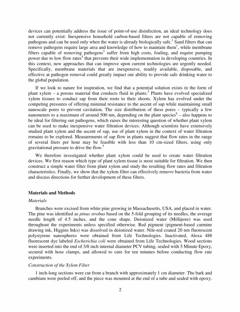
Xylem Structure and Rationale for Use of Conifer Xylem
The flow of sap in plants is driven primarily by transpiration from the leaves to the
atmosphere, which creates negative pressure in the xylem. Therefore, xylem evolution has
occurred under competing pressures of providing minimal resistance to the flow of sap, while
protecting against cavitation (i.e. nucleation) and growth of bubbles that could stop the flow of
sap and kill the plant, and to do this while maintaining mechanical strength.5 The xylem structure
comprises many small conduits that work in parallel and operate in a manner that is robust to
cavitation5, 6
(Figure 1). In woody plants, the xylem tissue is called the sapwood, which often
surrounds the heartwood (i.e. inactive, non-conducting lignified tissue found in older branches
and trunks) and is in turn surrounded by the bark (Figure 1b,c). The xylem conduits in
gymnosperms (conifers) are formed from single dead cells and are called tracheids (Figure 1c),
with the largest tracheids reaching diameters up to 80 µm and lengths up to 10 mm.5
Angiosperms (flowering plants) have xylem conduits called vessels that are derived from several
cells in a single file, having diameters up to 0.5 mm and lengths ranging from a few millimeters
to several meters.5 These parallel conduits have closed ends and are connected to adjacent
4
conduits via “pits”6 (Figure 1d,e). The pits have membranes with nanoscale pores that perform
the critical function of preventing bubbles from crossing over from one conduit to another. Pits
occur in a variety of configurations; Figure 1d,e show torus-margo pit membranes that are
shaped like a donut (margo) with an impermeable part in the center called torus, occurring in
conifers.6 More interestingly, the porosity of the pit membranes ranges in size from a few
nanometers to a few hundred nanometers, with pore sizes in the case of angiosperms tending to
be smaller than those in gymnosperms.6, 7
Pit membrane pore sizes have been estimated by
examining whether gold colloids or particles of different sizes can flow through.6, 8
Remarkably,
it was observed that 20 nm gold colloids could not pass through inter-vessel pit membranes of
some deciduous tree species,8 indicating an adequate size rejection to remove viruses from water.
Furthermore, inter-tracheid pit membranes were found to exclude particles in the 200 nm range,6
as required for removal of bacteria and protozoa.
Since angiosperms (flowering plants, including hardwood trees) have larger xylem vessels
that are more effective at conducting sap, xylem tissue constitutes a smaller fraction of the cross-
section area of their trunks or branches, which is not ideal in the context of filtration. The long
Figure 1. Xylem structure. a) Structure of xylem vessels in flowering plants and tracheids in conifers. Longer
length of the vessels can provide pathways that can bypass filtration through pit membranes that decorate their
circumference. b) Photograph of ~ 1 cm diameter pine (pinus strobus) branch used in the present study. c)
Scanning electron microscope (SEM) image of cut section showing tracheid cross section and lengthwise
profile. Scale bar is 40 µm. d) SEM image showing pits and pit membranes. Scale bar is 20 µm. e) Pit
membrane with inset showing a cartoon of the pit cross-section. The pit cover has been sliced away to reveal the
permeable margo surrounding the impermeable torus. Arrow indicates observed hole-like structures that may be
defects. The margo comprises radial spoke-like structures that suspend the torus that are only barely visible
overlaying the cell wall in the background. Scale bar 1 µm. f) Dependence of the area amplification, defined as
the pit membrane area divided by the nominal filter area, on the tracheid aspect ratio L/D and fractional area α
occupied by pit membranes.

5
length of their xylem vessels also implies that a large thickness (centimeters to meters) of xylem
tissue will be required to achieve any filtration effect at all – filters that are thinner than the
average vessel length will just allow water to flow through the vessels without filtering it through
pit membranes (Figure 1a). In contrast, gymnosperms (conifers, including softwood trees) have
short tracheids that would force water to flow through pit membranes even for small thicknesses
(< 1 cm) of xylem tissue (Figure 1a). Since tracheids have smaller diameters and are shorter, they
offer higher resistance to flow, but typically a greater fraction of the stem cross-section area is
devoted to conducting xylem tissue. For example, in the pine branch shown in Figure 1b used in
this study, fluid-conducting xylem constitutes the majority of the cross-section. This reasoning
leads us to the conclusion that in general the xylem tissue of coniferous tress – i.e. the sapwood –
is likely to be the most suitable xylem tissue for construction of a water filtration device, at least
for filtration of bacteria, protozoa, and other pathogens on the micron or larger scale.
The resistance to fluid flow is an important consideration for filtration. Pits can contribute a
significant fraction (as much as 30-80%)5, 6
of the resistance to sap flow, but this is remarkably
small considering that pit membrane pore sizes are several orders of magnitude smaller than the
tracheid or vessel diameter. The pits and pit membranes form a hierarchical structure where the
thin, highly-permeable pit membranes are supported across the microscale pits that are arranged
around the circumference of the tracheids (Figure 1a). This arrangement permits the pit
membranes to be thin, offering low resistance to fluid flow. Furthermore, the parallel
arrangement of tracheids with pits around their circumference provides a high packing density
for the pit membranes. For a given tracheid with diameter D and length L, where pit membranes
occupy a fraction α of the tracheid wall area, each tracheid effectively contributes a pit
membrane area of πDLα/2, where the factor of 2 arises as each membrane is shared by two
Figure 2. Xylem filter. a) Construction of xylem filter. b) Effect of applied pressure on the water flow rate
through the xylem filter. c) Hydrodynamic conductivity of the filter extracted at each measured pressure using
the total filter cross-section area and length as defined by Equation 1.

6
tracheids. However, the nominal area of the tracheid is only πD2/4, and therefore, the structure
effectively amplifies the nominal filter area by a factor of 2α(L/D) (Figure 1f). The images in
Figure 1c indicate D ~ 10-15 µm, α ~ 0.2, yielding an effective area amplification of ~20 for
tracheid lengths of 1-2 mm. Therefore, for a filter made by cutting a slice of thickness ~L of the
xylem, the actual membrane area is greater by a large factor due to vertical packing of the pit
membranes. Larger filter thicknesses further increase the total membrane area, but the additional
area of the membrane is positioned in series rather than in parallel and therefore decreases the
flow rate, but potentially improves the rejection performance of the filter.
Construction of the Xylem Filter and Measurement of Flow Rate
The xylem filter device was constructed by simply peeling off the bark from a section of the
pine branch and inserting it into a tube (Figure 2a). Although a simple tube fastener could
provide a leak-tight seal between the tube and the xylem, we used epoxy to ensure that there was
no inadvertent leakage. When deionized water was loaded into the tube above the xylem and
subjected to pressure in the 0.5-5 psi range, we found that water readily flowed through the
xylem. The flow rate was proportional to applied pressure (Figure 2b), which allowed for the
extraction of the hydrodynamic conductivity K (m2/Pa-s) of the filter, defined by
l
PKAQ
∆= (1)
Figure 3. Filtration performance of the xylem filter. a) Feed solution of a pigment dye before filtration,
compared to the filtrate. b) Size distribution of the pigment particles in the feed and filtrate solutions measured
by dynamic light scattering. c) Dependence of the rejection on the particle size estimated from the data in (c). d)
Cross-section of the xylem filter after filtration. Scale is in centimeters and inches.

7
where Q is the volumetric flow rate (in m3/s) under pressure difference ∆P across the filter, while
l and A are the thickness and the cross-section area of the filter, respectively. The observed
conductivities for three different filters were in the range of ~5-6×10⁻¹⁰
m2/ Pa-s (Figure 2c), or
equivalently, ~0.5-0.6 kg s-1
m-1
MPa-1
when defined with respect to mass flow rate of water.
Biologists have performed similar flow rate measurements by cutting a section of a plant
stem under water, flushing to remove any bubbles, and applying a pressure difference to measure
the flow rate.9, 10
Xylem conductivities of conifers5 typically range from 1-4 kg s
-1 m
-1 MPa
-1,
which compares very well with the conductivities measured in our experiments. Lower
conductivities can easily result from introduction of bubbles9 or the presence of some non-
conducting heartwood. We can therefore conclude that the flow rate measurements in our
devices are consistent with those expected from prior literature on conductivity of conifer xylem.
Filtration of Pigment Dye
After construction of the filter, we tested its ability to filter a pigment dye with a broad
particle size distribution. The red color of the feed solution disappeared upon filtration (Figure
3a) indicating that the xylem filter could effectively filter out the dye.
Since the dye had a broad pigment size distribution, we investigated the size-dependence of
filtration by quantifying the pigment size distribution before and after filtration using dynamic
light scattering. We found that the feed solution comprised particles ranging in size from ~70 nm
to ~500 nm, with some larger aggregates (Figure 3b). In contrast, the filtrate particle size
distribution peaked at that ~80 nm, indicating that larger particles were filtered out. In a separate
experiment, we observed that 20 nm fluorescent polystyrene nanoparticles could not be filtered
by the xylem filter, confirming this size dependence of filtration. Assuming that pigment
particles 70 nm or less in size were not rejected, the size distributions before and after filtration
enable calculation of the rejection performance of the xylem filter as a function of particle size
(Figure 3c). We find that the xylem filter exhibits excellent rejection for particles with diameters
exceeding 100 nm, with the estimated rejection exceeding 99% for particles over 150 nm.
Smaller particles are expected to pass through the larger porosity of the pit membrane: SEM
images in Figure 1e indicate sub-micron spacing between the radial spoke-like margo membrane
through which the pigment particles can pass, although the porosity is difficult to resolve.
After filtration, we cut the xylem filter parallel to the direction of flow to investigate the
distribution of dye in the filter. We observed that the dye was confined to the top 2-3 millimeters
of the xylem filter (Figure 3d), which compares well with the tracheid lengths on the millimeter
scale expected for coniferous trees.5 These results show that the majority of the filtration
occurred within this length scale, and suggests that the thickness of the xylem filter may be
decreased to below 1 cm while still rejecting the majority of the dye.
Filtration of Bacteria from Water
Finally, we investigated the ability of the xylem filter to remove bacteria from water. As a
model bacterium, we used fluorescently labeled and inactivated Escherichia coli bacteria that are
cylindrical in shape with a diameter of ~1 µm. Use of fluorescently labeled particles enabled
easy enumeration of their concentrations, as well as allowed for tracking of the location in the
xylem filter where they were trapped. Filtration using three different xylem filters showed nearly
complete rejection of the bacteria (Figure 4a). Using a hemacytometer to count the bacteria, we
estimate that the rejection was at least 99.9%.

8
To investigate the mechanism of filtration, after filtration we cut the xylem filter parallel to
the direction of flow. When examined under a fluorescence microscope, we observed the
bacteria accumulated over the donut-shaped margo pit membranes (Figure 4b). This distribution
is consistent with the expectation that the bacteria are filtered by the pit membranes. The
distribution of trapped bacteria was not uniform across the cross section. Similar to the case of
the dye, bacteria were observed only within the first few millimeters from the end through which
the solution was infused (indicated by white arrow in Figure 4c). In addition, the low-
magnification fluorescence image suggests that the bacteria may accumulate primarily over pit
membranes at the bottom of the tracheids, which is again not unexpected. Further investigation
Figure 4. Filtration of model bacteria by the xylem filter. a) Concentrations of bacteria in the feed and filtrate
solutions. Inset shows fluorescence images of the two solutions. Scale bar is 200 µm. b) Fluorescence image of
xylem filter cross-section showing accumulation of bacteria over the margo pit membranes. Scale bar is 20 µm. c)
Low-magnification fluorescence image shows that bacteria are trapped at the bottoms of tracheids within the first
few millimeters of the top surface. Scale bar is 400 µm. Arrow indicates top surface of the xylem filter and also
the direction of flow during filtration. Autofluorescence of the xylem tissue also contributes to the fluorescence
signal in (b) and (c). d,e) SEM images showing bacteria accumulated on the margo pit membranes after filtration.
Scale bars are 10 µm and 2 µm, respectively.

9
by SEM confirmed that the bacteria had accumulated on the pit membranes (Figure 4d,e). These
results confirm the pit membranes as the functional units that provide the filtration effect in the
xylem filter.
Discussion
Wood has been investigated recently as a potential filtration material,11
showing moderate
improvement of turbidity. While we showed filtration using freshly cut xylem, we found that the
flow rate dropped irreversibly by over a factor of 100 if the xylem was dried, even when the
xylem was flushed with water before drying. We also examined flow through commercially
available kiln-dried wood samples cut to similar dimensions. We found that wood samples that
exhibited filtration also had flow rates that were two orders of magnitude smaller than in the
fresh xylem filter, and those that had high flow rates did not exhibit much filtration effect and
seemed to have ruptured tracheids and membranes when observed under SEM. Wetting using
ethanol or vacuum to remove air did not significantly increase flow rate, suggesting that the pit
membranes may have a tendency to become clogged during drying. These results are consistent
with literature showing that the pit membranes can become irreversibly aspirated against the cell
wall, blocking the flow.12
In fact, the pit membranes in the SEM images (Figure 1d,e and Figure
4d,e), which were acquired after drying the samples, appear to be stuck to the walls. Regardless,
our results demonstrate that excellent rejection (>99.9%) of bacteria is possible using the pit
membranes of fresh plant xylem, and also provide insight into the mechanism of filtration as well
as guidelines for selection of the xylem material.
The vertical arrangement of the tracheids makes them susceptible to concentration
polarization and fouling (i.e. accumulation of the rejected particles in the tracheids), and
therefore xylem filters are likely to be most advantageous for removal of pathogens from water
that is not significantly turbid. The pressures of 1-5 psi used here are easily achievable using a
gravitational pressure head of 0.7 – 3.5 m, implying that no pumps are necessary for filtration.
The measured flow rates of about 0.05 mL/s using only ~ 1 cm2 filter area correspond to a flow
rate of over 4 L/d, sufficient to meet the drinking water requirements of one person. While
membranes are easily fouled, xylem filters could be easily replaced due to their biodegradability
and low cost.
Wood is an easily available material. While use of fresh xylem does not preclude its use as a
filter material, dried membranes have definite practical advantages. Therefore, the process of
wood drying and its influence on xylem conductivity needs further study. In particular, processes
that yield intact yet permeable xylem tissues that can be stored long-term will be helpful for
improving the supply chain if these filters are to be widely adopted. In addition, flow through
xylem of different plants needs to be studied to identify locally available sources of xylem,
which will truly enable construction of filters from locally available materials. In the present
study, we report results using xylem derived from only one species. These xylem filters could
not filter out 20 nm nanoparticles, which is a size comparable to that of the smallest viruses. It
will be interesting to explore whether there exist any coniferous species that have pit membranes
with smaller pore sizes that can filter out viruses. In their absence, angiosperms with short
vessels that yield practical filter lengths may be the best alternative due to their smaller pit
membrane pore sizes.6 Further exploration of xylem tissues from different plants with an
10
engineering perspective is needed to construct xylem filters that can effectively reject viruses,
exhibit improved flow rates, or that are amenable to easy storage.
Conclusions
Plant xylem is a porous material with membranes comprising nanoscale pores. We have
reasoned that xylem from the sapwood of coniferous trees is suitable for disinfection by filtration
of water. The hierarchical arrangement of the membranes in the xylem tissue effectively
amplifies the available membrane area for filtration, providing high flow rates. Xylem filters
were prepared by simply removing the bark of pine tree branches and inserting the xylem tissue
into a tube. Pigment filtration experiments revealed a size cutoff of about 100 nm, with most of
the filtration occurring within the first 2-3 mm of the xylem filter. The xylem filter could
effectively filter out bacteria from water with rejection exceeding 99.9%. Pit membranes were
identified as the functional unit where actual filtration of the bacteria occurred. Flow rates of
about 4 L/d were obtained through ~ 1 cm2 filter areas at applied pressures of about 5 psi, which
is sufficient to meet the drinking water needs of one person. The simple construction of xylem
filters, combined with their fabrication from an inexpensive, biodegradable, and disposable
material suggests that further research and development of xylem filters could lead to their
widespread use and greatly reduce the incidence of waterborne infectious disease in the world.
Acknowledgements
This work was supported by the James H. Ferry, Jr. Fund for Innovation in Research Education
award to R.K. for the proposal entitled “Can Plant Xylem Provide Safe Drinking Water?” SEM
imaging was performed at the Harvard Center for Nanoscale Systems, a member of the National
Nanotechnology Infrastructure Network (NNIN), which is supported by the National Science
Foundation under NSF award no. ECS-0335765. The authors thank Yukiko Oka for assistance
with preparation of illustrations and Sunandini Chopra for help with dynamic light scattering
measurements.
References
1. Gadgil, A. Drinking water in developing countries. Annu Rev Energ Env 1998, 23, 253-
286.
2. http://www.who.int/water_sanitation_health/mdg1/en/
3. Madaeni, S. S. The application of membrane technology for water disinfection. Water
Research 1999, 33, 301-308.
4. Loo, S. L.; Fane, A. G.; Krantz, W. B.; Lim, T. T. Emergency water supply: A review of
potential technologies and selection criteria. Water Research 2012, 46, 3125-3151.
5. Sperry, J. S. Evolution of water transport and xylem structure. Int J Plant Sci 2003, 164,
S115-S127.
6. Choat, B.; Cobb, A. R.; Jansen, S. Structure and function of bordered pits: new
discoveries and impacts on whole-plant hydraulic function. New Phytol 2008, 177, 608-
625.
7. Jansen, S.; Choat, B.; Pletsers, A. Morphological Variation of Intervessel Pit Membranes
and Implications to Xylem Function in Angiosperms. Am J Bot 2009, 96, 409-419.
11
8. Choat, B.; Ball, M.; Luly, J.; Holtum, J. Pit membrane porosity and water stress-induced
cavitation in four co-existing dry rainforest tree species. Plant Physiol 2003, 131, 41-48.
9. Espino, S.; Schenk, H. J. Mind the bubbles: achieving stable measurements of maximum
hydraulic conductivity through woody plant samples. J Exp Bot 2011, 62, 1119-1132.
10. van Ieperen, W.; van Meeteren, U.; van Gelder, H. Fluid ionic composition influences
hydraulic conductance of xylem conduits. J Exp Bot 2000, 51, 769-776.
11. Sens, M. L.; Emmendoerfer, M. L.; Muller, L. C. Water filtration through wood with
helical cross-flow. Desalination and Water Treatment 2013.
12. Petty, J. A. Aspiration of Bordered Pits in Conifer Wood. Proc R Soc Ser B-Bio 1972,
181, 395-406.