washington state dental association’s 2015 pacific ... pacific northwest dental conference...
TRANSCRIPT

Washington State Dental Association’s 2015 Pacific Northwest Dental Conference
Presents
I Hate Love Complete Dentures Lecture Dr. Ronnie Schnell
Friday, June 12, 2015 8:30 a.m. – 11:30 a.m.
Official Disclaimer
Neither the content of a program nor the use of the specific products in courses should be construed as indication endorsement or approval by the
Pacific Northwest Dental Conference or Washington State Dental Association

6/1/2015
1
Ronni A. Schnell, DMD, MAGD, FICDBoston University Henry M Goldman School of Dental Medicine
Friday June 12th
8:30‐11:30 AMLecture
I Hate Love Dentures © 2015
Knowledge of edentulous anatomy and their predicted effects on the outcome of the denture
Knowledge of the musculature is the key to successful denture borders and optimal impressions
Knowledge of optimal esthetics and how to achieve desirable results
Knowledge and skill to manage denture occlusion; whichcontrasts natural occlusion.
Key to Successful Dentures
α Preliminary Impressions
Custom trays
α Final Impressions
Record bases & Occlusion rims
α Intermaxillary Records
Set‐up
α Trial Denture
Processing & Lab Remount
α Insertion
Clinical (patient) Remount
Sequence of Patient and Lab Visits
=
Patient attitude
Patient expectations (vs what you can deliver)
Is there a “significant other” who play an important role in decisions?
Chief complaint Loose, sore, cannot chew, appearance, time to replace
Smoker?
Prescription Meds?
How much water does the patient drink daily?
How long has the patient been edentulous? U? L?
Does the patient have an existing prosthesis? How long ago was that made? How many dentures were made in the patient’s lifetime?
Esthetics and function: What does patient like and not like? What do you like and not like?
The initial data gathering appointment is EXTREMELY important to the success of the case… Do not rush through it – especially the space analysis
1st Clinical Visit: Initial Patient Exam
When naturalteeth are in occlusion,
ridge crests are approximately aminimum of
12 mm apart in the anterior
Inter‐arch Space
Inter-ridge Space?Anterior -12 mm minimum
Posterior– 1-2 mm minimum
Evaluate with a tongue blade during Initial Exam
1st Clinical Visit: Initial Patient Exam
α Saliva ‐ Ropy, Viscous, Absent, Average
α Oral Tolerance – Average, Sensitive, Very sensitive, Gagger
α TMJ Status – Symptomatic, Asymptomatic
α Oral Pathology Review – WNL, Suspected Areas …
α Tongue Size – Average, Large
α Throat Form –α U‐House I, II, IIIα L‐Neil’s Lateral I, II, III
6/1/2015
2
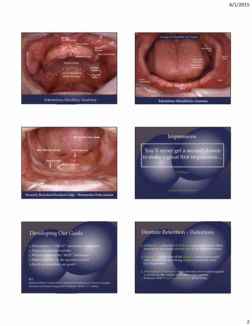
Edentulous Maxillary Anatomy
Broad Labial Frenum
Muco‐labial Fold
Buccal Frenum
Muco‐buccal Fold
Maxillary Tuberosity
Residual Ridge
Hamular Notch
Incisive Papilla
Anterior Vibrating Line
(House Throat Form)
Retro‐molar Pad
Retro mylohyoid space
(Neil’s Lateral Throat Form)
FoldsFrenal Attachments
Buccal Shelf
Edentulous Mandibular Anatomy
(average size Mandible and Tongue)
Residual ridge
Genial Tubercles
Retro molar pad
Severely Resorbed Residual ridge – Retromolar Pads remain
Mental Foramen
Mylohyoid Ridge You’ll never get a second chance to make a great first impression…
‐Will Rogers
Impressions
Luckily for us we often do…!
What makes a “GREAT” edentulous impression‐
Types of materials available
What is currently the “BEST” technique?
What works best in the operators hands?
Has it accomplished our goals?
Ref.
Zarb & Hobkirk: Prosthodontic Treatment for Edentulous Patients: Complete
Dentures and Implant‐Supported Prostheses, 13h Ed., C V Mosby
Developing Our Goals
Adhesion—attraction of different molecules to each other. Increase by increasing surface area of the tray/impression.
Cohesion ‐‐‐attraction of the identical molecules to each other. Increase by increasing contact (closeness) of the tray/impression.
Atmospheric Pressure ‐‐ force per unit area exerted against a surface by the weight of air above that surface. Enhance with Posterior Palatal Seal (Post Dam).
Denture Retention ‐ Definitions

6/1/2015
3
Area of over
compression
Captured are the incisive papilla, frenal attachments, folds, rugae, palate. Extended posteriorly to the hamular notches bilaterally and distal to the
anterior vibrating line.
May be corrected with green or grey stick compound
Maxillary Preliminary Impression for Diagnostic Cast and/or Custom Tray
Upper and Lower
Preliminary (1°) Alginate Impressions
Indications:
Undercuts
Existing dentition
α Mucostatic for hard tissue
α Mucodynamic for soft tissue
α Undergo syneresis if left in the air
α Undergo imbibition if too wet
α Heavy body best for edentulous
α Cannot bead and box, may deform
• under weight of stone if
• unsupported
α Low cost
Laboratory steps‐
Beading, Boxing and Pouring
(alginate cannot be beaded and boxed)
2nd Clinical Visit:Final Impressions
Are BOTH mucostatic and mucodynamic
Where?
Borders – MucodynamicIntaglio – Mucostatic
The borders of the denture are formed by the muscle attachments
We must capture the functional movements of the muscle attachment during impressioning
This process is called border molding
Why border mold?
For Custom tray fabrication
Surface adaptation for adhesion and cohesion
Diagnose Patient
General Preliminary (1º) Impression Objectives

6/1/2015
4
Prior removal of old dentures and tissue conditioner
to rest and or treat tissues ‐ if contributing to problems
Tray selection
1/4” larger or custom for thickness of impression material
Head position
Upright for safety / tissues vertical
Dentist position
Upper behind pt. / Lower in front of pt.
Muscle movements
Saliva control – suction & astringent mouthwash
Syringe teeth for immediate/interim/partial denture impressions
Clinical Hints for ImpressionsNever Block Out Anterior Undercuts
Alter Path NEVER block out Anterior undercuts !& block out Posterior undercuts
X
Dynamic Evaluation of Buccal Fold
Over‐extended At rest
When Evaluating folds…Do not pull down and away Pull out and up
Custom Tray Try‐in:Evaluation and Adjustment of Borders for Intra‐oral Anatomy
Overextension Relief of frenae
Border molding ‐ Required Armamentarium: Gray or green stick compound (red stick if needed to extend custom tray), Bunsen
Burner, Hanau torch, water bath @ 140 degrees
Intra oral Evaluation
Tray must not “rebound”
The Severely resorbed mandible
Tray can be retentive! Border molded custom traySeverely Resorbed Mandible
Final Impression ‐ PSR

6/1/2015
5
?
?
Tiny voids with sharp edges may be
filled in with Physiolologic Wax
Fill voids even with surface of impression
Wax softens @ mouth temperature
Leave in mouth for 1 minute to establish contour
Full arch border molded final
impression “picking up” the impression
copings
Final Impression for Overlay Partial
Denture
We can create a successful impression if we know:
α What anatomical landmarks must be captured
α How to activate the muscles
α Materials and technique
α Patient management
Criteria for Success
α Stability
α Border thickness
α Tissue detail
α Tray penetration
α Folds or creases
α Surface texture
α Voids
α Retention
Upper‐Y
Lower‐Y/N
Evaluation of Impressions
Rubber base adhesive painted 3 mm below height of border will facilitate Beading & Boxing
All‐acrylic prostheses are poured with YELLOW stone
Beading, Boxing and Pouring the final Cast
LABORATORY STEP
The
Final
Cast
Sufficient Land is critical
Formed by Beading Wax

6/1/2015
6
α Border Extension ‐ into the full width& depth of the fold
Record Base Criteria
Shellac
Formatray
Triad/Eclipse
α Stability during function
α ‐no rocking
α Retention
If a record base is not
all of the above, visits will be
challenging to the Doctor
and discouraging to the pt.
…revisit your final impression…
Record Base Criteria
Information from the initial patient exam Current denture
Patient experience
What we already know patient landmarks / anatomy
Esthetics
Phonetics
Clinician experience
How do we do it?
Locate guidelines on the final cast
Fabricate occlusion rims
Adjust rims according to guidelines and then intraorally
Occlusion Rims
Foveae palatinae
Incisive papilla
Locating Guidelines Helps Determine Tooth Position
α Midline (from the face)
α Internal Land Line
α Incisive papilla line (upper)
α Posterior crest of ridge lines
α Tuberosity lines
α Hamular Notch Line
ow the pe of the h! Maxillary Centrals are ALWAYS anterior to the Incisive Papilla
with the facial surface approximately 6 mm in front of the Papilla.
The remainder of the Maxillary Anterior teeth generally follow the shape of the arch.
Maxillary Posterior Teeth are generally set slightly labial to the crest of ridge….
To prevent cheek biting maxillary teeth should be offset approx2mm to the buccal of the mandibular teeth. If monoplane teeth are used, as in most cases, cross bites are also acceptable to prevent cheek biting
Maxillary Tooth Position

6/1/2015
7
α Retromolar Pad Height Lines
α Posterior Crest of Ridge Lines
α Anterior Crest of Ridge Line
or
Mandibular Anterior Teeth are centered over the anterior crest of ridge.
Mandibular anterior ridge bone resorption happens posterior to anterior
Mandibular teeth are set on or slightly lingual to the lower ridge crest….
**To help stabilize lower denture**and to prevent cheek biting
Mandibular Tooth Position
End of tuberosity
Parallel to ridge crest
End of Pad
Parallel to ridge crest
Maxillary & Mandibular End of Occlusal Table and Posterior crest of ridge line
We do not set teeth over the Tuberosity or Retromolar Pads
The VERTICAL height of mandibular posterior teeth is determined relative to the retromolar pad.
The distal of the second molar is generally 2/3 the vertical height of the Retromolar Pad.
This requires a frontal view
All four lines should line up
The Occlusal Plane
Note the RM Pad does note appear to be divided evenly in 1/3’s. That it because both pads level off at different slopes and rates.
Comparison of Rims
UPPER IS SLIGHTLY BUCCAL LOWER IS CENTERED TO RIDGE TO RIDGE
Max ant @ 22 mm Mandib ant @ 18 mmPost parallel to residual ridge Post = to 2/3 height RMP
Rims must be FLAT and FLUSH
Comparison of Rims
UPPER IS SLIGHTLY BUCCAL LOWER IS CENTERED TO RIDGE TO RIDGE
Max ant @ 22 mm Mandib ant @ 18 mmPost parallel to residual ridge Post = to 2/3 height RMP
Rims must be FLAT and FLUSH

6/1/2015
8
Why bases cover pads but rims do not
Alveolar bone
Basal bone
Mandibular occlusion rim is lowto stabilize denture
Mandibular occlusion rim is shortto distribute stress
AKA
The Post Dam
Posterior Palatal Seal
HNL= Hamular Notch Line
An arbitrary line connecting R & L Hamular Notches
AVL = Anterior Vibrating Line
Junction of movable and non‐movable tissue
CTL = Compressible Tissue Line
Junction of compressible and non‐compressible tissue
PPS = Posterior Palatal Seal (Post Dam)
A bead located posteriorly on the internal surface of the upper denture which completes the border seal
Definitions:α Locate hamular notches with T‐ball burnisher and mark with indelible
pencil.
α Place a perpendicular line on palate with indelible pencil. Observe repeated phonetic ‘ah’.
α Scribe “ah” line (AVL) with indelible pencil.
α Insert record base and transfer AVL pencil line.
α Cut record base back to AVL.
α Palpate outline of compressible tissue.
α Draw CTL on model.
Post Dam Clinical TechniqueOn Patient
α Score AVL transfer through both Hamular notches to ½ depth measured.
α Score CTL transfer just enough to break surface of cast.
α Connect CTL and AVL by tapering from zero depth at the CTL to AVL depth.
α Blend smoothly into cast.
Post Dam Clinical TechniqueOn Cast

6/1/2015
9
CTL = Shallowest = 0)
AVL ‐ DeepestHN
Postdam outlinetransferred to cast
Note: PPS extendspast hamular notch and is BLENDED into the DB fold
3rd Clinical Visit
Inter‐Maxillary Records
α Occlusal Plane
α Vertical Dimension of Occlusion
α Facebow Registration
α Centric Relation Registration
α Tooth Selection
Facial midlineLip fullnessLip lengthSmile lineCanine linePhonetics (sibilants, fricatives)Ridge relationship (I, II, III)VDOVDRFlush contact @ VDO
Occlusion Rims: Clinical Adjustment
Inter‐ridge Space
Freeway Space
Closest Speaking Space
The Neutral Zone
Space of Donders
VDO & Space
Freeway SpaceInter‐occlusal Distance
VDO occurs during swallowingVDR occurs at the end of swallowing
VDR occurs at the end of the “M” sound or smiling
VDOVDR
The Freeway Space is the spacebetween the teeth when the mandible is at rest (VDR)
“M”
Closest Speaking Space
• The Closest Speaking Space is the small space between the occlusal surfaces during sibilant sounds
• Teeth should not contact during sibilant “S” sounds or “clicking” of the teeth together will result
• Clicking of teeth is NOT normal and if VDO is correct, even if the patient is wearing porcelain teeth, there will be none
Other sibilant sounds include:
“ch” – church, sandwichMassachusett
“j” – jelly, judge

6/1/2015
10
Cheek
The Neutral Zone
The neutral zone is the area where the displacing forces of the lips cheeks and tongue are in balance.
It is in this zone that the natural dentition and flanges lie, and this is where the denture teeth should be positioned so that conflict does not occur and the dentures remain in equilibrium.
The influence of tooth position and flange contour on denture stability is equal to or greater than any other factor.
This is particularly critical in patients with atrophic ridges.
Cheek
The Space of Donders
The space betweentongue & palate during occlusionis called theSpace of Donders
Cheek
SPACE OF DONDERS
during occlusionSpace of Donders
↑VDO → ↑Space of Donders ↓VDO → ↓Space of Donders
Additionally, and independent of VDO… if the palate is too THICK… Space of Donders will be decreased
and the patient will have trouble swallowing
The occlusal plane is determined by areas “A” and “C”
“A” = Esthetics + Phonetics“C” = Anatomy of Retromolar Pad
What determines the Occlusal Plane?
What determines the Occlusal Plane?
Max- Start @ 22 mm and adjust
Mand- Start @ 18mm and 2/3 RMP adjust
These areas should not be altered to change VDO
The VDO is determined by “A” + “B” and “C” + “D”The VDO is altered by areas “B ” and “D”
“B” = Sibilants with “A”“D” = Sibilants with “C”
What determines the Occlusal Plane?
What areas may be altered to change VDO?
Max- Start @ 22 mm and adjust
Mand- Start @ 18mm and 2/3 RMP adjust
Your Rim = Your Rx
Canine Lines:
α ½ Ala‐Modiolus
α Mid‐pupillary Line
α Canine eminence on cast
α Consider each side separately
α Lines represent the M‐D height of contou
α Notice the asymmetry
α Midline
α Canine Lines
α High Smile Line
Lines Scribed to mark Anatomical Landmarks

6/1/2015
11
A mounting with a Facebow Registration
on a semi‐adjustable articulatorpermits small changes in VDO +/‐ 3mm without having to
remount
Bow should reflect the plane of the adjusted wax rim which should be parallel to the interpupillary line
Pt. photo courtesy of Dr. Shervin Tabeshfar
For a denture patient * – the first contact must be the full and final contact
Although CR is defined at least 7 different ways in the Glossary of Prosthodontic Terms, one thing that is agreed upon is that The position is difficult to define anatomically but is determined clinically by assessing when the jaw can hinge on a fixed terminal axis. This summary suggests that the CR position is therefore reproducible.
We therefore strive to mount all cases of unknown “centric”* in CR because of it’s reproducibility and continue to adjust ongoing cases to maintain CO=CR.
Key is reproducibility
* no habitual natural posterior stops
CR vs. CO
Denture movement
Denture displacement
Pain
Bone loss
95% of problem dentures have an undiagnosed occlusal discrepancy
Discrepancy in denture occlusion will cause:
Natural Dentition: the chin can be held during retrusion.If teeth are in dentures: the dentures must be held during retrusion.
Natural Dentition – OKDenture – NOT OK
Index finger on buccal shelfThumb under mandible
Always Re‐Verify CR in and out of Patient’s mouth
Check for stability of bite registration Check for impression of notch
Insure there is 1mm space in anterior Check for Heel Interference
Rim Guidelines = Your RX
Midline
High smile line
Canine lines
Inter‐pupillary line
Incisal Edge Contour
Tooth Selection & Arrangement

6/1/2015
12
Face Shape (О,∆, ) Smile Line (length) Canine Distance (width) Gender (Masculine/Feminine tooth set‐up) Complexion (shade) Old Denture (likes/dislikes) Old Photos (graduation/wedding) Old Xrays Old Casts Patent Opinion / Significant Other
Patient Analysis ‐Anterior
Trubyte Rim Selector Kit
Then evaluate your selections intra-orally
Denture Base and Custom Denture Base Shade Mixes
Fibered Dark
Fibered Light
Lucitone 199
{
Eliminate Maxillary Buccal Cusp interferencesTo eliminate additional lateral forces
Anatomical (lingualized) vs Lingual Contact
Occlusion
=ridge crest =ridge crest
Lingual Contact Occlusion

6/1/2015
13
Rules of Denture Occlusion:
No Anterior Contact in CR
No incisal guidance in protrusive
No canine guidance in lateral
CR=CO
Occlusal Plane = 1/2 to 2/3 height RMP
NO porcelain teeth unless the opposing is fixed (and even then, use caution)
Lingual Contact Occlusion
The Occlusal Scheme Natural vs. Denture
• Anterior contact in CO• Incisal guidance• Canine guidance• CO ≠ CR
• No anterior contact in CO• No incisal guidance• No canine guidance• CO = CR• Bilateral Balance (if
anatomical)
Complete Denture in Protrusion: Guidance
Leads to denture instability, movement and bone loss
Natural Dentition in Protrusion: Mutually Protected Occlusion or…
Complete Denture in Protrusion: Balance
@ VDO
In Protrusion In Balance
Verify VDO
Verify CR
Does CR (or CO) in patients mouth match with CR (or CO) on Articulator? (They MUST match or lower MUST be remounted to match the patient!)
Esthetics
Phonetics
Patient Approval Signature (EDR)
4th Clinical Visit:Trial denture
Sequence:
Adjust upper aloneAdjust lower aloneInsert U/L w/o occludingRehearse/record CRMount lowerPerform Patient remount‐ refine occlusionRe‐polishReview home care instructionsReappoint for 1st adjustment
5th Clinical Visit:Insertion

6/1/2015
14
Note areas of overextension & excessive thickness
Using pastes @ insertion or for
Dx/adjust‐ment of sore spots
Note “show through” in area of overextensionU/L inserted individually but prevented from occluding by cotton rolls
Centric Relation Record
Centric Relation recorded w/ Aluwax Lower denture mounting using CR
Patient Remount Technique
Objectives of Equilibration
To have CO = CR
To maintain VDO
To distribute stress
To retain cusp shape
To achieve balanced occlusion
To smooth contacting surfaces
Lingual of upperis last resort!
If wear facetIs insufficien
Adjusting Monoplane Occlusion*
Perfect lower Plane to 2/3 RMP (Area “C” & “B”)
Adjust Maxillary (Area “D”)
If significant adjustment is needed or to reduce VDO adjust both.
Remember‐ flat adjustments. Do not create inclines or cusps!
If incisal or canine guidance, create wear facets
√ pad
Anatomical Occlusal Equilibration
Interferences
Only one side contacts
Posteriors not in CR
Only one side contacts
Only one side contacts
Only one tooth contacts
Only anteriors contact
Only some teeth contactor anteriors are too far apart

6/1/2015
15
Equilibration Sequence
1st Centric interferences
2nd Lateral interferences
3rd Protrusive interferences
Maxilla is stationery - 1°Mandible is movable - 2°
Curve of Wilson
Ground Rules
●
Supporting Cusps
●
1º Supporting Cusps do not move
2º Supporting Cusps are movable
1º2º
1º
2º
Grinding supporting cusps will result in loss of CO & VDO
• Before grinding…
• Check excursions
Right lateral
Left lateral
Protrusion
Centric Occlusion
Centric Occlusion
If Right Lateral excursion has no interference and...
●
If Left Lateral excursion has no interference and...
Centric Occlusion
●

6/1/2015
16
Centric Occlusion
If Protrusive excursion has no interferencethen grind Fossa
Centric Occlusion
Centric interference:grind Fossa if interference occurs only in Centric
●
But…If Centric & Right Lateral excursion have interferences or…
Centric Occlusion
●
Centric Occlusion
If Centric & Left Lateral excursion have interferences or...
●
Centric Occlusion
If Centric & Protrusive excursions have interferencesthen grind Cusp
Centric Occlusion
Centric interferencegrind Cusp if interference occurs in Centric & Eccentric
●

6/1/2015
17
Centric Occlusion
Centric interference:grind Cusp or Fossa ?
●
Centric Interference Rules
Centric only: FossaCentric & Eccentric: Cusp
Supporting Cusps
Curve of Spee
Maxilla is stationary
Mandible is movable
Grinding supporting cusps can change CR or VDO
2º
2º
2º 2º
Centric Occlusion Centric Occlusion
● = MUDL (Mesial of Upper, Distal of Lower Buccal cusps)
● = DUML (Distal of Upper, Mesial of Lower Buccal cusps)
Centric Occlusion
For Horizontal interferences: MUDL
CO
CR
VERT
H O R I Z
CO=Vertical Tooth position
CR=Horizontal Jaw position
Centric Interference Rules
• Vertical direction:Centric only: FossaCentric & Eccentric: Cusp
• Horizontal direction: MUDL

6/1/2015
18
Left Working (Right Balancing)
1º
2º
1º
2º
1º Supporting Cusps do not move2º Supporting Cusps are movable
LR
Working Side
Each Supporting Cusp opposes a non-supporting cusp
Working Side
Grinding a non-supporting cusp will not change CR or VDO
● = centric stops
Lateral
Lateral
For Working interference: grind BULL(Buccal of Upper or Lingual of Lower)
√
√
Lateral
Restoration of bilateral balance

6/1/2015
19
● = centric stopsNo loss of CO or VDO
Lateral Right Balancing (Left Working)
1º
2º
1º
2º
1º Supporting Cusps do not move2º Supporting Cusps are movable
LR
Balancing Side
1º
2º
There is a pair of contacting supporting cusps on the balancing side
Grinding a Supporting Cusp will change CR or VDO
Balancing Side
1º
2º
To maintain CR & VDO, grind only the inner incline of the Secondary Supporting cusp
● = centric stops● = eccentric contacts
Lateral Lateral
For Balancing interference: grind only incline of BL(Buccal of Lower)

6/1/2015
20
Lateral
Removal of lateral interferenceusing 2 colors of articulating paper
● = centric stopsNo loss of CO or VDO
following grinding
Lateral
Lateral
Reason for posterior disclusion in Lateral?
Lateral
Canine Guidance in Lateral
Lateral
Protrusion
1st2nd
3rd
For Canine Guidance in Lateral:grind lowers (1>2>3)
Lateral Interference Rules
• Working side: BULL
• Balancing side: BL inclines only(buccal cusp – lingual incline only)
• Canine: 1st lower canine2nd lower premolar
3rd upper canine

6/1/2015
21
Equilibration Sequence
1st Centric interferences
2nd Lateral interferences
3rd Protrusive interferences
Curve of Spee
Maxilla is stationary - 1°
Mandible is movable- 2°
Protrusion
2º 2º
Contact between Supporting and non-supporting cusp inclines
Buccal cusps only – There is no lingual cusp contact in protrusion
2º 2º2º
Protrusion
Grind non-supporting cusp inclines whenever possible
In MOST cases – 99+% - Buccal cusps only
2º 2º2º 2º2º
Protrusion
For Protrusive interferences: grind DUML
Protrusion
Anterior & Posterior Balance in Protrusion

6/1/2015
22
Protrusion
Reason for posterior disclusion?
Incisal Guidance
↑ Horizontal Overlap or↓ Vertical Overlap
Cut – if denture
OR Move – if in wax
“Wear Facets”
Before During Aftera = U/L overlapb = ½ overlapc = ½ beveld = full bevel
If full bevel of Lower incisalis insufficientthen Upper lingualmay be groundas a last resort
Protrusive Interference Rules
• Anterior: Lower wear facets
• Posterior: DUML
Final Balanced Occlusion
Whenever the patient needs you!
First adjustment within 48 hours
Follow ups 1 week, 1 month and PRN
Immediate denture wearers: 24, 48 and either 72 hours to 1 week. 24 hour Is REQUIRED!!!
Follow up adjustments depend on individual and difficulty of case
Post Insertion AdjustmentsWhen?
6/1/2015
23
Sore spots
Burning sensation
Tongue & cheek biting
Tissue redness
TMJ pain
Instability
Interferences
Esthetics
Phonetics
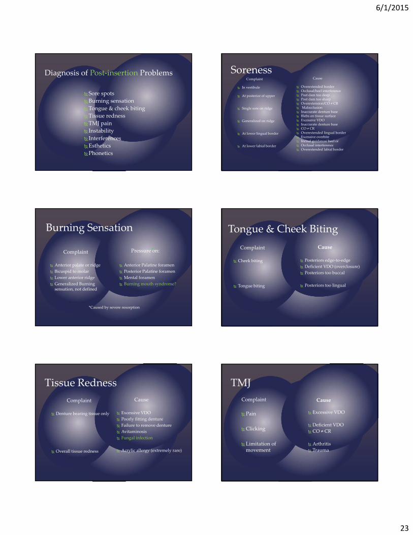
Diagnosis of Post‐insertion Problems SorenessComplaint
In vestibule
At posterior of upper
Single sore on ridge
Generalized on ridge
At lower lingual border
At lower labial border
Cause
Overextended border Occlusal/heel interference Post dam too deep Post dam too sharp Overextension/CO ≠ CR Malocclusion Inaccurate denture base Blebs on tissue surface Excessive VDO Inaccurate denture base CO ≠ CR Overextended lingual border Excessive overbite Incisal guidance/ heel or Occlusal interference Overextended labial border
Burning Sensation
Complaint
Anterior palate or ridge
Bicuspid to molar
Lower anterior ridge
Generalized Burning sensation, not defined
Pressure on:
Anterior Palatine foramen
Posterior Palatine foramen
Mental foramen
Burning mouth syndrome?
*Caused by severe resorption
*
Tongue & Cheek Biting
Complaint
Cheek biting
Tongue biting
Cause
Posteriors edge‐to‐edge
Deficient VDO (overclosure)
Posteriors too buccal
Posteriors too lingual
Tissue Redness
Complaint
Denture bearing tissue only
Overall tissue redness
Cause
Excessive VDO
Poorly fitting denture
Failure to remove denture
Avitaminosis
Fungal infection
Acrylic allergy (extremely rare)
TMJ
Complaint
Pain
Clicking
Limitation of movement
Cause
Excessive VDO
Deficient VDO
CO ≠ CR
Arthritis
Trauma

6/1/2015
24
Complaint
When not occluding
When incising
When occluding in centric
Cause
Border overextension Border underextension Loss of posterior palatal seal Tissue dehydration Flabby tissue displacement Loss of posterior palatal seal Anteriors too labial Poor denture foundation Improper incising habits Malocclusion Flabby tissues Teeth too buccal CO ≠ CR
InstabilityComplaint
Swallowing
Gagging Clicking
Deafness Muscle fatigue General uneasy feeling
Cause
U posterior too long or thick L lingual too long or thick Posteriors too lingual Excessive VDO Too long or thick or moving Excessive VDO Unstable denture Deficient VDO Excessive VDO Malocclusion Incorrect VDO CO ≠ CR
Interferences
Complaint
Fullness under nose
Depressed filtrum
Sunken upper lip
Shows too much teeth
Artificial appearance
Cause
Labial border too thick or long
Labial border too thin or short
Upper anteriors too lingual
Excessive VDO
Incisal plane too low
Cuspids, laterals too prominent
Poor set up
Lack of wear facets
Lack of custom gingiva
Esthetics Phonetics
Complaint
Whistle on “S” sound
Lisp on “S” sound
Indistinct “T” and “Th”
Cause
Anterior palate too narrow
Anterior palate too wide
Inadequate interocclusaldistance
Most Common
Sore Throat (pt is not sick)
It hurts when I open wide…
My upper denture pops out when I open to bite a sandwich…
My upper denture pops out when I bite into a sandwich…
Something is wrong, but I cant figure out what…
Clicking when speaking
Dentures are “loose”
Overextended DL Flange (in Mylohoidspace‐lower!)
DB flange too wide – maxillary
DB flange too wide – maxillary
Poor post dam
Occlusion
Excessive VDO Occlusion – usually incisal or canine
guidance
Problem Usual Solution

ADJUSTING RIMS FOR VDO

MOUNTING RIMS @ VDO
