volume 68 | issue 1 | january - march 2017
TRANSCRIPT

1acta OrthOpaedica et traumatOlOgica hellenica
MonographyVOLUME 68 | ISSUE 1 | JANUARY - MARCH 2017ActAScapulothoracic disorders
2
1MD, M.Sci, Ph.D, FEBOT, Ass. Professor of Orthopaedics2
Ioannis K. Triantafyllopoulos, MD, M.Sci, Ph.D, FEBOTAssistant Professor of Orthopaedics, Laboratory for the Research of Musculoskeletal Disorders, National and Kapodestrian University of Athens, Greece, E-mail: [email protected], Mob. +30 6937266639
Scapulothoracic articulation disorders may cause significant malfunction of the shoulder girdle. Scapular winging, dyskinesia, crepitus and bursitis are different pathological entities associated with the scapulothoracic joint. Their pathogenesis is a combination of anatomic, posture, traumatic and neuromuscular alterations. In this monography, the causes, diagnosis and treatment of the scapulothoracic disorders will be discussed.
KEY WORDS: scapula; winging; dyskinesia; crepitus; bursitis
abstract
IntroductionThe scapulothoracic articulation is essential in the kinesiology of the shoulder girdle. Few papers are referred to scapulothoracic disorders compared to glenohumeral and acromiclavicular joints of the shoulder girdle. Disorders associated with scapulo-thoracic joint are often poorly understood or difficult to diagnose. They are rather common in heavy man-ual workers as well as in water sport and overhead sport athletes, as the continuous and intense move-ment of upper limbs and trunk makes the area of the scapula the most functionally active site. The patho-genesis of scapulothoracic disorders involves ana-tomic and neuromuscular alterations that affect the biomechanics of the shoulder.
A. Surgical anatomyScapula is a thin bone which is the site of attachment of 17 muscles of trunk and upper limb and plays a key role in the coordinated movement of the upper limb (Fig. 1).
The muscles attached to scapula can be divided into three groups (Table Ι): (a) The scapulothoracic muscles adjust the scapulothoracic movement and include the major and minor rhomboids, the levator scapula, the anterior serratus, the trapezius, the omo-hyoid and the pectoralis minor. Conditions of these muscles present with winged scapula or scapulotho-racic dyskinesia. (b) The scapulohumeral muscles provide functional strength to the humerus and in-clude the deltoid, the long and short heads of biceps
corresponding author, guarantor
Ioannis K. Triantafyllopoulos 1 , Chrysa Argyrou
MD, Resident in Orthopaedics

2 acta OrthOpaedica et traumatOlOgica hellenica
VOLUME 68 | ISSUE 1 | JANUARY - MARCH 2017
Triantafyllopoulos IK, Argyrou C. Scapulothoracic disorders
brachii, the coracobrachialis, the long head of the tri-ceps brachii and the teres major. (c) The rotator cuff muscles control the movement of glenohumeral ar-ticulation and consist of the supraspinatus, infraspi-natus, subscapularis and teres minor. Disorders of these muscles are common and compose a special subject of study not included in this issue.
B. BiomechanicsThe interplay of 4 articulations (Sternoclavicu-lar Joint, Acromioclavicular Joint, Scapulothorac-ic Joint and Glenohumeral Joint) of the shoulder complex, results in an coordinated movement pat-tern of the arm elevation. The involved movements at each joint are continuous, although occurring at various rates and at different phases of arm eleva-tion. The movement of the scapula can be described by rotations in relation to the thorax. The scapula moves around a dorso-ventral axis, resulting in a ro-tation in the frontal plane. In this movement the gle-noid cavity is turned cranially (upward rotation) or caudally (downward rotation). In the sagittal plane, around a latero-lateral axis the scapula rotates pos-teriorly (posterior tilting) or anteriorly (anterior tilt-ing). External and internal rotation occurs around a cephalo-caudal (longitudinal) axis. The external ro-tation brings the glenoid cavity more into the frontal plane, whereas the internal rotation turns the gle-noid cavity more to the sagittal plane.
At rest, the scapula is rotated 30o anteriorly in frontal plane. The inferior angle of the medial bor-der of the scapula is also deviated 3o away from
the midline. When observing the lateral side of the scapula, there is an anterior inclination of 20o in the sagittal plane (Fig. 2).
Almost every upper limb movement includes ele-ments of scapulothoracic and glenohumeral motion. In the first 30o of humeral abduction, most of the movement is provided by the glenohumeral joint. The rest 60o are equally distributed to glenohumer-al and scapulothoracic joints. Overall, the gleno-humeral to scapulothoracic ratio during abduction of the shoulder up to 90o is 2:1.
During abduction, in the scapular level, the point of rotation of the scapula moves so that for the first 0o - 30o the scapula rotates around its actual center. From 30o to 60o rotates around the glenoid fossa, leading to medial and superior displacement of the inferior angle of the scapula (Fig. 3). Moreover, dur-ing arm abduction, the coracoid process and the ac-
Fig. 1. Muscular attachments to scapula
table 1. Muscles attached to scapula
SCAPULOTHORACIC SCAPULOHUMERAL ROTATOR CUFF
1. Levator scapula 1. Long head of biceps brachii 1. Supraspinatus
2. Omohyoid 2. Short head of biceps brachii 2. Infraspinatus
3. Rhomboid major 3. Deltoid 3. Subscapularis
4. Rhomboid minor 4. Coracobrachialis 4. Teres minor
5. Serratus anterior 5. Teres major
6. Trapezius 6. Long head of triceps brachii
7. Pectoralis minor

3acta OrthOpaedica et traumatOlOgica hellenica
VOLUME 68 | ISSUE 1 | JANUARY - MARCH 2017
Triantafyllopoulos IK, Argyrou C. Scapulothoracic disorders
romion move superiorly, reducing the sub-acromi-al impingement (Fig. 4).
C. Muscle function around the scapulaActivation of the upper fibers of trapezius, which insert at the lateral part of the scapular spine, the acromion and the lateral end of the clavicle, moves the scapula superiorly. This action is opposed by the gravity and the latissimus dorsi muscle, which acts as the main stabilizer of the scapula. Its action is re-inforced by the inferior fibers of the anterior serra-tus, the pectoralis minor and the lower trapezius.
The upward rotation of the scapula begins with the activation of middle trapezius, which stabiliz-es the scapula by its attachment to the medial part of the scapular spine. At the 45o of scapular protrac-tion the serratus anterior draws the inferior angle of the scapula outwards. The upper trapezius draws the lateral angle of the scapula superiorly, while the lower trapezius which inserts to the medial part of the scapular spine, draws the scapula downwards, leading to upward rotation (Fig. 5).
The downward rotators of the scapula are main-ly the rhomboids and the levator scapula, which draw superiorly the medial border of the scapula, while the pectoralis minor, the lower part of pecto-ralis major and the latissimus dorsi stabilize the lat-eral border of the scapula (Fig. 6).
The anterior movement of the scapula is per-formed by the serratus anterior, pectoralis major and minor, as they move the scapula anteriorly and
outward. The posterior movement of the scapula is performed by the middle trapezius and rhom-boids (Fig. 7).
D. Bursae of the scapulaThe bursae of the scapula ensure the smooth scap-ulothoracic movement. There have been described
Fig. 2: Stereoscopic position of scapula Fig. 3: Movements of scapula
Fig. 4: Scapular-humeral synergy

4 acta OrthOpaedica et traumatOlOgica hellenica
VOLUME 68 | ISSUE 1 | JANUARY - MARCH 2017
Triantafyllopoulos IK, Argyrou C. Scapulothoracic disorders
2 main and 4 accessory bursae in this area (Table 2, Fig. 8). The first main bursa is located between the serratus anterior muscle and the thoracic wall. The second is located between the subscapularis and the serratus anterior muscles.
Bursitis of the scapulothoracic joint is clinically manifested in two locations: At the superior me-dial angle and the inferior angle of the scapula. At the inferior angle of the scapula, the inflamed bur-sa is located between the serratus anterior and the thoracic wall. Many names have been attributed to this bursa so far, like infraserratus bursa, bursa mucosa serrata etc. Initially, Codman believed that the bursa of the superior medial angle is located between the three first ribs and the scapula[1]. Fi-nally, Von Grueber was the one who identified the bursa between the serratus anterior and the sub-scapularis muscle and named it bursa mucosa angu-lae superioris scapulae[2].
An accessory bursa is found in the triangular sur-face of the inner part of the scapular spine and be-low the trapezius. Codman believed that this bur-sa was responsible for the scapulothoracic crepitus and named it trapezoid bursa. Many authors believe that the accessory bursae developed in order to coun-teract the abnormal biomechanics of the scapulotho-racic joint and this is the reason that are not steadily found at the same region or at the same tissue plane.D. Scapulothoracic disorders
1. WINGED SCAPULAWinged scapula is the most common disorder of the scapulothoracic articulation and various factors are implicated in its pathogenesis. It is classified to pri-mary, secondary and voluntary (Table 3). Primary winging refers mainly to anatomic disorders that directly affect the joint. The secondary form is usu-ally related to pathology of the glenohumeral joint. Finally, the voluntary form is rather rare and is at-tributed to psychological causes.
Ι. PRIMARY WINGED SCAPULAΑ. Neurological disorders1) Major and minor rhomboids palsyThis is a rare cause of winged scapula. These mus-cles are innervated by the dorsal scapular nerve (C5 root). This nerve passes deeply or through the le-
Fig. 5-7: Muscular forces applied to the scapula
Fig. 8: Bursae of scapula

5acta OrthOpaedica et traumatOlOgica hellenica
VOLUME 68 | ISSUE 1 | JANUARY - MARCH 2017
Triantafyllopoulos IK, Argyrou C. Scapulothoracic disorders
vator scapula muscle and then reaches the rhom-boids. Winging of the scapula can be the result of C5 pathology or damage to the dorsal scapular nerve.
Patients with weakening of the rhomboids are presented with pain along the medial border of the scapula. The winging of the scapula is not very ob-vious at rest. It is possible, though, to resemble with the winged scapula caused by the trapezius weak-ening with depression of the scapula, outward dis-placement and outward rotation of the inferior an-gle. Furthermore, atrophy along the medial border of the scapula may exist. During arm abduction, the inferior angle of the scapula is drawn downwards and outwards due to the action of anterior serra-tus. The winging becomes even more evident when the arm is slowly adducted starting from the posi-tion of anterior flexion, so that the inferior angle of the scapula is drawn outwards and dorsally while hands are lying on the hips. Electromyography and other tests of neurological conduction can be very helpful in the differential diagnosis and the distinc-tion from other neurological disorders.
Treatment is based on the strengthening of the tra-pezius. If symptoms persist and conservative meas-ures fail, patients can benefit by the Dickson proce-dure [3]. In this procedure, two cylindrical grafts of the fascia lata connect the lower part of the medi-al border of the scapula with the paraspinal mus-cles and the inferior angle of the scapula with fib-ers of the latissimus dorsi. This technique stabilizes the scapula and partially inhibits the high thorac-
ic scoliosis that can emerge due to the paralysis of the rhomboids and the levator of scapula. Howev-er, there is a possibility that the grafts will eventu-ally become loose and elongated.
table 2. Bursae of the scapula
CLASSIFICATION ANATOMIC SITE INTERSPACE
Major/Anatomic Below SA SA-Thoracic wall
Above SA SA-Subscapularis
Minor/Ectopic Superior Medial angle Below SA SA- Thoracic wall
Above SA SA-Subscapularis
Inferior angle Below SA SA-Thoracic wall
Scapular spine Trapezius Inner part of spine-Τ
SA: Serratus Anterior, T: Trapezius
table 3. Classification of winged scapula
Ι. PRIMARY
Α. Neurological causes
1. Long thoracic nerve: Anterior serratus palsy
2. Accessory nerve: Trapezius palsy
3. Dorsal scapular nerve: Rhomboids palsy
Β. Osseous causes
1. Osteochondroma
2. Poor fracture healing
C. Soft tissues
1. Flexion deformities
2. Muscular detachments
3. Muscular agenesis
4. Scapulothoracic bursitis
ΙΙ. SECONDARY
1. Disorders of glenohumeral joint
2. Disorders of the subacromial space
ΙΙΙ. VOLUNTARY
1. Psychological causes

6 acta OrthOpaedica et traumatOlOgica hellenica
VOLUME 68 | ISSUE 1 | JANUARY - MARCH 2017
Triantafyllopoulos IK, Argyrou C. Scapulothoracic disorders
2) Serratus anterior palsyPalsy of the serratus anterior can cause a painful winged scapula. The long thoracic nerve that inner-vates the serratus anterior is formed by the C5,6,7 roots, passes below the brachial plexus and the clav-icle and above the first rib. It then runs superficially along the lateral thoracic wall, where it is more sus-ceptible to trauma (Fig. 9). Blunt trauma or protrac-tion of the nerve is rather common in athletes, es-pecially those involved with tennis, golf, hockey on ice, soccer, basketball, bowling, javelin throw, wres-tlers etc. Repeated minor injuries in workers that use their shoulder extensively have been reported as causes of nerve paralysis. Penetrating trauma can rarely cause nerve damage. Nevertheless, surgical procedures like radical mastectomy, first rib resec-tion and transaxillary sympathectomy have been associated with long thoracic nerve damage. The nerve can also get paralyzed due to non-traumatic causes, like the faulty positioning of the surgical pa-tient under general anesthesia, viral causes, inocula-tion, brachial plexus or long thoracic neuritis. More-over, sleeping with the arm placed under the head so that supports it in order to facilitate reading of a book has been shown to cause nerve dysfunction. Lastly, C7 radiculitis can also be a cause of serratus anterior weakening and winged scapula.
Patients with serratus anterior palsy usually com-plain about pain, as the rest of the scapular mus-
cles are trying to compensate for the winging of the scapula. Extreme pain should lead us to consider the possibility of brachial plexus neuritis or mon-oneuritis of the long thoracic nerve (Parsonage-Turn-er syndrome) [4]. The scapula is located in a more su-perior position, displaced inwards with the inferior angle facing inwards (Fig. 10). The patient presents with difficulty abducting the arm over 120o, where the winged scapula is more obvious. Pain is even worse in arm abduction when the head of the hu-merus tilts to the ipsilateral shoulder.
Electromyography is considered gold standard for the confirmation of the diagnosis. It should be repeated every 1-3 months to monitor the healing of the long thoracic nerve, since most cases with pa-resis are resolved spontaneously within the first 1 to 2 years.
Conservative treatment starts immediately after the diagnosis and includes exercises for the mainte-nance of the range of movement of the glenohumer-al joint. There are many types of braces that keep the scapula attached to the thoracic wall, but their role is controversial.
Penetrating trauma should be treated with early nerve repair. On the other hand, late surgical repair with neurolysis and neuronal grafts does not always give satisfactory results. In patients with sympto-matic winged scapula for over a year, surgical re-pair can alleviate the pain and repair the functional-
Fig. 9: Course of long thoracic nerve. Fig. 10: Serratus anterior (left) and trapezius (right) palsy

7acta OrthOpaedica et traumatOlOgica hellenica
VOLUME 68 | ISSUE 1 | JANUARY - MARCH 2017
Triantafyllopoulos IK, Argyrou C. Scapulothoracic disorders
ity of the scapula. Historically, these procedures are classified into three categories: (a) arthrodesis of the scapulothoracic joint, (b) use of fascial support and (c) muscle-tendon transfers. For the muscle-tendon transfer, graft sources are usually the pectoralis ma-jor, the sternocostal and clavicular heads of pecto-ralis major, the rhomboids and combinations of the above muscles.
The arthrodesis procedures for the scapulotho-racic joint can relieve the pain, but can also lead to loss of scapular mobility. Other complications of arthrodesis are pseudoarthrosis and pneumotho-rax. The indications of this method are limited to cases where other techniques have failed, simulta-neous paralysis of many muscles of the shoulder and in workers that perform heavy tasks and apply too much pressure on the shoulder. Fascial grafts have the drawback of loosening and failure of their supportive ability. Consequently, muscle-tendon transfers have earned interest in the treatment of the winged scapula due to anterior serratus paraly-sis. Transfer of various muscles has been used, with that of sternocostal head of pectoralis major to be the most popular (Fig. 11). The patient is placed at the lateral decubitus position and the incision runs through the axilla, from the pectoralis major up to the inferior angle of the scapula. Alternatively, two different incisions can be made and the pectoralis major is reversed subcutaneously. The sternocostal
head of the pectoralis major is released from its in-sertion biceps groove at the humerus. Then, an au-tologous graft from the ipsilateral fascia lata (18x15 cm) is prepared. The 18 cm tube-shaped autograft is then side-to-side sutured at the free end of the ten-don of the pectoralis major. An aperture is made at the inferior end of the medial border of the scapu-la and the graft is passed through it sutured under mild tension. Postoperatively, the arm is suspend-ed with a triangular bandage and passive mobili-zation begins. Active mobilization starts after the first 6 weeks and strengthening exercises after 12 weeks. Early complications of this technique in-clude pneumothorax, while fracture of the inferi-or angle of the scapula and graft failure has been reported as late complications. Arthrodesis of the scapulothoracic joint can be performed in case of severe complications.
3) Trapezius muscle palsyThe accessory nerve innervates the majority of the trapezius muscle. Its course is superficial, found in the subcutaneous tissue of the posterior cervi-cal triangle, making it susceptible to trauma (Fig. 12). The causes of the accessory nerve trauma in-clude blunt trauma, traction, sharp trauma (during lymph node biopsy) and the radical cervical lymph node excision.
Patients suffering from trapezius palsy usually
Fig. 11: Dissection of the sternocostal head of the pectoralis major and enhancement with fascia lata autograft for the treatment of the serratus anterior palsy
Fig. 12: Course of the accessory nerve
8 acta OrthOpaedica et traumatOlOgica hellenica
VOLUME 68 | ISSUE 1 | JANUARY - MARCH 2017
Triantafyllopoulos IK, Argyrou C. Scapulothoracic disorders
present with pain due to the counterbalancing ac-tion of the levator of the scapula and the rhomboids. Other causes of pain in these patients are the pro-gression to frozen shoulder, the subacromial fric-tion and cervical radiculitis due to protraction of the brachial plexus as a result of the shoulder drop.
Clinical evaluation reveals inability to shrunk the shoulder, as well as inability of abduction and forward flexion of the ipsilateral arm. The scapu-la is found in a lower level than normal, displaced outwards with the inferior angle headed outwards (Fig. 10). Electromyography is used to confirm the diagnosis.
Treatment of patients with winged scapula due to accessory nerve palsy depends on the duration and the intensity of the symptoms. Initial treatment is conservative, with the suspension of the arm in a tri-angular bandage in order to provide relaxation for the rest of the muscles. Physical therapy aims at the preservation of range of motion of the glenohumer-al joint and the avoidance of the progression to fro-zen shoulder. In cases of blunt trauma-related pa-ralysis, regular follow-up with electromyography every 4-6 weeks is necessary to monitor nerve func-tion. In cases of palsy due to sharp trauma or when no nerve function can be detected, surgical investi-gation with neurolysis, placement of nerve grafts or combination of the above techniques is the treat-ment of choice. The results of those methods are
rather variable, while they seem to be better when neurolysis is performed within 6 months.
Patients that present with symptoms lasting up to a year are not likely to benefit from conserva-tive treatment. Historically, a great variety of sur-gical techniques has been reported for the treatment of winged scapula due to trapezius muscle palsy. These techniques can be divided in (a) static stabili-zation, including arthrodesis of the scapulothoracic joint and (b) dynamic stabilization, including mus-cle-tendon transfers.
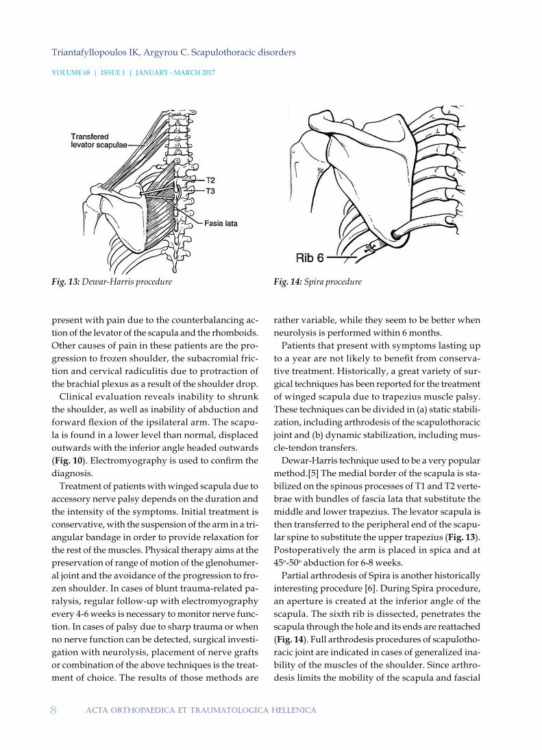
Dewar-Harris technique used to be a very popular method.[5] The medial border of the scapula is sta-bilized on the spinous processes of T1 and T2 verte-brae with bundles of fascia lata that substitute the middle and lower trapezius. The levator scapula is then transferred to the peripheral end of the scapu-lar spine to substitute the upper trapezius (Fig. 13). Postoperatively the arm is placed in spica and at 45ο-50ο abduction for 6-8 weeks.
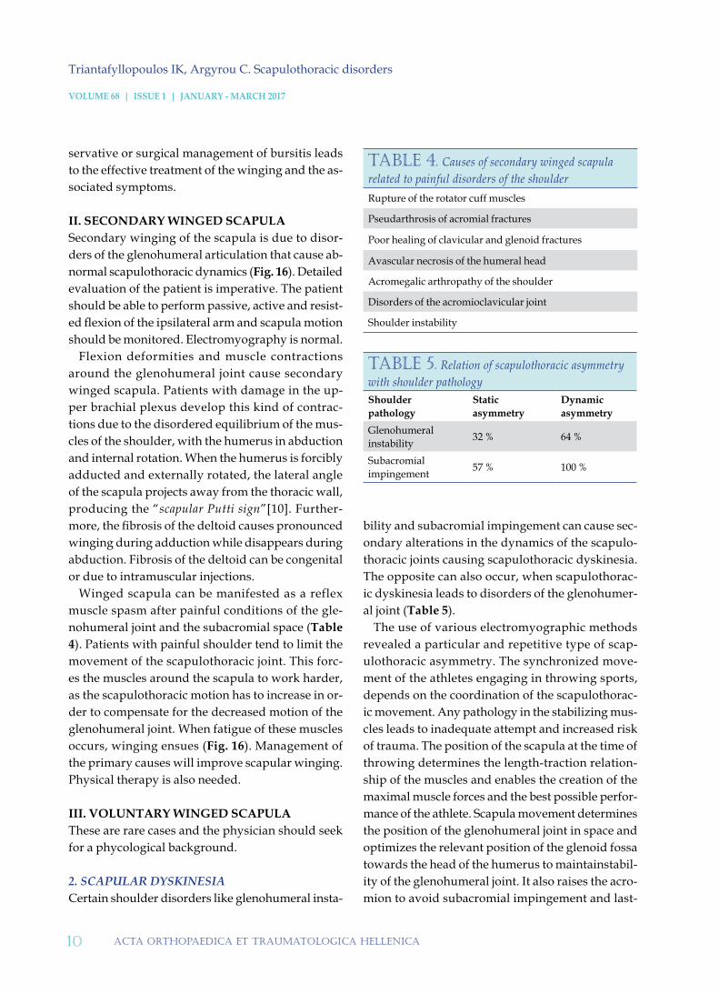
Partial arthrodesis of Spira is another historically interesting procedure [6]. During Spira procedure, an aperture is created at the inferior angle of the scapula. The sixth rib is dissected, penetrates the scapula through the hole and its ends are reattached (Fig. 14). Full arthrodesis procedures of scapulotho-racic joint are indicated in cases of generalized ina-bility of the muscles of the shoulder. Since arthro-desis limits the mobility of the scapula and fascial
Fig. 13: Dewar-Harris procedure Fig. 14: Spira procedure

9acta OrthOpaedica et traumatOlOgica hellenica
VOLUME 68 | ISSUE 1 | JANUARY - MARCH 2017
Triantafyllopoulos IK, Argyrou C. Scapulothoracic disorders
grafts usually fail due to loosening after 2-3 years, dynamic stabilization with muscle-tendon transfers is currently the treatment of choice.
In the Eden-Lange technique, the levator scapula and the rhomboids are transferred outwards (Fig. 15) [7-9]. The levator of the scapula substitutes the upper trapezius, the rhomboid minor substitutes the middle and the rhomboid major the lower tra-pezius. The lateral relocation of the insertions of these three muscles optimizes the biomechanical outcome and deceases the winging. This method includes two incisions. The first one is performed along the medial border of the scapula and the sec-ond one above the scapular spine. The insertions of the three muscles are detached along with an osse-ous segment. The rhomboids are then directed pe-ripherally under the infraspinatus and fixated with intraosseous sutures (via osseous holes made 5 cm peripherally of the medial border of the scapula). The levator of the scapula is directed subcutane-ously towards the second incision and gets fixat-ed to the spine through osseous apertures. Postop-eratively, the patient uses an arm abduction brace for 4-6 weeks, followed by physical therapy pro-gram with passive and active mobilization. The outcomes of this method are considered excellent, with 91% reporting pain alleviation and 87% sig-nificant improvement in the functionality of the shoulder.
Β. Osseous malformationsOsteochondroma of the scapula or the ribs is the main osseous causes that can lead to winged scap-ula. This type of winging is due to structural rath-er than functional causes and can be accompanied by scapular crepitus. Patients usually present with winging that does not change in respect to upper limb movement. Electromyography is normal and the osteochondroma is revealed when either a tan-gential X-ray of the scapula or a CT scan are per-formed. Surgical excision of the osteochondroma is the treatment of choice.
Primary causes of the winged scapula include poor positioning during scapula and clavicular frac-ture healing. These patients can remain asympto-matic due to intact muscle functioning.
C. Soft tissues disordersMuscular disorders causing winged scapula include traumatic muscle ruptures and congenital muscle agenesis. Electromyography is normal and MRI re-veals the cause of winging. Muscle detachments are treated with direct repair. In cases of congenital muscle agenesis of the trapezius, anterior serratus and rhomboids, patients usually compensate for the functional deficiency without the need of surgery.
Winged scapula can also present in 50% of pa-tients suffering from scapular bursitis. Bursitis is usually accompanied with pain and crepitus. Con-
Fig. 15: Eden-Lange procedure Fig. 16: Secondary winging cataract
10 acta OrthOpaedica et traumatOlOgica hellenica
VOLUME 68 | ISSUE 1 | JANUARY - MARCH 2017
Triantafyllopoulos IK, Argyrou C. Scapulothoracic disorders
servative or surgical management of bursitis leads to the effective treatment of the winging and the as-sociated symptoms.
ΙΙ. SECONDARY WINGED SCAPULASecondary winging of the scapula is due to disor-ders of the glenohumeral articulation that cause ab-normal scapulothoracic dynamics (Fig. 16). Detailed evaluation of the patient is imperative. The patient should be able to perform passive, active and resist-ed flexion of the ipsilateral arm and scapula motion should be monitored. Electromyography is normal.
Flexion deformities and muscle contractions around the glenohumeral joint cause secondary winged scapula. Patients with damage in the up-per brachial plexus develop this kind of contrac-tions due to the disordered equilibrium of the mus-cles of the shoulder, with the humerus in abduction and internal rotation. When the humerus is forcibly adducted and externally rotated, the lateral angle of the scapula projects away from the thoracic wall, producing the “scapular Putti sign”[10]. Further-more, the fibrosis of the deltoid causes pronounced winging during adduction while disappears during abduction. Fibrosis of the deltoid can be congenital or due to intramuscular injections.
Winged scapula can be manifested as a reflex muscle spasm after painful conditions of the gle-nohumeral joint and the subacromial space (Table 4). Patients with painful shoulder tend to limit the movement of the scapulothoracic joint. This forc-es the muscles around the scapula to work harder, as the scapulothoracic motion has to increase in or-der to compensate for the decreased motion of the glenohumeral joint. When fatigue of these muscles occurs, winging ensues (Fig. 16). Management of the primary causes will improve scapular winging. Physical therapy is also needed.
ΙΙΙ. VOLUNTARY WINGED SCAPULAThese are rare cases and the physician should seek for a phycological background.
2. SCAPULAR DYSKINESIACertain shoulder disorders like glenohumeral insta-
bility and subacromial impingement can cause sec-ondary alterations in the dynamics of the scapulo-thoracic joints causing scapulothoracic dyskinesia. The opposite can also occur, when scapulothorac-ic dyskinesia leads to disorders of the glenohumer-al joint (Table 5).
The use of various electromyographic methods revealed a particular and repetitive type of scap-ulothoracic asymmetry. The synchronized move-ment of the athletes engaging in throwing sports, depends on the coordination of the scapulothorac-ic movement. Any pathology in the stabilizing mus-cles leads to inadequate attempt and increased risk of trauma. The position of the scapula at the time of throwing determines the length-traction relation-ship of the muscles and enables the creation of the maximal muscle forces and the best possible perfor-mance of the athlete. Scapula movement determines the position of the glenohumeral joint in space and optimizes the relevant position of the glenoid fossa towards the head of the humerus to maintainstabil-ity of the glenohumeral joint. It also raises the acro-mion to avoid subacromial impingement and last-
table 4. Causes of secondary winged scapula related to painful disorders of the shoulderRupture of the rotator cuff muscles
Pseudarthrosis of acromial fractures
Poor healing of clavicular and glenoid fractures
Avascular necrosis of the humeral head
Acromegalic arthropathy of the shoulder
Disorders of the acromioclavicular joint
Shoulder instability
table 5. Relation of scapulothoracic asymmetry with shoulder pathologyShoulder pathology
Static asymmetry
Dynamic asymmetry
Glenohumeral instability 32 % 64 %
Subacromial impingement 57 % 100 %

11acta OrthOpaedica et traumatOlOgica hellenica
VOLUME 68 | ISSUE 1 | JANUARY - MARCH 2017
Triantafyllopoulos IK, Argyrou C. Scapulothoracic disorders
ly, the muscles around the scapula are important in order to ensure strength against the eccentric load at the various stages of throwing.
Kibler was the first to describe the lateral slide test for the assessment of the scapulothoracic dys-kinesia.[11-13] This test is designed to evaluate the patient’s ability to stabilize the medial border of the scapula during different positioning and load-ing. Arms are placed in three different positions: (a) at resting position, (b) hands on hips with fin-gers anterior and thumbs posterior and (c) arms at 90ο with internal rotation. In asymptomatic pa-tients, asymmetry is less than 1 cm. In symptomat-ic patients with pain and limited range of motion, there is significant asymmetry more than 1 cm in positions (a) and (b).
Clinically, the outward position of the scapula leads to greater anteversion of the glenoid fossa, leading to an increase in the anterior interspace of the glenohumeral joint, and therefore to instability and cartilage lesions. Moreover, disorders of shoul-der motion lead to elevation of the acromion and subsequent subacromial crepitus.
Management of the scapulothoracic dyskinesia consists of strengthening exercises of the scapu-lar muscles, while strengthening of the rotator cuff muscles should be avoided until the functionality of the scapula has been restored.
3. SCAPULOTHORACIC CREPITUSOver time, many names have been attributed to the symptomatic scapulothoracic crepitus, like snapping scapula, washboard syndrome, scapulothoracic syndrome, rolling scapula, grating scapula, scapulocostal syndrome, while many causes have been identified (Table 6). Although Codman [1] was the one who said that he was able to make his scapula produce a noise loud enough to be heard in the whole room without feel-ing any pain, Boinet was the first to describe this dis-order at 1867. It wasn’t until 37 years later, when Mauclaire classified the scapulothoracic crepitus in three groups: (a) froissement, is described as a normal mild friction sound, (b) frottement, is a more intense sound of coarse friction which is usually patholog-ic and (c) craquement is the intense and loud noise of scapular popping, which is always abnormal [14]. These scapular sounds arise from two sources: either from the tissue between the scapula and the thorac-ic wall, or from congruence disorders of the scapulo-thoracic joint. Milch states that the frottement is asso-ciated with soft tissue pathology or bursitis, while the craquement indicates osseous causes [14].
Muscular causes consist of atrophy, fibrosis and abnormal muscle insertions, while tuberculous and syphilitic lesions represent other soft tissue causes.
Osteochondroma of the ribs or scapula is the most common cause of scapulothoracic crepitus. Other causes include poorly healed rib fractures, abnormalities of superomedial angle of the scap-ula (hooked angle), Lushka’s tubercle and reac-tive osseous spurs from repetitive chronic muscle avulsion. Any osseous cause that produces scap-ulothoracic crepitus can lead to bursitis. On the other hand, an inflamed bursa can lead to painful crepitus. Finally, disorders of the scapulothoracic congruence, like scoliosis and thoracic kyphosis, can also be a cause of crepitus.
Diagnosis is made through meticulous history
table 6. Causes of scapulothoracic crepitus
Connective tissue
MuscleAtrophyFibrosisAnatomic abnormalities
Bone
Rib osteochondromaScapular osteochondromaRib fractureHooked superomedial angle of the scapulaLushka’s tubercleReactive bone spurs from muscle avulsion
Other soft tissuesBursitisTuberculosisSyphilis
Disorders of the scapulothoracic congruence
Spine scoliosisThoracic spine kyphosis

12 acta OrthOpaedica et traumatOlOgica hellenica
VOLUME 68 | ISSUE 1 | JANUARY - MARCH 2017
Triantafyllopoulos IK, Argyrou C. Scapulothoracic disorders
and clinical evaluation. Patients are often athletes engaging in throwing sports and workers involved in overhead activities. At inspection, winging im-plies a space-occupying lesion. Palpation and aus-cultation during shoulder motion will locate the crepitus. Palpation of a mass, crepitus and eminence at resting position combined with normal scapulo-thoracic motion are key features in differential di-agnosis from winged scapula due to neurological causes. The tangent X-ray and CT scan are also help-ful tools in diagnosis.
It is important to note that scapulothoracic crepitus is found in 35% of normal individuals. Furthermore, it is possible that patients with psychiatric background will not respond to treatment. Finally, crepitus is con-sidered an abnormal finding, when associated with pain, winging or other scapulothoracic disorders.
When osseous causes are involved, like osteo-chondroma, surgical resection is the treatment of choice. In cases of soft tissue pathology, initial management is conservative, with exercises that aim to avoid downward inclination of the scapu-la (use of figure-of-eight bandage), strengthen ex-ercises of the adjacent muscles and corticosteroid infusion in painful sites.
Surgical techniques include muscle transfers for muscle palsy cases, such as the transfer of rhom-boids, trapezius and their re-attachment under the scapula. These procedures are associated with mus-cle atrophy and failure. Other procedures are the
partial resections of the scapula, mainly the medial border or the superior medial angle.
During the superior medial angle resection, the patient is placed at prone position (Fig. 17). The in-cision is made over the medial part of the scapu-lar spine and the overlying soft tissues are dissect-ed. The periosteum of the spine is then elevated to create space between trapezius and scapula. The supraspinatus, rhomboids and levator scapula are subperiostically dissected starting from the spine. The superomedial angle of the scapula is resected with the use of a scapular saw. Care is taken not to injure the suprascapular nerve and the dorsal scap-ular artery. The muscles and periosteum are then reattached in place and sutured through osseous tunnels. Postoperatively, the arm is suspended in triangular bandage and patients start passive mo-bilization immediately. Active mobilization starts at 8 weeks following the operation and strengthen-ing exercises at 12 weeks.
Complications of the partial scapular excision are pneumothorax and postoperative hematoma. Re-currence is more common in younger patients, but are rarely symptomatic.
4. SCAPULOTHORACIC BURSITISScapulothoracic bursitis can either accompany scap-ular crepitus or be a separate entity. Patients often complain of pain related to activities or present with audible or palpable crepitus. They usually de-
Fig. 17: Superomedial scapular angle resection Fig. 18: Scapulothoracic bursitis

13acta OrthOpaedica et traumatOlOgica hellenica
VOLUME 68 | ISSUE 1 | JANUARY - MARCH 2017
Triantafyllopoulos IK, Argyrou C. Scapulothoracic disorders
scribe a repetitive activity that makes scapula move against the posterior thoracic wall. A chronic in-flammation is then developed that leads to fibrosis and scarring followed by crepitus and pain (Fig. 18).
Initial management is conservative with rest, analgesics and NSAIDs. Physical therapy, with strengthening exercises and stretching of the dorsal musculature, improves posture. Heat patches and cortisone injections offer pain. If symptoms persist, surgical treatment is indicated.
Sisti & Jobe performed bursectomy of the inferior scapular angle in athletes. An oblique incision dis-tal to the inferior angle is made and trapezius and latissimus dorsi are dissected along their muscle fib-ers. Physical therapy started 1 week postoperative-ly and within 6 weeks the athletes were able to per-form mild throwing exercises.
McCluskey & Bigliani performed open bursecto-my of the superomedial angle with vertical incision on the inside of the medial border of the scapula. Af-ter dividing the trapezius, the levator scapula and the rhomboids were subperiostically released from the scapular medial border. The space created be-tween the latissimus dorsi and the thoracic wall en-abled the resection of the thickened bursa. Muscles are then reattached. Triangular bandage is used to suspend the arm and after 3 weeks active range of motion exercises begin, while 12 weeks postopera-tively strengthening begins.
Ciullo & Jones conducted endoscopic bursae re-section with debridement and reconstruction of the superomedial or inferior scapular angle. Arthro-
scopic bursae resection was described by Matthews. Patient is placed at either lateral or prone position-ing, which enables the arthroscopic evaluation of the glenohumeral articulation and the subacromial space. Furthermore, the scapula moves away from the thoracic wall with abduction and internal rota-tion, facilitating access to the bursa. Three trocar in-sertion sites are created and the trocars are placed at least 2 cm on the inside of the medial scapular bor-der and between the spine of the scapula and the inferior angle. For the middle insertion site, a nee-dle is placed inside the bursa between the latissi-mus dorsi and the thoracic wall. The needle should enter between the spine and the inferior angle and at least 3 fingers on the inside of the medial border in order to avoid injury of the suprascapular artery and nerve. The bursa first gets larger with fluid in-fusion, before the insertion of the instruments. The upper insertion site is created 3 fingers on the inside of the medial scapular border, right under the spine and penetrates the space between the two rhom-boids. This site enables access at the superomedi-al angle. A more medial positioning of the inser-tion site would jeopardize the suprascapular artery and nerve, the accessory nerve and the circumflex scapular artery. The lower insertion site is placed in a similar way on the inferior angle. Shaver is used for bursa resection. Postoperatively, active mobili-zation begins immediately. a
Conflict of interest: The authors declared no conflicts of interest.
1. Codman EA: The Shoulder, Boston: G.Miller & Amp Company, 1934.
2. Dickson FD. Fascial transplants in paralytic and oth-er conditions. J Bone Joint Surg Am 1937; 19: 405-412.
3. Dewar FP, Harris RI. Restoration of function of the shoulder following paralysis of the trapezius by fas-cial sling fixation and transplantation of the levator scapulae. Ann Surg 1950; 132: 1111-1115.
4. Eden R. Zür behandlung der Trapeziuslahmung mit-telst, 397.
5. Lange M. Die behandlung der irreparablem trapezi-uslahmung. Langenbecks Arch Klin Chir Ver Dtsch Z Chir 1951; 270: 437-439.
6. Lange M. Die operative Behandlung der irrepara-blem Trapeziuslahmung. TIP Fakult Mecmuasi 1959; 22: 137-141.
references

14 acta OrthOpaedica et traumatOlOgica hellenica
VOLUME 68 | ISSUE 1 | JANUARY - MARCH 2017
Triantafyllopoulos IK, Argyrou C. Scapulothoracic disorders
7. Kibler WB. The role of the scapula in athletic shoulder function. Am J Sports Med 1998; 26: 325-337.
8. Kibler WB, Uhl TL, Maddux JW, et al. Qualitative clin-ical evaluation of scapu-lar dysfunction: A reliability study. Shoulder Elbow Surg 2002; 11: 550-556.
9. Kibler WB, Sciascia AD. Disordres of the Scapula and Their Role in Shoulder Injury. Spinger 2017.
10. Rockwood CA Jr, Matsen FA III. The Shoulder. (5th ed) Elsevier, 2017.
Η ωμική ζώνη κινείται μέσω τριών αρθρώσεων, της γληνο-βραχιονίου, της ακρωμιο-κλειδικής και της θώρα-κο-ωμοπλατιαίας. Η τελευταία, αν και αποτελεί σημαντικό στοιχείο της λειτουργίας του ώμου, δεν έχει λά-βει την απαραίτητη προσοχή σε σχέση με τις δύο πρώτες, τόσο στην ιατρική βιβλιογραφία όσο και στην κα-θημερινή ιατρική πρακτική Οι παθήσεις της θωρακο-ωμοπλατιαίας άρθρωσης είναι δύσκολα κατανοητές ή και διαγιγνώσκονται ακόμα πιο δύσκολα. Συμβαίνουν συχνά σε χειρώνακτες αλλά και σε αθλητές υγρού στί-βου και ρίπτες όπου η διαρκής και έντονη κίνηση των άνω άκρων και του κορμού καθιστά την ωμοπλατιαία χώρα την πλέον ενεργή λειτουργικά περιοχή. Η αιτιοπαθογένεια της θωρακο-ωμοπλατιαίας δυσλειτουργί-ας είναι συνήθως συνδυασμός ανατομικών, μυϊκών και νευρολογικών διαταραχών που επηρεάζουν την εμ-βιομηχανική της άρθρωσης αυτής.
ΛΕΞΕΙΣ ΚΛΕΙΔΙΑ: ωμοπλάτη, πτερυγοειδής, δυσκινησία, κριγμός, ορογονοθυλακίτιδα
ΠΕΡΙΛΗΨΗ
Triantafyllopoulos IK, Argyrou C. Scapulothoracic disorders.Acta Orthop Trauma Hell 2017; 68(1): 1-14.
ready - Madecitation