volume 19 number 20 organic & 28 may 2021 biomolecular
TRANSCRIPT
Organic &BiomolecularChemistryrsc.li/obc
Volume 19Number 2028 May 2021Pages 4371-4600
ISSN 1477-0520
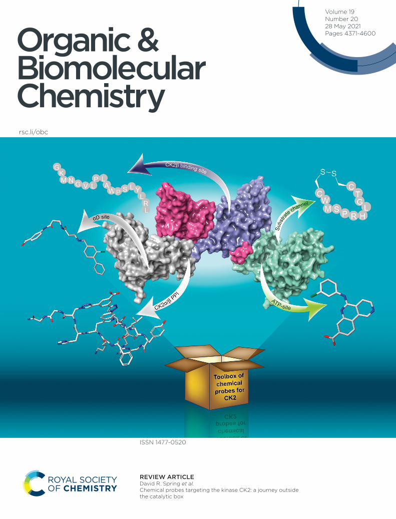
REVIEW ARTICLE David R. Spring et al. Chemical probes targeting the kinase CK2: a journey outside the catalytic box
Organic &Biomolecular Chemistry
REVIEW
Cite this: Org. Biomol. Chem., 2021,19, 4380
Received 9th February 2021,Accepted 29th March 2021
DOI: 10.1039/d1ob00257k
rsc.li/obc
Chemical probes targeting the kinase CK2:a journey outside the catalytic box
Jessica Iegre, †a Eleanor L. Atkinson, †a Paul D. Brear,b Bethany M. Cooper, a
Marko Hyvönen b and David R. Spring *a
CK2 is a protein kinase that plays important roles in many physio-pathological cellular processes. As such,
the development of chemical probes for CK2 has received increasing attention in the past decade with
more than 40 lead compounds developed. In this review, we aim to provide the reader with a compre-
hensive overview of the chemical probes acting outside the highly-conserved ATP-site developed to date.
Such probes belong to different classes of molecules spanning from small molecules to peptides, act with
a range of mechanisms of action and some of them present themselves as promising tools to investigate
the biology of CK2 and therefore develop therapeutics for many disease areas including cancer and
COVID-19.
Introduction
Protein kinases are considered the master regulators of thecell machinery. They exert their functions through the phos-phorylation of other proteins with consequent activation or de-activation of crucially important cellular signaling pathways.1
The essential cellular processes that kinases regulate comprisecell growth, proliferation, differentiation, and death.Therefore, it is unsurprising that gene mutations that alter thefunction or expression levels of protein kinases lead to theinsurgence of several diseases including neurological,immunological, hematological, endocrine, and skeletal dis-orders, as well as cancer.2 The implication of protein kinasesin disease generation and progression has been widely recog-nized by the pharmaceutical community, and, as of today,there are 75 FDA approved drugs targeting this class ofproteins.3
Jessica Iegre
Jessica Iegre was born in Italyand obtained her MSci inMedicinal Chemistry andPharmaceutical Technology atthe University of Pisa, Italy in2013. The same year, she joinedthe AstraZeneca IMED Graduateprogramme in Göteborg, Swedenwhere she spent 2 years workingacross three different depart-ments: medicinal chemistry,DMPK and computational chem-istry. In 2015 she joined theSpring group at the University of
Cambridge, and she obtained her PhD in chemical biology in2019. Jessica is currently a Postdoctoral Research Associate in thegroup, and she is developing novel stapled peptides to inhibit med-icinally relevant protein–protein interactions.
Eleanor L. Atkinson
Eleanor L. Atkinson received herMChem degree from theUniversity of Oxford in 2018after completing her Masters’project under the supervision ofProfessor Angela Russell, investi-gating the metabolism of noveldrug candidates. In 2018, shestarted her PhD studies at theUniversity of Cambridge underthe supervision of ProfessorDavid Spring, where her researchfocuses on the development ofnovel stapled peptides for theinhibition of protein–proteininteractions.
†These authors contributed equally to the work.
aDepartment of Chemistry, University of Cambridge, Lensfield Road, Cambridge, CB2
1EW, UK. E-mail: [email protected] of Biochemistry, University of Cambridge, 80 Tennis Court Road,
Cambridge, CB2 1GA, UK
4380 | Org. Biomol. Chem., 2021, 19, 4380–4396 This journal is © The Royal Society of Chemistry 2021
Ope
n A
cces
s A
rtic
le. P
ublis
hed
on 2
9 M
arch
202
1. D
ownl
oade
d on
12/
5/20
21 1
2:43
:24
AM
. T
his
artic
le is
lice
nsed
und
er a
Cre
ativ
e C
omm
ons
Attr
ibut
ion
3.0
Unp
orte
d L
icen
ce.
View Article OnlineView Journal | View Issue
Among the >500 protein kinases identified in the humanproteome, there is CK2s (formerly referred to as Casein Kinase2).4 CK2α and its very similar paralog CK2α′ are a serine/threo-nine kinases belonging to the CMCG family. Unlike othereukaryotic kinases, CK2α is constitutively active and as such,does not require upstream phosphorylation to exert its func-tion.5 They form a tetrameric complex with a non-catalyticscaffolding subunit CK2β. CK2 phosphorylates more than 300substrates and is thus one of the most promiscuous kinase weknow.6 CK2 is found in both healthy and cancerous cells –
whilst the protein levels in healthy cells increase during prolifer-ation only, cancerous cells are characterized by overexpressionof the different subunits which contributes to their rapidgrowth, proliferation, and their ability to escape apoptosis.7
Breast, lung, prostate, colorectal, renal cancer, leukemia, andglioblastoma are among the cancerous tissues featuring particu-larly high CK2 content, and hence, CK2 inhibitors could proveparticularly useful as therapeutic agents for the treatment ofthese types of cancer.8 Indeed, an orally available CK2α inhibi-tor, named CX4945 (silmitasertib), is currently undergoing clini-cal studies for the treatment of cholangiocarcinoma paving theway for CK2 to be validated as an anticancer target.9
Although the role of CK2 in cancer progression is the moststudied, it should be noted that the kinase is also involved inother pathological processes. For example, recently CK2 hasbeen found to be interacting with the nucleocapsid ofSARS-CoV-2, possibly contributing to COVID-19.10,11 Studiesprobing this relationship are in their infancy, however, it ispossible that CK2 may represent a valid target for the treat-ment of coronaviruses in the future.
Many are the mechanisms by which CK2 can be inhibited,and therefore, there is a large number of inhibitors and chemi-cal probes developed to date: from small molecules to peptideand peptidomimetics, to polyoxometalates.12 Similar to otherkinases, the most studied mode of inhibition is ATP-competi-tive, where a ligand competes with ATP and hence preventssubstrate phosphorylation. Though effective, one of the draw-
backs of this strategy is that the ATP binding site is highly con-served across the kinome, leading to numerous challengeswhen developing a selective chemical probe for the targetkinase.13,14 Most CK2α inhibitors serve as an example to thisproblem, including the clinical trial candidate CX4945.9 Inaddition, cellular concentration of ATP is high and thereforehigh-affinity ATP-competitive inhibitors are needed to effec-tively reduce the activity of the target. To overcome this issue,more selective strategies of CK2 inhibition have been exploredincluding targeting of sites outside the ATP pocket, inhibitionof the formation of the holoenzyme, and displacement ofendogenous substrates.
In this review, we aim to provide a comprehensive overviewof the molecular and structural biology of CK2, mechanisms ofinhibition, and chemical probes for CK2 developed to datewith emphasis on those acting outside the catalytic box(Fig. 1). In addition to describing their discovery and appli-cations, this review provides a critical view of the challengesand opportunities of this promising class of chemical probes.
CK2 kinaseProtein structure and characteristics
The CK2 holoenzyme is a tetramer constituted by two catalyticsubunits, α and/or α′ connected by a dimer of the two regulat-ory subunits, β (Fig. 1). The α subunit presents 20 additionalamino acids at the C-terminus that are absent in α′. The cata-lytic domain (∼48 kDa) is responsible for the phosphorylationof protein substrates and comprises the ATP binding sitebetween the N- and C-terminal domains, linked via the so-called αD region. The N-terminal domain comprises of oneα-helix, named αC and five β-sheets, β1–β5. The αC helix con-tacts the kinase substrates and, due to its lysine-rich content,imparts to the kinase a preference for acidic substrates. TheC-terminal domain of CK2α comprises seven α-helices and twoβ-sheets that form the floor of the ATP binding site and the
Paul D. Brear
Paul Brear obtained his Mchemdegree from the University ofDurham in 2008 and obtainedhis PhD from the University ofSt Andrews in 2012 under thesupervision of Prof. NicholasWestwood and Dr RupertJ. Russell. He joined the group ofDr Marko Hyvönen at theUniversity of Cambridge in 2012and from 2018 is also the X-raycrystallography facility managerat the biochemistry departmentof the University of Cambridge.
Bethany M. Cooper
Bethany M. Cooper received herMChem degree from theUniversity of Leeds in 2018,having completed her 3rd year ofstudy at Lubrizol Ltd,Hazelwood. On return to theUniversity of Leeds her final yearwas spent under the supervisionof Professor Steve Marsden. In2019, she started her PhDstudies at the University ofCambridge under the supervi-sion of Professor David Springand industrial supervisor
Dr Maria Ölwegård-Halvarsson, where her research has focusedon peptide stapling methodologies for use within therapeutics.
Organic & Biomolecular Chemistry Review
This journal is © The Royal Society of Chemistry 2021 Org. Biomol. Chem., 2021, 19, 4380–4396 | 4381
Ope
n A
cces
s A
rtic
le. P
ublis
hed
on 2
9 M
arch
202
1. D
ownl
oade
d on
12/
5/20
21 1
2:43
:24
AM
. T
his
artic
le is
lice
nsed
und
er a
Cre
ativ
e C
omm
ons
Attr
ibut
ion
3.0
Unp
orte
d L
icen
ce.
View Article Online
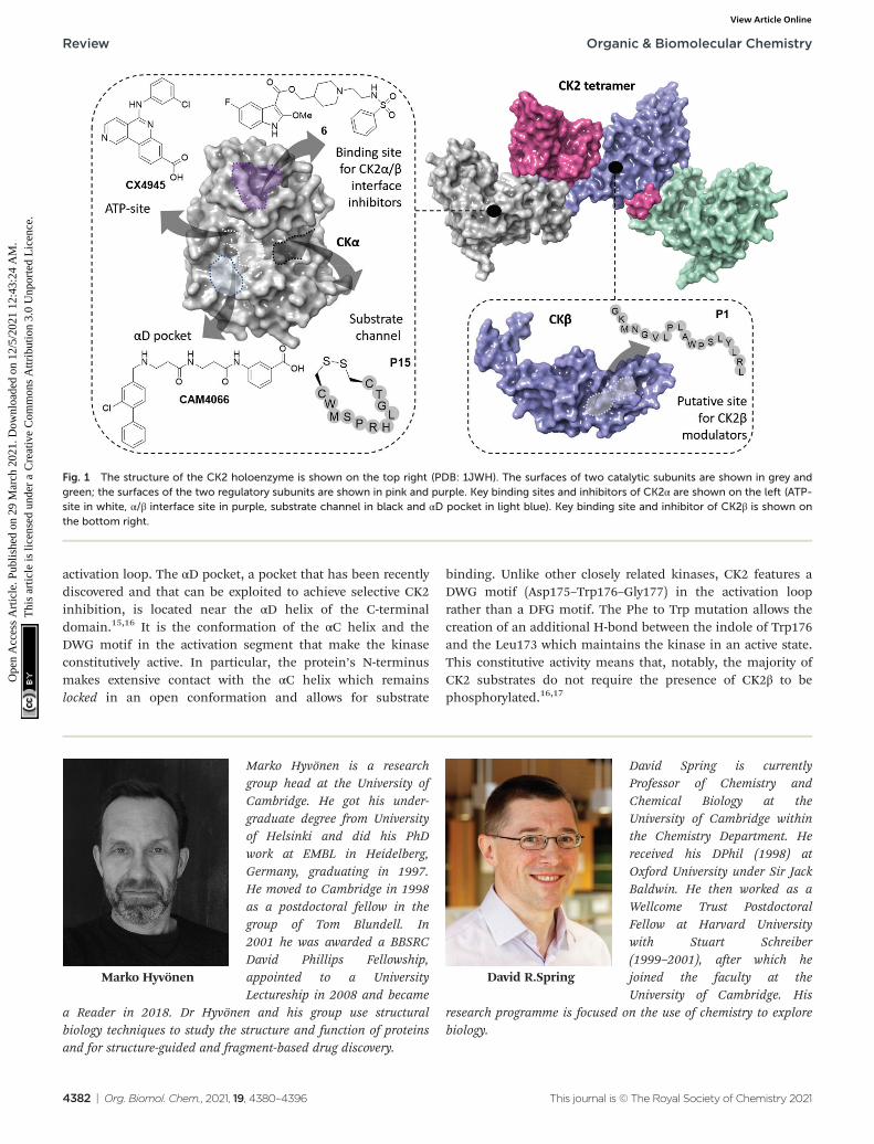
activation loop. The αD pocket, a pocket that has been recentlydiscovered and that can be exploited to achieve selective CK2inhibition, is located near the αD helix of the C-terminaldomain.15,16 It is the conformation of the αC helix and theDWG motif in the activation segment that make the kinaseconstitutively active. In particular, the protein’s N-terminusmakes extensive contact with the αC helix which remainslocked in an open conformation and allows for substrate
binding. Unlike other closely related kinases, CK2 features aDWG motif (Asp175–Trp176–Gly177) in the activation looprather than a DFG motif. The Phe to Trp mutation allows thecreation of an additional H-bond between the indole of Trp176and the Leu173 which maintains the kinase in an active state.This constitutive activity means that, notably, the majority ofCK2 substrates do not require the presence of CK2β to bephosphorylated.16,17
Marko Hyvönen
Marko Hyvönen is a researchgroup head at the University ofCambridge. He got his under-graduate degree from Universityof Helsinki and did his PhDwork at EMBL in Heidelberg,Germany, graduating in 1997.He moved to Cambridge in 1998as a postdoctoral fellow in thegroup of Tom Blundell. In2001 he was awarded a BBSRCDavid Phillips Fellowship,appointed to a UniversityLectureship in 2008 and became
a Reader in 2018. Dr Hyvönen and his group use structuralbiology techniques to study the structure and function of proteinsand for structure-guided and fragment-based drug discovery.
David R.Spring
David Spring is currentlyProfessor of Chemistry andChemical Biology at theUniversity of Cambridge withinthe Chemistry Department. Hereceived his DPhil (1998) atOxford University under Sir JackBaldwin. He then worked as aWellcome Trust PostdoctoralFellow at Harvard Universitywith Stuart Schreiber(1999–2001), after which hejoined the faculty at theUniversity of Cambridge. His
research programme is focused on the use of chemistry to explorebiology.
Fig. 1 The structure of the CK2 holoenzyme is shown on the top right (PDB: 1JWH). The surfaces of two catalytic subunits are shown in grey andgreen; the surfaces of the two regulatory subunits are shown in pink and purple. Key binding sites and inhibitors of CK2α are shown on the left (ATP-site in white, α/β interface site in purple, substrate channel in black and αD pocket in light blue). Key binding site and inhibitor of CK2β is shown onthe bottom right.
Review Organic & Biomolecular Chemistry
4382 | Org. Biomol. Chem., 2021, 19, 4380–4396 This journal is © The Royal Society of Chemistry 2021
Ope
n A
cces
s A
rtic
le. P
ublis
hed
on 2
9 M
arch
202
1. D
ownl
oade
d on
12/
5/20
21 1
2:43
:24
AM
. T
his
artic
le is
lice
nsed
und
er a
Cre
ativ
e C
omm
ons
Attr
ibut
ion
3.0
Unp
orte
d L
icen
ce.
View Article Online
Although not required for CK2α activation, the regulatory βsubunit (∼28 kDa) enhances the catalytic activity of CK2α bymaking the kinase more thermostable.12 Moreover, CK2β regu-lates the activity of the kinase by allowing the holoenzyme toshuttle between intracellular compartments, to dock to andpenetrate the nucleus where the majority of the substrates arelocated.18–20 Furthermore, CK2β acts as a docking station forsubstrates including p53, eIF2β, Nopp140, FGF-II amongstothers.19 CK2β forms an obligate dimer, each protomer ofwhich comprises of a Zn2+ coordinating globular domain withthe elongated C-terminus wrapping around the opposite proto-mer’s globular part. A short, linear epitope in the C-terminusforms a β-turn and is essential for the formation of the holoen-zyme assembly, with residues Arg186, Tyr188, Phe190, andHis193 being crucial for this interaction.15,19,21
In cells, the formation of the holoenzyme involves theassembly of the two regulatory subunits into a dimer, followedby their interaction with a surface of approximately ∼830 Å2 inthe N-lobe of the catalytic subunits.22 Consequent to the for-mation of the protein–protein interaction (PPI) between thetwo subunits, conformational changes occur in CK2α.Specifically, the β4–β5 loop is found to be in a closed form inisolated CK2α whereas it switches to an open form upon com-plexation with CK2β. The latter, on the other hand, does notundergo drastic conformational changes upon holoenzymeformation.23
Physiopathology of CK2
CK2 is involved in a multitude of cellular functions, the mostimportant of which are cell survival, apoptosis, and cell cycleregulation. This can be easily understood if we consider thewide-spread localisation of CK2 within the cell compartmentincluding in the nucleus, cytoplasm, Golgi, ER, ribosomes,and plasma membrane.19 The mechanisms by which CK2regulates the cell decisions of life and death are extremelycomplex and, in part, not fully understood.
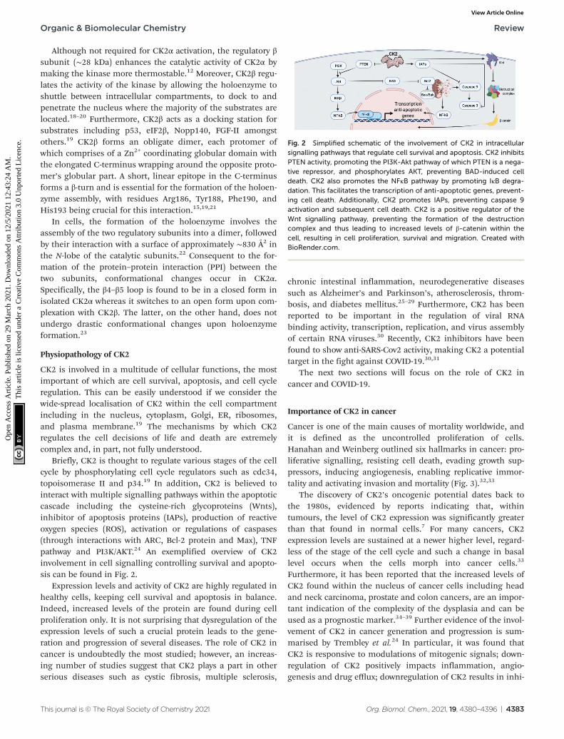
Briefly, CK2 is thought to regulate various stages of the cellcycle by phosphorylating cell cycle regulators such as cdc34,topoisomerase II and p34.19 In addition, CK2 is believed tointeract with multiple signalling pathways within the apoptoticcascade including the cysteine-rich glycoproteins (Wnts),inhibitor of apoptosis proteins (IAPs), production of reactiveoxygen species (ROS), activation or regulations of caspases(through interactions with ARC, Bcl-2 protein and Max), TNFpathway and PI3K/AKT.24 An exemplified overview of CK2involvement in cell signalling controlling survival and apopto-sis can be found in Fig. 2.
Expression levels and activity of CK2 are highly regulated inhealthy cells, keeping cell survival and apoptosis in balance.Indeed, increased levels of the protein are found during cellproliferation only. It is not surprising that dysregulation of theexpression levels of such a crucial protein leads to the gene-ration and progression of several diseases. The role of CK2 incancer is undoubtedly the most studied; however, an increas-ing number of studies suggest that CK2 plays a part in otherserious diseases such as cystic fibrosis, multiple sclerosis,
chronic intestinal inflammation, neurodegenerative diseasessuch as Alzheimer’s and Parkinson’s, atherosclerosis, throm-bosis, and diabetes mellitus.25–29 Furthermore, CK2 has beenreported to be important in the regulation of viral RNAbinding activity, transcription, replication, and virus assemblyof certain RNA viruses.30 Recently, CK2 inhibitors have beenfound to show anti-SARS-Cov2 activity, making CK2 a potentialtarget in the fight against COVID-19.30,31
The next two sections will focus on the role of CK2 incancer and COVID-19.
Importance of CK2 in cancer
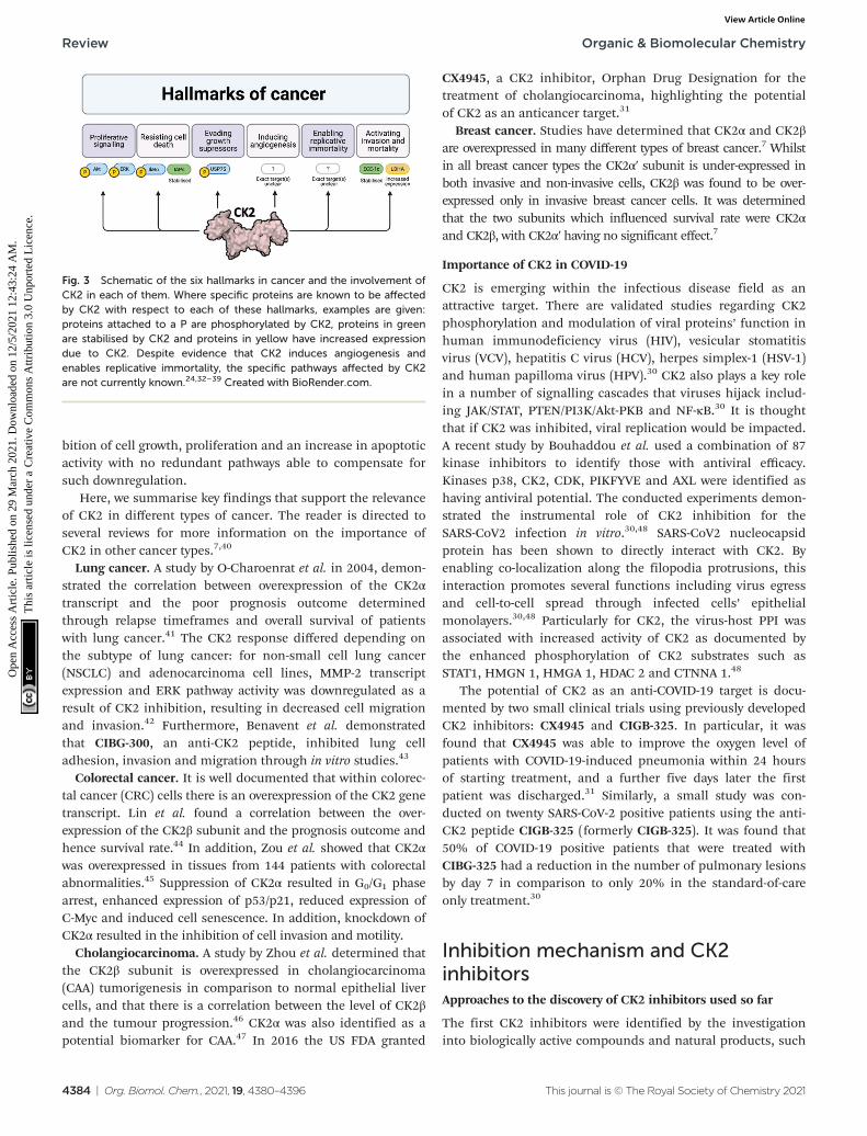
Cancer is one of the main causes of mortality worldwide, andit is defined as the uncontrolled proliferation of cells.Hanahan and Weinberg outlined six hallmarks in cancer: pro-liferative signalling, resisting cell death, evading growth sup-pressors, inducing angiogenesis, enabling replicative immor-tality and activating invasion and mortality (Fig. 3).32,33
The discovery of CK2’s oncogenic potential dates back tothe 1980s, evidenced by reports indicating that, withintumours, the level of CK2 expression was significantly greaterthan that found in normal cells.7 For many cancers, CK2expression levels are sustained at a newer higher level, regard-less of the stage of the cell cycle and such a change in basallevel occurs when the cells morph into cancer cells.33
Furthermore, it has been reported that the increased levels ofCK2 found within the nucleus of cancer cells including headand neck carcinoma, prostate and colon cancers, are an impor-tant indication of the complexity of the dysplasia and can beused as a prognostic marker.34–39 Further evidence of the invol-vement of CK2 in cancer generation and progression is sum-marised by Trembley et al.24 In particular, it was found thatCK2 is responsive to modulations of mitogenic signals; down-regulation of CK2 positively impacts inflammation, angio-genesis and drug efflux; downregulation of CK2 results in inhi-
Fig. 2 Simplified schematic of the involvement of CK2 in intracellularsignalling pathways that regulate cell survival and apoptosis. CK2 inhibitsPTEN activity, promoting the PI3K-Akt pathway of which PTEN is a nega-tive repressor, and phosphorylates AKT, preventing BAD-induced celldeath. CK2 also promotes the NFκB pathway by promoting IκB degra-dation. This facilitates the transcription of anti-apoptotic genes, prevent-ing cell death. Additionally, CK2 promotes IAPs, preventing caspase 9activation and subsequent cell death. CK2 is a positive regulator of theWnt signalling pathway, preventing the formation of the destructioncomplex and thus leading to increased levels of β-catenin within thecell, resulting in cell proliferation, survival and migration. Created withBioRender.com.
Organic & Biomolecular Chemistry Review
This journal is © The Royal Society of Chemistry 2021 Org. Biomol. Chem., 2021, 19, 4380–4396 | 4383
Ope
n A
cces
s A
rtic
le. P
ublis
hed
on 2
9 M
arch
202
1. D
ownl
oade
d on
12/
5/20
21 1
2:43
:24
AM
. T
his
artic
le is
lice
nsed
und
er a
Cre
ativ
e C
omm
ons
Attr
ibut
ion
3.0
Unp
orte
d L
icen
ce.
View Article Online
bition of cell growth, proliferation and an increase in apoptoticactivity with no redundant pathways able to compensate forsuch downregulation.
Here, we summarise key findings that support the relevanceof CK2 in different types of cancer. The reader is directed toseveral reviews for more information on the importance ofCK2 in other cancer types.7,40
Lung cancer. A study by O-Charoenrat et al. in 2004, demon-strated the correlation between overexpression of the CK2αtranscript and the poor prognosis outcome determinedthrough relapse timeframes and overall survival of patientswith lung cancer.41 The CK2 response differed depending onthe subtype of lung cancer: for non-small cell lung cancer(NSCLC) and adenocarcinoma cell lines, MMP-2 transcriptexpression and ERK pathway activity was downregulated as aresult of CK2 inhibition, resulting in decreased cell migrationand invasion.42 Furthermore, Benavent et al. demonstratedthat CIBG-300, an anti-CK2 peptide, inhibited lung celladhesion, invasion and migration through in vitro studies.43
Colorectal cancer. It is well documented that within colorec-tal cancer (CRC) cells there is an overexpression of the CK2 genetranscript. Lin et al. found a correlation between the over-expression of the CK2β subunit and the prognosis outcome andhence survival rate.44 In addition, Zou et al. showed that CK2αwas overexpressed in tissues from 144 patients with colorectalabnormalities.45 Suppression of CK2α resulted in G0/G1 phasearrest, enhanced expression of p53/p21, reduced expression ofC-Myc and induced cell senescence. In addition, knockdown ofCK2α resulted in the inhibition of cell invasion and motility.
Cholangiocarcinoma. A study by Zhou et al. determined thatthe CK2β subunit is overexpressed in cholangiocarcinoma(CAA) tumorigenesis in comparison to normal epithelial livercells, and that there is a correlation between the level of CK2βand the tumour progression.46 CK2α was also identified as apotential biomarker for CAA.47 In 2016 the US FDA granted
CX4945, a CK2 inhibitor, Orphan Drug Designation for thetreatment of cholangiocarcinoma, highlighting the potentialof CK2 as an anticancer target.31
Breast cancer. Studies have determined that CK2α and CK2βare overexpressed in many different types of breast cancer.7 Whilstin all breast cancer types the CK2α′ subunit is under-expressed inboth invasive and non-invasive cells, CK2β was found to be over-expressed only in invasive breast cancer cells. It was determinedthat the two subunits which influenced survival rate were CK2αand CK2β, with CK2α′ having no significant effect.7
Importance of CK2 in COVID-19
CK2 is emerging within the infectious disease field as anattractive target. There are validated studies regarding CK2phosphorylation and modulation of viral proteins’ function inhuman immunodeficiency virus (HIV), vesicular stomatitisvirus (VCV), hepatitis C virus (HCV), herpes simplex-1 (HSV-1)and human papilloma virus (HPV).30 CK2 also plays a key rolein a number of signalling cascades that viruses hijack includ-ing JAK/STAT, PTEN/PI3K/Akt-PKB and NF-κB.30 It is thoughtthat if CK2 was inhibited, viral replication would be impacted.A recent study by Bouhaddou et al. used a combination of 87kinase inhibitors to identify those with antiviral efficacy.Kinases p38, CK2, CDK, PIKFYVE and AXL were identified ashaving antiviral potential. The conducted experiments demon-strated the instrumental role of CK2 inhibition for theSARS-CoV2 infection in vitro.30,48 SARS-CoV2 nucleocapsidprotein has been shown to directly interact with CK2. Byenabling co-localization along the filopodia protrusions, thisinteraction promotes several functions including virus egressand cell-to-cell spread through infected cells’ epithelialmonolayers.30,48 Particularly for CK2, the virus-host PPI wasassociated with increased activity of CK2 as documented bythe enhanced phosphorylation of CK2 substrates such asSTAT1, HMGN 1, HMGA 1, HDAC 2 and CTNNA 1.48
The potential of CK2 as an anti-COVID-19 target is docu-mented by two small clinical trials using previously developedCK2 inhibitors: CX4945 and CIGB-325. In particular, it wasfound that CX4945 was able to improve the oxygen level ofpatients with COVID-19-induced pneumonia within 24 hoursof starting treatment, and a further five days later the firstpatient was discharged.31 Similarly, a small study was con-ducted on twenty SARS-CoV-2 positive patients using the anti-CK2 peptide CIGB-325 (formerly CIGB-325). It was found that50% of COVID-19 positive patients that were treated withCIBG-325 had a reduction in the number of pulmonary lesionsby day 7 in comparison to only 20% in the standard-of-careonly treatment.30
Inhibition mechanism and CK2inhibitorsApproaches to the discovery of CK2 inhibitors used so far
The first CK2 inhibitors were identified by the investigationinto biologically active compounds and natural products, such
Fig. 3 Schematic of the six hallmarks in cancer and the involvement ofCK2 in each of them. Where specific proteins are known to be affectedby CK2 with respect to each of these hallmarks, examples are given:proteins attached to a P are phosphorylated by CK2, proteins in greenare stabilised by CK2 and proteins in yellow have increased expressiondue to CK2. Despite evidence that CK2 induces angiogenesis andenables replicative immortality, the specific pathways affected by CK2are not currently known.24,32–39 Created with BioRender.com.
Review Organic & Biomolecular Chemistry
4384 | Org. Biomol. Chem., 2021, 19, 4380–4396 This journal is © The Royal Society of Chemistry 2021
Ope
n A
cces
s A
rtic
le. P
ublis
hed
on 2
9 M
arch
202
1. D
ownl
oade
d on
12/
5/20
21 1
2:43
:24
AM
. T
his
artic
le is
lice
nsed
und
er a
Cre
ativ
e C
omm
ons
Attr
ibut
ion
3.0
Unp
orte
d L
icen
ce.
View Article Online

as heparin and emodin.49,50 However, with the rapid growth inthe field of computational chemistry and structural biology,more recently inhibitors have been designed specifically forCK2 using structural-based approaches such as fragment-based drug discovery and phage display. The rapid growth intechnologies facilitating the guided and rational design ofprotein modulators has had a drastic impact upon the develop-ment of CK2 inhibitors, as is self-evident from the rapidgrowth in the field over the past 20 years.
From the many inhibitor development studies targetingCK2, multiple different binding sites have been identified onCK2α. These sites all have different advantages and disadvan-tages which have led to them being utilised with varyingsuccess for the inhibition of CK2. These sites can be initiallygrouped into four broad categories: the ATP site, CK2βprotein–protein interaction site, the substrate binding channeland potential allosteric sites outside of the classic ATP site(Fig. 4a).
In the next sections, we will focus on the approaches usedto discover CK2 inhibitors targeting sites outside the catalyticbox and we aim to provide a comprehensive overview of themolecules reported to date. A brief overview of the ATP com-petitive inhibitors is also provided.
Overview of ATP competitive inhibitors
The site where the most development has happened is the ATPsite. This site is the where the protein naturally binds thesmall molecule cofactor, and as such, is seen as the easiestsite to target due to its characteristics. Firstly, the site is pre-formed in the apo structure meaning that there is no barrierto ligand binding. Secondly, the site is composed of a deephydrophobic cleft sandwiched by Lys68 and the hinge regionat opposite ends. This layout is ideally set up for sandwichingan aromatic ring system with hydrogen bonding substituentsprotruding off either side (Fig. 4b).
In a recent crystallographic fragment screen conducted byBrear et al., the ATP site was the only site identified as bindingfragments.51 This is significant as fragment screens are oftenused to probe the surface of proteins for small-molecule
binding sites. The ATP site has been used with great success inidentifying ligands that bind with high affinity and displacethe endogenous ATP hence preventing the phosphorylation ofthe substrates. Indeed, a number of small molecules with pico-molar affinities have been identified for this site.52
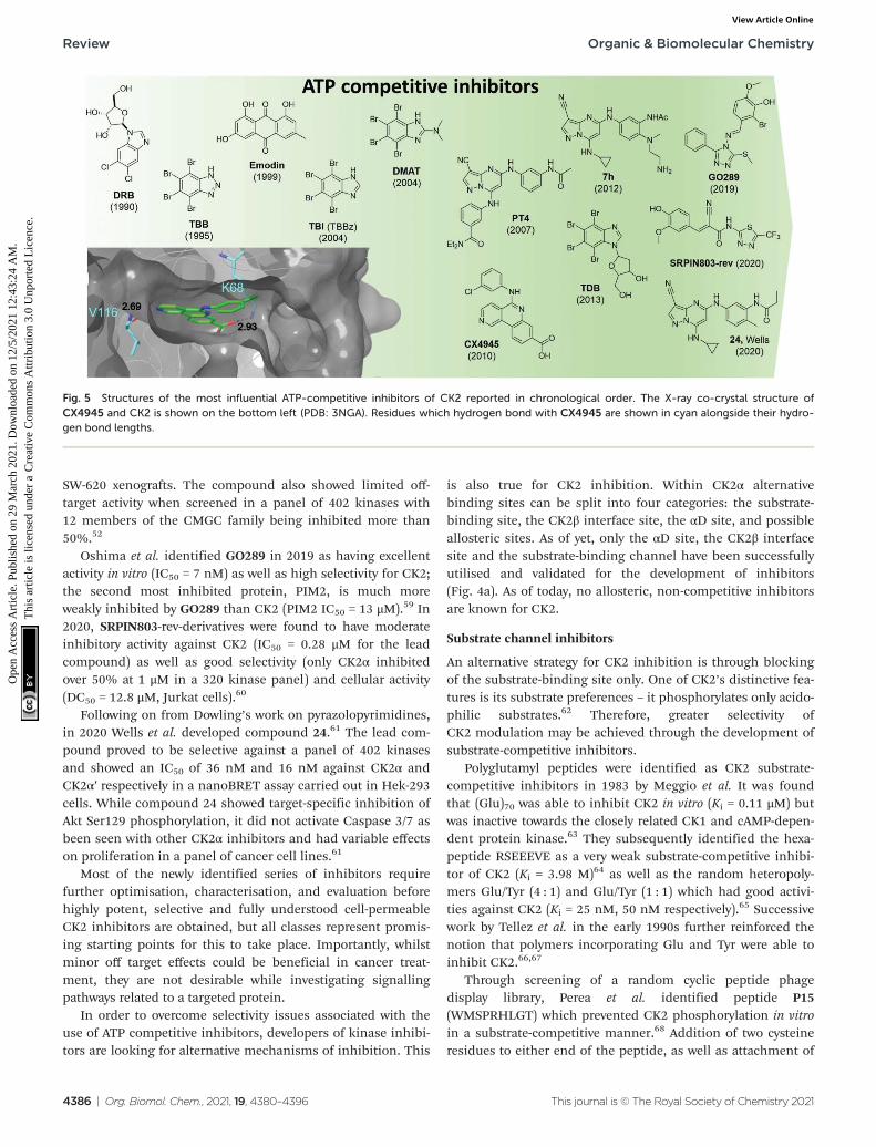
CK2 ATP-competitive inhibitors can broadly be divided intofour main classes: halogenated compounds such as DRB andits derivatives,53,54 condensed polyphenolic compounds suchas emodin and its derivatives,50,55 pyrazolo-triazines and pyra-zolo-pyrimidines56,57 and indoloquinazolines such asCX4945.9 The chronology of the most influential inhibitorsdeveloped thus far is detailed in Fig. 5.
The reader is redirected to a recent review for an up to datecomprehensive review on ATP-competitive inhibitors forCK2.58
Unfortunately, there are a number of drawbacks associatedwith the use of this site, with the primary issue being selecti-vity. The ATP site has evolved to bind ATP which is a verycommon cofactor amongst the genome. Indeed, Uniprot anno-tates 1391 human genes as binding ATP. Therefore, any smallmolecule that targets just the ATP site is likely to bind andinhibit any number of these other ATP-binding proteins. Anexample highlighting the selectivity issue of this class of com-pounds is given by the small molecule clinical candidateCX4945 which is frequently described in the literature ashighly selective for CK2 with an IC50 of 1 nM. However, whenscreened in a selectivity panel of 238 kinases, CX4945 alsoinhibits another seven kinases more than 90% (tested at 500nM). The most significant is the effect seen with Dyrk1A and1B where CX4945 showed IC50 values of 6.8 and 6.4 nM,respectively.9 A number of efforts are currently ongoing toidentify new selective series of CK2 ATP-competitive inhibitors.
In 2016, Dowling et al. identified compound 7h as the mostpromising CK2α inhibitors out of a pyrazolepyrimidine series.The compound showed picomoloar activity in an SPR assayand was found to inhibit the Wnt pathways with an IC50 of 50nM. Despite its limited oral bioavailability, 7h presented prom-ising pharmacokinetic properties after IV administration, andit was effective as monotherapy in both HCT116 cells and
Fig. 4 Crystal structure of CK2α in complex with ADP (PDB: 6YPN). (a) The small-molecule binding sites on CK2α. The ATP site is shown in red andthe sites outside the ATP pocket are shown in blue. (b) and (c) The ATP site of CK2α. The hinge residues are shown in cyan, the polar phosphatebinding residues are shown in green and the hydrophobic sandwiching residues are shown in grey.
Organic & Biomolecular Chemistry Review
This journal is © The Royal Society of Chemistry 2021 Org. Biomol. Chem., 2021, 19, 4380–4396 | 4385
Ope
n A
cces
s A
rtic
le. P
ublis
hed
on 2
9 M
arch
202
1. D
ownl
oade
d on
12/
5/20
21 1
2:43
:24
AM
. T
his
artic
le is
lice
nsed
und
er a
Cre
ativ
e C
omm
ons
Attr
ibut
ion
3.0
Unp
orte
d L
icen
ce.
View Article Online
SW-620 xenografts. The compound also showed limited off-target activity when screened in a panel of 402 kinases with12 members of the CMGC family being inhibited more than50%.52
Oshima et al. identified GO289 in 2019 as having excellentactivity in vitro (IC50 = 7 nM) as well as high selectivity for CK2;the second most inhibited protein, PIM2, is much moreweakly inhibited by GO289 than CK2 (PIM2 IC50 = 13 μM).59 In2020, SRPIN803-rev-derivatives were found to have moderateinhibitory activity against CK2 (IC50 = 0.28 μM for the leadcompound) as well as good selectivity (only CK2α inhibitedover 50% at 1 μM in a 320 kinase panel) and cellular activity(DC50 = 12.8 μM, Jurkat cells).60
Following on from Dowling’s work on pyrazolopyrimidines,in 2020 Wells et al. developed compound 24.61 The lead com-pound proved to be selective against a panel of 402 kinasesand showed an IC50 of 36 nM and 16 nM against CK2α andCK2α′ respectively in a nanoBRET assay carried out in Hek-293cells. While compound 24 showed target-specific inhibition ofAkt Ser129 phosphorylation, it did not activate Caspase 3/7 asbeen seen with other CK2α inhibitors and had variable effectson proliferation in a panel of cancer cell lines.61
Most of the newly identified series of inhibitors requirefurther optimisation, characterisation, and evaluation beforehighly potent, selective and fully understood cell-permeableCK2 inhibitors are obtained, but all classes represent promis-ing starting points for this to take place. Importantly, whilstminor off target effects could be beneficial in cancer treat-ment, they are not desirable while investigating signallingpathways related to a targeted protein.
In order to overcome selectivity issues associated with theuse of ATP competitive inhibitors, developers of kinase inhibi-tors are looking for alternative mechanisms of inhibition. This
is also true for CK2 inhibition. Within CK2α alternativebinding sites can be split into four categories: the substrate-binding site, the CK2β interface site, the αD site, and possibleallosteric sites. As of yet, only the αD site, the CK2β interfacesite and the substrate-binding channel have been successfullyutilised and validated for the development of inhibitors(Fig. 4a). As of today, no allosteric, non-competitive inhibitorsare known for CK2.
Substrate channel inhibitors
An alternative strategy for CK2 inhibition is through blockingof the substrate-binding site only. One of CK2’s distinctive fea-tures is its substrate preferences – it phosphorylates only acido-philic substrates.62 Therefore, greater selectivity ofCK2 modulation may be achieved through the development ofsubstrate-competitive inhibitors.
Polyglutamyl peptides were identified as CK2 substrate-competitive inhibitors in 1983 by Meggio et al. It was foundthat (Glu)70 was able to inhibit CK2 in vitro (Ki = 0.11 μM) butwas inactive towards the closely related CK1 and cAMP-depen-dent protein kinase.63 They subsequently identified the hexa-peptide RSEEEVE as a very weak substrate-competitive inhibi-tor of CK2 (Ki = 3.98 M)64 as well as the random heteropoly-mers Glu/Tyr (4 : 1) and Glu/Tyr (1 : 1) which had good activi-ties against CK2 (Ki = 25 nM, 50 nM respectively).65 Successivework by Tellez et al. in the early 1990s further reinforced thenotion that polymers incorporating Glu and Tyr were able toinhibit CK2.66,67
Through screening of a random cyclic peptide phagedisplay library, Perea et al. identified peptide P15(WMSPRHLGT) which prevented CK2 phosphorylation in vitroin a substrate-competitive manner.68 Addition of two cysteineresidues to either end of the peptide, as well as attachment of
Fig. 5 Structures of the most influential ATP-competitive inhibitors of CK2 reported in chronological order. The X-ray co-crystal structure ofCX4945 and CK2 is shown on the bottom left (PDB: 3NGA). Residues which hydrogen bond with CX4945 are shown in cyan alongside their hydro-gen bond lengths.
Review Organic & Biomolecular Chemistry
4386 | Org. Biomol. Chem., 2021, 19, 4380–4396 This journal is © The Royal Society of Chemistry 2021
Ope
n A
cces
s A
rtic
le. P
ublis
hed
on 2
9 M
arch
202
1. D
ownl
oade
d on
12/
5/20
21 1
2:43
:24
AM
. T
his
artic
le is
lice
nsed
und
er a
Cre
ativ
e C
omm
ons
Attr
ibut
ion
3.0
Unp
orte
d L
icen
ce.
View Article Online
the known cell-penetrating peptide TAT to the N-terminus andcyclisation with a disulfide bridge afforded the cell-permeableCK2 substrate-competitive inhibitor P15-TAT (subsequentlyrenamed and hereafter referred to as CIGB-325). CIGB-325showed moderate inhibition of cell viability against a range oftumour cell lines with an EC50 of between 20 and 136 μMdepending on the cell line.69 Furthermore, it was found thatdaily intra-tumoural administration of CIGB-325 into TC-1lung epithelial tumours (C57BL6 mice) led to a significantreduction in tumour growth.69
Proteomics studies also revealed the up- and down-regu-lation of proteins involved in the apoptotic intrinsic pathwaysin the presence of this peptide. In 2008, the first-in-man clini-cal trial with CIGB-325 was conducted: 75% of the patients (31women with cervical cancer) had significant lesion reductionand, most promisingly, 19% of the patients exhibited fullhistological regression. Additionally, the effect of CIGB-325 onspontaneous lung metastasis was investigated; systemic treat-ment with CIGB-325 prevented breast cancer colonization inthe lung.70 In 2019, Winiewska-Szajewska et al. identified a6-mer peptide (KESEEE-NH2) which weakly binds to the CK2αsubstrate binding-site (Kd = 0.39 mM).71 They showed thatboth the peptide and the ATP-competitive inhibitor TBI couldbind simultaneously to CK2 without a drastic loss in bindingaffinity (Kd = 0.45 mM for KESEEE-NH2 with TBI present).71
Therefore, the novel peptide KESEEE-NH2 represents a poten-tial substrate-competitive inhibitor.
CIGB-325 currently represents the only highly potent sub-strate-competitive inhibitor of CK2 that is effective in vivo onmultiple cancer cell lines. In particular, the promising resultsshown against cancer cell metastasis make CIGB-325 a promis-ing candidate for limiting metastatic cancer spread. It shouldbe noted that the lack of structural data on the substrate-binding makes the development of inhibitors at this siteharder to achieve.
Bi-specific ATP/substrate competitive inhibitors
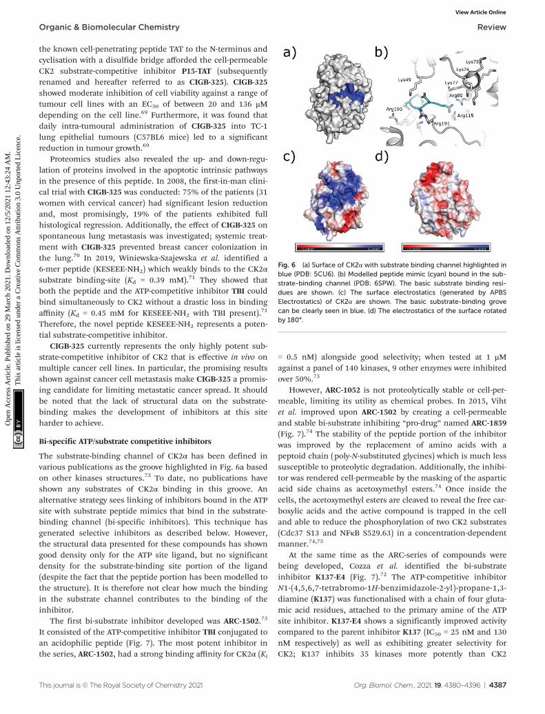
The substrate-binding channel of CK2α has been defined invarious publications as the groove highlighted in Fig. 6a basedon other kinases structures.72 To date, no publications haveshown any substrates of CK2α binding in this groove. Analternative strategy sees linking of inhibitors bound in the ATPsite with substrate peptide mimics that bind in the substrate-binding channel (bi-specific inhibitors). This technique hasgenerated selective inhibitors as described below. However,the structural data presented for these compounds has showngood density only for the ATP site ligand, but no significantdensity for the substrate-binding site portion of the ligand(despite the fact that the peptide portion has been modelled tothe structure). It is therefore not clear how much the bindingin the substrate channel contributes to the binding of theinhibitor.
The first bi-substrate inhibitor developed was ARC-1502.73
It consisted of the ATP-competitive inhibitor TBI conjugated toan acidophilic peptide (Fig. 7). The most potent inhibitor inthe series, ARC-1502, had a strong binding affinity for CK2α (Ki
= 0.5 nM) alongside good selectivity; when tested at 1 μMagainst a panel of 140 kinases, 9 other enzymes were inhibitedover 50%.73
However, ARC-1052 is not proteolytically stable or cell-per-meable, limiting its utility as chemical probes. In 2015, Vihtet al. improved upon ARC-1502 by creating a cell-permeableand stable bi-substrate inhibiting “pro-drug” named ARC-1859(Fig. 7).74 The stability of the peptide portion of the inhibitorwas improved by the replacement of amino acids with apeptoid chain (poly-N-substituted glycines) which is much lesssusceptible to proteolytic degradation. Additionally, the inhibi-tor was rendered cell-permeable by the masking of the asparticacid side chains as acetoxymethyl esters.74 Once inside thecells, the acetoxymethyl esters are cleaved to reveal the free car-boxylic acids and the active compound is trapped in the celland able to reduce the phosphorylation of two CK2 substrates(Cdc37 S13 and NFκB S529.63) in a concentration-dependentmanner.74,75
At the same time as the ARC-series of compounds werebeing developed, Cozza et al. identified the bi-substrateinhibitor K137-E4 (Fig. 7).72 The ATP-competitive inhibitorN1-(4,5,6,7-tetrabromo-1H-benzimidazole-2-yl)-propane-1,3-diamine (K137) was functionalised with a chain of four gluta-mic acid residues, attached to the primary amine of the ATPsite inhibitor. K137-E4 shows a significantly improved activitycompared to the parent inhibitor K137 (IC50 = 25 nM and 130nM respectively) as well as exhibiting greater selectivity forCK2; K137 inhibits 35 kinases more potently than CK2
Fig. 6 (a) Surface of CK2α with substrate binding channel highlighted inblue (PDB: 5CU6). (b) Modelled peptide mimic (cyan) bound in the sub-strate-binding channel (PDB: 6SPW). The basic substrate binding resi-dues are shown. (c) The surface electrostatics (generated by APBSElectrostatics) of CK2α are shown. The basic substrate-binding grovecan be clearly seen in blue. (d) The electrostatics of the surface rotatedby 180°.
Organic & Biomolecular Chemistry Review
This journal is © The Royal Society of Chemistry 2021 Org. Biomol. Chem., 2021, 19, 4380–4396 | 4387
Ope
n A
cces
s A
rtic
le. P
ublis
hed
on 2
9 M
arch
202
1. D
ownl
oade
d on
12/
5/20
21 1
2:43
:24
AM
. T
his
artic
le is
lice
nsed
und
er a
Cre
ativ
e C
omm
ons
Attr
ibut
ion
3.0
Unp
orte
d L
icen
ce.
View Article Online
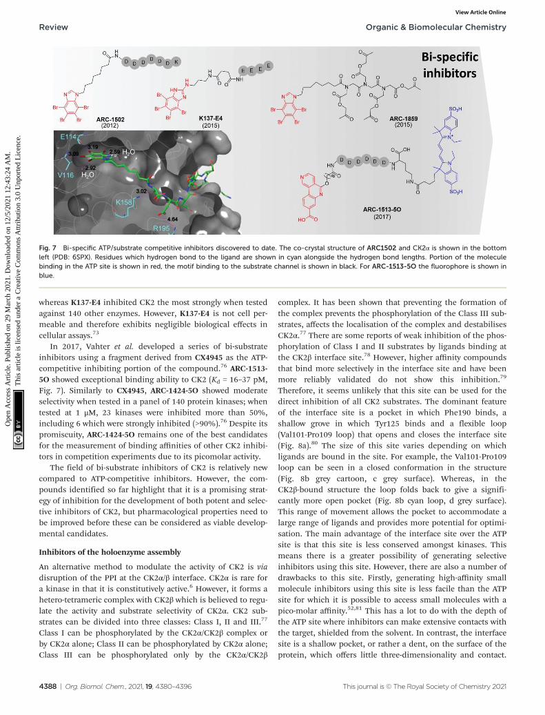
whereas K137-E4 inhibited CK2 the most strongly when testedagainst 140 other enzymes. However, K137-E4 is not cell per-meable and therefore exhibits negligible biological effects incellular assays.73
In 2017, Vahter et al. developed a series of bi-substrateinhibitors using a fragment derived from CX4945 as the ATP-competitive inhibiting portion of the compound.76 ARC-1513-5O showed exceptional binding ability to CK2 (Kd = 16–37 pM,Fig. 7). Similarly to CX4945, ARC-1424-5O showed moderateselectivity when tested in a panel of 140 protein kinases; whentested at 1 μM, 23 kinases were inhibited more than 50%,including 6 which were strongly inhibited (>90%).76 Despite itspromiscuity, ARC-1424-5O remains one of the best candidatesfor the measurement of binding affinities of other CK2 inhibi-tors in competition experiments due to its picomolar activity.
The field of bi-substrate inhibitors of CK2 is relatively newcompared to ATP-competitive inhibitors. However, the com-pounds identified so far highlight that it is a promising strat-egy of inhibition for the development of both potent and selec-tive inhibitors of CK2, but pharmacological properties need tobe improved before these can be considered as viable develop-mental candidates.
Inhibitors of the holoenzyme assembly
An alternative method to modulate the activity of CK2 is viadisruption of the PPI at the CK2α/β interface. CK2α is rare fora kinase in that it is constitutively active.6 However, it forms ahetero-tetrameric complex with CK2β which is believed to regu-late the activity and substrate selectivity of CK2α. CK2 sub-strates can be divided into three classes: Class I, II and III.77
Class I can be phosphorylated by the CK2α/CK2β complex orby CK2α alone; Class II can be phosphorylated by CK2α alone;Class III can be phosphorylated only by the CK2α/CK2β
complex. It has been shown that preventing the formation ofthe complex prevents the phosphorylation of the Class III sub-strates, affects the localisation of the complex and destabilisesCK2α.77 There are some reports of weak inhibition of the phos-phorylation of Class I and II substrates by ligands binding atthe CK2β interface site.78 However, higher affinity compoundsthat bind more selectively in the interface site and have beenmore reliably validated do not show this inhibition.79
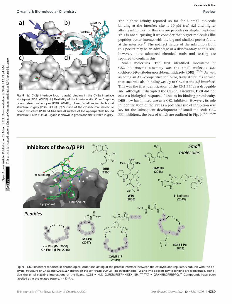
Therefore, it seems unlikely that this site can be used for thedirect inhibition of all CK2 substrates. The dominant featureof the interface site is a pocket in which Phe190 binds, ashallow grove in which Tyr125 binds and a flexible loop(Val101-Pro109 loop) that opens and closes the interface site(Fig. 8a).80 The size of this site varies depending on whichligands are bound in the site. For example, the Val101-Pro109loop can be seen in a closed conformation in the structure(Fig. 8b grey cartoon, c grey surface). Whereas, in theCK2β-bound structure the loop folds back to give a signifi-cantly more open pocket (Fig. 8b cyan loop, d grey surface).This range of movement allows the pocket to accommodate alarge range of ligands and provides more potential for optimi-sation. The main advantage of the interface site over the ATPsite is that this site is less conserved amongst kinases. Thismeans there is a greater possibility of generating selectiveinhibitors using this site. However, there are also a number ofdrawbacks to this site. Firstly, generating high-affinity smallmolecule inhibitors using this site is less facile than the ATPsite for which it is possible to access small molecules with apico-molar affinity.52,81 This has a lot to do with the depth ofthe ATP site where inhibitors can make extensive contacts withthe target, shielded from the solvent. In contrast, the interfacesite is a shallow pocket, or rather a dent, on the surface of theprotein, which offers little three-dimensionality and contact.
Fig. 7 Bi-specific ATP/substrate competitive inhibitors discovered to date. The co-crystal structure of ARC1502 and CK2α is shown in the bottomleft (PDB: 6SPX). Residues which hydrogen bond to the ligand are shown in cyan alongside the hydrogen bond lengths. Portion of the moleculebinding in the ATP site is shown in red, the motif binding to the substrate channel is shown in black. For ARC-1513-5O the fluorophore is shown inblue.
Review Organic & Biomolecular Chemistry
4388 | Org. Biomol. Chem., 2021, 19, 4380–4396 This journal is © The Royal Society of Chemistry 2021
Ope
n A
cces
s A
rtic
le. P
ublis
hed
on 2
9 M
arch
202
1. D
ownl
oade
d on
12/
5/20
21 1
2:43
:24
AM
. T
his
artic
le is
lice
nsed
und
er a
Cre
ativ
e C
omm
ons
Attr
ibut
ion
3.0
Unp
orte
d L
icen
ce.
View Article Online
The highest affinity reported so far for a small moleculebinding at the interface site is 30 μM (ref. 82) and higheraffinity inhibitors for this site are peptides or stapled peptides.This is not surprising if we consider that bigger molecules likepeptides better interact with the big and shallow pocket foundat the interface.83 The indirect nature of the inhibition fromthis pocket may be an advantage or a disadvantage to this site;however, more advanced chemical tools and testing arerequired to confirm this.
Small molecules. The first identified modulator ofCK2 holoenzyme assembly was the small molecule 5,6-dichloro-1-β-D-ribofuranosyl-benzimidazole (DRB).78,84 As wellas being an ATP-competitive inhibitor, X-ray structures showedthat DRB was also binding weakly to CK2α at the α/β interface.This was the first identification of the CK2 PPI as a druggablesite. Although it disrupted the CK2α/β assembly, DRB did notcause a biological response.78 Due to its binding promiscuity,DRB now has limited use as a CK2 inhibitor. However, its rolein identification of the PPI as a potential site of inhibition waskey for the subsequent development of small molecule CK2PPI inhibitors, the best of which are outlined in Fig. 9.78,82,85,86
Fig. 8 (a) CK2β interface loop (purple) binding in the CK2α interfacesite (grey) (PDB: 4MD7). (b) Flexibility of the interface site. Open/peptidebound structure in cyan (PDB: 6Q4Q), closed/small molecule boundstructure in grey (PDB: 5CU6). (c) Surface of the closed/small moleculebound structure (PDB: 5CU6) and (d) surface of the open/peptide boundstructure (PDB: 6Q4Q). Ligand is shown in green and the surface in grey.
Fig. 9 CK2 inhibitors reported in chronological order and acting at the protein interface between the catalytic and regulatory subunit with the co-crystal structure of CK2α and CAM7117 shown on the left (PDB: 6Q4Q). The hydrophobic Tyr and Phe pockets key to binding are highlighted, along-side the pi–pi stacking interactions of the ligand. sC18 = H2N-GLRKRLRKFRNKIKEK-NH2;
93 TAT = GRKKRRQRRRPPQ.96 Compounds have beenlabelled as in the related papers. r = D-Arg.
Organic & Biomolecular Chemistry Review
This journal is © The Royal Society of Chemistry 2021 Org. Biomol. Chem., 2021, 19, 4380–4396 | 4389
Ope
n A
cces
s A
rtic
le. P
ublis
hed
on 2
9 M
arch
202
1. D
ownl
oade
d on
12/
5/20
21 1
2:43
:24
AM
. T
his
artic
le is
lice
nsed
und
er a
Cre
ativ
e C
omm
ons
Attr
ibut
ion
3.0
Unp
orte
d L
icen
ce.
View Article Online

The second small molecule modulator of the PPI was thepodophyllotoxin indole-analogue W16, identified as having amodest IC50 of 30–40 μM in enzymatic assays (Fig. 9).86 Kineticanalysis showed that W16 is a non-competitive inhibitor ofCK2α and that this inhibition was reversed upon the additionof CK2β or the CK2β-mimicking peptide Pc, suggesting thatW16 binds in or close to the CK2β-binding pocket on CK2α.The inhibition of CK2α may result from small conformationalchanges of the protein upon binding of W16.86
Recently, Kröger et al. found that the enantiomer of W16was six times more potent than its analogue (Ki = 4.9 μM vs.31 μM respectively).87 They also replaced the labile anhydridegroup of W16 with a more stable imide and N-methylimide,increasing stability without the loss of binding affinity (Ki =3.6 μM vs. 2.8 μM respectively). However, the increased inhi-bition of the PPI did not result in a greater biological effect asthe new analogues of W16 were not seen to inhibit CK2. Thisclearly highlights that perturbation to the equilibrium of theCK2 holoenzyme complex formation does not necessarily leadto enzyme inhibition.87 Due to the large aromatic structures ofthe podophyllotoxin indolo-analogues, the compounds sufferfrom poor solubility making the likelihood of developingtherapeutics derived from W16 low.86,87
A high-concentration X-ray crystallography fragment screenidentified 1 (in Brear et al.) binding at both the CK2α/β inter-face and in the ATP-pocket of CK2 with a weak IC50 (900 μM).88
Iterative design-test cycles resulted in the development of thesmall molecule CAM187 which had greatly improved bioactiv-ity (IC50 = 44 μM) and did not show promiscuity (Fig. 9). Dueto its significantly smaller size and, as such, more favourablephysical properties than W16, the fragment CAM187 is amuch-improved starting point for the development of selectivesmall molecule drug-like compounds to target the CK2 PPI.However, further analyses of its activity and selectivity againstother kinases, including evaluation of its biological effect incells are required.
With a virtual screening of a compound library followed bysurface-plasmon resonance (SPR), NMR and crystallography,Kufareva et al. identified compound 6 (Kd = 30 μM) a novelCK2 PPI inhibiting molecule (Fig. 9).82 Compound 6 (inKufareva et al.) successfully inhibited the phosphorylation ofCK2β-dependent substrates, suppressed MDA-MB231 triple-negative breast cancer cell growth and induced apoptosis inthe μM range.82 As such, compound 6 is the first example of asmall molecule inhibitor of the CK2α/β interaction which hasbeen shown to be effective in cells. Alongside CAM187, com-pound 6 represents an excellent starting point for the develop-ment of selective and potent small molecule therapeutics forthe inhibition of CK2 via the perturbation of the CK2 PPI.
Peptides. In 2008, Laudet et al. developed a peptide to bindto CK2α at the site of the CK2β binding pocket.107 Peptidesrepresent good candidates for PPI modulators as their flexi-bility allows them to adapt to the large surfaces involved inPPIs.89 They can be designed to mimic the section of thepartner protein binding to the desired target region, henceovercoming the difficulties associated with de novo design.90
Using the prior knowledge of residues Tyr188, Gly189 andPhe190 being the main source of affinity between the β-hairpinloop and CK2α, in combination with the crystal structure ofCK2α bound to CK2β, Laudet et al. designed a handful ofCK2β-derived peptides, the most promising of which was Pc(Fig. 9).15,91 Pc consists of the central interacting segment ofCK2β C-terminal loop cyclised by a disulfide bridge betweentwo cysteine residues. The peptide was shown to antagonisethe assembly of the holoenzyme complex (IC50 = 3.0 μM) aswell as altering its substrate specificity.91 Pc represented thefirst peptide antagonist of CK2 which exerted its effect bybinding to CK2α and preventing the formation of the holo-enzyme.91 However, Pc did not successfully exhibit cellularactivity. To improve the cellular activity of the peptide, pre-dicted to suffer from poor stability of disulfides in reducingintracellular environment, the disulfide bond was replacedwith a head-to-tail cyclisation.92 Subsequently, the cyclicpeptide was fused with the TAT cell-penetrating peptide whichis known to facilitate transport across cell membranes.89 Theresulting compound, TAT-Pc, was shown to enter cells, inhibitthe phosphorylation of CK2β-dependent substrates and lead tocaspase independent cell death.92 In 2019, three simultaneousbut separate optimisations of the Pc and TAT-Pc peptidesoccurred leading to the development of sC18-I-Pc,93 I192F94
and CAM7117.79
In 2015, Hochscherf et al. altered the Phe residue in thestructure of Pc to include para-halogens. The iodinated-Pcpeptide was the most effective, showing a slight improvementin Kd compared to Pc (0.24 vs. 0.56 μM)95 In an attempt tomake I-Pc cell-permeable, in 2019, the same research groupintroduced a cell-penetrating tag, namely, sC1896 to translocatethe peptide into cells. Although the introduction of sC18resulted in a 4-time increase of the Ki, sC18-I-Pc successfullyinhibited the phosphorylation of CK2β-dependent substratesas well as evoking cell death in the mid-μM range (Fig. 9).93
Tang et al. used structure-based computational design incombination with experimental evaluation to develop pointmutated Pc derivatives.108 The most promising mutant, I192Fexhibited a 10-fold improvement in the predicted bindingaffinity to CK2α compared to Pc (0.5 vs. 8.9 μM). When theTAT-conjugated cell-permeable derivative of the peptide wassynthesised, TAT-I192F showed comparable anti-proliferativeeffects against HepG2 cells to TAT-Pc (20.1 μM vs.30.4 μM).96,108
The development of CAM7117 took a different approach tothe aforementioned peptides in that it was the cyclic constraintthat was the main site of optimisation due to the lability of thedisulfide bond in the Pc derivatives. Using a structure-basedapproach, a variety of covalent constraints were designed andtested to determine which held the peptide in the optimal con-formation. In addition, molecular modelling and X-ray crystal-lography of the peptide sequence directed a point mutation inthe central sequence of Pc, namely Ile 192 to Trp. Movementof one of the cyclising residues by one position and the use ofCuAAC chemistry gave CAM7117 (Fig. 9).79 The peptide had animproved Kd value compared to Pc (0.2 vs. 1 μM respectively) in
Review Organic & Biomolecular Chemistry
4390 | Org. Biomol. Chem., 2021, 19, 4380–4396 This journal is © The Royal Society of Chemistry 2021
Ope
n A
cces
s A
rtic
le. P
ublis
hed
on 2
9 M
arch
202
1. D
ownl
oade
d on
12/
5/20
21 1
2:43
:24
AM
. T
his
artic
le is
lice
nsed
und
er a
Cre
ativ
e C
omm
ons
Attr
ibut
ion
3.0
Unp
orte
d L
icen
ce.
View Article Online
an isothermal titration calorimetry (ITC) assay, was stable inhuman serum and cell-permeable. CAM7117 was able toinhibit cell growth with a GI50 of 33 μM in U2OS cells. It waspostulated that the difference between the enzymatic and thecellular activity was due to partial trapping in the Golgiapparatus.79
In just over a decade, there has been a movement from theidentification of CK2α/CK2β interaction site as a potentialtarget for small molecule-directed inhibition of CK2 to thedevelopment of a handful of potent CK2 PPI inhibitors. Boththe peptide and small molecule inhibitors have their limit-ations and require further optimisation before a sufficientlystable, extremely potent and highly selective cellular chemicalprobe or therapeutic is obtained for this site. However, themost promising inhibitors currently known are an excellentplace for optimisation to continue from.
Inhibitors of CK2α acting outside the ATP pocket
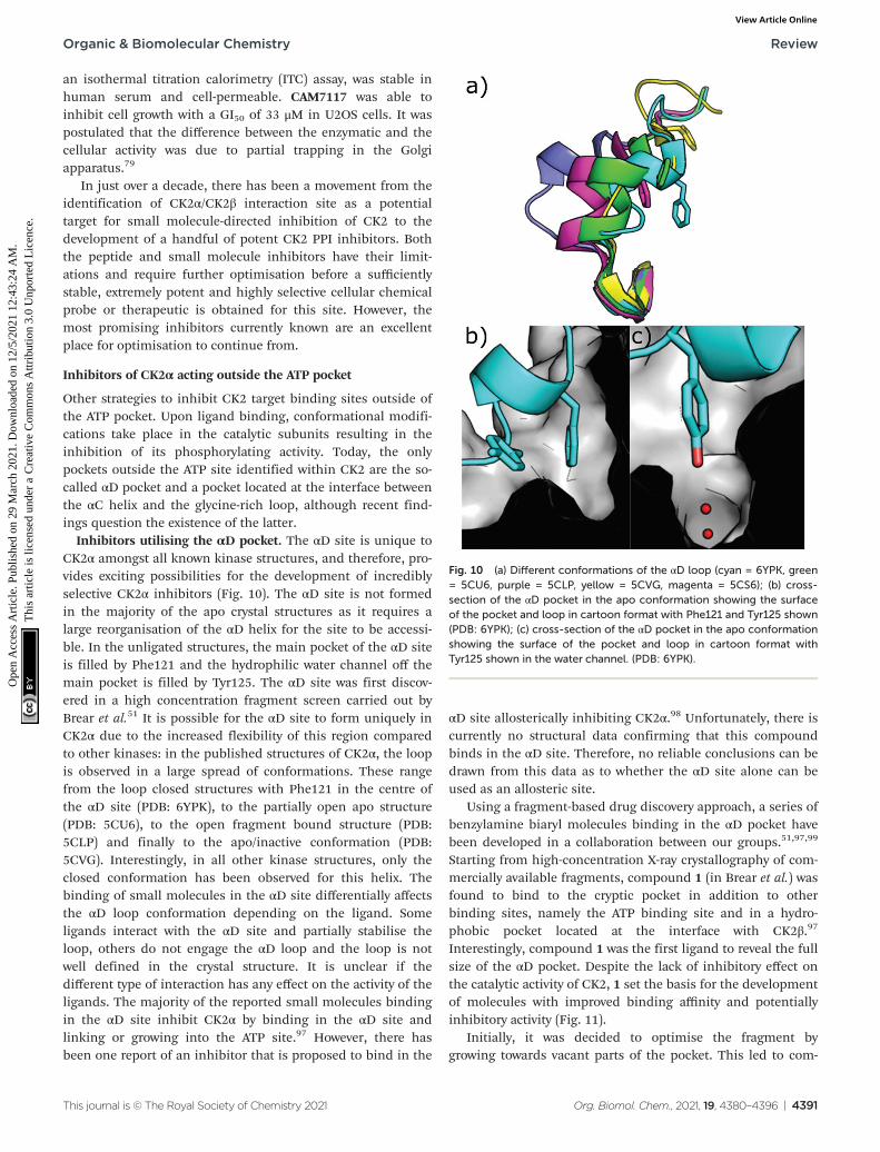
Other strategies to inhibit CK2 target binding sites outside ofthe ATP pocket. Upon ligand binding, conformational modifi-cations take place in the catalytic subunits resulting in theinhibition of its phosphorylating activity. Today, the onlypockets outside the ATP site identified within CK2 are the so-called αD pocket and a pocket located at the interface betweenthe αC helix and the glycine-rich loop, although recent find-ings question the existence of the latter.
Inhibitors utilising the αD pocket. The αD site is unique toCK2α amongst all known kinase structures, and therefore, pro-vides exciting possibilities for the development of incrediblyselective CK2α inhibitors (Fig. 10). The αD site is not formedin the majority of the apo crystal structures as it requires alarge reorganisation of the αD helix for the site to be accessi-ble. In the unligated structures, the main pocket of the αD siteis filled by Phe121 and the hydrophilic water channel off themain pocket is filled by Tyr125. The αD site was first discov-ered in a high concentration fragment screen carried out byBrear et al.51 It is possible for the αD site to form uniquely inCK2α due to the increased flexibility of this region comparedto other kinases: in the published structures of CK2α, the loopis observed in a large spread of conformations. These rangefrom the loop closed structures with Phe121 in the centre ofthe αD site (PDB: 6YPK), to the partially open apo structure(PDB: 5CU6), to the open fragment bound structure (PDB:5CLP) and finally to the apo/inactive conformation (PDB:5CVG). Interestingly, in all other kinase structures, only theclosed conformation has been observed for this helix. Thebinding of small molecules in the αD site differentially affectsthe αD loop conformation depending on the ligand. Someligands interact with the αD site and partially stabilise theloop, others do not engage the αD loop and the loop is notwell defined in the crystal structure. It is unclear if thedifferent type of interaction has any effect on the activity of theligands. The majority of the reported small molecules bindingin the αD site inhibit CK2α by binding in the αD site andlinking or growing into the ATP site.97 However, there hasbeen one report of an inhibitor that is proposed to bind in the
αD site allosterically inhibiting CK2α.98 Unfortunately, there iscurrently no structural data confirming that this compoundbinds in the αD site. Therefore, no reliable conclusions can bedrawn from this data as to whether the αD site alone can beused as an allosteric site.
Using a fragment-based drug discovery approach, a series ofbenzylamine biaryl molecules binding in the αD pocket havebeen developed in a collaboration between our groups.51,97,99
Starting from high-concentration X-ray crystallography of com-mercially available fragments, compound 1 (in Brear et al.) wasfound to bind to the cryptic pocket in addition to otherbinding sites, namely the ATP binding site and in a hydro-phobic pocket located at the interface with CK2β.97
Interestingly, compound 1 was the first ligand to reveal the fullsize of the αD pocket. Despite the lack of inhibitory effect onthe catalytic activity of CK2, 1 set the basis for the developmentof molecules with improved binding affinity and potentiallyinhibitory activity (Fig. 11).
Initially, it was decided to optimise the fragment bygrowing towards vacant parts of the pocket. This led to com-
Fig. 10 (a) Different conformations of the αD loop (cyan = 6YPK, green= 5CU6, purple = 5CLP, yellow = 5CVG, magenta = 5CS6); (b) cross-section of the αD pocket in the apo conformation showing the surfaceof the pocket and loop in cartoon format with Phe121 and Tyr125 shown(PDB: 6YPK); (c) cross-section of the αD pocket in the apo conformationshowing the surface of the pocket and loop in cartoon format withTyr125 shown in the water channel. (PDB: 6YPK).
Organic & Biomolecular Chemistry Review
This journal is © The Royal Society of Chemistry 2021 Org. Biomol. Chem., 2021, 19, 4380–4396 | 4391
Ope
n A
cces
s A
rtic
le. P
ublis
hed
on 2
9 M
arch
202
1. D
ownl
oade
d on
12/
5/20
21 1
2:43
:24
AM
. T
his
artic
le is
lice
nsed
und
er a
Cre
ativ
e C
omm
ons
Attr
ibut
ion
3.0
Unp
orte
d L
icen
ce.
View Article Online
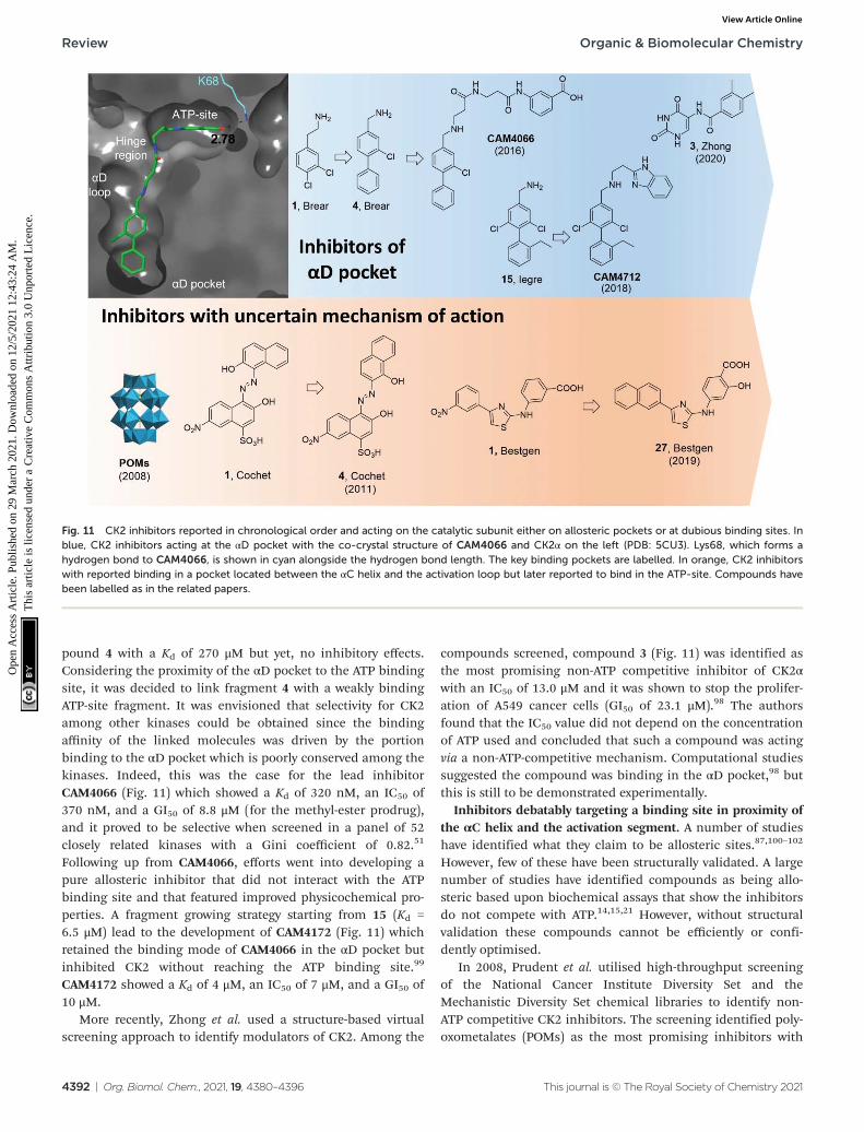
pound 4 with a Kd of 270 μM but yet, no inhibitory effects.Considering the proximity of the αD pocket to the ATP bindingsite, it was decided to link fragment 4 with a weakly bindingATP-site fragment. It was envisioned that selectivity for CK2among other kinases could be obtained since the bindingaffinity of the linked molecules was driven by the portionbinding to the αD pocket which is poorly conserved among thekinases. Indeed, this was the case for the lead inhibitorCAM4066 (Fig. 11) which showed a Kd of 320 nM, an IC50 of370 nM, and a GI50 of 8.8 μM (for the methyl-ester prodrug),and it proved to be selective when screened in a panel of 52closely related kinases with a Gini coefficient of 0.82.51
Following up from CAM4066, efforts went into developing apure allosteric inhibitor that did not interact with the ATPbinding site and that featured improved physicochemical pro-perties. A fragment growing strategy starting from 15 (Kd =6.5 μM) lead to the development of CAM4172 (Fig. 11) whichretained the binding mode of CAM4066 in the αD pocket butinhibited CK2 without reaching the ATP binding site.99
CAM4172 showed a Kd of 4 μM, an IC50 of 7 μM, and a GI50 of10 μM.
More recently, Zhong et al. used a structure-based virtualscreening approach to identify modulators of CK2. Among the
compounds screened, compound 3 (Fig. 11) was identified asthe most promising non-ATP competitive inhibitor of CK2αwith an IC50 of 13.0 μM and it was shown to stop the prolifer-ation of A549 cancer cells (GI50 of 23.1 μM).98 The authorsfound that the IC50 value did not depend on the concentrationof ATP used and concluded that such a compound was actingvia a non-ATP-competitive mechanism. Computational studiessuggested the compound was binding in the αD pocket,98 butthis is still to be demonstrated experimentally.
Inhibitors debatably targeting a binding site in proximity ofthe αC helix and the activation segment. A number of studieshave identified what they claim to be allosteric sites.87,100–102
However, few of these have been structurally validated. A largenumber of studies have identified compounds as being allo-steric based upon biochemical assays that show the inhibitorsdo not compete with ATP.14,15,21 However, without structuralvalidation these compounds cannot be efficiently or confi-dently optimised.
In 2008, Prudent et al. utilised high-throughput screeningof the National Cancer Institute Diversity Set and theMechanistic Diversity Set chemical libraries to identify non-ATP competitive CK2 inhibitors. The screening identified poly-oxometalates (POMs) as the most promising inhibitors with
Fig. 11 CK2 inhibitors reported in chronological order and acting on the catalytic subunit either on allosteric pockets or at dubious binding sites. Inblue, CK2 inhibitors acting at the αD pocket with the co-crystal structure of CAM4066 and CK2α on the left (PDB: 5CU3). Lys68, which forms ahydrogen bond to CAM4066, is shown in cyan alongside the hydrogen bond length. The key binding pockets are labelled. In orange, CK2 inhibitorswith reported binding in a pocket located between the αC helix and the activation loop but later reported to bind in the ATP-site. Compounds havebeen labelled as in the related papers.
Review Organic & Biomolecular Chemistry
4392 | Org. Biomol. Chem., 2021, 19, 4380–4396 This journal is © The Royal Society of Chemistry 2021
Ope
n A
cces
s A
rtic
le. P
ublis
hed
on 2
9 M
arch
202
1. D
ownl
oade
d on
12/
5/20
21 1
2:43
:24
AM
. T
his
artic
le is
lice
nsed
und
er a
Cre
ativ
e C
omm
ons
Attr
ibut
ion
3.0
Unp
orte
d L
icen
ce.
View Article Online

IC50 < 10 nM and the highest inhibition obtained forK6[P2Mo18O62].
103 Doubts remained around the mechanism ofaction of this probe which is known to hydrolyse in the assaymedia into the individual MoO4
2− and PO43− components
which proved inactive when tested individually. Interestingly,degradation was not observed when POM was in the presenceof CK2. The lead POM compound showed exquisite selectivityfor CK2 when tested in a panel of 29 Ser/Thr kinases. Whenthe mechanism of action of POMs was investigated moreclosely with steady-state kinetic analysis, it was found thatPOMs were not ATP site or peptide site-directed inhibitors.Kinase assays with CK2 holoenzyme, affinity chromatography,trypsin proteolysis, and site-directed mutagenesis excludedbinding of POMs to the α/β interface and confirmed lack ofbinding at the ATP/peptide-binding pocket. In addition, it wasfound that mutations around the Gly-rich loop, helix αC, andthe activation segment weakened the sensitivity of CK2 toPOM inhibition. Despite all these studies, a clear binding sitefor POMs has not yet been identified. Similarly, the efficacy ofPOMs on CK2 inhibition in a cellular context remains to beseen.103
In 2011, a screening of some 3000 small molecules fromthe National Cancer Institute Developmental TherapeuticsProgram led Cochet et al. to the identification of azanaptha-lene compounds as hits against CK2α.86 Through SAR studies,hit compound 1 (in Cochet et al.) was modified into com-pound 4 which showed an IC50 of 0.4 μM and it was found notto act via an aggregation mechanism despite its chemicalstructure (Fig. 11). Furthermore, steady-state kinetic analysisshowed that the compound was non-competitive towards ATPand peptide substrate. Mutagenesis studies indicated that resi-dues located on helix αC and the activation segment might bepart of compound 4’s binding site. As seen for the POM inhibi-tors, the lack of further conclusive evidence (NMR orX-crystallography) of the inhibitor in complex with the proteinleaves uncertainty around the actual binding site. The effect ofcompound 4 in cells was also investigated and it was found topromote cell cycle arrest. The compound proved to be selectivefor CK2 with a Gini coefficient of 0.803 when screened at 5 μMin a panel of 42 related kinases.86
Recently, Bestgen et al. reported aryl aminothiazole deriva-tives as allosteric modulators of CK2 by targeting the interfacebetween the αC helix and the glycine-rich loop (G-loop).100,101
Starting from a virtual ligand screening campaign of around2 million compounds, compound 1 (in Bestgen et al.) wasidentified as a hit and it was able to inhibit CK2 with an IC50
of 28 μM. SAR studies initially led to the discovery of com-pound 2 (IC50 of 7 μM) and later to the lead compound 27 (inBestgen et al.) with an IC50 of 0.6 μM, Kd of 0.3 μM, and EC50
of 5 μM (Fig. 11). Enzymatic kinetic studies, native mass spec-troscopy, circular dichroism experiments together with STDNMR studies hinted that compound i was acting as an allo-steric inhibitor with a non-ATP-competitive mechanism ofaction able to stabilise the inactive conformation of CK2.100,101
However, Brear et al. used a combination of crystal structures,competitive ITC and NMR, hydrogen–deuterium exchange
(HDX) mass spectrometry, and computational analyses of theamino thiazole compounds to confirm that these moleculeswere binding to the ATP binding site of the kinase, and henceacted as type II inhibitors.104 Independently, Lindenblatt et al.also obtained co-crystal structures of these compounds withCK2 showing binding to the ATP site and used kinase enzymeassay to demonstrate that these inhibitors do indeed act via anATP-competitive mechanism.105
Modulators of the CK2β subunit
Lastly, another approach to inhibit CK2 outside its catalyticpocket is to interact with the CK2β subunit.
Back in 2006, Cochet et al. made use of a yeast two-hybridapproach to identify molecules that bind at the N-terminus ofCK2β.103 The peptide aptamer identified was an 18-mer uncon-strained peptide (GKMNGVLPLAWPSLYLRL) showing a Kd of0.4 μM in an SPR assay. Interestingly, P1 had high sequencehomology with the cytomegalovirus IE2 protein, known tointeract with CK2β and consequently arrest the cell cycle andtrigger apoptosis (Fig. 1). The peptide identified did neitherdisrupt nor prevent the formation of the CK2 holoenzyme.ELISA assays with truncated versions of CK2β suggested that itwas interacting at a site in between residues 1 and 55 of theN-terminus. Treatment of NIH3T3, HCT116, and MCF-10Acells transfected with GFP-P1 showed typical signs of apoptosisand cell cycle arrest. It was postulated that P1 was able toinduce apoptosis by interacting with the p53-dependent apop-tosis pathways.106 Although P1 represented the first peptide ofthis kind of inhibitor, it should be noted that more mechanis-tic studies would be needed to fully assess its potential. Inaddition, cell-permeable variants will also be needed for P1 tobe regarded as a chemical probe.
Conclusions
In this review we described the key features of CK2, its involve-ment in a variety of diseases with particular focus on cancerand COVID-19, and we aimed to provide the reader with a com-prehensive review on all the chemical probes reported to date.
Despite the numerous efforts gone into developing chemi-cal tools to inhibit such a crucial kinase, clinical candidatesare yet to reach the market. Only one molecule, CX4945, is cur-rently undergoing clinical studies; however, due to its limitedselectivity, even if approved, there will still be a long way to gobefore CK2 can be considered a truly validated target in oncol-ogy. For this reason, scientists have recently shifted their atten-tion to the development of CK2 inhibitors that interact selec-tively with CK2. Strategies have seen the development ofchemical probes acting at sites located outside the ATP-pocketon CK2α, with the site at the interface with CK2β and the αDsite being the most validated. With the increasing amount ofinformation around these sites and advances in biochemicaltechniques, it is reasonable to believe that potent and selectiveCK2 inhibitors with suitable pharmacokinetic and pharmaco-dynamic properties will soon be discovered. In addition,
Organic & Biomolecular Chemistry Review
This journal is © The Royal Society of Chemistry 2021 Org. Biomol. Chem., 2021, 19, 4380–4396 | 4393
Ope
n A
cces
s A
rtic
le. P
ublis
hed
on 2
9 M
arch
202
1. D
ownl
oade
d on
12/
5/20
21 1
2:43
:24
AM
. T
his
artic
le is
lice
nsed
und
er a
Cre
ativ
e C
omm
ons
Attr
ibut
ion
3.0
Unp
orte
d L
icen
ce.
View Article Online
recent advances in chemistry make it possible to foresee thatdifferent types of chemical probes will soon be developedincluding PROTACs acting outside the catalytic box, covalentinhibitors and small molecule/peptide hybrids to target mul-tiple sites simultaneously.
Based upon the explosion of interest in the field of CK2inhibition over the past two decades, it is not then unrealisticto imagine that CK2 inhibition will remain a key and activearea of research over the coming years, as the search for noveltherapeutics for cancer, COVID-19 and a multitude of otherdiseases in which CK2 is implicated continues.
Conflicts of interest
There are no conflicts to declare.
Notes and references
1 F. Ardito, M. Giuliani, D. Perrone, G. Troiano and L. LoMuzio, Int. J. Mol. Med., 2017, 40, 271–280.
2 P. Lahiry, A. Torkamani, N. J. Schork and R. A. Hegele,Nat. Rev. Genet., 2010, 1, 60–74.
3 http://www.kinase-screen.mrc.ac.uk/phosphorylation-ubi-quitylation-drug-discovery Accessed January 2021.
4 G. Manning, D. B. Whyte, R. Martinez, T. Hunter andS. Sudarsanam, Science, 2002, 298, 1912–1934.
5 N. N. Singh and D. P. Ramji, J. Mol. Med., 2008, 86, 227–897.
6 L. A. Pinna, J. Cell Sci., 2002, 115, 3873–3878.7 C. E. Ortega, Y. Seidner and I. Dominguez, PLoS One,
2014, 9, e115609.8 J. Zhang, P. L. Yang and N. S. Gray, Nat. Rev. Cancer, 2009,
9, 28–39.9 A. Siddiqui-Jain, D. Drygin, N. Streiner, P. Chua, F. Pierre,
S. E. O′ Brien, J. Bliesath, M. Omori, N. Huser, C. Ho,et al., Cancer Res., 2010, 70, 10288–10299.
10 M. Bouhaddou, D. Memon, B. Meyer, K. M. White,V. V. Rezelj, M. Correa Marrero, B. J. Polacco, J. E. Melnyk,S. Ulferts, R. M. Kaake, et al., Cell, 2020, 182, 685–712.
11 D. E. Gordon, G. M. Jang, M. Bouhaddou, J. Xu,K. Obernier, K. M. White, M. J. O’Meara, V. V. Rezelj,J. Z. Guo, D. L. Swaney, et al., Nature, 2020, 583, 459–468.
12 R. Prudent and C. Cochet, Chem. Biol., 2009, 16, 112–120.13 Z. A. Knight and K. M. Shokat, Chem. Biol., 2005, 12, 621–
637.14 M. I. Davis, J. P. Hunt, S. Herrgard, P. Ciceri,
L. M. Wodicka, G. Pallares, M. Hocker, D. K. Treiber andP. P. Zarrinkar, Nat. Biotechnol., 2011, 29, 1046–1051.
15 K. Niefind, B. Guerra, I. Ermakowa and O. G. Issinger,EMBO J., 2001, 20, 5320–5331.
16 K. Niefind, J. Raaf and O. G. Issinger, Cell. Mol. Life Sci.,2009, 66, 1800–1816.
17 E. Papinutto, A. Ranchio, G. Lolli, L. A. Pinna andR. Battistutta, J. Struct. Biol., 2012, 177, 382–391.
18 A. C. Bibby and D. W. Litchfield, Int. J. Biol. Sci., 2005, 1,67–79.
19 D. W. Litchfield, Biochem. J., 2003, 369, 1–15.20 O. Filhol, J. L. Martiel and C. Cochet, EMBO Rep., 2004, 5,
351–355.21 G. Poletto, J. Vilardell, O. Marin, M. A. Pagano, G. Cozza,
S. Sarno, A. Falqués, E. Itarte, L. A. Pinna and F. Meggio,Biochemistry, 2008, 32, 8317–8325.
22 J. Raaf, N. Bischoff, K. Klopffleisch, E. Brunstein,B. B. Olsen, G. Vilk, D. W. Litchfield, O.-G. Issinger andK. Niefind, Biochemistry, 2011, 50, 512–522.
23 J. Raaf, E. Brunstein, O.-G. Issinger and K. Niefind, ProteinSci., 2008, 12, 2180–2186.
24 J. H. Trembley, Z. Chen, G. Unger, J. Slaton, B. T. Kren,C. Van Waes and K. Ahmed, BioFactors, 2010, 36, 187–195.
25 S. A. Gibson and E. N. Benveniste, Trends Immunol., 2018,38, 82–85.
26 J. Castello, A. Ragnauth, E. Friedman and H. Rebholz,Pharmaceuticals, 2017, 10(1), 7.
27 S. Koch, C. T. Capaldo, R. S. Hilgarth, B. Fournier,C. A. Parkos and A. Nusrat, Mucosal Immunol., 2013, 6,136–145.
28 E. Ampofo, L. Nalbach, M. D. Menger, M. Montenarh andC. Götz, Int. J. Mol. Sci., 2019, 18, 4398.
29 K. J. Treharne, R. M. Crawford, Z. Xu, J. H. Chen,O. G. Best, E. A. Schulte, D. C. Gruenert, S. M. Wilson,D. N. Sheppard, K. Kunzelmann and A. Mehta, J. Biol.Chem., 2007, 282, 10804–10813.
30 L. R. Cruz, I. Baladrón, A. Rittoles, P. A. Díaz, R. Santana,M. M. Vázquez, A. García, D. Chacón, G. Perera,A. González, et al., ACS Oharmacol. Transl. Sci., 2021, 4(1),206–212.
31 R. Turner, BeingWell Online, https://medium.com/being-well/is-silmitasertib-the-covid-19-treatment-breakthrough-weve-been-waiting-for-263cf9c696d5 (accessed January2021).
32 D. Hanahan and R. A. Weinberg, Cell, 2011, 144, 646–674.33 D. Hanahan and R. A. Weinberg, Cell, 2000, 100,
57–70.34 R. A. Faust, G. Niehans, M. Gapany, D. Hoistad, D. Knapp,
D. Cherwitz, A. Davis, G. L. Adams and K. Ahmed,Int. J. Biochem. Cell Biol., 1999, 31, 941–949.
35 J. H. Trembley, G. Wang, G. Unger, J. Slaton andK. Ahmed, Cell. Mol. Life Sci., 2009, 66, 1858–1867.
36 R. A. Faust, M. Gapany, P. Tristani, A. Davis,G. L. Adams and K. Ahmed, Cancer Lett., 1996, 101, 31–35.
37 S. Yenice, A. T. Davis, S. A. Goueli, A. Akdas, C. Limas andK. Ahmed, Prostate, 1994, 24, 11–16.
38 G. Seitz, U. Münstermann, H. R. Schneider andO. G. Issinger, Biochem. Biophys. Res. Commun., 1989, 163,635–641.
39 M. Gapany, R. A. Faust, S. Tawfic, A. Davis, G. L. Adamsand K. Ahmed, Mol. Med., 1995, 1, 659–666.
40 M. Chua, C. Ortega, A. Sheikh, M. Lee, H. Abdul-Rassoul,K. Hartshorn and I. Dominguez, Pharmaceuticals, 2017,10, 18.
Review Organic & Biomolecular Chemistry
4394 | Org. Biomol. Chem., 2021, 19, 4380–4396 This journal is © The Royal Society of Chemistry 2021
Ope
n A
cces
s A
rtic
le. P
ublis
hed
on 2
9 M
arch
202
1. D
ownl
oade
d on
12/
5/20
21 1
2:43
:24
AM
. T
his
artic
le is
lice
nsed
und
er a
Cre
ativ
e C
omm
ons
Attr
ibut
ion
3.0
Unp
orte
d L
icen
ce.
View Article Online

41 P. O-Charoenrat, V. Rusch, S. G. Talbot, I. Sarkaria,A. Viale, N. Socci, I. Ngai, P. Rao and B. Singh, Clin.Cancer Res., 2004, 10, 5792–5803.
42 M. J. Ku, J. W. Park, B. J. Ryu, Y. J. Son, S. H. Kim andS. Y. Lee, Bioorg. Med. Chem. Lett., 2013, 23, 5609–5613.
43 F. Benavent Acero, C. S. Capobianco, J. Garona,S. M. Cirigliano, Y. Perera, A. J. Urtreger, S. E. Perea,D. F. Alonso and H. G. Farina, Lung Cancer, 2017, 107, 14–21.
44 K. Y. Lin, C. L. Fang, Y. Chen, C. F. Li, S. H. Chen,C. Y. Kuo, C. Tai and Y. H. Uen, Ann. Surg. Oncol., 2010,17, 1695–1702.
45 J. Zou, H. Luo, Q. Zeng, Z. Dong, D. Wu and L. Liu,J. Transl. Med., 2011, 9, 97.
46 F. Zhou, J. Xu, G. Ding and L. Cao, Pathol. Oncol. Res.,2014, 20, 73–79.
47 G. Di Maira, A. Gentilini, M. Pastore, A. Caligiuri,B. Piombanti, C. Raggi, E. Rovida, M. Lewinska,J. B. Andersen, C. Borgo, et al., Oncogenesis, 2019, 8, 61.
48 M. Bouhaddou, D. Memon, B. Meyer, K. M. White,V. V. Rezelj, M. C. Marrero, B. J. Polacco, J. E. Melnyk,S. Ulferts, R. M. Kaake, et al., Cell, 2020, 182, 685–712.
49 G. M. Hathaway, T. H. Lubben and J. A. Traugh, J. Biol.Chem., 1980, 255, 8038–8041.
50 H. Yim, Y. H. Lee, C. H. Lee and S. K. Lee, Planta Med.,1999, 65, 009–013.
51 C. De Fusco, P. Brear, J. Iegre, K. H. Georgiou, H. F. Sore,M. Hyvönen and D. R. Spring, Bioorg. Med. Chem., 2017,13, 3471–3482.
52 J. E. Dowling, M. Alimzhanov, L. Bao, C. Chuaqui,C. R. Denz, E. Jenkins, N. A. Larsen, P. D. Lyne, T. Pontz,Q. Ye, G. A. Holdgate, L. Snow, N. O’Connell andA. D. Ferguson, ACS Med. Chem. Lett., 2016, 7, 300–305.
53 R. Zandomeni, M. C. Zandomeni, D. Shugar andR. Weinmann, J. Biol. Chem., 1986, 261, 3414–3419.
54 S. Sarno, E. de Moliner, M. Ruzzene, M. A. Pagano,R. Battistutta, J. Bain, D. Fabbro, J. Schoepfer, M. Elliott,P. Furet, F. Meggio, G. Zanotti and L. A. Pinna, Biochem. J.,2003, 374, 639–646.
55 F. Meggio, M. A. Pagano, S. Moro, G. Zagotto, M. Ruzzene,S. Sarno, G. Cozza, J. Bain, M. Elliott, A. D. Deana,A. M. Brunati and L. A. Pinna, Biochemistry, 2004, 43,12931–12936.
56 Z. Nie, C. Perretta, P. Erickson, S. Margosiak, R. Almassy,J. Lu, A. Averill, K. M. Yager and S. Chu, Bioorg. Med.Chem. Lett., 2007, 17, 4191–4195.
57 J. E. Dowling, C. Chuaqui, T. W. Pontz, P. D. Lyne,N. A. Larsen, M. H. Block, H. Chen, N. Su, A. Wu,D. Russell, et al., ACS Med. Chem. Lett., 2012, 3, 278–283.
58 E. L. Atkinson, J. Iegre, P. D. Brear, E. A. Zhabina,M. Hyvönen and D. R. Spring, Molecules, 2021, 26,1977.
59 T. Oshima, Y. Niwa, K. Kuwata, A. Srivastava, T. Hyoda,Y. Tsuchiya, M. Kumagai, M. Tsuyuguchi, T. Tamaru,A. Sugiyama, N. Ono, N. Zolboot, Y. Aikawa, S. Oishi,A. Nonami, F. Arai, S. Hagihara, J. Yamaguchi, F. Tama,
Y. Kunisaki, K. Yagita, M. Ikeda, T. Kinoshita, S. A. Kay,K. Itami and T. Hirota, Sci. Adv., 2019, 5, 9060–9083.
60 A. Dalle Vedove, F. Zonta, E. Zanforlin, N. Demitri,G. Ribaudo, G. Cazzanelli, A. Ongaro, S. Sarno, G. Zagotto,R. Battistutta, M. Ruzzene and G. Lolli, Eur. J. Med. Chem.,2020, 195, 112267.
61 C. Wells, D. H. Drewry, J. E. Pickett, D. W. Litchfield,S. Knapp and A. D. Axtman, Cell Chem. Biol., 2021, DOI:10.1016/j.chembiol.2020.12.013.
62 B. Kobe, T. Kampmann, J. K. Forwood, P. Listwan andR. I. Brinkworth, Biochim. Biophys. Acta, ProteinsProteomics, 2005, 1754, 200–209.
63 F. Meggio, L. A. Pinna, F. Marchiori and G. Borin, FEBSLett., 1983, 162, 235–238.
64 F. Meggio, F. Marchiori, G. Borin, G. Chessa andL. A. Pinna, J. Biol. Chem., 1984, 259, 14576–14579.
65 F. Meggio and L. A. Pinna, Biochim. Biophys. Acta, Mol.Cell Res., 1989, 1010, 128–130.
66 R. Tellez, M. Gatica, C. C. Allende and J. E. Allende, FEBSLett., 1990, 265, 113–116.
67 R. Tellez, C. C. Allende and J. E. Allende, FEBS Lett., 1992,308, 113–115.
68 Y. Perera, H. G. Farina, I. Hernandez, O. Mendoza,J. M. Serrano, O. Reyer, D. E. Gomez, R. E. Gomez,B. E. Acevedo, D. F. Alfonso and S. E. Perea, Int. J. Cancer,2008, 122, 57–62.
69 S. E. Perea, O. Reyes, Y. Puchades, O. Mendoza,N. S. Vispo, I. Torrens, A. Santos, R. Silva, B. Acevedo,E. López, V. Falcón and D. F. Alonso, Cancer Res., 2004,64, 7127–7129.
70 M. F. Gottardo, C. S. Capobianco, J. E. Sidabra, J. Garona,Y. Perera, S. E. Perea, D. F. Alonso and H. G. Farina, Sci.Rep., 2020, 10, 14689.
71 M. Winiewska-Szajewska, D. Płonka, I. Zhukov andJ. Poznański, Sci. Rep., 2019, 9, 11018.
72 G. Cozza, S. Zanin, S. Sarno, E. Costa, C. Girardi,G. Ribaudo, M. Salvi, G. Zagotto, M. Ruzzene andL. A. Pinna, Biochem. J., 2015, 471, 415–430.
73 E. Enkvist, K. Viht, N. Bischoff, J. Vahter, S. Saaver,G. Raidaru, O. G. Issinger, K. Niefind and A. Uri, Org.Biomol. Chem., 2012, 10, 8645–8653.
74 K. Viht, S. Saaver, J. Vahter, E. Enkvist, D. Lavogina,H. Sinijärv, G. Raidaru, B. Guerra, O. G. Issinger andA. Uri, Bioconjugate Chem., 2015, 26, 2324–2335.
75 R. Y. Tsien, Nature, 1981, 290, 527–528.76 J. Vahter, K. Viht, A. Uri and E. Enkvist, Bioorg. Med.
Chem., 2017, 25, 2277–2284.77 T. N. de Villavicencio-Diaz, Y. Mazola, Y. P. Negrín,
Y. C. García, O. G. Cruz and S. E. Perea Rodríguez,Biochem. Biophys. Rep., 2015, 4, 20–27.
78 F. Meggio, D. Shugar and L. A. Pinna, Eur. J. Biochem.,1990, 187, 89–94.
79 J. Iegre, P. Brear, D. J. Baker, Y. S. Tan,E. L. Atkinson, H. F. Sore, D. H. O’Donovan, C. S. Verma,M. Hyvönen and D. R. Spring, Chem. Sci., 2019, 10, 5056–5063.
Organic & Biomolecular Chemistry Review
This journal is © The Royal Society of Chemistry 2021 Org. Biomol. Chem., 2021, 19, 4380–4396 | 4395
Ope
n A
cces
s A
rtic
le. P
ublis
hed
on 2
9 M
arch
202
1. D
ownl
oade
d on
12/
5/20
21 1
2:43
:24
AM
. T
his
artic
le is
lice
nsed
und
er a
Cre
ativ
e C
omm
ons
Attr
ibut
ion
3.0
Unp
orte
d L
icen
ce.
View Article Online
80 G. Lolli, A. Ranchio and R. Battistutta, ACS Chem. Biol.,2014, 9, 366–371.
81 S. Zanin, C. Borgo, C. Girardi, S. E. O’Brien, Y. Miyata,L. A. Pinna, A. Donella-Deana and M. Ruzzene, PLoS One,2012, 11, e49193.
82 I. Kufareva, B. Bestgen, P. Brear, R. Prudent, B. Laudet,V. Moucadel, M. Ettaoussi, C. F. Sautel, I. Krimm,M. Engel, O. Filhol, M. Le Borgne, T. Lomberget,C. Cochet and R. Abagyan, Sci. Rep., 2019, 9, 15893.
83 J. C. Fuller, N. J. Burgoyene and R. M. Jackson, DrugDiscovery Today, 2009, 14, 155–161.
84 J. Raaf, E. Brunstein, O. G. Issinger and K. Niefind, Chem.Biol., 2008, 2, 111–117.
85 B. Laudet, V. Moucadel, R. Prudent, O. Filhol, Y. S. Wong,D. Royer and C. Cochet, Mol. Cell. Biochem., 2008, 316, 63–69.
86 V. Moucadel, R. Prudent, C. F. Sautel, F. Teillet, C. Barette,L. Lafanechere, V. Receveur-Brechot and C. Cochet,Oncotarget, 2011, 2, 997–1010.
87 L. Kröger, C. G. Daniliuc, D. Ensan, S. Borgert,C. Nienberg, M. Lauwers, M. Steinkrüger, J. Jose,M. Pietsch and B. Wünsch, ChemMedChem, 2020, 15, 871–881.
88 P. Brear, A. North, J. Iegre, K. Hadje Georgiou, A. Lubin,L. Carro, W. Green, H. F. Sore, M. Hyvonen andD. R. Spring, Bioorg. Med. Chem., 2018, 26, 3016–3020.
89 L. Nevola and E. Giralt, Chem. Commun., 2015, 51, 3302–3315.
90 J. A. Wells and C. L. McClendon, Nature, 2007, 450, 1001–1009.
91 B. Laudet, C. Barette, V. Dulery, O. Renaudet, P. Dumy,A. Metz, R. Prudent, A. Deshiere, O. Dideberg, O. Filholand C. Cochet, Biochem. J., 2007, 408, 363–373.
92 B. Bestgen, Z. Belaid-Choucair, T. Lomberget, M. LeBorgne, O. Filhol and C. Cochet, Pharmaceuticals, 2017, 1,16.
93 D. Lindenblatt, M. Horn, C. Götz, K. Niefind, I. Neundorfand M. Pietsch, ChemMedChem, 2019, 14, 833–841.
94 S. Tang, N. Zhang, Y. Zhou, W. A. Cortopassi,M. P. Jacobson, L. Zhao and R. Zhong, Mol. Inf., 2019, 38,1800089.
95 J. Hochscherf, D. Lindenblatt, M. Steinkrüger, E. Yoo,Ö. Ulucan, S. Herzig, O.-G. G. Issinger, V. Helms, C. Götz,I. Neundorf, K. Niefind and M. Pietsch, Anal. Biochem.,2015, 468, 4–14.
96 I. Neundorf, R. Rennert, J. Hoyer, F. Schramm, K. Löbner,I. Kitanovic and S. Wölfl, Pharmaceuticals, 2009, 2,49–65.
97 P. Brear, C. De Fusco, K. Hadje-Georgiou, N. J. Francis-Newton, C. J. Stubbs, H. F. Sore, A. R. Venkitaraman,C. Abell, D. R. Spring and M. Hyvönen, Chem. Sci., 2016,7, 6839–6845.
98 C. Li, X. Zhang, N. Zhang, Y. Zhou, G. Sun, L. Zhao andR. Zhong, Molecules, 2020, 25, 237.
99 J. Iegre, P. Brear, C. De Fusco, M. Yoshida, S. L. Mitchell,M. Rossmann, L. Carro, H. F. Sore, M. Hyvönen andD. R. Spring, Chem. Sci., 2018, 9, 3041–3049.
100 B. Bestgen, I. Krimm, I. Kufareva, A. A. M. Kamal,W. G. Seetoh, C. Abell, R. W. Hartmann, R. Abagyan,C. Cochet, M. Le Borgne, M. Engel and T. Lomberget,J. Med. Chem., 2019, 62, 1803–1816.
101 B. Bestgen, I. Kufareva, W. Seetoh, C. Abell,R. W. Hartmann, R. Abagyan, M. Le Borgne, O. Filhol,C. Cochet, T. Lomberget and M. Engel, J. Med. Chem.,2019, 62, 1817–1836.
102 X. Chen, C. Li, D. Wang, Y. Chen and N. Zhang, Molecules,2020, 25, 870.
103 R. Prudent, V. Moucadel, B. Laudet, C. Barette,L. Lafanechère, B. Hasenknopf, J. Li, S. Bareyt, E. Lacôte,S. Thorimbert, M. Malacria, P. Gouzerh and C. Cochet,Chem. Biol., 2008, 15, 683–692.
104 P. Brear, D. Ball, K. Stott, S. D’Arcy and M. Hyvönen,J. Med. Chem., 2020, 21, 12786–12798.
105 D. Lindenblatt, A. Nickelsen, V. M. Applegate, J. Jose andK. Niefind, J. Med. Chem., 2020, 14, 7766–7772.
106 V. Martel, O. Filhol, P. Colas and C. Cochet, Oncogene,2006, 25, 7343–7353.
107 B. Laudet, C. Barette, V. Dulery, O. Renaudet, P. Dumy,A. Metz, R. Prudent, A. Deshiere, O. Dideberg, O. Filholand C. Cochet, Biochem. J., 2007, 408, 363–373, 17714077.
108 S. Tang, N. Zhang, Y. Zhou, W. A. Cortopassi andM. P. Jacobson, Mol. Inf., 2018, 38, 1800089.
Review Organic & Biomolecular Chemistry
4396 | Org. Biomol. Chem., 2021, 19, 4380–4396 This journal is © The Royal Society of Chemistry 2021
Ope
n A
cces
s A
rtic
le. P
ublis
hed
on 2
9 M
arch
202
1. D
ownl
oade
d on
12/
5/20
21 1
2:43
:24
AM
. T
his
artic
le is
lice
nsed
und
er a
Cre
ativ
e C
omm
ons
Attr
ibut
ion
3.0
Unp
orte
d L
icen
ce.
View Article Online