virtual microscopy system - olympus corporation · a real slide-mounted sample and microscope can...
TRANSCRIPT

For Research and Education purposes only.
VS110™
Virtual Microscopy System

EXPERIENCE A WHOLE NEW DIMENSION... of digital imagingMicroscopy and image analysis are generally considered to be two separate processes, the former feeding into the latter. In some fields though, such as pathology, the two are synonymous, with analyzing preparations directly through the eyepiece. This is due to the “look and feel” that only a real slide-mounted sample and microscope can offer. The VS110 systems from Olympus bring microscopy and imaging closer together. The result is an advanced and extremely versatile “virtual” slide, which is a copy of the real specimen. Furthermore, “virtual” microscopy generates a high-resolution image of the whole specimen, which can be viewed and analyzed from the overview image at anything from low magnification up to maximum magnification via seamless zoom. Stored electronically on a central server, samples can be evaluated around the world instantly and simultaneously – the future of imaging is here.
VS110 IN RESEARCH AND EDuCATION 4–11Virtual microscopy enables the information from one glass slide to be shared across the globe for teleconsultation, supraregional collaboration, archiving of slide materials and teaching. The VS110 features include: remote access to virtual slides with the functionality of a microscope, eliminating the need for the instrument itself, and the ability to see and analyze macro overviews of slides not possible using traditional microscopes.
VS110 – FuNCTIONS, DATA MANAgEMENT 12–19 AND SySTEMAll of the VS110 system components are designed to interact seamlessly, producing a fully automated high-speed scanning system with excellent flexibility and simple operation. This ensures that users get the best results for each type of specimen with the minimum of effort. The Net Image Server SQL for the Olympus VS110 system is a real client-server-based data management system and storage solution developed especially for microscopy. This, together with the VS110 platform, assists the user by keeping everything clearly structured and easy to manage and ensures efficient communication through images and data.
For Research and Education purposes only.
60 µm thick section of bone examined using polarized transmitted light and a sensitively tinted auxiliary quartz workpiece. The bone section was taken from the shaft of the left femur of a 20–30 year-old woman from the Neolithic burial ground in Wandersleben (Thuringia, Germany) who suffered from chronic inflammation of the bones of the lower extremity.
Image courtesy of Prof. Michael Schultz, University of Göttingen, Germany.
2 CONTENTS

Sample preparation
Sample digitalization
Evaluation and teleconsultation
VIRTuAL MICROSCOPyWith the increasing demand for collaboration and networking in hospitals comes the need for the correct tools. The new VS110 concept from Olympus has introduced a unique high-speed, whole-slide scanning system, producing digital slide images at high-end microscope quality. The user can now work more flexibly and efficiently to evaluate slides and consult colleagues at separate times and in different locations. Virtual slide Taking a digital copy of an entire slide using the VS110 system increases the flexibility of analysis. This unique ability is due to the careful combination of precision optical components, the high-quality scanning stage and the camera with advanced application software. The whole picture The careful integration of all the components creates a highly flexible system that enables the user to quickly and effortlessly acquire digital slides. The acquisition process starts with generating an overview image of the entire slide. It includes the automatic detection of the specimen to avoid time-consuming scanning of areas where there is no sample on the slide. As a second step, the high-magnification scan is automatically performed over the whole slide or over smaller, predefined areas of interest using the preselected objective. During the acquisition, thousands of individual images are simultaneously compressed without any loss in quality and stitched together to create a seamless, magnified version of the slide. As a result, this is carried out within minutes, even using high-magnification objectives. High-capacity and automatic throughput Many research and teaching laboratories are faced with a growing number of samples to process and it is therefore more efficient to scan slides in batches and review them later. The Olympus VS110 system can be fitted with a slide loader, giving full traceability and hands-off scanning. This capability also makes it quick and easy to archive slides and produce permanent records. Scan it right With the Olympus VS110, users will be guided through the entire virtual slide acquisition process step-by-step by an intuitive Scan Wizard. Full-slide scanning can be set up with just three mouse clicks. With the VS110, it also is possible to review the overview slide images and assign individual magnifications, scan areas and other parameters to the slides, even in a batch scan. Although several modes arepredefined, the advanced software allows experienced users to be in full control of the details of the scanning process. Therefore, different scan settings can be assigned to individual slides, saving significant scanning time, as well as offering the possibility of only scanning areas of interest, reducing the data file size.
VS110 IN RESEARCH AND EDuCATIONMoving beyond conventional digital microscopyVirtual slide microscopy offers researchers and students in professional education the unique potential to acquire complete slides at high magnification and resolution to enable review, analysis and archiving of samples for discussion both remotely and in online conferences. Virtual microscopy offers new ways of teaching students to analyze pathology samples by enabling the tutor to work with all students in real time and to analyze the same virtual sample, as if they were using a real microscope. This flexibility of virtual slide technology within all clinical teaching environments enables users to move beyond the limits of conventional digital microscopy and telepathology in terms of both the size and the resolution of image files that can be discussed, as well as in terms of time and location of discussion with colleagues.
4 ChAPTER 1 VS110 IN RESEARCh AND EDUCATION

Online conferencing in real time,regardless of location
Worldwide accessA The virtual slide image is stored with all the relevant information in such a way that it enables instant, controlled access from anywhere in the world. This makes it an excellent tool for remote collaboration, second-opinion consultations and group discussions. The system provides each user with an easy-to-use on-screen control interface, allowing free navigation within the sample. Moreover, users can control multiple virtual slides and/or other image file formats simultaneously for easy comparison. In this way, they can access virtual slides from wherever they are and therefore do not need to be in their office, or even their home country, to screen a slide. Meeting room
B More importantly, as well as giving individual access to the virtual slides, the system also enables collaboration around the globe via the access-controlled conferencing tool. This allows users to be assigned different roles such as moderator, speaker or client. Therefore, second opinions can be requested or difficult case discussions can happen immediately and in real time, with the added benefit of annotation, highlighting and minute taking – a truly revolutionary tool ensuring a new level of efficiency. Laboratory crosstalkC The integrated, versatile Net Image Server SQL expands the VS110 slide scanner family with a client-server database and allows users to manage any kind of image in a simple and convenient way. This powerful tool enables scanned images to be automatically uploaded to the database, making them readily available for immediate remote access and multiple keyword queries. Customized database fields and user-defined database structure offer greater flexibility, and Net Image Server SQL also supports multiple file repository systems to allow secure, easy networking between different scanning units within one database. Connecting to laboratory environment
D The Olympus VS110 software offers the possibility of converting standard image file formats into JPEG2000 images after image acquisition, which dramatically reduces the image size and allows compatibility with many software systems.
Storage and preservationAccess and security E When handling a large number of virtual slides, high-quality, secure and rapid access data storage is an important consideration. Olympus, therefore, has worked with leading electronic storage providers to establish local, regional and international data access and storage network capabilities. With patients’ samples, privacy is paramount and the system therefore has integrated security settings, giving controlled access to the slide image and related data. The old and the new A clear advantage of electronic storage is that slides can be found and loaded quickly and easily, enabling users to rapidly reopen a slide for further investigation, access slides for reference purposes or prepare presentations for conferences and lectures. Furthermore, there is no degradation of the sample over time – the slide image will always be as crisp and clear as the day it was recorded. This is especially advantageous when archiving old or rare samples, which then can be made available to everyone as if they had been copied. Looking to the future The advances of the VS110 system are based on the versatile nature of Olympus microscopes, which means it can be upgraded to match future requirements. Whatever applies to the microscope also applies to future analysis of digitized specimens. With the ongoing development of the application software, it is expected that computer-assisted analysis software will become available which will help research pathologists to improve the detection and interpretation of tumors and other disease patterns.
Virtual digital slides can be easilyaccessed and viewed with an Internet browser
Guided by annotations and “views,”colleagues and collaborators can easily identify regions of interest (ROIs)
Assignment of user roles and access rights
E user management
The database window: database struc-ture, image gallery, image information and query tool window at a glance
Distribution of virtual slide information
D Network
NIS server
Communication
JPEg 2000
IMAgE STORAgE LIMS ARCHIVE
Users
A
B
B
C
VS110 IN RESEARCh AND EDUCATION6
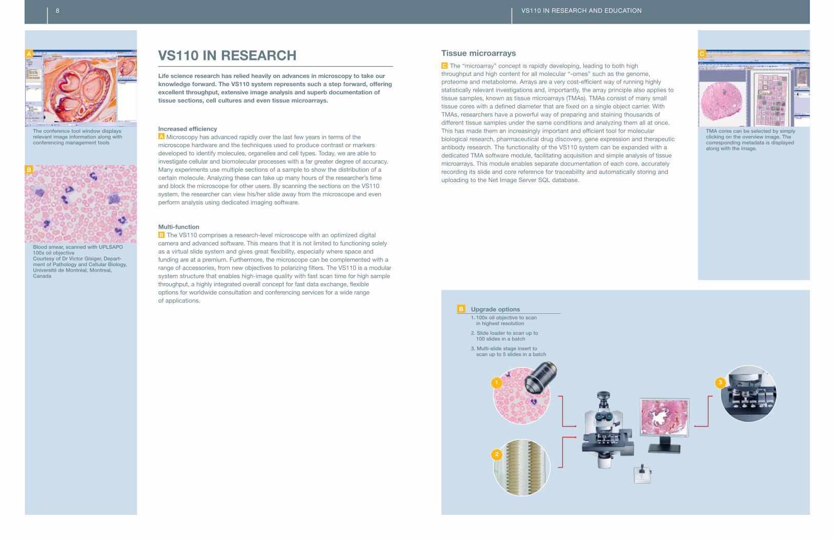
Tissue microarraysC The “microarray” concept is rapidly developing, leading to both high throughput and high content for all molecular “-omes” such as the genome, proteome and metabolome. Arrays are a very cost-efficient way of running highly statistically relevant investigations and, importantly, the array principle also applies to tissue samples, known as tissue microarrays (TMAs). TMAs consist of many small tissue cores with a defined diameter that are fixed on a single object carrier. With TMAs, researchers have a powerful way of preparing and staining thousands of different tissue samples under the same conditions and analyzing them all at once. This has made them an increasingly important and efficient tool for molecular biological research, pharmaceutical drug discovery, gene expression and therapeutic antibody research. The functionality of the VS110 system can be expanded with a dedicated TMA software module, facilitating acquisition and simple analysis of tissue microarrays. This module enables separate documentation of each core, accurately recording its slide and core reference for traceability and automatically storing and uploading to the Net Image Server SQL database.
VS110 IN RESEARCh AND EDUCATION
VS110 IN RESEARCHLife science research has relied heavily on advances in microscopy to take our knowledge forward. The VS110 system represents such a step forward, offering excellent throughput, extensive image analysis and superb documentation of tissue sections, cell cultures and even tissue microarrays.
Increased efficiency A Microscopy has advanced rapidly over the last few years in terms of the microscope hardware and the techniques used to produce contrast or markers developed to identify molecules, organelles and cell types. Today, we are able to investigate cellular and biomolecular processes with a far greater degree of accuracy. Many experiments use multiple sections of a sample to show the distribution of a certain molecule. Analyzing these can take up many hours of the researcher’s time and block the microscope for other users. By scanning the sections on the VS110 system, the researcher can view his/her slide away from the microscope and even perform analysis using dedicated imaging software. Multi-function B The VS110 comprises a research-level microscope with an optimized digital camera and advanced software. This means that it is not limited to functioning solely as a virtual slide system and gives great flexibility, especially where space and funding are at a premium. Furthermore, the microscope can be complemented with a range of accessories, from new objectives to polarizing filters. The VS110 is a modular system structure that enables high-image quality with fast scan time for high sample throughput, a highly integrated overall concept for fast data exchange, flexible options for worldwide consultation and conferencing services for a wide range of applications.
Blood smear, scanned with UPLSAPO 100x oil objective Courtesy of Dr Victor Gisiger, Depart-ment of Pathology and Cellular Biology, Université de Montréal, Montreal, Canada
TMA cores can be selected by simply clicking on the overview image. The corresponding metadata is displayed along with the image.
1. 100x oil objective to scan in highest resolution
2. Slide loader to scan up to 100 slides in a batch
3. Multi-slide stage insert to scan up to 5 slides in a batch
B upgrade options
The conference tool window displays relevant image information along with conferencing management tools
A
B
C
1 3
2
8

VS110 IN RESEARCh AND EDUCATION
Live discussions The OlyVIA viewer software includes tools to add annotations and notes, record files and directly link them to the slide. This enables the tutor to prepare images and guide students through each key feature. Furthermore, the software enables users to navigate multiple images simultaneously for easy comparision, ideal for teaching and conferencing purposes. Easy access C Scanned images are automatically uploaded to a database, resulting in virtual slides being readily available for immediate remote access. This means that it is possible to access and view slides stored anywhere on the Web, which is ideal for whole-class consistency in homework and tests, as well as allowing courses and assessments to be organized quickly and easily. Easy upload D The advanced software technology also allows the easy and quick uploading of virtual slide files to educational institutes’ homepages, enabling students to quickly access slides via fast intra-networks with integrated security settings, giving tutors controlled access to slide images and related data.
VS110 IN EDuCATIONTeaching new students to analyze pathology samples is an important process. It, however, is beset with the difficult decision of how best to demonstrate the differences between normal samples and the many different disease stages that exist for each type of tissue. up until now, this choice has been restricted to either giving each student a different, but very similar slide, so that they can get the feel of working with a whole sample, or presenting a single image of one sample via a projector or online tutorial. Both of these options have severe limitations for teaching in groups. With the introduction of the Olympus VS110 system, however, this has now changed. Whole-class access A Each student can review the same sample and control the position and magnification individually – as if it were a real slide. The tutor also can work with all the students in real-time, showing them how best to analyze effectively and pick out rare cases, or test all students on the same slide. Real-time viewing B The conferencing facility allows teachers to set up class discussions using synchronized screens that enable all students to see and discuss individual slides at the same time. Students can participate in the conference by using the specialized free OlyVIA® software.
Designed for multi-user access
A Whole-class access
Fast and easy creation of online content
D Easy upload
Instant information distribution
C Easy access
Synchronized screen conferencingfacility
B Real-time viewing
10

VS110 – THE FuNCTIONSThe real power of the VS110 software lies in its functionality. It is designed to provide all the features needed to make both education and research use as easy as possible. These functions are presented to the user via an intuitive and easy-to-use interface. Easy to use A The user is guided step-by-step through the virtual slide acquisition process by an intuitive Scan Wizard. This graphical user interface (GUI) features large control icons and enables even inexperienced users to immediately produce the perfect image results they require – in just a few steps and with minimal training! Several modes are predefined, like the quick mode which automatically acquires the entire slide, or to control all the settings during the acquisition completely. Region definition B To define the area to be scanned, the user can identify a region of interest (ROI) using the simple on-screen freehand tool to outline the area to be scanned, or leave the system to automatically detect the extent of the sample or samples. VS110 supports automatic sample detection, which reduces the data size by only scanning the areas needed. The freehand tool further allows the user to reduce the scan area to the minimum required to save time and hard disk space. Keeping focused C To make the scan process as fast as possible, the software generates a focus map using the fast, software-based autofocus function. This autofocus capability allows the optimal z-position to be determined and saved on various parts of the sample, permitting a height profile of the sample to be compiled before acquiring the detailed scans. Different maps are used depending on the type of sample being scanned, providing the best solution whatever the specimen. Numerous coordinates are selected to create the map, which gives the system an instant focus reference when scanning the slide. High speed and resolution Including the generation of the focus map, it takes less than two minutes for the VS110 to scan a 10 x 10 mm sample area using a 20x objective with a 0.63x camera adapter. Moreover, VS110 scans with outstanding resolution at 0.16 µm/pixel at 40x.
The clear Scan Wizard guides the user step-by-step through the scanning process
A Scan Wizard
The scan area can be defined by rectangles, circles and polygons
The focus map calculates the specimen’s topography to ensure precise focusing while scanning and superb scanning results
C Focus map
B
VS110 – FUNCTIONS, DATA MANAGEMENT AND SYSTEM
VS110 – FuNCTIONS, DATA MANAgEMENT AND SySTEMLeveraging potentialThe VS110 system is a versatile platform for clinical research, teaching and online conferences. It offers an intuitive scanning wizard, fast-acquisition speed, high-resolution images with brilliant image quality, secure image and user management and straightforward creation of online image galleries. In addition to these features, the VS110 system also offers annotation functions, fast, easy navigation, reviewed batch scan and the flexibility offered by a whole range of additional modules. The system consists of robust and carefully selected hardware integrated with the Olympus BX® upright microscope, offering the user reliability and flexibility.
ChAPTER 212

Memory and file management With such high-quality images being produced for each slide, file storage and access are very important considerations with the VS110 system. The special memory management overcomes the limitations of the operating system and can handle the very large images generated by the scanning system. This has made it possible to acquire images with large uncompressed file sizes. The first step used by VS110 is on-the-fly lossless compression, which means that images can be compressed to a tenth of their original size. Secondly, with the VS110 system fully prepared for mass data transfer and storage, it can be easily integrated with ultra high-capacity storage systems, for which Olympus works with the leading companies in the electronic storage market. Measuring functions E The Olympus VS110 software offers a wide range of measurement functions. Measurement results are saved together with the image in the “measurements layer,” and are displayed in a sheet which can be exported in XLS format for further editing in a spreadsheet program, such as Microsoft Excel. Access everywhere F Virtual slides can be accessed via a number of different methods and can be published to online databases in a special file format to enable very easy teleconsultation or international collaboration. Similarly, the virtual slide can be recreated on DVD so that it can be distributed to colleagues and students without the need for specialist viewing software. The system ensures the ideal properties for processing, data transfer and image display, since parameters such as resolution are automatically adjusted to what is needed for the user’s monitor. In this way, there is no time delay between changing the view and seeing the image.
Multi-layer images A A multi-layer image is made up of two or more image layers. Each of these layers contains the microscope image of a sample (or part of one) acquired at a different magnification. This means that if an image is acquired at a magnification of 2x and 10x, an image will have been created with two image layers. Furthermore, several regions of interest that are scanned with the same or different objectives can be combined in one image. The software manages the images in such a way that the image layer with the highest magnification will be displayed automatically. This ensures that users will always obtain the maximum available image information without needing to keep in mind which image layers exist. Real virtual microscopy B The VS110 system is capable of scanning multiple large specimens in up to 15 z-planes. By selecting the Virtual-Z™ scan mode, the reviewer can just simply focus through the sample, as well as examining regions of interest in different dimensions. At each position of the whole sample, the VS110 system acquires a series of images at various focus positions, and these z-stacks are stored within the images and can be focused virtually on the monitor. This enables better observation for remote conferencing, as well as consistent training for students and pathologists. Always sharp C For evaluation of distinct structures, such as the shapes of cell nuclei or complex specimens with different types of tissue, or for the analysis of thick samples, the extended focal imaging (EFI) feature can improve the image quality. The software produces a perfectly focused image with greatly increased depth of field at any point within the virtual sample. This is achieved by taking a z-series of images through the sample and combining them, in focus elements of each other, into a single ultra sharp image. Immediate review The image is displayed without delay on the screen while being scanned and can be immediately reviewed without affecting the acquisition. This is due to the high-performance multi-threading Olympus software. Annotations can be immediately added to the file in the form of shapes, arrows and text, while audio commentary and an automated scale bar can be displayed in the image. Easy navigation D The multi-function screen not only displays the digital slide image, but also relevant data and an overview image that can be used for navigation, along with buttons for x and y movements and zooming. The Properties tool window also shows any additional information about the file being displayed.
The unique multi-layer format allows the scanning and combining of multiple scanning areas in one image. Colored frames outline different resolutions and z-stack scans in the overview image.
The dynamic algorithm of the Virtual-Z scan mode follows the 3-D structure of the specimen. The resulting image is in focus – plane by plane.
B Virtual-Z™
Aspect of whole mouse embryo,20x magnificationEFI image (left), non-EFI image (right)
The navigation window allows easyorientation and navigation within the slide
A large set of measurement functions is available in the software
E
Virtual slides can be stored in a Web format and be viewed with an Internet browser
A
C
D
F
VS110 – FUNCTIONS, DATA MANAGEMENT AND SYSTEM14

VS110 – FUNCTIONS, DATA MANAGEMENT AND SYSTEM
Security/access rights D In some cases, the security of data is not just important, but essential, and therefore NIS SQL has a number of solutions to provide watertight protection. Access to a project or to individual project data can be restricted to a group or to individual users. NIS SQL offers standard authentication via the SQL or Windows® authentication login with username and password, meaning that it will accept the user’s Windows login for single sign-on (SSO) access. For the ultimate security, a secure file repository is established by default, which ensures that files are only visible to users with permission to access them. Therefore, there is no way for unauthorized users to know that the files exist – even if they are using Windows Explorer. Furthermore, passwords and privileges can be set by the main user so that team members, collaborators and students can have differing access to the files. This means that changes can only be made by authorized people. System integration E NIS SQL can be set up to interface directly and exchange information with existing LIMS (Laboratory Information Management Software). The software is designed to integrate with existing data sources on a network. As a result, existing data, such as slide properties and related information, is available at every VS110 workstation. Furthermore, optional JPEG2000 conversion of virtual slide images makes them compatible with many other software solutions.
VS110 – DATA MANAgEMENTWith a growing amount of data to manage, multiple users and the desire for greater data security, conventional data managing solutions quickly become inadequate. The Net Image Server (NIS) SQL for the Olympus VS110 system is a versatile image access and storage solution for digital virtual microscopy. The NIS SQL is a real client-server-based data management system developed especially for microscopy and ensures that even with large amounts of data and many users workflows run efficiently and securely. Data management When image acquisition plays a major role, storing and maintaining traceability on individual data sources such as images, reports and analytical results becomes extremely important, as does the sharing of images and information – independent of time and place. Therefore, the NIS SQL platform assists the user by keeping everything clearly structured, easy to manage and ensures efficent communication through images and data via an independent Web browser or free-of-charge viewer software. Simultaneous multi-user access A NIS SQL is designed to cater to any situation, from single acquisition stations to larger workgroups. In the latter cases NIS SQL avoids the common user overload problems of conventional data storage systems by utilizing careful architecture design and leading SQL systems. The client-server system leaves the data on the server and only downloads image data which is being viewed or edited to the client computer. Therefore, smooth data access is ensured even when there is a maximum number of users online. Manages data B NIS SQL has the capacity to manage very large amounts of data. The built-in volume management enables the system to write on different hard disks or to access data from multiple hard disk sources. Fast data access C NIS SQL allows users to manage any kind of image in a simple and convenient way. Scanned images can be automatically uploaded to the database, making them readily available for immediate remote access and multiple keyword queries. Customized database fields and a user defined database structure offer greater flexibility, and NIS SQL also supports multiple file repository systems to allow secure, easy networking between different scanning units within one database. NIS SQL uses the excellent DBMS facilities of Microsoft® SQL Server® 2005 (or SQL Server 2005 Express Edition).
The NIS SQL offers a predefined, but fully customizable database structure
NIS SQL can be set up for web access with an Internet browser
C Web accessMaximum data security
D Secure file repository
S F
Structured database and extensive query functions
C Fast data access
ELarge labs and universities can use data jointly
A Multi-user access
Integrated volume management
B Manages data
16

VS110 – FUNCTIONS, DATA MANAGEMENT AND SYSTEM
VS110 – THE SySTEMThe VS110 system is built on the highly flexible Olympus “optical bench” microscope concept, enabling each system to be right for every requirement. Every component is made to the highest standards and ready for any task. In the frame The VS110 system is based on an upright motorized BX2 Olympus microscope, which enables a range of modules to be easily added to meet the desired specification. Objectives A The objectives used with the VS110 system are all high-end research objectives from the Olympus UIS2 range, offering excellent transmission across the entire spectrum. All objectives are plan-corrected, displaying high color fidelity with optimized contrast to make stained specimens stand out against a bright, natural background. The motorized nosepiece integrates fully with the system for fast, hands-free objective changes. Illumination B The 100-watt halogen light source provides the perfect illumination for high- fidelity brightfield observation and can be polarized using the correct filters. The stability and quality of the light provided by the illumination system, coupled with the imaging camera, produce unmatched color reproduction, giving users the best quality of image through high-end microscope optics with the convenience of a computer monitor.
High-power controlA top-specification PC-based workstation provides all the power required forcomplex control and processing of both microscope and analysis functions. The large, high-resolution monitor is driven by a dedicated graphics board anda multi-format DVD burner provides easy-to-use optical storage.
Accurate movementIn combination with a dedicated stage controller, a high-precision X, Y stage gives the system excellent accuracy and repeatability and ensures optimal performance. Standard as well as large slide formats can be handled.
The VS110 scanner familyVirtual slide microscopy offers pathologists and researchers the unique potential to acquire complete slides at high magnification and resolution in the shortest possible time. This enables analysis and archiving of samples for discussion both remotely and in online conferences.
Three VS110 models are available as part of a comprehensive series of high-quality and mutually compatible solutions to match different applications and workloads. VS110-S1 The VS110-S1 system allows standard slides to be loaded manually along with any associated metadata. All system components are designed to interact seamlessly, producing a fully automated, high-speed scanning system with excellent flexibility and simple operation. The system is perfectly suited to education, documentation, and teleconferencing, as well as tissue microarray scanning.
VS110-S5C The VS110-S5 is designed for medium workload requirements in teleconferencing, education and research. It can hold up to five standard slides or two 2 x 3 slides simultaneously on the stage for semi-automated batch scanning. VS110-L100 D The VS110-L100 is engineered for high-workload research environments. The system features a dependable, robustly designed slide loader containing two racks, each holding 50 slides. This offers users careful and reliable slide handling, as well as fully automated scanning of up to 100 slides. The integrated barcode scanner reads all common 1-D and 2-D code formats and can be used to automate slide data entry.
High-end UIS2 objectives offering excellent transmission across the entire spectrum
A Objectives
Excellent resolution and natural color reproduction
B Peltier-cooled camera
The handle and safety cover enable safe transport from the bench to the slide loader
D Slide rack
High-capacity system with robust integrated slide loader
D VS110-L100
The system can hold up to five standard slides or two 2 x 3 slides
C VS110-S5
18

638
519
720
625
174.5*
588
598
OLY
VS
110B
RO
1002
359 483
341
174.5*
334
514
487
209
VS110-S1/VS110-S5 VS110-L100
Weight: 100 kg, power consumption: 1,024 W
* This measurement may vary according to interpupillary distance. The measurement given pertains to a distance of 62 mm.
Specifications
Fully automatic virtual slide scanner
Motorized BX® microscope frame
Objectives included: 2x PLAPON, 10x, 20x, 40x UPLSAPO, other objectives may be added optionally
Illumination: brightfield with 100 W halogen lamphouse
VS110-S1 with slide holder for one 1 x 3 slide
VS110-S5 with slide holder for five 1 x 3 slides or for two 2 x 3 slides
VS110-L100 with slide loader for 100 3 x 1’’ slides, with integrated barcode scanner
Digital CCD camera: 2/3 CCD camera, 6.45 µm x 6.45 µm pixel size, high-sensitivity, high-resolution,
Peltier-cooled, 14-bit dynamic range
Workstation with Dual XEON® technology and 500 GB data storage capacity,
network interface 100/1,000 Mbit/s Ethernet, 4 GB RAM and high-resolution TFT monitor
Data storage solution (optional)
CE compliant
Resolution
high-resolution scanning (standard 1x camera adapter): 0.32 µm/pixel (20x/NA 0.75)
and 0.16 µm/pixel (40x/NA 0.95)
Fast scanning (0.63x camera adapter option): 0.51 µm/pixel (20x/NA 0.75)
and 0.26 µm/pixel (40x/NA 0.95)
Scanning speed
10 x 10 mm sample area with 20x objective: Optional: fast scanning mode < 2 min
high-resolution scanning < 3 min
Image acquisition
Acquisition scanning wizard
Automatic sample and tissue finding
Autofocus routines
Multi-layer scanning: multiple scans in one image
Seamless stitching
Virtual Z™-acquisition
Single button for automatic scanning of up to 100 slides (with VS110-L100)
Reviewed batch scan
Software functions and modules
View images locally, zoom, crop and rotate images
Compress images and save images in different file formats
Insert text annotations and drawings; create views of regions of interest, record audio annotations
(sound card, microphone and speakers required)
Net Image Server software for remote access to virtual slides with VS110 software, OlyVIA® viewer
or Web browser and management of virtual slides and user access to images
Conferencing module for simultaneous and synchronized viewing of virtual slides, conference access
control and user management, minute-taking tool, chat function
TMA module to acquire individual images of TMA cores
Analysis
Integrated measurement functions: distances, areas, circumferences, point count, export results
to Microsoft® Excel®
Analysis with software packages, e.g. phase analysis to measure shares of tissue types, particle
analysis to identify content of tumor cells
Dimensions (mm)
Weight: 52 kg, power consumption: 952 W
For Research and Education purposes only.
©2010 Olympus America Inc. All rights reserved. Printed in the United States of America. Olympus, VS, Virtual-Z, OlyVIA, and BXare trademarks or registered trademarks of Olympus Corporation, Olympus America Inc. and/or their affiliates. All other trademarks are the property of their respective owners.
Olympus America Inc.3500 Corporate ParkwayP.O. Box 610Center Valley, PA 18034-0610