viktor's notes – subdural hematomaneurosurgeryresident.net/trh. head trauma/trh13. subdural...
TRANSCRIPT

SUBDURAL HEMATOMA TrH13 (1)
Subdural Hematoma Last updated: September 5, 2017
EPIDEMIOLOGY ...................................................................................................................................... 1 ETIOLOGY .............................................................................................................................................. 1
PATHOLOGY, PATHOGENESIS ................................................................................................................. 1
Location ................................................................................................................................. 1 Classification ......................................................................................................................... 1
CLINICAL FEATURES .............................................................................................................................. 3 DIAGNOSIS ............................................................................................................................................. 3
TREATMENT ........................................................................................................................................... 8 Conservative Management ............................................................................................................... 8
Chronic SDH ......................................................................................................................... 8
Hematoma Evacuation ..................................................................................................................... 8 Acute SDH ............................................................................................................................ 8
Chronic SDH ......................................................................................................................... 8 PROGNOSIS ............................................................................................................................................ 8
SDH IN INFANTS ....................................................................................................................................... 9 Newborns ......................................................................................................................................... 9 Older infants ..................................................................................................................................... 9
Subdural Tap ......................................................................................................................... 9
SDH – (rapidly clotting) blood collection in plane between dura and arachnoid.
EPIDEMIOLOGY
men : women = 3:1
most patients > 70 yrs.
more common than EDH.
ETIOLOGY
1. Vigorous head motion (acceleration-deceleration injury*) – may be trivial!
2. Spontaneous (30% of chronic SDH, i.e. intrinsic susceptibility – prone to recur)
1) coagulopathies
2) (occult) CSF leak / CSF shunts causing ventricular decompression (→ stretching of
bridging veins) and low ICP (intracranial venous congestion – seen on MRI as
meningeal enhancement)
3) intracerebral hemorrhage, ruptured intracranial aneurysm (blood may dissect into
subdural space, esp. PComA → convexital SDH; distal ACA → parafalcine SDH)
4) intermittent bleeding from dural AVF (recurrent subdural hematomas)
5) bleeding from intracranial tumors
*e.g. falls & assaults (≈ 72% SDH cases!), vehicular trauma
(only ≈ 24% - automobile absorbs some of energy - so
deceleration rate is less!), shaken baby syndrome
SDH is not usually associated with skull fractures*;
direct impact is not necessary!
*if skull fracture is present, it is commonly contralateral to SDH
PATHOLOGY, PATHOGENESIS
- movement of brain relative to skull → rupture (via shearing mechanism) of BRIDGING VEINS (cross
subdural space - run from cortical surface to dural sinus; commonly found along sagittal sinus and
around anterior tip of temporal lobe).
– rarely, bleeding source may be cortical artery or oozing brain laceration.
N.B. bleeding is most commonly venous (vs. EDH - arterial)
as hematoma expands in subdural space, it raises ICP (→ global ischemia) and compresses brain
(→ regional ischemia → herniation).
brain atrophy (e.g. elderly, chronic alcoholism, dementia) predisposes to SDH even after minor
trauma - brain has additional space for movement, bridging veins are stretched, atrophic brain
cannot tamponade beginning hematoma; SDH may reach > 100 mL before becoming symptomatic!
LOCATION
1) along cerebral convexities - most common! (most often frontotemporal).
2) along interhemispheric fissure and tentorium* (often associated with shaken baby
syndrome). *i.e. between occipital lobe and tentorium
3) posterior fossa (< 1%) - cerebellum undergoes little movement; most SDHs here are
result of parenchymal cerebellar injury – posterior fossa SDHs have highest mortality!
subdural space (unlike epidural space) is not confined by cranial sutures and has no adhesions –
SDH rapidly spreads along entire hemisphere and into hemispheric fissure, limited only by dural
reflections at midline / tentorium.
bilateral SDHs (≈ 10%) are more common in infants - adhesions in subdural space are absent at
birth and develop with aging.
CLASSIFICATION
Acute SDH manifests during first 72 hours; most common type of traumatic intracranial hematoma (5-
30% of severe head injuries; ≈ 1% of mild head injuries)
commonly (> 50%) associated with extensive primary brain injury!!! (vs. EDH) - diffuse
parenchymal injury, contusions, lacerations, intracerebral hematomas - play major role in
outcome!!!
more common in elderly and in infants (both have larger subarachnoid space - allows for
more movement between brain and dura).
SDH is more common in very young and elderly (vs. EDH)
average age of trauma patient without acute SDH - 26 years;
average age of patient with acute SDH - 41 years.
MORTALITY:
simple SDH (if no other brain injury) ≈ 20%;
complicated SDH (e.g. with contusions) ≈ 60%.
GCS 12-15 ≈ 0%;
GCS 3-5 ≈ 76%.
Subacute SDH manifests when 3-20 days old (surgical literature favors > 3 days; radiological
literature favors > 7).
Chronic SDH manifests when > 20 days old
most common after age 50 with apparently insignificant head trauma.
SUBDURAL HEMATOMA TrH13 (2)
most are derived from subdural hygroma; minority develop from untreated acute SDH.
commonly associated with cerebral atrophy.
risk factors for chronic SDH - elderly with cerebral atrophy, chronic alcoholism, epilepsy,
bleeding disorders, arachnoid cysts, cardiovascular disease (hypertension, arteriosclerosis).
8.7-32% are BILATERAL.
MORTALITY ≈ 5-10%.
small SDHs often spontaneously resorb; larger SDHs liquefy (in ≈ 1 week) and form
encapsulating vascular membranes (fibroblasts grow from dural surface and form thicker
outer membrane by about 7 days, and thinner inner membrane after 2-3 weeks), rarely
calcifies.
– blood in chronic SDH has liquid consistency, typically resembling crank case oil (can
be drained through burr holes!).
– membrane consists of many fragile capillaries, intact and lysed RBCs, hemosiderin-
laden macrophages, and granulation tissue.
– organized hematoma is firmly attached by fibrous tissue to dura and is not at all
adherent to arachnoid (arachnoid does not contribute to membrane formation).
at some point, critical mass is reached (hematoma assumes biconvex shape and becomes
symptomatic):
a) bleeding into chronic SDH (small recurrent hemorrhages from thin-walled vessels
within membrane due to repeated minor trauma); up to 45% chronic SDHs rebleed;
risk of rebleeding is greatest in first few months.
b) osmotic swelling – due to blood break down and increased protein content (draws
water osmotically across subdural membrane → clot enlargement).
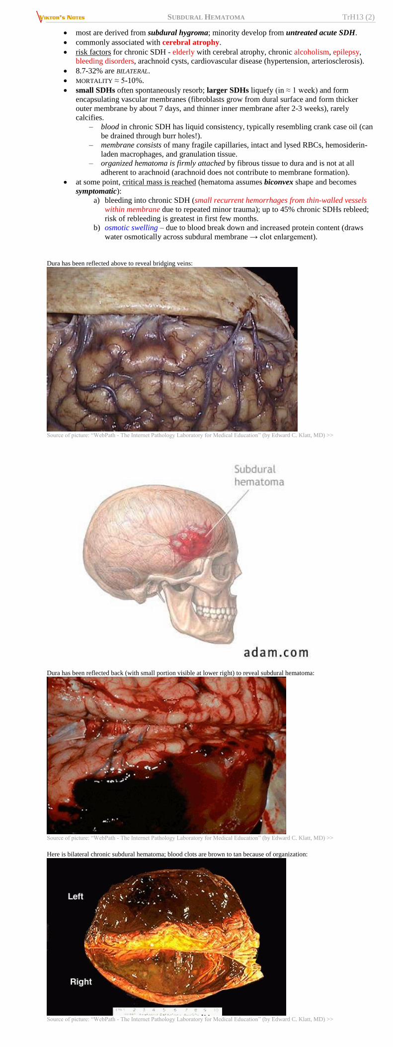
Dura has been reflected above to reveal bridging veins:
Source of picture: “WebPath - The Internet Pathology Laboratory for Medical Education” (by Edward C. Klatt, MD) >>
Dura has been reflected back (with small portion visible at lower right) to reveal subdural hematoma:
Source of picture: “WebPath - The Internet Pathology Laboratory for Medical Education” (by Edward C. Klatt, MD) >>
Here is bilateral chronic subdural hematoma; blood clots are brown to tan because of organization:
Source of picture: “WebPath - The Internet Pathology Laboratory for Medical Education” (by Edward C. Klatt, MD) >>

SUBDURAL HEMATOMA TrH13 (3)
Source of picture: Ramzi S. Cotran “Robbins Pathologic Basis of Disease”, 6th ed. (1999);
W. B. Saunders Company; ISBN-13: 978-0721673356 >>
CLINICAL FEATURES
A. Clinically silent.
B. Brain compression (slow venous bleeding enables large hematomas to form before clinical signs
appear) - can progress rapidly or slowly.
Most acute SDHs manifest within 48 hours.
≈ 50% patients are unconscious from time of injury; LUCID INTERVAL is observed in 30-70
cases.
there may be focal signs* (due to prolonged brain tissue compression under hematoma), but
often clinical manifestations are nonlocalizing (due to ICP↑); later, brain herniation may
develop.
*deficits are soft (not as profound as in other hematomas);
hemianesthesia, hemianopsia are seldom observed (anatomic
structures are deep and not easily compressed)
In pre-CT era, chronic SDHs earned label "great imitator" because of variable course and
presentation (sometimes mistaken for dementia, stroke, or brain tumor!).
in 25-50% cases, there is no clear history of head trauma.
signs or symptoms fluctuate in ≈ 24% cases (mimic TIAs).
headache (90%), mild hemiparesis (45-58%), confusion (56%), drowsiness (40-50%),
personality changes, papilledema, gait dysfunction, seizures are most common presenting
features.
N.B. after hematomas have exerted pressure on brain for long time (perhaps year or more),
removing them does little to improve cognitive function.
DIAGNOSIS
Hemostasis (PT, aPTT, platelet count)
LP (absolutely contraindicated) – xanthochromia, variable number of RBCs.
Noncontrast CT - crescentic collection over hemispheral convexity without extension into depths of
sulci; can cross suture lines and continue along falx and tentorium (do not cross midline!):
N.B. blood pressure is less and is less restricted (vs. EDH) → crescentic shape.
Acute SDH – hyperdense* (40-90 HU); rarely, can appear isodense:
a. low hematocrit (anemia)
b. hyperacute clot (< 1 h old)
c. active bleeding
small hematomas may not be depicted because attenuation similar to adjacent inner table
of skull (H: wider CT window and level, e.g. 240 and 80 HU).
Subacute SDH – isodense*; important signs - effacement of cortical sulci, displacement of
gray matter–white matter junction (‘buckling’); membranes are not vascularized.
better visualization - MRI (high signal on T1) or contrast-enhanced CT (opacification
of cortical vessels - definition of brain margins).
Chronic SDH – hypodense* (15-30 HU, i.e. isodense to CSF); vascularized membranes
enhance with contrast.
*compared to brain.
Rule of thumb: blood remains denser than brain for 1 week, and is less dense after 3 weeks.
underlying brain is flattened (mass effect), and subarachnoid space is often clear.
absent midline shift - suspect contralateral mass (e.g. ≈ 20% chronic SDHs are bilateral);
useful sign – ventricular frontal horns lie closer together (‘rabbit’s ear’ configuration).
excessive midline shift - suspect underlying cerebral edema.
in interhemispheric SDH, falx cerebri appears thickened and irregular.
– interhemispheric SDH may mimic SAH (subarachnoid blood clears after several days; SDH
remains wedge-shaped, smooth-bordered, hyperattenuating lesion).
– interhemispheric SDH may spread onto tentorium → characteristic ‘comma shape’ on axial
CT.
posterior fossa SDH does not cross midline or extend above tentorium (vs. EDH).
temporal and tentorial SDHs are better detected on coronal MRI (than on axial CT).
rebleeding into subacute / chronic SDH makes hematoma biconvex and heterogeneous density
(mixed old and fresh blood, sedimentation levels) – in general, looks like EDH with heterogeneous
density.
Size of extra-axial hematoma is more important factor than whether blood is epidural or
subdural in location!
Suspect child abuse! (esp. if posterior interhemispheric & tentorial SDHs)
osmotic swelling of chronic SDH makes hematoma biconvex and water density – in general, looks
like EDH with water density.
MRI is most sensitive imaging test! (esp. in subacute and chronic phase). further see p. D51 >>
chronic SDH - high signal on T1; membranes have low signal intensities (on T1 and T2).
Absence of clear history of trauma → angiography (search for ruptured aneurysm or dural AV
fistula).
angiographic signs of SDH – avascular zone between skull and brain with away dislocation of
major vessels.
T1 & T2-MRI - bilateral subacute SDHs (increased signal intensity; areas of intermediate intensity represent more acute
hemorrhage into subacute collections):

SUBDURAL HEMATOMA TrH13 (4)
Skull fracture with adjacent, small acute SDH (window and level values are widened over standard values to aid detection):
MRI - subacute SDH with extension into anterior interhemispheric cistern (note that sutures do not contain spread):
SDH with adjacent SAH due to ruptured MCA aneurysm:
SDH due to ruptured right PComA aneurysm:
Right frontal subacute SDH; note displaced gray matter–white matter junction, and midline shift:
Subacute-on-chronic SDH with blood-fluid level (acute hemorrhage into chronic collection):
SUBDURAL HEMATOMA TrH13 (5)
Acute-on-chronic SDH:
Source of picture: H. Richard Winn “Youmans Neurological Surgery”, 6th ed. (2011); Saunders; ISBN-13: 978-1416053163 >>
Subacute SDH - less dense than brain but denser than CSF; it is denser posteriorly; midline displacement is greater than
would be expected from size of lesion - suggests extensive underlying swelling; contralateral (left) ventricle is dilated:
Bilateral SDHs, right greater in size than left:
Source of picture: “WebPath - The Internet Pathology Laboratory for Medical Education” (by Edward C. Klatt, MD) >>
Acute SDH:
MRI - bilateral SDHs (arrows) with suboccipital extension:

SUBDURAL HEMATOMA TrH13 (6)
Interhemispheric acute SDH:
Bilateral chronic SDHs; midline shift is absent:
Chronic SDH with no mass effect:
Bilateral chronic SDHs with acute rebleeding:
Acute SDH extending over entire left hemisphere; sedimentation level is seen (patient has clotting dysfunction):

SUBDURAL HEMATOMA TrH13 (7)
Source of picture: “WebPath - The Internet Pathology Laboratory for Medical Education” (by Edward C. Klatt, MD) >>
Subacute SDHs: A) Noncontrast CT inward displacement of gray-white matter junction of left cerebral hemisphere; small sedimentation
level is apparent in posterior portion.
B) Contrast CT at same level as A - good visualization of lateral margin of left cerebral hemisphere (opacified cortical
veins):
Acute and subacute SDHs:
A. T1-MRI - large fluid collections (white arrows) consistent with large subacute SDHs.
B. T2-MRI - hyperintense signal (white arrows) representing subacute blood and hypointense signal (black arrows)
representing more recent hematoma.
Calcified SDHs:
A. In frontal view, bilateral shell-like calcifications form cast of cerebral hemispheres.
B. In lateral view, outer calcified membranes are near bony skull, and inner membranes are separated from outer by
relatively clear zones.

SUBDURAL HEMATOMA TrH13 (8)
TREATMENT
In all cases, hematoma complete resolution should be documented (conservatively treated acute SDH
can evolve into chronic SDH).
CONSERVATIVE MANAGEMENT
surgical drainage is not required in many cases (acute SDH may disappear spontaneously, but may
evolve into subacute or chronic lesion!).
seizure prophylaxis and other measures → see p. TrH1 >>
monitor coagulation parameters!
platelet transfusion for patient on ASA/Plavix – probably no effect! “Reversal of Antiplatelet Therapy May Not Benefit TBI” Congress of Neurological Surgeons
(CNS) 2013 Annual Meeting. Abstract #164 and #195. Presented October 22, 2013. >>
CHRONIC SDH
chronic SDH can be treated with TRANEXAMIC ACID (TXA) 650-750 mg PO daily for 30 days
without concomitant surgery; tranexamic acid might simultaneously inhibit fibrinolytic and
inflammatory (kinin-kallikrein) systems, which might consequently resolve CSDH; some experts
administer it postop (after bur hole washout) J Neurosurg. 2013 Aug;119(2):332-7. doi: 10.3171/2013.3.JNS122162. Epub 2013 May 3.
Nonsurgical treatment of chronic subdural hematoma with tranexamic acid.
Kageyama H, Toyooka T, Tsuzuki N, Oka K.
EG-1964 (Edge Therapeutics, Inc.) - polymer-based filament that contains APROTININ - pancreatic
trypsin inhibitor.
— indicated to prevent rebleeding and reduce the need for blood transfusions
following cardiac bypass surgery
— IV administration has serious thrombotic side effects
— EG-1964 delivers sustained dose of aprotinin over 21-28 days directly to site of
SDH
chronic SDH growth is due to the highly friable nature of the vascularized membrane that forms
after initial injury; proposed treatment for recurrent symptomatic SDH - middle meningeal artery
(MMA) embolization with the goal of eliminating the arterial supply to this vascularized
membrane.
HEMATOMA EVACUATION
ACUTE SDH
indications for surgery → see p. TrH1 >>
technique → see p. Op320 >>
CHRONIC SDH
any coagulopathy must be reversed ASAP.
must be surgically evacuated if symptomatic* (+ significant mass effect on imaging):
*except mild headaches
A. Burr hole craniostomy (“burr hole washout”) – procedure of choice. see p. Op320 >>
B. Twist drill craniostomy – at bedside for very sick patient. see p. Op320 >>
C. Craniotomy – indicated for:
a) multilocular / calcified SDHs.
b) burr hole / twist drill drainage failures.
N.B. some experts advocate craniotomy as first choice for cSDH but studies show that all results
(complications, dispo, recurrences requiring reoperation*, mortality) are worse than with BHWO.
*24% (vs. 6-7% for BHWO)
Postoperatively see p. Op320 >>
PROGNOSIS
Most important prognostic factors:
1. Concomitant primary brain injury
2. GCS score
3. Age (esp. > 40 yrs).

SUBDURAL HEMATOMA TrH13 (9)
4. Time from trauma to surgical evacuation of hematoma (esp. > 4 hours)
After surgical evacuation, subdural hematoma recurs in 5-30% patients.
Chronic SDH increases mortality 17-fold at 1 year.
SDH IN INFANTS
NEWBORNS
SDH was once considered most common intracranial birth injury (but now ↓ with improved
obstetric care).
in majority of cases, bleeding is BILATERAL and located over dorsolateral surfaces of frontal and
parietal lobes.
clinically: seizures, pallor, tense anterior fontanel, rapidly enlarging head, hypotonia, poor Moro
reflex.
– may be abnormal at birth (no spontaneous respiration, severe hypotension, seizures, retinal
hemorrhage).
– initial manifestation may be generalized seizures within first 6 months of life.
– acute cases may progress to herniation.
diagnosis – ultrasound, CT, funduscopy (50% show retinal or subhyaloid hemorrhages).
– if SDH interfered with brain growth, skull vault may be thick, with paucity of convolutional
impressions, and hypertrophy of air cells and paranasal sinuses.
– calcification streaks / plaques, often parallel to vault, may be visible in capsule of chronic
SDH.
treatment – evacuation through craniotomy.
Ultrasound - left echogenic acute subdural hematoma (H), associated with right subarachnoid anechogenic effusion
(E). Falx cerebri (arrow) remains straight:
OLDER INFANTS
SDH is most common intracranial lesion in children < 2 years:
a) shaken baby syndrome!!! (chronic SDH in infants who do not yet walk)
see p. TrH20 >>
b) complication of shunt procedures
c) bleeding disorders.
clinically: macrocrania (symmetrical or asymmetrical), tense anterior fontanel, lethargy /
irritability, seizures, poor feeding, vomiting, failure to thrive.
diagnosis – CT, diagnostic subdural tap (should be done bilaterally, even if positive on one side).
treatment – repeated daily subdural taps monitored by CT, head circumference measurements.
– resorption of hematoma must occur over weeks ÷ months.
– if > 10 taps are done (symptoms persist after 2 wk of daily drainage) → surgical treatment
(e.g. subdural-peritoneal shunting or subdural-subgaleal shunting for 6 months;
historically – subtemporal craniectomy – to let fluid drain into pterygoid fossa).
– removal of membranes was once thought to be important to avoid brain growth restriction,
but this no longer seems necessary.
25 have some psychomotor retardation.
SUBDURAL TAP
anterior fontanelle becomes effectively closed between 9 and 18 months.
equipment - sterile prep and drape, gloves, razor, blunt short-beveled spinal needle with stylet (20
G, 1½ or 2½ inch), manometer, CSF collection tubes, and local anesthesia (if desired).
scalp is shaved anterior to ears (over lateral margins of anterior fontanelle).
restrain infant by bundling him in sheet; supine, head firmly held by attendant (avoid excessive
neck flexion).
raise skin wheal of local anesthetic at puncture site.
insert needle through skin at extreme lateral limit of anterior fontanelle where it meets coronal
suture (i.e. at least 2-3 cm from midline - to prevent sagittal sinus injury).
– use ZIGZAG PUNCTURE to prevent later fluid leakage (puncture dislocated skin at right
angle, then aim needle laterally).
– dura is entered with sudden "popping" sensation.
– cerebral cortex is ≈ 1.5 cm from skin surface (attachment of hemostat 5-7 mm from beveled
end of needle should provide adequate safeguard).
remove stylet - subdural fluid is allowed to drain spontaneously (fluid is never aspirated - risk of
drawing pial vessels into point of needle).
– only 10-20 mL of subdural fluid should be removed from each side at one time (removing
larger amounts may precipitate rebleeding or shock).
– if pial vessel is punctured, bleeding will usually cease spontaneously.
– pressure is measured with manometer.
– specimens are collected for Gram staining, culture, cells, glucose, and protein.
remove needle → apply pressure → firm sterile dressing → place in sitting position (to prevent
leakage).
repeat on opposite side.

SUBDURAL HEMATOMA TrH13 (10)
continued leakage from puncture site → apply collodion-impregnated cotton fluff over puncture
wound + elevate head 20-30°.
BIBLIOGRAPHY for ch. “Head Trauma” → follow this LINK >>
Viktor’s Notes℠ for the Neurosurgery Resident
Please visit website at www.NeurosurgeryResident.net