digital.csic.esdigital.csic.es/bitstream/10261/137065/1/biotechnology... · web viewstructural...
TRANSCRIPT

Structural traits and catalytic versatility of the lipases from the Candida rugosa-like
family: A review
Jorge Barriuso, María Eugenia Vaquero, Alicia Prieto*, and Mª Jesús Martínez*
Centro de Investigaciones Biológicas, Consejo Superior de Investigaciones Científicas, Ramiro
de Maeztu 9, 28040 Madrid, Spain
*Corresponding authors: María Jesús Martínez ([email protected]); Alicia Prieto
Abstract
Lipases and sterol esterases are enzymes with broad biotechnological applications,
which catalyze the hydrolysis or synthesis of long-chain acylglycerols and sterol esters,
respectively. In this paper, we review the current knowledge on the so-called Candida rugosa-
like family of enzymes, whose members display in most cases affinity against the two substrates
mentioned above. The family includes proteins with the α/β-hydrolase folding, sharing
conserved motifs in their sequences, and common structural features. We will go through their
production and purification, relate their described structures and catalytic activity, and discuss
the influence of the hydrophobic character of these lipases on their aggregation state and
activity. On the basis of the few crystal structures available, the role of each of the functional
areas in catalysis will be analyzed. Considering the particular characteristics of this group, we
propose their classification as “Versatile Lipases” (EC 3.1.1.x).
Key words: Lipase, sterol-esterase, Candida rugosa, biocatalysts, hydrophobic enzymes.
1
2
3
4
5
6
7
8
9
10
11
12
13
14
15
16
17
18
19
20
21
22
23
24
25
26
27
28
29
30
31

1. Introduction
Lipases, also known as triacylglycerol lipases (EC. 3.1.1.3), act on ester bonds of
several compounds, with acylglycerols as their natural substrates. These enzymes catalyze the
hydrolysis of triglycerides to produce free fatty acids, diglycerides and/or monoglycerides under
aqueous conditions, but they can also carry out synthesis reactions, such as esterification and
transesterification, in the presence of organic solvents (Houde et al., 2004; Reis et al., 2009).
On the other hand, sterol esterases (EC 3.1.1.13) are defined as enzymes that hydrolyze
sterol esters releasing free sterols and fatty acids in aqueous media, being also able to perform
synthesis reactions in the presence of organic solvents (Barba Cedillo et al., 2013; Morinaga et
al., 2011). Most of the known sterol esterases have been reported to have both lipase and sterol
esterase activity (Calero-Rueda et al., 2002; Maeda et al., 2008; Vaquero et al., 2016).
Lipases and sterol esterases are carboxylic ester hydrolases (EC 3.1.1) that share the
α/β-hydrolase fold (Grochulski et al., 1993; Holmquist, 2000; Nardini and Dijkstra, 1999).
Hence, their catalytic machinery includes the residues of the catalytic triad (serine, histidine and
aspartic or glutamic acid) and the oxyanion hole, a pocket in the active site involved in
catalysis. They compose a very diverse group of ubiquitous enzymes in nature and are
represented from microbes to plants and animals. Nevertheless, bacterial and fungal lipases are
of special interest as they are easily produced and applicable for industrial processes due to their
versatility and stability to harsh conditions (Gupta et al., 2015; Jaeger and Eggert, 2002; Schmid
and Verger, 1998; Singh and Mukhopadhyay, 2012).
These enzymes can perform a variety of reactions, have wide substrate specificity and
good selectivity, and are regio- and stereoselective. The most important lipases from a
commercial point of view belong to yeasts, such as Candida rugosa (synonym Candida
cylindracea) and Candida antarctica, or filamentous fungi, such as Aspergillus niger, Humicola
lanuginosa, Mucor miehei, and Rhizopus species (Bornscheuer, 2002; Domínguez de María et
al., 2006; Gupta et al., 2015; Hasan et al., 2006; Singh and Mukhopadhyay, 2012). Among
them, some of the characterized lipase isoenzymes from C. rugosa show also sterol esterase
activity, as those from other microorganisms, still not produced at commercial level, which we
will see across this review.
Esterol esterases and lipases are included in ESTHER
(http://bioweb.ensam.inra.fr/esther), a broad database that collects very complete information on
the members of the α/β-hydrolase fold superfamily (Lenfant et al., 2013received). In addition, a
sequence-based comparatively simplified version is available in the Lipase Engineering
Database (LED) (http://www.led.uni-stuttgart.de/). LED lists sequences of all the available
microbial enzymes with lipase activity, putative or not, and provides links to 22 published lipase
structures. This database serves as a bioinformatics tool for the systematic analysis of sequence,
structure and function of diverse lipases, and for designing variants with optimized properties
32
33
34
35
36
37
38
39
40
41
42
43
44
45
46
47
48
49
50
51
52
53
54
55
56
57
58
59
60
61
62
63
64
65
66
67
68

(Fischer and Pleiss, 2003). Based on different characteristics, such as the presence of specific
conserved motifs in their amino acidic sequence, microbial enzymes with lipase activity are
grouped under several classes or subclasses in these databases. Yeast and fungal lipases fall into
five different subclasses: Yarrowia lipolytica-like lipase, C. rugosa-like lipase, filamentous
fungi lipases, C. antarctica lipase B-like and C. antarctica lipase A-like. Moreover, on the basis
of their sequence, structure, and function, these enzymes are classified in LED into the GX,
GGGX and Y classes.
In this review we will focus on a very versatile group of enzymes that generally show activity
towards acylglycerols and sterol esters, the so-called C. rugosa-like or abH03.01 lipase family.
Only a few members of this group have been characterized, although recent findings have
shown their potential for engineering future industrial applications. The characterized proteins
belong to ascomycetes and basidiomycetes, and little is known about their phylogenetic
affiliation and evolution (Barriuso et al., 2013). According to LED, the 336 protein sequences
currently assigned to this family belong to the class GGGX. However, most of them correspond
to hypothetical proteins and, according to this sequence-based classification, some enzymes can
be wrongly ascribed to this group. This was the case of the hypothetical protein EstA from A.
niger that, once expressed, showed to have structural characteristics and substrate preferences
different to those characteristic of the C. rugosa-like family (Bourne et al., 2004). The enzymes
described within this group are glycoproteins with a common overall structure. Their active
sites are hidden under a mobile region denominated lid or flap that, in a lipid-water interface or
in the presence of substrates or inhibitors, rearranges its position leaving an open gate to the
active center. Then, the position of the lid marks the difference between the open (active) or
closed (inactive) forms of these proteins. Other interesting properties of these catalysts as their
temperature and pH stabilities (T50= 40–60 °C, and pH 4-10), or their stereo- and
regioselectivity (Colton et al., 2011; Lee et al., 2007; Palocci et al., 2007; Vaquero et al.,
2015a), make them very attractive, and the current or proposed application of these catalysts
affect a wide range of industrial sectors, such as biofuels, oleochemical, food, detergents,
cosmetics, pharmaceutical, textile and paper industry (Hasan et al., 2006; Houde et al., 2004;
Reetz, 2002; Singh and Mukhopadhyay, 2012). Excellent reviews covering the biotechnological
applications of lipases, including the best known enzymes from the C. rugosa-like family, have
been recently published (Gupta et al., 2015; Stergiou et al., 2013). Here, we will revise their
structural and catalytic properties, their production and characterization in different hosts, and
the applicability of tools as genome mining for in silico search of novel catalysts or protein
engineering for tailoring enzyme activity.
2. Ecophysiological role
69
70
71
72
73
74
75
76
77
78
79
80
81
82
83
84
85
86
87
88
89
90
91
92
93
94
95
96
97
98
99
100
101
102
103
104

Fungi producing C. rugosa-like enzymes share a plant-associated habitat, and most of
them have been isolated from natural soils where plant material is frequent (Tomizuka et al.,
1966). In these cases, the ecological role of the extracellular lipases can be related to the attack
of esters from the epicuticular waxes and cuticle (Juniper and Jeffree 1983), improving the
accessibility to cell-wall polysaccharides that can serve as carbon sources for plant-associated
fungi. In pathogenic species these enzymes may act also on such components, located in the
surface of leaves, facilitating fungal colonization (de Vries et al., 1997; Doss, 1999).
The only studied lipase of this family from a basidiomycete is produced by Pleurotus
sapidus, a white-rot ligninolytic fungus (Zorn et al., 2005), while several have been
characterized form saprophytic ascomycetes, isolated from forest, agricultural, or composting
soils. Among them, we can mention C. rugosa, a non-sporogenic imperfect hemiascomycete, a
number of filamentous fungi, as the thermophilic Melanocarpus albomyces (Kontkanen et al.,
2006a), isolates from Trichoderma (Schuster and Schmoll, 2010received) or A. niger (Hu et al.,
2011), and the dimorphic ascomycetous yeast-like fungi Geotrichum candidum and Ophiostoma
piceae (Yan et al., 2007). The habitat of G. candidum is often associated with all kinds of soft
plant tissues, but it is ubiquitous in soils and pupal galleries of bark beetles (de Hoog and Smith,
2004). The environment of the wood-staining fungus O. piceae is much more restricted. This
species disseminates its spores using bark beetles as vectors and lives as saprobe in the
superficial layers of conifers’ sapwood. There, it metabolizes wood lipids, releasing hydrolysis
products as glycerol that seem to play an important role in the formation of the dark pigments
responsible for the “blue stain” (Eagen et al., 1997), which causes severe losses to the forestry
industry (Calero-Rueda et al., 2002; Haridas et al., 2013). Some extracellular fungal lipases are
also gaining attention for their potential role as virulence factors in relation to colonization,
adhesion, biofilm formation and pathogenesis (Gupta et al., 2015). Plant pathogens as Fusarium
solani are also producers of these enzymes (Vaquero et al., 2015a), and the causal agent of the
bunch rot disease of grapes, Botritys cinerea, releases lipases during the early phases of
parasitism induced by the components of the cuticle, being capable of entering its host directly
through an undamaged plant cuticle (Comménil et al., 1999).
Many other hypothetical proteins from this family have been described in the genomes
of fungi usually living as vegetal saprobes or parasites, such as ascomycetes from the genera
Alternaria, Pyrenophora and Neurospora, or basidiomycetes belonging to Postia, Laccaria and
Puccinia, although these enzymes have not yet been produced and characterized (Barriuso et al.,
2013; Fischer and Pleiss, 2003).
3. Production, purification and biochemical characterization of the C. rugosa-like enzymes
Due to their wide applicability, enzymes from this family have been produced in a
variety of hosts, from their native producers to recombinant bacteria, yeasts or filamentous
105
106
107
108
109
110
111
112
113
114
115
116
117
118
119
120
121
122
123
124
125
126
127
128
129
130
131
132
133
134
135
136
137
138
139
140
141
fungi. As explained in the following sections, it has been shown that the expression of a given
enzyme in different systems may induce changes in its properties (Barba Cedillo et al., 2012;
Kontkanen et al., 2006b; Vaquero et al., 2015b) and then the choice of the appropriate host is
not a minor decision. Once the functional protein has been successfully released to the
extracellular medium it must be purified, although the existence of several lipase isoforms with
similar chemical characteristics in some of the natural producers complicates their isolation as
single molecular species and generally requires a combination of techniques. In these cases, the
expression of recombinant proteins can make protein purification much easier.
Preparation of pure protein samples is generally accomplished using ultrafiltration,
precipitation and/or solubilization protocols, followed by one or several chromatographic
strategies as ion-exchange, hydrophobic interaction or size-exclusion chromatography (SEC). It
is important to keep in mind that the hydrophobic character of these enzymes facilitates the
formation of protein aggregates in solution and that in many occasions the C. rugosa-like
enzymes purified by SEC may correspond to aggregated forms, as well as the molecular mass
calculated from this technique.
3.1. The C. rugosa isoenzymes
The best known members of this family are some of the C. rugosa isoenzymes
(hereinafter, CRL), which have been profusely investigated since their discovery and even
today. This yeast secretes a variety of closely related enzymes (Benjamin and Pandey, 1998;
Ferrer et al., 2001; Lotti et al., 1994), commercialized as lipases or sterol esterases depending on
their catalytic efficiency. Nevertheless, comparison of data on the biochemical properties of
these isoenzymes, reported from different research groups, is difficult due to the use of different
methodologies, substrates, and even solvents for substrates solubilization, which is not a trivial
parameter for its influence on the interface where the enzyme exerts its action (Domínguez de
María et al., 2006; Pernas et al., 2009).
Tomizuka et al. (1966) described for the first time the isolation and purification of an
extracellular lipase from C. rugosa as a hydrophobic glycoprotein of 62 kDa with 4.2% neutral
sugars. The subsequent research of several groups showed inconsistency about the properties of
these enzymes and it was soon corroborated that the C. rugosa lipase commercialized by Sigma
and Amano consisted of a mixture of proteins with different catalytic properties. Veeraragavan
and Gibbs (1989) separated two lipases (I and II) and Rúa et al. (1993) demonstrated the
presence of isoenzymes A and B (currently known as isoforms CRL3 and CRL1, respectively),
which were later separated by hydrophobic interaction chromatography as a result of the higher
hydrophobicity of CRL3 against CRL1 (Rúa, 1994). Seven or more genes codify for different
lipase isoforms (Brocca et al., 1995), and five of the proteins (CRL1-CRL5) have been
characterized (Lotti and Alberghina, 1996; Lotti et al., 1994) . The expression levels of these
142
143
144
145
146
147
148
149
150
151
152
153
154
155
156
157
158
159
160
161
162
163
164
165
166
167
168
169
170
171
172
173
174
175
176
177
178

genes was analyzed measuring the abundance of lip mRNAs in non-induced C. rugosa cultures,
finding that it was in the order lip1, lip3, lip2, lip5, and lip4 (Lee et al., 1999). The expression of
lip1 and lip3 is high and constitutive, whereas for the remaining isoenzymes it depends on the
culture medium and conditions. Therefore, the expression profile of individual genes could be
modulated adding inducers such as olive oil or Tween 20 (Ferrer et al., 2001; Lee et al., 1999;
Pernas et al., 2000). Commercial cocktails of CRLs containing different proportions of each
isoform may have been obtained from industrial strains as a result of mutations or using
different substrates (Ferrer et al., 2001; López et al., 2004) and can be suitable for diverse
biotechnological applications. For instance, CRL1 is the major isoenzyme in the crude
commercialized by Sigma as lipase, whereas CRL3 is the principal isoenzyme in the crude
commercialized by Roche as cholesterol esterase.
For heterologous production, the use of universal hosts to express the native genes of C.
rugosa isoforms as functional lipases is unfeasible because this organism uses the non-universal
codon CUG-Leu for serine (Kawaguchi et al., 1989). This fact forced to resort to alternative
approaches. One of them, proposed by Mileto et al. (1998), consisted on using Candida maltosa
as host for its similar codon usage, but the recombinant protein secreted was heavily hyper
glycosylated. Brocca et al. (1998) carried out the total synthesis of a codon-optimized
nucleotidic sequence of the lip1 gene, and its functional over expression in Saccharomyces
cerevisiae and Pichia pastoris. Low expression levels were achieved in the first host, but not in
P. pastoris, that secreted a recombinant CRL1 with similar physico-chemical properties and
substrate specificity toward triglycerides of different chain length to the native CRL1. From this
finding, many groups have reported the heterologous expression of C. rugosa isoforms in P.
pastoris using the same strategy (Chang et al., 2006a; Chang et al., 2006b; Chang et al., 2005;
Ferrer et al., 2009; Lee et al., 2002; Lee et al., 2007; Lee et al., 2011; Tang et al., 2001; Yen et
al., 2010; Zhao et al., 2008). On the other hand, Tang et al. (2000) described the functional
expression of the lip4 gene in Escherichia coli, which synthesizes non-glycosylated protein,
after converting 19 CUG codons into a universal serine codon. The enzymatic activity of this
protein was similar to that of the glycosylated lipase from P. pastoris, although the CRL4 from
the yeast had higher thermal stability.
Isoenzymes CRL1 to CRL5 were separated from the crudes of C. rugosa by means of
several chromatographic techniques and characterized. They are glycoproteins with high
sequence identity (77-88%), molecular masses around 57-61 kDa, and isoelectric points in the
range of 4.5-5.7. Despite these similarities, their stability to temperature and pH, and their
substrate specificity are different (López et al., 2004; Mancheño et al., 2003). SEC analysis
showed that, in all cases, a proportion of the proteins’ population forms aggregates in solution,
whose molecular mass was estimated to be over 200 kDa even in the presence of detergents
(Pernas et al., 2000; Pernas et al., 2009). Nevertheless, among the CRLs currently characterized
179
180
181
182
183
184
185
186
187
188
189
190
191
192
193
194
195
196
197
198
199
200
201
202
203
204
205
206
207
208
209
210
211
212
213
214
215

only CRL3 is dimeric in its active form, while CRL1 and CRL2 are monomers (Kaiser et al.,
1994; Otero et al., 2005; Pernas et al., 2009; Pernas et al., 2001).
According to the literature, CRL1 and CRL5 showed the highest affinity for
triglycerides among the CRL isoenzymes. CRL1 displays the maximum lipolytic activity
towards middle-chain (C8 and C10) substrates (Chang et al., 2006a; López et al., 2004), and its
activity against p-nitrophenyl and cholesteryl esters was very poor when compared with CRL2,
CRL3 and CRL4 (Chang et al., 2006b; López et al., 2004; Mancheño et al., 2003). CRL5 seems
to be even less active than CRL1 against these substrates (Lee et al., 2011). On the other hand,
CRL2 had the highest cholesterol esterase activity (Mancheño et al., 2003) and its efficiency
towards p-nitrophenyl esters and triglycerides is intermediate between those of CRL1 and CRL3
(Chang et al., 2006b; López et al., 2004; Pernas et al., 2000), preferring long-chain fatty acids
(C12-C18:1) in all substrates tested. In spite of having the highest sequence homology (86%),
CRL3 and CRL1 differed drastically in their hydrolytic activity. CRL3 was the most active
isoenzyme on p-nitrophenyl esters regardless of the length of their acyl chain, though the best
results were reported for medium-chain substrates (Chang et al., 2006b; López et al., 2004;
Pernas et al., 2000). Its cholesterol esterase activity showed intermediate values between those
of CRL1 and CRL2 for long-chain substrates (Mancheño et al., 2003). Similarly, the CRL4
isoform displayed more activity against p-nitrophenyl esters than the commercial CRL lipase
(mostly containing CRL1), but especially towards C16 and C18 esters (Tang et al., 2001).
3.2. The G. candidum lipases
Several papers describe the purification to homogeneity of lipases produced by G.
candidum (GCL). Tsujisaka et al. (1973) applied ammonium sulfate precipitation, anion
exchange chromatography and two SEC steps to obtain a supposedly pure lipase. Nevertheless,
subsequent works demonstrated that a mixture of enzymes existed in that preparation (Shimada
et al., 1990; Shimada et al., 1989). Sugihara et al. (1990) isolated lipases I and II (hereinafter
GCL1 and GCL2) but, after incorporating an additional hydrophobic interaction
chromatography step, the same group reported four molecular forms of lipase, with GCL1
representing 84% of the total lipase activity (Sugihara et al., 1991). In parallel, Sidebottom et al.
(1991) purified and characterized four different GCLs from two G. candidum strains (two of
each), which were described as closely related monomeric glycoproteins with similar molecular
masses (54-62 kDa, from SDS-PAGE), pI (4.5-4.7), and around 8% N-linked carbohydrates. It
is worthy to mention that the strategy followed to purify each one of the crudes had to be
different. In one of them the two enzymes were separated after hydrophobic interaction and ion
exchange chromatography (Mono Q) while using the same protocol for the other one the
activity resulted irreversibly bound to the second column (a strong anion exchanger) and the
proteins were finally separated using a weak anion exchanger.
216
217
218
219
220
221
222
223
224
225
226
227
228
229
230
231
232
233
234
235
236
237
238
239
240
241
242
243
244
245
246
247
248
249
250
251
252

These isoenzymes have 86% sequence identity, but GCL2 has two potential N-
glycosylation sites, while GCL1 has three (Bertolini et al., 1995; Schrag and Cygler, 1993).
Their identity with the isoforms of C. rugosa is around 40% (Bertolini et al., 1995; Holmquist et
al., 1997a).
The expression of recombinant enzymes from this organism made easier their
purification. Two works from the same research group (Bertolini et al., 1995; Vernet et al.,
1993) described the production, purification and characterization of GCL1 and GCL2 in a
glycosylation extension-deficient S. cerevisiae strain. Inclusion of a His-tag in the gene
sequences of the two isoenzymes before their expression allowed their easy purification by
metal-affinity chromatography. Substrate specificity tests were carried out with the purified
enzymes, demonstrating the higher specific activity of GCL1 for long chain, unsaturated fatty
acid substrates with a double bond at position 9 (cis configuration, as oleate esters) whereas
GCL2 was more efficient against saturated, short-chain fatty acid substrates. These enzymes
have also been expressed in P. pastoris under the control of the methanol-inducible AOX
promoter and purified using the same strategy reported by Bertolini (1995), with higher yields
(60-fold) than in S. cerevisiae (Holmquist et al., 1997b). Recently, another lipase from
Galactomyces candidus, the teleomorph of G. candidum, has been produced in the same host.
This protein showed high sequence identity with GCL1 and GCL2 (>80%) and the same
substrate preferences, but higher values of optimum pH and temperature (Yan et al., 2007).
3.3. The O. piceae sterol esterase/lipase
After a screening on ascomycetes, basidiomycetes and conidial fungi, O. piceae was
selected for secreting lipase and sterol esterase activities (Calero-Rueda et al., 2002). The
responsible for these activities resulted to be a single enzyme of broad substrate specificity that
was isolated from the liquid cultures of this dimorphic ascomycete, and has been carefully
studied and characterized (Fig. 4A). This enzyme (OPE) was purified in a single step by
hydrophobic interaction chromatography as a 56.5 kDa-glycoprotein with 8% of N-
glycosylation, and pI of 3.3, and it forms multi-aggregates in aqueous solution (Calero-Rueda et
al., 2002), as confirmed by sedimentation velocity analyses (Barba Cedillo et al., 2012).
According to its sequence the protein contains three predicted glycosylation sites and shows
high identity (~45%) to C. rugosa lipases (Calero-Rueda et al., 2009).
The enzyme was successfully expressed in different heterologous hosts, resulting in
different yields and catalytic properties. The best yields were reached upon expression in P.
pastoris, higher than those obtained with the saprophytic fungus (~30-fold) and in a shorter
growth period (Barba Cedillo et al., 2012; Vaquero et al., 2015b). This recombinant protein had
a molecular mass of 75 kDa, 28% N-linked carbohydrates and presented improved stability at
basic pH values. Its kinetic parameters were also better when compared with the native enzyme
253
254
255
256
257
258
259
260
261
262
263
264
265
266
267
268
269
270
271
272
273
274
275
276
277
278
279
280
281
282
283
284
285
286
287
288
289
(Fig. 4B) due to its enhanced solubility, as confirmed by the presence of monomers and dimers
in solution. This behavior was attributed to the presence of 6-8 extra amino acids in the N-
terminus of the recombinant form that affect its aggregation and solubility (Barba Cedillo et al.,
2012). Recombinant forms of OPE were later obtained in other hosts (Vaquero et al., 2015b). A
sterol esterase with similar molecular mass and glycosylation degree was successfully expressed
in the Generally Recognized as Safe yeast S. cerevisiae. The enzyme produced in this host
presented an intermediate aggregation state between those of the proteins secreted by P.
pastoris and the native producer. However, only an inactive form of this enzyme was produced
in E. coli, probably because this organism cannot perform posttranslational modifications
(glycosylation, formation of disulfide bridges, etc), which can affect the correct protein folding.
The native sterol esterase from O. piceae and its recombinant form expressed in P.
pastoris display very broad substrate specificity (Barba Cedillo et al., 2012; Calero-Rueda et al.,
2002; Vaquero et al., 2015b), with higher activity towards medium-chain fatty acid p-
nitrophenyl esters (C12) and triglycerides (C8-C12), while among cholesterol esters the
preference shifts for long, unsaturated (cis-9) fatty acid acyl chains (C18:1, C18:2). Comparison
of their activity with those of other lipases or sterol esterases, under the same reaction
conditions, revealed the high efficiency and versatility of both OPE forms against most of the
substrates assayed.
3.4. The M. albomyces sterol esterase/lipase
The thermophilic fungus M. albomyces secretes a sterol esterase (STE1) with high
activity toward cholesterol esters. The enzyme was purified using hydrophobic interaction and
anion exchange chromatography after extraction with 0.1% Triton X-100, since around 30% of
the total activity was mycelium-bound (Kontkanen et al., 2006a). The monomeric protein had
around 64 kDa molecular mass as determined by SDS-PAGE, 5% glycosylation, and pI 4.5, and
its sequence presented around 47% homology with CRL1-CRL5. On the other hand, the enzyme
eluted as a single peak around 238 kDa after SEC, suggesting that it could be tetrameric in
aqueous solution. The sterol esterase STE1 was produced in two heterologous hosts, P. pastoris
GS115 (under the AOX promoter control) and Trichoderma reesei. In the first case, poor yields
of extracellular protein were recovered (Table I) since it was intracellular in a large proportion
(Kontkanen et al., 2006b). Production in T. reesei gave similar yields as M. albomyces, but the
activity was again bound to the mycelium or detected as aggregates, probably due to the high
content of hydrophobic amino acid residues in the protein (41.1%).
The steryl esterase from M. albomyces prefers unsaturated, long-chain fatty acid
substrates and its specific activity increases with the number of unsaturations
(C18:3>C18:2>C18:1>C18:0). In the case of p-nitrophenyl esters this enzyme presented the
290
291
292
293
294
295
296
297
298
299
300
301
302
303
304
305
306
307
308
309
310
311
312
313
314
315
316
317
318
319
320
321
322
323
324
325

highest substrate specificity for those with a short-chain (C4) fatty acid moiety (Kontkanen et
al., 2006a).
3.5. The B. cinerea lipase
An extracellular lipase produced by the fungus B. cinerea was purified by ammonium
sulfate fractionation and chromatography (Comménil et al., 1995). The protein had pI 6.5 and
its molecular mass was 60 kDa if determined by PAGE-SDS and 72 kDa by SEC, which
suggests that it is monomeric in solution. The amino acid composition of the lipase disclosed its
high homology with C. rugosa, G. candidum and O. piceae enzymes (46%, 38% and 56%,
respectively) and that, as them, it is very hydrophobic. Reis et al. (2005) expressed this enzyme
in P. pastoris and confirmed that the recombinant protein had properties virtually identical to
those reported for the B. cinerea culture supernatants. This lipase has activity against p-
nitrophenyl butyrate, but it shows a marked predilection for unsaturated, long-chain fatty acid
substrates (Comménil et al., 1995).
3.6. Other sterol estases/lipases from ascomycetes
The C. rugosa-like enzymes from ascomycetes most recently reported in the literature
are three hypothetical proteins from Nectria haematococca (teleomorph of F. solani), T. reesei
and A. niger, respectively, selected from in silico search in public databases of fungal genomes
(Barriuso et al., 2013). Their sequences resulted to be very similar to those of the sterol
esterases secreted by O. piceae and M. albomyces. The three proteins were cloned, expressed
and produced as functional proteins in P. pastoris with different yields, and purified by
hydrophobic interaction chromatography (Vaquero et al., 2015a). They are glycoproteins with
molecular masses between 63-67 kDa and 6-20% N-linked carbohydrates. N. haematococca and
T. reesei were active against all substrates tested, although with very different efficiency. Both
enzymes hydrolyzed cholesterol esters of long and short-chain fatty acids with low efficiency,
while their catalytic efficiency towards long and short-chain fatty acid triglycerides was one
order of magnitude higher, and were very active against p-nitrophenyl esters of short-, medium-
and long-chain fatty acid, although they were less efficient than OPE, with which they were
compared under the same experimental conditions. N. haematococca and T. reesei resembled
OPE in terms of their broad substrate specificity, but in most cases the catalytic efficiency of
OPE was similar or superior. On the other hand, the enzyme from A. niger hydrolyzed p-
nitrophenyl esters (preferably of short-chain fatty acids) and triglycerides, but it could not act on
cholesterol esters under the conditions assayed, revealing that in spite of their versatility not all
enzymes of the C. rugosa-like family are active towards sterol esters.
3.7. The P. sapidus lipase
326
327
328
329
330
331
332
333
334
335
336
337
338
339
340
341
342
343
344
345
346
347
348
349
350
351
352
353
354
355
356
357
358
359
360
361
362

Only one protein belonging to the C. rugosa-like family has been characterized so far
from basidiomycetes. This enzyme, secreted by the white rot fungus P. sapidus, was purified by
ion exchange chromatography and SEC, has isoelectric point of 4.5 and molecular mass of 54
kDa, although under native conditions the measured value was 430 kDa. From this information,
the authors suggested that the active form of the protein is a homo-octamer (Zorn et al., 2005),
although this point is not confirmed by other means and the result can simply owe to enzyme
aggregation. The sequence homology of the native lipase with C. rugosa isoenzymes is around
43%. The heterologous expression of the His-tagged protein sequence in E. coli rendered high
production of the recombinant lipase, mainly as inclusion bodies, although a small part was
soluble and active (Zelena et al., 2009). Hence, refolding was necessary to express active
enzyme from inclusion bodies (Krugener et al., 2009). Purification by metal-affinity
chromatography gave a protein of similar molecular mass to the native form. Apparently,
neither the particular codon usage of P. sapidus nor the lacking of glycosylation in this host
restricted the active expression of P. sapidus lipase in E. coli.
This lipase hydrolyzes triglycerides and xanthophyll esters, and its ability to hydrolyze
steryl esters has been suggested but never tested (Zorn et al., 2005).
4. Structural traits of C. rugosa-like lipases
The members of this family contain a similar number of amino acids (Table 1), have a
conserved glycosylation site in Asn351, and share several structural features (Figs. 1 and 2)
(Ghosh et al., 1995; Grochulski et al., 1993; Gutiérrez-Fernández et al., 2014; Mancheño et al.,
2003; Schrag and Cygler, 1993). Their protein scaffold, with the α/β hydrolase fold, is
composed of an 11-stranded mixed β-sheet, a small and nearly perpendicular N-terminal 3-
stranded β-sheet, and 16 helices (17 in the case of GCL2). In addition, they form two disulfide
bridges and several salt bridges for the stabilization of the N- and C-terminus. Similarly to
serine-proteases, the CRL-like enzymes display a catalytic mechanism based on a transfer
charge system through the so-called “catalytic triad” which is formed by Ser, His and Glu
residues in all family members. This establishes a difference with most esterases and lipases that
have Asp as the acidic residue of the triad. The catalytic machinery includes also the oxyanion
hole, a pocket in the active site involved in catalysis. The nucleophilic serine is located in a very
sharp turn, the nucleophilic elbow, composed of the conserved residues GESAG (Fig. 1). Other
characteristic of these enzymes is the presence of a substrate-binding site located in a long
internal tunnel formed by aromatic and aliphatic residues (Figs. 2 and 3) that confer a highly
hydrophobic environment all along it (Domínguez de María et al., 2006; Gutiérrez-Fernández et
al., 2014; Mancheño et al., 2003; Pleiss et al., 1998). The access to the active site is covered by
an amphipathic α-helix that serves as a lid, fixed by one of the disulfide bonds.
363
364
365
366
367
368
369
370
371
372
373
374
375
376
377
378
379
380
381
382
383
384
385
386
387
388
389
390
391
392
393
394
395
396
397
398

Only five enzymes from the C. rugosa-like lipase family have been structurally
characterized so far, and 14 three-dimensional structures from these lipases are currently
available. Crystals of C. rugosa CRL1 (PDBs: 1CRL, 1TRH, 1LPP, 1LPN, 1LPM, 1LPO) were
obtained in both its open and closed conformations (Grochulski et al., 1993; Grochulski et al.,
1994a; Grochulski et al., 1994b), while CRL2 (PDB: 1GZ7) was crystallized in its closed state
(Mancheño et al., 2003) and CRL3 (PDBs: 1CLE, 1LPS, 1LLF) in an open, dimeric
conformation (Ghosh et al., 1995; Pletnev et al., 2003) (Fig. 4D). As previously explained,
although these three isoforms have sequence identities of 77-88%, some amino acid changes at
strategic positions may be responsible for their different catalytic properties and substrate
specificity. The lipase GCL2 of G. candidum was crystallized in its closed conformation (PDB:
1TGH) (Schrag and Cygler, 1993) and very recently, the crystal structure of the O. piceae sterol
esterase/lipase (OPE) was elucidated in the open (dimer Fig. 4D) and closed (monomer, Fig.
4A) conformations (PDBs: 4BE4, 4BE9 and 4UPD) (Gutiérrez-Fernández et al., 2014).
4.1. The substrate-induced movement of the lid causes the transition to an open, active state
The flap region is one of the less conserved areas across the C. rugosa-like members,
and its structure is different in the crystallized enzymes, even within CRL isoforms. In CRLs,
the flap contains one α-helix with 10 residues and one 310 α-helix with 3 residues (Domínguez
de María et al., 2006; Mancheño et al., 2003), in OPE it has one α-helix with 6 residues and two
310 α-helices with 8 residues (Gutiérrez-Fernández et al., 2014), and in GCL2 it is formed by 3
α-helices with 12, 4, and 10 residues, respectively (Schrag and Cygler, 1993). The resolution of
four structures in their closed conformation allowed a more quantitative description of the
conformational changes that are likely associated with the transition to the active form of the
protein in water. In the closed conformation (Figs. 2 and 4), the amphiphilic α-helix is
positioned in such a way that the hydrophilic side of the lid faces the solvent, and the
hydrophobic side is directed towards the protein core (Grochulski et al., 1993; Gutiérrez-
Fernández et al., 2014; Schrag and Cygler, 1993). The transition to an open form in an aqueous
environment results from the displacement of the flap in the presence of a substrate, leaving a
large hydrophobic area exposed around the active site that contributes to recognition and
binding of substrates (Mancheño et al., 2003). This change increases the total hydrophobic
surface of the protein and opens the access to an internal tunnel. The movement of the flap of
CRL1 was analyzed by comparing its structure in the open and closed conformations,
concluding that the transition to the open state is associated exclusively to a rearrangement of
the secondary structure of the lid and to the cis-trans isomerization of the Pro92 peptidic bond
(Grochulski et al., 1994a), while the stem region is conformationally restricted by the presence
of a disulfide bridge (Cys60 – Cys97) and an ionic interaction (Glu96 and Arg37). These
structural components are also present in GCL2 and OPE, in which the disulfide bridge may
399
400
401
402
403
404
405
406
407
408
409
410
411
412
413
414
415
416
417
418
419
420
421
422
423
424
425
426
427
428
429
430
431
432
433
434
435

serve as a hinge for lid’s opening. The flap displacement in OPE is the largest (~30 Å) among
the related enzymes (Gutiérrez-Fernández et al., 2014). The highest degree of sequence
dissimilarity between CRLs is found in the flap and in the loop connecting strand β3 and helix
α3,4. This loop makes numerous contacts with the hydrophobic face of the flap and, in the case
of CRL3, it directly interacts with the sterol moiety of the substrate (Ghosh et al., 1995;
Grochulski et al., 1993; Mancheño et al., 2003).
4.2. S ubstrate-binding site and catalytic machinery: the internal tunnel, the catalytic triad, and
the oxyanion hole
It has already been mentioned that the catalytic machinery is highly conserved within
the members of the C. rugosa-like family. The substrate-binding site is located in a long and
extremely hydrophobic internal tunnel (Gutiérrez-Fernández et al., 2014; Holmquist, 2000;
Mancheño et al., 2003; Pleiss et al., 1998) and, once a compound enters this region, catalysis
takes place in two consecutive steps. In the acylation phase the hydroxyl group of the catalytic
Ser initiates the nucleophilic attack of the substrate´s carbonyl group, forming a first tetrahedral
intermediate negatively charged that is stabilized by specific residues of the oxyanion hole.
Then, the alcohol moiety of the substrate accepts a proton and is released, while the acidic part
forms a covalent acyl-enzyme intermediate that is finally broken by one water, alcohol, acid, or
ester molecule.
According to the crystal structures available, variations in substrate specificity observed
within this group of enzymes could be related to amino acid changes in these regions. For
example, few differences among the highly homologous CRL1 to CRL3 isoenzymes affecting
this area, namely one residue of the flap, three of the substrate binding pocket area, and two of
the tunnel’s mouth, are partly responsible for their variations in substrate specificity and
lipase/esterase preference. In the substrate-binding site area, the change of Ser450 in CRL1 for
Ala in CRL3 may prevent direct steric conflict to accommodate the strongly hydrophobic
cholesteryl moiety in the hydrophobic cleft, deviating enzyme specificity from triglycerides to
cholesteryl esters (Ghosh et al., 1995). OPE, with relevant sterol esterase activity, has also Ala
in this position, while in CRL2 and GCL2 it is occupied by Gly (Gutiérrez-Fernández et al.,
2014; Mancheño et al., 2003; Schrag and Cygler, 1993). In addition, comparison of the
structures of the three CRL isoenzymes disclosed that two regions of different hydrophobicity
could be differentiated along the internal tunnel (Mancheño et al., 2003). At the entrance, in
close proximity to the catalytic triad and the substrate-binding site, there is a phenylalanine-rich
region that was associated to substrate recognition and then to the lipase/esterase character of
the isoenzymes since their Phe content in this area (CRL1>CRL3≈CRL5>CRL2≈CRL4) is
inversely correlated to their activity on cholesterol esters. The aliphatic-rich region, at the
bottom of the tunnel, does not vary among CRL isoforms. OPE contains similar amount of Phe
436
437
438
439
440
441
442
443
444
445
446
447
448
449
450
451
452
453
454
455
456
457
458
459
460
461
462
463
464
465
466
467
468
469
470
471
472

as CRL2 in the first part of the tunnel, which agrees with its sterol esterase activity (Gutiérrez-
Fernández et al., 2014). Nevertheless, this enzyme is far superior to the commercial sterol
esterase of C. rugosa (Calero-Rueda et al., 2009). One of the reasons for this enhanced
efficiency has been related to the size and orientation of the tunnel (Fig. 4C) that is wide and
follows a straight trajectory in OPE (Gutiérrez-Fernández et al., 2014), while in CRL enzymes it
is narrower and bent, forming a 150º angle due to a change on Ser377 for Tyr (Mancheño et al.,
2003). A computational search for intramolecular tunnels among these enzymes and other
members of the abH03.01 family (Barriuso et al., 2013) showed that lipases from M. albomyces,
N. haematococca, T. reesei, A. niger, B. cinerea and P. sapidus have cavities comparable to
those from CRL isoforms or GCL2, although oriented towards different regions of the protein
surface (Fig. 3). The first half of the tunnels followed the same pattern in all members.
According to the structural models, the enzyme from A. niger has a bent, L-shaped internal
tunnel, similar to those of CRLs, which may be related to its lower specific activity compared
with the extracellular lipases/sterol esterases from O. piceae, N. haematococca and T. reesei.
These three enzymes have a quite straight internal tunnel, but the wider and straighter tunnel of
OPE may influence its better activity.
During catalysis, the tetrahedral intermediate could be stabilized by the main-chain
amide groups of residues located at the oxyanion hole. In CRL-like enzymes this region belongs
to the class GGGX- and in the CRLs, OPE, and GCL2 the positions Gly123, Gly124 and
Ala210 are conserved (Gutiérrez-Fernández et al., 2014; Monecke et al., 2016; Schrag and
Cygler, 1993). The possibility of these proteins having an exit tunnel has been suggested in
several works. Structural analysis of CRL3-substrate complexes showed that the fatty acid-
chain appears imbedded in a deep hydrophobic cleft of the protein that goes from the internal
catalytic site to the C-terminus, near the outer surface of the enzyme (Ghosh et al., 1995) and
the results from computational analysis of CRL1 reported by Foresti and Ferreira (2004) also
supported that this protein may have an exit tunnel. Similarly, the existence of a 30 Å-long
tunnel in OPE that connected the substrate-binding pocket with the outer surface of the protein
on the opposite side was demonstrated from its crystal structure (Gutiérrez-Fernández et al.,
2014).
4.3. Proteins’ surface: h ydrophobic patches, tertiary structure and N-linked carbohydrates
The proteins of the family abH03.01 possess several hydrophobic superficial areas that
generally locate in the surroundings of the lid and the catalytic pocket. These hydrophobic
patches have been related to interfacial activation, lid’s movement, substrate stabilization, and
protein aggregation. When opening, the flap of CRL isoforms interacts with a superficial
hydrophobic patch formed by residues 439 to 459 (Grochulski et al., 1994a). CRL2 contains a
second hydrophobic patch, located near the mouth of the substrate tunnel (Val296, Leu297,
473
474
475
476
477
478
479
480
481
482
483
484
485
486
487
488
489
490
491
492
493
494
495
496
497
498
499
500
501
502
503
504
505
506
507
508
509

Leu344 and Phe345), that prevents strong interactions of the substrate in the binding pocket,
modifying its catalytic behavior (Mancheño et al., 2003). OPE holds a hydrophobic patch
located close to the lid (Ile407, Phe408, Phe458, Pro459 and Phe460), with a unique Phe-Pro-
Phe pattern that is not present in other C. rugosa-like enzymes. Transition to the active form of
OPE involves two main structural rearrangements. The interaction of two of the superficial Phe
with two Leu residues of the lid, together with a conformational change in the α16-α17 loop,
induce stabilization of the substrate into a groove that leads to the active site (Gutiérrez-
Fernández et al., 2014). The composition of this loop differs in other family members. In the
structures of CRLs and GCLs a Trp and Phe residue, respectively, are filling this cavity and
then the channel does not exist, being a unique characteristic of OPE.
In the same way as the content of the aromatic Phe in the tunnel seems to be related to
the higher sterol esterase or lipase activity, it has been suggested that the amount of
hydrophobic residues in the lid area of CRL isoenzymes (CRL2 > CRL3 > CRL1) influences
their affinity for very hydrophobic substrates as sterol esters (Domínguez de María et al., 2006;
Mancheño et al., 2003). This hypothesis also agrees with the data reported for OPE, which has
higher content in hydrophobic amino acids and improved sterol esterase activity than the CRL
isoforms (Gutiérrez-Fernández et al., 2014), and with the data from structural models for the
hypothetical proteins expressed by Vaquero et al. (2015b), since that from A. niger had the least
amount of hydrophobic amino acids in the lid region and was not active on sterol esters under
the assayed conditions.
Among the crystallized enzymes of this family, CRL1, CRL2 and GCL2 are monomeric
in their open, active state. Evidences for dimerization have been reported exclusively for the
open forms of CRL3 and OPE (Gutiérrez-Fernández et al., 2014; Otero et al., 2005; Pernas et
al., 2001). In the presence of a substrate or inhibitor, CRL3 monomers interact to form a tight
homodimer, with the flaps of the two monomers open and the active-site grooves from the two
subunits facing each other. This arrangement of the two protein molecules creates a 7.3 Å-
radius cavity composed of hydrophobic and aromatic residues (Pletnev et al., 2003), with four
openings to the surface (Fig. 4D). The organization of the OPE dimer is completely different
since the aliphatic residues from the lid of one monomer interact with the hydrophobic patch of
the opposite chain in such a way that contacts between monomers are scarce. This disposition
conforms a pacman-like structure (Fig. 4D), creating a wide hydrophobic opening (23 Å × 38
Å) exposed to the solvent where large and hydrophobic substrates can interact, allowing also the
quick release of the products upon catalysis (Gutiérrez-Fernández et al., 2014). This type of
dimeric organization can be related to the high activity of OPE towards many substrates.
The phenomenon of interfacial activation, consistent in the increase of the lipolytic
activity of certain enzymes upon binding to the lipid-water interface, has usually been related to
the displacement of the lid (Otero et al., 2005; Pernas et al., 2001). Nevertheless, some enzymes
510
511
512
513
514
515
516
517
518
519
520
521
522
523
524
525
526
527
528
529
530
531
532
533
534
535
536
537
538
539
540
541
542
543
544
545
546

of this family as CRL3, CRL4 and OPE don’t show interfacial activation (Calero-Rueda et al.,
2002) in spite of having lids that must be opened to allow the access to the active center.
Curiously, these three enzymes possess relevant esterase activity and two of them are dimeric
(there is not information on the tertiary structure of CRL4), with their flap participating at more
or less extent in dimer formation.
All members of this family are glycoproteins, and their amino acidic sequences contain
one conserved N-glycosylation site (Asn351 in CRLs) with the only exception of the T. reesei
lipase that bears a glycidic chain in a different position (Vaquero et al., 2015a). The presence of
carbohydrates usually enhances proteins’ stability (Hung et al., 2011; Tang et al., 2000) and,
particularly, the sugar chain attached at the conserved glycosylation site has shown to provide
additional stabilization to the open conformation of the flap in CRL1 (Brocca et al., 2000;
Grochulski et al., 1994a). However, sugars may not be indispensable in these proteins once
secreted, as deduced for full activity retention of the deglycosylated OPE (Vaquero et al.,
2015a). On the other hand, a non-glycosylated recombinant form of CRL4 expressed in E. coli
showed the same catalytic properties as the glycosylated protein expressed in P. pastoris (Tang
et al., 2001) while expression of non-glycosylated OPE in the prokaryotic host gave inactive
intracellular or extracellular protein (Vaquero et al., 2015b).
5. Protein engineering of C. rugosa-like lipases to decipher the role of structural areas in
enzyme´s function
As explained above, sterol esterases and lipases from the C. rugosa-like family usually
display activity on triglycerides, p-nitrophenyl esters and cholesterol esters (López et al., 2004;
Mancheño et al., 2003; Vaquero et al., 2015a), although their catalytic efficiency towards these
compounds is variable. Many studies have tried to relate the structural elements or key residues
of the CRL isoenzymes with their different catalytic properties. These structure-function studies
can be attempted only with the previous knowledge of the crystal structure of the protein, and
are mostly based in two approaches. One is the replacement of a whole region of a given
isoform for the same region of other isoenzyme with different properties. The other analyzes the
effects of changing specific residues, located in strategic positions around the active center, by
sequence-based directed mutagenesis. Then, the changes introduced in the new variants may
serve not only to gain information on the role of particular areas or amino acids of the proteins,
but also to tune the enzyme’s activity.
As the flap area is not very conserved among CRL isoenzymes, its role in modulating
their activity and selectivity was analyzed. Brocca et al. (2003) reported that swapping the lid of
CRL3 to CRL1 (their sequences differ in 6 out to 27 amino acids) was sufficient to confer
higher cholesterol esterase activity to the chimeric CRL1lid3, although without any specific
shift in its chain-length specificity. Particular residues of the lid of CRL3, namely Phe69,
547
548
549
550
551
552
553
554
555
556
557
558
559
560
561
562
563
564
565
566
567
568
569
570
571
572
573
574
575
576
577
578
579
580
581
582
583
Gly74, Thr76 and Gln88, seem to have a role in facilitating the access to the substrate-binding
pocket, allowing the recognition of very hydrophobic substrates as cholesterol esters. The flap
region of CRL2 differs from those of CRL1 and CRL3 in 11 and 9 positions (out of 27),
respectively, and it is more hydrophobic (Mancheño et al., 2003). Lid swapping in CRL4 (Akoh
et al., 2004) resulted in increased hydrolytic activities on tributyrin of the chimeric CRL4/lid2
and CRL4/lid3 by 14% and 32%, respectively, and affected also the substrate specificity of the
enzymes to various unsaturated fatty acids (C18:1>C18:2>>C18.0).
Similarly, the implication of the C-terminal region of the CRLs on enzymatic activity
was investigated by Hung et al. (2011) by generating a chimeric lipase made up with the C-
terminus of CRL1 on a CRL4 scaffold. While the native CRL4 possess high esterase activity,
low lipolytic activity, and lacks interfacial activation, the chimera showed to have similar
activity than commercial CRL on triolein, enhanced lipolytic activity over CRL4, and interfacial
activation.
Several studies tackle the effects derived from modification of amino acids located in
the tunnel region. Schmitt et al. (2002) studied the molecular basis of chain-length specificity of
CRL1, showing that placing a bulky amino acid in different areas of this channel restricts the
specificity of CRL1 towards substrates of different acyl-chain length. For example, mutants
P246F and L413F had a strong preference for short chain lengths whereas substrates longer than
C10 were hardly hydrolyzed. In the same way, increasing the bulkiness of the amino acid at
position 410 led to mutants that showed a reduced activity on substrates of chain lengths longer
than C14, since the protein activity sharply decreased as the acyl chain became long enough to
reach the mutated site. Similarly, in OPE the change of a Ile residue for a Trp at the end of the
tunnel (Fig. 4C) increased its specificity towards substrates of medium acyl-chain length and
abolished its activity against long acyl-chain length substrates (Gutiérrez-Fernández et al.,
2014). Similar results have recently been reported in LIP1 mutants of CRL expressed in P.
pastoris (Zhang et al., 2016).
In CRL4, residues 296 and 344 are located at a hydrophobic pocket, near the entrance to
the hydrophobic tunnel, and then they are good targets for single-residue mutations in structure-
function studies. Some experiments suggest that the small Ala residue at the 296 position of
CRL4 might be responsible for its lower hydrolytic activity towards triglycerides. The
replacement of A296 by Ile, a more hydrophobic residue that resembles those of other CRL
isoforms at the same position, increased the hydrophobic interaction with triglycerides, showing
highly improved activity against these substrates. On the other hand, the results of mutating
V344 indicated that a hydrophobic residue is required at this position for binding of the
medium- or long-chain triglycerides (Lee et al., 2007). Furthermore, it has been reported that
mutations in the substrate-binding site of CRL2 shifted the specificity of this enzyme from
short- to medium-chain triglycerides (Yen et al., 2010). The substitution of L132 for a smaller
584
585
586
587
588
589
590
591
592
593
594
595
596
597
598
599
600
601
602
603
604
605
606
607
608
609
610
611
612
613
614
615
616
617
618
619
620

side-chain amino acid could enhance the accommodation of medium- to long-chain triglycerides
in the active site, while the replacement of G450 by the hydrophobic Ala, which is the residue
present in other CRL isoforms at this position, might increase the hydrophobic interaction with
triglycerides resulting in improved catalytic activity.
Enantioselectivity is a very interesting characteristic of some enzymes of this group,
especially in the pharmacological context where they are used for the resolution of racemic
mixtures. Protein engineering helped to show that in CRLs specific phenylalanine residues of
the tunnel are responsible for this property. For example, the work from Berglund et al. (1995)
concluded that Phe345 is essential in CRL1 and CRL3 for providing high enantioselectivity
toward naproxen and 2-arylpropionic acid, while the adjacent Phe344 is not. Piamtongkam et al.
(2011) studied the enantioselectivity of CRL1, CRL3 and CRL4 in the resolution of
enantiomers of 2-bromo-arilacetic acid esters, an important class of chemical intermediates in
the pharmaceutical industry. All exhibited a high enantioselectivity, with CRL4 preferring the
R-enantiomer, while CRL1 and CRL3 showed S–enantioselectivity. The construction of CRL1
and CRL4 variants in position 296 revealed that the bulkier is the amino acid introduced, the
larger the selectivity towards the S-enantiomer.
Few structure-function studies concerning other enzymes from the C. rugosa-like
family have been published. Crucial elements were found to be located at the entrance and the
bottom of the active site cavity of the G. candidum lipase GCL1. Mutations affecting
exclusively target residues of the active site access increased the specific activity towards
trioctanoin, whereas amino acid replacements at the end of the tunnel gave active proteins
against both triolein and trioctanoin (Holmquist, 1998). Computational analysis of the GCLs
revealed that a region that shows high conformational flexibility, comprising residues 349 – 406
and the flap, is involved in substrate recognition and catalysis. These segments have a crucial
role for the high oleate preference of GCL1.
6. Conclusions and future prospects
Among extracellular lipases, those belonging to family C. rugosa-like share a number
of well defined structural traits, as the presence of a Glu residue instead of Asp in the catalytic
triad, the conserved GESAG sequence where the catalytic Ser is accommodated, or the GGGX
sequence in the oxyanion hole. They display wide substrate specificity and, in this sense, the
literature review evidences the difficulty for defining a given enzyme of this group as lipase,
aryl esterase, or sterol esterase, as many of them are able to act, more or less efficiently, on all
these substrates. The use of standardized protocols to determine their activity levels and
substrate specificity would enable a better comparison of the catalysts, since these parameters
are hardly comparable among the papers examined. The outcomes from structure-function
621
622
623
624
625
626
627
628
629
630
631
632
633
634
635
636
637
638
639
640
641
642
643
644
645
646
647
648
649
650
651
652
653
654
655
656

studies suggest that the morphology, dimensions, and amino acid composition of the internal
tunnel and the lid are crucial to the catalytic versatility of this lipase family.
Some of the C. rugosa-like enzymes have proved to be efficient catalysts from a
biotechnological perspective (Benjamin and Pandey, 1998) and for this reason there is a
renewed interest in discovering novel enzymes from this group or improving the activity of the
currently known. These enzymes are extremely hydrophobic, and although this feature causes in
many occasions unwanted effects, such as aggregation, it can also simplify their
chromatographic purification from other proteins in crude extracts. Nevertheless, when pools of
hydrophobic proteins are secreted (e.g. CRL isoforms) their purification to homogeneity is
difficult. Cloning and heterologous expression of a specific protein helps to solve this problem
since specific tags can be introduced in the recombinant species.
Among the systems tested so far for heterologous enzyme production within this family,
P. pastoris has proved to be the most promising host in terms of easy manipulation, yields/costs
and product quality. Since the recombinant enzyme does not necessarily have the same
properties as the native protein, its characteristics should be analyzed. With this technology,
hypothetical lipases from this group have also been cloned and produced to assess their true
structure and properties. Moreover, mutants for amino acids relevant to function, or chimeric
enzymes with changes in entire structural domains, can be designed on the scaffold of known
enzymes, and expressed in order to study the effect of such modifications or to get a catalyst
with enhanced properties for a particular application, expanding also the basic knowledge on C.
rugosa-like enzymes. Additionally, recent reports have shown the usefulness of in silico data
mining and its associated bioinformatics tools to unveil potential members of this family. The
increasingly high availability of fungal genomes makes feasible extending the search for the
conserved motifs and structural characteristics common to these versatile enzymes to
unexplored fungal groups. Further expression and analysis of the hypothetical proteins likely
related to this group may provide catalysts with new or improved capacities. Finally, and
considering the unique structural characteristics and substrate versatility of the enzymes
grouped under this common denomination, we propose their classification as “Versatile
Lipases” (EC 3.1.1.x).
Acknowledgements
This work was supported by the Spanish projects BIO2012-36372, BIO2015-68387-R, RTC-
2014-1777-3 from MINECO and S2013/MAE-2972 from Comunidad de Madrid.
657
658
659
660
661
662
663
664
665
666
667
668
669
670
671
672
673
674
675
676
677
678
679
680
681
682
683
684
685
686
687
688
689
690
691

References
Akoh, C.C., Lee, G.C., Shaw, J.F., 2004. Protein engineering and applications of Candida rugosa lipase isoforms. Lipids 39, 513-526.
Barba Cedillo, V., Plou, F.J., Martínez, M.J., 2012. Recombinant sterol esterase from Ophiostoma piceae: an improved biocatalyst expressed in Pichia pastoris. Microb Cell Fact 11, 73
Barba Cedillo, V., Prieto, A., Martínez, Á.T., Martínez, M.J. 2013. Acylation method for producing food and/or pharmaceutical compounds of interest using fungal sterol esterases. WO 2013/001126 A1.
Barriuso, J., Prieto, A., Martinez, M.J., 2013. Fungal genomes mining to discover novel sterol esterases and lipases as catalysts. BMC Genomics 14, 712
Benjamin, S., Pandey, A., 1998. Candida rugosa lipases: Molecular biology and versatility in biotechnology. Yeast 14, 1069-1087.
Berglund, P., Holmquist, M., Hult, K., Högberg, H.E., 1995. Alcohols as enantioselective inhibitors in a lipase catalysed esterification of a chiral acyl donor. Biotechnol Lett 17, 55-60.
Bertolini, M.C., Schrag, J.D., Cygler, M., Ziomek, E., Thomas, D.Y., Vernet, T., 1995. Expression and characterization of Geotrichum candidum lipase I gene - comparison of specificity profile with lipase II. Eur J Biochem 228, 863-869.
Bornscheuer, U.T., 2002. Microbial carboxyl esterases: classification, properties and application in biocatalysis. FEMS Microbiol Rev 26, 73-81.
Bourne, Y., Hasper, A.A., Chahinian, H., Juin, M., de Graaff, L.H., Marchot, P., 2004. Aspergillus niger protein EstA defines a new class of fungal esterases within the α/ hydrolase fold superfamily of proteins. Structure 12, 677-687.
Brocca, S., Persson, M., Wehtje, E., Adlercreutz, P., Alberghina, L., Lotti, M., 2000. Mutants provide evidence of the importance of glycosydic chains in the activation of lipase 1 from Candida rugosa. Protein Sci 9, 985-990.
Brocca, S., Schmidt-Dannert, C., Lotti, M., Alberghina, L., Schmid, R.D., 1998. Design, total synthesis, and fuctional overexpression of the Candida rugosa lip1 gene coding for a major industrial lipase. Protein Sci. 7, 1415-1422.
Brocca, S., Secundo, F., Ossola, M., Alberghina, L., Carrea, G. et al., 2003. Sequence of the lid affects activity and specificity of Candida rugosa lipase isoenzymes. Protein Sci. 12, 2312-2319.
Brocca, S., Grandori, R., Breviario, D., Lotti, M., 1995. Localization of lipase genes on Candida rugosa chromosomes. Current Genetics 28, 454-457.
Calero-Rueda, O., Barba, V., Rodriguez, E., Plou, F., Martínez, A.T., Martínez, M.J., 2009. Study of a sterol esterase secreted by Ophiostoma piceae: Sequence, model and biochemical properties. BBA-Proteins Proteomics 1794, 1099-1106.
Calero-Rueda, O., Plou, F.J., Ballesteros, A., Martínez, A.T., Martínez, M.J., 2002. Production, isolation and characterization of a sterol esterase from Ophiostoma piceae. BBA Proteins Proteomics 1599, 28-35.
692
693694
695696697
698699700
701702
703704
705706707
708709710
711712
713714715
716717718
719720721
722723724
725726
727728729
730731732

Chang, S.W., Lee, G.C., Shaw, J.F., 2006a. Codon optimization of Candida rugosa lip1 Gene for improving expression in Pichia pastoris and biochemical characterization of the purified recombinant LIP1 lipase. J Agr Food Chem 54, 815-822.
Chang, S.W., Lee, G.C., Shaw, J.F., 2006b. Efficient production of active recombinant Candida rugosa LIP3 lipase in Pichia pastoris and biochemical characterization of purified enzyme. J Agr Food Chem 54, 5831-5838.
Chang, S.W., Shieh, C.J., Lee, G.C., Shaw, J.F., 2005. Multiple mutagenesis of the Candida rugosa lip1 gene and optimum production of recombinant LIP1 expressed in Pichia pastoris. App Microbiol Biot 67, 215-224.
Colton, I.J., Yin, D., Grochulski, P., Kazlauskas, R., 2011. Molecular basis of chiral acid recognition by Candida rugosa lipase: X-ray xtructure of transition state analog and modeling of the hydrolysis of methyl 2-methoxy-2-phenylacetate. Adv Synth Catal 353, 2529-2544.
Comménil, P., Belingheri, L., Bauw, G., Dehorter, B., 1999. Molecular characterization of a lipase induced in Botrytis cinerea by components of grape berry cuticle. Physiol Mol Plant P 55, 37-43.
Comménil, P., Belingheri, L., Sancholle, M., Dehorter, B., 1995. Purification and properties of an extracellular lipase from the fungus Botrytis cinerea. Lipids 30, 351-356.
de Hoog, G.S., Smith, M.T., 2004. Ribosomal gene phylogeny and species delimitation in Geotrichum and its teleomorphs. Stud Mycol 50, 489-516.
de Vries, R.P., Michelsen, B., Poulsen, C.H., Kroon, P.A., van den Heuvel, R.H., Faulds, C.B. et al., 1997. The faeA genes from Aspergillus niger and Aspergillus tubingensis encode ferulic acid esterases involved in degradation of complex cell wall polysaccharides. Appl Environ Microbiol 63, 4638-4644.
Domínguez de María, P., Sanchez-Montero, J.M., Sinisterra, J.V., Alcántara, A.R., 2006. Understanding Candida rugosa lipases: An overview. Biotechnol Adv 24, 178-194.
Doss, R.P., 1999. Composition and enzymatic activity of the extracellular matrix secreted by germlings of Botrytis cinerea. Appl Environ Microbiol 65, 404-408.
Eagen, R., Brisson, A., Breuil, C., 1997. The sap-staining fungus Ophiostoma piceae synthesizes different types of melanin in different growth media. Can J Microbiol 43, 592-595.
Ferrer, P., Alarcón, M., Ramón, R., Benaiges, M.D., Valero, F., 2009. Recombinant Candida rugosa LIP2 expression in Pichia pastoris under the control of the AOX1 promoter. Biochem Eng J 46, 271-277.
Ferrer, P., Montesinos, J.L., Valero, F., Sola, C., 2001. Production of native and recombinant lipases by Candida rugosa - A review. Appl Biochem Biotechnol 95, 221-255.
Fischer, M., Pleiss, J., 2003. The Lipase Engineering Database: a navigation and analysis tool for protein families. Nucleic Acids Res 31, 319-321.
Foresti, M.L., Ferreira, M.L., 2004. Computational approach to solvent-free synthesis of ethyl oleate using Candida rugosa and Candida antarctica B lipases. I. Interfacial activation and substrate (ethanol, oleic acid) adsorption. Biomacromolecules 5, 2366-2375.
733734735
736737738
739740741
742743744745
746747748
749750
751752
753754755756
757758
759760
761762763
764765766
767768
769770
771772773

Ghosh, D., Wawrzak, Z., Pletnev, V.Z., Li, N.Y., Kaiser, R., Pangborn, W. et al., 1995. Structure of uncomplexed and linoleate-bound Candida cylindracea cholesterol esterase. Structure 3, 279-288.
Grochulski, P., Li, Y., Schrag, J.D., Bouthillier, F., Smith, P., Harrison, D. et al., 1993. Insights into interfacial activation from an open structure of Candida rugosa lipase. J Biol Chem 268, 12843-12847.
Grochulski, P., Li, Y., Schrag, J.D., Cygler, M., 1994a. Two conformational states of Candida rugosa lipase. Protein Sci 3, 82-91.
Grochulski, P., Bouthillier, F., Kazlauskas, R., Serreqi, A., Schrag, J., Ziomek, E. et al., 1994b. Analogs of reaction intermediates identify a unique substrate binding site in Candida rugosa lipase. Biochemistry-US 33, 3494-3500.
Gupta, R., Kumari, A., Syal, P., Singh, Y., 2015. Molecular and functional diversity of yeast and fungal lipases: Their role in biotechnology and cellular physiology. Prog Lipid Res 57, 40-54.
Gutiérrez-Fernández, J., Vaquero, M.E., Prieto, A., Barriuso, J., Martinez, M.J., Hermoso, J.A., 2014. Crystal structures of Ophiostoma piceae sterol esterase: Structural insights into activation mechanism and product release. J Struct Biol 187, 215-222.
Haridas, S., Wang, Y., Lim, L., Alamouti, S., Jackman, S., Massoumi Alamouti, S. et al., 2013. The genome and transcriptome of the pine saprophyte Ophiostoma piceae, and a comparison with the bark beetle-associated pine pathogen Grosmannia clavigera. BMC Genomics 14, 373-373.
Hasan, F., Shah, A.A., Hameed, A., 2006. Industrial applications of microbial lipases. Enzyme Microb Tech 39, 235-251.
Holmquist, M., 2000. Alpha/beta-hydrolase fold enzymes: structures, functions and mechanisms. Curr Protein Pept Sc 1, 209-235.
Holmquist, M., Tessier, D.C., Cygler, M., 1997b. High-level production of recombinant Geotrichum candidum lipases in yeast Pichia pastoris. Prot Expres Purif 11, 35-40.
Holmquist, M., Tessier, D.C., Cygler, M., 1997a. Identification of residues essential for differential fatty acyl specificity of Geotrichum candidum lipases I and II. Biochemistry-US 36, 15019-15025.
Holmquist, M., 1998. Insights into the molecular basis for fatty acyl specificities of lipases from Geotrichum candidum and Candida rugosa. Chem Phys Lipids 93, 57-65.
Houde, A., Kademi, A., Leblanc, D., 2004. Lipases and their industrial applications. Appl Biochem Biotechnol 118, 155-170.
Hu, H.L., van den Brink, J., Gruben, B.S., Wösten, H.A.B., Gu, J.D., de Vries, R.P., 2011. Improved enzyme production by co-cultivation of Aspergillus niger and Aspergillus oryzae and with other fungi. Int Biodeter Biodegr 65, 248-252.
Hung, K.S., Chen, S.Y., Liu, H.F., Tsai, B.R., Chen, H.W., Huang, C.Y. et al., 2011. C-Terminal region of Candida rugosa lipases affects enzyme activity and interfacial activation. J Agr Food Chem 59, 5396-5401.
Jaeger, K.E., Eggert, T., 2002. Lipases for biotechnology. Curr Opin Biotech 13, 390-397.
774775776
777778779
780781
782783784
785786787
788789790
791792793794
795796
797798
799800
801802803
804805
806807
808809810
811812813
814
Juniper, B.E., Jeffree, C.E.1983. Plant Surfaces, Eduard Arnold, Baltimore, MD.
Kaiser, R., Erman, M., Duax, W.L., Ghosh, D., Jörnvall, H., 1994. Monomeric and dimeric forms of cholesterol esterase from Candida cylindracea: primary structure, identity in peptide patterns, and additional microheterogeneity. FEBS Lett 337, 123-127.
Kawaguchi, Y., Honda, H., Taniguchi-Morimura, J., Iwasaki, S., 1989. The codon CUG is read as serine in an asporogenic yeast Candida cylindracea. Nature 341, 164-166.
Kontkanen, H., Reinikainen, T., Saloheimo, M., 2006b. Cloning and expression of a Melanocarpus albomyces steryl esterase gene in Pichia pastoris and Trichoderma reesei. Biotechnol Bioeng 94, 407-415.
Kontkanen, H., Tenkanen, M., Reinikainen, T., 2006a. Purification and characterisation of a novel steryl esterase from Melanocarpus albomyces. Enzyme Microb Tech 39, 265-273.
Krugener, S., Zelenaa, K., Zorn, H., Nimtz, M., Bergera, R.G., 2009. Heterologous expression of an extra-cellular lipase from the basidiomycete Pleurotus sapidus. J Mol Catal B 57, 16-21.
Lee, G.C., Lee, L.C., Sava, V., Shaw, J.F., 2002. Multiple mutagenesis of non-universal serine codons of the Candida rugosa lip2 gene and biochemical characterization of purified recombinant LIP2 lipase overexpressed in Pichia pastoris. Biochem J 366, 603-611.
Lee, G.-C., Tang, S.-J., Sun, K.-H., Shaw, J.-F., 1999. Analysis of the gene family encoding lipases in Candida rugosa by competitive reverse transcription-PCR. Appl Environ Microbiol 65, 3888-3895.
Lee, L.C., Chen, Y.T., Yen, C.C., Chiang, T.C.Y., Tang, S.J., Lee, G.C. et al., 2007. Altering the substrate specificity of Candida rugosa LIP4 by engineering the substrate-binding sites. J Agr Food Chem 55, 5103-5108.
Lee, L.C., Yen, C.C., Malmis, C.C., Chen, L.F., Chen, J.C., Lee, G.C. et al., 2011. Characterization of codon-optimized recombinant Candida rugosa lipase 5 (LIP5). J Agr Food Chem 59, 10693-10698.
Lenfant, N., Hotelier, T., Velluet, E., Bourne, Y., Marchot, P., Chatonnet, A., 2013received. ESTHER, the database of the a/-hydrolase fold superfamily of proteins: tools to explore diversity of functions. Nucleic Acids Res 41, D423-D429.
López, N., Pernas, M.A., Pastrana, L.M., Sánchez, A., Rúa, M.L., 2004. Reactivity of pure Candida rugosa lipase isoenzymes (Lip1, Lip2, and Lip3) in aqueous and organic media. Influence of the isoenzymatic profile on the lipase performance in organic media. Biotechnol Prog 20, 65-73.
Lotti, M., Alberghina, L., 1996. Candida rugosa lipase isozymes. In: Engineering of/with lipases. Malcata,F.X. NATO Advanced Science Institutes Series. Series E, Applied Sciences 317, 115-124.
Lotti, M., Tramontano, A., Longhi, S., Fusetti, F., Brocca, S., Pizzi, E. et al., 1994. Variability within the Candida rugosa lipases family. Protein Eng 7, 531-535.
Maeda, A., Mizuno, T., Bunya, M., Sugihara, S., Nakayama, D., Tsunasawa, S. et al., 2008. Characterization of novel cholesterol esterase from Trichoderma sp. AS59 with high ability to synthesize steryl esters. J Biosci Bioeng 105, 341-349.
815
816817818
819820
821822823
824825
826827828
829830831
832833834
835836837
838839840
841842843
844845846847
848849850
851852
853854855

Mancheño, J.M., Pernas, M.A., Martínez, M.J., Ochoa, B., Rua, M.L., Hermoso, J.A., 2003. Structural insights into the lipase/esterase behavior in the Candida rugosa lipases family: crystal structure of the lipase 2 isoenzyme at 1.97 Å resolution. J Mol Biol 332, 1059-1069.
Mileto, D., Brocca, S., Lotti, M., Takagi, M., Alquati, C., Alberghina, L., 1998. Characterization of the Candida rugosa lipase system and overexpression of the lip1 isoenzyme in a non-conventional yeast. Chem Phys Lipids 93, 47-55.
Monecke, P., Friedemann, R., Naumann, S., Csuk, R., 2016. Molecular modelling studies on the catalytic mechanism of Candida rugosa lipase. J Mol Model 4, 395-404.
Morinaga, N., Maeda, A., Mizuno, T., Bunya, M., Sugihara, S., Sugihara, A., 2011. Synthesis of fatty acid sterol esters using cholesterol esterase from Trichoderma sp. AS59. Enzyme Microb Tech 48, 498-504.
Nardini, M., Dijkstra, B.W., 1999. a/ hydrolase fold enzymes: the family keeps growing. Curr Opin Struc Biol 9, 732-737.
Otero, C., Fernandez-Perez, M., Hermoso, J.A., Ripoll, M.M., 2005. Activation in the family of Candida rugosa isolipases by polyethylene glycol. J Mol Catal B 32, 225-229.
Palocci, C., Falconi, M., Alcaro, S., Tafi, A., Puglisi, R., Ortuso, F. et al., 2007. An approach to address Candida rugosa lipase regioselectivity in the acylation reactions of trytilated glucosides. J Biotechnol 128, 908-918.
Pernas, M.A., Lopez, C., Pastrana, L., Rua, M.L., 2000. Purification and characterization of Lip2 and Lip3 isoenzymes from a Candida rugosa pilot-plant scale fed-batch fermentation. J Biotechnol 84, 163-174.
Pernas, M.A., Pastrana, L., Fucinos, P., Rua, M.L., 2009. Regulation of the interfacial activation within the Candida rugosa lipase family. J Phys Org Chem 22, 508-514.
Pernas, M.A., López, C., Rúa, M.L., Hermoso, J., 2001. Influence of the conformational flexibility on the kinetics and dimerisation process of two Candida rugosa lipase isoenzymes. FEBS Lett 501, 87-91.
Piamtongkam, R., Duquesne, S., Bordes, F., Barbe, S., André, I., Marty, A. et al., 2011. Enantioselectivity of Candida rugosa lipases (Lip1,Lip3, and LIp4) towards 2-bromo phenylacetic acid octyl esters controlled by a single amino acid. Biotechnol. Bioeng. 108, 1749-1756.
Pleiss, J., Fischer, M., Schimd, R.D., 1998. Anatomy of lipase binding sites: the scissile fatty acid binding site. Chem Phys Lip 93, 67-80.
Pletnev, V., Addlagatta, A., Wawrzak, Z., Duax, W., 2003. Three-dimensional structure of homodimeric cholesterol esterase-ligand complex at 1.4 angstrom resolution. Acta Crystallogr. Sect D -Biol Crystallogr 59, 50-56.
Reetz, M.T., 2002. Lipases as practical biocatalysts. Curr Opin Chem Biol 6, 145-150.
Reis, H., Pfiffi, S., Hahn, M., 2005. Molecular and functional characterization of a secreted lipase from Botrytis cinerea. Mol Plant Pathol 6, 257-267.
Reis, P., Holmberg, K., Watzke, H., Leser, M.E., Miller, R., 2009. Lipases at interfaces: a review. Adv Colloid Interface Sci 147-148, 237-250.
856857858859
860861862
863864
865866867
868869
870871
872873874
875876877
878879
880881882
883884885886
887888
889890891
892
893894
895896

Rua, L., Diaz-Mauriño, T., Fernández, V.M., Otero, C., Ballesteros, A., 1993. Purification and characterization of two distinct lipases from Candida cylindracea. Biochim Biophys Acta 1156, 181-189.
Rúa, M.L., 1994. Rapid purification of two lipase isoenzymes from Candida rugosa. Biotechnol Tech 8, 21-26.
Schmid, R.D., Verger, R., 1998. Lipases: Interfacial enzymes with atractive applications. Angew Chem Int Ed 37, 1609-1633.
Schmitt, J., Brocca, S., Schmid, R.D., Pleiss, J., 2002. Blocking the tunnel: engineering of Candida rugosa lipase mutants with short chain length specificity. Protein Eng 15, 595-601.
Schrag, J.D., Cygler, M., 1993. 1.8 Å Refined structure of the lipase from Geotrichum candidum. J Mol Biol 230, 575-591.
Schuster, A., Schmoll, M., 2010received. Biology and biotechnology of Trichoderma. Appl Microbiol Biotechnol 87, 787-799.
Shimada, Y., Sugihara, A., Iizumi, T., Tominaga, Y., 1990. cDNA cloning and characterization of Geotrichum candidum lipase II. J Biochemistry 107, 703-707.
Shimada, Y., Sugihara, A., Tominaga, Y., Iizumi, T., Tsunasawa, S., 1989. cDNA molecular cloning of Geotrichum candidum lipase. J Biochemistry 106, 383-388.
Sidebottom, M., Charton, E., Dunn, P.P.J., Mycock, G., Davies, C., Sutton, J.L. et al., 1991. Geotrichum candidum produces several lipases with markedly different sustrate specifities. Eur. J. Biochem. 202, 485-491.
Singh, A.K., Mukhopadhyay, M., 2012. Overview of fungal lipase: a review. Appl Biochem Biotechnol 166, 486-520.
Stergiou, P.Y., Foukis, A., Filippou, M., Koukouritaki, M., Parapouli, M., Theodorou, L.G. et al., 2013. Advances in lipase-catalyzed esterification reactions. Biotechnol Adv 31, 1846-1859.
Sugihara, A., Shimada, Y., Tominaga, Y., 1990. Separation and characterization of two molecular forms of Geotrichum candidum lipase. J. Biochem. (Tokyo) 107, 426-430.
Sugihara, A., Shimada, Y., Tominaga, Y., 1991. A novel Geotrichum candidum lipase with some preference for the 2-position on a triglyceride molecule. Appl Microbiol Biotechnol 35, 738-740.
Tang, S.J., Shaw, J.F., Sun, K.H., Sun, G.H., Chang, T.Y., Lin, C.K. et al., 2001. Recombinant expression and characterization of the Candida rugosa LIP4 lipase in Pichia pastoris: comparison of glycosilation, activity, and stability. Arch Biochem Biophys 387, 93-98.
Tang, S.J., Sun, K.H., Sun, G.H., hang, T.Y., Lee, G.C., 2000. Recombinant Expression of the Candida rugosa lip4 Lipase in Escherichia coli. Protein Expr Purif 20, 308-313.
Tomizuka, N., Ota, Y., Yamada, K., 1966. Studies on lipase from Candida cylindracea: amino acid composition, carbohydrate component and some physical properties. Agric. Biol. Chem. 30, 1090-1096.
897898899
900901
902903
904905906
907908
909910
911912
913914
915916917
918919
920921922
923924
925926927
928929930
931932
933934935

Tsujisaka, Y., Iwai, M., Tominaga, Y., 1973. Purification, crystallization and some properties of lipase from Geotrichum candidum Link. Agric. Biol. Chem. 37, 1457-1464.
Vaquero, M.E., Barriuso, J., Martinez, M.J., Prieto, A., 2016. Properties, structure, and applications of microbial sterol esterases. App Microbiol Biot 100, 2047-2061.
Vaquero, M.E., Barriuso, J., Medrano, F., Prieto, A., Martinez, M.J., 2015b. Heterologous expression of a fungal sterol esterase/lipase in different hosts: Effect on solubility, glycosylation and production. J Biosci Bioeng 120, 637-643.
Vaquero, M.E., Prieto, A., Barriuso, J., Martinez, M.J., 2015a. Expression and properties of three novel fungal lipases/sterol esterases predicted in silico: comparison with other enzymes of the Candida rugosa-like family. App Microbiol Biot 99, 10057-10067.
Veeraragavan, K., Gibbs, B.F., 1989. Detection and partial purification of two lipases from Candida rugosa. Biotechnol Lett 11, 345-348.
Vernet, T., Ziomek, E., Recktenwald, A., Schrag, J.D., Demontigny, C., Tessier, D.C. et al., 1993. Cloning and expression of Geotrichum candidum lipase II gene in yeast: probing of the enzyme active site by site-directed mutagenesis. J Biol Chem 268, 26212-26219.
Yan, J.Y., Yang, H.K., Xu, L., Yan, Y.J., 2007. Gene cloning, overexpression and characterization of a novel organic solvent tolerant and thermostable lipase from Galactomyces geotrichum Y05. J Mol Catal B 49, 28-35.
Yen, C.C., Malmis, C.C., Lee, G.C., Lee, L.C., Shaw, J.F., 2010. Site-specific saturation mutagenesis on residues 132 and 450 of Candida rugosa LIP2 enhances catalytic efficiency and alters substrate specificity in various chain lengths of triglycerides and esters. J Agr Food Chem 58, 10899-10905.
Zelena, K., Krugener, S., Lunkenbein, S., Zorn, H., Berger, R.G., 2009. Functional expression of the lipase gene Lip2 of Pleurotus sapidus in Escherichia coli. Biotechnol Lett 31, 395-401.
Zhang, X., Zhang, Y., Yang, G., Xie, Y., Xu, L., An, J. et al., 2016. Modulation of the thermostability and substrate specificity of Candida rugosa lipase1 by altering the acyl-binding residue Gly414 at the a-helix-connecting bend. Enzyme Microb Tech 82, 34-41.
Zhao, W., Wang, J.W., Deng, R.Q., Wang, X.Z., 2008. Scale-up fermentation of recombinant Candida rugosa lipase expressed in Pichia pastoris using the GAP promoter. J Ind Microbiol Biot 35, 189-195.
Zorn, H., Bouws, H., Takenberg, M., Nimtz, M., Getzlaff, R., Breithaupt, D.E. et al., 2005. An extracellular carboxylesterase from the basidiomycete Pleurotus sapidus hydrolyses xanthophyll esters. Biol Chem 386, 435-440.
936937
938939
940941942
943944945
946947
948949950
951952953
954955956957
958959960
961962963964
965966967
968969970971
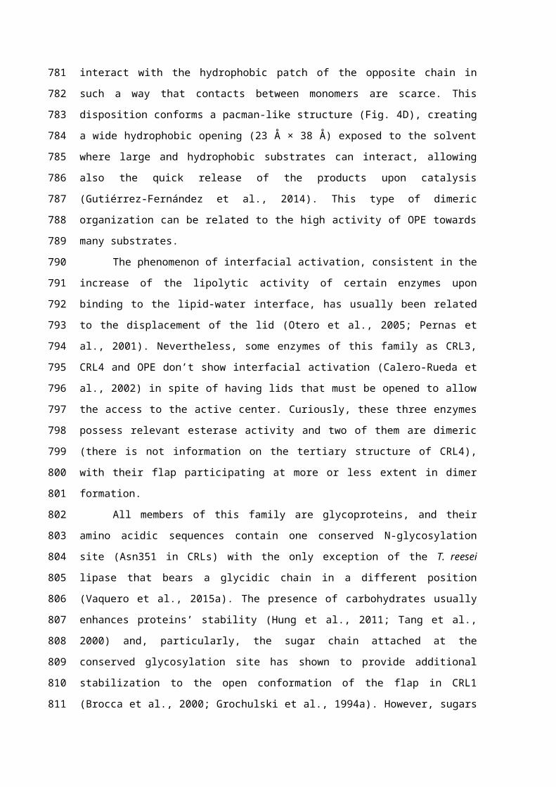
Figure captions
Figure 1. Sequence alignment of the characterized enzymes from the C. rugosa-like family.
Consensus residues at a given position are highlighted in blue. C. rugosa lipases 1, 2, 3 and 4
(CRL1-4), O. piceae sterol esterase (OPE), M. albomyces sterol esterase (STE1), T. reesei lipase
(Trire2), A. niger lipase (Aspni5), N. haematococca lipase (Necha2), P. sapidus lipase (Lip2),
B. cinerea lipase (BCL1), G. candidum lipases 1 and 2 (GCL1 and 2). In a red boxes are
highlighted the lid region (between the two cysteines, positions 97-141), the conserved residues
from the catalytic elbow (GESAG, positions 251-255), the other two residues from the catalytic
triad (Gln at position 389 and His at position 503) and the conserved N-glycosylation site (Asn
at position 399).
Figure 2. Common structural features of the members of the C. rugosa-like family. 3D structure
of the lipase 3 from C. rugosa (CRL3) is presented as model. A) Cartoon representations of
CRL3. B) Surface representation of CRL3. The core of the protein is formed by with
antiparallel β-sheets that form the α/β-hydrolase fold, the α-helices forming the lid is
highlighted in magenta, and the intramolecular tunnel is represented by a mesh. The residue
depicted in the centre of the protein corresponds to the catalytic Ser.
Figure 3. Models of the intramolecular tunnels modeled using Caver 2.0 in the C. rugosa-like
lipases. Cartoon and surface representations are presented for all the proteins, the lid of each
enzyme is highlighted in magenta, and the intramolecular tunnels are represented by a mesh. A)
C. rugosa lipase 3 (CRL3), B) O. piceae sterol esterase (OPE), C) M. albomyces sterol esterase
(STE1), D) T. reesei lipase (Trire2), E) A. niger lipase (Aspni5), F) N. haematococca lipase
(Necha2), G) P. sapidus lipase, H) B. cinerea lipase (BCL1), I) G. candidum lipase 2 (GCL2).
Figure 4. Case study: O. piceae sterol esterase (OPE). A) Detail on the structure of OPE with a
large internal tunnel that exits opposite to the lid region near a glycosylation site. The lid
undergoes a great displacement (~30 Å) from its closed to its open conformation (Gutiérrez-
Fernández et al., 2014). B) Heterologous expression of OPE in E. coli renders a non-active
unfolded protein as shown by circular dichroism (far UV spectrum) (Vaquero et al., 2015b),
while expression in P. pastoris increases its catalytic properties due to a modification of the N-
terminus of the protein. C) Introduction of a bulky residue (Ile544Trp) at the end of the internal
tunnel of OPE modified its substrate specificity. The activity of OPE against p-nitrophenyl
esters of different acyl-chain length was modified, abolishing its activity against these long-
chain fatty acid esters. D) Monomers association in CRL3 and OPE to form a dimer in their
active form.
972973974
975
976
977
978
979
980
981
982
983
984
985
986
987
988
989
990
991
992
993
994
995
996
997
998
999
1000
1001
1002
1003
1004
1005
1006
1007
1008

Organism Habitat % id* Similarity % aac Reference
C. rugosa Soil 79-88a 88-89b 534 Lotti et al., 1994
G. candidum Cosmopolitan 41 57 544 Bertolini et al., 1995
O. piceae Wood 44 62 537 Calero-Rueda et al., 2009
M. albomyces Soil 43 58 558 Kontkanen et al., 2006a
B. cinerea Vine pathogen 46 61 558 Comménil et al., 1999
P. sapidus Wood pathogen 42 61 528 Zorn et al., 2005
T. reesei Soil 41 54 551 Vaquero et al., 2015a
N. haematococca Ubiquitous 40 54 570 Vaquero et al., 2015a
A. niger Soil 41 57 544 Vaquero et al., 2015a* Sequence identity with C. rugosa lipases a Sequence identity among the isoenzymes CRL1-4 b Structural similarity among the isoenzymes CRL1-4 c Number of amino acids in the mature protein
Table I. Known lipases/sterol esterases belonging to the C. rugosa-like family1009
1010
1011
1012

Fig 11013
1014
1015

Fig 21016
1017
1018
1019
1020
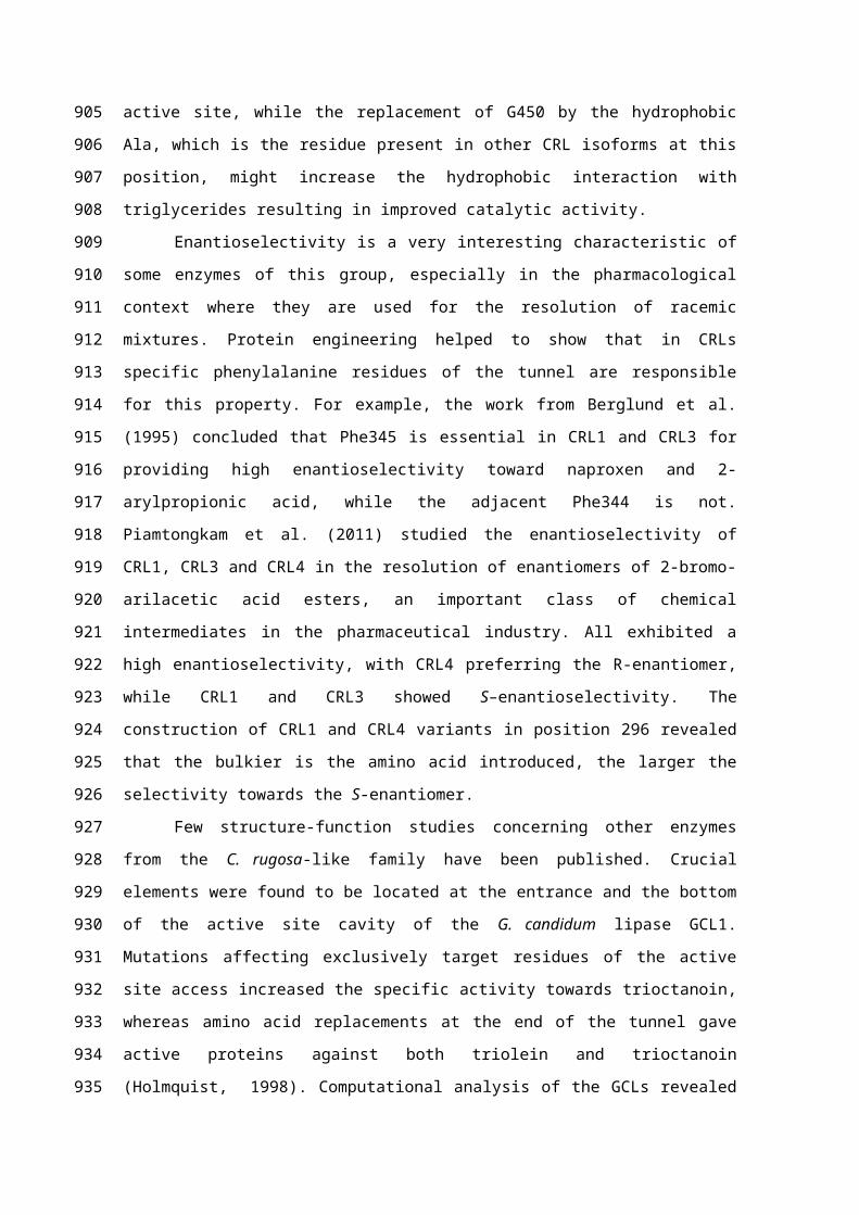
Fig 31021
1022
1023
1024

Fig 4102510261027
1028