viewpoints complex receptive fields in primary visual...
TRANSCRIPT

Volume 9, Number 5, 2003 THE NEUROSCIENTIST 317Copyright © 2003 Sage PublicationsISSN 1073-8584
In 1938, Hartline introduced the term “receptive field”to name a region of the retina where a change in lightbrightness modified the firing rate of a retinal ganglioncell. Kuffler (1953) would later demonstrate thatHartline’s receptive field had a specific spatial structureknown as center surround, and in a series of studies tofollow, Hubel and Wiesel (1959, 1961, 1962, 1965)would extend the term receptive field to other cells invisual cortex and would demonstrate that receptive-fieldstructures become increasingly complex at successivestages of the visual pathway.
In comparison with the retina and the lateral genicu-late nucleus (LGN), the primary visual cortex turned outto have a remarkable variety of receptive fields. Hubeland Wiesel classified cortical receptive fields into twomain categories—simple cells and complex cells—afteradmitting that “new varieties [of receptive fields] arecontinually appearing, and it is unlikely that the ones wehave listed give anything like a complete picture of thestriate cortex” (Hubel and Wiesel 1962, p 109). Hubeland Wiesel’s (1962) simple receptive fields had a verycharacteristic spatial structure. Like cells in the LGN,they had separate on and off subregions that could be
mapped with small spots of light. Unlike geniculatecells, the on and off subregions were elongated and par-allel instead of circular and concentric (Fig. 1). Simplereceptive fields were identified based on four criteria:
1. they were subdivided into distinct excitatory andinhibitory regions,
2. there was summation within the separate excita-tory and inhibitory parts,
3. there was antagonism between excitatory andinhibitory regions, and
4. it was possible to predict responses to stationaryor moving spots of various shapes from a map ofthe excitatory and inhibitory areas.
In contrast, complex receptive fields formed a muchmore diverse population and were identified by exclu-sion. Figure 2 shows three different types of complexreceptive fields, as illustrated in Hubel and Wiesel(1962). Those three examples share very little in com-mon. Cell A generates on-off responses throughout theentire receptive field. Cell B responds exclusively to ablack horizontal bar. Cell C has partially separated onand off regions, but unlike a simple cell, its receptivefield cannot be mapped with light spots.
Hubel and Wiesel’s (1962) description did not providea quantitative test to clearly distinguish between simpleand complex receptive fields. The lack of such a testwould later become a serious problem and eventuallywould lead to the proposal of many different classifica-tion criteria (Palmer and Rosenquist 1974; Schiller andothers 1976; Henry 1977; Henry and others 1983;Tanaka 1983; Toyama and others 1981a, 1981b; see
Complex Receptive Fields in Primary Visual CortexLUIS M. MARTINEZ and JOSE-MANUEL ALONSO
In the early 1960s, Hubel and Wiesel reported the first physiological description of cells in cat primary visu-al cortex. They distinguished two main cell types: simple cells and complex cells. Based on their distinctresponse properties, they suggested that the two cell types could represent two consecutive stages inreceptive-field construction. Since the 1960s, new experimental and computational evidence provided seri-ous alternatives to this hierarchical model. Parallel models put forward the idea that both simple and com-plex receptive fields could be built in parallel by direct geniculate inputs. Recurrent models suggested thatsimple cells and complex cells may not be different cell types after all. To this day, a consensus among hier-archical, parallel, and recurrent models has been difficult to attain; however, the circuitry used by all mod-els is becoming increasingly similar. The authors review theoretical and experimental evidence for each lineof models emphasizing their strengths and weaknesses. NEUROSCIENTIST 9(5):317–331, 2003. DOI:10.1177/1073858403252732
KEY WORDS Thalamus, Thalamocortical, Corticocortical, Cortical circuitry, Striate cortex
This study was supported by NIH and HFSP. We thank Dario Ringachand Casto Rivadulla for their comments on the article.
Neuroscience and Motor Control Group (Neurocom), Department ofMedicine, Campus de Oza, Universidade de A Coruña (LMM).Department of Biological Sciences, SUNY College of Optometry(J-MA).
Address correspondence to: Jose-Manuel Alonso, Department ofBiological Sciences, SUNY-Optometry, New York, NY 10036 (e-mail:[email protected]).
VIEWPOINTS ■

318 THE NEUROSCIENTIST Complex Receptive Fields
Orban 1984 for review). Simple cells and complex cellshave been classified based on the presence and degree ofoverlap of on and off subregions (Fig. 3A), spontaneousactivity level, response amplitude, length summation,responses to patterns of random dots, responses to mov-ing light and dark bars, responses to moving edges,responses to drifting or contrast-reversal gratings, andresponses to flashed bars and reverse correlation maps(Fig. 3B; see Skottun and others 1991a); Mechler andRingach 2002, for review). To give an example of theconfusion, cell B of Figure 2 could probably be classi-fied by different authors as complex (e.g., Hubel andWiesel 1962), simple (e.g., Skottun and others 1991a),S1 (e.g., Schiller and others 1976; Martin andWhitteridge 1984; Jones and Palmer 1987; see Orban1984, for review) or Eoff (e.g., Tanaka 1983). Similarly,cell C could probably be classified either as complex(e.g., Hubel and Wiesel 1962) or simple (e.g., Debanneand others 1998).
A successful quantitative test to classify simple andcomplex cells was introduced by De Valois and col-leagues (1982) and further refined by Skottun and others(1991a). The so-called response modulation method pro-vided a mixed quantification of the 2nd and 3rd criteriaof Hubel and Wiesel (1962) and rendered two cell popu-lations that roughly corresponded to simple and complexcells (Fig. 3C). This new classification was based on thedifferent response modulation of simple cells and com-plex cells to drifting sinusoidal gratings. Although sim-ple cells tend to modulate their firing rate in phase withthe stimulus, complex cells elevate their firing with littleor no modulation. Thus, the ratio between the amplitudeof the first Fourier harmonic and the mean spike rate canbe used as a quantitative index of “response non-linearity”or “receptive-field complexity” (Fig. 3C). Unfortunate-ly, this test is not free of limitations. The “response mod-
ulation” does not provide any information on the recep-tive-field geometry (e.g., number and shape of subre-gions), and therefore it cannot distinguish an X-genicu-late cell from a simple cell even if thalamic and corticalcells are clearly different populations. Moreover, grant-ed a nonlinear relationship between synaptic current andfiring rate, cells with identical synaptic inputs and intrin-sic properties can still show a bimodal distribution ofresponse modulation (Mechler and Ringach 2002).
The difficulty in classifying receptive fields is notsimply a problem of semantics. It is closely related tofundamental questions such as, how are simple and com-plex receptive fields generated? And what is the role ofsimple and complex receptive fields in visual process-ing? Possible answers to the first question have beenintensively investigated over the past decades by experi-mental and computational neuroscientists. In this article,we review the history of ideas and receptive-field mod-els that, for didactic purposes, we divide into three cate-gories: hierarchical, parallel, and recurrent. Each ofthese models has made important contributions to ourcurrent understanding of how complex receptive fieldsare generated, and, as recently noticed by Kevan Martin(2002), all models have become progressively similar. Inthis review, we attempt to identify the barriers that keepthem apart, driving us away from consensus.
What Makes a Complex Cell Complex?
Before we review the different models, it is important tofurther clarify what we mean by “complex cell.”Although simple cells (as defined by Hubel and Wiesel1962) have relatively similar receptive-field structures(Fig. 1), complex cells are very diverse, and the termclearly embraces different populations (Fig. 2). Here, a
Fig. 1. Simple receptive fields (colored version of Figure 2 from Hubel and Wiesel 1962). Red crosses represent on subregions andblue triangles off subregions.

Volume 9, Number 5, 2003 THE NEUROSCIENTIST 319
simple cell is defined by the four Hubel and Wiesel(1962) criteria (see above), and a complex cell is definedby exclusion: any cortical neuron that does not have asimple receptive field.
In our view, a large body of evidence indicates thatsimple cells are a separate population from the rest ofcortical cells in cat visual cortex.
1. Simple cells are segregated in specific corticallayers (e.g., the overwhelming majority of cellsin layer 4 have simple receptive fields; Hubeland Wiesel 1962; Gilbert 1977; Gilbert andWiesel 1979; Hirsch and others 1998, 2002;Martinez and others 2002; but see Orban 1984).
2. The overwhelming majority of spiny stellatecells in cat area 17 are simple cells (Kelly andVan Essen 1974; Gilbert and Wiesel 1979;Martin and Whitteridge 1984; Hirsch and others1998, 2002; Martinez and others 1999, 2002).
3. At least in layer 4, all simple cells have recep-tive-field structures consistent with a push-pullorganization (e.g., within each subregion, stim-uli of the opposite contrast evoke synapticresponses of the opposite sign; Ferster 1988;Hirsch and others 1998, 2002; Martinez and oth-ers 1999, 2002).
4. All simple cells receive direct geniculate input;in contrast, only a minority of complex cells does(most complex cells in the superficial layers andlayer 5 do not receive direct geniculate input[e.g., Ferster and Lindstrom 1983; Martin andWhitteridge 1984; Alonso and Martinez 1998]).
5. The majority of simple cells generate responsesthat are roughly linear (Hubel and Wiesel 1962;Movshon and others 1978b; Skottun and others
1991a; Ferster 1994; Carandini and others 1997;Lampl and others 2001a).
6. Mechler and Ringach (2002) have recently sug-gested that it is necessary to show a bimodal dis-tribution of cortical response properties to provethat simple cells are a separate class from com-plex cells. Although their argument is valid, sta-tistical tools cannot be taken as the only possiblesource of proof. Neuroscientists do not discusswhether pyramidal cells and spiny stellate cellsare different classes, and to our knowledge,nobody has shown that soma shapes arebimodally distributed.
It should also be emphasized that most of the evi-dence cited above comes from studies in cat visual cor-tex (using Hubel and Wiesel criteria for classification).Over the past two decades, we have accumulated enormousknowledge on the anatomy and physiology of cat area17, although essential data in primate are still missing.Following are a few examples of unknowns in primate:
1. There are very few studies that measured thereceptive-field structure of cells that receivedirect geniculate input (as identified by electri-cal stimulation or cross-correlation analysis;Bullier and Henry 1980).
2. We have almost no data on intracellular physiol-ogy in vivo (Anderson and others 1993).
3. There are very few studies correlating neuronalmorphology and receptive-field properties(McGuire and others 1991; Anderson and others1993).
4. There is still no agreement on whether there is alaminar segregation of simple cells and complex
Fig. 2. Three different types of complex receptive fields (modified version of Figures 4, 6, and 7 of Hubel and Wiesel 1962). Complexcells are a very heterogeneous population. Cell A generates on-off responses throughout the entire receptive field. Cell B respondsexclusively to a black horizontal bar. Cell C has partially separated on and off regions but the receptive field cannot be mapped withsmall spots of light. The green icons on the left represent the complex receptive field with the stimuli (flashed bars or borders) over-laid.

320 THE NEUROSCIENTIST Complex Receptive Fields
cells in layer 4C (e.g., layer 4C has mostly sim-ple cells and unoriented cells [Hubel and Wiesel1968; Bullier and Henry 1980]; layer 4C hassimple cells and complex cells in similar pro-portions (Ringach and others 2002]).
Consistent with this reasoning, most of the work revisedhere is based on studies of cat visual cortex or computa-tional models based on the physiology of the cat visualcortex.
Fig. 3. Simple and complex cells differ intheir receptive-field structure and theirresponses to static and moving stimuli. A,Idealized receptive-field maps of a simplecell and a complex cell. B, Cartoon of theintracellular responses from a simple cellto small spots (light/dark) presented oneach subregion (left and middle, thetraces show a push-pull arrangement ofexcitatory and inhibitory inputs in the sim-ple receptive field). Complex cells lacksegregated on and off regions; smallspots (light/dark) evoke on and off (push-push) responses throughout the entirereceptive field (right). C, Cartoon repre-senting the responses of a simple cell anda complex cell to drifting sinusoidalgratings.
Volume 9, Number 5, 2003 THE NEUROSCIENTIST 321
Complex Receptive-Field Models
Hierarchical Models
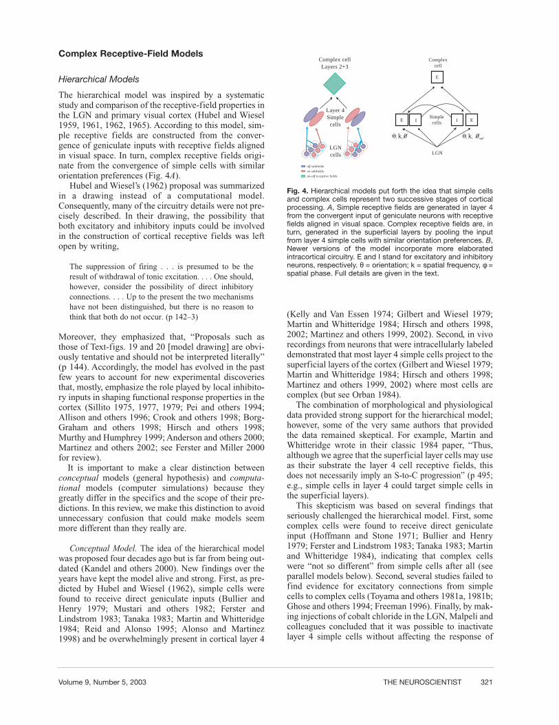
The hierarchical model was inspired by a systematicstudy and comparison of the receptive-field properties inthe LGN and primary visual cortex (Hubel and Wiesel1959, 1961, 1962, 1965). According to this model, sim-ple receptive fields are constructed from the conver-gence of geniculate inputs with receptive fields alignedin visual space. In turn, complex receptive fields origi-nate from the convergence of simple cells with similarorientation preferences (Fig. 4A).
Hubel and Wiesel’s (1962) proposal was summarizedin a drawing instead of a computational model.Consequently, many of the circuitry details were not pre-cisely described. In their drawing, the possibility thatboth excitatory and inhibitory inputs could be involvedin the construction of cortical receptive fields was leftopen by writing,
The suppression of firing . . . is presumed to be theresult of withdrawal of tonic excitation. . . . One should,however, consider the possibility of direct inhibitoryconnections. . . . Up to the present the two mechanismshave not been distinguished, but there is no reason tothink that both do not occur. (p 142–3)
Moreover, they emphasized that, “Proposals such asthose of Text-figs. 19 and 20 [model drawing] are obvi-ously tentative and should not be interpreted literally”(p 144). Accordingly, the model has evolved in the pastfew years to account for new experimental discoveriesthat, mostly, emphasize the role played by local inhibito-ry inputs in shaping functional response properties in thecortex (Sillito 1975, 1977, 1979; Pei and others 1994;Allison and others 1996; Crook and others 1998; Borg-Graham and others 1998; Hirsch and others 1998;Murthy and Humphrey 1999; Anderson and others 2000;Martinez and others 2002; see Ferster and Miller 2000for review).
It is important to make a clear distinction betweenconceptual models (general hypothesis) and computa-tional models (computer simulations) because theygreatly differ in the specifics and the scope of their pre-dictions. In this review, we make this distinction to avoidunnecessary confusion that could make models seemmore different than they really are.
Conceptual Model. The idea of the hierarchical modelwas proposed four decades ago but is far from being out-dated (Kandel and others 2000). New findings over theyears have kept the model alive and strong. First, as pre-dicted by Hubel and Wiesel (1962), simple cells werefound to receive direct geniculate inputs (Bullier andHenry 1979; Mustari and others 1982; Ferster andLindstrom 1983; Tanaka 1983; Martin and Whitteridge1984; Reid and Alonso 1995; Alonso and Martinez1998) and be overwhelmingly present in cortical layer 4
(Kelly and Van Essen 1974; Gilbert and Wiesel 1979;Martin and Whitteridge 1984; Hirsch and others 1998,2002; Martinez and others 1999, 2002). Second, in vivorecordings from neurons that were intracellularly labeleddemonstrated that most layer 4 simple cells project to thesuperficial layers of the cortex (Gilbert and Wiesel 1979;Martin and Whitteridge 1984; Hirsch and others 1998;Martinez and others 1999, 2002) where most cells arecomplex (but see Orban 1984).
The combination of morphological and physiologicaldata provided strong support for the hierarchical model;however, some of the very same authors that providedthe data remained skeptical. For example, Martin andWhitteridge wrote in their classic 1984 paper, “Thus,although we agree that the superficial layer cells may useas their substrate the layer 4 cell receptive fields, thisdoes not necessarily imply an S-to-C progression” (p 495;e.g., simple cells in layer 4 could target simple cells inthe superficial layers).
This skepticism was based on several findings thatseriously challenged the hierarchical model. First, somecomplex cells were found to receive direct geniculateinput (Hoffmann and Stone 1971; Bullier and Henry1979; Ferster and Lindstrom 1983; Tanaka 1983; Martinand Whitteridge 1984), indicating that complex cellswere “not so different” from simple cells after all (seeparallel models below). Second, several studies failed tofind evidence for excitatory connections from simplecells to complex cells (Toyama and others 1981a, 1981b;Ghose and others 1994; Freeman 1996). Finally, by mak-ing injections of cobalt chloride in the LGN, Malpeli andcolleagues concluded that it was possible to inactivatelayer 4 simple cells without affecting the response of
Layer 4Simplecells
LGNcells
Complex cellLayers 2+3
Complexcell
off subfields
on subfields
on-off receptive fields
E E
E
I ISimplecells
0 k 01 1 0 k 01 1 90
LGN
Fig. 4. Hierarchical models put forth the idea that simple cellsand complex cells represent two successive stages of corticalprocessing. A, Simple receptive fields are generated in layer 4from the convergent input of geniculate neurons with receptivefields aligned in visual space. Complex receptive fields are, inturn, generated in the superficial layers by pooling the inputfrom layer 4 simple cells with similar orientation preferences. B,Newer versions of the model incorporate more elaboratedintracortical circuitry. E and I stand for excitatory and inhibitoryneurons, respectively. θ = orientation; k = spatial frequency, φ =spatial phase. Full details are given in the text.

322 THE NEUROSCIENTIST Complex Receptive Fields
complex cells in the superficial layers (Malpeli 1983;Malpeli and others 1986; Mignard and Malpeli 1991).
Some of the criticisms to the hierarchical model havebeen addressed over the years. By recording from verti-cally aligned layer 4 and layers 2+3 cells within the cor-tex, evidence for excitatory connections from simplecells to complex cells was found (Alonso and Martinez1998). Furthermore, by making injections of GABA inthe LGN, the complex cells described by Malpeli werefound to be a minority; most layers 2+3 complex cellsare not visually driven when the thalamic input to layer4 is inactivated (Martinez and Alonso 2001; Callaway2001). Finally, additional support for the hierarchicalmodel came from another discipline that would becomeincreasingly important over the years: computationalneuroscience.
Computational Model. Most theoretical approachesto complex receptive-field generation were open up bythe elegant “two-bar interaction” experiments of TonyMovshon and colleagues (1978a). They argued that ifcomplex cells respond to stationary or moving forms ina way that cannot be predicted from their first-orderresponses (e.g., responses to single bars or small spots),it must be because complex-cell responses depend onnonlinear interactions between at least two positions inspace and time. Movshon and colleagues showed thatwhen sets of two bars were applied to complex cells,their responses displayed on and off linear subunitsresembling simple-cell subregions (see also Baker andCynader 1986; Gaska and others 1994; Szulborski andPalmer 1990; Heggelund 1981). Emerson and others(1992) extended this finding by demonstrating that thesubunits of directionally selective complex-cells werealso directional selective. Similarly, Ohzawa andFreeman (1986) demonstrated that the subunits ofbinocular complex cells were also binocular and had thesame optimal disparity as the complex cell (see alsoOhzawa and others 1990 1997; Anzai and others 1999).
Based on these findings, many authors modeled com-plex cells’ responses as a square sum of simple cells withsimilar orientation and spatial frequency but with phas-es that differed by 90 degrees (e.g., Emerson and others1992; Fleet and others 1996; Ohzawa and others 1990,1997; Pollen and others 1989; Qian and Zhu 1997; Sakaiand Tanaka 2000; Okajima and Imaoka 2001; Shams andMalsburg 2002a, 2002b). Mathematically, this can bedescribed as a square sum of two linear operators, eachcharacterized by a Gabor function of the same frequen-cy but with phases 90 degrees apart from each other.Because a linear filter followed by a squaring device andthen an integrator is considered an energy detector(Green and Swets 1966), these models are collectivelyknown as energy models (Adelson and Bergen 1985).Recently, Okajima and Imaoka (2001) demonstrated thatenergy models render complex cells that are optimallydesigned from an information-theory point of view.However, as an interesting alternative, Lampl and others(2001b) suggested that the pooling of simple cell inputs
may be better described by a MAX operation. That is,the strongest input determines the response of the com-plex cell (Riesenhuber and Poggio 1999, 2002) in whichthe strongest input determines the response of the com-plex cell. Linear or nonlinear, both proposals rest on theassumption that simple and complex receptive fieldsrepresent two successive stages of cortical processing. Acartoon version of the core circuitry predicted by energymodels is shown in Figure 4B.
Although energy models seem to be good news for thehierarchical hypothesis, a key question remains: how isthis precise synaptic connectivity achieved during devel-opment? Recently, it has been shown that a two-layernetwork fed with natural images can learn the phase-invariance characteristic of complex cells (Einhauserand others 2002). The model uses layers of neurons thatlearn their properties from competitive Hebbian algo-rithms (based on the relative timing of pre- and postsy-naptic spikes). Interestingly, neurons in the first layerlearn to respond like simple cells, and then simple cellresponses are used by neurons in the second layer tolearn to respond as complex cells. Similar approacheshave been used in the past to model the emergence ofocular dominance and orientation columns (e.g., Miller1996; Miller and others 1999). Hyvarinen and Hoyer(2001) also used natural images to make neurons learnto respond as complex cells in a network organizationthat is very similar to cat area 17 (cells clustered accord-ing to retinotopy, spatial frequency, and orientation butindependently of spatial phase; see DeAngelis and oth-ers 1999). Again, in this model, simple-cell responsesemerge in the first layer (see also Olshausen and Field1996) and complex cells in the second layer.
The hierarchical model has been very successful ingreat part because of its appealing simplicity. The essen-tial idea that simple cells and complex cells representtwo stages in receptive-field construction is likely toremain correct even if newer models incorporateincreasingly more realistic circuits (see below).However, the hierarchical has model also been justlycritized. One of these criticisms is based on an experi-mental finding that is now unanimously accepted: Somecomplex cells do receive direct geniculate input. Thiscriticism makes it necessary for the hierarchical modelto embrace other ideas and evolve.
Parallel Models
Conceptual Model. Historically, the first strong evi-dence against the hierarchical model was the discoverythat some complex cells, like simple cells, receive mono-synaptic input from the thalamus (Hoffmann and Stone1971). Based on this discovery, Hoffman and Stone pro-posed that both cell types, simple and complex, weregenerated in parallel by separate thalamocortical path-ways (Hoffman and Stone 1971; Stone and others 1979;see Fig. 5A). But how could complex cells become non-linear without pooling simple cell inputs? It was already
Volume 9, Number 5, 2003 THE NEUROSCIENTIST 323
known that there were linear and nonlinear cells in theretina and the LGN (X and Y cells, respectively [Shapleyand Hochstein 1975]) and that the two cell types formedseparate parallel channels: X cells in the retina projectedto X cells in LGN and Y retinal cells projected to Ygeniculate cells (see Stone and others 1979 for review).Hoffman and Stone used this evidence to propose thatlinear simple cells were built from the convergence oflinear X-cell inputs and nonlinear complex cells fromthe convergence of nonlinear Y-cell inputs.
A striking experimental result soon came in supportof the new parallel model: some complex cells seemedto respond to visual stimuli that were not effective indriving simple cells (Hammond and Mackay 1975,1977). This result was reinforced by a series of remark-able studies done by Malpeli and colleagues (Malpeli1983; Malpeli and others 1986; Mignard and Malpeli1991). Malpeli and others (1986) found that injections ofcobalt chloride in layer A of LGN inactivated layer 4simple cells but not layer 2+3 complex cells (Malpeli1983); responses at layers 2+3 cells were only affectedwhen the inactivation of LGN (layer A) was combinedwith large lesions in area 18 (Mignard and Malpeli1991).
The results of Malpeli and colleagues fitted very nice-ly with the idea that simple and complex receptive fieldswere built in parallel and independently. However, paral-lel models soon found serious criticisms also. First, theidea of two cleanly segregated parallel channels (X →simple cell, Y → complex cell) was discarded by a largenumber of studies (Singer and others 1975; Bullier andHenry 1979; Tanaka 1983; Ferster and Lindstrom 1983;Martin and Whitteridge 1984). Second, some authorsshowed that the impact of the entire Y pathway in catarea 17 was weak (Spitzer and Hochstein 1987; Ferster1990a, 1990b; Burke and others 1992). Third, Skottunand colleagues (1988, 1991b) presented evidenceagainst the Hammond and Mackay (1975, 1977) find-ings showing that most simple cells do respond to thestimuli that drive complex cells (but see Hammond1991). Finally, with the availability of new techniques,Malpeli’s results were found to be correct only for aminority of cells in the superficial layers (Martinez andAlonso 2001; Callaway 2001)—most layer 2+3 complexcells cannot be driven from within the classical receptivefield when the main thalamic input to layer 4 is inacti-vated (see also Rivadulla and Sur 2000).
Regardless of the specifics, the main idea of parallelmodels remained valid: at least some complex receptivefields could be constructed from direct geniculateinputs. However, an important question was still withoutan answer. How can a complex cell be orientation selec-tive if its main inputs are nonoriented and its receptivefield does not have elongated subregions? Once again,computational neuroscience came to the rescue (Mel andothers 1998).
Computational Model. Mel and colleagues (1998)clearly showed that a cortical neuron could receive inputfrom geniculate cells with overlapping on and off centers
and still produce phase-invariant orientation tuning. Inthis model, the arrangement of geniculate inputs, origi-nated by a Hebbian developmental rule, produces func-tional subunits scattered across the dendrites. Thesefunctional subunits respond more efficiently to specificline orientations by activating local voltage-dependentexcitatory currents (see also Spitzer and Hochstein1985, 1988; see Fig. 5B, left).
In a similar line of thinking, a more recent parallelmodel combined thalamic and cortical inputs to generatelinear and nonlinear visual responses (Tao and others2001, forthcoming; see also Wielaard and others 2001).In Tao and others’ (2001, forthcoming) model, nonlinearresponses originate in cells that receive weak geniculateinput and linear responses in cells that receive stronggeniculate input (Fig. 5B, right). In agreement with Taoand others model, complex cells (that generate nonlinearresponses) are abundant in cortical layers that receive theweakest thalamic input, and the converse is true for sim-ple cells that generate linear responses. Also consistentwith this model, a large number of complex cells in catprimary visual cortex do not receive measurable directgeniculate input (e.g., Ferster and Lindstrom 1983;Martin and Whitteridge 1984; Alonso and Martinez1998).
Simple cells and complex cells are far from being twoparallel cortical pathways in the same way that X and Ycells are parallel thalamic pathways. However, the ideathat some complex receptive fields can be generated atleast in part by direct thalamic inputs is likely to be cor-rect. In support of this idea, recent computational mod-els have provided plausible mechanisms to generatecomplex receptive fields directly from geniculate inputs(Spitzer and Hochstein 1988; Mel and others 1998; Taoand others 2001). Some of these models (Tao and others2001, forthcoming) also emphasize another possible keyplayer in the construction of complex receptive fields:the intracortical circuitry.
Recurrent Models
Conceptual Model. Perhaps the most widely usedargument against any thalamocortical model (hierarchi-cal or parallel) is the nature of the cortical circuit itself.The number of geniculate synapses is only a small frac-tion of the total excitatory synapses made onto corticalcells (LeVay and Gilbert 1976; Kisvarday and others1986; Peters and Payne 1993; Ahmed and others 1994).Therefore, cortical responses should be determinedmostly by cortical inputs and not by thalamic inputs(Martin 2002).
Douglas and Martin (Douglas and others 1989;Douglas and Martin 1991; see also Douglas and others1995) developed the conceptual frame for a new type ofmodel that is known as a recurrent model (Fig. 6A).Their proposal rests on three key assumptions:
1. Thalamic excitatory inputs are weak becausegeniculate synapses account for less than 10%

324 THE NEUROSCIENTIST Complex Receptive Fields
of the total excitatory synapses onto layer 4 cells(Kisvarday and others 1986; Peters and Payne1993; Ahmed and others 1994).
2. Cortical excitation from neighboring neurons isvery strong and serves to amplify the weak thal-amic input.
3. Weak cortical inhibition controls the gain of the“cortical amplifier” preventing runaway excita-tion and adjusting the network performance tochanges in visual stimulation.
In initial versions of recurrent models, the predomi-nance of intracortical excitation over intracortical inhibi-tion was emphasized because 80% of the synapses oncortical cells are excitatory (Braitenberg and Schuz1991) and because intracellular measurements failed tofind evidence for strong inhibition in response to visualstimuli (Berman and others 1991; Douglas and others1991; Ferster 1988). However, our view of cortical inhi-bition has changed over the years (Sillito 1975, 1977,1979; Pei and others 1994; Allison and others 1996; Crookand others 1998; Borg-Graham and others 1998; Hirschand others 1998; Murthy and Humphrey 1999; Andersonand others 2000; Martinez and others 2002), and,accordingly, new recurrent models are incorporatingstronger inhibitory inputs (Somers and others 1995;Ben-Yishai and others 1995; reviewed in Sompolinskiand Shapley 1997).
Recurrent models claim to be more faithful to thestructure of the cortical network than hierarchical mod-els, and, to some extent, they are. Cortical neurons (sim-ple or complex) receive abundant input from other corti-cal neurons both inhibitory and excitatory (e.g., Ahmedand others 1994; Callaway 1998; Fitzpatrick 1996;Braitenberg and Schuz 1998). Therefore, any model thatincorporates more elaborate intracortical circuitry is insome way closer to the reality than are models that useprimarily one type of input. That being said, the idea thatgeniculate inputs are much weaker than cortical inputs isunlikely to be correct. Although the number of genicu-late synapses is a small percentage of the total excitato-ry synapses made on cortical cells (5% to 25% depend-ing on which study is cited; e.g., Peters and Payne 1993;Levay and Gilbert 1976), geniculocortical connectionshave many other features that make them strong.
1. Geniculate synapses are bigger than corticalsynapses (e.g., Ahmed and others 1994).
2. Geniculate synapses are located proximally inthe dendrites, whereas excitatory corticalsynapses tend to be located more distally (e.g.,Ahmed and others 1994).
3. Geniculate synapses have more release sitesthan cortical synapses do (Gil and others 1999).
4. Thalamocortical excitatory postsynaptic poten-tials (EPSPs) are likely to be larger than cortico-
Fig. 5. A, Parallel models advocate that simple cells and complex cells are both created in parallel from different thalamocortical path-ways. B, The most recent versions of parallel models come in two sorts that exploit the preponderance of the single cells over the cir-cuit (Mel and others 1998) or vice versa (Tao and others 2001, forthcoming). Mel and others’ (1998) proposal generates complex-cellresponses that are orientation selective through specific intradendritic computations. Tao and others’ scheme obtains simple cells andcomplex cells by modulating the gain of the thalamocortical and corticocortical inputs. The line thickness represents the strength ofthe connection. E and I stand for excitatory and inhibitory neurons, respectively. See the text for details.

Volume 9, Number 5, 2003 THE NEUROSCIENTIST 325
cortical EPSPs (Gil and others 1999; Stratfordand others 1996).
5. Geniculate cells have generally higher firingrates than cortical cells do (e.g., Bullier and oth-ers 1982).
6. Many of the geniculate cells that converge onthe same cortical cell generate precise andstrong synchronous firing (Alonso and others1996; such precise and strong synchrony is notfound within the cortex).
7. The inactivation of a tiny region of LGN isenough to silence the activity of most corticalcells in layers 4 and 2+3 (Martinez and Alonso2001; see also Malpeli 1983). The inactivationof a similarly tiny region in the cortex has only asubtle effect on the response of a cortical cell(Bolz and Gilbert 1986; Grieve and Sillito1995).
In general, it is risky to estimate the strength of agiven pathway based solely on the number of synapticcontacts. Indeed, if this type of assumption were correct,the geniculate receptive fields should resemble morecortical receptive fields than retinal receptive fields, andthis is clearly not the case (Hubel and Wiesel 1961;Cleland and others 1971; Usrey and others 1998; genic-ulate cells receive 7% of excitatory synapses from theretina and about 40% from the cortex; Van Horn and oth-ers 2000).
Although the relative contribution of thalamic andcortical inputs to cortical receptive-field generation isstill a matter of debate (Ferster and Miller 2000; Martin2002), recent models have chosen to focus on a differentquestion: how do recurrent networks generate simpleand complex receptive fields? This focus is the seed of anew bold idea: Simple cells and complex cells may notbe different cell types after all (e.g., Mechler andRingach 2002; Abbott and Chance 2002). Supportingthis proposal, the work of Debanne and others (1998)showed that the relative strength of cortical on-offresponses could be modified by pairing visual stimuliwith current injections. In theory, if we can modify thestrength of on-off responses, we should also be able totransform a simple cell into a complex cell and viceversa. In reality, changes in the strength of on-offresponses are rare in the adult (four cases in Debanneand others 1998), and they are usually subtle. More pro-nounced changes are generated when the precise balancebetween cortical excitation and inhibition is manipulat-ed (Sillito 1975; Nelson and others 1994; Rivadulla andothers 2001). However, these changes are not inconsis-tent with a hierarchical model. In a hierarchical model, astimulus presented in an on subregion of a simple cellshould generate on responses because it activates mostlythe receptive-field centers from on-center geniculatecells. In the absence of cortical inhibition, the samestimulus should also activate off responses that originatefrom two main different sources:
Fig. 6. Recurrent models changed the focus of attention from single cells to networks of cortical connections. A, Most models areinspired by Douglas and Martin’s (1991) canonical microcircuit that emphasized the predominance of the input from intracortical con-nections over the thalamocortical input (bottom). The most recent addenda to the family of recurrent models are the so-called state-dependent models. State-dependent models propose that simple and complex cells originate from the same cortical circuit operat-ing at different amplification gains (top). B, Debanne and others’ (1998) model (left) emphasizes the balance between excitatory andinhibitory inputs as the origin of different cortical receptive fields. Chance and others’ (1999) model (right) emphasizes the strength oflocal excitatory connections within the superficial layers of the cortex. The line thickness represents the strength of the connection. Eand I stand for excitatory and inhibitory neurons, respectively. A more detailed explanation is given in the text.
326 THE NEUROSCIENTIST Complex Receptive Fields
1. the off surrounds from on-center geniculate cellsthat overlap the on subregion and
2. the off-center borders from off-center geniculatecells that overlap the adjacent off subregion (seeAlonso and others 2001 for multiple examples).
The fact that on-off responses are observed in simplecells after blocking inhibition is exactly what we wouldexpect from a hierarchical model and the properties ofthalamocortical connections (Reid and Alonso 1995;Alonso and others 2001). Also as expected from a hier-archical model, when on-off responses are quantitativelymapped and averaged, simple receptive fields still look“very simple” even if cortical inhibition is blocked(Murthy and Humphrey 1999; see also Rivadulla andothers 2001).
The idea that simple cells and complex cells are thesame cell type (or generated by the same basic circuit)seems hard to accept based on a large body of literaturein cat visual cortex (see the introduction of this review).Recent anatomical and physiological data also suggestthat thalamic inputs are not weaker than cortical inputs(see above). In spite of these criticisms, the “intracorti-cal emphasis” that emerged from recurrent models isimportant and is leading to powerful computationalmodels.
Computational Model. Recurrent models moved thefocus of attention from thalamo-cortical connections tonetworks of cortical neurons that are reciprocally con-nected (Martin 2002; Nelson 2002). With differencesregarding mainly the role of inhibitory inputs, recurrentmodels used networks of cortical neurons to explain theemergence of orientation and direction selectivity in thecortex (Somers and others 1995; Ben-Yishai and others1995; reviewed in Sompolinski and Shapley 1997).Somers and others (1995) developed a recurrent modelthat was especially successful at explaining a large bodyof experimental data. In this model, cortical excitationlinks cells with similar orientation preferences, whereascortical inhibition links cells with a broader range of ori-entations. The model was challenged by recent datademonstrating that the tuning for excitatory andinhibitory inputs in layers 4 and 2+3 is similar (Nelsonand others 1994; Anderson and others 2000; Martinezand others 2002). Also, against the model predictions,local excitatory connections have been found to extendfarther laterally than inhibitory connections do (Roerigand Chen 2001; Yousef and others 1999, 2001; Buzasand others 2001; see Carandini and Ringach 1997 forother criticisms). Another recurrent model was proposedby Ben Yishai and others (1995) to generate contrastinvariant orientation tuning. In this model, feedback con-nections are so strong that the cortical network becomesan orientation attractor (i.e., the output of the network isdetermined simply by the balance between excitationand inhibition independently of the properties of theinput). This type of orientation attractors (see alsoTsodyks 1999) has been challenged by data showing that
cortical responses to oriented gratings do depend on thespatial frequency of the input (Vidyasagar and Siguenza1985; Webster and De Valois 1985; Jones and others1987; Hammond and Pomfrett 1990).
The models of Somers and others (1995) and Ben-Yishai and others (1995) did not address specifically thegeneration of complex receptive fields; however, theywere very influential in setting the basis for future recur-rent models that did address the issue. Debanne and oth-ers (1998) used recurrent circuits to generate simple andcomplex cells. In their model, both cell types receiveinput from geniculate cells with overlapping on- and off-receptive-field centers (and other cortical cells). Simplecells are generated when the cortical inhibitory inputsare strong enough to impose a bias toward either on oroff responses. Complex cells are generated when inhibi-tion is reduced to unmask on-off responses. Debanne andothers’ model explains why simple cells generate on-offresponses when cortical inhibition is pharmacologicallyblocked (Sillito 1975; Ramoa and others 1988; Nelsonand others 1994; Shulz and others 1993). However, asdiscussed earlier, these findings can also be explainedwith a hierarchical model (see above). Moreover, someof the details of Debanne and others’ model are at oddswith experimental data. For example, not all smoothinhibitory neurons in layer 4 have S1 receptive fields(e.g., Gilbert and Wiesel 1979; Martin and others 1983;Kisvarday and others 1985, 1987; Azouz and others1997; Hirsch and others 2000), and the model fails toexplain why many layer 4 cells have receptive fields withpush-pull organization (Ferster 1988; Hirsch and others1998; Martinez and others 1999; Anderson and others2000).
Although Debanne and others’ (1998) model made abridge between parallel and recurrent models (Tao andothers 2001, forthcoming are also in this category), themodel of Chance and others (1999) abandoned thisbridge to adopt elements from a hierarchical organiza-tion. As in a hierarchical model, Chance and others usea first layer of simple cells (equivalent to layer 4) thatfeeds into another layer of cells with similar orientationpreferences (equivalent to layers 2+3). Chance and oth-ers model departs from a hierarchical organization bymaking the connections from layer 4 to layers 2+3 weak-er than the connections within layers 2+3 and by makingthe vertical connections link cells with similar spatialphases, whereas strong recurrent connections link cellswith different spatial phase (Fig. 6B, right). Althoughprevious recurrent models used cortical amplification togenerate orientation tuning and direction selectivity(Douglas and others 1995; Ben-Yishai and others 1995;Somers and others 1995), Chance et al. used corticalamplification to generate nonlinear responses. In sup-port of Chance et al.’s model, it has been suggested thatsimple cells and complex cells cannot be separated intotwo different populations (Mechler and Ringach 2002;Abbott and Chance 2002) and that complex cells canbehave like simple cells when the balance between exci-tation and inhibition changes (Sillito 1975; Nelson and

Volume 9, Number 5, 2003 THE NEUROSCIENTIST 327
others 1994; Shulz and others 1993; Rivadulla and oth-ers 2001). We have already criticized these two sugges-tions in detail (see above). Here, we could also add thatthe specificity of vertical connections (layer 4 → layers2+3) for spatial phase is difficult to reconcile with thelack of clustering for this property (DeAngelis and oth-ers 1999) and with the finding that some layer 4 simplecells with different spatial phases do converge on thesame superficial complex cells (Alonso and Martinez,unpublished observations). Furthermore, it has beenrecently shown that the synaptic connections betweenlayer 4 and layers 2+3 are among the strongest connec-tions in the cortical network (stronger than local hori-zontal connections between cells in the superficial lay-ers; Feldmeyer and others 2002).
As with any model, the circuitry predicted by recur-rent models will have to pass the test of time and exper-imental data. In any case, the idea that response lineari-ty could be modulated by the gain of the cortical networkis interesting and may prove to be correct in some extent.New experimental data will tell us whether this modula-tion exists, how strong it is, and which cortical layers itinvolves (see Rivadulla and others 2001).
Searching for a Consensus
Of all the criticisms that the hierarchical model hasreceived over the years, two have remained sufficientlystrong as to promote the birth of new conceptual frame-works (Fig. 7). First, as suggested by parallel models,some complex receptive fields can be generated at leastin part from direct geniculate inputs. Second, as sug-gested by recurrent models, cortical cells are heavilyinterconnected, and their responses are likely to be mod-ulated by recurrent connections. In spite of these criti-cisms, the idea that simple cells and complex cells rep-
resent two stages in receptive-field construction is likelyto remain correct even if models evolve and becomemore precise. Eventually, all models will mix to adoptelements from each other and finally converge into one.For example, Troyer and others (1998) introduced stronginhibitory inputs and more elaborate local connectivityin their hierarchical model of orientation selectivity incat layer 4 (see also Miller and others 2001 for review).Chance and others (1998) adopted a hierarchical organ-ization by using a first layer of simple cells that feedsinto a second layer of cells with more elaboratedresponses (simple or complex depending on networkgain). Also, Martin (2002) has recently described hisrecurrent model as an “alternative hierarchy” (the wordhierarchy was not used in the original description ofrecurrent or canonical microcircuits [e.g., Douglas andMartin 1991]). Finally, Tao and others (2001, forthcom-ing) used strong/weak geniculate inputs to model simplecells/complex cells, making simple cells “closer” to theirthalamic inputs than complex cells are (in previous par-allel models, complex cells received strong geniculateinputs, e.g., Mel and others 1998).
It seems clear that each line of models—hierarchical,parallel, or recurrent—can no longer be considered inisolation. Here, we propose a circuit diagram to integratethe main ideas/models discussed above (Fig. 7). The dia-gram cannot be easily ascribed to any of the main mod-els, although some could see it as clearly hierarchical,others as clearly parallel, and still others as clearly recur-rent. The diagram gives a general framework of the mainconnections as a function of cortical layers (layer 4, 2+3)and receptive-field properties (simple/complex). Layer 4simple cells and layers 2+3 complex cells are two stagesin receptive-field construction as proposed by the hier-archical model (Hubel and Wiesel 1962; Adelson andBergen 1985) and some recurrent models (Chance and
Fig. 7. Hubel and Wiesel’s (1962) hierarchical model (left) has been extensively challenged over the years. Two main criticisms (mid-dle) have propelled the emergence of alternative frameworks that accounted for the new experimental findings. Here, we propose anew circuit diagram for cat primary visual cortex (right) that embraces all current ideas of hierarchical, parallel, and recurrent models.Layer 4 simple cells and superficial complex cells form two successive stages in receptive-field construction. In addition, some cellsin layer 4 have complex receptive fields derived, in parallel, from direct geniculate inputs. At each layer, local intracortical circuits (bothexcitatory and inhibitory) modulate the gain of the network. See the text for a detailed explanation.

328 THE NEUROSCIENTIST Complex Receptive Fields
others 1998). At the same time, a subpopulation of com-plex cells has their receptive fields constructed fromdirect geniculate inputs (Spitzer and Hochstein 1988;Mel and others 1998; Tao and others 2001, forthcom-ing). Finally, at each layer, local intracortical circuits(both excitatory and inhibitory) modulate the gain of thecortical network (Debanne and others 1998; Chance andothers 1998; Tao and others 2001, forthcoming) generat-ing different results depending on the layer. Within lay-ers 2+3, response linearity would be modulated in someextent by local connections as proposed by recurrentmodels; within layer 4 (simple cells with push-pullreceptive fields), response linearity would not be so eas-ily modulated. Forty years had gone by since Hubel andWiesel proposed the hierarchical model; one wondershow many times the pendulum will swing back and forthbefore it finally sets at a standstill and we reach a con-sensus on how complex receptive fields are generated.
References
Abbott LF, Chance FS. 2002. Rethinking the taxonomy of visual neu-rons. Nat Neurosci 5:391–2.
Adelson EH, Bergen JR. 1985. Spatiotemporal energy models for theperception of motion. J Opt Soc Am A 2:284–99.
Ahmed B, Anderson JC, Douglas RJ, Martin KA, Nelson JC. 1994.Polyneuronal innervation of spiny stellate neurons in cat visual cor-tex. J Comp Neurol 341:39–49.
Allison JD, Kabara JF, Snider RK, Casagrande VA, Bonds AB. 1996.GABA B-receptor-mediated inhibition reduces the orientationselectivity of the sustained response of striate cortical neurons incats. Vis Neurosci 13:559–66.
Alonso JM, Martinez LM. 1998. Functional connectivity between sim-ple cells and complex cells in cat striate cortex. Nat Neurosci1:395–403.
Alonso JM, Usrey WM, Reid RC. 1996. Precisely correlated firing incells of the lateral geniculate nucleus. Nature 383:815-819.
Alonso JM, Usrey WM, Reid RC. 2001. Rules of connectivity betweengeniculate cells and simple cells in cat primary visual cortex. JNeurosci 21:4002–15.
Anderson JC, Martin KA, Whitteridge D. 1993. Form, function, andintracortical projections of neurons in the striate cortex of the mon-key Macacus nemestrinus. Cereb Cortex 3:412–20.
Anderson JS, Carandini M, Ferster D. 2000. Orientation tuning ofinput conductance, excitation and inhibition in cat primary visualcortex. J Neurophysiol 84:909–26.
Anzai A, Ohzawa I, Freeman RD. 1999. Neural mechanisms for pro-cessing binocular information II. Complex cells. J Neurophysiol82:909–24.
Azouz R, Gray CM, Nowak LG, McCormick DA. 1997. Physiologicalproperties of inhibitory interneurons in cat striate cortex. CerebCortex 7:534–45.
Baker CL Jr, Cynader MS. 1986. Spatial receptive-field properties ofdirection-selective neurons in cat striate cortex. J Neurophysiol55:1136–52.
Ben-Yishai R, Bar-Or RL, Sompolinsky H. 1995. Theory of orienta-tion tuning in visual cortex. Proc Natl Acad Sci U S A 92:3844–8.
Berman NJ, Douglas RJ, Martin KA, Whitteridge D. 1991.Mechanisms of inhibition in cat visual cortex. J Physiol (Lond)440:697–722.
Bolz J, Gilbert CD. 1986. Generation of end-inhibition in the visualcortex via interlaminar connections. Nature 320:362–5.
Borg-Graham LJ, Monier C, Fregnac Y. 1998. Visual input evokes tran-sient and strong shunting inhibition in visual cortical neurons.Nature 393:369–73.
Bouzas P, Eysel UT, Adorjan P, Kisvarday ZF. 2001. Axonal topogra-phy of cortical basket cells in relation to orientation, direction, andocular dominance maps. J Comp Neurol 437:259––85.
Braitenberg V, Schuz A. 1991. Anatomy of the cortex. Berlin: Springer-Verlag.
Braitenberg V, Schuz A. 1998. Cortex: statistics and geometry of neu-ronal connectivity. 2nd ed. Berlin: Springer.
Bullier J, Henry GH. 1979. Ordinal position of neurons in cat striatecortex. J Neurophysiol 42:1251–1263.
Bullier J, Henry GH. 1980. Ordinal position and afferent input of neu-rons in monkey striate cortex. J Comp Neurol 193:913–35.
Bullier J, Mustari MJ, Henry GH. 1982. Receptive-field transforma-tions between LGN neurons and S-cells of cat-striate cortex. JNeurophysiol 47:417–38.
Burke W, Dreher B, Michalski A, Cleland BG, Rowe MH. 1992.Effects of selective pressure block of Y-type optic nerve fibers onthe receptive-field properties of neurons in the striate cortex of thecat. Vis Neurosci 9:47–64.
Callaway EM. 1998. Local circuits in primary visual cortex of themacaque monkey. Annu Rev Neurosci 21:47–74.
Callaway EM. 2001. Neural mechanism for the generation of visualcomplex cells. Neuron 32:378–80.
Carandini M, Heeger DJ, Movshon JA. 1997. Linearity and normaliza-tion in simple cells of the macaque primary visual cortex. JNeurosci 17:8621–44.
Carandini M, Ringach DL. 1997. Predictions of a recurrent model oforientation selectivity. Vision Res 37:3061–71.
Chance FS, Nelson SB, Abbott LF. 1999. Complex cells as corticallyamplified simple cells. Nat Neurosci 2:277–82.
Cleland BG, Dubin MW, Levick WR. 1971. Simultaneous recording ofinput and output of lateral geniculate neurones. Nature—NewBiology 231:191–2.
Crook JM, Kisvarday ZF, Eysel UT. 1998. Evidence for a contributionof lateral inhibition to orientation tuning and direction selectivityin cat visual cortex: reversible inactivation of functionally charac-terized sites combined with neuroanatomical tracing techniques.Eur J Neurosci 10:2056–75.
DeAngelis GC, Ghose GM, Ohzawa I, Freeman RD. 1999. Functionalmicro-organization of primary visual cortex: receptive field analy-sis of nearby neurons. J Neurosci 19:4046–64.
Debanne D, Shultz DE, Fregnac Y. 1998. Activity-dependent regula-tion of “on” and “off” responses in cat visual cortical repretivefields. J Physiol (Lond) 508:523–48.
De Valois RL, Albrecht DG, Thorell LG. 1982. Spatial frequencyselectivity of cells in macaque visual cortex. Vision Res22:545–59.
Douglas RJ, Koch C, Mahowald M, Martin KA, Suarez HH. 1995.Recurrent excitation in neocortical circuits. Science 269:981–5.
Douglas RJ, Martin KA. 1991. A functional microcircuit for cat visualcortex. J Physiol (Lond) 440:735–69.
Douglas RJ, Martin KAC, Whitteridge D. 1989. A canonical microcir-cuit for neocortex. Neural Comput 1:480–8.
Douglas RJ, Martin KAC, Whitteridge D. 1991. An intracellular analy-sis of the visual responses of neurones in cat visual cortex. JPhysiol (Lond) 440:659–96.
Einhauser W, Kayser C, Koning P, Kording KP. 2002. Learning theinvariance properties of complex cells from their responses to nat-ural stimuli. Eur J Neurosci 15:475–486.
Emerson RC, Bergen JR, Adelson EH. 1992. Directionally selectivecomplex cells and the computation of motion energy in cat visualcortex. Vision Res 32:203–18.
Feldmeyer D, Lubke J, Silver RA, Sakmann B. 2002. Synaptic con-nections between layer 4 spiny neurone-layer 2/3 pyramidal cellpairs in juvenile rat barrel cortex: physiology and anatomy of inter-laminar signaling within a cortical column. J Physiol (Lond)583:803–22.
Ferster D. 1988. Spatially opponent excitation and inhibition in simplecells of the cat visual cortex. J Neurosci 8:1172–80.
Ferster D. 1990a. X- and Y-mediated current sources in areas 17 and 18of cat visual cortex. Vis Neurosci 4:135–45.
Ferster D. 1990b. X- and Y-mediated synaptic potentials in neurons ofareas 17 and 18 of cat visual cortex. Vis Neurosci 4:115–33.
Ferster D. 1994. Linearity of synaptic interactions in the assembly ofreceptive fields in cat visual cortex. Curr Opin Neurobiol 4:563–8.

Volume 9, Number 5, 2003 THE NEUROSCIENTIST 329
Ferster D, Lindstrom S. 1983. An intracellular analysis of geniculo-cortical connectivity in area 17 of the cat. J Physiol (Lond)342:181–215.
Ferster D, Miller KD. 2000. Neural mechanisms of orientation selec-tivity in the visual cortex. Ann Rev Neurosci 23:441–71.
Fitzpatrick D. 1996. The functional organization of local circuits invisual cortex: insights from the study of tree shrew striate cortex.Cereb Cortex 6:329–41.
Fleet DJ, Wagner H, Heeger DJ. 1996. Neural encoding of binoculardisparity: energy models, position shifts and phase shifts. VisionRes 36:1839–57.
Freeman RD. 1996. Studies of functional connectivity in the develop-ing and mature visual cortex. J Physiol (Paris) 90:199–203.
Gaska JP, Jacobson LD, Chen HW, Pollen DA. 1994. Space-time spec-tra of complex cell filters in the macaque monkey: a comparison ofresults obtained with pseudowhite noise and grating stimuli. VisNeurosci 11:805–21.
Ghose GM, Freeman RD, Ohzawa I. 1994. Local intracortical connec-tions in the cat’s visual cortex: postnatal development and plastici-ty. J Neurophysiol 72:1290–303.
Gil Z, Connors BW, Amitai Y. 1999. Efficacy of thalamocortical andintracortical synaptic connections: quanta, innervation, and relia-bility. Neuron 23:385–97.
Gilbert CD. 1977. Laminar differences in receptive field properties ofcells in cat primary visual cortex. J Physiol (Lond) 268:391–421.
Gilbert CD, Wiesel TN. 1979. Morphology and intracortical projec-tions of functionally characterised neurones in the cat visual cor-tex. Nature 280:120–5.
Green DM, Swets JA. 1966. Signal detection theory. New York: JohnWiley.
Grieve KL, Sillito AM. 1995. Non-length-tuned cells in layers II/IIIand IV of the visual cortex: the effect of blockade of layer VI onresponses to stimuli of different lengths. Exp Brain Res 104:12–20.
Hammond P. 1991. On the response of simple and complex cells torandom dot patterns: a reply to Skottun, Grosof and De Valois.Vision Res 31:47–50.
Hammond P, MacKay DM. 1975. Response of cat visual cortical cellsto kinetic contours and static noise. J Physiol (Lond) 252:43P–4P.
Hammond P, MacKay DM. 1977. Differential responsiveness of sim-ple and complex cells in cat striate cortex to visual texture. ExpBrain Res 30:275–96.
Hammond P, Pomfrett CJ. 1990. Influence of spatial frequency on tun-ing and bias for orientation and direction in the cat’s striate cortex.Vision Res 30:359–69.
Hartline HK. 1938. The response of single optic nerve fibers of thevertebrate eye to illumination of the retina. Am J Physiol121:400–15.
Heggelund P. 1981. Receptive field organization of complex cells incat striate cortex. Exp Brain Res 42:90–107.
Henry GH. 1977. Receptive field classes of cells in the striate cortexof the cat. Brain Res 133:1–28.
Henry GH, Mustari MJ, Bullier J. 1983. Different geniculate inputs toB and C cells of cat striate cortex. Exp Brain Res 52:179–89.
Hirsch JA, Alonso JM, Reid RC, Martinez LM. 1998. Synaptic inte-gration in striate cortical simple cells. J Neurosci 18:9517–28.
Hirsch JA, Martinez LM, Alonso JM, Desai K, Pillai C, Pierre C. 2002.Synaptic physiology of the flow of information in the cat’s visualcortex in vivo. J Physiol (Lond) 540:335–50.
Hirsch JA, Martinez LM, Alonso JM, Pillai C, Pierre C. 2000. Simpleand complex inhibitory cells in layer 4 of cat visual cortex. Societyfor Neuroscience Abstracts 26:1083.
Hoffmann KP, Stone J. 1971. Conduction velocity of afferents to catvisual cortex: a correlation with cortical receptive field properties.Brain Res 32:460–6.
Hubel DH, Wiesel TN. 1959. Receptive fields of single neurones in thecat’s striate cortex. J Physiol (Lond) 148:574–91.
Hubel DH, Wiesel TN. 1961. Integrative action in the cat’s lateralgeniculate body. J Physiol (Lond) 155:385–98.
Hubel DH, Wiesel TN. 1962. Receptive fields, binocular interactionand functional architecture in the cat’s visual cortex. J Physiol(Lond) 160:106–54.
Hubel DH, Wiesel TN. 1965. Receptive fields and functional architec-ture in two non-striate visual areas (18 and 19) of the cat. JNeurophysiol 28:229–89.
Hubel DH, Wiesel TN. 1968. Receptive fields and functional architec-ture of monkey striate cortex. J Physiol 195:215–43.
Hyvarinen A, Hoyer PO. 2001. A two-layer sparse coding model learnssimple and complex cell receptive fields and topography from nat-ural images. Vision Res 41:2413–23.
Jones JP, Palmer LA. 1987. The two-dimensional spatial structure ofsimple receptive fields in cat striate cortex. J Neurophysiol58:1187–211.
Jones JP, Stepnoski A, Palmer LA. 1987. The two-dimensional spectralstructure of simple receptive fields in cat striate cortex. JNeurophysiol 58:1212–32.
Kandel ER, Schwartz JH, Jessel TM. 2000. Principles of neural sci-ence. New York: McGraw-Hill.
Kelly JP, Van Essen DC. 1974. Cell structure and function in the visu-al cortex of the cat. J Physiol 238:515–47.
Kisvarday ZF, Martin KA, Freund TF, Magloczky Z, Whitteridge D,Somogyi P. 1986. Synaptic targets of HRP-filled layer III pyrami-dal cells in the cat striate cortex. Exp Brain Res 64:541–52.
Kisvarday ZF, Martin KA, Friedlander MJ, Somogyi P. 1987. Evidencefor interlaminar inhibitory circuits in the striate cortex of the cat. JComp Neurol 260:1–19.
Kisvarday ZF, Martin KA, Whitteridge D, Somogyi P. 1985. Synapticconnections of intracellularly filled clutch cells: a type of smallbasket cell in the visual cortex of the cat. J Comp Neurol241:111–37.
Kuffler SW. 1953. Discharge patterns and functional organization ofmammalian retina. J Neurophysiol 16:37–68.
Lampl I, Anderson JS, Gillespie DC, Ferster D. 2001a. Prediction oforientation selectivity from receptive field architecture in simplecells of cat visual cortex. Neuron 30:263–74.
Lampl I, Riesenhuber M, Poggio T, Ferster D. 2001b. The Max opera-tion in cells in the cat visual cortex. In: Society for Neuroscience,Program number 619.30; 2001 Nov 10–15; San Diego, CA.
LeVay S, Gilbert CD. 1976. Laminar patterns of geniculocortical pro-jection in the cat. Brain Res 113:1–19.
Malpeli JG. 1983. Activity of cells in area 17 of the cat in absence ofinput from layer A of lateral geniculate nucleus. J Neurophysiol49:595–610.
Malpeli JG, Lee C, Schwark HD, Weyand TG. 1986. Cat area 17. I.Pattern of thalamic control of cortical layers. J Neurophysiol56:1062–73.
Martin KA, Somogyi P, Whitteridge D. 1983. Physiological and mor-phological properties of identified basket cells in the cat’s visualcortex. Exp Brain Res 50:193–200.
Martin KAC. 2002. Microcircuits in visual cortex. Curr OpinNeurobiol 12:418–25.
Martin KAC, Whitteridge D. 1984. Form, function and intracorticalprojections of spiny neurones in the striate visual cortex of the cat.J Physiol (Lond) 353:463–504.
Martinez LM, Alonso JM. 2001. Construction of complex receptivefields in cat primary visual cortex. Neuron 32:515–25.
Martinez LM, Alonso JM, Reid RC, Hirsch JA. 2002. Laminar pro-cessing of stimulus orientation in cat visual cortex. J Physiol(Lond) 540:321–33.
Martinez LM, Reid RC, Alonso JM, Hirsch JA. 1999. The synapticstructure of the simple receptive field. In: Society forNeuroscience; 1999 Oct 23–28; Miami, FL.
McGuire BA, Gilbert CD, Rivlin PK, Wiesel TN. 1991. Targets of hor-izontal connections in macaque primary visual cortex. J CompNeurol 305:370–92.
Mechler F, Ringach DL. 2002. On the classification of simple andcomplex cells. Vision Res 40:1017–33.
Mel BW, Ruderman DL, Archie KA. 1998. Translation-invariant ori-entation tuning in visual complex cells could derive from intraden-dritic computations. J Neurosci 18:4325–34.
Mignard M, Malpeli JG. 1991. Paths of information flow through visu-al cortex. Science 251:1249–51.

330 THE NEUROSCIENTIST Complex Receptive Fields
Miller KD. 1996. Receptive fields and maps in the visual cortex: mod-els of ocular dominance and orientation columns. New York:Springer-Verlag.
Miller KD, Erwin E, Kaiser A. 1999. Is the development of orientationselectivity instructed by activity? J Neurophysiol 41:44–57.
Miller KD, Pinto DJ, Simons DJ. 2001. Processing in layer 4 of theneocortical circuit: new insights from visual and somatosensorycortex. Curr Opin Neurobiol 11:488–97.
Movshon JA, Thompson ID, Tolhurst DJ. 1978a. Receptive field organ-ization of complex cells in the cat’s striate cortex. J Physiol (Lond)283:79–99.
Movshon JA, Thompson ID, Tolhurst DJ. 1978b. Spatial summation inthe receptive fields of simple cells in the cat’s striate cortex. JPhysiol (Lond) 283:53–77.
Murthy A, Humphrey AL. 1999. Inhibitory contributions to spatiotem-poral receptive-field structure and direction selectivity in simplecells of cat area 17. J Neurophysiol 81:1212–24.
Mustari MJ, Bullier J, Henry GH. 1982. Comparison of response prop-erties of three types of monosynaptic S-cell in cat striate cortex. JNeurophysiol 47:439–54.
Nelson S. 2002. Cortical microcircuits: diverse or canonical? Neuron36:19–27.
Nelson S, Toth L, Sheth B, Sur M. 1994. Orientation selectivity of cor-tical neurons during intracellular blockade of inhibition. Science265:774–77.
Ohzawa I, DeAngelis GC, Freeman RD. 1990. Stereoscopic depth dis-crimination in the visual cortex: neurons ideally suited as disparitydetectors. Science 249:1037–41.
Ohzawa I, DeAngelis GC, Freeman RD. 1997. Encoding of binoculardisparity by complex cells in the cat’s visual cortex. J Neurophysiol77:2879–909.
Ohzawa I, Freeman RD. 1986. The binocular organization of complexcells in the cat’s visual cortex. J Neurophysiol 56:243–59.
Okajima K, Imaoka H. 2001. A complex cell-like receptive fieldobtained by information maximization. Neural Comput 13:547–62.
Olshausen BA, Field DJ. 1996. Emergence of simple-cell receptivefield properties by learning a sparse code for natural images.Nature 381:607–9.
Orban GA. 1984. Neuronal operations in the visual cortex. New York:Springer-Verlag.
Palmer LA, Rosenquist AC. 1974. Visual receptive fields of single stri-ate corical units projecting to the superior colliculus in the cat.Brain Res 67:27–42.
Pei X, Vidyasagar TR, Volgushev M, Creutzfeldt OD. 1994. Receptivefield analysis and orientation selectivity of postsynaptic potentialsof simple cells in cat visual cortex. J Neurosci 14:7130–40.
Peters A, Payne BR. 1993. Numerical relationships between geniculo-cortical afferents and pyramidal cell modules in cat primary visualcortex. Cereb Cortex 3:69–78.
Pollen DA, Gaska JP, Jacobson LD. 1989. Physiological constrains onmodels of visual cortical function. In: Cotterill RMJ, editor.Models of brain function. New York: Cambridge. p 115–35.
Qian N, Zhu Y. 1997. Physiological computation of binocular dispari-ty. Vision Res 37:1811–27.
Ramoa AS, Paradiso MA, Freeman RD. 1988. Blockade of intracorti-cal inhibition in kitten striate cortex: effects on receptive fieldproperties and associated loss of ocular dominance plasticity. ExpBrain Res 73:285–96.
Reid RC, Alonso JM. 1995. Specificity of monosynaptic connectionsfrom thalamus to visual cortex. Nature 378:281–4.
Riesenhuber M, Poggio T. 1999. Hierarchical models of object recog-nition in cortex. Nat Neurosci 2:1019–25.
Riesenhuber M, Poggio T. 2002. Neural mechanisms of object recog-nition. Curr Opin Neurobiol 12:162–8.
Ringach DL, Shapley RM, Hawken MJ. 2002. Orientation selectivityin macaque V1: diversity and laminar dependence. J Neurosci22:5639–51.
Rivadulla C, Sharma J, Sur M. 2001. Specific roles of NMDA andAMPA receptors in direction-selective and spatial phase-selectiveresponses in visual cortex. J Neurosci 21:1710–9.
Rivadulla C, Sur M. 2000. Contribution of corticocortical connectionsto the generation of orientation maps in V1. In: Society for
Neuroscience, Program number 53.11; 2000 Nov 4–9; NewOrleans, LA.
Roerig B, Chen B. 2002. Relationship of local inhibitory and excitato-ry circuits to orientation preference maps in ferret visual cortex.Cereb Cortex 12:187–98.
Sakai K, Tanaka S. 2000. Spatial pooling in the second-order spatialstructure of cortical complex cells. Vision Res 40:855–71.
Schiller PH, Finlay BL, Volman SF. 1976. Quantitative studies of sin-gle-cell properties in monkey striate cortex. I. Spatiotemporalorganization of receptive fields. J Neurophysiol 39:1288–319.
Shams L, von der Malsburg C. 2002a. Acquisition of visual shapeprimitives. Vision Res 42:2105.
Shams L, von der Malsburg C. 2002b. The role of complex cells inobject recognition. Vision Res 42:2547–54.
Shapley R, Hochstein S. 1975. Visual spatial summation in two class-es of geniculate cells. Nature 256:411–3.
Shulz D, Debanne D, Fregnac Y. 1993. Cortical convergence of ON-and OFF-pathways and functional adaptation of receptive fieldorganization in cat area 17. Prog Brain Res 95:191–205.
Sillito AM. 1975. The contribution of inhibitory mechanisms to thereceptive field properties of neurones in the striate cortex of thecat. J Physiol (Lond) 250:305–29.
Sillito AM. 1977. Inhibitory processes underlying the directionalspecificity of simple, complex and hypercomplex cells in the cat’svisual cortex. J Physiol (Lond) 271:699–720.
Sillito AM. 1979. Inhibitory mechanisms influencing complex cell ori-entation selectivity and their modification at high resting dischargelevels. J Physiol (Lond) 289:33–53.
Singer W, Tretter F, Cynader M. 1975. Organization of cat striate cor-tex: a correlation of receptive-field properties with afferent andefferent connections. J Neurophysiol 38:1080–98.
Skottun BC, De Valois RL, Grosof DH, Movshon JA, Albrecht DG,Bonds AB. 1991a. Classifying simple and complex cells on thebasis of response modulation. Vision Res 31:1079–86.
Skottun BC, Grosof DH, De Valois RL. 1988. Responses of simple andcomplex cells to random dot patterns: a quantitative comparison. JNeurophysiol 59:1719–35.
Skottun BC, Grosof DH, De Valois RL. 1991b. On the responses ofsimple and complex cells to random dot patterns. Vision Res31:43–6.
Somers DC, Nelson SB, Sur M. 1995. An emergent model of orienta-tion selectivity in cat visual cortical simple cells. J Neurosci15:5448–65.
Sompolinsky H, Shapley R. 1997. New perspectives on the mecha-nisms for orientation selectivity. Curr Opin Neurobiol 77:514–522.
Spitzer H, Hochstein S. 1985. A complex-cell receptive-field model. JNeurophysiol 53:1266–86.
Spitzer H, Hochstein S. 1987. Visual receptive fields of cat corticalneurons lack the distinctive geniculate Y cell signature. Isr J MedSci 23:69–74.
Spitzer H, Hochstein S. 1988. Complex-cell receptive field models.Prog Neurobiol 31:285–309.
Stone J, Dreher B, Leventhal A. 1979. Hierarchical and parallel mech-anisms in the organization of visual cortex. Brain Res 180:345–94.
Stratford KJ, Tarczy-Hornoch K, Martin KA, Bannister NJ, Jack JJ.1996. Excitatory synaptic inputs to spiny stellate cells in cat visu-al cortex. Nature 382:258–61.
Szulborski RG, Palmer LA. 1990. The two-dimensional spatial struc-ture of nonlinear subunits in the receptive fields of complex cells.Vision Res 30:249–54.
Tanaka K. 1983. Cross-correlation analysis of geniculostriate neuronalrelationships in cats. J Neurophysiol 49:1303–18.
Tao L, Shelley M, McLaughlin D, Shapley R. 2002. An egalitarian net-work model for the emergence of simple and complex cells in visu-al cortex. Forthcoming.
Tao L, Shelley MJ, Shapley RM, McLaughlin DW. 2001. How complexcells are made in a simple cell network. In: Society forNeuroscience, Program number 349.6; 2001 Nov 10–15; SanDiego, CA.
Toyama K, Kimura M, Tanaka K. 1981a. Cross-correlation analysis ofinterneuronal connectivity in cat visual cortex. J Neurophysiol46:191–201.

Volume 9, Number 5, 2003 THE NEUROSCIENTIST 331
Toyama K, Kimura M, Tanaka K. 1981b. Organization of cat visualcortex as investigated by cross-correlation technique. JNeurophysiol 46:202–14.
Troyer TW, Krukowski AE, Priebe NJ, Miller KD. 1998. Contrast-invariant orientation tuning in cat visual cortex: thalamocorticalinput tuning and correlation-based intracortical connectivity. JNeurosci 18:5908–27.
Tsodyks M. 1999. Attractor neural network models of spatial maps inhippocampus. Hippocampus 9:481–9.
Usrey WM, Reppas JB, Reid RC. 1998. Paired-spike interactions andsynaptic efficacy of retinal inputs to the thalamus. Nature395:384–7.
Van Horn SC, Erisir A, Sherman SM. 2000. Relative distribution ofsynapses in the A-laminae of the lateral geniculate nucelus of thecat. J Comp Neurol 416:509–20.
Vidyasagar TR, Siguenza JA. 1985. Relationship between orientationtuning and spatial frequency in neurones of cat area 17. Exp BrainRes 57:628–31.
Webster MA, De Valois RL. 1985. Relationship between spatial-fre-quency and orientation tuning of striate-cortex cells. J Opt Soc Am[A] 2:1124–32.
Wielaard DJ, Shelley M, Mclaughlin D, Shapley R. 2001. How simplecells are made in a nonlinear network model of the visual cortex. JNeurosci 21:5203–11.
Yousef T, Bonhoeffer T, Kim DS, Eysel UT, Toth E, Kisvarday ZF.1999. Orientation topography of layer 4 lateral networks revealedby optical imaging in cat visual cortex (area 18). Eur J Neurosci11:4291–308.
Yousef T, Toth E, Rausch M, Eysel UT, Kisvarday ZF. 2001.Topography of orientation centre connections in the primary visu-al cortex of the cat. Neuroreport 12:1693–9.