contents · web viewmelanoma surveillance photography – total body photography and digital...
TRANSCRIPT

Melanoma surveillance photography – total body photography and digital dermoscopy
June 2017
MSAC application no. 1356
Assessment report

© Commonwealth of Australia 2017
ISSN (Online) 1443-7139
Internet site http://www.msac.gov.au/
Electronic copies of the report can be obtained from the Medical Service Advisory Committee’s Internet site at http://www.msac.gov.au/ Electronic copies of the report can be obtained from the Medical Service Advisory Committee’s Internet site at http://www.msac.gov.au/
Enquiries about the content of the report should be emailed to [email protected]
The technical information in this document is used by the Medical Services Advisory Committee (MSAC) to inform its deliberations. MSAC is an independent committee which has been established to provide advice to the Minister for Health on the strength of evidence available on new and existing medical technologies and procedures in terms of their safety, effectiveness and cost effectiveness. This advice will help to inform government decisions about which medical services should attract funding under Medicare.
MSAC’s advice does not necessarily reflect the views of all individuals who participated in the MSAC evaluation.
This report was prepared by the NHMRC Clinical Trials Centre. Clinical advice was provided by A/Prof Rob Miller. The report was commissioned by the Australian Government Department of Health.
The suggested citation for this document is:
Carrillo de Albornoz S, Lewis S, Wilson M, Agresta B(2017). Melanoma surveillance photography – total body photography and digital dermoscopy. MSAC Application 1356, Assessment Report. Commonwealth of Australia, Canberra, ACT.

CONTENTS
Contents iii
Tables....................................................................................................................................vi
Boxes....................................................................................................................................vii
Figures..................................................................................................................................vii
Executive Summary.......................................................................................................................ix
Purpose of the application...........................................................................................xProposal for Public Funding..........................................................................................x
Comparator..............................................................................................................xxivBackground..............................................................................................................xxiv
Clinical need.............................................................................................................xxivClinical evaluation.....................................................................................................xxv
Economic evaluation.................................................................................................xxvFinancial implications...............................................................................................xxix
Acronyms and Abbreviations......................................................................................................xxxi
Section A Context...................................................................................................................32
A.1. Items in the agreed PICO Confirmation....................................................................32
A.2. Proposed Medical Service........................................................................................33
A.3. Proposal for Public Funding.....................................................................................36
A.4. Proposed population................................................................................................50
Population 1: High risk population.............................................................................52Population 2: Very high risk population.....................................................................53
A.5. Comparator Details..................................................................................................53
A.6. Clinical management Algorithm(s)...........................................................................55
A.7. Key Differences in the Delivery of the Proposed Medical Service and the Main Comparator.................................................................................................................58
A.8. Clinical Claim............................................................................................................58
A.9. Summary of the PICO...............................................................................................59
A.10. Consumer impact statement....................................................................................62
Section B Clinical Evaluation...................................................................................................63
B.1. Literature Sources and Search Strategies...................................................................63
B.2. Results of Literature Search.......................................................................................63
Appraisal of the evidence...........................................................................................65
Melanoma surveillance photography – total body photography and digital dermoscopy – MSAC Contracted Assessment 1356 iii

B.3. Risk of Bias Assessment..............................................................................................65
B.4. Characteristics of the Evidence Base..........................................................................67
B.4.1. High Risk Population........................................................................................67
B.5. Outcome Measures and Analysis...............................................................................68
B.5.1. High Risk Population........................................................................................68
B.5.2. Very High Risk Population................................................................................68B.6. Results of the Systematic Literature review...............................................................69
Is it safe?..............................................................................................................................69
Is it effective?.......................................................................................................................70
Effectiveness Outcomes.............................................................................................70B.5.1. High Risk Population........................................................................................70
B.6.2. Very High Risk Population................................................................................71B.7. Extended Assessment of Harms.................................................................................71
B.7.1. High Risk Population........................................................................................71B.7.2. Very High Risk Population................................................................................71
B.8. Interpretation of the Clinical Evidence.......................................................................71
B.8.1. High Risk Population........................................................................................71
B.8.2.Very High Risk Population.................................................................................73
Section C Translation Issues...................................................................................................74
C.1. Overview...................................................................................................................74
C.2. Applicability translation issues..................................................................................74
C.3. Extrapolation translation issues................................................................................75
C.4. Transformation issues...............................................................................................75
C.5. Any other translation issues......................................................................................76
Section D Economic Evaluation...............................................................................................77
D.1. Overview..................................................................................................................77
D.2. Populations and settings..........................................................................................77
D.3. Structure and rationale of the economic evaluation................................................77
Literature review........................................................................................................78
Structure of the economic evaluation........................................................................79D.4. Inputs to the economic evaluation..........................................................................83
D.5. Results of the Economic Evaluation.........................................................................87
Incremental costs and effectiveness..........................................................................87
D.6. Sensitivity analyses..................................................................................................88
Section E Financial Implications.............................................................................................94
Melanoma surveillance photography – total body photography and digital dermoscopy – MSAC Contracted Assessment 1356 iv

E.1. Justification of the Selection of Sources of Data........................................................94
E.2. Use and Costs of Melanoma Surveillance..................................................................95
E.3. Changes in Use and Cost of Other Medical Services..................................................97
E.4. Financial Implications for the MBS.............................................................................97
E.5. Financial Implications for Government Health Budgets.............................................98
The Broader Impact on the MBS................................................................................98Other Government Impacts.......................................................................................98
State and Territory Government Health Budgets.......................................................99E.6. Identification, Estimation and Reduction of Uncertainty...........................................99
Section F Other relevant considerations...............................................................................100
Appendix 1 Clinical Experts and Assessment Group.....................................................................101
Clinical Expert.....................................................................................................................101
Assessment group..............................................................................................................101
Appendix 2 Search strategies......................................................................................................102
Appendix 3 Studies included in the systematic review................................................................103
Appendix 4 MBS Item Descriptors...............................................................................................105
Appendix 5 Updated costs..........................................................................................................112
Appendix 6 Excluded studies.......................................................................................................116
Appendix 7 References...............................................................................................................250
Melanoma surveillance photography – total body photography and digital dermoscopy – MSAC Contracted Assessment 1356 v

TABLES
Table 1 Proposed MBS item descriptors for the high risk population.....................................38
Table 2 Proposed MBS item descriptors for the very high risk population.............................44
Table 3 Relevant MBS item for standard care........................................................................54
Table 4 Search terms used in the systematic review (Ovid platform).....................................63
Table 5 Summary of quality assessment of included studies..................................................66
Table 6 Key features of the included evidence comparing MSP with standard care..............67
Table 7 Mean Breslow Index (mm).........................................................................................70
Table 8 Frequency of in-situ and invasive melanoma, n (%)...................................................71
Table 9 Melanoma stage according to Breslow thickness (mm).............................................75
Table 10 Summary of the economic evaluation......................................................................78
Table 11 Literature search for cost-effectiveness evaluation.................................................78
Table 12 Mean Total Costs ($) and QALYs per patient over 10 years......................................79
Table 13 Baseline characteristics of patients included in the cost-effectiveness
analysis................................................................................................................82
Table 14 Annual probability inputs in specialised surveillance and standard care.................83
Table 15 Annual cost per patient of specialised surveillance, 85% of scheduled fee.............84
Table 16 Annual cost per patient of unresectable stage III and stage IV melanoma..............84
Table 17 Cost-effectiveness results MSP compared with Standard care (10-year time horizon)...............................................................................................................86
Table 18 Epidemiological parameters used in the calculation of the eligible populations.........................................................................................................90
Table 19 Estimation of the number of people at high risk and very high risk of melanoma (2018–2022)......................................................................................90
Table 20 Cost of MSP for individuals at high risk and very high risk of melanoma ($155 per patient, per year); 100% utilisation – 100% uptake and
compliance..........................................................................................................91
Table 21 Cost of MSP for individuals at high risk of melanoma ($155 per patient, per
year); low utilisation (20% to 60% uptake, 22.2% compliance)...........................92
Melanoma surveillance photography – total body photography and digital dermoscopy – MSAC Contracted Assessment 1356 vi

Table 22 Cost of MSP for individuals at high risk and very high risk of melanoma
($155 per patient, per year); moderate utilisation – 20% to 60% uptake*, 90% compliance..................................................................................................92
Table 23 Total cost ($) to the MBS associated with MSP in high risk patients; moderate and low utilisation..............................................................................93
Table 24 Total costs ($) to the MBS associated with MSP in very high risk patients; moderate utilisation............................................................................................94
Table 25 Registered dermatologists in Australia.....................................................................97
Table 26 Profiles of studies comparing MSP to standard care included in the
systematic literature review..............................................................................100
Table 27 Updated Annual health system costs in the economic model, 2017......................109
Table 28 Updated Treatment costs, 2017.............................................................................110
BOXES
Box 1 Criteria for identifying and selecting studies to determine the
effectiveness and safety, and cost-effectiveness of MSP in patients with high risk of melanoma.........................................................................................59
Box 2 Criteria for identifying and selecting studies to determine the effectiveness and safety, and cost-effectiveness of MSP in patients with
very high risk of melanoma.................................................................................61
FIGURES
Figure 1 Relative survival (%) by tumour thickness.................................................................50
Figure 2 Melanoma incidence and mortality age-standardised rates (per 100,000) by
sex, 1982-2018....................................................................................................51
Figure 3 Incidence and mortality rates (per 100,000) for melanoma by age group and
sex (2013-2014)...................................................................................................51
Figure 4 Five-year prevalence by cancer site/type (total number of cases) in 2012...............52
Figure 5 Clinical management algorithm for the proposed MSP relative to current clinical practice....................................................................................................57
Figure 6 Summary of the process used to identify and select studies for the assessment..........................................................................................................64
Melanoma surveillance photography – total body photography and digital dermoscopy – MSAC Contracted Assessment 1356 vii

Figure 7 Decision analytic structure of the economic evaluation...........................................80
Figure 8 Markov model showing the transition between health states from patient presenting for MSP..............................................................................................81
Figure 9 One way sensitivity analysis: incremental cost-effectiveness as cost of MSP increases..............................................................................................................87
Figure 10 Tornado diagram: one-way sensitivity analysis of MSP versus standard care........88
Figure 11 Monte Carlo simulation: Incremental cost-effectiveness of MSP versus
standard care......................................................................................................89
Melanoma surveillance photography – total body photography and digital dermoscopy – MSAC Contracted Assessment 1356 viii

EXECUTIVE SUMMARY
Main issues for MSAC consideration
This assessment examines the evidence to support the listing of Melanoma Surveillance
Photography (MSP), including total body photography (TBP) and digital dermoscopy (DD), on the Medicare Benefits Schedule (MBS). The service would be used in a dermatology setting for the target
population at high and/or very risk of melanoma in addition to current clinical practice. The applicant has claimed that the successful listing of the technology in the target population and
setting will lead to:
Early detection of melanoma and subsequent improvement in prognosis
Less aggressive skin surgery as definitive treatment, and fewer benign lesion excisions Reduced need for sentinel lymph node biopsy, and investigation and treatment of
metastatic disease
Clinical evidence from three retrospective comparative studies in patients at high risk of melanoma
suggests MSP conducted in a specialist dermatology setting might lead to earlier melanoma detection compared with standard care, but the quality of these studies was very low. There were
no data regarding the number of excisions (Salerni, Lovatto et al. 2011, Salerni, Teran et al. 2014, Mintsoulis and Beecker 2016).
There was no comparative clinical evidence for the very high risk patient population.
A cost-effectiveness evaluation of MSP in patients at very high risk of melanoma was conducted,
based on a previously published economic model (Watts, Cust et al. 2017). Results showed that MSP in this population was more effective and less costly than standard care. This analysis used real-
world data from the Australian healthcare system and a single-arm prospective Australian study of very high risk patients receiving MSP. Therefore, it is directly relevant to the Australian setting.
However, the transferability of these results to the high-risk population remains uncertain.
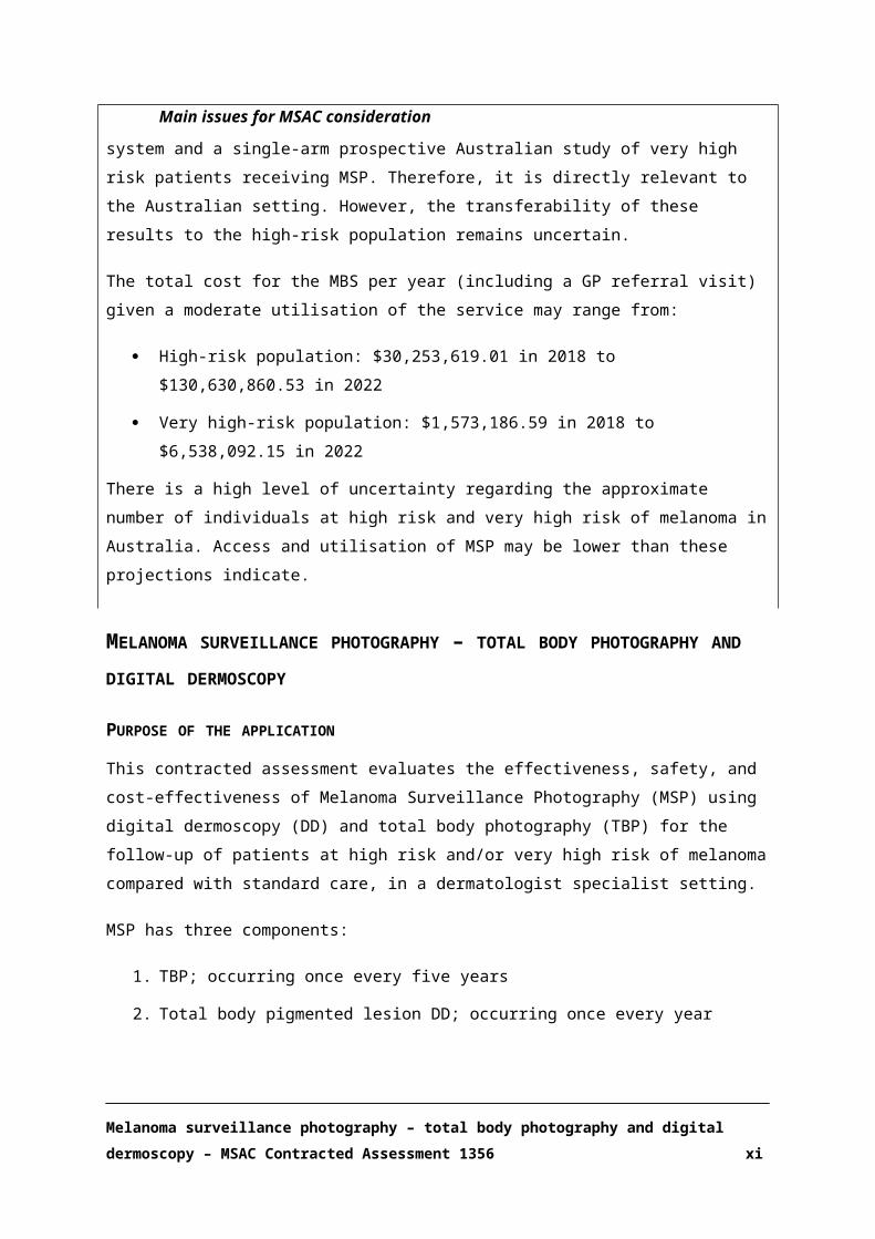
The total cost for the MBS per year (including a GP referral visit) given a moderate utilisation of the
service may range from:
High-risk population: $30,253,619.01 in 2018 to $130,630,860.53 in 2022
Very high-risk population: $1,573,186.59 in 2018 to $6,538,092.15 in 2022
There is a high level of uncertainty regarding the approximate number of individuals at high risk and
very high risk of melanoma in Australia. Access and utilisation of MSP may be lower than these projections indicate.
Melanoma surveillance photography – total body photography and digital dermoscopy – MSAC Contracted Assessment 1356 ix

MELANOMA SURVEILLANCE PHOTOGRAPHY – TOTAL BODY PHOTOGRAPHY AND DIGITAL DERMOSCOPY
PURPOSE OF THE APPLICATION
This contracted assessment evaluates the effectiveness, safety, and cost-effectiveness of Melanoma
Surveillance Photography (MSP) using digital dermoscopy (DD) and total body photography (TBP) for the follow-up of patients at high risk and/or very high risk of melanoma compared with standard
care, in a dermatologist specialist setting.
MSP has three components:
1. TBP; occurring once every five years
2. Total body pigmented lesion DD; occurring once every year
3. Follow-up DD of a previously photographed (by DD) pigmented lesion within 8-16 weeks of digital dermoscopy; limited to once per year
The populations considered include Individuals aged 18 years or older and with a minimum of 15 or more pigmented lesions for photography. They must also satisfy at least one of the criteria outlined
below:
High risk population Very High risk population
1. Personal history of melanoma OR2. Family history of two or more first-
degree relatives having had melanoma OR
3. Personal history of CDKN2A genetic mutation and at least one first- or second- degree relative with melanoma OR
4. 100 or more common naevi OR5. Six or more atypical/dysplastic naevi
1. Personal history of two or more primary melanomas OR
2. CDKN2A mutation OR3. One past melanoma AND one of
100 or more naevi OR Six or more atypical naevi OR
Family history of three or more first- or second-degree relatives
PROPOSAL FOR PUBLIC FUNDING
There is currently no MBS item for MSP (and it is therefore self-funded). The proposed MBS item
descriptors for the high-risk and very high-risk populations are summarised below in Table 1 and Table 2, respectively.
It is important to highlight that this assessment does not evaluate MSP as a population screening strategy, but the use of MSP for the monitoring of patients at high risk or very high risk of
melanoma.
Melanoma surveillance photography – total body photography and digital dermoscopy – MSAC Contracted Assessment 1356 x
The proposed medical services would be implemented in addition to current clinical practice. Ideally
patients at high and very high risk of melanoma perform their own self-examination at home, have a spouse or relative/friend look at inaccessible places such as the back regularly, see their general
practioner (GP) regularly and are under the care of a dermatologist. Those who would benefit from MSP would have their TBP and DD images in addition to the above measures routinely done as a
baseline, and repeated as per the attending dermatologist’s recommendations.
A private fee for comprehensive MSP as part of an initial or long-term follow-up study in Australia is
$300-450 per person per year. A current fee schedule for Molemap Australia quotes $449 for an initial study (1 hour) and $329 for follow up (Molemap 2017).
The proposed MBS fee for TBP is $100.00, and the fee for DD ranges from $117.65 to $205.90, depending on the number of lesions photographed. The suggested fee for short-term follow-up has
been costed at $70 per visit. It is therefore important to note that the proposed MBS fee does not cover the total cost of MSP and patients would likely still incur out of pocket costs.
Melanoma surveillance photography – total body photography and digital dermoscopy – MSAC Contracted Assessment 1356 xi

Table 1 Proposed MBS item descriptors for the high-risk population
Melanoma surveillance photography – total body photography and digital dermoscopy – MSAC Contracted Assessment 1356 xii

MBS item Descriptors
Category 2 – Diagnostic Procedures and InvestigationsGroup: D1 – Miscellaneous diagnostic procedures and investigationsSubgroup 10–Other diagnostic procedures and investigations[Item A]TOTAL BODY PHOTOGRAPHY performed by a specialist dermatologist, or on behalf of a specialist dermatologist by a registered nurse:a) Only if performed in association with MBS Items Bi, Bii, Biii or Biv, andb) Only using image capture and processing equipment approved by the Therapeutic Goods Administrationc) Only claimable once in a 5 year periodd) Only if referred patient is 18 years of age or older and has a minimum of 15 lesions for photography
To be eligible:a) the specialist dermatologist must hold current status on the Australasian College of Dermatologists register of
Reporting Dermatologists andb) the reporting dermatologist must provide a diagnostic report* to the referring doctor andc) any registered nurse involved in the process must hold current status on the Joint Register of Registered Nurses
Accredited in Melanoma Surveillance Photography held by the Australian Dermatology Nurses Association/Australasian College of Dermatologists
d) the item is only claimable once per calendar yeare) the service must be delivered at the reporting dermatologists practice site
(See para D1.10 of explanatory notes to this Category)Fee: $100.00 Benefit 75%= $75.00, 85%=$85.00
Category 2 – Diagnostic Procedures and InvestigationsGroup: D1 – Miscellaneous diagnostic procedures and investigationsSubgroup 10–Other diagnostic procedures and investigations[Item Bi]TOTAL BODY PIGMENTED LESION DIGITAL DERMOSCOPY performed by a specialist dermatologist or on behalf of a specialist dermatologist by a registered nurse using an image capture and processing device approved by the Therapeutic Goods Administration. To be eligible the subject must be 18 years of age or older, have 15 or more pigmented lesions for photography and be at high risk of melanoma as evidenced by:i. past personal history of melanoma orii. family history of 2 first degree relatives with melanoma oriii. personal history of CDKN2A mutation and at least 1 first or second degree relative diagnosed with melanoma oriv. 6 or more atypical melanocytic naevi orv. 100 or more common melanocytic naevi or
Number of lesions photographed: 15-49To be eligible:f) the specialist dermatologist must hold current status on the Australasian College of Dermatologists register of
Reporting Dermatologists andg) the reporting dermatologist must provide a diagnostic report* to the referring doctor andh) any registered nurse involved in the process must hold current status on the Joint Register of Registered Nurses
Accredited in Melanoma Surveillance Photography held by the Australian Dermatology Nurses Association/Australasian College of Dermatologists
i) the item is only claimable once per calendar yearj) the service must be delivered at the reporting dermatologists practice site
(See para D1.10 of explanatory notes to this Category)Fee: $117.65, Benefit 75%=$88.25, 85%=$100
Melanoma surveillance photography – total body photography and digital dermoscopy – MSAC Contracted Assessment 1356 xiii
MBS item Descriptors
Category 2 – Diagnostic Procedures and InvestigationsGroup: D1 – Miscellaneous diagnostic procedures and investigationsSubgroup 10–Other diagnostic procedures and investigations[Item Bii]TOTAL BODY PIGMENTED LESION DIGITAL DERMOSCOPY performed by a specialist dermatologist or on behalf of a specialist dermatologist by a registered nurse using an image capture and processing device approved by the Therapeutic Goods Administration. To be eligible the subject must be 18 years of age or older, have 15 or more pigmented lesions for photography and be at high risk of melanoma as evidenced by:i. past personal history of melanoma orii. family history of 2 first degree relatives with melanoma oriii. personal history of CDKN2A mutation and at least 1 first or second degree relative diagnosed with melanoma oriv. 6 or more atypical melanocytic naevi orv. 100 or more common melanocytic naevi or
Number of lesions photographed: 50-99To be eligible:a) the specialist dermatologist must hold current status on the Australasian College of Dermatologists register of
Reporting Dermatologists andb) the reporting dermatologist must provide a diagnostic report* to the referring doctor andc) any registered nurse involved in the process must hold current status on the Joint Register of Registered Nurses
Accredited in Melanoma Surveillance Photography held by the Australian Dermatology Nurses Association/Australasian College of Dermatologists
d) the item is only claimable once per calendar yeare) the service must be delivered at the reporting dermatologists practice site
(See para D1.10 of explanatory notes to this Category)Fee: $147.05, Benefit 75%=$110.30, 85%=$125
Category 2 – Diagnostic Procedures and InvestigationsGroup: D1 – Miscellaneous diagnostic procedures and investigationsSubgroup 10–Other diagnostic procedures and investigations[Item Biii]TOTAL BODY PIGMENTED LESION DIGITAL DERMOSCOPY performed by a specialist dermatologist or on behalf of a specialist dermatologist by a registered nurse using an image capture and processing device approved by the Therapeutic Goods Administration. To be eligible the subject must be 18 years of age or older, have 15 or more pigmented lesions for photography and be at high risk of melanoma as evidenced by:i. past personal history of melanoma orii. family history of 2 first degree relatives with melanoma oriii. personal history of CDKN2A mutation and at least 1 first or second degree relative diagnosed with melanoma or
1. 6 or more atypical melanocytic naevi2. Or 100 or more common melanocytic naevi or
Number of lesions photographed: 100-149To be eligible:a) the specialist dermatologist must hold current status on the Australasian College of Dermatologists register of
Reporting Dermatologists andb) the reporting dermatologist must provide a diagnostic report* to the referring doctor andc) any registered nurse involved in the process must hold current status on the Joint Register of Registered Nurses
Accredited in Melanoma Surveillance Photography held by the Australian Dermatology Nurses Association/Australasian College of Dermatologists
d) the item is only claimable once per calendar yeare) the service must be delivered at the reporting dermatologists practice site*
(See para D1.10 of explanatory notes to this Category)Fee: $176.45, Benefit 75%=$132.35, 85%=$150
Melanoma surveillance photography – total body photography and digital dermoscopy – MSAC Contracted Assessment 1356 xiv

MBS item Descriptors
Category 2 – Diagnostic Procedures and InvestigationsGroup: D1 – Miscellaneous diagnostic procedures and investigationsSubgroup 10–Other diagnostic procedures and investigations[Item Biv]TOTAL BODY PIGMENTED LESION DIGITAL DERMOSCOPY performed by a specialist dermatologist or on behalf of a specialist dermatologist by a registered nurse using an image capture and processing device approved by the Therapeutic Goods Administration. To be eligible the subject must be 18 years of age or older, have 15 or more pigmented lesions for photography and be at high risk of melanoma as evidenced by:i. past personal history of melanoma orii. family history of 2 first degree relatives with melanoma oriii. personal history of CDKN2A mutation and at least 1 first or second degree relative diagnosed with melanoma oriv. 6 or more atypical melanocytic naevi orv. 100 or more common melanocytic naevi or
Number of lesions photographed: ≥150To be eligible:a) the specialist dermatologist must hold current status on the Australasian College of Dermatologists register of
Reporting Dermatologists andb) the reporting dermatologist must provide a diagnostic report* to the referring doctor andc) any registered nurse involved in the process must hold current status on the Joint Register of Registered Nurses
Accredited in Melanoma Surveillance Photography held by the Australian Dermatology Nurses Association/Australasian College of Dermatologists
d) the item is only claimable once per calendar yeare) the service must be delivered at the reporting dermatologists practice site*
(See para D1.10 of explanatory notes to this Category)Fee: $205.90, Benefit 75%=$154.40, 85%=$175
Category 2 – Diagnostic Procedures and InvestigationsGroup: D1 – Miscellaneous diagnostic procedures and investigationsSubgroup 10–Other diagnostic procedures and investigations[Item C]SHORT TERM FOLLOW UP DIGITAL DERMOSCOPY of selected pigmented lesions previously identified via digital dermoscopy and claimed under Items Bi, Bii, Biii or Biv, performed by a specialist dermatologist, or on behalf of a specialist dermatologist by a registered nurse.
To be eligible:a) the specialist dermatologist must hold current status on the Australasian College of Dermatologists Register of
Reporting Dermatologists andb) the reporting dermatologist must provide a diagnostic report* to the referring doctor andc) any registered nurse involved in the process must hold current status on the Joint Register of Nurses Accredited
in Melanoma Surveillance Photography held by the Australian Dermatology Nurses Association/Australasian College of Dermatologists
d) the item is only claimable once per calendar yeare) the service must have been expressly recommended by the reporting dermatologist in the preceding Total Body
Pigmented Lesion Digital Dermoscopy (Item B) reportf) the service must be performed in the time period 8-16 weeks from the date MBS service Item Bi, Bii, Biii, or Biv
was performedg) the service must be delivered at the reporting dermatologists practice site
(See para D1.10 of explanatory notes to this Category)Fee: $70.00 Benefit 75%=52.50 85%=$59.50
Melanoma surveillance photography – total body photography and digital dermoscopy – MSAC Contracted Assessment 1356 xv

Explanatory Notes
D1.10 Melanoma Surveillance Photography
Item A. Item A may only be claimed if performed in conjunction with Item B. Item A total body photographic findings do not require a report from the reporting dermatologist however if Item B is performed with Item A the report relating to Item B must note that item A has been performed. If Item A has been claimed previously the date of the last Item A procedure must appear on the patient’s invoice/Medicare claim such as for example “previous Item A 1/1/2003”
Item B. The report to the referring doctor must document the Medicare eligible criteria, patient details, image capture technology used, time and date of image capture, name and qualifications of person performing the image capture, diagnosis and suggested management of specific lesions and whether short term follow up (Item C) is subsequently required. A referral is only valid for a single episode of Item B and any associated Item A or C that is required in association with that item B. The reporting dermatologist or their locum must be physically present at the practice site at the time of image capture.
Item C. The report to the referring doctor must document the Medicare eligible criteria, patient details, image capture technology used, time and date of image capture, name and qualifications of person performing the image capture, diagnosis and suggested management of specific lesions. A referral is only valid for a single episode of Item C. The reporting dermatologist or their locum must be physically present at the practice site at the time of image capture.
Melanoma surveillance photography – total body photography and digital dermoscopy – MSAC Contracted Assessment 1356 xvi

Table 2 Proposed MBS item descriptors for the very high-risk population
Melanoma surveillance photography – total body photography and digital dermoscopy – MSAC Contracted Assessment 1356 xvii

MBS item Descriptors
Category 2 – Diagnostic Procedures and InvestigationsGroup: D1 – Miscellaneous diagnostic procedures and investigationsSubgroup 10–Other diagnostic procedures and investigations[Item A]TOTAL BODY PHOTOGRAPHY performed by a specialist dermatologist, or on behalf of a specialist dermatologist by a registered nurse:a) Only if performed in association with MBS Items Bi, Bii, Biii or Biv, andb) Only using image capture and processing equipment approved by the Therapeutic Goods Administrationc) Only claimable once in a 5 year periodd) Only if referred patient is 18 years of age or older and has a minimum of 15 lesions for photography
To be eligible:a) the specialist dermatologist must hold current status on the Australasian College of Dermatologists register of
Reporting Dermatologists andb) the reporting dermatologist must provide a diagnostic report* to the referring doctor andc) any registered nurse involved in the process must hold current status on the Joint Register of Registered Nurses
Accredited in Melanoma Surveillance Photography held by the Australian Dermatology Nurses Association/Australasian College of Dermatologists
d) the item is only claimable once per calendar yeare) the service must be delivered at the reporting dermatologists practice site
(See para D1.10 of explanatory notes to this Category)Fee: $100.00 Benefit 75%= $75.00, 85%=$85.00
Category 2 – Diagnostic Procedures and InvestigationsGroup: D1 – Miscellaneous diagnostic procedures and investigationsSubgroup 10–Other diagnostic procedures and investigations[Item Bi]TOTAL BODY PIGMENTED LESION DIGITAL DERMOSCOPY performed by a specialist dermatologist or on behalf of a specialist dermatologist by a registered nurse using an image capture and processing device approved by the Therapeutic Goods Administration. To be eligible the subject must be 18 years of age or older, have 15 or more pigmented lesions for photography and be at high risk of melanoma as evidenced by:(i) past personal history of 2 or more melanoma or(ii) CDKN2A mutation carrier or(iii) Past personal history of 1 melanoma AND
a) Family history of melanoma in 3 first or second degree relatives ORb) 100 or more common naevi ORc) 6 or more atypical naevi
Number of lesions photographed: 15-49To be eligible:f) the specialist dermatologist must hold current status on the Australasian College of Dermatologists register of
Reporting Dermatologists andg) the reporting dermatologist must provide a diagnostic report* to the referring doctor andh) any registered nurse involved in the process must hold current status on the Joint Register of Registered Nurses
Accredited in Melanoma Surveillance Photography held by the Australian Dermatology Nurses Association/Australasian College of Dermatologists
i) the item is only claimable once per calendar yearj) the service must be delivered at the reporting dermatologists practice site
(See para D1.10 of explanatory notes to this Category)Fee: $117.65, Benefit 75%=$88.25, 85%=$100
Melanoma surveillance photography – total body photography and digital dermoscopy – MSAC Contracted Assessment 1356 xviii

MBS item Descriptors
Category 2 – Diagnostic Procedures and InvestigationsGroup: D1 – Miscellaneous diagnostic procedures and investigationsSubgroup 10–Other diagnostic procedures and investigations[Item Bii]TOTAL BODY PIGMENTED LESION DIGITAL DERMOSCOPY performed by a specialist dermatologist or on behalf of a specialist dermatologist by a registered nurse using an image capture and processing device approved by the Therapeutic Goods Administration. To be eligible the subject must be 18 years of age or older, have 15 or more pigmented lesions for photography and be at high risk of melanoma as evidenced by:(i) past personal history of 2 or more melanoma or(ii) CDKN2A mutation carrier or(iii) Past personal history of 1 melanoma AND
a) Family history of melanoma in 3 first or second degree relatives ORb) 100 or more common naevi ORc) 6 or more atypical naevi
Number of lesions photographed: 50-99To be eligible:a) the specialist dermatologist must hold current status on the Australasian College of Dermatologists register of
Reporting Dermatologists andb) the reporting dermatologist must provide a diagnostic report* to the referring doctor andc) any registered nurse involved in the process must hold current status on the Joint Register of Registered Nurses
Accredited in Melanoma Surveillance Photography held by the Australian Dermatology Nurses Association/Australasian College of Dermatologists
d) the item is only claimable once per calendar yeare) the service must be delivered at the reporting dermatologists practice site
(See para D1.10 of explanatory notes to this Category)Fee: $147.05, Benefit 75%=$110.30, 85%=$125
Category 2 – Diagnostic Procedures and InvestigationsGroup: D1 – Miscellaneous diagnostic procedures and investigationsSubgroup 10–Other diagnostic procedures and investigations[Item Biii]TOTAL BODY PIGMENTED LESION DIGITAL DERMOSCOPY performed by a specialist dermatologist or on behalf of a specialist dermatologist by a registered nurse using an image capture and processing device approved by the Therapeutic Goods Administration. To be eligible the subject must be 18 years of age or older, have 15 or more pigmented lesions for photography and be at high risk of melanoma as evidenced by:(i) past personal history of 2 or more melanoma or(ii) CDKN2A mutation carrier or(iii) Past personal history of 1 melanoma AND
a) Family history of melanoma in 3 first or second degree relatives ORb) 100 or more common naevi ORc) 6 or more atypical naevi
Number of lesions photographed: 100-149To be eligible:a) the specialist dermatologist must hold current status on the Australasian College of Dermatologists register of
Reporting Dermatologists andb) the reporting dermatologist must provide a diagnostic report* to the referring doctor andc) any registered nurse involved in the process must hold current status on the Joint Register of Registered Nurses
Accredited in Melanoma Surveillance Photography held by the Australian Dermatology Nurses Association/Australasian College of Dermatologists
d) the item is only claimable once per calendar yeare) the service must be delivered at the reporting dermatologists practice site*
(See para D1.10 of explanatory notes to this Category)Fee: $176.45, Benefit 75%=$132.35, 85%=$150
Melanoma surveillance photography – total body photography and digital dermoscopy – MSAC Contracted Assessment 1356 xix

MBS item Descriptors
Category 2 – Diagnostic Procedures and InvestigationsGroup: D1 – Miscellaneous diagnostic procedures and investigationsSubgroup 10–Other diagnostic procedures and investigations[Item Biv]TOTAL BODY PIGMENTED LESION DIGITAL DERMOSCOPY performed by a specialist dermatologist or on behalf of a specialist dermatologist by a registered nurse using an image capture and processing device approved by the Therapeutic Goods Administration. To be eligible the subject must be 18 years of age or older, have 15 or more pigmented lesions for photography and be at high risk of melanoma as evidenced by:
(i) past personal history of 2 or more melanoma or(ii) CDKN2A mutation carrier or(iii) Past personal history of 1 melanoma AND
a) Family history of melanoma in 3 first or second degree relatives ORb) 100 or more common naevi ORc) 6 or more atypical naevi
Number of lesions photographed: ≥150To be eligible:a) the specialist dermatologist must hold current status on the Australasian College of Dermatologists register of
Reporting Dermatologists andb) the reporting dermatologist must provide a diagnostic report* to the referring doctor andc) any registered nurse involved in the process must hold current status on the Joint Register of Registered Nurses
Accredited in Melanoma Surveillance Photography held by the Australian Dermatology Nurses Association/Australasian College of Dermatologists
d) the item is only claimable once per calendar yeare) the service must be delivered at the reporting dermatologists practice site*
(See para D1.10 of explanatory notes to this Category)Fee: $205.90, Benefit 75%=$154.40, 85%=$175
Melanoma surveillance photography – total body photography and digital dermoscopy – MSAC Contracted Assessment 1356 xx

MBS item Descriptors
Category 2 – Diagnostic Procedures and InvestigationsGroup: D1 – Miscellaneous diagnostic procedures and investigationsSubgroup 10–Other diagnostic procedures and investigations[Item C]SHORT TERM FOLLOW UP DIGITAL DERMOSCOPY of selected pigmented lesions performed by a specialist dermatologist, or on behalf of a specialist dermatologist by a registered nurse. Lesions selected may only be identified via:a) Identification on total body pigmented lesion dermoscopy by the reporting Dermatologist and claimed under MBS items Bi-ivb) Identification by the referring medical practitioner following physical examination used in conjunction with past total body photography (claimed under Item A) and past total body pigmented lesion dermoscopy (claimed under Items Bi-iv) as a clinical aid
To be eligible:a) the specialist dermatologist must hold current status on the Australasian College of Dermatologists Register of
Reporting Dermatologists andb) the reporting dermatologist must provide a diagnostic report* to the referring doctor andc) any registered nurse involved in the process must hold current status on the Joint Register of Nurses Accredited
in Melanoma Surveillance Photography held by the Australian Dermatology Nurses Association/Australasian College of Dermatologists
d) the item is only claimable twice per calendar yeare) if lesion/s identified at the time of total body pigmented lesion dermoscopic photography the service must be
performed in the time period 8-16 weeks from the date of MBS service Item Bi,Bii, Biii, or Biv was performedf) if lesion/s identified at physical examination using past photography (Items A, Bi-iv) as a clinical aid the lesion/s
require photography at the time of referral for short term monitoring and this must be repeated in the time period 8-16 weeks to allow a comparative report to be produced
g) the service must be delivered at the reporting dermatologists practice site
(See para D1.10 of explanatory notes to this Category)Fee: $70.00 Benefit 75%=52.50 85%=$59.50
Explanatory Notes
D1.10 Melanoma Surveillance Photography
Item A. Item A may only be claimed if performed in conjunction with Item B. Item A total body photographic findings do not require a report from the reporting dermatologist however if Item B is performed with Item A the report relating to Item B must note that item A has been performed. If Item A has been claimed previously the date of the last Item A procedure must appear on the patient’s invoice/Medicare claim such as for example “previous Item A 1/1/2003”
Item B. The report to the referring doctor must document the Medicare eligible criteria, patient details, image capture technology used, time and date of image capture, name and qualifications of person performing the image capture, diagnosis and suggested management of specific lesions and whether short term follow up (Item C) is subsequently required. A referral is only valid for a single episode of Item B and any associated Item A or C that is required in association with that item B. The reporting dermatologist or their locum must be physically present at the practice site at the time of image capture.
Item C. The report to the referring doctor must document the Medicare eligible criteria, patient details, image capture technology used, time and date of image capture, name and qualifications of person performing the image capture, diagnosis and suggested management of specific lesions. A referral is only valid for a single episode of Item C. The reporting dermatologist or their locum must be physically present at the practice site at the time of image capture.
Melanoma surveillance photography – total body photography and digital dermoscopy – MSAC Contracted Assessment 1356 xxi

COMPARATOR
The comparator is standard care without MSP conducted by a GP or dermatologist, or self-examination:
Self-examination at home without use of comparative photographic images
GP clinical examination without access to photography for real-time comparison (once or
twice per year)
Dermatologist clinical examination (including dermoscopy) without access to digital
photography for real time comparison (once or twice a year)
BACKGROUND
Dermoscopy is a diagnostic technique that uses a hand-held magnifying device with cross-polarised
light filters. It does not use any form of radiation or exposure of the skin to oil or alcohol, and reveals features of the skin not visible through normal lighting and magnification. The superior
diagnostic accuracy of dermoscopy for melanoma detection compared with clinical naked-eye examination has been previously established (National Institute for Health and Care Excellence
2015), and therefore will not be investigated in this application.
Nevertheless, melanoma may be clinically and dermoscopically indistinguishable from melanocytic
nevi, especially in early stages. In these cases, digital follow-up of melanocytic lesions may allow the identification of dermoscopic changes from baseline and early diagnosis of melanoma while
minimising the excision of benign lesions (Salerni, Teran et al. 2013). Follow-up DD is used to monitor melanocytic lesions over a period of time, comparing new dermoscopic images to digitally-
stored ones. The initial MSP consultation comprises TBP and DD. Follow-up involves repeat DD for comparison over time, which can be over a short-term or long-term period.
CLINICAL NEED
Melanoma is the fourth most prevalent cancer in Australia after prostate, breast and colorectal cancer. Early detection of melanoma is a key predictor of survival. In the earliest phase, melanoma is
confined to the top layer of the skin, the epidermis, and is non-invasive. Five-year survival in patients with tumours thicker than 4 mm is only 55% compared with almost 100% survival for patients with
tumours ≤1 mm thick (Australian Institute of Health and Welfare 2017).
In Australia, the Clinical Practice Guidelines for the Management of Melanoma in Australia and New
Zealand recommend surveillance for people at high-risk (National Health & Medical Research Council/Australian Cancer Network 2008), stating: “Individuals at high risk of melanoma and their
partner or carer be educated to recognise and document lesions suspicious of melanoma, and to be
Melanoma surveillance photography – total body photography and digital dermoscopy – MSAC Contracted Assessment 1356 xxii

regularly checked by a clinician with six-monthly full body examination supported by total body
photography and dermoscopy as required”.
The applicant has claimed that the successful listing of MSP for the target population and specialist
setting will lead to earlier detection of melanoma and subsequent improvement in prognosis, as well as reduced requirement for sentinel lymph node biopsy, treatment of metastatic disease, less
aggressive skin surgery, and fewer benign lesion excisions.
CLINICAL EVALUATION
Three retrospective comparative studies of MSP versus standard care in patients at high risk of
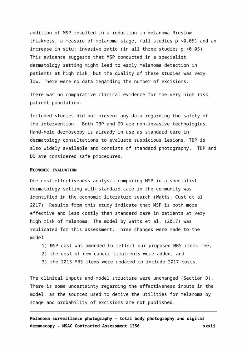
melanoma were identified in the systematic review and included in Section B (Salerni, Lovatto et al. 2011, Salerni, Teran et al. 2014, Mintsoulis and Beecker 2016). The addition of MSP resulted in a
reduction in melanoma Breslow thickness, a measure of melanoma stage, (all studies p <0.05) and an increase in situ: invasive ratio (in all three studies p <0.05). This evidence suggests that MSP
conducted in a specialist dermatology setting might lead to early melanoma detection in patients at high risk, but the quality of these studies was very low. There were no data regarding the number of
excisions.
There was no comparative clinical evidence for the very high risk patient population.
Included studies did not present any data regarding the safety of the intervention. Both TBP and DD are non-invasive technologies. Hand-held dermoscopy is already in use as standard care in
dermatology consultations to evaluate suspicious lesions. TBP is also widely available and consists of standard photography. TBP and DD are considered safe procedures.
ECONOMIC EVALUATION
One cost-effectiveness analysis comparing MSP in a specialist dermatology setting with standard care in the community was identified in the economic literature search (Watts, Cust et al. 2017).
Results from this study indicate that MSP is both more effective and less costly than standard care in patients at very high risk of melanoma. The model by Watts et al. (2017) was replicated for this
assessment. Three changes were made to the model: 1) MSP cost was amended to reflect our proposed MBS items fee,
2) the cost of new cancer treatments were added, and 3) the 2013 MBS items were updated to include 2017 costs.
The clinical inputs and model structure were unchanged (Section D). There is some uncertainty
regarding the effectiveness inputs in the model, as the sources used to derive the utilities for melanoma by stage and probability of excisions are not published.
Melanoma surveillance photography – total body photography and digital dermoscopy – MSAC Contracted Assessment 1356 xxiii

As per the published model, the replicated model showed that MSP in patients at very high risk of
melanoma is more effective and less costly than standard care. The overall costs and effectiveness, and incremental costs and effectiveness as calculated for the intervention and comparator in the
model are shown in Table 3.
Table 3 Base case cost-effectiveness results (10-year time horizon)MSP Standard care Incremental
Cost ($) 7,172.63 15,558.83 8,386.19Effectiveness (QALY) 7.53 7.32 0.21QALY: quality-adjusted life year; MSP: melanoma surveillance photography
Results from a one-way sensitivity analysis indicate that MSP is less costly and more effective than
standard care up to a threshold of $1100 annual MSP cost per patient (Figure 1), which is a higher cost than the annual estimate per patient (based on the proposed MBS items fees) of $155 included
in the model. Beyond this threshold, specialised surveillance (MSP) becomes more costly than standard care (given an annual cost of standard care of $70), but MSP remains cost-effective up to
an annual cost of $2700 for a willingness to pay of $50,000 per quality adjusted life years (QALY) gained. It is important to note these results were obtained from a model of MSP in a population at
very high risk of melanoma attending a highly specialised clinic within an urban centre, and these results might not be generalisable to the high-risk population across the country.
Melanoma surveillance photography – total body photography and digital dermoscopy – MSAC Contracted Assessment 1356 xxiv

Figure 1 One way sensitivity analysis, incremental cost-effectiveness as cost of specialised surveillance (MSP) increases.
The model was most sensitive to variations in the probability of excisions in standard care, with a
slight increase in the incremental cost-effectiveness ratio (ICER) as the probability of excision in standard care was changed from 0.8 to 0.1. However, none of the variables had a substantial impact
on the ICER. A potential increase in the cost of MSP from $155 to $1000 did not significantly alter the cost-effectiveness of the intervention. Other key drivers of the economic model were the probability
of early detection of melanoma (in-situ and stage I) by MSP, treatment costs for melanoma stage III and IV, and the probability of benign excisions in standard care (Figure 2).
Melanoma surveillance photography – total body photography and digital dermoscopy – MSAC Contracted Assessment 1356 xxv
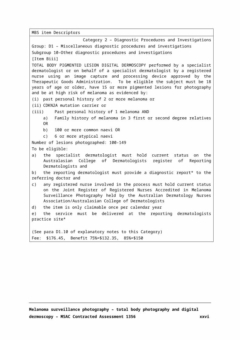
Figure 2 Tornado diagram: one-way sensitivity analysis of MSP versus standard careICER: incremental cost-effectiveness ratio, EV: expected value
A probabilistic sensitivity analysis (Figure 3) was conducted to evaluate the spread of results across
the cost-effectiveness plane, which represents a measure of the ICER’s degree of uncertainty. The incremental costs and QALY points were predominantly spread in the south-east quadrant,
confirming that MSP is less costly and more effective than standard care.
Melanoma surveillance photography – total body photography and digital dermoscopy – MSAC Contracted Assessment 1356 xxvi

Figure 3 Monte Carlo simulation. Incremental cost-effectiveness of MSP versus standard care
FINANCIAL IMPLICATIONS
An epidemiological approach was used to estimate the financial implications of the introduction of
MSP in high-risk and very high-risk patients. The calculation of the number of patients who would be eligible to receive MSP was based on data from the Australian Institute of Health and Welfare
(AIHW) (Australian Institute of Health and Welfare 2017, Australian Institute of Health and Welfare 2017) and the Australian Bureau of Statistics (Australian Bureau of Statistics 2017).
Two scenarios were estimated to evaluate low and moderate utilisation of the proposed service in both populations, as 100% participation of the total eligible population in screening and monitoring
services is not realistic. In the moderate utilisation scenario, we assumed an increase in MSP uptake from 20% to 60% over 5 years, and 90% compliance with follow-up MSP every year. Table 4 presents
the estimated number or individuals at high risk and very high risk at 100% participation and moderate utilisation of MSP.
The total costs for the high-risk and very high-risk populations given a moderate utilisation, including
an annual GP referral visit are presented below in Table 5.
Melanoma surveillance photography – total body photography and digital dermoscopy – MSAC Contracted Assessment 1356 xxvii

Table 4 Number of eligible individuals at high-risk and very high-risk of melanoma in the Australian population; 100% and moderate utilisation of MSP
Population 2018 2019 2020 2021 2022Total eligible (n); High risk 787,650 798,378 809,106 819,835 830,563
Total eligible (n), very high risk
40,958 41,068 41,179 41,289 41,399
Moderate utilisation (n); High risk
157,530 334,031 490,658 606,181 680,192
Moderate utilisation (n); Very high risk
8,192 17,235 25,089 30,680 34,044
Moderate utilisation: uptake 20% in 2018, increasing by 10% every year, and reaching 60% in 2022, 90% compliance;
Table 5 Total costs ($) to the MBS associated with MSP in high-risk and very high-risk patients; moderate utilisation
Population
Cost per patient
2018 2019 2020 2021 2022
High riskGP ($37.05) + MSP ($155)
$30,253,619.01 $64,150,713.75 $94,230,898.45 $116,416,976.07 $130,630,860.53
Very high risk
GP ($37.05) + MSP ($155)
$1,573,186.59 $3,310,053.80 $4,818,365.30 $5,892,122.39 $6,538,092.15
Moderate utilisation: uptake 20% in 2018, increasing by 10% every year, and reaching 60% in 2022, 90% compliance GP: general practitioner; MSP: melanoma surveillance photography3816
There is a high level of uncertainty regarding the approximate number of individuals at high risk and
very high risk of melanoma in Australia; therefore, these estimates are only approximate. Access and utilisation of these medical services may be lower than these projections indicate, especially in rural
areas.
Melanoma surveillance photography – total body photography and digital dermoscopy – MSAC Contracted Assessment 1356 xxviii

ACRONYMS AND ABBREVIATIONS
Acronym/abbreviation Meaning
ABS Australian Bureau of statistics
AJCC American Joint Commission on Cancer
APDC Admitted Patient Data Collection
AIHW Australian Institute of Health and Welfare
CTC Clinical Trials Centre
DD Digital dermoscopy
HESP Health Expert Standing Panel
HTA Health technology assessment
ICER Incremental cost-effectiveness ratio
MBS Medicare Benefits Schedule
MSAC Medical Services Advisory Committee
MSP Melanoma surveillance photography
NHMRC National Health and Medical Research Council
PASC PICO Confirmation Advisory Sub-Committee of the MSAC
PBS Pharmaceutical Benefits Scheme
QALY Quality adjusted life year
TBP Total Body Photography
TGA Therapeutic Goods Administration
Melanoma surveillance photography – total body photography and digital dermoscopy – MSAC Contracted Assessment 1356 xxix

SECTION A CONTEXT
This contracted assessment of total body photography (TBP) and digital dermoscopy (DD) for the
surveillance of melanoma is intended for the Medical Services Advisory Committee (MSAC). MSAC evaluates new and existing health technologies and procedures for which funding is sought under
the Medicare Benefits Schedule (MBS) in terms of their safety, effectiveness and cost-effectiveness, while taking into account other issues such as access and equity. MSAC adopts an evidence-based
approach to its assessments, based on reviews of the scientific literature and other information sources, including clinical expertise.
The NHMRC Clinical Trials Centre (CTC) has been commissioned by the Australian Government Department of Health to conduct a systematic literature review and economic evaluation of
melanoma surveillance photography (MSP) using TBP and DD. This assessment has been undertaken in order to inform MSAC’s decision-making regarding whether the proposed medical service should
be publicly funded.
Appendix A provides a list of the people involved in the development of this assessment report.
The proposed use of MSP in Australian clinical practice was outlined in a PICO Confirmation that was presented to, and accepted by, the PICO Confirmation Advisory SubCommittee (PASC). The PICO
Confirmation was released for public comment on 3 March 2014. The protocol was ratified by PASC in January 2017.
A.1. ITEMS IN THE AGREED PICO CONFIRMATION
The PICO for MSP was first submitted in 2014. Since this time, the PICO has changed a number of times and was ratified in an out-of-PASC session in 2017. The agreed population within the ratified
PICO was a high risk population defined as:
Adults (aged ≥ 18 years) with minimum of 15 naevi and at least one of the following:
Personal history of melanoma
Family history of two or more first degree relatives having had melanoma
Personal history of gene mutation CDKN2A and one first or second degree relative with
melanoma
100 or more common naevi
Six or more atypical/dysplastic naevi
Melanoma surveillance photography – total body photography and digital dermoscopy – MSAC Contracted Assessment 1356 30

Several interventions and comparators were listed within the PICO confirmation. The interventions
listed were:
Total Body Photography (TBP) once every 5 years
Total body pigmented lesion digital dermoscopy (DD)/sequential DD once a year (‘Long-term follow-up DD; all lesions’)
Follow-up DD of a previously photographed (by DD) pigmented lesion within 8-16 weeks of DD but limited to once per year (‘short-term follow-up DD’)
The comparators listed were: Self-examination at home without the use of photography (monthly);
GP clinical examination/skin excisions without access to photography for real time comparison (once or twice per year);
Dermatologist clinical examination/skin excisions (including dermoscopy), without access to photography for real time comparison (once or twice per year)
For the context of this assessment, it was imperative that these interventions be grouped as MSP and the comparator as standard practice. This was because the effectiveness of the MSP would be
determined by the combination of all the individual elements described above rather than the incremental benefit of each element separately.
Limited comparative evidence was found using these criteria. An additional study was identified which assessed MSP in a very high risk Australian population (Watts, Cust et al. 2017). To include the
evidence from this study, a more restrictive population was also included in this assessment. This very high risk population was defined as having:
1. A personal history of two or more primary melanomas OR
2. A CDKN2A mutation OR
3. One past melanoma AND one of
100 or more naevi OR
Six or more atypical naevi OR
Family history of three or more first- or second-degree relatives
A.2. PROPOSED MEDICAL SERVICE
This application will focus on the assessment of Melanoma Surveillance Photography (MSP) – DD and TBP – in the follow-up of patients at high risk and very high risk of melanoma, and conducted in
a dermatologist specialist setting.
MSP has three components:
Melanoma surveillance photography – total body photography and digital dermoscopy – MSAC Contracted Assessment 1356 31
Total body photography (TBP); occurring once every five years – Proposed Item A
Total body pigmented lesion digital dermoscopy (DD); occurring once every year –Proposed Item B
Follow-up DD of a previously photographed (by DD) pigmented lesion within 8-16 weeks of DD (limited to once per year) –Proposed Item C
Dermoscopy is a diagnostic technique that uses a hand-held magnifying device with cross-polarised light filters. It does not use any form of radiation or exposure of the skin to oil or alcohol, and
reveals features of the skin not visible through normal lighting and magnification. The superior diagnostic accuracy of dermoscopy for melanoma detection compared with the naked eye
examination has been previously established (National Institute for Health and Care Excellence 2015), and therefore will not be investigated in this application.
Nevertheless, melanoma may be clinically and dermoscopically indistinguishable from melanocytic nevi, especially in early stages. In these cases, DD follow-up of melanocytic lesions may allow the
identification of dermoscopic changes from baseline and early diagnosis of melanoma while minimising the excision of benign lesions (Salerni, Teran et al. 2013). Follow-up DD is used to
monitor melanocytic lesions over a period of time, comparing new dermoscopic images to digitally-stored ones. The initial MSP consultation comprises TBP and DD. Follow-up involves repeat DD for
comparison over time, which can be short-term or long-term:
Short-term follow-up DD: consists of only a few lesions re-examined photographically by DD
at a set time (usually 3 months). It is usually only performed once on a specific lesion. If there is no change in a lesion within the specified 3 months period, there is no longer the
need to continue short-term monitoring. If there is significant change in a lesion under short-term DD follow-up, it should be excised for histopathological examination for evidence
of melanoma.
Long-term follow-up DD: involves all the lesions under surveillance at routine yearly
intervals. It may be recommended on a continued basis over many years.
TBP is performed to photograph all the body regions. Different areas of the body are photographed
in standard poses to give approximately 25 “long shot” photographs (19-36 depending on the system used and occasionally more). All the existing naevi can be seen in those photographs (but not
the very fine detail of each).
Once the TBP shots are performed, individual melanocytic naevi are photographed up close (macro
images) and then additionally through DD. All pigmented lesions of any size that have any irregularity in them at all are photographed. It also includes all naevi and pigmented lesions on the
body approximately ≥3mm in lateral diameter. In many cases, there are more than 100 macro and dermoscopic photographs taken for a single individual. Each individual close-up and dermoscopic
Melanoma surveillance photography – total body photography and digital dermoscopy – MSAC Contracted Assessment 1356 32

image is orientated and “tagged” to the TBP shots to show its exact anatomical location. That being
the case, TBP must be performed at baseline to determine the correct anatomical position of each naevus photographed with DD.
The images are uploaded to a computer. The body shots and all individual naevus photographs are viewed by the reporting dermatologist, and those that are suspicious for melanoma are reported as
requiring excision. Any other individual lesion that the referring doctor, the patient or the melanographer has a specific question about is also formally commented on in the report. The
diagnoses are based on the appearance of each lesion specifically looked at by the reporting dermatologist and not via computer generated algorithm diagnosis.
A melanographer, by definition for the purposes of this evaluation, is a registered nurse with experience in both dermatology practice and photography. The melanographer works under the
instruction of the dermatologist, who is responsible for the supervision and quality of the melanographer’s clinical work. Currently, the only suggested requirement for a melanographer is
that they be an AHPRA (Australian Health Practitioner Regulation Agency)-registered nurse and participate in self-directed continuing medical education.
At the time of the follow-up consultation, the previous DD images are directly compared to the patient examination, going through each body segment. This examination will usually be performed
by the melanographer. This requires time and a methodical approach. Any new lesion requires repeat photography of that specific body shot/segment as well as the close up and DD image of the
new lesion. Each individual naevus that was photographed at baseline is photographed as a close-up shot and through DD. All the images are reviewed by the dermatologist. Each individual lesion is
compared to its previous image (or images if there has been more than 1 previous study). Any new changes are noted by the dermatologist, and specifically if any changes suggestive of melanoma are
observed then surgical excision is recommended. The subtle comparative changes seen over time can help diagnose “featureless” melanoma that cannot be diagnosed any other way. A report is
produced similar in nature to the initial study (specific lesion comments, treatment recommendations and whether further photographic short term follow up is required).
The images may be made available to the patient and/or their doctor to use for their own comparison over time at home or at the attending clinician’s surgery. The patients themselves can
use the body shots and macro images for their own comparison at home. The patient’s GP can do this too, but additionally can use a hand-held dermatoscope to directly compare real-time
dermoscopy to the stored DD images. Various storage and transfer methods are available, such as images on a data storage device such as a compact disc or via a password protected secure website.
The over-riding principle is that any report or images are only available to those with a bone fide interest and that they are secure. The applicant advocates for patient confidentiality and a robust
security system around access to the patient’s images. This is a major focus of the Australasian
Melanoma surveillance photography – total body photography and digital dermoscopy – MSAC Contracted Assessment 1356 33

College of Dermatologists Teledermatology Guidelines, which are in development (PICO
confirmation).
MSP is time consuming. This is the reason the task of photography may be delegated to a
melanographer under the instruction and supervision of the reporting dermatologist. Although clinically trained, the melanographer makes no management decisions during the process regarding
specific lesions or the interpretation of overall risk. If the melanographer feels that a lesion requires immediate action for some reason, the melanographer directly informs the reporting dermatologist
for their urgent determination. The interpretation of the images and reporting is always performed by the dermatologist, and a report is issued once the dermatologist has reviewed the recorded
referral, demographic data and all the images. A referring GP can have a written report returned within 24 hours of the photography taking place.
Access to dermatologist-led MSP is usually much quicker than access to face-to-face dermatologist consultation. This is because of the availability of the melanographer to do the time-consuming
photography and the fact the dermatologist can generate a report at any time of the day (not just restricted to daytime working hours). Therefore, GPs who have high-risk patients can refer them
directly to a dermatologist for MSP only (not clinical consultations) and the GP will receive an opinion more quickly on whether their patients have a melanoma or not, and which (if any) lesions
require excision. A suggestion will also be made by the reporting dermatologist regarding the requirement for short-term monitoring of one or more atypical lesions. It is common for the
reporting dermatologist to also identify non-melanoma skin cancer and recommend its treatment.
Follow-up examinations in subsequent years after initial studies require repeat full-body shots (TBP)
after 5 years due to natural body, skin, hair, naevi and pigmentation changes over time. This has been factored in as part of the proposal, i.e. proposed item A.
TBP is expected to be used once every 5 years per patient, while total body pigmented lesion DD, initial and long-term follow-up, is expected to be used a maximum of once a year. Short-term DD
follow-up of a previously photographed pigmented lesion within 8-16 weeks of DD is also limited to once per year.
A.3. PROPOSAL FOR PUBLIC FUNDING
There is currently no MBS item for MSP and it is therefore self-funded. The proposed MBS item descriptors for the high-risk and very high-risk populations are summarised in Table 1 and Table 2,
respectively.
It is important to highlight that this assessment does not evaluate MSP as a population screening
strategy, but the use of MSP for the monitoring of patients at high risk or very high risk of melanoma.
Melanoma surveillance photography – total body photography and digital dermoscopy – MSAC Contracted Assessment 1356 34

The proposed medical service would be implemented in addition to current clinical practice. Ideally
patients at high and very high risk of melanoma perform their own self-examination at home, have a spouse or relative/friend look at inaccessible places such as the back regularly, see their GP regularly
and are under the care of a dermatologist. Those who would benefit from MSP would have their TBP and DD images in addition to the above measures routinely done as a baseline, and repeated as per
the attending dermatologist’s recommendations.
A private fee for comprehensive MSP, initial or long-term follow-up study in Australia is $300-450
per person per year. A current fee schedule for Molemap Australia quotes $449 for an initial study (1 hour) and $329 for follow-up (Molemap 2017). The fee for a similar service in the USA is $US400-
450.
The proposed MBS fee for TBP is $100.00, and the fee for DD ranges from $117.65 to $205.90,
depending on the number of lesions photographed. The suggested fee for short-term follow-up has been costed at $70. It is therefore important to note that the proposed MBS fee does not cover the
total cost of MSP and patients would likely still incur out of pocket costs.
Melanoma surveillance photography – total body photography and digital dermoscopy – MSAC Contracted Assessment 1356 35

Table 1 Proposed MBS item descriptors for the high risk population
Melanoma surveillance photography – total body photography and digital dermoscopy – MSAC Contracted Assessment 1356 36

MBS item Descriptors
Category 2 – Diagnostic Procedures and InvestigationsGroup: D1 – Miscellaneous diagnostic procedures and investigationsSubgroup 10–Other diagnostic procedures and investigations[Item A]TOTAL BODY PHOTOGRAPHY performed by a specialist dermatologist, or on behalf of a specialist dermatologist by a registered nurse:a) Only if performed in association with MBS Items Bi, Bii, Biii or Biv, andb) Only using image capture and processing equipment approved by the Therapeutic Goods Administrationc) Only claimable once in a 5 year periodd) Only if referred patient is 18 years of age or older and has a minimum of 15 lesions for photography
To be eligible:a) the specialist dermatologist must hold current status on the Australasian College of Dermatologists register of
Reporting Dermatologists andb) the reporting dermatologist must provide a diagnostic report* to the referring doctor andc) any registered nurse involved in the process must hold current status on the Joint Register of Registered Nurses
Accredited in Melanoma Surveillance Photography held by the Australian Dermatology Nurses Association/Australasian College of Dermatologists
d) the item is only claimable once per calendar yeare) the service must be delivered at the reporting dermatologists practice site
(See para D1.10 of explanatory notes to this Category)Fee: $100.00 Benefit 75%= $75.00, 85%=$85.00
Category 2 – Diagnostic Procedures and InvestigationsGroup: D1 – Miscellaneous diagnostic procedures and investigationsSubgroup 10–Other diagnostic procedures and investigations[Item Bi]TOTAL BODY PIGMENTED LESION DIGITAL DERMOSCOPY performed by a specialist dermatologist or on behalf of a specialist dermatologist by a registered nurse using an image capture and processing device approved by the Therapeutic Goods Administration. To be eligible the subject must be 18 years of age or older, have 15 or more pigmented lesions for photography and be at high risk of melanoma as evidenced by:i. past personal history of melanoma orii. family history of 2 first degree relatives with melanoma oriii. personal history of CDKN2A mutation and at least 1 first or second degree relative diagnosed with melanoma oriv. 6 or more atypical melanocytic naevi orv. 100 or more common melanocytic naevi or
Number of lesions photographed: 15-49To be eligible:f) the specialist dermatologist must hold current status on the Australasian College of Dermatologists register of
Reporting Dermatologists andg) the reporting dermatologist must provide a diagnostic report* to the referring doctor andh) any registered nurse involved in the process must hold current status on the Joint Register of Registered Nurses
Accredited in Melanoma Surveillance Photography held by the Australian Dermatology Nurses Association/Australasian College of Dermatologists
i) the item is only claimable once per calendar yearj) the service must be delivered at the reporting dermatologists practice site
(See para D1.10 of explanatory notes to this Category)Fee: $117.65, Benefit 75%=$88.25, 85%=$100
Melanoma surveillance photography – total body photography and digital dermoscopy – MSAC Contracted Assessment 1356 37

MBS item Descriptors
Category 2 – Diagnostic Procedures and InvestigationsGroup: D1 – Miscellaneous diagnostic procedures and investigationsSubgroup 10–Other diagnostic procedures and investigations[Item Bii]TOTAL BODY PIGMENTED LESION DIGITAL DERMOSCOPY performed by a specialist dermatologist or on behalf of a specialist dermatologist by a registered nurse using an image capture and processing device approved by the Therapeutic Goods Administration. To be eligible the subject must be 18 years of age or older, have 15 or more pigmented lesions for photography and be at high risk of melanoma as evidenced by:i. past personal history of melanoma orii. family history of 2 first degree relatives with melanoma oriii. personal history of CDKN2A mutation and at least 1 first or second degree relative diagnosed with melanoma oriv. 6 or more atypical melanocytic naevi orv. 100 or more common melanocytic naevi or
Number of lesions photographed: 50-99To be eligible:a) the specialist dermatologist must hold current status on the Australasian College of Dermatologists register of
Reporting Dermatologists andb) the reporting dermatologist must provide a diagnostic report* to the referring doctor andc) any registered nurse involved in the process must hold current status on the Joint Register of Registered Nurses
Accredited in Melanoma Surveillance Photography held by the Australian Dermatology Nurses Association/Australasian College of Dermatologists
d) the item is only claimable once per calendar yeare) the service must be delivered at the reporting dermatologists practice site
(See para D1.10 of explanatory notes to this Category)Fee: $147.05, Benefit 75%=$110.30, 85%=$125
Category 2 – Diagnostic Procedures and InvestigationsGroup: D1 – Miscellaneous diagnostic procedures and investigationsSubgroup 10–Other diagnostic procedures and investigations[Item Biii]TOTAL BODY PIGMENTED LESION DIGITAL DERMOSCOPY performed by a specialist dermatologist or on behalf of a specialist dermatologist by a registered nurse using an image capture and processing device approved by the Therapeutic Goods Administration. To be eligible the subject must be 18 years of age or older, have 15 or more pigmented lesions for photography and be at high risk of melanoma as evidenced by:i. past personal history of melanoma orii. family history of 2 first degree relatives with melanoma oriii. personal history of CDKN2A mutation and at least 1 first or second degree relative diagnosed with melanoma or
3. 6 or more atypical melanocytic naevi4. Or 100 or more common melanocytic naevi or
Number of lesions photographed: 100-149To be eligible:a) the specialist dermatologist must hold current status on the Australasian College of Dermatologists register of
Reporting Dermatologists andb) the reporting dermatologist must provide a diagnostic report* to the referring doctor andc) any registered nurse involved in the process must hold current status on the Joint Register of Registered Nurses
Accredited in Melanoma Surveillance Photography held by the Australian Dermatology Nurses Association/Australasian College of Dermatologists
d) the item is only claimable once per calendar yeare) the service must be delivered at the reporting dermatologists practice site*
(See para D1.10 of explanatory notes to this Category)Fee: $176.45, Benefit 75%=$132.35, 85%=$150
Melanoma surveillance photography – total body photography and digital dermoscopy – MSAC Contracted Assessment 1356 38

MBS item Descriptors
Category 2 – Diagnostic Procedures and InvestigationsGroup: D1 – Miscellaneous diagnostic procedures and investigationsSubgroup 10–Other diagnostic procedures and investigations[Item Biv]TOTAL BODY PIGMENTED LESION DIGITAL DERMOSCOPY performed by a specialist dermatologist or on behalf of a specialist dermatologist by a registered nurse using an image capture and processing device approved by the Therapeutic Goods Administration. To be eligible the subject must be 18 years of age or older, have 15 or more pigmented lesions for photography and be at high risk of melanoma as evidenced by:i. past personal history of melanoma orii. family history of 2 first degree relatives with melanoma oriii. personal history of CDKN2A mutation and at least 1 first or second degree relative diagnosed with melanoma oriv. 6 or more atypical melanocytic naevi orv. 100 or more common melanocytic naevi or
Number of lesions photographed: ≥150To be eligible:a) the specialist dermatologist must hold current status on the Australasian College of Dermatologists register of
Reporting Dermatologists andb) the reporting dermatologist must provide a diagnostic report* to the referring doctor andc) any registered nurse involved in the process must hold current status on the Joint Register of Registered Nurses
Accredited in Melanoma Surveillance Photography held by the Australian Dermatology Nurses Association/Australasian College of Dermatologists
d) the item is only claimable once per calendar yeare) the service must be delivered at the reporting dermatologists practice site*
(See para D1.10 of explanatory notes to this Category)Fee: $205.90, Benefit 75%=$154.40, 85%=$175
Category 2 – Diagnostic Procedures and InvestigationsGroup: D1 – Miscellaneous diagnostic procedures and investigationsSubgroup 10–Other diagnostic procedures and investigations[Item C]SHORT TERM FOLLOW UP DIGITAL DERMOSCOPY of selected pigmented lesions previously identified via digital dermoscopy and claimed under Items Bi, Bii, Biii or Biv, performed by a specialist dermatologist, or on behalf of a specialist dermatologist by a registered nurse.
To be eligible:a) the specialist dermatologist must hold current status on the Australasian College of Dermatologists Register of
Reporting Dermatologists andb) the reporting dermatologist must provide a diagnostic report* to the referring doctor andc) any registered nurse involved in the process must hold current status on the Joint Register of Nurses Accredited
in Melanoma Surveillance Photography held by the Australian Dermatology Nurses Association/Australasian College of Dermatologists
d) the item is only claimable once per calendar yeare) the service must have been expressly recommended by the reporting dermatologist in the preceding Total Body
Pigmented Lesion Digital Dermoscopy (Item B) reportf) the service must be performed in the time period 8-16 weeks from the date MBS service Item Bi, Bii, Biii, or Biv
was performedg) the service must be delivered at the reporting dermatologists practice site
(See para D1.10 of explanatory notes to this Category)Fee: $70.00 Benefit 75%=52.50 85%=$59.50
Melanoma surveillance photography – total body photography and digital dermoscopy – MSAC Contracted Assessment 1356 39

Explanatory Notes
D1.10 Melanoma Surveillance Photography
Item A. Item A may only be claimed if performed in conjunction with Item B. Item A total body photographic findings do not require a report from the reporting dermatologist however if Item B is performed with Item A the report relating to Item B must note that item A has been performed. If Item A has been claimed previously the date of the last Item A procedure must appear on the patient’s invoice/Medicare claim such as for example “previous Item A 1/1/2003”
Item B. The report to the referring doctor must document the Medicare eligible criteria, patient details, image capture technology used, time and date of image capture, name and qualifications of person performing the image capture, diagnosis and suggested management of specific lesions and whether short term follow up (Item C) is subsequently required. A referral is only valid for a single episode of Item B and any associated Item A or C that is required in association with that item B. The reporting dermatologist or their locum must be physically present at the practice site at the time of image capture.
Item C. The report to the referring doctor must document the Medicare eligible criteria, patient details, image capture technology used, time and date of image capture, name and qualifications of person performing the image capture, diagnosis and suggested management of specific lesions. A referral is only valid for a single episode of Item C. The reporting dermatologist or their locum must be physically present at the practice site at the time of image capture.
Melanoma surveillance photography – total body photography and digital dermoscopy – MSAC Contracted Assessment 1356 40

Table 2 Proposed MBS item descriptors for the very high risk population
Melanoma surveillance photography – total body photography and digital dermoscopy – MSAC Contracted Assessment 1356 41

MBS item Descriptors
Category 2 – Diagnostic Procedures and InvestigationsGroup: D1 – Miscellaneous diagnostic procedures and investigationsSubgroup 10–Other diagnostic procedures and investigations[Item A]TOTAL BODY PHOTOGRAPHY performed by a specialist dermatologist, or on behalf of a specialist dermatologist by a registered nurse:a) Only if performed in association with MBS Items Bi, Bii, Biii or Biv, andb) Only using image capture and processing equipment approved by the Therapeutic Goods Administrationc) Only claimable once in a 5 year periodd) Only if referred patient is 18 years of age or older and has a minimum of 15 lesions for photography
To be eligible:a) the specialist dermatologist must hold current status on the Australasian College of Dermatologists register of
Reporting Dermatologists andb) the reporting dermatologist must provide a diagnostic report* to the referring doctor andc) any registered nurse involved in the process must hold current status on the Joint Register of Registered Nurses
Accredited in Melanoma Surveillance Photography held by the Australian Dermatology Nurses Association/Australasian College of Dermatologists
d) the item is only claimable once per calendar yeare) the service must be delivered at the reporting dermatologists practice site
(See para D1.10 of explanatory notes to this Category)Fee: $100.00 Benefit 75%= $75.00, 85%=$85.00
Category 2 – Diagnostic Procedures and InvestigationsGroup: D1 – Miscellaneous diagnostic procedures and investigationsSubgroup 10–Other diagnostic procedures and investigations[Item Bi]TOTAL BODY PIGMENTED LESION DIGITAL DERMOSCOPY performed by a specialist dermatologist or on behalf of a specialist dermatologist by a registered nurse using an image capture and processing device approved by the Therapeutic Goods Administration. To be eligible the subject must be 18 years of age or older, have 15 or more pigmented lesions for photography and be at high risk of melanoma as evidenced by:(i) past personal history of 2 or more melanoma or(ii) CDKN2A mutation carrier or(iii) Past personal history of 1 melanoma AND
a) Family history of melanoma in 3 first or second degree relatives ORb) 100 or more common naevi ORc) 6 or more atypical naevi
Number of lesions photographed: 15-49To be eligible:f) the specialist dermatologist must hold current status on the Australasian College of Dermatologists register of
Reporting Dermatologists andg) the reporting dermatologist must provide a diagnostic report* to the referring doctor andh) any registered nurse involved in the process must hold current status on the Joint Register of Registered Nurses
Accredited in Melanoma Surveillance Photography held by the Australian Dermatology Nurses Association/Australasian College of Dermatologists
i) the item is only claimable once per calendar yearj) the service must be delivered at the reporting dermatologists practice site
(See para D1.10 of explanatory notes to this Category)Fee: $117.65, Benefit 75%=$88.25, 85%=$100
Melanoma surveillance photography – total body photography and digital dermoscopy – MSAC Contracted Assessment 1356 42

MBS item Descriptors
Category 2 – Diagnostic Procedures and InvestigationsGroup: D1 – Miscellaneous diagnostic procedures and investigationsSubgroup 10–Other diagnostic procedures and investigations[Item Bii]TOTAL BODY PIGMENTED LESION DIGITAL DERMOSCOPY performed by a specialist dermatologist or on behalf of a specialist dermatologist by a registered nurse using an image capture and processing device approved by the Therapeutic Goods Administration. To be eligible the subject must be 18 years of age or older, have 15 or more pigmented lesions for photography and be at high risk of melanoma as evidenced by:(i) past personal history of 2 or more melanoma or(ii) CDKN2A mutation carrier or(iii) Past personal history of 1 melanoma AND
a) Family history of melanoma in 3 first or second degree relatives ORb) 100 or more common naevi ORc) 6 or more atypical naevi
Number of lesions photographed: 50-99To be eligible:a) the specialist dermatologist must hold current status on the Australasian College of Dermatologists register of
Reporting Dermatologists andb) the reporting dermatologist must provide a diagnostic report* to the referring doctor andc) any registered nurse involved in the process must hold current status on the Joint Register of Registered Nurses
Accredited in Melanoma Surveillance Photography held by the Australian Dermatology Nurses Association/Australasian College of Dermatologists
d) the item is only claimable once per calendar yeare) the service must be delivered at the reporting dermatologists practice site
(See para D1.10 of explanatory notes to this Category)Fee: $147.05, Benefit 75%=$110.30, 85%=$125
Category 2 – Diagnostic Procedures and InvestigationsGroup: D1 – Miscellaneous diagnostic procedures and investigationsSubgroup 10–Other diagnostic procedures and investigations[Item Biii]TOTAL BODY PIGMENTED LESION DIGITAL DERMOSCOPY performed by a specialist dermatologist or on behalf of a specialist dermatologist by a registered nurse using an image capture and processing device approved by the Therapeutic Goods Administration. To be eligible the subject must be 18 years of age or older, have 15 or more pigmented lesions for photography and be at high risk of melanoma as evidenced by:(i) past personal history of 2 or more melanoma or(ii) CDKN2A mutation carrier or(iii) Past personal history of 1 melanoma AND
a) Family history of melanoma in 3 first or second degree relatives ORb) 100 or more common naevi ORc) 6 or more atypical naevi
Number of lesions photographed: 100-149To be eligible:a) the specialist dermatologist must hold current status on the Australasian College of Dermatologists register of
Reporting Dermatologists andb) the reporting dermatologist must provide a diagnostic report* to the referring doctor andc) any registered nurse involved in the process must hold current status on the Joint Register of Registered Nurses
Accredited in Melanoma Surveillance Photography held by the Australian Dermatology Nurses Association/Australasian College of Dermatologists
d) the item is only claimable once per calendar yeare) the service must be delivered at the reporting dermatologists practice site*
(See para D1.10 of explanatory notes to this Category)Fee: $176.45, Benefit 75%=$132.35, 85%=$150
Melanoma surveillance photography – total body photography and digital dermoscopy – MSAC Contracted Assessment 1356 43

MBS item Descriptors
Category 2 – Diagnostic Procedures and InvestigationsGroup: D1 – Miscellaneous diagnostic procedures and investigationsSubgroup 10–Other diagnostic procedures and investigations[Item Biv]TOTAL BODY PIGMENTED LESION DIGITAL DERMOSCOPY performed by a specialist dermatologist or on behalf of a specialist dermatologist by a registered nurse using an image capture and processing device approved by the Therapeutic Goods Administration. To be eligible the subject must be 18 years of age or older, have 15 or more pigmented lesions for photography and be at high risk of melanoma as evidenced by:
(i) past personal history of 2 or more melanoma or(ii) CDKN2A mutation carrier or(iii) Past personal history of 1 melanoma AND
a) Family history of melanoma in 3 first or second degree relatives ORb) 100 or more common naevi ORc) 6 or more atypical naevi
Number of lesions photographed: ≥150To be eligible:a) the specialist dermatologist must hold current status on the Australasian College of Dermatologists register of
Reporting Dermatologists andb) the reporting dermatologist must provide a diagnostic report* to the referring doctor andc) any registered nurse involved in the process must hold current status on the Joint Register of Registered Nurses
Accredited in Melanoma Surveillance Photography held by the Australian Dermatology Nurses Association/Australasian College of Dermatologists
d) the item is only claimable once per calendar yeare) the service must be delivered at the reporting dermatologists practice site*
(See para D1.10 of explanatory notes to this Category)Fee: $205.90, Benefit 75%=$154.40, 85%=$175
Melanoma surveillance photography – total body photography and digital dermoscopy – MSAC Contracted Assessment 1356 44

MBS item Descriptors
Category 2 – Diagnostic Procedures and InvestigationsGroup: D1 – Miscellaneous diagnostic procedures and investigationsSubgroup 10–Other diagnostic procedures and investigations[Item C]SHORT TERM FOLLOW UP DIGITAL DERMOSCOPY of selected pigmented lesions performed by a specialist dermatologist, or on behalf of a specialist dermatologist by a registered nurse. Lesions selected may only be identified via:a) Identification on total body pigmented lesion dermoscopy by the reporting Dermatologist and claimed under MBS items Bi-ivb) Identification by the referring medical practitioner following physical examination used in conjunction with past total body photography (claimed under Item A) and past total body pigmented lesion dermoscopy (claimed under Items Bi-iv) as a clinical aid
To be eligible:a) the specialist dermatologist must hold current status on the Australasian College of Dermatologists Register of
Reporting Dermatologists andb) the reporting dermatologist must provide a diagnostic report* to the referring doctor andc) any registered nurse involved in the process must hold current status on the Joint Register of Nurses Accredited
in Melanoma Surveillance Photography held by the Australian Dermatology Nurses Association/Australasian College of Dermatologists
d) the item is only claimable twice per calendar yeare) if lesion/s identified at the time of total body pigmented lesion dermoscopic photography the service must be
performed in the time period 8-16 weeks from the date of MBS service Item Bi,Bii, Biii, or Biv was performedf) if lesion/s identified at physical examination using past photography (Items A, Bi-iv) as a clinical aid the lesion/s
require photography at the time of referral for short term monitoring and this must be repeated in the time period 8-16 weeks to allow a comparative report to be produced
g) the service must be delivered at the reporting dermatologists practice site
(See para D1.10 of explanatory notes to this Category)Fee: $70.00 Benefit 75%=52.50 85%=$59.50
Explanatory Notes
D1.10 Melanoma Surveillance Photography
Item A. Item A may only be claimed if performed in conjunction with Item B. Item A total body photographic findings do not require a report from the reporting dermatologist however if Item B is performed with Item A the report relating to Item B must note that item A has been performed. If Item A has been claimed previously the date of the last Item A procedure must appear on the patient’s invoice/Medicare claim such as for example “previous Item A 1/1/2003”
Item B. The report to the referring doctor must document the Medicare eligible criteria, patient details, image capture technology used, time and date of image capture, name and qualifications of person performing the image capture, diagnosis and suggested management of specific lesions and whether short term follow up (Item C) is subsequently required. A referral is only valid for a single episode of Item B and any associated Item A or C that is required in association with that item B. The reporting dermatologist or their locum must be physically present at the practice site at the time of image capture.
Item C. The report to the referring doctor must document the Medicare eligible criteria, patient details, image capture technology used, time and date of image capture, name and qualifications of person performing the image capture, diagnosis and suggested management of specific lesions. A referral is only valid for a single episode of Item C. The reporting dermatologist or their locum must be physically present at the practice site at the time of image capture.
Melanoma surveillance photography – total body photography and digital dermoscopy – MSAC Contracted Assessment 1356 45

A.4. PROPOSED POPULATION
Melanoma is a cancer of the pigment cells of the skin (melanocytes). Melanoma may grow out of an
existing melanocytic naevus (“mole”) or from a single melanocyte on otherwise clear skin. Sun exposure is a major risk factor for melanoma and Australians have a high risk due to our
geography/sun exposure. It is estimated that the risk of an individual dying from melanoma skin cancer by their 85th birthday will be 1 in 120 (1 in 78 males and 1 in 228 females) in 2016 (Australian
Institute of Health and Welfare 2016).
The single most effective method of preventing metastasis is to surgically cut the melanoma out of
the skin at the earliest point in its development. In the earliest phase, melanoma is confined to the top layer of the skin, the epidermis, and is non-invasive. Melanoma in the non-invasive phase can be
difficult to diagnose clinically. As melanoma becomes more advanced and penetrates the layers of the skin sequentially it is designated “invasive”. The melanoma is given a vertical thickness
measurement in millimetres, the Breslow thickness, once it becomes invasive. The Breslow thickness of the melanoma correlates with risk of metastasis and mortality (Figure 1). Five-year survival in
patients with tumours thicker than 4 mm is only 55% compared with almost 100% survival for patients with tumours ≤1 mm thick. Melanoma can also be measured using the Clark’s level of
invasion and the American Joint Commission on Cancer (AJCC) classification of melanoma (Balch, Gershenwald et al. 2009).
Figure 1 Relative survival (%) by tumour thicknessSource: (Australian Institute of Health and Welfare 2012)
Melanoma surveillance photography – total body photography and digital dermoscopy – MSAC Contracted Assessment 1356 46

The incidence of melanoma has increased over the past three decades, while mortality rates have
not varied substantially during the same period (Figure 2). Men and older patients have higher incidence and mortality (Figure 3). In 2013, there were 12,744 new cases of melanoma diagnosed in
Australia (7,513 males and 5,232 females). In 2017, this number is estimated to increase to 13,941 new cases of melanoma (8,392 males and 5,549 females). Five-year survival after diagnosis is
estimated at 90.4% (88.3% in males and 93.3% in women), and after 10-years of diagnosis 98.6% of patients are expected to survive another 5 years (98.3% in males and 99.0% in females)(Australian
Institute of Health and Welfare 2017). Despite high survival rates of melanoma overall, survival decreases considerably as tumour thickness increases (Figure 1).
Figure 2 Melanoma incidence and mortality age-standardised rates (per 100,000) by sex, 1982-2018Source: (Australian Institute of Health and Welfare 2017)
Figure 3 Incidence and mortality rates (per 100,000) for melanoma by age group and sex (2013-2014)Source: (Australian Institute of Health and Welfare 2017)
Melanoma surveillance photography – total body photography and digital dermoscopy – MSAC Contracted Assessment 1356 47

Although annual incidence of melanoma is well documented, it can be harder to determine how
many patients have melanoma at any given time (point prevalence), the number of people at high/very high risk or how individual risk factors contribute to the development of melanoma.
Prevalence is a product of incidence and survival, defined as the number of people alive who have previously been diagnosed with cancer (Australian Institute of Health and Welfare 2017). It is
important to consider prevalence data for health-care planning and service delivery, as it indicates the number of people receiving treatment or ongoing monitoring for their cancer. Melanoma is the
fourth most prevalent cancer in Australia after prostate, breast and colorectal cancer. The 5-year prevalence by the end of 2012 was estimated at 51,697 persons (29,567 males and 22,130 females)
(Australian Institute of Health and Welfare 2017).
Figure 4 Five-year prevalence by cancer site/type (total number of cases) in 2012Source: (Australian Institute of Health and Welfare 2017) NHL: non-hodgkin lymphoma
POPULATION 1: HIGH RISK POPULATION
The proposed population as per the PICO confirmation is Medicare eligible individuals aged 18 years or over and at high risk of developing melanoma. The patient would have a minimum of 15 or more
pigmented lesions for photography and would be referred by a medical practitioner to the dermatologist. They must also satisfy at least one of the criteria outlined below:
1. Personal history of melanoma OR
2. Family history of two or more first degree relatives having had melanoma OR
3. Personal history of CDKN2A genetic mutation and at least one first or second degree relative with melanoma OR
4. 100 or more common naevi OR
5. Six or more atypical/dysplastic naevi
Melanoma surveillance photography – total body photography and digital dermoscopy – MSAC Contracted Assessment 1356 48

POPULATION 2: VERY HIGH RISK POPULATION
Watts et al. (Watts, Cust et al. 2017) have recently conducted a cost-effectiveness analysis of MSP for
patients at very high risk of melanoma. This cohort represents a more restricted proportion of the total Australian population compared to the high-risk population presented above, but given the
relevance of these data (presented in detail in Section C and D) we are also considering this very high risk patient group in this assessment. The suggested definition of the very high risk population
includes patients with:
1. A personal history of two or more primary melanomas OR
2. A CDKN2A mutation OR
3. One past melanoma AND one of
100 or more naevi OR
Six or more atypical naevi OR
Family history of three or more first- or second-degree relatives
A.5. COMPARATOR DETAILS
The comparator is standard care without MSP conducted by a GP or dermatologist, or self-
examination. This may include:
Self-examination at home without the use of comparative photographic images
GP clinical examination without access to photography (TBP, DD) for real time comparison
Dermatologist clinical examination (including hand-held dermoscopy) without access to
TBP/DD for real-time comparison
Many high-risk patients do not examine themselves and in fact do not visit their GP, let alone see a
dermatologist. They might not be identified as high-risk by their GPs, or if they are identified as high-risk they might not offered a dermatologist consultation by the GP.
Table 3 describes the relevant MBS items for standard care in melanoma prevention.
Melanoma surveillance photography – total body photography and digital dermoscopy – MSAC Contracted Assessment 1356 49

Table 3 Relevant MBS item for standard careMBS Item Descriptors
Category 1 - PROFESSIONAL ATTENDANCES
23 Group A1 - GENERAL PRACTITIONER ATTENDANCES TO WHICH NO OTHER ITEM APPLIESSubheading 2 - LEVEL BProfessional attendance by a general practitioner at consulting rooms (other than a service to which another item in the table applies), lasting less than 20 minutes and including any of the following that are clinically relevant:(a) taking a patient history;(b) performing a clinical examination;(c) arranging any necessary investigation;(d) implementing a management plan;(e) providing appropriate preventive health care;for one or more health-related issues, with appropriate documentation-each attendanceFee: $37.05 Benefit: 100% = $37.05(See para A5 of explanatory notes to this Category)Extended Medicare Safety Net Cap: $111.15
Category 1 - PROFESSIONAL ATTENDANCES
36 Group A1 - GENERAL PRACTITIONER ATTENDANCES TO WHICH NO OTHER ITEM APPLIESSubheading 3 - LEVEL CProfessional attendance by a general practitioner at consulting rooms (other than a service to which another item in the table applies), lasting at least 20 minutes and including any of the following that are clinically relevant:(a) taking a detailed patient history;(b) performing a clinical examination;(c) arranging any necessary investigation;(d) implementing a management plan;(e) providing appropriate preventive health care;for one or more health-related issues, with appropriate documentation-each attendanceFee: $71.70 Benefit: 100% = $71.70(See para A5 of explanatory notes to this Category)Extended Medicare Safety Net Cap: $215.10
Category 1 - PROFESSIONAL ATTENDANCES
104 Group A3 - SPECIALIST ATTENDANCES TO WHICH NO OTHER ITEM APPLIESProfessional attendance at consulting rooms or hospital by a specialist in the practice of his or her specialty after referral of the patient to him or her-each attendance, other than a second or subsequent attendance, in a single course of treatment, other than a service to which item 106, 109 or 16401 appliesFee: $85.55 Benefit: 75% = $64.20 85% = $72.75Extended Medicare Safety Net Cap: $256.65
Category 1 - PROFESSIONAL ATTENDANCES
105 Group A3 - SPECIALIST ATTENDANCES TO WHICH NO OTHER ITEM APPLIESProfessional attendance by a specialist in the practice of his or her specialty following referral of the patient to him or her-an attendance after the first in a single course of treatment, if that attendance is at consulting rooms or hospitalFee: $43.00 Benefit: 75% = $32.25 85% = $36.55
Melanoma surveillance photography – total body photography and digital dermoscopy – MSAC Contracted Assessment 1356 50

MBS Item Descriptors
Extended Medicare Safety Net Cap: $129.00
Category 1 - PROFESSIONAL ATTENDANCES
110
Group A4 - CONSULTANT PHYSICIAN ATTENDANCES TO WHICH NO OTHER ITEM APPLIES
Professional attendance at consulting rooms or hospital, by a consultant physician in the practice of his or her specialty (other than psychiatry) following referral of the patient to him or her by a referring practitioner-initial attendance in a single course of treatment
Fee: $150.90 Benefit: 75% = $113.20 85% = $128.30
A.6. CLINICAL MANAGEMENT ALGORITHM(S)
The proposed services are an addition to current clinical management of high-risk and very high risk
patients for melanoma with the aim to diagnose melanoma at an early stage. The more advanced the melanoma is at the time of diagnosis (measured by Breslow thickness in mm) the more extensive
the surgery, further assessment, treatment, morbidity, mortality and cost. Dermatologic consultation, skin excisions (including biopsy), and pathology costs are funded by the MBS, but
surveillance including the use of digital dermoscopy is not funded under the MBS.
The proposed service is specifically for people at either:
High risk or, Very high risk of melanoma
Currently, a patient at risk of melanoma is presented to a GP (MBS item 23 or 26). The patient may not seek further follow-up with a GP or dermatologist, or may continue with GP follow-up only (1 or
2 visits per year), or the GP may refer the patient to a dermatologist for further examination (MBS item 104). The patient may then either continue with follow-up visits with the dermatologist (1 or 2
per year; MBS item 105), continue with follow-up with the GP (1 or 2 visits per year; MBS item 23 or 26), or continue follow-up with both the GP and dermatologist (1 or 2 visits per year; MBS items 23,
36 and 105).
Clinical assessment by either the GP or dermatologist would result in 1 of 3 conclusions: that no
pigmented lesions are suspicious for melanoma and no surgical excisions are needed, a lesion is suspicious for melanoma and is removed (histology assessment would show the lesion is benign), or
a lesion is suspicious, removed and is found to be malignant through histology assessment. If melanoma is found further assessment and surgery occurs.
Clinical examination by a doctor may result in one or more lesions being identified as possible melanoma and being excised for diagnosis. On average, several suspected lesions will be excised for
each melanoma identified. Benign lesions excised result in a cost to the health system and morbidity
Melanoma surveillance photography – total body photography and digital dermoscopy – MSAC Contracted Assessment 1356 51
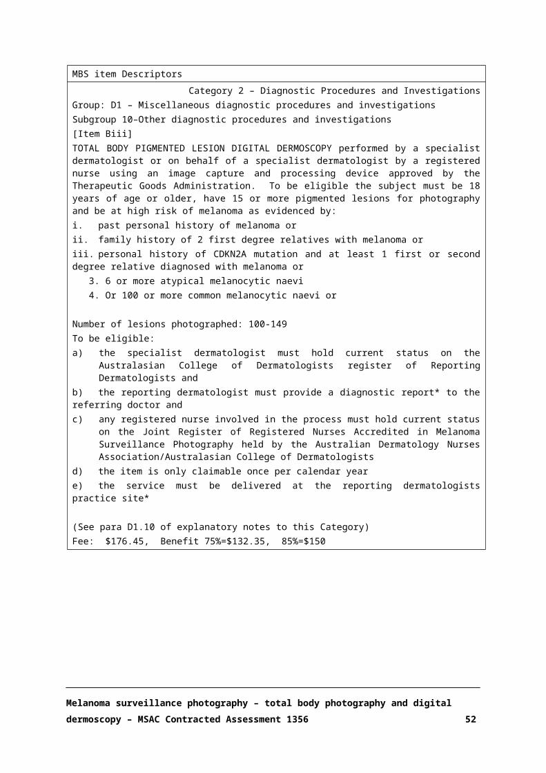
to the patient. Early diagnosis of melanoma leads to excisions with relatively narrow surgical margins
(0.5 cm usually), and a less invasive intervention. More advanced melanoma often requires large amounts of tissue to be excised, sentinel lymph node biopsy, general anaesthetic and longer term
investigative follow up – all involving significant cost, morbidity and mortality.
In the proposed clinical pathway, eligible patients would have been assessed as high or very high risk
by either a GP or by both a GP and a dermatologist. Once a person has been assessed as high/very high-risk, he/she is photographed using TBP. Macro-imaging then occurs; individual melanocytic
naevi are photographed at close range and these naevi are then examined through DD. TBP occurs every five years to refresh images. All naevi and pigmented lesions on the body approximately ≥
3mm in lateral diameter are photographed. Short-term follow-up may occur within 8-16 weeks of total body DD, and only occur once in a year, at the discretion of the reporting dermatologist. Short-
term DD follow-up may be requested for a select few naevi that do not have the dermoscopic features of melanoma, but have an unusual dermoscopic appearance or a patient-reported history
of change. Short-term DD follow-up may also be requested for atypical lesions that for anatomical reasons would be difficult or disfiguring to biopsy or excise. Once a lesion has been identified
through MSP, treatment and/or management is determined based on the size and histology of the melanoma. Surveillance continues after lesion identification.
The current clinical algorithm pathway is shown in black in Figure 5. The additional MSP program proposed in this assessment is shown in dotted red.
Melanoma surveillance photography – total body photography and digital dermoscopy – MSAC Contracted Assessment 1356 52

Figure 5 Clinical management algorithm for the proposed MSP relative to current clinical practice
Melanoma surveillance photography – total body photography and digital dermoscopy – MSAC Contracted Assessment 1356 53
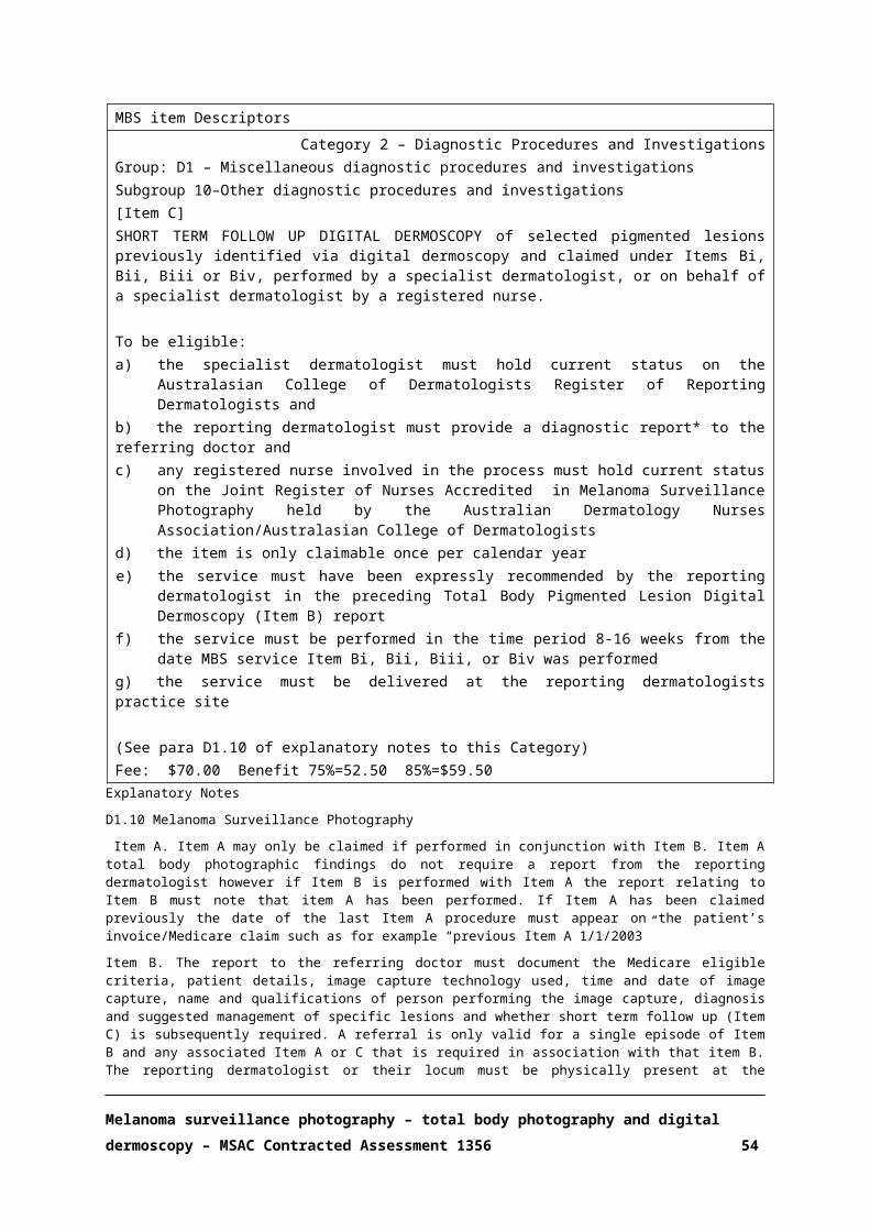
A.7. KEY DIFFERENCES IN THE DELIVERY OF THE PROPOSED MEDICAL SERVICE AND THE MAIN COMPARATOR
MSP is conducted in addition to current practice to monitor high-risk and/or very high-risk patients,
and is proposed to allow accurate comparison of melanocytic naevi over time. MSP may identify early histological changes and early-stages melanoma in the absence of specific criteria for
malignancy, which could be missed by standard care alone.
Dermoscopy has been proved to be more sensitive and more specific at detecting melanoma than a
naked eye clinical examination (National Institute for Health and Care Excellence 2015), but some melanocytic lesions remain indistinguishable from melanoma at early stages. MSP allows
dermatologists to identify incipient changes over time and improve early diagnosis of melanoma (Salerni, Teran et al. 2013).
MSP has been previously described in Section A.2 and Section A.6.
The reasons all pigmented lesions are photographed with digital dermoscopy are:
1. Approximately 50% of naevus-derived melanomas arise from atypical naevi and 50% from common naevi. Therefore, if only atypical naevi are photographed, the common naevi are not
available for comparison in the future. Up to 30% of melanomas in high-risk patients may develop in unmonitored lesions if only clinically atypical lesions are photographed (Salerni,
Carrera et al. 2012).
2. If only naevi that appear clinically atypical are examined and photographed dermoscopically,
only 62% of dermoscopically abnormal naevi would be identified; i.e. many common naevi may have suspicious dermoscopic appearances which can only be identified if they are
examined/photographed with a dermatoscope (Seidenari, Longo et al. 2006).
A.8. CLINICAL CLAIM
Some international guidelines recommend that individuals at high risk of melanoma should receive
surveillance as a standard of practice (Watts, Dieng et al. 2015). In Australia, the Clinical Practice Guidelines for the Management of Melanoma in Australia and New Zealand recommend surveillance
for people at high-risk, stating: “Individuals at high risk of melanoma and their partner or carer be educated to recognise and document lesions suspicious of melanoma, and to be regularly checked
by a clinician with six-monthly full body examination supported by total body photography and dermoscopy as required” (National Health & Medical Research Council/Australian Cancer Network
2008). However, MSP recommendations are largely based on consensus due to the lack of strong evidence.
Melanoma surveillance photography – total body photography and digital dermoscopy – MSAC Contracted Assessment 1356 54

The potential benefits of MSP for high-risk and very high-risk patient groups include:
Melanoma detection at an earlier stage in its development;
Less aggressive skin surgery as definitive treatment;
Fewer benign lesion excisions;
Reduced requirement for sentinel lymph node biopsy and investigation and treatment of
metastatic disease;
Improved diagnosis of non-melanoma skin cancer;
Improved capacity for self-examination and early diagnosis at home; and
Increased sense of well-being following a comprehensive photography session and written
report
A.9. SUMMARY OF THE PICO
The guiding framework of a PICO Confirmation is recommended by MSAC for each assessment. The
Population, Investigation/Index test, Comparator and Outcomes (PICO) Confirmation describes current clinical practice and reflects the likely future practice with the proposed medical service.
Two separate PICOs are presented in Box 1 and Box 2 to guide the systematic literature review for
direct evidence and account for the high-risk and very high-risk populations.
Box 1 Criteria for identifying and selecting studies to determine the effectiveness and safety, and cost-effectiveness of MSP in patients with high risk of melanomaSelection criteria DescriptionPopulations High-risk – Adults (aged ≥ 18 years) with minimum of 15 naevi and at least one of the
following:1. Personal history of melanoma2. Family history of two or more first degree relatives having had melanoma3. Personal history of gene mutation CDKN2A and one first or second degree relative with
melanoma4. 100 or more common naevi5. Six or more atypical/dysplastic naevi
Intervention Total Body Photography (once every 5 years) Total body pigmented lesion digital dermoscopy (DD) (once a year) (‘Long-term follow-up
sequential DD; all lesions’) Follow-up digital dermoscopy of a previously photographed (by DD) pigmented lesion
within 8-16 weeks of DD but limited to once per year (‘short-term follow-up sequential DD’)Comparators Self-examination at home without the use of photography (monthly);
GP clinical examination/skin excisions without access to photography for real time comparison (once or twice per year);
Dermatologist clinical examination/skin excisions (including dermoscopy), without access to photography for real time comparison (once or twice per year)
Melanoma surveillance photography – total body photography and digital dermoscopy – MSAC Contracted Assessment 1356 55

Selection criteria DescriptionOutcomes Safety:
Efficacy/effectiveness:Average Breslow thickness of detected melanomaFrequency distribution of Breslow thickness – in situ, <1mm, >1mmRatio for benign:malignant excisions for melanoma diagnosisTime to diagnosisQuality of Life (QoL)Rate of non-melanoma skin cancer such as basal cell skin cancer and squamous cell skin cancer identified.Healthcare resources:GP and specialist consultationsSkin surgery benign (diagnostic) excisionSkin surgery benign (diagnostic) biopsySkin surgery melanoma wide excisionSkin surgery defect repairHistopathologyDiagnostic surgical sentinel lymph node biopsyTherapeutic lymph node dissectionDiagnostic radiology ultrasoundDiagnostic radiology PET/CT scanCost-effectiveness:Cost/QALYCost/Life YearTotal Australian Government healthcare costs.Cost to the PBSCost to the MBSCost to other Government Healthcare providers
Systematic review question
What is the value of melanoma surveillance photography in addition to standard practice for the detection of melanoma in high risk patients?
Box 2 Criteria for identifying and selecting studies to determine the effectiveness and safety, and cost-effectiveness of MSP in patients with very high risk of melanoma
Selection criteria DescriptionPopulations Very High-risk – Adults (aged ≥ 18 years) with minimum of 15 naevi and:
1. Personal history of two or more primary melanomas OR2. CDKN2A mutation OR3. One past melanoma AND one of
• 100 or more naevi OR• Six or more atypical naevi OR• Family history of three or more first- or second-degree relatives
Intervention Total Body Photography (once every 5 years) Total body pigmented lesion digital dermoscopy (DD) (once a year) (‘Long-term follow-up
sequential DD; all lesions’) Follow-up digital dermoscopy of a previously photographed (by DD) pigmented lesion
within 8-16 weeks of DD but limited to once per year (‘short-term follow-up sequential DD’)Comparators Self-examination at home without the use of photography (monthly);
GP clinical examination/skin excisions without access to photography for real time comparison (once or twice per year);
Dermatologist clinical examination/skin excisions (including dermoscopy), without access to photography for real time comparison (once or twice per year)
Melanoma surveillance photography – total body photography and digital dermoscopy – MSAC Contracted Assessment 1356 56

Selection criteria DescriptionOutcomes Safety:
Efficacy/effectiveness:Average Breslow thickness of detected melanomaFrequency distribution of Breslow thickness – in situ, <1mm, >1mmRatio for benign:malignant excisions for melanoma diagnosisTime to diagnosisQuality of Life (QoL)Rate of non-melanoma skin cancer such as basal cell skin cancer and squamous cell skin cancer identified.Healthcare resources:GP and specialist consultationsSkin surgery benign (diagnostic) excisionSkin surgery benign (diagnostic) biopsySkin surgery melanoma wide excisionSkin surgery defect repairHistopathologyDiagnostic surgical sentinel lymph node biopsyTherapeutic lymph node dissectionDiagnostic radiology ultrasoundDiagnostic radiology PET/CT scanCost-effectiveness:Cost/QALYCost/Life YearTotal Australian Government healthcare costs.Cost to the PBSCost to the MBSCost to other Government Healthcare providers
Systematic review question
What is the value of melanoma surveillance photography in addition to standard practice for the detection of melanoma in high risk patients?
A.10. CONSUMER IMPACT STATEMENT
No consumer responses were identified in the literature.
Melanoma surveillance photography – total body photography and digital dermoscopy – MSAC Contracted Assessment 1356 57

SECTION B CLINICAL EVALUATION
B.1. LITERATURE SOURCES AND SEARCH STRATEGIES
The medical literature was searched on 17 February 2017 to identify relevant studies and systematic
reviews published with no date limits. Searches were conducted in the databases and sources described in Appendix 2 Search strategies. The search terms used are described below in Table 4.
Table 4 Search terms used in the systematic review (Ovid platform)Element of clinical question Search terms
Population exp Melanoma/exp Skin Neoplasms/(pigmented adj2 (lesion$ or mole$ or nevus or nevi or naevus or naevi or skin)).mp.(melanom$ or melanocyt$).mp.cutaneous melanoma.tw.suspicious melanocyt$ lesion$.tw.melanoma$.tw.invasive melanoma.tw.
Intervention exp Dermoscopy/dermoscop$.mp.dermatoscop$.mp.photomicrograph$.mp.(epiluminescence adj2 microscopy).mp.total body photograph$.mp.screening.mp. or exp Mass Screening/population surveillance.mp. or exp Population Surveillance/exp "Early Detection of Cancer"/surveillance.mp.diagnosis.mp.detection.mp.
Limits None applied
B.2. RESULTS OF LITERATURE SEARCH
A PRISMA flowchart (Figure 6) provides a graphic depiction of the results of the literature search and the application of the study selection criteria (listed in Box 1 and Box 2) (Liberati, Altman et al. 2009).
Studies were selected independently by a single reviewer with a random sample receiving independent assessment by a second reviewer. Disagreements regarding study selection were
resolved by discussion.
The selection process firstly included screening of titles and abstracts where the following exclusion
criteria were used:
Editorials; letters; research notes; case studies
Melanoma surveillance photography – total body photography and digital dermoscopy – MSAC Contracted Assessment 1356 58

Conference abstracts/presentations with insufficient details
Non-systematic reviews, non-comparative studies, diagnostic accuracy studies only
Publications in languages other than English
Studies that could not be retrieved or that met the inclusion criteria but contained insufficient or inadequate data for inclusion are listed in Appendix 6 Excluded studies. All other studies that met
the inclusion criteria are listed in Appendix 3 Studies included in the systematic review.
Figure 6 Summary of the process used to identify and select studies for the assessment
A profile of each included study is given in Appendix 3 Studies included in the systematic review. This study profile describes the authors, study ID, publication year, study design and quality (level of
evidence and risk of bias), study location, setting, length of follow-up of patients, study population characteristics, description of the intervention, description of the comparator and the relevant
outcomes assessed. Study characteristics are also summarised in a shorter format in Section B.4.
Melanoma surveillance photography – total body photography and digital dermoscopy – MSAC Contracted Assessment 1356 59

The three included studies compared MSP with standard care using hand-held dermoscopy in
patients at high risk of melanoma, were retrospective in design, and conducted in a dermatology specialist setting. Reported outcomes across the studies included Breslow index and percentage of
in-situ/invasive melanoma. There were no data on the number of excisions or patient-follow-up.
APPRAISAL OF THE EVIDENCE
Appraisal of the evidence was conducted in 2 stages:
Stage 1: Appraisal of the risk of bias within individual studies included in the review (Section B.3).
Stage 2: Integration of this evidence for conclusions about the net clinical benefit of the intervention in the context of Australian clinical practice (Sections B.6-B.8).
Following stage 1, the included studies were found to be at high risk of bias and the quality of the evidence too low to perform a meta-analysis of the reported outcomes. We were therefore unable
to use GRADE to assess the estimate of effect per outcome across the studies.
B.3. RISK OF BIAS ASSESSMENT
We assessed the quality of the included studies using the NICE checklist for cohort studies (National
Institute for Health and Care Excellence 2012), which examines the risk of bias and internal validity of cohort studies designed to answer questions about the relative effects of interventions. Each of
the items was checked as ‘yes’, ‘no’ or ‘unclear’. A summary of the quality assessment is presented in Table 5. This checklist was developed to critically appraise studies that include comparisons
involving a test and subsequent management, and does not cover comparisons of diagnostic test accuracy. Therefore, this checklist is directly applicable to our review question, as we aim to evaluate
key clinical outcomes following surveillance with TBP and digital dermoscopy over time compared to standard care, not the diagnostic accuracy of dermoscopy.
All of the included studies evaluated medical records from patients with melanoma, were retrospective in design and at high risk of bias (Salerni, Lovatto et al. 2011, Salerni, Teran et al. 2014,
Mintsoulis and Beecker 2016). Two of the studies were conducted by the same lead author in two different countries, Spain and Argentina (Salerni, Lovatto et al. 2011, Salerni, Teran et al. 2014).
Selection bias was of concern across the studies and patient populations in the comparison groups differed significantly in some key baseline parameters, such as age. There were no studies which
reported whether patients were consecutively selected into the surveillance programme. Patients in the surveillance groups for each of the three studies met the high-risk of melanoma criteria specified
in our protocol, but no inclusion criteria was provided for patients in the non-surveillance cohorts.
Melanoma surveillance photography – total body photography and digital dermoscopy – MSAC Contracted Assessment 1356 60

The increased risk of melanoma in the control group can therefore not be established or compared
with the population of interest.
The non-surveillance comparison group in Salerni, Lovatto et al. (2011) included patients referred to
the dermatology department for evaluation of suspicious lesions. These patients were significantly older and less likely to have atypical mole syndrome (AMS) or previous melanoma than individuals in
the surveillance programme (Salerni, Lovatto et al. 2011). Salerni, Teran et al. (2014) had two comparison groups – the first included patients who visited the dermatologist for a melanoma
consultation, and the second included patients who attended routine control examinations using hand-held dermoscopy – but the baseline characteristics of these cohorts were not reported.
Mintsoulis and Beecker (2016) evaluated medical records from two different dermatology clinics; patients in the control clinic had a previous history of melanoma and were older than patients in the
MSP surveillance group, and most of them had received yearly skin checks in a general dermatology clinic with hand-held dermatoscopy. There may be systematic differences between the groups being
compared that are not accounted for in the analysis and are likely to interfere with the effect of the intervention.
A further issue is the potential difference in care received by patients in the non-surveillance cohorts. Patients in the standard care group included in Mintsoulis and Beecker (2016) were
monitored by a dermatologist at yearly intervals. Salerni et.al (2011) and (2014) did not report any details on the frequency of examinations or standard care details of patients in the non-surveillance
groups (Salerni, Lovatto et al. 2011, Salerni, Teran et al. 2014).
The three studies clearly reported the outcomes of interest – Breslow thickness and frequency of in-
situ melanomas – but none of them adjusted their analyses for potential prognostic/confounding factors (e.g. age).
None of the studies reported patient follow-up, attrition or compliance data. In summary, all three studies were at high risk of bias.
Table 5 Summary of quality assessment of included studiesCohort Study Checklist Mintsoulis
and Beecker (2016)
Salerni, Lovatto et al. (2011)
Salerni, Teran et al. (2014)
Method of allocation to treatment groups was unrelated to potential confounding factors
No No No
Attempts were made within the design or analysis to balance the comparison groups for potential confounders
No No No
Groups were comparable at baseline No No UnclearComparison groups received the same care apart from the intervention
Unclear Unclear Unclear
Blinding No No NoFollowed up for an equal length of time Unclear Unclear Unclear
Melanoma surveillance photography – total body photography and digital dermoscopy – MSAC Contracted Assessment 1356 61

Comparable for treatment completion Unclear Unclear UnclearComparable with respect to the availability of outcome data Unclear Unclear UnclearAppropriate length of follow-up Unclear Unclear UnclearPrecise definition of outcome Yes Yes YesInvestigators were kept 'blind' to participants' exposure No No NoInvestigators were kept 'blind' to other important confounding and prognostic factors
No No No
B.4. CHARACTERISTICS OF THE EVIDENCE BASE
B.4.1. HIGH-RISK POPULATION
The literature search identified three retrospective comparative cohort studies assessing patient
outcomes. These studies provided comparative data on Breslow thickness and tumour stages from melanoma biopsy samples of patients who had undergone MSP and those receiving standard care
without MSP surveillance. The population undergoing MSP was comparable to the high risk population in our protocol (Salerni, Lovatto et al. 2011, Salerni, Teran et al. 2014, Mintsoulis and
Beecker 2016).
See Appendix 3 Studies included in the systematic review for details on the individual studies
included in the evidence base. A summary is provided below in Table 6.
Table 6 Key features of the included evidence comparing MSP with standard care
Trial/Study N Design/ duration
Risk of bias
Patient population Key outcome(s) Result used in
economic model
Salerni, Lovatto et al. (2011)
Total: 201
MSP: 40SC: 161
Retrospective cohort/2 years
High High-risk of melanoma
• Breslow index• In situ melanoma•Invasive melanoma
Not used
Salerni, Teran et al. (2014)
Total: 89
MSP: NRSC: NR
Retrospective cohort/4 years
High High-risk of melanoma
• Breslow index• In situ melanoma•Invasive melanoma
Not used
Mintsoulis and Beecker (2016)
Total: 224
MSP: 9SC: 215
Retrospective cohort/10 years
High High-risk of melanoma
• Breslow index• In situ melanoma•Invasive melanoma
Not used
MSP: melanoma surveillance photography, NR: not reported, SC: standard care
B.4.2. Very High-Risk population
No available evidence.
Melanoma surveillance photography – total body photography and digital dermoscopy – MSAC Contracted Assessment 1356 62

B.5. OUTCOME MEASURES AND ANALYSIS
B.5.1. HIGH-RISK POPULATION
All the included studies retrospectively assessed melanoma biopsies from patients who received
MSP and compared these results to biopsies from high-risk patients who did not receive MSP.
Breslow index and frequency of in-situ: invasive melanoma were compared between the surveillance
intervention and control groups.
T-tests were used in the statistical analysis for continuous variables in all three studies. No
confidence intervals were reported for any of the studies. The small sample size in these studies suggests high variance. As this variance is not reported, reporting bias is assumed.
There is a high risk of selection bias that could have affected the results, as the comparability of the two populations is unclear and no attempt was made to adjust the analysis for potential
confounders in the studies. Patient characteristics differed between the two comparison groups in two studies (Salerni, Lovatto et al. 2011, Mintsoulis and Beecker 2016), and the study by Salerni,
Teran et al. (2014) did not provide any data on patient characteristics. Furthermore, strict inclusion criteria only applied to the intervention cohorts, and risk of melanoma might have been different
between the comparison groups.
B.5.2. VERY HIGH-RISK POPULATION
No available evidence.
Melanoma surveillance photography – total body photography and digital dermoscopy – MSAC Contracted Assessment 1356 63

B.6. RESULTS OF THE SYSTEMATIC LITERATURE REVIEW
IS IT SAFE?
Summary – MSP is a non-invasive surveillance technology.
There are no safety concerns with providing MSP using TBP and DD
Included studies did not present any data regarding the safety of the intervention. Both TBP and DD
are non-invasive technologies. Hand-held dermoscopy is already in use as standard care in dermatology consultations to evaluate suspicious lesions. TBP is also widely available and consists of
standard photography. TBP and DD are considered safe procedures.
Melanoma surveillance photography – total body photography and digital dermoscopy – MSAC Contracted Assessment 1356 65

IS IT EFFECTIVE?
Summary –
High risk population:
Mean breslow index was significantly reduced in three studies in the surveillance arm compared to the standard care arm (in all three studies p<0.05). The in-situ:invasive melanoma ratio was also significantly higher in the MSP arm than in standard care (in all three studies p<0.05).
All included studies are retrospective in design and at high risk of bias. These results should be interpreted with caution.
Very high-risk population:
No evidence was identified.
EFFECTIVENESS OUTCOMES
B.5.1. HIGH RISK POPULATION
In all the included studies, patients undergoing MSP had significantly higher numbers of melanomas
in-situ and fewer invasive melanomas compared to those receiving usual care (Table 7).These differences in the number of in situ and invasive melanomas indicate that these lesions were
detected at an earlier stage in the MSP surveillance cohorts. However, these studies had a retrospective design and high risk of bias, so the results should be interpreted with caution. It is
important to note that standard care was not clearly defined in the studies and these populations might be potentially different regarding clinical and demographic characteristics as well as patterns
of care (details discussed in Section B.8).
Table 7 Mean Breslow Index (mm)
Study MSP Standard care pSalerni, Lovatto et al. (2011) 0.54 1.71 <0.001
Salerni, Teran et al. (2014) 0.52 1.43a 0.77b <0.05*
Mintsoulis and Beecker (2016) 0.03 0.34 0.02
MSP: melanoma surveillance photographyaMelanoma consultation; bMelanoma routine control* Not specified which comparator this refers to
Breslow index was significantly lower in melanomas from patients in the MSP surveillance
programme than in patients receiving standard care without MSP surveillance (Table 7). The ratio in-situ:invasive melanoma was higher in the MSP surveillance groups compared to standard care (Table
8).
Melanoma surveillance photography – total body photography and digital dermoscopy – MSAC Contracted Assessment 1356 66
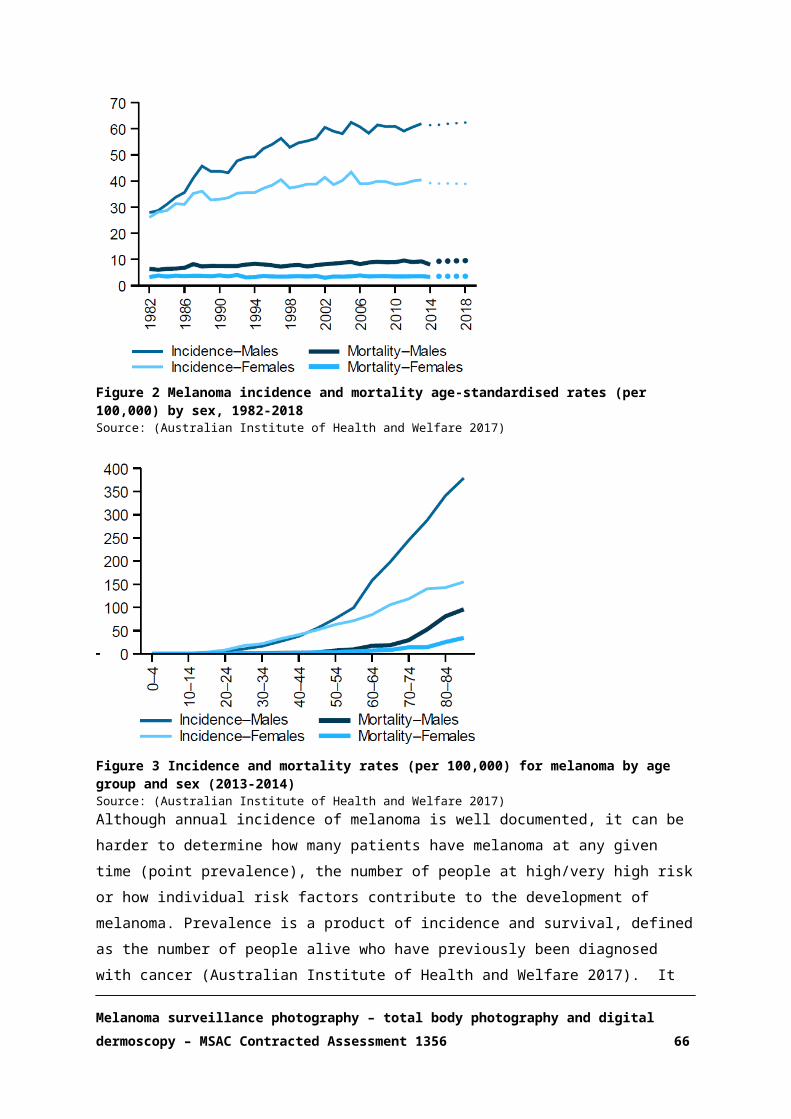
No other outcomes specified in the protocol were reported.
Table 8 Frequency of in-situ and invasive melanoma, n (%)
Study Melanoma MSP Standard care pIn-situ 35 (70) 46 (27.8) <0.001
Salerni, Lovatto et al. (2011) Invasive 15 (30) 119 (72.2) <0.001
Ratio in-situ:invasive 2.33:1 0.38:1
In-situ 7 (58.3) 8 (22.9)a 26 (50.0)b <0.05Salerni, Teran et al. (2014) Invasive 5 (41.7) 27 (77.1)a 26 (50.0)b <0.005*
Ratio in-situ:invasive 1.40:1 0.29:1a 1:1b
In-situ 13 (92.8) 171 (61.7) 0.02Mintsoulis and Beecker (2016) Invasive 1 (7.14) 106 (38.3) NR
Ratio in-situ:invasive 13:1 1.61:1MSP: melanoma surveillance photographyaMelanoma consultation; bMelanoma routine control*not specified which comparator this refers to
B.6.2. VERY HIGH-RISK POPULATION
No available evidence.
B.7. EXTENDED ASSESSMENT OF HARMS
B.7.1. HIGH RISK POPULATION
There was no comparative evidence on the number of nevi biopsies following MSP versus standard
care. Clinical opinion estimates that MSP is associated with a reduced number of biopsies and benign:malignant excision ratio compared with standard care, which might help to reduce patient
anxiety and morbidity associated with multiple procedures.
B.7.2. VERY HIGH RISK POPULATION
No available evidence.
B.8. INTERPRETATION OF THE CLINICAL EVIDENCE
B.8.1. HIGH-RISK POPULATION
Evidence about the relative effectiveness of adding MSP to standard care is limited to three low-quality level III-3 studies reporting Breslow thickness and in-situ: invasive melanoma ratio. On the
basis of the included evidence (Table 7 and Table 8), it is suggested that, relative to the comparator the intervention has non-inferior safety and uncertain effectiveness.
Melanoma surveillance photography – total body photography and digital dermoscopy – MSAC Contracted Assessment 1356 67

The decision not to conduct a meta-analysis was made due to the high risk of bias and the low
number of included studies – three studies of poor quality, two of them by the same author. We were therefore unable to use GRADE to assess the estimate of effect per outcome across the studies.
The effectiveness of the intervention can not be estimated due to the following features of the included studies:
1. Patient populations were heterogeneous. The demographic and clinical characteristics of the cohorts within and across studies might not be comparable.
2. Important risk factors such as age or previous melanoma were uncontrolled (Salerni, Lovatto et al. 2011, Mintsoulis and Beecker 2016) or unreported (Salerni, Teran et al. 2014). In the two
studies that reported information on participant’s age, patients in the control groups were significantly older than those undergoing MSP surveillance (Salerni, Lovatto et al. 2011,
Mintsoulis and Beecker 2016). Salerni, Lovatto et al. (2011) reported a patient population within the MSP group who were significantly more likely to have atypical mole syndrome, a family
history of melanoma or previous melanoma than the standard care group. These differences are likely to bias the results towards the intervention. None of the studies adjusted their statistical
analysis for potential confounders. The definition of standard care was unclear and inconsistent across the studies. Mintsoulis and Beecker (2016) reported patients treated with standard care
had a previous history of melanoma and underwent yearly dermoscopy surveillance (without digital photography) at a hospital dermatology clinic. This control population might differ from
control patients in the other studies, where patients were referred to a dermatologist for melanoma evaluation but no information regarding their routine care were provided. Salerni,
Teran et al. (2014) included two groups in standard care – one was identified as “melanoma consultation” and the second as “melanoma routine control”, but no data was reported on
frequency of examinations or baseline patient characteristics. Salerni, Lovatto et al. (2011) reported patients in the standard care group who were referred to the dermatology hospital
department, but there was no discussion on prior patient care. Furthermore, none of the studies included any data on patient follow-up.
3. The patient population in the MSP groups across the studies was relatively small. The study by Mintsoulis and Beecker (2016) only evaluated medical records from nine patients (14
melanomas) in the surveillance arm during the time period from 2010 to 2014, compared to 215 patients (277 melanomas) in the standard care group over a 10-year period (2004-2014). Salerni
et. al (2011) included data from 40 patients in the MSP group and 161 patients who had received standard care. Salerni, Teran et al. (2014) included 89 patients, but the exact number of patients
in each comparison group was not reported, only the number of melanomas analysed (12 in MSP and 35 in standard care).
Mintsoulis and Beecker (2016) reported better outcomes – lower Breslow thickness and frequency of invasive melanoma in both MSP and standard care than the other two studies. Regular specialist
Melanoma surveillance photography – total body photography and digital dermoscopy – MSAC Contracted Assessment 1356 68
monitoring in the standard care group and the limited number of patients in the MSP group might
have had an impact on these results. Salerni, Teran et al. (2014) reported patients in the non-MSP standard care group who received routine melanoma controls also had thinner Breslow index and a
higher number of in-situ melanomas than those who were classified as having a melanoma consultation, suggesting a positive effect of melanoma monitoring without MSP.
None of the studies reported the number of excisions or ratio for benign:malignant excisions for melanoma diagnosis. These are important measures of effectiveness as the surveillance program is
proposed to reduce the number of excisions leading to better quality of life for patients and reduced costs, however, increased excisions due to more intensive monitoring is also possible.
In summary, the addition of MSP resulted in a reduction in mean breslow thickness in all three studies (all p<0.05) and an increase in situ: invasive ratio (all p<0.05). A low Breslow index and higher
frequency of in-situ lesions indicate early stage melanoma, which is a significant predictor of survival (Balch, Gershenwald et al. 2009, Australian Institute of Health and Welfare 2016, Wernli, Henrikson
et al. 2016). Therefore, the findings are supportive of a benefit from MSP in the proposed population.
However, the quality of the evidence was very low and the applicability of the studies is limited. The findings should therefore be interpreted with caution and the true effect of the proposed
intervention remains unclear. Further research in the form of randomised controlled trials is warranted to determine the impact of MSP on patient’s outcomes.
B.8.2.VERY HIGH-RISK POPULATION
No available evidence.
Melanoma surveillance photography – total body photography and digital dermoscopy – MSAC Contracted Assessment 1356 69

SECTION C TRANSLATION ISSUES
C.1. OVERVIEW
The clinical evaluation suggested that, relative to standard care, MSP has non-inferior safety and
uncertain effectiveness based on the evidence profile given in Section B. Given the poor quality and limited data presented in Section B Clinical Evaluation, the clinical evidence was not considered
sufficient to be used in the economic analysis.
The cost-effectiveness model is presented in Section D is based on a published economic evaluation
(Watts, Cust et al. 2017). The study by Watts et al. (2017) is a cost-effectiveness analysis from the Australian healthcare system perspective, comparing the cost and benefits of MSP for very high-risk
patients in a High-risk dermatology clinic at the Royal Prince Alfred Hospital in Sydney with standard care in the community.
The key translation issues for this model are:
1. Validity of the clinical evidence used in the model;
2. Applicability of the modelled population to the high risk population proposed for funding; and
3. Applicability of the model of care to those proposed for funding.
C.2. APPLICABILITY TRANSLATION ISSUES
All the studies presented in Section B include a patient population at high risk of melanoma, but no comparative evidence was found for patients at very high risk. In contrast, the study by Watts et al.
(2017) focused on patients at very high risk, comparable to the very high-risk population in the agreed protocol, defined as individuals with at least one of the following characteristics:
Confirmed family history of three or more first- or second-degree relatives with melanoma and a confirmed personal history of invasive melanoma;
Dysplastic nevus syndrome and a confirmed personal history of invasive melanoma;
Personal history of at least two confirmed invasive melanomas, one of which diagnosed in
the past 10 years; and
Confirmed high-penetrance mutation affecting melanoma risk.
The model by Watts (2017) is highly applicable to current clinical practice as it used data from the Australian healthcare system and a prospective Australian study of very high risk patients. Therefore,
it is directly relevant for the Australian setting. However, the MSP intervention was conducted in a
Melanoma surveillance photography – total body photography and digital dermoscopy – MSAC Contracted Assessment 1356 70

cohort of very high risk individuals attending a highly specialised clinic in a tertiary hospital within an
urban setting; therefore, the generalizability of the model’s cost-effectiveness results to patients at high risk (for whom the evidence in Section B is relevant) is uncertain.
C.3. EXTRAPOLATION TRANSLATION ISSUES
Watts et al. (2017) developed a Markov model to simulate the management and progression of melanoma over a 10-year time horizon and six health states that would represent long-term follow-
up (details of the model are described in Section D Economic Evaluation). The overall relative survival for melanoma has been reported to stabilise after 9 years (Criscione and Weinstock 2010).
Therefore, the selected 10-year timeframe is appropriate for this analysis as it maximises the availability of data without increasing uncertainty about future projections.
The cost of treatment of late stage melanoma is highly variable and rapidly changing. New immunotherapy treatments were introduced in Australia after the study by Watts et al. (2017) was
finalised. The model presented in Section D has included these new cancer immunotherapies, but these treatments and their utilisation rates may vary in the following years.
C.4. TRANSFORMATION ISSUES
Clinical effectiveness data used to derive the model probabilities of excisions or melanoma by stage inputs for the standard care group were obtained from linked data of patients participating in the
“45 and Up” population-based study (Banks, Redman et al. 2008). These data were compared to that derived from a single-arm prospective study (Moloney, Guitera et al. 2014). However, the standard
care data inputs have not been published, and not all the inputs used in the model were published in Moloney et al. (2014). Therefore, these clinical inputs can not be assessed with the same rigour as
those considered in section B and their validity remains uncertain.
Breslow thickness was used as a surrogate to determine disease stage at diagnosis if spread of the
disease was not documented (Table 9). The validation of this surrogate measure with AJCC stage was conducted in 77 primary melanomas with 92% agreement (weighted k statistic 0.93, 95% CI 0.87-
0.89), indicating good concordance between the two staging methods.
Table 9 Melanoma stage according to Breslow thickness (mm)Stage Breslow thickness (mm) or disease spreadIn-situ No invasionStage I 0 to 1Stage II 1.01 to ≤4Stage III ≥4.01 or lymph node involvementStage IV distant metastatic diseaseSource: (Watts, Cust et al. 2017)
Melanoma surveillance photography – total body photography and digital dermoscopy – MSAC Contracted Assessment 1356 71

C.5. ANY OTHER TRANSLATION ISSUES
No other translation issues were identified.
Melanoma surveillance photography – total body photography and digital dermoscopy – MSAC Contracted Assessment 1356 72
SECTION D ECONOMIC EVALUATION
D.1. OVERVIEW
A cost-effectiveness analysis of MSP compared to standard care from the Australian health system
perspective was identified in the economic literature search (Watts, Cust et al. 2017), and was considered to be key evidence for this application. This study used clinical data from a High-risk
dermatology clinic within a tertiary hospital in Sydney and compared it to linked data from a population-based cohort of selected patients at very high risk of melanoma. The decision was,
therefore, to conduct a cost-effectiveness analysis based on this model, keeping the model structure and updating the cost inputs with 2017 data.
D.2. POPULATIONS AND SETTINGS
The patient population in the economic evaluation includes individuals at very high risk of melanoma, as previously defined in the protocol:
1. Personal history of two or more primary melanomas OR2. CDKN2A mutation OR
3. One past melanoma AND one of• 100 or more naevi OR
• Six or more atypical naevi OR• Family history of three or more first- or second-degree relatives
The setting in the intervention group was a specialised surveillance clinic within a public hospital
where patients at very high risk were seen by dermatologists or dermatology registrars. The standard care group included patients at very high risk who were monitored in the community by
GPs or dermatologists. All patients had a previous melanoma.
There were insufficient data to assess the cost-effectiveness of this intervention in patients at high
risk of melanoma.
D.3. STRUCTURE AND RATIONALE OF THE ECONOMIC EVALUATION
A summary of the key characteristics of the economic evaluation is given in Table 10 below.
Melanoma surveillance photography – total body photography and digital dermoscopy – MSAC Contracted Assessment 1356 73

Table 10 Summary of the economic evaluation Perspective Australian Health System
Comparator Standard care in the community
Type of economic evaluation Cost-effectiveness evaluation
Sources of evidence Indirect comparison of a prospective single-arm study and population-based linked data
Time horizon 10 years in the model base case
Outcomes QALYs
Methods used to generate results Markov model
Health states Patient presents for surveillance, dead from other causes, dead due to melanoma, recurrence of melanoma, stage III disease, stage IV disease
Cycle length 12 months
Discount rate 5%
Software packages used TreeAgePro 2017QALY: quality adjusted life year
LITERATURE REVIEW
Medline, Embase, and the Cochrane Database of Systematic Reviews (without time limits) were used to identify relevant studies, and this search was conducted on the 3rd April, 2017. HTA websites were
also searched, but no relevant assessments were identified.
Table 11 Literature search for cost-effectiveness evaluation# Search terms Results1 exp Melanoma/ 2214592 exp Skin Neoplasms/ 4537383 (pigmented adj2 (lesion$ or mole$ or nevus or nevi or naevus or naevi or skin)).mp. 190754 (melanom$ or melanocyt$).mp. 3093425 cutaneous melanoma.tw. 118916 suspicious melanocyt$ lesion$.tw. 407 melanoma$.tw. 2211928 invasive melanoma.tw. 13319 or/1-8 68895710 surveillance.ab. 41756111 9 and 10 875312 "cost*".ab,ti. 106347613 exp "Costs and Cost Analysis"/ 54532814 "cost benefit analys*".ab,ti. 865615 Cost-Benefit Analysis/ 16527116 exp "health care costs"/ 31852017 12 or 13 or 14 or 15 or 16 135284918 11 and 17 372
One relevant cost-effectiveness analysis was identified in the literature review(Watts, Cust et al. 2017).
Melanoma surveillance photography – total body photography and digital dermoscopy – MSAC Contracted Assessment 1356 74

Watts et al (2017) reported that specialised surveillance was less expensive and more effective than
standard care (Table 12) with mean savings of $6,828 per patient and mean gain in health-related quality of life of 0.31 quality-adjusted life years (QALY) over 10 years. The key drivers for these
differences were earlier detection of melanoma with MSP, which resulted in lower treatment costs, and fewer excisions compared with standard care.
Table 12 Mean Total Costs ($) and QALYs per patient over 10 yearsSpecialised surveillance
Standard care Difference (95% CI)
Mean cost per patient $13,468 $20,296 $6,828 ($5,564 to $8,092)QALY 7.87 7.56 0.31 (0.27 to 0.35)Source: (Watts, Cust et al. 2017)QALY, quality-adjusted life years
STRUCTURE OF THE ECONOMIC EVALUATION
The Markov model developed by Watts et al. 2017 was replicated for this assessment (Table 10). In addition, the cost of MSP was amended to reflect the proposed MBS fee for this intervention, the
cost of new cancer immunotherapies were added to the model, and 2013 MBS items were updated to include 2017 costs. The clinical inputs and model structure were unchanged. TreeAgePro 2017
R1.1 was used to conduct this economic evaluation.
The model compares the use of MSP for patients at very high risk of melanoma in a specialist dermatology setting with standard care in the community (). All patients start the model in the first
year of surveillance and present with a lesion that is either excised or not, in which case the patient would return to surveillance at the specified interval. The excised lesions could be benign,
keratinocytic or melanoma at different stages of development. A patient may have two types of lesions and several excisions within a year.
Patients in the MSP group had a skin examination every six months, and those in standard care once a year. In standard care, the model assumes all lesions identified for excision occur during the one
skin surveillance appointment. Lesion thickness was used to determine melanoma stage (See section C. C.4. Transformation issues)
Melanoma surveillance photography – total body photography and digital dermoscopy – MSAC Contracted Assessment 1356 75

Figure 7 Decision analytic structure of the economic evaluationSource: (Watts, Cust et al. 2017)
The primary outcome from the economic model is the cost per QALY gained over a 10-year time horizon. Six health states () are presented in the model structure:
1) Patient presents for surveillance
2) Dead from other causes
3) Dead due to melanoma
4) Recurrence of melanoma
5) Stage III disease
Melanoma surveillance photography – total body photography and digital dermoscopy – MSAC Contracted Assessment 1356 76

6) Stage IV disease
Depending on the type of lesion excised, the patient may return for surveillance or move to another health state. If the lesion was a melanoma, the health state was determined by melanoma stage at
diagnosis.
Figure 8 Markov model showing the transition between health states from patient presenting for MSPSource: (Watts, Cust et al. 2017)
D.4. INPUTS TO THE ECONOMIC EVALUATION
The updated 2017 costs included in the economic evaluation are provided in Appendix 5 Updated costs.
Clinical data for the specialised surveillance group were obtained from a prospective 5-year (2006-2009) single-arm study conducted in the high-risk clinic (Moloney, Guitera et al. 2014). A total of 311
patients at very high risk of melanoma attended MSP examinations with a dermatologist or a dermatology registrar every 6-months, or returned for a 3-month short follow-up if a suspicious
lesion was identified and not excised.
The standard care arm included data from 607 patients participating in the Sax Institute’s “45 and
Up” cohort study during the same period. The “45 and Up” is a population-based study that recruited 250,000 individuals aged 45 years and older from NSW from 2006 to 2009 with the aim of
providing socio-demographic and health information on an ageing population in Australia (Banks, Redman et al. 2008). Patients at very high risk of melanoma, based on a personal history of
Melanoma surveillance photography – total body photography and digital dermoscopy – MSAC Contracted Assessment 1356 77

melanoma and reported family history of two first-degree relatives with melanoma, were identified
from this study. Only those with linked data to NSW Cancer Registry and Admitted Patient Data Collection (APDC) were included in the analysis.
Deterministic matching was conducted to minimise baseline differences between the two patient groups. However, some baseline characteristics remained unbalanced, such as higher proportion of
men, older age and lower socioeconomic status in the standard care group compared to the MSP group (Table 13). Therefore, we are not able to rule out systematic differences between the two
patient groups, which might have affected the results.
Table 13 Baseline characteristics of patients included in the cost-effectiveness analysisBaseline characteristics MSP
n=311Standard careN=607
Males, n (%) 179 (58) 402 (66)Age in 2006, years
Males, median (IQR) 58 (50-67) 68 (60-76)Females, median (IQR) 50 (41-58) 60 (50-68)Family and personal history, n (%) 52 (16) 93 (15)Multiple primary melanomas, n (%) 146 (47) 514 (85)Naevi and history of melanoma, n (%) 219 (70) NRCDKN2A mutation, n (%) 17 (5) NRIndex of relative socio-economic disadvantage
Ist quintile (most disadvantaged), n (%) 24 (8) 95 (16)2nd quintile, n (%) 43(14) 135 (22)3rd quintile, n (%) 55 (18) 125 (21)4th quintile, n (%) 46 (15) 89 (15)5th quintile (least disadvantaged), n (%) 143 (46) 136 (22)Unrecorded, n (%) NA 27(4)IQR: interquartile range; MSP: melanoma surveillance photographySource: (Watts, Cust et al. 2017)
The annual probabilities (Table 14) for melanoma by stage and excisions in the model were derived from the two sources described above. However, not all the included data used in the model to
derive the probabilities for patients undergoing MSP were reported in Moloney et al. (2014). Excision probabilities were obtained from data collected in the first year of surveillance in the MSP
arm, and 2008 linked data were used in the standard care group. The probabilities of melanoma by stage at diagnosis were calculated from Breslow thickness data from the high risk clinic and based on
a weighted average over 5 years. In the standard care arm, linked MBS data with NSW Cancer Registry and APDC data were used to derive the probabilities. However, these data are not published
and, therefore, it is not possible to adequately assess the validity of these inputs.
Life tables of the Australian Bureau of Statistics (ABS) were used to obtain overall mortality
estimates, and AJCC staging data were used to estimate melanoma-specific mortality. Disease
Melanoma surveillance photography – total body photography and digital dermoscopy – MSAC Contracted Assessment 1356 78
progression probabilities and utility scores reflecting the quality of life for each health state were
obtained from the published literature.
Table 14 Annual probability inputs in specialised surveillance and standard careMSP Standard care
Melanoma by stage at diagnosis Base case
Low High Base Case
Low High
In situ melanoma 0.45 0.02 0.9 0.15 0.08 0.3Stage I melanoma 0.42 0.02 0.84 0.59 0.29 0.9
Stage II melanoma 0.09 0.04 0.18 0.22 0.11 0.44Stage III melanoma 0.03 0.02 0.06 0.05 0.02 0.09Stage IV melanoma 0 NA NA 0.005 0.003 0.01Excisions under surveillanceProbability of an excision 0.4 0.21 0.84 0.64 0.32 0.99Melanoma only 0.12 0.06 0.24 0.05 0.02 0.09Keratinocytic lesion only 0.18 0.09 0.36 0.13 0.07 0.26Benign lesions only 0.5 0.25 0.99 0.28 0.14 0.55Melanoma and keratinocytic lesion 0.02 0.01 0.04 0.03 0.01 0.06Melanoma and benign lesion 0.07 0.03 0.14 0.08 0.04 0.17Keratinocytic and benign lesion 0.09 0.04 0.18 0.33 0.15 0.66Melanoma and keratinocytic and bening lesion 0.01 0.008 0.04 0.1 0.04 0.2Source: (Watts, Cust et al. 2017)MSP: melanoma surveillance photographyLow and high calculations for sensitivity analyses on the basis of multiplier equation (0.5 and 2) for excision probabilities and from published literature for other values.
Resource use, cost and clinical pathways for the surveillance arm were estimated from published data from the high-risk clinic (Watts, Cust et al. 2015). Clinical pathways for the standard care arm
were obtained through telephone surveys of 10 GP clinics in NSW, and costs from MBS items. Patients in the standard care group were assumed to undergo an annual skin examination conducted
by their GP or dermatologist, but only the MBS item for a GP consultation was used as the annual base care cost for standard care. A dermatologist consultation item was not considered. Additional
follow-up costs were estimated for patients with disease progression.
MBS items for out-of-hospital costs in standard care were calculated at 100% of the scheduled fee,
whereas a standard fee for service of 85% of the scheduled fee was applied for the high-risk clinic, as it was part of a public hospital. Costs of excisions were obtained from MBS item numbers and costs
for ongoing surveillance were based on predicted frequency of consultation. Indirect costs (e.g. out-of-pocket costs for travel, productivity loss) were not included. Hospital costs were obtained from
the APDC in NSW. Costs associated to death in hospital, palliative care and treatment of metastatic disease were obtained from published studies. All costs were adjusted to 2017 dollars and a
standard discount rate of 5% was applied to all future costs and benefits.
Melanoma surveillance photography – total body photography and digital dermoscopy – MSAC Contracted Assessment 1356 79

The input costs for specialised surveillance included in the replicated model were calculated at 85%
of the scheduled fee based on the proposed MBS items for this intervention (Table 15). The digital dermoscopy items (by number of naevi) were assumed to be evenly divided across the patient
population (25% of patients accessing each of the 4 items). It was estimated that 10% of patients may undergo short-term follow up every year. The total body photography item was divided by 5 to
estimate an annual cost. The total annual cost per patient undergoing MSP was estimated to be $155. This is considerably less than the cost used in the published model of $884.
Table 15 Annual cost per patient of specialised surveillance, 85% of scheduled feeIntervention Proposed
MBS item cost ($)
Frequency of use
% Accessing Annual Cost (2017 $) per patient
Total Body Photography 85 1 every 5 years 100 17
Digital Dermoscopy (15-49 naevi) 100 annual 25 25
Digital Dermoscopy (50-99 naevi) 125 annual 25 31.25
Digital Dermoscopy (100-149 naevi)
150 annual 25 37.5
Digital Dermoscopy (150+ naevi) 175 annual 25 43.75
Short-Term Follow-up 59.5 annual 10 0.595
Total cost 155
*The following assumptions were applied: 25% usage of each digital dermoscopy item (i.e. of 100 patients using digital dermoscopy, 25 patients would be stratified to each dermoscopy item), and 10% of patients may undergo short-term follow-up every year
It should be highlighted that the proposed annual MBS fee of $155 does not cover the total cost of MSP and patients would likely still incur out of pocket costs. Indirect costs, such as patient out-of-
pocket costs for travel, specialist visits or treatment, and productivity loss were not included in the model. In addition, adherence to the surveillance intervention was not taken into account.
The input for the annual cost for standard care was the same as that published in Watts et al. 2017, at $70 per patient (based on costs for a skin examination GP appointment, estimated from a
telephone survey of skin cancer clinics and mixed practice clinics in NSW).
Treatment costs for stage III and IV were updated to include new immunotherapies that were not
approved by PBS (Pharmaceutical Benefits Scheme) when the Watts et al. (2017) model was developed (Table 16). These costs are approximate, as there were no definite data on the
percentage of patients receiving new immunotherapy treatments in Australia, and such treatment is likely to be highly individualised and rapidly changing.
Table 16 Annual cost per patient of unresectable stage III and stage IV melanoma
Melanoma surveillance photography – total body photography and digital dermoscopy – MSAC Contracted Assessment 1356 80

Treatment items, melanoma stage III and IV Annual cost (2017 $) per patientStaging 20,161.34Follow-up 3,087.54
Unresectable Stage III/Stage IV 8,477.18BRAF immunotherapy 59,118.76
NRAS immunotherapy 40,928.37
Chemotherapy 1,550.55
Adjuvant radiation 6,234.98
Palliative chemotherapy 694.47
Palliative radiotherapy 982.29
Supportive care 2,815.73
Complications 384.61
Total 144,435.83
ASSUMPTIONS INCORPORATED INTO THE MODEL STRUCTURE:
In the Watts (2017) model, the cost of MSP was based on the assumption that 36% of individuals
undergoing MSP would be seen by a dermatologist and 64% would be examined by a GP. In the replicated model, the cost of MSP only accounts for the proposed MBS items and the assumption
that all patients would go to a dermatologist.
It was assumed that patients at very high risk of melanoma were evenly distributed across the four
digital dermoscopy MBS items according to number of naevi, and 10% of patients would need short-term follow-up (Table 15). This distribution might vary in a real-world setting.
The cost of standard care was estimated at $70, which might be an underestimate as it is likely that some patients at very high risk of melanoma would be already under surveillance by a
dermatologist. Nevertheless, an increase in the baseline cost of standard care would favour the cost-effectiveness of the specialised surveillance intervention.
It was assumed that patients in standard care had one skin consultation per year, while those in the MSP group were monitored twice a year. However, the number of GP consultations in standard care
might be higher given that patients in standard care had more excisions than those under specialised surveillance. In addition, there are uncertainties regarding the standard care intervention, such as
details regarding frequency of follow-up or the proportion of patients who might be treated by GPs or dermatologists following their melanoma diagnosis.
No additional follow-up costs were applied for patients with in-situ, stage I and II melanoma. Costs for ongoing follow-up in patients with stage III and IV melanoma were based on the predicted
frequency of specialist consultations: based on 3 monthly specialist consultation, 3-monthly ultrasound and 6-monthly positron emission tomography (PET) in the first and second year following
diagnosis. Surveillance costs in the third and fourth year post-diagnosis included 6 monthly specialist
Melanoma surveillance photography – total body photography and digital dermoscopy – MSAC Contracted Assessment 1356 81

consultation, 6-monthly ultrasound and annual PET. From year five, a cost for an annual specialist
consultation and annual ultrasound was applied.
D.5. RESULTS OF THE ECONOMIC EVALUATION
INCREMENTAL COSTS AND EFFECTIVENESS
As per the published model, the replicated model showed that MSP in patients at very high risk of
melanoma is more effective and less costly than standard care. The overall costs and effectiveness (QALY), and incremental costs and effectiveness as calculated for the intervention and comparator in
the model, with the base case assumptions, are shown in Table 17. The mean cost per patient over 10 years in specialised surveillance and standard care was $7,172.63 and $15,558.83, respectively.
The corresponding QALYs were 7.53 and 7.32. The mean saving associated with the use of MSP over 10 years was estimated at $8,386.19, and the mean QALY gain was 0.21.
Table 17 Cost-effectiveness results MSP compared with Standard care (10-year time horizon)MSP Standard care Incremental
Cost ($) 7,172.63 15,558.83 8,386.19Effectiveness (QALY) 7.53 7.32 0.21QALY: quality adjusted life year, MSP: melanoma surveillance photography
D.6. SENSITIVITY ANALYSES
Results from a one-way sensitivity analysis indicate that MSP is less costly and more effective than standard care up to a threshold of $1,100 for the annual cost of MSP, which is a higher cost than the proposed estimate of $155 (). Beyond this threshold, specialised surveillance with MSP becomes more costly than standard care (given an annual cost of standard care of $70), but MSP remains cost-effective up to an annual cost of $2,700 for a willingness to pay of $50,000 per quality adjusted life years (QALY) gained.
It is important to note this threshold was obtained from a model of MSP in a population at very high risk of melanoma attending a highly specialised clinic within an urban centre, and these results might not be generalisable to the high-risk population in other areas of the country.
There is some uncertainty around the effectiveness inputs in the model. The key drivers for the cost-effectiveness of the model were earlier detection of melanoma and fewer excisions with MSP, but we could not validate the data used to derive these variables.
Melanoma surveillance photography – total body photography and digital dermoscopy – MSAC Contracted Assessment 1356 82

Figure 9 One way sensitivity analysis: incremental cost-effectiveness as cost of MSP increases
As a visual aid to determining which parameters have the greatest influence over the model’s cost-effectiveness results, one-way sensitivity analysis scenarios are presented as a tornado diagram in
Figure 10. The model was most sensitive to variations in the probability of excisions in standard care, with a slight increase in the incremental cost-effectiveness ratio (ICER) as the probability of excision
in standard care was changed from 0.8 to 0.1. However, none of the variables had a substantial impact on the ICER. A potential increase in the cost of MSP from $155 to $1,000 did not significantly
alter the cost-effectiveness of the intervention. Other key drivers of the economic model were the probability of early detection of melanoma (in-situ and stage I) by MSP, treatment costs for
melanoma stage III and IV, and the probability of benign excisions in standard care.
Melanoma surveillance photography – total body photography and digital dermoscopy – MSAC Contracted Assessment 1356 83
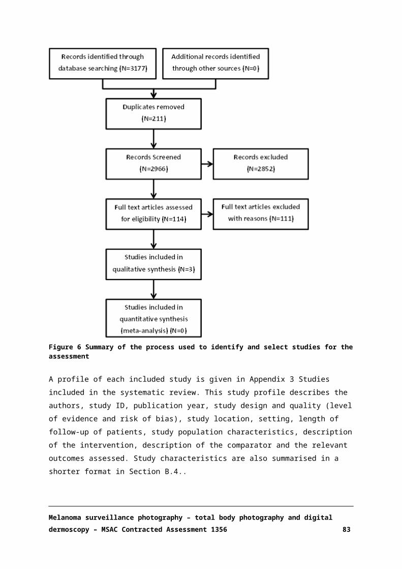
Figure 10 Tornado diagram: one-way sensitivity analysis of MSP versus standard careICER: incremental cost-effectiveness ratio, EV: expected value
A probabilistic sensitivity analysis () was conducted to evaluate the spread of results across the cost-
effectiveness plane, which represents a measure of the ICER’s degree of uncertainty. The incremental costs and QALY points were predominantly spread in the south-east quadrant,
confirming that MSP is dominant, less costly and more effective than standard care.
Melanoma surveillance photography – total body photography and digital dermoscopy – MSAC Contracted Assessment 1356 84

Figure 11 Monte Carlo simulation: Incremental cost-effectiveness of MSP versus standard care
Melanoma surveillance photography – total body photography and digital dermoscopy – MSAC Contracted Assessment 1356 85

SECTION E FINANCIAL IMPLICATIONS
E.1. JUSTIFICATION OF THE SELECTION OF SOURCES OF DATA
An epidemiological approach has been used to estimate the financial implications of the introduction
of MSP in high risk and very high risk patients.
The calculation of the number of patients who would be eligible to receive MSP has been based on
data from the Australian Institute of Health and Welfare (AIHW) (Australian Institute of Health and Welfare 2017, Australian Institute of Health and Welfare 2017) and the Australian Bureau of
Statistics(Australian Bureau of Statistics 2017).
To estimate the size of the eligible populations, the AIHW projections have been combined with
epidemiological parameters (Table 18) derived from published studies (Nordlund, Kirkwood et al. 1985, Newton, Bataille et al. 1993, Aitken, Welch et al. 1999, Vuong, Armstrong et al. 2016,
Australian Institute of Health and Welfare 2017, Australian Institute of Health and Welfare 2017).
Table 18 Epidemiological parameters used in the calculation of the eligible populationsParameter description Input SourcePopulation with previous melanoma Annual incidence, survival AIHW (2017)Population with 100+ naevi 3%* Newton (1993)Population with 6 or more atypical naevi 4%* Nordlund (1985)Population with family history 7%* Vuong (2016)Population CDKN2A positive 0.2%ψ Aitken (1999)*percentage of the population at risk, ψpercentage of the population with previous melanoma
An estimation of the number of patients at high risk and very high risk of melanoma targeted by the proposed medical service is reported in Table 19. These calculations are an approximation given the
lack of direct epidemiological data regarding the proportion of people at high risk and very high risk. The total number of individuals in the Australian population with the risk factors described in Table
18 was calculated with an overlap expectation for each risk factor of 50%, based on clinical advice from the applicant. As no prevalence data were found to establish the number of people at very high
risk in the Australian population, clinical advice was sought, and it was suggested that individuals at very high risk would comprise approximately 40% of those with previous melanoma.
Table 19 Estimation of the number of people at high risk and very high risk of melanoma (2018–2022)Description 2018 2019 2020 2021 2022Population with previous melanoma (10 years)
102,394 102,670 102,946 103,223 103,499
Population with 100+ naevi 315,622 320,161 324,700 329,239 333,777Population with 6 or more atypical naevi 420,830 426,882 432,933 438,985 445,036Population with a family history 736,452 747,043 757,633 768,223 778,814
Melanoma surveillance photography – total body photography and digital dermoscopy – MSAC Contracted Assessment 1356 86
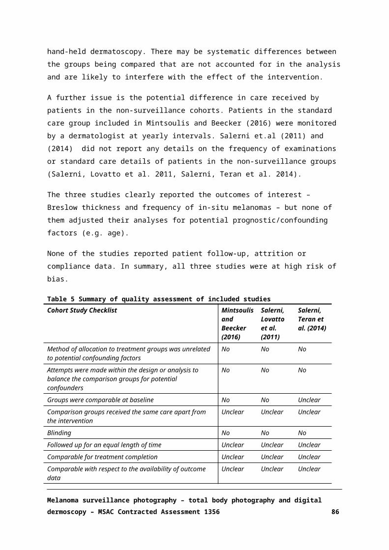
Description 2018 2019 2020 2021 2022Population CDKN2A positive 205 205 206 206 207Overlap expectation 50% 50% 50% 50% 50%
Total population at high risk 787,650 798,378 809,106 819,835 830,563
Total population at very high risk* 40,958 41,068 41,179 41,289 41,399*Population at very high risk was estimated to be approximately 40% of that at high riskψIt was assumed that individuals with CDKN2A would already be included within the population with previous melanoma and was not included in the overlap calculation
E.2. USE AND COSTS OF MELANOMA SURVEILLANCE
The annual cost of MSP is calculated by multiplying the number of patients in each population eligible for MSP by the proposed annual cost per patient. In our model, the annual cost per patient
of the proposed MSP intervention at 85% of the scheduled fee was estimated to be $155 (Section D, Table 15). The total annual cost of MSP using this proposed MBS fee for the population at high risk
was estimated at $122,085,678.70 in 2018, rising to $128,737,255.70 in 2022. The corresponding total annual costs for the high risk population were $6,348,448.88 to $6,416,910.47 (Table 20).
These numbers, however, are estimates given a 100% utilisation rate of MSP and may not reflect the real world.
Table 20 Cost of MSP for individuals at high risk and very high risk of melanoma ($155 per patient, per year); 100% utilisation – 100% uptake and compliancePopulation 2018 2019 2020 2021 2022Number of individuals at high risk
787,650 798,378 809,106 819,835 830,563
Annual MSP cost, population at high risk
$122,085,679.41 $123,748,573.53 $125,411,467.64 $127,074,361.76 $128,737,255.87
Number of individuals at very high risk
40,958 41,068 41,179 41,289 41,399
Annual MSP cost, population at very high risk
$6,348,448.88 $6,365,564.28 $6,382,679.67 $6,399,795.07 $6,416,910.47
Note: rounding has been appliedMSP: melanoma surveillance photography
Uptake at a population level and compliance with MSP remain uncertain. Utilisation of MSP may be
considerably lower than 100% if we take into account the relatively low participation observed in screening programmes (e.g. cervical screening 57.2%) (Australian Institute of Health and Welfare
2013) and low annual rates of compliance (22.2% to 26%) reported by some studies of individuals at high risk of melanoma attending annual digital dermoscopy follow-up examinations for melanocytic
nevi (Schiffner, Schiffner-Rohe et al. 2003, Gadens 2014). Across studies, MSP compliance rates in patients at high risk of melanoma range from 22.2% to over 90% (Schiffner, Schiffner-Rohe et al.
2003, Salerni, Carrera et al. 2012, Gadens 2014).
Melanoma surveillance photography – total body photography and digital dermoscopy – MSAC Contracted Assessment 1356 87

Therefore, we conducted separate analyses to explore annual MSP costs in the scenarios of low and
moderate utilisation. In the low-utilisation scenario (Table 21), we estimated that uptake would increase by 10% every year as more patients become aware of the service, thereby increasing from
20% in 2018 to 60% in 2022. Low compliance in the high-risk population was defined as 22.2%; that is the proportion of people who would return for follow-up. In the first year, we estimated that only
20% of the eligible population would undergo MSP; in the second year, 30% of the eligible population would uptake the service and 22.2% of those who had MSP the year before would
return; in the third year, 40% new patients would uptake the service and 22.2% of those who received MSP the previous year would attend their follow-up; in the fourth and fifth year, uptake
would go up to 50% and 60%, respectively, but compliance would stay low, at 22.2%.
No compliance data were found in the literature for the very high-risk population, so we did not
include them in this analysis under the assumption these patients would be more compliant with the intervention given their elevated risk.
Table 21 Cost of MSP for individuals at high risk of melanoma ($155 per patient, per year); low utilisation (20% to 60% uptake, 22.2% compliance)Population 2018 2019 2020 2021 2022
Number of individuals at high risk (low utilisation) 157,530 274,485 384,578 495,294 608,293
Annual MSP cost, population at high risk (low utilisation)
$24,417,135.88
$42,545,176.22
$59,609,616.18
$76,770,515.67
$94,285,408.00
Note: rounding has been appliedMSP: melanoma surveillance photography Uptake was estimated as 20% in 2018, increasing by 10% every year, and reaching 60% in 2022
A scenario of moderate utilisation of services was estimated for both populations, assuming an
increase on uptake from 20% to 60% over 5 years and 90% compliance. This was calculated as described above for the low utilisation scenario, but with 90% of patients returning for follow-up
(Table 22).
Table 22 Cost of MSP for individuals at high risk and very high risk of melanoma ($155 per patient, per year); moderate utilisation – 20% to 60% uptake*, 90% compliance
Population 2018 2019 2020 2021 2022Number of individuals at high risk (moderate utilisation)
157,530 334,031 490,658 606,181 680,192
Annual MSP cost, population at high risk (moderate utilisation)
$24,417,135.88 51,774,853.59 $76,052,013.85 93,957,986.42 $105,429,749.45
Number of individuals at very high risk (moderate utilisation)
8,192 17,235 25,089 30,680 34,044
Annual MSP cost ($), population at very high risk (moderate utilisation)
$1,269,689.78 $2,671,483.15 $3,888,813.44 $4,755,422.91 $5,276,773.15
Melanoma surveillance photography – total body photography and digital dermoscopy – MSAC Contracted Assessment 1356 88

Note: rounding has been appliedMSP: melanoma surveillance photography*Uptake was estimated as 20% in 2018, increasing by 10% every year, and reaching 60% in 2022
E.3. CHANGES IN USE AND COST OF OTHER MEDICAL SERVICES
Early diagnosis of melanoma may reduce hospital costs and the need for immunotherapy treatment,
which could be translated in savings for the State Government and PBS. It is assumed that GP visits and referrals to dermatologists may increase as a result of MSP uptake, which may increase Federal
costs. The impact of MSP on excision rates remains unknown.
E.4. FINANCIAL IMPLICATIONS FOR THE MBS
It is estimated that patients may need an annual referral from their GP to a dermatologist and this
cost has been added to the 5-year projections. The fee for a GP consultation ≤20 min (MBS00023) is $37.05 at a 100% rebate. Assuming the need for an annual referral to a dermatologist for MSP, the
total cost for patients at high risk by 2022 would be estimated to be $82,115,548.69 if utilisation of services is moderate, and $20,255,168.68 if utilisation is low (Table 23). The corresponding costs for
patients at very high risk assuming moderate utilisation are presented in Table 24.
Table 23 Total cost ($) to the MBS associated with MSP in high risk patients; moderate and low utilisationCost per patient
2018 2019 2020 2021 2022
Intervention - - - - - -Annual MSP cost; high risk (low utilisation)
$155 $24,417,135.88
$42,545,176.22
$59,609,616.18 $76,770,515.67 $94,285,408.00
Annual MSP cost; high risk (moderate utilisation)
$155 $24,417,135.88
$51,774,853.59
$76,052,013.85 $93,957,986.42
$105,429,749.45
Any co-administered services currently MBS listed
- - - - - -
GP consultation ≤20 min (low utilisation)
$37.05 $5,836,483.13 $10,169,669.54
$14,248,621.16 $18,350,629.71 $22,537,253.98
GP consultation ≤20 min (moderate utilisation)
$37.05 $5,836,483.13 $12,375,860.16
$18,178,884.60 $22,458,989.66 $25,201,111.08
Total services - - - - - -
Melanoma surveillance photography – total body photography and digital dermoscopy – MSAC Contracted Assessment 1356 89

Cost per patient
2018 2019 2020 2021 2022
Total cost: High risk (low utilisation)
GP + MSP
$30,253,619.01
$52,714,845.77
$73,858,237.34 $95,121,145.38
$116,822,661.98
Total cost: High risk (moderate utilisation)
GP + MSP
$30,253,619.01
$64,150,713.75
$94,230,898.45
$116,416,976.07
$130,630,860.53
Moderate utilisation: uptake 20% in 2018, increasing by 10% every year, and reaching 60% in 2022, 90% compliance; Low utilisation: uptake 20% in 2018, increasing by 10% every year, and reaching 60% in 2022, 22.2% complianceGP: general practitioner; MSP: melanoma surveillance photography
Table 24 Total costs ($) to the MBS associated with MSP in very high risk patients; moderate utilisationCost per patient 2018 2019 2020 2021 2022
Intervention - - - - - -
Annual MSP cost; very high risk (Moderate utilisation)
$155 $1,269,689.78
$2,671,483.15
$3,888,813.44
$4,755,422.91
$5,276,773.15
Any co-administered services currently MBS listed
- - - - - -
GP consultation ≤20 min (moderate utilisation)
$37.05 $303,496.81 $638,570.65 $929,551.86 $1,136,699.48
$1,261,319.00
Total services - - - - - -
Total cost: very high risk (moderate utilisation)
GP + MSP $1,573,186.59
$3,310,053.80
$4,818,365.30
$5,892,122.39
$6,538,092.15
Moderate utilisation: uptake 20% in 2018, increasing by 10% every year, and reaching 60% in 2022, 90% compliance; Low utilisation: uptake 20% in 2018, increasing by 10% every year, and reaching 60% in 2022, 22.2% complianceGP: general practitioner; MSP: melanoma surveillance photography
E.5. FINANCIAL IMPLICATIONS FOR GOVERNMENT HEALTH BUDGETS
The model results indicate that MSP is less costly than standard care through early detection of melanoma and subsequent reduction in hospital costs and need for metastatic disease treatment.
This would be translated into savings for the State Government.
THE BROADER IMPACT ON THE MBS
The model inputs (unpublished) indicate that the number of excisions was reduced with MSP in a
specialised setting compared with standard care, but we found no comparative published studies
Melanoma surveillance photography – total body photography and digital dermoscopy – MSAC Contracted Assessment 1356 90

that reported excisions rates. Therefore, we are unable to estimate the impact of this item on the
budget.
OTHER GOVERNMENT IMPACTS
The overall cost of treatment for patients with melanoma stage III and IV was estimated in the
economic model at a total cost of $144,436 per patient in 2017 dollars. High-cost targeted immunotherapies have appeared in the Australian market in the past 5 years, and treatment costs
for advance melanoma might increase in the following years as new agents are approved and they are incorporated in clinical practice. A reduction in the number of people with late-stage melanoma
would bring significant savings for the PBS, but the proportion of stage III and IV melanomas prevented in the eligible populations is difficult to estimate.
STATE AND TERRITORY GOVERNMENT HEALTH BUDGETS
Early detection of melanoma may result in fewer hospital admissions, fewer surgeries, and reduced need for monitoring of oncology patients with advanced disease. However, the specific impact of
early detection on hospital savings remains uncertain.
E.6. IDENTIFICATION, ESTIMATION AND REDUCTION OF UNCERTAINTY
There is a high level of uncertainty regarding the approximate number of individuals at high risk and
very high risk of melanoma in Australia. There were no studies that identified the proportion of people at high risk and very high risk, and we estimated these populations based on the prevalence
of risk factors and clinical advice from the applicant.
There is significant variation in MSP compliance rates across studies. It is therefore not possible to
estimate the proportion of individuals within the eligible population who may return for follow-up surveillance after the first consultation. The potential uptake of MSP in these populations is also
uncertain, as many individuals may not identify themselves as high risk or seek medical attention. In addition, access to a dermatologist and out-of-pocket costs may be a barrier for widespread uptake
of MSP and long-term compliance.
Watts et al. (2017) estimated in their model that 64% of patients at very high risk may visit a GP for
skin examinations. Under this scenario, only 36% of eligible individuals at high or very high risk of melanoma may visit a dermatologist. This would significantly reduce our MSP-eligible population
below the 57.2% estimated uptake in a dermatology setting. It is important to note that this participation rate included about half of the eligible population for cervical screening, even though
reminders were sent.
Uptake and compliance may be higher if health promotion programmes in melanoma prevention
and automated patient recalls are implemented along with MSP in these populations.
Melanoma surveillance photography – total body photography and digital dermoscopy – MSAC Contracted Assessment 1356 91

The economic model identified that savings in hospital costs and PBS costs due to early detection of
melanoma were driving the cost-effectiveness of MSP, along with the lower number of excisions in the surveillance group compared with standard care. However, we are not able to assess the impact
of each individual factor on total savings.
Melanoma surveillance photography – total body photography and digital dermoscopy – MSAC Contracted Assessment 1356 92
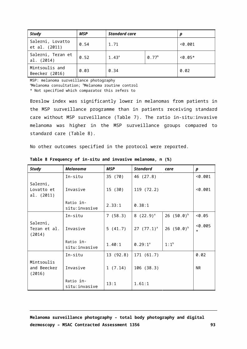
SECTION F OTHER RELEVANT CONSIDERATIONS
Medical models of care are affected by the patient’s geography, socioeconomic status, and patient’s
mobility and risk. Availability of health resources is variable throughout the country, and many patients who are at high risk do not access medical care (PICO confirmation).
Access to a dermatologist may be a barrier to long-term MSP given the low proportion of dermatologists in the Australian population (Table 25), especially in rural areas (The Australasian
College of Dermatologists 2017).
Table 25 Registered dermatologists in AustraliaState Total number of
DermatologistsTotal Population (2016) Number of dermatologists per
100,000 populationAustralian Capital Territory 5 396,294 1.26New South Wales 250 7,726,924 3.24Queensland 112 4,843,303 2.31Northern Territory 1 245,191 0.41Western Australia 60 2,617,074 2.29South Australia 64 1,708,135 3.75Victoria 203 6,069,636 3.34Tasmania 6 519,063 1.16Total 701 24,128,876 2.91Source: (Australian bureau of Statistics 2017, The Australasian College of Dermatologists 2017)
Potential out-of-pocket costs raise the issue of equitable access to high-quality healthcare. The proposed MBS fee does not cover the total cost of MSP. A current fee schedule for Molemap
Australia quotes $449 for an initial DD study (1 hour) and $329 for follow up (Molemap 2017), while the proposed MBS fee for DD ranges from $117.65 to $205.90, depending on the number of lesions
photographed. The out-of-pocket costs to patients might be higher if they have to pay for GP or dermatologist visits, as the service would be implemented in addition to current clinical practice.
Travel costs may also have a substantial impact for patients who live far from main urban centres. In a cost analysis of 102 patients undergoing MSP in a High-Risk clinic in Sydney, Watts. et al. calculated
that mean travel and accommodation costs (return trip) per patient per year were $407, and mean opportunity cost (time not at work or lost personal time) per visit was $502 (Watts, Cust et al. 2015).
This population included patients at very high risk who may have required more frequent follow-up and surgical interventions than what we might expect in patients at high risk. Overall, individuals
who live in rural areas could potentially incur out-of-pocket costs over $500, and people from a low socioeconomic status may not be able to afford MSP.
Melanoma surveillance photography – total body photography and digital dermoscopy – MSAC Contracted Assessment 1356 93
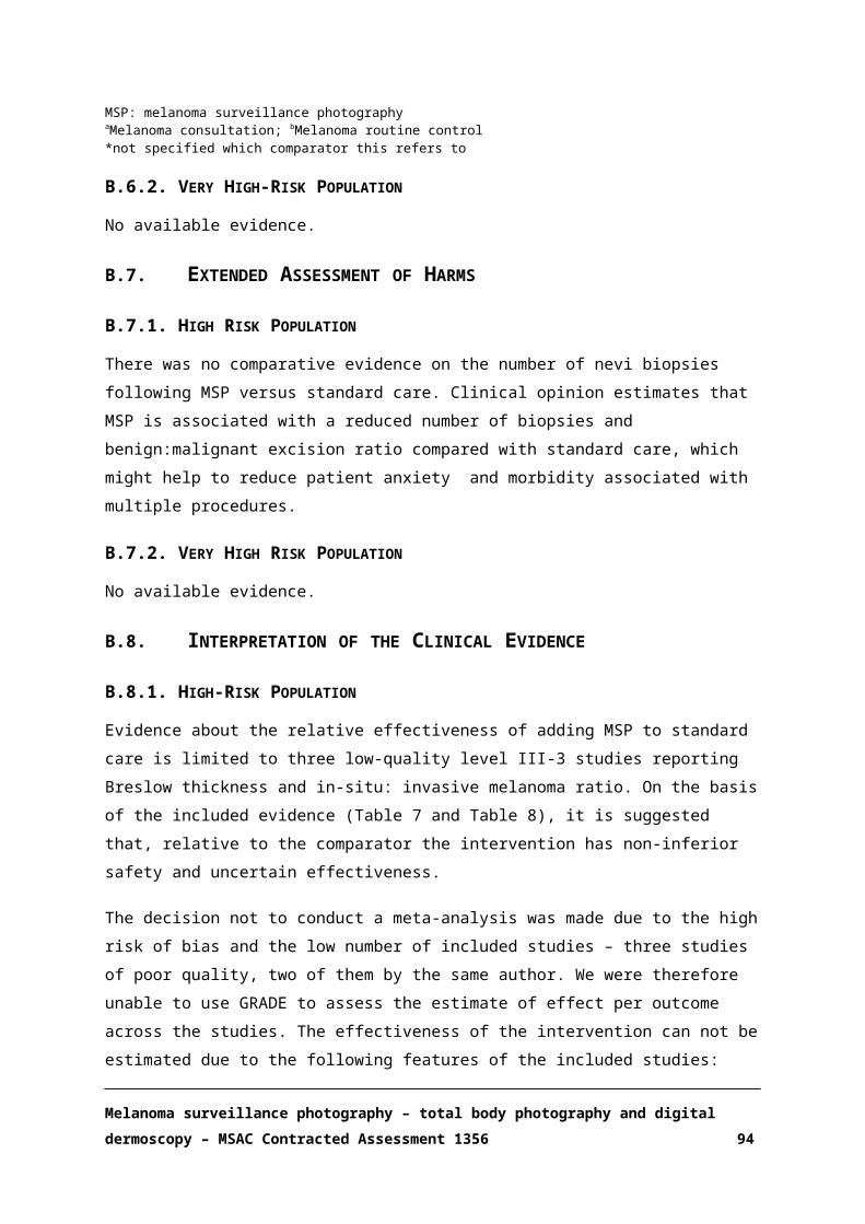
APPENDIX 1 CLINICAL EXPERTS AND ASSESSMENT GROUP
CLINICAL EXPERT
Name Expertise
A/Prof Rob Miller Australasian College of Dermatologists
ASSESSMENT GROUP
NHMRC Clinical Trials Centre
Name Position
Sara Carrillo de Albornoz, MPH Project Officer
Samara Lewis, PhD Project Manager
Melina Wilson, PhD Project Manager
Blaise Agresta, MPH Project Manager
Noted conflicts of interest
There were no conflicts of interest.
Melanoma surveillance photography – total body photography and digital dermoscopy – MSAC Contracted Assessment 1356 94

APPENDIX 2 SEARCH STRATEGIES
Bibliographic databases
Electronic database Time period searchedEmbase February 2017
Medline February 2017
The Cochrane Library (CDSR, Central, DARE, HTA, HEED) February 2017
Additional sources of literature (including websites)
Source LocationAustralian Institute of Health and Welfare (AIHW) http://www.aihw.gov.au/
National Institute for Care and Care Excellence (NICE) https://www.nice.org.uk/
Australian Bureau of Statistics http://www.abs.gov.au/
Medicare Benefits Schedule http://www.mbsonline.gov.au/
Cancer Council Australia http://www.cancer.org.au/
Melanoma surveillance photography – total body photography and digital dermoscopy – MSAC Contracted Assessment 1356 95

APPENDIX 3 STUDIES INCLUDED IN THE SYSTEMATIC REVIEW
Table 26 Profiles of studies comparing MSP to standard care included in the systematic literature review
AuthorsStudy IDPublication Year
Study design/ duration
and risk of bias assessmentb LocationSettingLength of follow-up
Study population characteristics
Measurement of outcomes and methods of analysis
Salerni 2011
Retrospective cohort2 years
Level III-3High risk of bias
SpainSingle hospital
Follow-up not reported
MSP: n=40 (50 melanomas)
Male, n (%): 20 (50)Age, y (range): 49.9 (23-83)AMS, n (%):12 (30)Previous melanoma n (%): 24 (60)Family history of melanoma, n (%):10 (25)
Control (referred patients): n=161 (165 melanomas)
Male, n (%): 85 (52.8)Age, y (range): 61.7 (23-95)AMS, n (%): 22 (13.7)Previous melanoma n (%): 8 (5)Family history of melanoma, n (%): 15 (9.3)
Total body photography+ digital dermoscopy with follow-up visits once or twice a year
Duration of surveillance not reported
Handheld dermoscopy (GP referred)
Breslow IndexIn-situ melanomaInvasive melanoma
Salerni 2014
Retrospective cohort4 years
Level III-3High risk of bias
ArgentinaSingle Hospital
Follow-up
MSP: n not reported (12 melanomas
Control (melanoma consultation): n not reported
Total body photography+ digital dermoscopy with follow-up visits once or twice a year
Handheld dermoscopy (melanoma consultation, melanoma routine control)
Breslow IndexIn-situ melanomaInvasive melanoma
Melanoma surveillance photography – total body photography and digital dermoscopy – MSAC Contracted Assessment 1356 96

AuthorsStudy IDPublication Year
Study design/ duration
and risk of bias assessmentb LocationSettingLength of follow-up
Study population characteristics
Measurement of outcomes and methods of analysis
not reported (35 melanomas)
Patient characteristics not reported
Duration of surveillance not reported
Mintsoulis and Beecker 2016
Retrospective cohort
10 years(4 years MSP and 10 years control)
Level III-3High risk of bias
CanadaPigmented lesion clinics (MSP)General dermatology clinics (control)
Follow-up not reported
MSP: n=9 (14 melanomas)Male, n (%): 5 (55.56)Age, mean (SD): 41.07 (12.62)
Control: n= 215 (277 melanomas)Male, n (%): 128 (59.53)Age, mean (SD): 60.85 (14.52)
Total body photography+ digital dermoscopy with follow-up visits once or twice a year
Duration of surveillance not reported
Handheld dermoscopy (dermatology routine control once a year)
Breslow IndexIn-situ melanomaInvasive melanoma
MSP: melanoma surveillance photographyasource:(NHMRC 2009) bsource:(National Institute for Health and Care Excellence 2012)
Melanoma surveillance photography – total body photography and digital dermoscopy – MSAC Contracted Assessment 1356 97

APPENDIX 4 MBS ITEM DESCRIPTORS
MBS Item Descriptors
Category 1 - PROFESSIONAL ATTENDANCES
23 Group A1 - GENERAL PRACTITIONER ATTENDANCES TO WHICH NO OTHER ITEM APPLIESSubheading 2 - LEVEL BProfessional attendance by a general practitioner at consulting rooms (other than a service to which another item in the table applies), lasting less than 20 minutes and including any of the following that are clinically relevant:(a) taking a patient history;(b) performing a clinical examination;(c) arranging any necessary investigation;(d) implementing a management plan;(e) providing appropriate preventive health care;for one or more health-related issues, with appropriate documentation-each attendanceFee: $37.05 Benefit: 100% = $37.05(See para A5 of explanatory notes to this Category)Extended Medicare Safety Net Cap: $111.15
Category 1 - PROFESSIONAL ATTENDANCES
36 Group A1 - GENERAL PRACTITIONER ATTENDANCES TO WHICH NO OTHER ITEM APPLIESSubheading 3 - LEVEL CProfessional attendance by a general practitioner at consulting rooms (other than a service to which another item in the table applies), lasting at least 20 minutes and including any of the following that are clinically relevant:(a) taking a detailed patient history;(b) performing a clinical examination;(c) arranging any necessary investigation;(d) implementing a management plan;(e) providing appropriate preventive health care;for one or more health-related issues, with appropriate documentation-each attendanceFee: $71.70 Benefit: 100% = $71.70(See para A5 of explanatory notes to this Category)Extended Medicare Safety Net Cap: $215.10
Category 1 - PROFESSIONAL ATTENDANCES
104 Group A3 - SPECIALIST ATTENDANCES TO WHICH NO OTHER ITEM APPLIESProfessional attendance at consulting rooms or hospital by a specialist in the practice of his or her specialty after referral of the patient to him or her-each attendance, other than a second or subsequent attendance, in a single course of treatment, other than a service to which item 106, 109 or 16401 appliesFee: $85.55 Benefit: 75% = $64.20 85% = $72.75Extended Medicare Safety Net Cap: $256.65
Melanoma surveillance photography – total body photography and digital dermoscopy – MSAC Contracted Assessment 1356 98
MBS Item Descriptors
Category 1 - PROFESSIONAL ATTENDANCES
105 Group A3 - SPECIALIST ATTENDANCES TO WHICH NO OTHER ITEM APPLIESProfessional attendance by a specialist in the practice of his or her specialty following referral of the patient to him or her-an attendance after the first in a single course of treatment, if that attendance is at consulting rooms or hospitalFee: $43.00 Benefit: 75% = $32.25 85% = $36.55Extended Medicare Safety Net Cap: $129.00
Category 1 - PROFESSIONAL ATTENDANCES
110
Group A4 - CONSULTANT PHYSICIAN ATTENDANCES TO WHICH NO OTHER ITEM APPLIES
Professional attendance at consulting rooms or hospital, by a consultant physician in the practice of his or her specialty (other than psychiatry) following referral of the patient to him or her by a referring practitioner-initial attendance in a single course of treatment
Fee: $150.90 Benefit: 75% = $113.20 85% = $128.30
Category 3 - THERAPEUTIC PROCEDURES
30071 Group T8 - SURGICAL OPERATIONSSubgroup 1 - GENERALDiagnostic biopsy of skin, as an independent procedure, if the biopsy specimen is sent for pathological examinationMultiple Services Rule(Anaes.) Fee: $52.20 Benefit: 75% = $39.15 85% = $44.40(See para T8.7 of explanatory notes to this Category)Extended Medicare Safety Net Cap: $41.80
Category 3 – THERAPEUTIC PROCEDURES
45203 GroupT8 - SURGICAL OPERATIONSSubgroup 13 - PLASTIC AND RECONSTRUCTIVE SURGERYSubheading 2 - SKIN FLAP SURGERYSingle stage local flap, if indicated to repair one defect, complicated or large, excluding flap for male pattern baldness and excluding H-flap or double advancement flap not in association with any of items 31356 to 31376Multiple Services Rule(Anaes.) (Assist.)Fee: $406.05 Benefit: 75% = $304.55 85% = $345.15(See para T8.95 of explanatory notes to this Category)Extended Medicare Safety Net Cap: $324.85
Melanoma surveillance photography – total body photography and digital dermoscopy – MSAC Contracted Assessment 1356 99

MBS Item Descriptors
Category 3 – THERAPEUTIC PROCEDURES
45669 Group T8 - SURGICAL OPERATIONSSubgroup 13 - PLASTIC AND RECONSTRUCTIVE SURGERYSubheading 4 - OTHER GRAFTS AND MISCELLANEOUS PROCEDURESVERMILIONECTOMY, using carbon dioxide laser or erbium laser excision-ablationMultiple Services Rule(Anaes.) Fee: $326.05 Benefit: 75% = $244.55 85% = $277.15(See para T8.108 of explanatory notes to this Category)
Category 3 – THERAPEUTIC PROCEDURES
30075 SpecialistGroup T8 - SURGICAL OPERATIONSSubgroup 1 - GENERALDIAGNOSTIC BIOPSY OF LYMPH GLAND, MUSCLE OR OTHER DEEP TISSUE OR ORGAN, as an independent procedure, where the biopsy specimen is sent for pathological examinationMultiple Services Rule(Anaes.) Fee: $149.75 Benefit: 75% = $112.35 85% = $127.30
Category 5 - DIAGNOSTIC IMAGING SERVICES
55808
GroupI1 - ULTRASOUND
Subgroup6 - MUSCULOSKELETAL
SHOULDER OR UPPER ARM, 1 or both sides, ultrasound scan of, where:
(a) the service is not associated with a service to which an item in Subgroups 2 or 3 of this Group applies; and
(b) the referring practitioner is not a member of a group of practitioners of which the providing practitioner is a member, and where the service is provided, for the assessment of one or more of the following conditions or suspected conditions:
- evaluation of injury to tendon, muscle or muscle/tendon junction; or
- rotator cuff tear/calcification/tendinosis (biceps, subscapular, suspraspinatus, infraspinatus); or
- biceps subluxation; or
- capsulitis and bursitis; or
- evaluation of mass including ganglion; or
- occult fracture; or
- acromioclavicular joint pathology.(R)
Bulk bill incentive
Fee: $109.10 Benefit: 75% = $81.85 85% = $92.75
Melanoma surveillance photography – total body photography and digital dermoscopy – MSAC Contracted Assessment 1356 100
MBS Item Descriptors
Category 5 – DIAGNOSTIC IMAGING SERVICES
55816 GroupI 1 - ULTRASOUNDSubgroup 6 - MUSCULOSKELETALHIP OR GROIN, 1 or both sides, ultrasound scan of, where:(a) the service is not associated with a service to which an item in Subgroups 2 or 3 of this Group applies; and(b) the referring practitioner is not a member of a group of practitioners of which the providing practitioner is a member (R)Bulk bill incentiveFee: $109.10 Benefit: 75% = $81.85 85% = $92.75(See para DIQ of explanatory notes to this Category)
Category 5 - DIAGNOSTIC IMAGING SERVICES
61553
GroupI4 - NUCLEAR MEDICINE IMAGING
Whole body FDG PET study, following initial therapy, performed for the evaluation of suspected metastatic or recurrent malignant melanoma in patients considered suitable for active therapy (R)
Bulk bill incentive
Fee: $999.00 Benefit: 75% = $749.25 85% = $918.80
Category 6 – PATHOLOGY SERVICES
72816 Group P5 - TISSUE PATHOLOGYExamination of complexity level 3 biopsy material with 1 or more tissue blocks, including specimen dissection, all tissue processing, staining, light microscopy and professional opinion or opinions - 1 separately identified specimen(Item is subject to rule 13)Fee: $86.35 Benefit: 75% = $64.80 85% = $73.40
Category 6 – PATHOLOGY SERVICE
72817
GroupP5 - TISSUE PATHOLOGY
Examination of complexity level 3 biopsy material with 1 or more tissue blocks, including specimen dissection, all tissue processing, staining, light microscopy and professional opinion or opinions - 2 to 4 separately identified specimens
(Item is subject to rule 13)
Fee: $96.80 Benefit: 75% = $72.60 85% = $82.30
Melanoma surveillance photography – total body photography and digital dermoscopy – MSAC Contracted Assessment 1356 101

MBS Item Descriptors
Category 6 – PATHOLOGY SERVICE
72818
GroupP5 - TISSUE PATHOLOGY
Examination of complexity level 3 biopsy material with 1 or more tissue blocks, including specimen dissection, all tissue processing, staining, light microscopy and professional opinion or opinions - 5 or more separately identified specimens
(Item is subject to rule 13)
Fee: $107.05 Benefit: 75% = $80.30 85% = $91.00
Category 6 – PATHOLOGY SERVICES
72823 Group P5 - TISSUE PATHOLOGYExamination of complexity level 4 biopsy material with 1 or more tissue blocks, including specimen dissection, all tissue processing, staining, light microscopy and professional opinion or opinions - 1 separately identified specimen(Item is subject to rule 13)Fee: $97.15 Benefit: 75% = $72.90 85% = $82.60
Category 6 – PATHOLOGY SERVICES
72830 GroupP5 - TISSUE PATHOLOGYExamination of complexity level 5 biopsy material with 1 or more tissue blocks, including specimen dissection, all tissue processing, staining, light microscopy and professional opinion or opinions - 1 or more separately identified specimens(Item is subject to rule 13)Fee: $274.15 Benefit: 75% = $205.65 85% = $233.05
Category 6 – PATHOLOGY SERVICES
72846 GroupP5 - TISSUE PATHOLOGYImmunohistochemical examination of biopsy material by immunofluorescence, immunoperoxidase or other labelled antibody techniques with multiple antigenic specificities per specimen - 1 to 3 antibodies except those listed in 72848(Item is subject to rule 13)Fee: $59.60 Benefit: 75% = $44.70 85% = $50.70
Category 6 – PATHOLOGY SERVICES
73049 GroupP6 - CYTOLOGYCytology of material obtained directly from a patient by fine needle aspiration of solid tissue or tissues - 1 identified siteFee: $68.15 Benefit: 75% = $51.15 85% = $57.95
Melanoma surveillance photography – total body photography and digital dermoscopy – MSAC Contracted Assessment 1356 102

MBS Item Descriptors
Category 6 - PATHOLOGY SERVICES
73924
Group P10 - PATIENT EPISODE INITIATION
Initiation of a patient episode that consists of 1 or more services described in items 72813, 72816, 72817, 72818, 72823, 72824, 72825, 72826, 72827, 72828, 72830, 72836 and 72838 (in circumstances other than those described in item 73925) from a person who is an in-patient of a hospital.
Fee: $14.65 Benefit: 75% = $11.00 85% = $12.50
Category 6 - PATHOLOGY SERVICES
73927
Group P10 - PATIENT EPISODE INITIATION
Initiation of a patient episode by a prescribed laboratory that consists of 1 or more services described in items, 72813, 72816, 72817, 72818, 72823, 72824, 72825, 72826, 72827, 72828, 72830, 72836 and 72838 from a person who is not a patient of a hospital.
Fee: $2.40 Benefit: 75% = $1.80 85% = $2.05
Melanoma surveillance photography – total body photography and digital dermoscopy – MSAC Contracted Assessment 1356 103

APPENDIX 5 UPDATED COSTS
Table 27 Updated Annual health system costs in the economic model, 2017Description Procedure
CodeCost per Item, $
No. in 12months
Annual cost $
Medical Consultation TypeStandard-length consultation ≤20 min (level B) MBS 00023 37.05 95 3520Extended-length consultation >20 min (level C) MBS 00036 71.70 176 12619Dermatologic or surgical specialistInitial consultation MBS 00104 72.75 6 437Subsequent consultation MBS 00105 36.55 10 366Total consultation 16,941Procedures for Excisions of SkinDiagnostic biopsy of skin MBS 30071 44.40 7.00 314.00Diagnostic excision of skin<10 mm MBS 31206 81.15 39.00 2930.0010 to <20 mm MBS 31211 104.65 2.00 209.30<6mm and nose, eyelid, eyebrow, lip, ear, digit or genitalia, or from a contiguous area
MBS 31356 188.20 1.00 188.20
<14mm and face, neck, scalp, nipple-areola complex, distal lower limb or distal upper limb
MBS 31361 158.70 2.00 215.00
<6mm and nose, eyelid, eyebrow, lip, ear, digit or genitalia, or from a contiguous area
MBS 31356 188.20 1.00 188.20
<14mm and face, neck, scalp, nipple-areola complex, distal lower limb or distal upper limb
MBS 31361 158.70 1.00 158.70
<14mm and face, neck, scalp, nipple-areola complex, distal lower limb or distal upper limb
MBS 31361 158.70 1.00 158.70
<14mm and face, neck, scalp, nipple-areola complex, distal lower limb or distal upper limb
MBS 31361 158.70 4.00 634.80
<14mm and face, neck, scalp, nipple-areola complex, distal lower limb or distal upper limb
MBS 31361 158.70 1.00 158.70
<14mm and face, neck, scalp, nipple-areola complex, distal lower limb or distal upper limb
MBS 31372 262.40 2.00 524.80
<15mm and other areas MBS 31374 239.65 1.00 239.65Total skin excisions 5920SurveillanceSequential digital dermoscopy imaging1-3 Lesions 70.49 43.00 3031.08>3 Lesions 81.33 3.00 244.00Total monitoring 3275.09Other Proceduresf
Single-stage local flap repair MBS 45203 345.15 1.00 345.15Vermilionectomy MBS 45669 277.15 1.00 277.15Diagnostic biopsy of lymph gland (by specialist) MBS 30075 127.30 1.00 127.30Ultrasonography of the groin MBS 55816 92.75 1.00 92.75
Melanoma surveillance photography – total body photography and digital dermoscopy – MSAC Contracted Assessment 1356 104
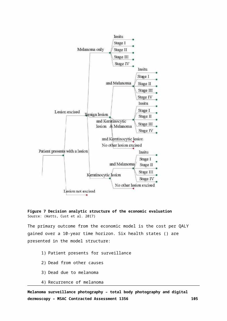
Inpatient stay in standard ward (mean length of stay,1.23 days)
DRG 11Z NA NA 2215.56
Pathological examinationLevel 3 MBS 72816 73.40 22.00 1614.80Level 4 MBS 72823 82.60 39.00 3221.40Level 5 MBS 72830 233.05 3.00 699.15Immunohistochemistry MBS 72846 50.70 1.00 50.70Cytology fine-needle aspiration MBS 73049 57.95 1.00 57.95Total other procedures 8701.91Total medical procedures (consultations, excisions, monitoring, and other)
NA NA NA 34,838.00
Mean Cost per Patient per Year (n patients=87) 400.44
Estimated total medical procedure costs per patient (MSP) Proposed MBS item
155
Estimated total health system costs per patient 556MSP: melanoma surveillance photographyAdapted from (Watts, Cust et al. 2015)
Table 28 Updated Treatment costs, 2017
Cost item MBS/DRG Item
Unit costs, $
No. in 12months
Annual costs, $
Diagnostic imaging techniques
PET scan n.a.Bone scan 61425 $510.60 1 $510.60Gallium scan 61442 $639.50 1 $639.50Surgery for lymph node recurrence with curative intent (lymphadenectomy)Other neoplastic disorders w major or procedures w/o catastrophic or severe complications DRG R02B $20,989 1 $20,988.7
9Surgery for lymph node recurrence with curative intent (lymphadenectomy) with complicationsOther neoplastic disorders w major or procedures w catastrophic or severe complications DRG R02A $38,832 1 $38,831.7
7Radiotherapy adjuvant
Planning episode of 3-dimensional conformal radiotherapyDosimetry for three dimensional conformal radiotherapy of level 3 complexity MBS 15562 $952.65 1 $953
Simulation for three dimensional conformal radiotherapy (w/o iv contrast) MBS 15550 $559.85 1 $560
TreatmentRadiation oncology treatment, 10 rounds (1 field) MBS 15254 $50.75 10 $508Radiation oncology treatment, 10 rounds (subsequent 2 fields) (at $37.95 per field) MBS 15269 $37.95 20 $759
Total cost for radiotherapy treatment $2,779
Immunotherapy adjuvant (for 3 months)induction therapy: Interferon alfa-2b (IV injection 5d/week for 4 weeks) PBS 6246R $1,861.68 20 $37,234
maintenance therapy: Interferon alfa-2b(IV injection 3d/week PBS 6246R $930.84 24 $22,340
Melanoma surveillance photography – total body photography and digital dermoscopy – MSAC Contracted Assessment 1356 105

Cost item MBS/DRG Item
Unit costs, $
No. in 12months
Annual costs, $
for 8 weeks)Initial specialist consultation MBS 104 $72.75 1 $73Follow-up special consultation MBS 105 $36.55 1 $37Total cost for immunotherapy treatment $168,646Palliative ChemotherapyDacarbazine 2 cycles (5 day-cycle (250 mg/m2/day), 1 cycle every 3 weeks) $48.94 10 $489
Anti-emetic: IV dexamethasone (8mg) single bolus per cycle (1 pack of 5 vials) PBS 1291Y $4.03 2 $8
Administration of chemotherapyInjection <1hr (once per cycle) MBS 13915 $55.30 2 $111Total cost for palliative chemotherapy $608Radiotherapy palliative
Planning episode of 3-dimensional conformal radiotherapyDosimetry for three dimensional conformal radiotherapy of level 3 complexity MBS 15562 $952.65 1 $953
Simulation for three dimensional conformal radiotherapy (w/o iv contrast) MBS 15550 $559.85 1 $560
TreatmentRadiation oncology treatment, 10 rounds (1 field) MBS 15254 $50.75 10 $508Radiation oncology treatment, 10 rounds (subsequent 1 field) (at $37.95) MBS 15269 $37.95 10 $380
Total cost for radiotherapy treatment $2,400Observation/ Follow-up at 3 months (in all patients)
Follow-up specialist consultation MBS 104 $72.75 1 $73Full blood count MBS 65070 $14.45 1 $14Liver function tests MBS 66512 $15.05 1 $15
CT scan (chest, abdomen, pelvis) with IV contrast MBS 56807/56847 $358.65 1 $359
CT scan (head) with IV contrast MBS 56007/56047 $159.85 1 $160
Total cost per follow-up $621Observation/ Follow-up at 3 months (in patients with incurable disease having undergone resection)
Follow-up specialist consultation MBS 104 $72.75 1 $73Full blood count MBS 65070 $14.45 1 $14Liver function tests MBS 66512 $15.05 1 $15
CT scan (chest, abdomen, pelvis) with IV contrast MBS 56807/56847 $358.65 1 $359
CT scan (head) with IV contrast MBS 56007/56047 $159.85 $160
Brain MRI performed by a consultant physician scan of head for tumour of the brain or meninges (Contrast) (Anaesthesia) MBS 63001 $342.75 $343
Total cost per follow-up $964CT: computerised tomography; IV: intravenous; PET: positron emission tomography; Adapted from (Watts, Cust et al. 2015)
Melanoma surveillance photography – total body photography and digital dermoscopy – MSAC Contracted Assessment 1356 106

APPENDIX 6 EXCLUDED STUDIES
Reason for exclusion: Abstracts
(2011) "Abstracts Presented at the Australasian College of Dermatologists Biennial Spring Conference 2011." Australasian journal of dermatology 52.
Anonymous (2011). "Abstracts Presented at the Australasian College of Dermatologists Biennial Spring Conference 2011." Australasian Journal of Dermatology. Conference: Australasian College of Dermatologists Biennial Spring Conference 52(no pagination).
Berryman, C., M. Haskett, S. Hannam and G. Largey (2012). "Melanoma shared care, a tripartite approach for survival. The patient, their GP and their specialist-a survivorship project." Asia-Pacific Journal of Clinical Oncology 8: 307.
Carrera, C., G. Salerni, S. Puig, R. Lorenzato, F. Cabrini, L. Lovato and J. Malvehy (2010). "Early detection of melanoma based on digital follow-up of high-risk patients. Ten years Spanish experience of a prospective surveillance." Pigment Cell and Melanoma Research 23 (6): 895.
Cedef (2012). "[Item 149--Epithelial and melanotic cutaneous tumors: nevus]." Annales de Dermatologie et de Venereologie 139(11 Suppl): A158-163.
Cheng, H. M. and P. Guitera (2015). "Assessment of the optimal interval for digital dermoscopy monitoring for the evaluation of pigmented nail lesions." Australasian Journal of Dermatology 56: 25.
Coates, E. and S. W. Menzies (2012). "Total body photography self-examination in patients at high risk of melanoma." Australasian Journal of Dermatology 53: 34.
Cosgarea, R., L. Ungureanu and L. Rogojanu (2013). "Our 9 years digital dermoscopy experience in the diagnosis of early melanoma." JDDG - Journal of the German Society of Dermatology 11: 2-3.
Frances, L., A. M. Bouret, I. Marin, J. Banuls and M. Leiva-Salinas (2014). "Longitudinal nevus: A distinctive type of nevus on the sole." Journal of the American Academy of Dermatology 1): AB128.
Gniadecki, R. (2005). "6<sup>th</sup> World Congress on Melanoma: September 6-10, 2005, Vancouver, B.C., Canada." Forum for Nordic Dermato-Venerology 10(4): 113-115.
Goldust, M. (2012). "Efficacy of dermatoscopy in the diagnosis of pigmented skin lesions as compared to histopathology." Journal of Investigative Dermatology 132: S135.
Goldust, M. (2013). "Efficacy of dermatoscopy in the diagnosis of pigmented skin lesions as compared to histopathology." JDDG - Journal of the German Society of Dermatology 11: 39.
Hofmann, L., S. Kraus, K. M. Thoms, S. Emmert, M. Schon, L. Kretschmer and H. Haenssle (2013). "Unravelling of difficult-to-diagnose skin tumors by analysis with in-vivo confocal laser scanning microscopy." JDDG - Journal of the German Society of Dermatology 11: 37.
Huang, A., S. Y. Kwan, W. Y. Chang, M. Y. Liu, M. H. Chi and G. S. Chen (2013). "A robust hair segmentation and removal approach for clinical images of skin lesions." Conference Proceedings: ... Annual International Conference of the IEEE Engineering in Medicine & Biology Society 2013: 3315-3318.
Hunt, D., C. Duff and S. Watso (2011). "An audit to evaluate the effectiveness of dermatoscopy." International Journal of Surgery 9 (5): 367.
Lin, C. Y., S. Hill, A. Yung, A. Oakley and M. Rademaker (2012). "Evaluation of naevi exposed to narrowband UVB during phototherapy." Australasian Journal of Dermatology 53 (4): A1-A2.
Longo, C., S. Ciardo, F. Farnetani, A. Cesinaro, G. Ponti and G. Pellacani (2012). "Feasibility of in vivo confocal microscopy in diagnosing nodular lesions." Journal of Investigative Dermatology 132: S132.
Luttrell, M. J., G. M. Williams, R. Hofmann-Wellenhof, R. Fink-Puches and H. P. Soyer (2010). "The AC Rule for Melanoma: A simple clinical and dermoscopic rule for the wider community." Pigment Cell and Melanoma Research 23 (6): 901-902.
Lynch, M., A. Syed and K. Lacy (2013). "Analysis of factors leading to the diagnosis of malignant melanoma in a tertiary centre high-risk melanoma screening clinic." JDDG - Journal of the German Society of Dermatology 11: 38.
Malvehy, J. and S. Puig (2011). "Mole mapping and digital photography." Pigment Cell and Melanoma Research 24 (5): 1009.
Manganoni, A. M., L. Pavoni, M. Alaibac and P. Calzavara-Pinton (2014). "Diagnostic utility of dermoscopy in BRAF inhibitorassociated verrucous keratoses." Pigment Cell and Melanoma Research 27 (6): 1209.
McGoldrick, D. M., K. M. Fogarty, M. Majeed, A. Amin Achakzai and H. P. Redmond (2016). "Audit: Correlation between dermoscopy score and final histology in skin lesions." Irish Journal of Medical Science 1): S111.
Melanoma surveillance photography – total body photography and digital dermoscopy – MSAC Contracted Assessment 1356 107

Reason for exclusion: Abstracts
Meani, R. E., R. J. Bloom and A. J. Chamberlain (2014). "Fibro-osseous pseudotumour of the digit: Nailing a difficult diagnosis." Australasian Journal of Dermatology 55: 40-41.
Moloney, F. J., P. Guitera, E. Coates, N. Haass, K. Ho, R. Khoury, G. Mann and S. W. Menzies (2012). "Observation of a five year high risk clinic for primary melanoma." Australasian Journal of Dermatology 53: 33.
Moloney, F., P. Guitera, E. Coates, N. Haass, K. Ho, R. Khoury, G. Mann and S. Menzies (2012). "Sequential digital dermoscopic imaging and total body photography aid detection of primary melanoma in individuals at high risk: A prospective 5-year follow-up study." British Journal of Dermatology 167: 4.
Moscarella, E., I. Zalaudek, H. Rabinovitz, S. Puig and G. Argenziano (2012). "Dermoscopy in multiple primary melanomas: A multicenter, retrospective, observational study." Journal of Investigative Dermatology 132: S134.
Murzaku, E. C., S. Hayan and B. K. Rao (2014). "Methods and rates of dermoscopy usage: A crosssectional survey of United States dermatologists stratified by years in practice." Pigment Cell and Melanoma Research 27 (5): 943-944.
Nagaoka, T., A. Nakamura, Y. Kiyohara, H. Koga, T. Saida and T. Sota (2013). "Hyperspectroscopic screening of melanoma." JDDG - Journal of the German Society of Dermatology 11: 33.
Nikolic, D., A. Nikolic and V. Stanimirovic (2011). "Challenges of early diagnosis of melanoma in Serbia." Pigment Cell and Melanoma Research 24 (5): 1063-1064.
Nikolic, D., A. Nikolic, V. Stanimirovic, T. Filipovic and J. Kostic (2013). "Screening of the pigmented tumors of the skin-single medical center experience." JDDG - Journal of the German Society of Dermatology 11: 28.
Nikolic, D., V. Stanimirovic, A. Nikolic, M. Aleksandar and S. Miljkovic (2010). "Great benefits of epiluminescence microscopy in early diagnosis of melanoma in situ in Serbia." Pigment Cell and Melanoma Research 23 (6): 904-905.
Puig, S., A. Diaz, C. Trullas, J. A. Puig-Butille and J. Malvehy (2012). "Evaluation of the effects of topical application of a film-forming medical device containing photolyase and UV filters in the repair of UV induced DNA damages in actinic keratosis patients." Journal of the American Academy of Dermatology 1): AB151.
Rice, S. A., V. J. Swale and R. Cerio (2012). "Are we relying too heavily on dermoscopy?" British Journal of Dermatology 167: 105.
Ruiz, D., V. J. Berenguer, A. Soriano and J. Martin (2008). "A cooperative approach for the diagnosis of the melanoma." Conference Proceedings: ... Annual International Conference of the IEEE Engineering in Medicine & Biology Society 2008: 5144-5147.
Shie, C., F. Fan and Y. Liao (2014). "Hybrid-featuring melanoma CAD for self-diagnosis." Journal of Investigative Dermatology 134: S126.
Smith, M., L. Kagemann, J. S. Schuman and J. L. Fine (2012). "Histopathologic correlation with spectral-domain optical coherence tomography (SD-OCT), a novel digital imaging technique." Laboratory Investigation 92: 394A.
Szalai, K., Z. Hatvani, J. Harsing, B. Somlai and S. Karpati (2011). "High frequency ultrasonography in the diagnosis of cutaneous pigmented lesions and melanoma reduce the possibilities of diagnostic pitfalls." Melanoma Research 21: e4-e5.
Urbancek, S., P. Simekova and J. Tomkova (2013). "Misdiagnosis of melanoma: A 5 years analysis." JDDG - Journal of the German Society of Dermatology 11: 31.
Verykiou, S., J. A. A. Langtry and N. Rajan (2012). "Cutaneous malignant melanoma: Does increased number of biopsies increase melanoma detection?" British Journal of Dermatology 167: 95-96.
Wray, E. V., B. Brant and F. Muller (2015). "Diagnostic accuracy of a store-and-forward teledermoscopy clinic for skin cancer referrals." British Journal of Dermatology 173: 182.
Zurac, S., D. Boda, A. Diaconeasa, R. Andrei, T. Tiberiu, C. Caruntu, R. Mustata, F. Staniceanu, V. Chitu and C. Solovan (2011). "Spitz tumor in pediatric and adult population: Clinical, dermoscopic, in vivo confocal microscopic and histopathologic study." Virchows Archiv 459: S97-S98.
Zurac, S., D. Boda, A. Diaconeasa, R. Andrei, T. Tiberiu, C. Caruntu, R. Mustata, F. Staniceanu, V. Chitu and C. Solovan (2011). "Spitz tumor in pediatric and adult population: Clinical, dermoscopic, in vivo confocal microscopic and histopathologic study." Virchows Archiv 459: S97-S98.
Reason for exclusion: Animals/not human
Ertam, I., B. G. Turk, A. Urkmez, A. Kazandi and F. Ozdemir (2009). "Linear and whorled nevoid hypermelanosis: dermatoscopic features." Journal of the American Academy of Dermatology 60(2): 328-331.
Nolte, T., P. Brander-Weber, C. Dangler, U. Deschl, M. R. Elwell, P. Greaves, R. Hailey, M. W. Leach, A. R. Pandiri, A. Rogers, C. C. Shackelford, A. Spencer, T. Tanaka and J. M. Ward (2016). "Nonproliferative and proliferative lesions of the gastrointestinal tract, pancreas and salivary glands of the rat and mouse." Journal of Toxicologic Pathology 29(1 Supplement):
Melanoma surveillance photography – total body photography and digital dermoscopy – MSAC Contracted Assessment 1356 108

Reason for exclusion: Abstracts
1S-124S.
Reason for exclusion: Insufficient data
Argenziano, G., J. Giacomel, I. Zalaudek, A. Blum, R. P. Braun, H. Cabo, A. Halpern, R. Hofmann-Wellenhof, J. Malvehy, A. A. Marghoob, S. Menzies, E. Moscarella, G. Pellacani, S. Puig, H. Rabinovitz, T. Saida, S. Seidenari, H. P. Soyer, W. Stolz, L. Thomas and H. Kittler (2013). "A clinico-dermoscopic approach for skin cancer screening: recommendations involving a survey of the International Dermoscopy Society." Dermatologic Clinics 31(4): 525-534, vii.Botti, E., C. Mercurio, G. Spallone, A. Di Stefani, M. Gabellini, A. Orlandi, S. Chimenti, S. Minucci and A. Costanzo (2011). "Histone deacetylases as new therapeutical targets for the treatment of Non-Melanoma Skin Cancer: Results of phase I/IIa trial with topical DAC060." Journal of Investigative Dermatology 131: S46.Gooderham, M., V. Nanda, C. Ko and R. H. Johr (2002). "Self-assessment examination of the American Academy of Dermatology." Journal of the American Academy of Dermatology 47(5): 803-808.Guillod, J. F., P. Schmid, S. Fischer, D. Salomon and J. H. Saurat (1996). "Detection and classification of pigmented skin lesions by dermatoscopic digital image processing." Dermatology 193(2): 169.Lemonnier, E., R. P. Braun, J. Guillod, D. Salomon and J. H. Saurat (1996). "Epiluminescence microscopy documents: Time-dependent structural changes in melanocytic nevi with atypia." Dermatology 193(2): 164.Pichon, R. A., F. Augustovski, M. S. Garcia, D. Glujovsky, A. Lopez, A. Bardach, P. Aruj and J. Galante (2010) "Digital dermoscopy in the diagnosis of melanoma (Structured abstract)." Health Technology Assessment Database.Wilkes, D. (2010). "The use of dermoscopy in medical photography for the early detection of skin cancer." Journal of Visual Communication in Medicine 33(4): 169-173.Reason for exclusion: non-English
Alvarez, C. C., P. Zaballos, S. Puig, J. Malvehy, J. M. Mascaro-Galy and J. Palou (2004). "Histological correlation in dermoscopy: Melanocytic and non-melanocytic lesions. Dermoscopic criteria in melanocytic nevi. [Spanish]." Medicina Cutanea Ibero-Latino-Americana 32(2): 47-60.Abellaneda, C. F., J. Dalmau, M. Gomez, R. Navarra and T. Martin-Urda Ma (2011). "Nail pigmentation: Dermoscopic examination. [Spanish]." Medicina Cutanea Ibero-Latino-Americana 39(6): 247-254.Alcalay, J. (2014). "[Lentigo maligna: a diagnostic and therapeutic challenge]." Harefuah 153(5): 259-260, 306, 305.Alekseenko, A., A. Wojas-Pelc, Z. Wisniowski and M. Czerwinska (2010). "Phenotype of patients with malignant melanoma and dysplastic and common nevi. [Polish]." Przeglad Dermatologiczny 97(6): 370-377.Anonymous (2000). "Fight against melanoma in lower saxony: Epidemiology and diagnosis. [German]." Arztliche Praxis Dermatologie(3): 34-35.Anonymous (2003). "Continuing education: Questionnaire. [German]." Hautarzt 54(3): 292-293.Anonymous (2003). "Question. [French]." Nouvelles Dermatologiques 22(5): 318+321.Anonymous (2008). "Item no. 149: Epithelial and melanic skin tumors: Nevus. [French]." Annales de Dermatologie et de Venereologie 135(11 SUPPL.): F154-F159.Anonymous (2008). "Reply. [Dutch]." Huisarts en Wetenschap 51(12): 593.Ari, S. and G. Gokdemir (2013). "Dermoscopic findings in cicatricial alopecia. [Turkish]." Turkderm Deri Hastaliklari ve Frengi Arsivi 47(4): 223-226.Arnon, O., O. Aharon, E. Silberstain, A. Bogdanov-Berezovsky, Y. Krieger, L. Rosenberg and I. Abdulhalim (2011). "[Description of differential optical spectropolarimetric imaging system (DOSI) for non-invasive detection of skin tumors]." Harefuah 150(12): 902-905, 936, 935.Asilian, A., I. Momeni and M. A. Nilforoushzadeh (2015). "Comparison between examination with naked eye, curretage and dermoscopy in determining tumor extension before Mohs micrographic surgery. [Persian]." Journal of Isfahan Medical School 32(318): 2360-2366.Astner, S. and M. Ulrich (2010). "[Confocal laser scanning microscopy]." Hautarzt 61(5): 421-428.Aviles-Izquierdo, J. A., D. Velazquez-Tarjuelo, M. Lecona-Echevarria and P. Lazaro-Ochaita (2009).
Melanoma surveillance photography – total body photography and digital dermoscopy – MSAC Contracted Assessment 1356 109

Reason for exclusion: Abstracts
"[Dermoscopic features of eccrine poroma]." Actas Dermo-Sifiliograficas 100(2): 133-136.Bahadoran, P. (2005). "Dubreuilh melanoma. [French]." Nouvelles Dermatologiques 24(5): 318.Bahadoran, P. (2005). "Fibrous histiocytofibroma. [French]." Nouvelles Dermatologiques 24(2): 82.Bahadoran, P. (2006). "Amelanotic melanoma. [French]." Nouvelles Dermatologiques 25(8): 574-576.Bahadoran, P. (2007). "Atypical nevus. [French]." Nouvelles Dermatologiques 26(9): 545-546.Bahadoran, P. (2008). "Basocellular carcinoma. [French]." Nouvelles Dermatologiques 27(5): 233-234.Bahadoran, P. and N. Erfan (2010). "Basal cell carcinomas. [French]." Nouvelles Dermatologiques 29(8 PART 1): 434-435.Bahadoran, P. and N. Erfan (2011). "Acral lentiginous naevus. [French]." Nouvelles Dermatologiques 30(1 PART 1): 17-18.Bahmer, F. A., P. Fritsch, J. Kreusch, H. Pehamberger, C. Rohrer, I. Schindera, J. Smolle, H. P. Soyer and W. Stolz (1990). "[Diagnostic criteria in epiluminescence microscopy. Consensus meeting of the professional committee of analytic morphology of the Society of Dermatologic Research, 17 November 1989 in Hamburg]." Hautarzt 41(9): 513-514.Balbin, E., P. De La Cueva, M. Valdivielso, C. Mauleon and J. M. Hernanz Hermosa (2008). "Diffuse neonatal hemangiomatosis. [Spanish]." Acta Pediatrica Espanola 66(9): 452-454.Basak, P. Y. and R. Hofmann-Wellenhof (2010). "[Early melanoma as opposed to a benign nevus, nevus-associated melanoma, or halo nevus with regression?]." Hautarzt 61(9): 785-786.Bauer, J. (2012). "Prevention of malignant melanoma. [German]." Onkologe 18(3): 224-229.Bauer, J. (2012). "Prevention of malignant melanoma. [German]." Onkologe 18(3): 224-229.Beaulieu, P., J. M. Amici, J. P. Claudel, V. Gassia, J. Martel, S. Monpoint, J. L. Riboulet, J. Savary and J. F. Sei (2004). "12th Meeting of the European Academy of Dermatology and Venereology (2nd part) Barcelona (Spain), October, 15-18, 2003. [French]." Nouvelles Dermatologiques 23(1): 31-34.Bello-Cisternas, C., L. Navajas-Galimany, A. Hasson-Nissis and S. Gonzalez-Bombardiere (2015). "Eccrine poroma and its dermoscopic patterns. [Spanish]." Revista Argentina de Dermatologia 96(3).Bergman, W. (2002). "[Dermatoscopy: an asset in the clinical assessment of pigmented skin lesions]." Nederlands Tijdschrift voor Geneeskunde 146(34): 1574-1578.Bergman, W. (2005). "Melanoma: New guideline. [Dutch]." Nederlands Tijdschrift voor Dermatologie en Venereologie 15(3): 135-138.Bergman, W. (2005). "Melanoma: New guideline. [Dutch]." Nederlands Tijdschrift voor Dermatologie en Venereologie 15(3): 135-138.Bergman, W. and K. Reenders (2008). "Early diagnosis of melanoma by general practitioners and dermatologists. [Dutch]." Huisarts en Wetenschap 51(10): 501-504.Binder, B., V. Ahlgrimm-Siess and R. Hofmann-Wellenhof (2006). "[Eruptive melanocytic nevi of the palms and soles in a patient with Crohn disease]." Journal der Deutschen Dermatologischen Gesellschaft 4(6): 486-488.Blum, A. (2006). "1st Congress of the International Dermoscopy Society. [German]." JDDG - Journal of the German Society of Dermatology 4(7): 607-609.Blum, A. (2009). "Diagnostics of malignant melanomas. [German]." Onkologe 15(8): 744-749.Blum, A. and R. Hofmann-Wellenhof (2012). "[Dermatofibroma or melanoma?]." Hautarzt 63(7): 584-586.Blum, A., C. Garbe and J. Bauer (2004). "Epidemiology and risk factors for malignant melanoma. [German]." Onkologe 10(7): 688-700.Blum, A., D. Thunemann and R. Hofmann-Wellenhof (2014). "[Comedo or basal cell carcinoma?]." Hautarzt 65(9): 835-837.Blum, A., G. Metzler, R. Hofmann-Wellenhof, H. P. Soyer, C. Garbe and J. Bauer (2003). "Correlation between dermoscopy and histopathology in pigmented and non-pigmented skin tumours. [German]." Hautarzt 54(3): 279-291.Blum, A., J. Bauer, G. Rassner and C. Garbe (2002). "Early detection of skin cancer. [German]." Onkologe 8(10): 1064-1071.Blum, A., K. W. Hartmann and A. Rutten (2010). "[Brownish coloration of the left nipple in a 60-year-old woman]." Hautarzt 61(1): 64-68.Bourra, H., S. Lemtibbet, F. Z. Lamchahab, A. Saidi, K. Senouci and B. Hassam (2013). "[Adult
Melanoma surveillance photography – total body photography and digital dermoscopy – MSAC Contracted Assessment 1356 110
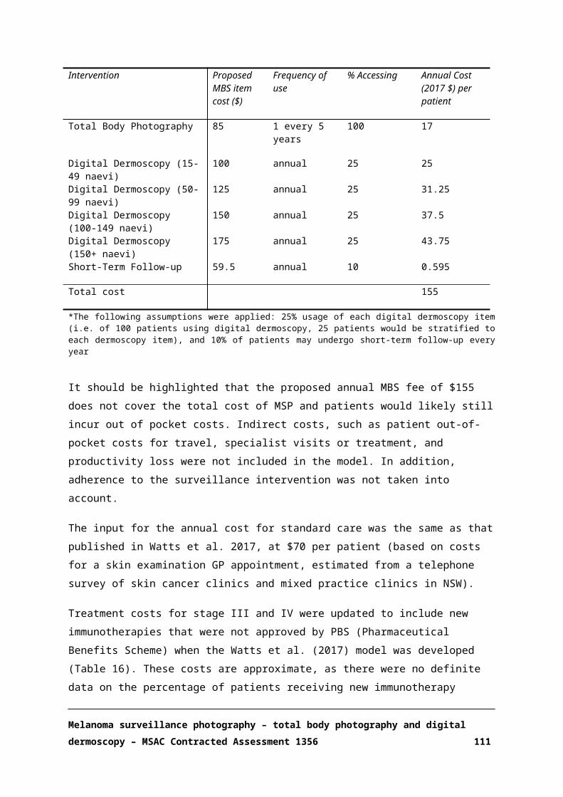
Reason for exclusion: Abstracts
xanthogranuloma at the base of the nose]." Annales de Dermatologie et de Venereologie 140(12): 835-837.Braun, R. P., A. M. Calza, J. Krischer and J. H. Saurat (2002). "The use of digital dermoscopy for the follow-up of congenital nevi. [French]." Medecine et Hygiene 60(2381): 465-466.Braun, R. P., H. Rabinovitz, M. Oliviero, A. W. Kopf, J. H. Saurat and L. Thomas (2002). "[Dermatoscopy of pigmented lesions]." Annales de Dermatologie et de Venereologie 129(2): 187-202.Braun, R. P., O. Gaide, F. A. Le Gal, J. H. Saurat and A. A. Marghoob (2007). "[Follow-up of melanoma lesions]." Revue Medicale Suisse 3(109): 1119-1123.Braun-Falco, O., W. Stolz, P. Bilek, T. Merkle and M. Landthaler (1990). "[The dermatoscope. A simplification of epiluminescent microscopy of pigmented skin changes]." Hautarzt 41(3): 131-136.Brinkhuijsen, M. (2008). "Early diagnosis of melanomas. [Dutch]." Huisarts en Wetenschap 51(12): 593.Broganelli, P., S. Titli, A. Lallas, M. A. A. Alaibac, V. Battarra, B. Brunetti, I. Castagno, S. Cavicchini, A. Ferrari, G. Ghigliotti, C. Landi, A. Manganoni, E. Moscarella, G. Pellacani, M. A. Pizzichetta, P. Rosina, P. Rubegni, R. Satta, M. Scalvenzi, I. Stanganelli, G. Stinco, I. Zalaudek, P. Zampieri, G. Argenziano and D. Dermoscopy Study Group of the Italian Society of (2014). "Spitz/Reed nevi: proposal of management recommendations by the Dermoscopy Study Group of the Italian Society of Dermatology (SIDeMaST)." Giornale Italiano di Dermatologia e Venereologia 149(5): 601-606.Calabro, G., F. De Natale and G. Coppola (1991). "In vivo epiluminescence microscopy in the differential diagnosis of thrombosed angioma versus malignant melanoma. [Italian]." Annali Italiani di Dermatologia Clinica e Sperimentale 45(3): 139-140.Canpolat, F., H. K. Akis, B. N. Akay and C. Erdem (2011). "Dermoscopic features of acral melanocytic nevi. [Turkish]." Turkderm Deri Hastaliklari ve Frengi Arsivi 45(4): 193-197.Carli, P., V. De Giorgi, D. Massi and E. Donati (1999). "A case of melanosis of the areola clinically simulating malignant melanoma. Dermoscopic analysis. [Italian]." Giornale Italiano di Dermatologia e Venereologia 134(3): 243-246.Carli, P., V. De Giorgi, E. Donati, E. Pestelli and B. Giannotti (1994). "Epiluminescence microscopy reduces the risk of removing clinically atypical, but histologically common, melanocytic lesions. [Italian]." Giornale Italiano di Dermatologia e Venereologia 129(12): 599-605.Carlos-Ortega, B. and F. Montes de Oca-Monroy (2009). "[The impact of dermoscopy in clinical practice]." Revista Medica del Instituto Mexicano del Seguro Social 47(6): 581-582.Carlos-Ortega, B., F. M. de Oca-Monroy and A. Isyta-Morales (2009). "[Dermoscopy findings in malignant skin melanoma with metastases. A case report]." Revista Medica del Instituto Mexicano del Seguro Social 47(1): 73-76.Casado, M. (2003). "Most frequent cutaneous disease in the elderly (and II): Presentation. [Spanish]." Revista Espanola de Geriatria y Gerontologia 38(SUPPL. 2): 1.Casari, A., E. Cinotti, A. M. Cesinaro, G. Pellacani and C. Longo (2014). "[Dermatoscopy and reflectance confocal microscopy aspects of a "nested melanoma of the elderly"]." Annales de Dermatologie et de Venereologie 141(6-7): 458-461.Cavalie, M., L. Sillard and P. Bahadoran (2011). "Degos' disease. [French]." Nouvelles Dermatologiques 30(10): 565.Cinotti, E., B. Labeille, C. Habougit, C. Douchet, F. Cambazard, J. L. Perrot and d. Groupe imagerie cutanee non invasive de la Societe francaise de (2015). "[Contribution of reflectance confocal microscopy for the diagnosis of junctional naevus]." Annales de Dermatologie et de Venereologie 142(10): 595-597.Cinotti, E., J. L. Perrot, B. Labeille, C. Douchet, F. Cambazard and d. au nom du groupe imagerie cutanee non invasive de la Societe francaise de (2014). "[Solar lentigo diagnosed by reflectance confocal microscopy]." Annales de Dermatologie et de Venereologie 141(1): 71-73.Ciudad, C., J. A. Aviles, R. Suarez and P. Lazaro (2011). "[Diagnostic utility of dermoscopy in pigmented actinic keratosis]." Actas Dermo-Sifiliograficas 102(8): 623-626.Ciudad-Blanco, C., J. A. Aviles-Izquierdo, P. Lazaro-Ochaita and R. Suarez-Fernandez (2014). "Dermoscopic findings for the early detection of melanoma: An analysis of 200 cases. [Spanish]." Actas Dermo-Sifiliograficas 105(7): 683-693.Contreras-Steyls, M., E. Herrera-Acosta, B. Moyano and E. Herrera (2011). "[Primary melanoma with multiple skin metastases]." Actas Dermo-Sifiliograficas 102(3): 226-229.Czeschik, J. C., U. Hillen and D. Schadendorf (2010). "Diagnosis of malignant melanoma. [German]."
Melanoma surveillance photography – total body photography and digital dermoscopy – MSAC Contracted Assessment 1356 111

Reason for exclusion: Abstracts
Onkologe 16(12): 1121-1130.Dabkowski, J. (2006). "Dermoscopic algorithms in the diagnosis of melanocytic lesions. [Polish]." Przeglad Dermatologiczny 93(4): 469-473.Dalle, S., A. Finet and L. Thomas (2012). "[Kaposi's sarcoma]." Annales de Dermatologie et de Venereologie 139(1): 73-74.Dalle, S., F. Julia and L. Thomas (2010). "[Bluish nodule]." Annales de Dermatologie et de Venereologie 137(10): 663-664.Dalle, S., M. Peyrier-Muzet and L. Thomas (2010). "[Pink nodule]." Annales de Dermatologie et de Venereologie 137(12): 839-840.De Argila Fernandez-Duran, D. (2010). "Skin cancer. [Spanish]." Medicine 10(48): 3213-3222.De La Cueva, P., E. Balbin, M. Valdivielso, C. Mauleon, J. M. Hernanz and J. M. H. Hermosa (2008). "Black heel (subcorneal hematoma). [Spanish]." Acta Pediatrica Espanola 66(7): 365-366.de Troya-Martin, M., N. Blazquez-Sanchez, I. Fernandez-Canedo, M. Frieyro-Elicegui, R. Funez-Liebana and F. Rivas-Ruiz (2008). "[Dermoscopic study of cutaneous malignant melanoma: descriptive analysis of 45 cases]." Actas Dermo-Sifiliograficas 99(1): 44-53.de Vries, E., J. W. Coebergh and H. van der Rhee (2006). "[Trends, causes, approach and consequences related to the skin-cancer epidemic in the Netherlands and Europe]." Nederlands Tijdschrift voor Geneeskunde 150(20): 1108-1115.Dejaco, D., U. Hauser, B. Zelger and H. Riechelmann (2015). "Actinic Keratosis. [German]." Laryngo- Rhino- Otologie 94(7): 467-481.Delaunay, M. M. (2000). "Diagnosis of melanoma. [French]." Oncologie 2(1): 25-28.Delfino, M., D. Farina, G. De Fata Salvatores, M. Mancini and M. S. Di Santolo (2013). "Role of high-resolution ultrasound asnd color-Doppler in the assessment of the grade of malignancy in the diagnosis of melanoma. [Italian]." Esperienze Dermatologiche 15(4): 163-168.Delfino, M., D. Farinag, G. De Fata Salvatores, M. Mancini and M. S. Di Santolo (2013). "Differentiation of benign and malignant skin lesions using high-resolution sonography and color-Doppler. [Italian]." Esperienze Dermatologiche 15(4): 171-175.Dominguez Cruz, J., L. Ferrandiz Pulido and D. Moreno Ramirez (2008). "Dermatoscopy of non-melanocytic lesions (I). [Spanish]." Piel 23(7): 367-374.Dominguez Cruz, J., L. Ferrandiz Pulido and D. Moreno Ramirez (2008). "Dermatoscopy of non-melanocytic lesions. Part 2. [Spanish]." Piel 23(8): 428-432.Dominguez-Espinosa, A. E. (2014). "History of dermoscopy. [Spanish]." Dermatologia Revista Mexicana 58(2): 165-172.Du Thanh, A. and B. Guillot (2012). "[Actinic keratosis: when is a skin biopsy necessary?]." European Journal of Dermatology 22 Suppl 1: 13-16.Dumke, A. K., M. Gina, S. Goetze, S. Schliemann and P. Elsner (2017). "[Ethnic nail pigmentations and differential diagnoses]." Hautarzt 68(1): 64-66.Dumke, A. K., M. Gina, S. Goetze, S. Schliemann and P. Elsner (2017). "Ethnic nail pigmentations and differential diagnoses. [German]." Hautarzt 68(1): 64-66.Dummer, W., K. A. Doehnel and W. Remy (1993). "[Videomicroscopy in differential diagnosis of skin tumors and secondary prevention of malignant melanoma]." Hautarzt 44(12): 772-776.Dupont, J., K. S. Culpepper and C. Curiel-Lewandrowski (2013). "[Red papule with collarette scale on thigh]." Hautarzt 64(8): 603-605.Elsner, P., S. Metz and S. Schliemann (2014). "[Importance of dermatopathology in the diagnosis of tropical and travel-associated skin diseases]." Hautarzt 65(10): 895-899.Enei Ma, L., F. Paschoal, R. Valdes and G. Valdes (2011). "A dermoscopic contribution to the establishment of minimal surgical margins of resection in basal cell carcinoma on the face and scalp. [Spanish]." Medicina Cutanea Ibero-Latino-Americana 39(4): 205-208.Ertam, I., B. G. Turk, I. K. Karaarslan, I. Unal and S. Alper (2014). "Effectiveness of Intense Pulsed Light treatment in solar lentigo: A retrospective study. [Turkish]." Turkderm Deri Hastaliklari ve Frengi Arsivi 48(1): 39-42.Espinosa, A. E. D. (2012). "Dermatoscopia en poroma ecrino. [Spanish]." Dermatologia Revista Mexicana 56(3): 221-224.Falcao, E. M., A. R. Catharino, C. M. Zompero and B. K. Kac (2012). "[Poroma: a typical presentation]." Dermatology Online Journal 18(11): 14.Fanti, P. A., R. Barducci, C. Misciali and C. Varotti (2003). "Dermatoscopy as viewed by the
Melanoma surveillance photography – total body photography and digital dermoscopy – MSAC Contracted Assessment 1356 112

Reason for exclusion: Abstracts
dermatopathologist. [Italian]." Giornale Italiano di Dermatologia e Venereologia 138(2): 160-161.Feldmann, R., C. Fellenz and F. Gschnait (1998). "[The ABCD rule in dermatoscopy: analysis of 500 melanocytic lesions]." Hautarzt 49(6): 473-476.Figueiredo Kopke, L. F. (1996). "Dermatoscopy in melanocytic lesions. [Portuguese]." Anais Brasileiros de Dermatologia 71(1): 63-67.Fikrle, T. and K. Pizinger (2005). "Use of dermatoscopy in the differential diagnosis of non-melanocytic lesions and malignant melanoma. [Czech]." Cesko-Slovenska Dermatologie 80(3): 162-168.Friedman, T., D. Klein, E. Hadad, M. Westreich and A. Shalom (2008). "[Diagnostic accuracy of skin lesions excised by a plastic surgeon]." Harefuah 147(4): 305-308, 375.Fuessl, H. S. (2013). "[Red melanoma]." MMW Fortschritte der Medizin 155(13): 33.Fujihara, M., T. Kurokawa, U. Otsu, S. Moriwaki and T. Nuri (2010). "A case of nevocellular nevus showing rapid growth during pregnancy, needed to required differentiation from malignant melanoma. [Japanese]." Skin Research 9(6): 532-535.Fuyuno, Y., T. Chiba, S. Takeuchi, M. Takahara, Y. Moroi and M. Furue (2014). "Basal cell carcinoma in two white men. [Japanese]." Nishinihon Journal of Dermatology 76(3): 202-205.Gallegos-Hernandez, J. F., A. L. Ortiz-Maldonado, G. G. Minauro-Munoz, H. Arias-Ceballos and M. Hernandez-Sanjuan (2015). "[Dermoscopy in cutaneous melanoma]." Cirugia y Cirujanos 83(2): 107-111.Garbe, C. (1996). "Perspectives in dermatologic oncology. [German]." H+G Zeitschrift fur Hautkrankheiten 71(6): 416-424.Garbe, C. (2008). "Early detection and primary prevention of skin cancer. [German]." Onkologe 14(2): 156-163.Garbe, C. (2016). "Early detection of skin cancer and skin cancer screening examinations. [German]." Onkologe 22(8): 537-549.Garmyn, M. (2007). "How to recognize a malignant melanoma between the other pigmented spots. [Dutch]." Tijdschrift voor Geneeskunde 63(24): 1240-1243.Garmyn, M. (2009). "How to recognize a malignant melanoma between the other pimented spots. [Dutch]." Tijdschrift voor Geneeskunde 65(1): 9.Gasparini, S., G. L. Giovene and C. Gasparini (2008). "Utility of dermoscopy in the diagnosis of collision tumours. [Italian]." Journal of Plastic Dermatology 4(2): 223-227.Gereli, M. C., N. Onsun and U. Atilganoglu (2008). "Comparison of two dermoscopic techniques in the melanoma diagnosis: 3-Point checklist and 7-point checklist. [Turkish]." Turkderm Deri Hastaliklari ve Frengi Arsivi 42(2): 45-50.Giacchero, D. and P. Bahadoran (2008). "Dermoscopy of a Trichoblastoma arising in nevus sebaceous of scalp. [French]." Nouvelles Dermatologiques 27(9): 437-438.Giannotti, B. and P. Carli (2004). "Improvement of early diagnosis of melanoma in a mediterranean population: The experience of the Florence melanoma clinic." Giornale Italiano di Dermatologia e Venereologia 139(2): 89-96.Giard, R. W. and H. A. Neumann (2004). "[Diagnosis of pigmented skin lesions: how to recognize a malignant melanoma]." Nederlands Tijdschrift voor Geneeskunde 148(46): 2261-2267.Gokdemir, A., M. Guler Ozden, Y. Bek, F. Aydin, N. Senturk, T. Canturk and A. Y. Turanli (2011). "Dermoscopic and histopathological correlation in melanocytic and non-melanocytic lesions. [Turkish]." Turkiye Klinikleri Dermatoloji 21(1): 7-16.Gonzalez, R., C. E. L. Gurrola and R. D. Canales (2012). "Eficacia de un curso intensivo basico en la ensenanza de la dermatoscopia: Estudio piloto en dermatologos mexicanos. [Spanish]." Dermatologia Revista Mexicana 56(3): 167-171.Goralska, A. and J. Btaszczyk (2013). "Atypical naevus, dysplastic naevus, dysplastic naevus syndrome - Nomenclature controversy, diagnostic difficulties and prognostic perspectives. [Polish]." Przeglad Dermatologiczny 100(1): 40-47.Gourhant, J. Y. (2011). "Pigmented Bowen's disease. [French]." Nouvelles Dermatologiques 30(9): 501.Gourhant, J. Y. (2011). "Superficial spreading melanoma. [French]." Nouvelles Dermatologiques 30(2): 85-86.Grob, J. J. (2012). "[Actinic keratosis: diagnostic difficulties and misinterpretation]." European Journal of Dermatology 22 Suppl 1: 10-12.
Melanoma surveillance photography – total body photography and digital dermoscopy – MSAC Contracted Assessment 1356 113

Reason for exclusion: Abstracts
Guillod, J. (1999). "[Clinical differential diagnosis of pigmented skin changes and contribution of epiluminescence microscopy]." Therapeutische Umschau 56(6): 312-317.Guitera-Rovel, P. and M. E. Vestergaard (2008). "[Diagnosis tools for cutaneous melanoma]." Annales de Dermatologie et de Venereologie 135(12): 828-834.Gutierrez Vidrio, R. M. and N. Cortes Lozano (2007). "Confronting melanoma in the 21<sup>st</sup> century. [Spanish]." Medicina Cutanea Ibero-Latino-Americana 35(1): 3-13.Haas, N., T. M. Ernst and G. Stuttgen (1984). "[Macrophotography in transmitted light. Contribution to an analysis of horizontal structures of pigmented skin tumors]." Zeitschrift fur Hautkrankheiten 59(15): 985-989.Haenssle, H. A. (2009). "Early detection of melanoma through digital epiluminescence microscopy (DELM). [German]." Aktuelle Dermatologie 35(3): 75-78.Haenssle, H. A. (2012). "[Enlarging pigmented facial lesion]." Hautarzt 63(2): 143-144.Haenssle, H. A., F. Brehmer, I. Zalaudek, R. Hofmann-Wellenhof, J. Kreusch, W. Stolz, G. Argenziano and A. Blum (2014). "[Dermoscopy of nails]." Hautarzt 65(4): 301-311.Hammami-Ghorbel, H., R. Benmously-Mlika and I. Mokhtar (2012). "Multiple blue tumors. [French]." Nouvelles Dermatologiques 31(9): 445-446.Hammami-Ghorbel, H., T. Badri and I. Mokhtar (2013). "Papulonodules on the face Contribution of dermoscopy. [French]." Nouvelles Dermatologiques 32(3): 106-107.Happe, M., M. Freitag, M. Stucker, P. Altmeyer and K. Hoffmann (1997). "[High resolution 20 MHz ultrasound diagnosis in dermatology for noninvasive imaging of malignant melanomas]." Zeitschrift fur Arztliche Fortbildung und Qualitatssicherung 91(4): 347-353.Harsh, A. (2000). "Innovation approaches in computer-assisted tumor diagnosis. [German]." Medizinische Welt 51(1-2): 48-49.Haspeslagh, M., N. Degryse and I. De Wispelaere (2013). "Ex vivo dermoscopy with dermdotting. [Dutch]." Nederlands Tijdschrift voor Dermatologie en Venereologie 23(9): 525-527.Held, L., T. K. Eigentler and C. Garbe (2009). "Early detection and prevention of cutaneous melanoma. [German]." Onkologe 15(8): 738-743.Hernandez-Salazar, A., J. Cordova-Lopez, L. Esquivel, C. Scholtes and R. Orozco-Topete (2006). "Malignant melanoma: Retrospective study in the Department of Dermatology of the National Institute of Medical Sciences and Nutrition Salvador Zubiran. [Spanish]." Dermatologia Cosmetica, Medica y Quirurgica 4(4): 242-246.Hoelt, P., S. Dalle and L. Thomas (2014). "[Cutaneous metastases of melanoma]." Annales de Dermatologie et de Venereologie 141(10): 640-642.Hofmann-Wellenhof, R. (2006). "Melanoma. [German]." Gynakologische Praxis 30(4): 665-667.Ihara, H., M. Koga, R. Tatsukawa, K. Koga, Y. Shibayama and S. Imafuku (2015). "A case of periungual spitz nevus with a characteristic clinical manifestation and a specific pattern on dermoscopy. [Japanese]." Nishinihon Journal of Dermatology 77(5): 483-486.Ilter, N., M. O. Oztas, E. Adisen, M. A. Gurer, O. Keseroglu, S. Unal, B. Kirac, J. Yuksek, C. Kevlekci, D. Cetinkunar, B. Ozturk and N. Eroglu (2009). "Evaluation of sun protection habits and melanocytic nevi of population screened in a shopping mall in Ankara. [Turkish]." Turkderm Deri Hastaliklari ve Frengi Arsivi 43(4): 155-159.Jacome, A. B., R. Q. Venegas and G. S. Ledesma (2012). "Caracteristicas dermatoscopicas del angioqueratoma solitario. [Spanish]." Dermatologia Revista Mexicana 56(3): 201-202.Kaatz, M. and K. Konig (2010). "[Multiphoton microscopy and in vivo tomography in dermatologic imaging]." Hautarzt 61(5): 397-409.Kaddu, S., H. P. Soyer, I. H. Wolf, E. Rieger and H. Kerl (1997). "[Reticular lentigo]." Hautarzt 48(3): 181-185.Kainz, J. T. and R. Hofmann-Wellenhof (2011). "[Trivial injury to the sole of the foot]." Hautarzt 62(12): 943-946.Kaminska-Winciorek, G. and R. Spiewak (2011). "[Basic dermoscopy of melanocytic lesions for beginners]." Postepy Higieny i Medycyny do Swiadczalnej (Online) 65: 501-508.Kaminska-Winciorek, G. and R. Spiewak (2013). "[Dermoscopy on subungual melanoma]." Postepy Higieny i Medycyny do Swiadczalnej (Online) 67: 380-387.Karaarslan, I. (2016). "Dermoscopy of actinic keratosis, Bowen's disease and squamous carcinoma. [Turkish]." Turk Dermatoloji Dergisi 10(1): 27-32.Karaarslan, I. K. (2014). "Patient management in dermoscopy. [Turkish]." Turkderm Deri Hastaliklari
Melanoma surveillance photography – total body photography and digital dermoscopy – MSAC Contracted Assessment 1356 114

Reason for exclusion: Abstracts
ve Frengi Arsivi 48(1): 2-6.Karaarslan, I. K., E. Y. Biyikh, T. Akalin and F. Ozdemir (2009). "Dermoscopic Features in Seborrheic Keratoses; What is the Value of the False Melanocytic Features?. [Turkish]." Turk Dermatoloji Dergisi 3(1): 1-4.Kaudewitz, P. (1996). "Diagnostic and treatment of malignant melanoma from a dermatological point of view. [German]." Aktuelle Chirurgie 31(5): 277-281.Kellner, C. and U. Reinhold (2015). "[Modern diagnostic procedures in dermatological oncology]." Pathologe 36(1): 11-15.Kerl, H., I. H. Wolf, W. Sterry and H. P. Soyer (1995). "[Dermatoscopy. A new method for the clinical diagnosis of malignant melanoma]." Deutsche Medizinische Wochenschrift 120(22): 801-805.Kilinc Karaarslan, I., H. E. Ercal, I. Ertam, M. Turkmen, B. G. Turk and F. Ozdemir (2009). "Geriatric patients in management of pigmented lesions: Should follow-up be recommended?. [Turkish]." Turk Geriatri Dergisi 12(3): 124-129.Kinashi, N., K. Ichihashi, H. Takenaka, N. Katoh, S. Kishimoto and K. Kagami (2007). "A case of thrombotic angiokeratoma. [Japanese]." Nishinihon Journal of Dermatology 69(3): 266-268.Kiriyak, N., G. Tchernev, P. Penev, J. Ananiev, I. Bakardzhiev, G. Pehlivanov, L. Zisova and J. W. Patterson (2012). "Simplified dermatoscopic criteria for diagnosing benign and malignant nonmelanocytic cutaneous lesions: Indications for surgical removal. [Bulgarian]." General Medicine 14(2): 41-51.Kofler, L., M. Egger and H. Kofler (2014). "[Sequential digital dermatoscopic imaging. How much time is required per patient?]." Hautarzt 65(5): 450-453.Kolm, I., G. Hofbauer and R. P. Braun (2010). "[Early diagnosis of skin cancer]." Therapeutische Umschau 67(9): 439-446.Kowalzick, L., T. Waldmann, D. Mischke and S. Suckow (2002). "Early detection of a malignant melanoma by computerized video image dermatoscopy in a patient with multiple melanocytic lesions. [German]." H+G Zeitschrift fur Hautkrankheiten 77(10): 487-490.Kreusch, J. and G. Rassner (1990). "[Epiluminescence image of lentiginous junctional nevi]." Hautarzt 41(5): 274-276.Kukutsch, N. and W. Bergman (2015). "Dermoscopy in general practice 2." Huisarts en Wetenschap 58(2): 90-93.Lacava, V., N. Salesi, L. Ferrone, A. Veri, R. Lembo, M. C. Masi, M. Rocchi, R. Rondinelli, E. Parisi, T. Grieco, A. Balzani, R. Clerico, D. Innocenzi, U. Bottoni and S. Calvieri (2001). "[Importance of dermatologic screening within the frame work of a general cancer prevention program]." Minerva Medica 92(2): 85-88.Le Duff, F. (2013). "Collision tumor. [French]." Nouvelles Dermatologiques 32(2): 64.Le Maitre, M. (2009). "Pigmented spot of the face: Dermoscopy before cryotherapy. [French]." Nouvelles Dermatologiques 28(9 PART 1): 371.Le, F. and P. Bahadoran (2012). "An achromic nodula. [French]." Nouvelles Dermatologiques 31(4): 170.Leiter, U. and C. Garbe (2009). "Surveillance of cutaneous melanoma. [German]." Onkologe 15(8): 777-783.Leiter, U. and C. Garbe (2016). "Basal cell carcinoma - Diagnosis and management. [German]." Klinikarzt 45(4): 190-195.Leiter, U., F. Meier, C. Garbe and M. Rocken (2012). "Secondary prevention of skin cancer. [German]." Onkologe 18(3): 237-242.Lejeune, F. J., M. Matter, D. Guggisberg, J. P. Cerottini and D. Lienard (2004). "Management of cutaneous malignant melanoma in the first stage. [French]." Medecine et Hygiene 62(2504): 2256-2263.Lemonnier, E., R. Braun, J. Krischer, J. Guillod, D. Salomon and J. H. Saurat (1996). "Dermatoscopy and videomicroscopy: Two complementary techniques in diagnosing pigmented lesions. [French]." Nouvelles Dermatologiques 15(6): 460.Levi, A., A. Ingber and D. C. Enk (2012). "[In vivo reflectance-mode confocal laser microscopy: basic principles and clinical and research employments in dermatology]." Harefuah 151(10): 576-580, 605.Li, W. W., P. Tu, S. X. Yang, H. Li and Y. Wang (2013). "Dermoscopic features of 100 cases of seborrheic keratoses. [Chinese]." Journal of Clinical Dermatology 42(11): 641-645.
Melanoma surveillance photography – total body photography and digital dermoscopy – MSAC Contracted Assessment 1356 115

Reason for exclusion: Abstracts
Lindelof, B., M. A. Hedblad and U. Ringborg (2008). "[Nevus or malignant melanoma? Correct diagnostic competence results in lower costs]." Lakartidningen 105(39): 2666-2669.Longo Imedio, I. and P. Lazaro Ochaita (2007). "Evidence-based review of studies in dermatology: A new section. [Spanish]." Piel 22(3): 107-108.Lopez Cruz, Z. C. (2007). "Basocellular and epidermoid carcinoma. [Spanish]." Dermatologia Revista Mexicana 51(4): 149-153.Lorentzen, H. F. and K. Weismann (2000). "[Dermatoscopic diagnosis of cutaneous malignant melanoma. Secondary prophylaxis]." Ugeskrift for Laeger 162(23): 3312-3316.Maciejewska, J., M. Luczak, L. Marek, S. K. Ewa, P. Krause, A. Cichewicz and W. Placek (2012). "Local actions in the prevention and the early diagnosis of melanoma and skin cancers, Dzialania lokalne w zapobieganiu oraz wczesnym wykrywaniu czerniaka oraz rakow skory. [Polish, English]." Przeglad Dermatologiczny 99 (4): 422-423.Malaczewska, J. and J. Dabkowski (2004). "[Melanocytic nevi as one of the risk factors of melanoma malignum]." Polski Merkuriusz Lekarski 16(93): 298-302.Malvehy Guilera, J. and S. Puig (2007). "The future of diagnostic imaging techniques in dermatology. [Spanish]." Piel 22(1): 1-2.Mang, R., D. Krahl and T. Assmann (2012). "[Dermoscopic changes in melanocytic nevi during use of melanotan II]." Hautarzt 63(11): 880-884.Martin, J. M., R. Bella-Navarro and E. Jorda (2012). "[Vascular patterns in dermoscopy]." Actas Dermo-Sifiliograficas 103(5): 357-375.Martinez, A. P. V., R. Q. Venegas and E. G. Gutierrez (2012). "Carcinoma basocelular pigmentado con patron dermatoscopico atipico: Reporte de tres casos de variedad azul-blanca. [Spanish]." Dermatologia Revista Mexicana 56(3): 203-205.Mateus, C., E. Routier, B. Reynaud-Mendel, A. Lacert, L. Maksimovic, M. Thomas, J. P. Arnault, G. Tomasic and C. Robert (2011). "Unlikely melanomas. [French]." Nouvelles Dermatologiques 30(8): 462-464.Matsuda, H., Y. Ito, Y. Kai, M. Goto, Y. Hatano, K. Katagiri, S. Fujiwara, A. Kato, F. Shimizu and S. Yokoyama (2010). "The diagnosis of subungual malignant melanoma in the early stage. [Japanese]." Nishinihon Journal of Dermatology 72(2): 101-105.Mayor, M., A. Pizarro, M. Siguenza, C. Vidaurrazaga, M. A. Garcia-Cabezas, F. Contreras and M. Casado (2003). "Pigmented trichoblastoma. [Spanish]." Actas Dermo-Sifiliograficas 94(6): 395-398.Mazzocchetti, G., A. Ferrari, D. Piccolo, K. Peris and S. Chimenti (1998). "New diagnostic parameters in dermatoscopic symptomatology. [Italian]." Giornale Italiano di Dermatologia e Venereologia 133(5): 319-324.Mazzocchetti, G., A. Ferrari, D. Piccolo, K. Peris and S. Chimenti (1998). "New diagnostic parameters in dermatoscopic symptomatology. [Italian]." Giornale Italiano di Dermatologia e Venereologia 133(5): 319-324.Meijer, S., P. A. van Leeuwen, M. P. van den Tol and B. G. Molenkamp (2005). "[Guideline 'Melanoma' (3rd revision)]." Nederlands Tijdschrift voor Geneeskunde 149(50): 2821; author reply 2821.Michel, J. L. (2011). "Dermoscopic follow-up of a conjunctival nevus. [French]." Nouvelles Dermatologiques 30(9): 497-499.Mikulska, D. (2009). "[Thermographic examination of cutaneous melanocytic nevi]." Annales Academiae Medicae Stetinensis 55(1): 31-38; discussion 38.Mikulska, D., R. Maleszka and M. Parafiniuk (2004). "The thermographic and dermatoscopic analyses of the skin melanocytic nevi. [Polish]." Przeglad Dermatologiczny 91(1): 15-21.Miquel, J., J. Chevrant-Breton, A. Dupuy, F. Le Gall, E. Bourdon-Lannoy, H. Adamski and C. Bodemer (2012). "[Eruptive naevi in epidermolysis bullosa hereditaria patients]." Annales de Dermatologie et de Venereologie 139(6-7): 435-443.Mizote, M., C. Mitoma and M. Furue (2015). "A case of pilomatricoma on the preauricular region in an aged woman. [Japanese]." Nishinihon Journal of Dermatology 77(2): 131-133.Morales-Callaghan, A. M., J. Castrodeza-Sanz, G. Martinez-Garcia, I. Peral-Martinez and A. Miranda-Romero (2008). "[Correlation between clinical, dermatoscopic, and histopathologic variables in atypical melanocytic nevi]." Actas Dermo-Sifiliograficas 99(5): 380-389.Nikolic, D. V., A. T. Nikolic, V. V. Stanimirovic, M. K. Granic, T. Randelovic and D. Bilanovic (2008). "[Efficient way in early detection of malignant skin tumors by applying epiluminescence microscopy in skin screening]." Medicinski Pregled 61(9-10): 507-511.Oda, Y., N. Ikkaku and T. Horikawa (2015). "A case with pigmented and non-pigmented basal cell
Melanoma surveillance photography – total body photography and digital dermoscopy – MSAC Contracted Assessment 1356 116

Reason for exclusion: Abstracts
carcinomas. [Japanese]." Skin Research 14(2): 62-66.Ono, Y., Y. Suzuki, Y. Togawa and H. Nagayama (2013). "A case of pigmented bowen's disease of the penis with caracteristic regular blue-gray globules on dermoscopy. [Japanese]." Nishinihon Journal of Dermatology 75(3): 227-229.Orendain-Koch, N., M. P. Ramos-alvarez, A. B. Ruiz-Leal, L. E. Sanchez-Duenas, A. B. Crocker-Sandoval, T. Sanchez-Tenorio and J. Izquierdo-alvarez (2015). "Melanoma in a private practice setting in Mexico: An early diagnosis. [Spanish]." Dermatologia Revista Mexicana 59(2): 89-97.Ozbatcivan, O., B. Lebe, S. Akarsu and E. Fetil (2015). "Investigation of the relationship between dermoscopic features and histopathological prognostic indicators in patients with cutaneous melanoma. [Turkish]." Turkderm Deri Hastaliklari ve Frengi Arsivi 49(3): 200-207.Ozmen, I., O. Kose and M. Demiriz (2010). "Spitz nevus on the earlobe mimicking cutaneous leishmaniasis. [Turkish]." Turk Dermatoloji Dergisi 4(2): 55-57.Paschoal, F. M. (1998). "Dermatoscopy of malignant melanoma. [Portuguese]." Anais Brasileiros de Dermatologia 73(SUPPL. 1): 23.Peccerillo, F., P. Di Caterino, D. Pitruzzella, V. C. Battarra and L. Boccia (2013). "Pilomatrixoma: Often only a postoperative diagnosis. [Italian]." Esperienze Dermatologiche 15(1): 23-25.Perrot, J. L., B. Labeille, C. Douchet, F. Cambazard, E. Cinotti and d. Groupe imagerie cutanee non invasive de Societe francaise de (2014). "[Contribution of reflectance confocal microscopy to the diagnosis of fibroepithelioma of Pinkus]." Annales de Dermatologie et de Venereologie 141(10): 643-645.Persechino, F., C. Longo, A. M. Cesinaro, E. Cinotti, S. Ciardo, G. Pellacani and S. Bassoli (2016). "Dermoscopic and reflectance confocal microscopic features of clonal seborrhoeic keratosis. [French]." Annales de Dermatologie et de Venereologie 143(2): 164-166.Persechino, S., M. Grimaldi, E. Palese, V. Panasiti and G. Panasiti (2005). "Clear cell acanthoma: Dermatoscopic observation. [Italian]." Dermatologia Clinica 25(3): 136-137.Perusquia, A. M., D. Metze and A. Blum (2005). "Diagnostic algorithms in dermatoscopy. [Spanish]." Dermatologia Cosmetica, Medica y Quirurgica 3(SUPPL. 1): 157-167.Perusquia, A. M., J. Ferrer-Bernat, D. Metze and A. Blum (2005). "Dermatoscopy and histopathology of pigmented melanocytic or non melanocytic lesions. [Spanish]." Dermatologia Cosmetica, Medica y Quirurgica 3(4): 331-343.Pezza, M. (2013). "Epiluminescence microscopy: A valid diagnostic aid for the dermatologist in the prevention of skin cancers. [Italian]." Journal of Plastic Dermatology 9(2): 157-158.Phan, A., S. Dalle and L. Thomas (2007). "[Acquired pigmented plantar lesion]." Annales de Dermatologie et de Venereologie 134(11): 892-893.Phan, A., S. Dalle and L. Thomas (2010). "[Thoracic pigmented lesion]." Annales de Dermatologie et de Venereologie 137(4): 334-335.Piccolo, D., H. P. Soyer, J. Smolle, I. H. Wolf, G. Argenziano, L. Cerroni, A. Ferrari, F. Magrini, G. Mazzocchetti, M. A. Pizzichetta, H. Kerl and S. Chimenti (2000). "Teledermatoscopy. Results of a multicentric study on pigmented skin lesions. [Italian]." Giornale Italiano di Dermatologia e Venereologia 135(2): 153-159.Pierard, G. E., C. Pierard-Franchimont, T. Hermanns-Le and S. L. Pierard (2015). "[Gazing into a Crystal Ball and Detecting Subjects at Risk for Developing Cutaneous Melanoma]." Revue Medicale de Liege 70(9): 446-449.Pierard-Franchimont, C. and G. E. Pierard (2011). "[Melanoma of older subjects]." Revue Medicale de Liege 66(1): 34-40.Pierard-Franchimont, C., V. Goffin and G. E. Pierard (1998). "[Dermoscopy: magnified imaging of pigmented cutaneous tumors]." Revue Medicale de Liege 53(4): 180-186.Pitarch, G. and R. Botella-Estrada (2015). "Dermoscopic findings in trichoblastoma. [Spanish]." Actas Dermo-Sifiliograficas 106(9): e45-e48.Pizarro, A., J. L. Santiago and D. I. Santiago (2015). "Dermoscopy in the prevention and early diagnosis of melanoma: A biological perspective. [Spanish]." Actas Dermo-Sifiliograficas 106(1): 3-6.Pizarro, A., M. Siguenza, M. Mayor, T. Lazaro, F. Contreras and M. Casado (2003). "Negative pigment network in a growing melanocytic nevus. [Spanish]." Actas Dermo-Sifiliograficas 94(2): 116-118.Pizinger, K. and T. Fikrle (2005). "Benign cutaneous pigmented lesions and the various possibilities in their examination. [Czech]." Prakticky Lekar 85(4): 202-206.Pock, L. and P. Zlosky (1996). "[Malignant melanoma in dermatoscopical picture]." Ceskoslovenska Patologie 32(4): 150-153.
Melanoma surveillance photography – total body photography and digital dermoscopy – MSAC Contracted Assessment 1356 117
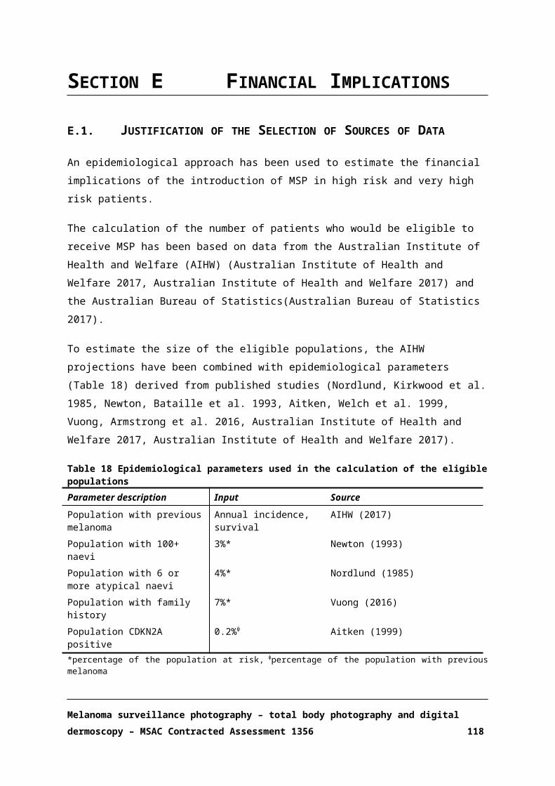
Reason for exclusion: Abstracts
Poulalhon, N., S. Dalle and L. Thomas (2008). "[Dermoscopic diagnosis of dermatofibromas]." Annales de Dermatologie et de Venereologie 135(12): 886-887.Poulsen, A. G., F. G. Larsen, K. Weismann, C. S. Petersen, L. R. Ravnborg, M. Heidenheim, T. E. Lauritzen, E. Held and A. L. Osterlind (1999). "[Investigation of malignant melanoma in an "open house" setting]." Ugeskrift for Laeger 161(12): 1758-1761.Pullmann, S. and H. J. Schulze (2005). "Early recognition of skin cancer. [German]." Haut 16(4): 154-156.Quatresooz, P., G. E. Pierard, C. Pierard-Franchimont and P. Groupe Mosan d'Etudes des Tumeurs (2010). "[Deciphering the "ugly duckling", but escaping the "telly belly". Exploratory algorithm of the diagnostic cutting edge of melanoma]." Revue Medicale de Liege 65(4): 180-185.Quatresooz, P., G. E. Pierard, C. Pierard-Franchimont, J. E. Arrese, G. Blaise, R. Bourguignon, M. Broux, N. Claessens, F. Cornil, M. Damseaux, J. M. Darcis, J. Dehavay, P. Delvoye, C. Devillers, A. L. Fraiture, C. Franchimont, V. Goffin, F. Henry, J. F. Hermanns, T. Le, M. Lesuisse, C. Letawe, B. Letot, O. Martalo, F. Mauhin, P. Paquet, M. A. Reginster, G. Szepetiuk, N. Tassoudji, L. Thirion, I. Uhoda, G. Vandenbossche and V. Willemaers (2010). "Deciphering the <<ugly duckling>>, but escaping the <<telly belly>>. Exploratory algorithm of the diagnostic cutting edge of melanoma. [French]." Revue Medicale de Liege 65(4): 180-185.Quinones-Venegas, R., X. Valenzuela-Barba and R. A. Gonzalez-Ramirez (2015). "Non-melanoma skin cancer of the helix: The dermatoscope is on our side!. [Spanish]." Dermatologia Revista Mexicana 59(2): 175-180.Rakowska, A., A. Kardynal, M. Maj, M. Slowinska, J. Czuwara and L. Rudnicka (2012). "Vascular structures in dermoscopy, Struktury naczyniowe w dermoskopii. [Polish, English]." Przeglad Dermatologiczny 99 (4): 462-463.Remy, W. and S. Golsch (1995). "Demonstration of horny layer cells by means of epiluminescence microscopy (x400) (following staining with silver nitrate solution). [German]." H+G Zeitschrift fur Hautkrankheiten 70(12): 896-898.Requena, C. and L. Requena (2009). "Keys for the diagnosis of spitzoid melanoma. [Spanish]." Piel 24(10): 542-548.Resende Moreira, R. and H. Friedman (1996). "Dermatoscopy: Basic concepts and importance in the diagnosis of pigmented skin lesions. [Portuguese]." Anais Brasileiros de Dermatologia 71(1): 51-57.Roberti, V., T. Lopez, R. Coppola, S. Zanframundo, A. Graziano, V. Devirgiliis and V. Panasiti (2013). "Dermoscopic features of acquired digital fibrokeratoma. [Italian]." Esperienze Dermatologiche 15(4): 169-170.Rodriguez Saa, S., R. Maria Ciancio and H. Cabo (2015). "Dermoscopic study of periorbital syringoma. [Spanish]." Medicina Cutanea Ibero-Latino-Americana 43(2): 132-135.Rodriguez, M. G. (2009). "Melanoma: Clinical and treatment. [Spanish]." Medicine 10(27): 1817-1829.Rodriguez-Cabral, R., G. Montoya-Sosa, N. E. Gomez-Bernal, R. Roldan-Marin and B. Carlos-Ortega (2014). "Histopathologic correlation of the melanocytic and no-melanocytic structures observed by dermoscopy. [Spanish]." Dermatologia Revista Mexicana 58(6): 560-567.Roesch, A., M. Landthaler and T. Vogt (2003). "[The dysplastic nevus. Separate entity, melanoma precursor or diagnostic dilemma?]." Hautarzt 54(9): 871-883; quiz 884-875.Roldan, L. M. C., M. V. Lopera and G. A. V. Suaza (2008). "Acral lentiginous melanoma: a variant of malignant melanoma of special interest in Colombia. [Spanish]." Iatreia 21(4): 386-397.Rudnicka, L., M. Olszewska, A. Baran and M. Slowinska (2006). "Dermoscopy and videodermoscopy in diagnosis of skin disease. [Polish]." Przeglad Dermatologiczny 93(2): 126-130.Ruiz-Leal, A. B. and A. E. Dominguez-Espinosa (2015). "Dermoscopy of blue nevus and its variants. [Spanish]." Dermatologia Revista Mexicana 59(2): 102-113.Ruiz-Villaverde, R., S. Arias-Santiago and J. B. Repiso-Jimenez (2015). "Ultrasound in the management of benign cutaneous tumours. [Spanish]." Actas Dermo-Sifiliograficas 106: 29-40.Rutkowski, P., P. J. Wysocki, A. Nasierowska-Guttmejer, J. Fijuth, E. Kalinka-Warzocha, T. witaj, A. Jeziorski, M. Szacht, W. Zegarski, W. M. Wysocki, L. Rudnicka, W. Owczarek and M. Krzakowski (2015). "Recommendations for diagnosis and therapy of cutaneous melanoma. [Polish]." Nowotwory 65(6): 501-516.Rutkowski, P., P. J. Wysocki, A. Nasierowska-Guttmejer, J. Fijuth, E. Kalinka-Warzocha, T. witaj, A. Jeziorski, M. Szacht, W. Zegarski, W. M. Wysocki, L. Rudnicka, W. Owczarek and M. Krzakowski (2016). "Cutaneous melanoma - Guidelines for diagnostics and therapy in 2016. [Polish]." Przeglad Dermatologiczny 103(1): 1-18.
Melanoma surveillance photography – total body photography and digital dermoscopy – MSAC Contracted Assessment 1356 118

Reason for exclusion: Abstracts
Rutkowski, P., P. J. Wysocki, Z. I. Nowecki, L. Rudnicka, A. Nasierowska-Guttmejer, J. Fijuth, E. Kalinka-Warzocha, T. Switaj, M. Wojtukiewicz, W. Zegarski, A. Jeziorski, K. Krzemieniecki, R. Zaucha, E. Filipczyk-Cisarz, K. Herman and M. Krzakowski (2013). "Cutaneous melanoma - Diagnostic and therapeutic guidelines in 2013. [Polish]." Przeglad Dermatologiczny 100(1): 1-15.Rutkowski, P., P. J. Wysocki, Z. I. Nowecki, L. Rudnicka, A. Nasierowska-Guttmejer, J. Fijuth, E. Kalinka-Warzocha, T. Switaj, M. Wojtukiewicz, W. Zegarski, A. Jeziorski, K. Krzemieniecki, R. Zaucha, E. Filipczyk-Cisarz, K. Herman and M. Krzakowski (2013). "Recommendations for diagnostics and therapy of cutaneous melanoma. [Polish]." Nowotwory 63(2): 157-170.Saida, T. (2008). "Recent advances in the diagnosis and treatment of malignant melanoma. [Japanese]." Biotherapy 22(2): 63-70.Saida, T. (2009). "[Dermoscopy: revolutionary method of diagnosis for melanoma]." Gan to Kagaku Ryoho [Japanese Journal of Cancer & Chemotherapy] 36(4): 573-576.Saita, T. (2013). "[Dermoscopic diagnosis of volar melanoma]." Gan to Kagaku Ryoho [Japanese Journal of Cancer & Chemotherapy] 40(4): 458-461.Salava, A., A. Ranki and O. Saksela (2010). "[Dysplastic melanocytic nevus]." Duodecim 126(21): 2492-2501.Samsoen, M. (2002). "Cultural revolution in dermatological practice. [French]." Nouvelles Dermatologiques 21(7): 319-320.Samsoen, M. (2005). "Digital "ectoscopy" and daily global dermatology. 1st Part: Numeric videoscope: New tool. [French]." Nouvelles Dermatologiques 24(1): 9-15.Sanchez Boix, A., C. Paniago, F. Macedo Paschoal, G. Landman, N. Y. Sakai Valante and N. Romiti (1998). "Dysplastic nevus. [Portuguese]." Anais Brasileiros de Dermatologia 73(2): 149-156.Sanchez-Duenas, L. E., R. Quinones-Venegas, R. A. Gonzalez-Ramirez, A. B. Crocker-Sandoval and A. B. Ruiz-Leal (2015). "Dermoscopy of special locations: Eyelid margin. [Spanish]." Dermatologia Revista Mexicana 59(2): 136-141.Sanchez-Salas Ma, P., I. Garcia Salces, H. Gimenez Serrano, P. Grasa Jordan Ma and F. J. Carapeto Marquez De Prado (2009). "Subungueal melanoma: A challenge for the diagnosis. [Spanish]." Medicina Cutanea Ibero-Latino-Americana 37(1): 47-51.Sarkissian, L., L. Thomas and S. Dalle (2015). "[Longitudinal melanonychia]." Annales de Dermatologie et de Venereologie 142(10): 607-608.Savary, J. (2002). "Dermatoscopy. [French]." Nouvelles Dermatologiques 21(7): 350.Savary, J. (2004). "A case of benign condition but difficult diagnosis. [French]." Nouvelles Dermatologiques 23(5): 292.Savary, J. (2004). "Dermatoscopy of lentigo melanoma. [French]." Nouvelles Dermatologiques 23(1): 23-24.Savary, J. (2006). "Dermoscopy together with clinical examination. [French]." Nouvelles Dermatologiques 25(9): 644.Savary, J. (2007). "A globule does not always mean melanocytic lesion. [French]." Nouvelles Dermatologiques 26(4): 274.Savary, J. (2007). "Blue veil. [French]." Nouvelles Dermatologiques 26(6): 391.Savary, J. (2008). "Benign junctional nevus. [French]." Nouvelles Dermatologiques 27(6): 291-292.Savary, J. (2008). "In situ melanoma. [French]." Nouvelles Dermatologiques 27(2): 71.Savary, J. (2009). "Basal cell carcinoma without tree-like telangiectasias and without maple leaf-like areas. [French]." Nouvelles Dermatologiques 28(2 PART 1): 70.Savary, J. (2009). "High risk of new melanoma in a melanoma patient. [French]." Nouvelles Dermatologiques 28(5 PART 1): 269.Savary, J. (2010). "A clinical picture surprising histopathology!. [French]." Nouvelles Dermatologiques 29(1): 22.Schein, O., M. Westreich and A. Shalom (2009). "[Effect of dermoscopy on diagnostic accuracy of pigmented skin lesions emphasizing malignant melanoma]." Harefuah 148(12): 820-823, 855.Schultz, H. (1991). "Differentiation of malignant melanoma by epiluminescence microscopy. [German]." Aktuelle Dermatologie 17(5): 134-136.Schulz, C., M. Stucker, H. Schulz, P. Altmeyer and K. Hoffmann (1999). "[Correlation between epiluminescence microscopy characteristics of malignant melanomas and Clark's level of invasion]." Hautarzt 50(11): 785-790.
Melanoma surveillance photography – total body photography and digital dermoscopy – MSAC Contracted Assessment 1356 119

Reason for exclusion: Abstracts
Schulz, H. (1991). "Epiluminescence microscopic correlatives of chronic UV-induced melanocytic lesions of the skin. [German]." Aktuelle Dermatologie 17(8): 227-229.Schulz, H. (1996). "[Dysplastic nevus in differential epiluminescence microscopy diagnosis of melanoma]." Hautarzt 47(2): 109-113.Schulz, H. (1997). "[Epiluminescence microscopic characteristics of small malignant melanoma]." Hautarzt 48(12): 904-909.Scope, A., A. Barzilai and I. Zilinsky (2014). "[Clinical utility of reflectance confocal microscopy for the non-invasive diagnosis of lentigo maligna melanoma]." Harefuah 153(5): 248-252, 306.Secchi, T. (2007). "Halonevus with darkening phenomena. Clinical and dermoscopic studies. [French]." Nouvelles Dermatologiques 26(3): 188-189.Secchi, T. (2011). "In rose and white: Telescopic dermoscopic diagnosis. [French]." Nouvelles Dermatologiques 30(3): 181.Secker, L., W. Bergman and N. Kukutsch (2015). "Dermoscopy in general practice 1." Huisarts en Wetenschap 58(1): 38-41.Shimoyama, H., C. Suzuki, Y. Sei and T. Saida (2016). "A case of compound blue nevus. [Japanese]." Nishinihon Journal of Dermatology 78(1): 33-35.Slowinska, M., A. Rakowska, E. Piekarczyk, J. Sicinska, M. Maj, J. Czuwara, N. G. Anna, M. Olszewska and L. Rudnicka (2012). "Dermoscopy diagnostics of pigmentary lesions of mucous membranes, Diagnostyka dermoskopowa zmian barwnikowych blon sluzowych. [Polish, English]." Przeglad Dermatologiczny 99 (4): 422.Slowinska, M., A. Rakowska, M. Maj, J. Sicinska, M. Kurzeja, O. Warszawik, A. Kardynal, J. Czuwara, N. G. Anna, M. Olszewska and L. Rudnicka (2012). "Dermoscopy of proliferative lesions of the scalp-12 years of experience, Dermoskopia zmian rozrostowych skory owlosionej glowy - podsumowanie 12 lat doswiadczen. [Polish, English]." Przeglad Dermatologiczny 99 (4): 461.Sobjanek, M., I. Michajlowski, W. Biernat, A. Wlodarkiewicz and R. Nowicki (2013). "Clinical and dermoscopic features of nail apparatus pigmentations. [Polish]." Przeglad Dermatologiczny 100(2): 65-79.Soyer, H. P. and H. Kerl (1993). "[Surface microscopy of pigmented cutaneous tumors]." Annales de Dermatologie et de Venereologie 120(1): 15-20.Stiefelhagen, P. (2008). "[Cafe-au-lait spot, seborrheic keratosis--or melanoma after all?]." MMW Fortschritte der Medizin 150(28-31): 22-23.Stolz, W. (2014). "Early detection of melanoma and automatic analysis of pigmented skin lesions. [German]." JDDG - Journal of the German Society of Dermatology 12(7): 535-536.Stolz, W. and M. Landthaler (1994). "[Classification, diagnosis and differential diagnosis of malignant melanoma]." Chirurg 65(3): 145-152.Stolz, W. and M. Landthaler (1995). "Diagnosis of pigmented lesions. [German]." Medizinische Welt 46(7): 351-356.Stolz, W., P. Bilek, T. Merkle, M. Landthaler and O. Braun-Falco (1991). "[Improving clinical diagnosis of pigmented skin changes in childhood with the dermatoscope]." Monatsschrift Kinderheilkunde. Organ der Deutschen Gesellschaft fur Kinderheilkunde 139(2): 110-113.Stolz, W., R. Pompl, T. Burgdorff, A. Horsch, W. Bunk, R. Schiffner, A. Glabl, G. Morfill and W. Abmayr (1998). "Computerized follow-up and image analysis of pigmented skin lesions. [German]." Zeitschrift fur Dermatologie 184(4): 170-175.Stucker, M., M. Hoffmann, U. Memmel, C. von Bormann, K. Hoffmann and P. Altmeyer (2002). "[In vivo differentiation of pigmented skin tumors with laser Doppler perfusion imaging]." Hautarzt 53(4): 244-249.Surinach, C., B. Balme, L. Thomas and S. Dalle (2014). "[Melanoma in regression]." Annales de Dermatologie et de Venereologie 141(2): 147-149.Szymanska, E., M. Maj, M. Majsterek, J. Litniewski, A. Nowicki and L. Rudnicka (2011). "[The usefulness of high frequency ultrasonography in dermatological practice--ultrasound features of selected cutaneous lesions]." Polski Merkuriusz Lekarski 31(181): 37-40.Takenouchi, T. (2006). "[Basal cell carcinoma]." Gan to Kagaku Ryoho [Japanese Journal of Cancer & Chemotherapy] 33(10): 1398-1403.Takenouchi, T. (2008). "[Diagnosis and treatment for squamous cell carcinoma and basal cell carcinoma of skin]." Gan to Kagaku Ryoho [Japanese Journal of Cancer & Chemotherapy] 35(4): 591-595.Teban, L., H. Pehamberger, K. Wolff, M. Binder and H. Kittler (2003). "[Clinical value of a
Melanoma surveillance photography – total body photography and digital dermoscopy – MSAC Contracted Assessment 1356 120

Reason for exclusion: Abstracts
dermatoscopic classification of Clark nevi]." Journal der Deutschen Dermatologischen Gesellschaft 1(4): 292-296.Theillac, C., L. Thomas and S. Dalle (2015). "[Lentigo maligna]." Annales de Dermatologie et de Venereologie 142(11): 714-715.Thomas, L. (1999). "Introduction to dermatoscopy. [French]." Nouvelles Dermatologiques 18(3): 112-114.Thomas, L., S. Ronger-Savle, R. Pandolfi, L. Cigale and S. Dalle (2006). "Dermoscopy: An help in the diagnosis of melanychia striata. [French]." Nouvelles Dermatologiques 25(5 II): 25-27.Torrijos-Aguilar, A., A. Torrijos-Aguilar, V. Ramos-Fernandez, P. Hernandez-Bel and J. M. Ricart-Vaya (2010). "[Asymptomatic pigmentation on the breast]." Actas Dermo-Sifiliograficas 101(2): 175-176.Tovar-Garza, A. P., M. P. Ramos-alvarez, R. Quinones-Venegas and J. G. Barrientos-Garcia (2015). "Dermoscopy in trichoblastoma associated to nevus sebaceous of Jadassohn. [Spanish]." Dermatologia Revista Mexicana 59(2): 166-169.Treiber, N., M. A. Huber, K. Scharffetter-Kochanek and L. A. Schneider (2014). "[Early detection of skin cancer]." MMW Fortschritte der Medizin 156(4): 37-40.Triller, R. (2000). "Interests and limits of dermatoscopy. [French]." Nouvelles Dermatologiques 19(2): 89.Triller, R. (2002). "Interest of digital dermatoscopy. [French]." Nouvelles Dermatologiques 21(7): 368.Triller, R. (2014). "[New tools for detection and diagnosis of skin cancers]." Revue du Praticien 64(1): 66-67.Tromme, I. (2007). "Multidisciplinary consultation of melanoma - 1st part. Introduction. [French]." Louvain Medical 126(6): 188-189.Tulli, A., P. Laraia, R. Capizzi and G. Massi (1999). "New details of congenital melanocytic nevus by 250-fold magnification in the surface videodermoscopy. [Italian]." Giornale Italiano di Dermatologia e Venereologia 134(4): 289-293.Uhoda, I., C. Pierard-Franchimont, J. E. Arrese, P. Quatresooz, F. Cornil, F. Deleixhe-Mauhin and G. E. Pierard (2003). "[How to investigate....A darkened skin lesion during pregnancy. A difficult task for the clinician]." Revue Medicale de Liege 58(12): 766-769.Ulrich, M. (2015). "[Confocal laser scanning microscopy]." Hautarzt 66(7): 504-510.Vaccari, S., C. Baraldi and P. A. Fanti (2016). "When dermoscopy is helpful in the diagnosis of pigmented genital lesions. [Italian]." Esperienze Dermatologiche 18(2): 105-106.Valdivielso-Ramos, M., C. Mauleon, E. Balbin, P. De La Cueva, E. Chavarria and J. M. Hernanz Hermosa (2009). "Congenital plantar nevus spilus. [Spanish]." Acta Pediatrica Espanola 67(9): 435-436.Van Der Rhee, J. I., W. Bergman and N. A. Kukutsch (2010). "Melanomas in disguise, replies to a case in the previous issue. [Dutch]." Nederlands Tijdschrift voor Dermatologie en Venereologie 20(6): 306-307.van Everdingen, J. J., H. J. van der Rhee, C. C. Koning, O. E. Nieweg, W. H. Kruit, J. W. Coebergh, D. J. Ruiter and W. Nederlandse Melanoom (2005). "[Guideline 'Melanoma' (3rd revision)]." Nederlands Tijdschrift voor Geneeskunde 149(33): 1839-1843.Vilar Coromina, N., L. Ferrandiz, D. Moreno- Ramirez and F. Camacho Martinez (2009). "Laugier-Hunziker syndrome. [Spanish]." Piel 24(6): 304-306.Villalon, G., C. Monteagudo, J. M. Martin, D. Ramon, V. Alonso and E. Jorda (2006). "[Chondroid syringoma: a clinical and histological review of eight cases]." Actas Dermo-Sifiliograficas 97(9): 573-577.Villarreal-Martinez, A. and J. Ocampo-Candiani (2015). "Current techniques for the diagnosis of basal cell carcinoma: Dermoscopy and reflectance confocal microscopy. [Spanish]." Dermatologia Revista Mexicana 59(2): 129-135.Wallon-Dumon, G. and B. Dreno (2003). "[The clinical diagnosis of melanoma]." Presse Medicale 32(1): 33-38.Wawrzynkiewicz, M., W. Wozniak, A. Wojas-Pelc, M. Chlebick and J. Ogiela (2013). "[Dermoscopic pattern analysis of acral melanocytic nevi]." Przeglad Lekarski 70(11): 911-915.Weigert, U. and W. Stolz (2008). "The six most important criteria for the diagnosis of malignant melanoma: Possibilities and limits of digital dermatoscopy. [German]." Haut 19(5): 205-208.Weigert, U., K. Ramrath and W. Stolz (2005). "Basic principles of epiluminescence microscopy.
Melanoma surveillance photography – total body photography and digital dermoscopy – MSAC Contracted Assessment 1356 121

Reason for exclusion: Abstracts
[German]." Haut 16(5): 207-211.Weismann, K., A. L. Osterlind and H. K. Thomsen (1995). "[Dermatoscopy. An investigative method for the diagnosis of pigmented skin tumors]." Ugeskrift for Laeger 157(2): 147-152.Wlodarkiewicz, A., M. Sobjanek, M. Urban, J. Tobola and I. Michajlowski (2007). "Congenital melanocytic nevi - Current diagnostic problems and treatment. [Polish]." Family Medicine and Primary Care Review 9(3): 892-896.Wobser, M., C. S. Seitz, J. C. Becker and E. B. Brocker (2009). "Umbilical endometriosis. [German]." Geburtshilfe und Frauenheilkunde 69(4): 331-333.Wolf, I. H., H. Kerl, H. P. Soyer, M. Binder, H. Pehamberger, P. Fritsch and K. Wolff (1997). "[Epiluminescence microscopy in diagnosis of pigmented skin tumors]." Hautarzt 48(5): 353-362.Wolf, O. and A. Shalom (2010). "[Dermoscopy--a glimpse into the skin]." Harefuah 149(8): 519-523, 550.Yoshikawa, S. (2010). "Aging of the skin and neoplastic lesion - Benign tumor. [Japanese]." Biotherapy 24(3): 227-231.Zaballos Diego, P., C. Carrera, S. Puig and J. Malvehy (2004). "Dermoscopic criteria in the diagnosis of melanoma. [Spanish]." Medicina Cutanea Ibero-Latino-Americana 32(1): 3-17.Zalaudek, I., G. Argenziano, G. Ferrara, R. Fink-Puches, H. Kerl, E. Richtig, J. Smolle, A. Di Stefani, H. P. Soyer, I. H. Wolf and R. Hofmann-Wellenhof (2005). "Melanoma. Guidelines for diagnosis and therapy. [German]." Internistische Praxis 45(1): 61-78.Zalaudek, I., G. Argenziano, G. Ferrara, R. Fink-Puches, H. Kerl, E. Richtig, J. Smolle, A. Di Stefani, H. P. Soyer, I. H. Wolf and R. Hofmann-Wellenhof (2005). "Melanoma. Guidelines for diagnosis and therapy. [German]." Internistische Praxis 45(1): 61-78.Zalaudek, I., P. Donati, C. Catricala and G. Argenziano (2011). "["Dying nevus" or regressing melanoma]." Hautarzt 62(4): 293-296.Zamberk-Majlis, P., D. Velazquez-Tarjuelo, J. A. Aviles-Izquierdo and P. Lazaro-Ochaita (2009). "[Dermoscopic characterization of 3 cases of fibroepithelioma of Pinkus]." Actas Dermo-Sifiliograficas 100(10): 899-902.Review/letterAbuzahra, F. and M. Hundeiker (1996). "Short remarks concerning the history of epiluminescence microscopy. [German]." H+G Zeitschrift fur Hautkrankheiten 71(5): 341-342.Ahlgrimm-Siess, V., M. Laimer, E. Arzberger and R. Hofmann-Wellenhof (2012). "New diagnostics for melanoma detection: from artificial intelligence to RNA microarrays." Future Oncology 8(7): 819-827.Alendar, F., H. Helppikangas and T. Alendar (2007). "First Bosnia and Herzegovina international dermatovenerology conference." Expert Review of Dermatology 2(4): 381-385.Amelard, R., J. Glaister, A. Wong and D. A. Clausi (2015). "High-level intuitive features (HLIFs) for intuitive skin lesion description." IEEE Transactions on Biomedical Engineering 62(3): 820-831.Amin, K. and G. R. Fraga (2012). "Ex vivo dermoscopy of cutaneous biopsies for melanocytic neoplasms: a retrospective review of 517 cases with histopathologic correlation." American Journal of Dermatopathology 34(7): 710-715.Amini, S., N. H. Kim, D. S. Zell, M. C. Oliviero and H. S. Rabinovitz (2008). "Dermoscopic-histopathologic correlation of cutaneous lymphangioma circumscriptum." Archives of Dermatology 144(12): 1671-1672.Andreassi, L., P. Rubegni, M. Burroni, G. Dell'Eva and M. Fimiani (2004). "Digital technology in dermatoscopy." Giornale Italiano di Dermatologia e Venereologia 139(5): 449-454.Anonymous (2009). "New tools aid in diagnosing and detecting skin cancer in earliest stages." Dermatology Nursing 21(4): 222-223.Anonymous (2015). "Nevus with central autoimmune depigmentation." European Journal of Pediatric Dermatology 25(4): 248.Anonymous (2015). "New Zealand Dermatological Society Inc. Annual Scientific Meeting 2015." Australasian Journal of Dermatology. Conference: New Zealand Dermatological Society Inc. Annual Scientific Meeting 56(4).Aoyagi, S., H. Hata, K. Izumi, M. M. Iitani and H. Shimizu (2010). "Diagnostic pitfalls of using dermoscopic features to differentiate between malignant melanoma and pigmented seborrhoeic keratosis." Acta Dermato-Venereologica 90(4): 440-441.Apalla, Z., A. Lallas, G. Argenziano, C. Ricci, S. Piana, E. Moscarella, C. Longo and I. Zalaudek (2013). "The light and the dark of dermatoscopy in the early diagnosis of melanoma: facts and
Melanoma surveillance photography – total body photography and digital dermoscopy – MSAC Contracted Assessment 1356 122

Reason for exclusion: Abstracts
controversies." Clinics in Dermatology 31(6): 671-676.Arenbergerova, M. and P. Arenberger (2005). "Malignant melanoma in the skin. [Czech]." Prakticky Lekar 85(SUPPL. 1): 17-19.Argenyi, Z. B. (1997). "Dermoscopy (epiluminescence microscopy) of pigmented skin lesions. Current status and evolving trends." Dermatologic Clinics 15(1): 79-95.Argenziano, G. and H. P. Soyer (2001). "Dermoscopy of pigmented skin lesions--a valuable tool for early diagnosis of melanoma." Lancet Oncology 2(7): 443-449.Argenziano, G., G. Albertini and I. Zalaudek (2012). "Commentary: improved detection of nonpigmented skin tumors." Dermatologic Surgery 38(9): 1445-1447.Argenziano, G., G. Ferrara, S. Francione, K. Di Nola, A. Martino and I. Zalaudek (2009). "Dermoscopy--the ultimate tool for melanoma diagnosis." Seminars in Cutaneous Medicine & Surgery 28(3): 142-148.Argenziano, G., H. P. Soyer, G. Ferrara, D. Piccolo, R. Hofmann-Wellenhof, K. Peris, S. Staibano and S. Chimenti (2001). "Superficial black network: an additional dermoscopic clue for the diagnosis of pigmented spindle and/or epithelioid cell nevus." Dermatology 203(4): 333-335.Argenziano, G., I. Zalaudek and J. Giacomel (2013). "Preface." Dermatologic Clinics 31(4): xiii-xiv.Ascierto, P. A., A. M. Grimaldi, A. C. Anderson, C. Bifulco, A. Cochran, C. Garbe, A. M. Eggermont, M. Faries, S. Ferrone, J. E. Gershenwald, T. F. Gajewski, R. Halaban, F. Stephen Hodi, R. Kefford, J. M. Kirkwood, J. Larkin, S. Leachman, M. Maio, R. Marais, G. Masucci, I. Melero, G. Palmieri, I. Puzanov, A. Ribas, Y. Saenger, B. Schilling, B. Seliger, D. Stroncek, R. Sullivan, A. Testori, E. Wang, G. Ciliberto, N. Mozzillo, F. M. Marincola and M. Thurin (2014). "Future perspectives in melanoma research: Meeting report from the "Melanoma Bridge", Napoli, December 5th-8th 2013." Journal of Translational Medicine 12 (1) (no pagination)(277).Assarian, Z., T. J. O'Brien and A. J. Chamberlain (2010). "When the hue is blue." Medicine Today 11(8): 73-74.Babino, G., A. Lallas, C. Longo, E. Moscarella, R. Alfano and G. Argenziano (2015). "Dermoscopy of melanoma and non-melanoma skin cancer." Giornale Italiano di Dermatologia e Venereologia 150(5): 507-519.Banuls, J., P. Arribas, L. Berbegal, F. J. DeLeon, L. Frances and P. Zaballos (2015). "Yellow and orange in cutaneous lesions: clinical and dermoscopic data." Journal of the European Academy of Dermatology & Venereology 29(12): 2317-2325.Barata, C., J. S. Marques and J. Rozeira (2011). "Detecting the pigment network in dermoscopy images: a directional approach." Conference Proceedings: ... Annual International Conference of the IEEE Engineering in Medicine & Biology Society 2011: 5120-5123.Barata, C., M. E. Celebi and J. S. Marques (2015). "Improving dermoscopy image classification using color constancy." IEEE Journal of Biomedical & Health Informatics 19(3): 1146-1152.Bataille, V. (2009). "Early detection of melanoma improves survival." Practitioner 253(1722): 29-32, 23.Bethanney Janney, J., G. Rohini and J. Pauline (2015). "Detection of malignancy of skin lesions based on local texture and fractal analysis." Research Journal of Pharmaceutical, Biological and Chemical Sciences 6(2): 408-412.Blum, A. (2004). "Pattern analysis, not simplified algorithms, is the most reliable method for teaching dermoscopy for melanoma diagnosis to residents in dermatology." British Journal of Dermatology 151(2): 511-512.Blum, A. (2004). "Some thoughts on the 'three-point checklist of dermoscopy' by Soyer et al." Dermatology 209(2): 167; author reply 167-168.Blum, A. and C. Garbe (2002). "Malignant melanoma - Epidemiology, risk factors and diagnostics. [German]." Klinikarzt 31(5): 105-109.Blum, A. and R. Hofmann-Wellenhof (2004). "Simplified dermoscopic diagnosis of acral melanocytic lesions: mountains and valleys." Australasian Journal of Dermatology 45(4): 235-236.Blum, A., J. Clemens and G. Argenziano (2004). "Three-colour test in dermoscopy: a re-evaluation." British Journal of Dermatology 150(5): 1040.Blum, A., J. Kreusch, W. Stolz and H. Haenssle (2015). "Skin cancer screening in Germany. The situation in 2014 with suggestions for the future. [German]." Hautarzt 66(7): 533-539.Bollea-Garlatti, L. A., G. N. Galimberti and R. L. Galimberti (2016). "Lentigo Maligna: Keys to Dermoscopic Diagnosis." Actas Dermo-Sifiliograficas 107(6): 489-497.Borsari, S., C. Longo, S. Piana, E. Moscarella, A. Lallas, R. Alfano and G. Argenziano (2015). "When
Melanoma surveillance photography – total body photography and digital dermoscopy – MSAC Contracted Assessment 1356 123

Reason for exclusion: Abstracts
the 'Ugly Duckling' Loses Brothers, It Becomes the 'Only Son of a Widowed Mother'." Dermatology 231(3): 222-223.Braun, R. P., H. S. Rabinovitz, M. Oliviero, A. W. Kopf and J. H. Saurat (2005). "Dermoscopy of pigmented skin lesions." Journal of the American Academy of Dermatology 52(1): 109-121.Braun, R. P., J. H. Saurat and L. E. French (2004). "Dermoscopy of pigmented lesions: a valuable tool in the diagnosis of melanoma." Swiss Medical Weekly 134(7-8): 83-90.Braun, R. P., L. French and J. H. Saurat (2005). "[State of the art in melanoma diagnosis]." Revue Medicale Suisse 1(16): 1086-1090, 1093.Braun, R. P., M. Oliviero, I. Kolm, L. E. French, A. A. Marghoob and H. Rabinovitz (2009). "Dermoscopy: what's new?" Clinics in Dermatology 27(1): 26-34.Brehmer, F., M. Ulrich and H. A. Haenssle (2012). "Strategies for early recognition of cutaneous melanoma-present and future." Dermatology Practical & Conceptual 2(3): 203a206.Bristow, I. R. and J. Bowling (2009). "Dermoscopy as a technique for the early identification of foot melanoma." Journal of Foot & Ankle Research 2: 14.Brown, N. (2000) "Exploration of diagnostic techniques for malignant melanoma: an integrative review (Structured abstract)." Clinical Excellence for Nurse Practitioners 4, 263-271.Brown, N. (2000). "Exploration of diagnostic techniques for malignant melanoma: an integrative review." Clinical Excellence for Nurse Practitioners 4(5): 263-271.Brunetti, B., M. Nino, E. Sammarco and M. Scalvenzi (2005). "Spitz naevus: a proposal for management." Journal of the European Academy of Dermatology & Venereology 19(3): 391-393.Bugatti, L., G. Filosa and R. De Angelis (2007). "The specific dermoscopical criteria of Bowen's disease." Journal of the European Academy of Dermatology & Venereology 21(5): 700-701.Burghout, K., V. Sigurdsson and J. Toonstra (2014). "Melanoma: Diagnosis." Huisarts en Wetenschap 57(9): 482-485.Busam, K. J. (2004). "The use and application of special techniques in assessing melanocytic tumours." Pathology 36(5): 462-469.Bystryn, J. C. (2003). "Dermoscopic and histopathologic diagnosis of equivocal melanocytic skin lesions: an interdisciplinary study on 107 cases." Cancer 97(7): 1817; author reply 1817-1818.Campos-do-Carmo, G. and M. Ramos-e-Silva (2008). "Dermoscopy: basic concepts." International Journal of Dermatology 47(7): 712-719.Carli, P., V. De Giorgi and B. Giannotti (2001). "Dermoscopy and early diagnosis of melanoma: the light and the dark." Archives of Dermatology 137(12): 1641-1644.Carli, P., V. De Giorgi and B. Giannotti (2003). "Why digital follow-up of dermoscopically equivocal pigmented lesions should be discouraged." British Journal of Dermatology 148(6): 1272-1273.Carrera, C. and A. A. Marghoob (2016). "Discriminating Nevi from Melanomas: Clues and Pitfalls." Dermatologic Clinics 34(4): 395-409.Celebi, M. E., H. Iyatomi, G. Schaefer and W. V. Stoecker (2009). "Lesion border detection in dermoscopy images." Computerized Medical Imaging & Graphics 33(2): 148-153.Chaidemenos, G., A. Stratigos, M. Papakonstantinou and F. Tsatsou (2008). "Prevention of malignant melanoma." Hippokratia 12(1): 17-21.Chamberlain, A. J. (2006). "A smudge on the sole - Safe or sinister?" Medicine Today 7(9): 65-66.Chamberlain, A. J. (2009). "Pods, dots and blobs." Medicine Today 10(7): 71-72.Chan, G. J. (2013). "Dermoscopy: The dermatologists' stethoscope?" Hong Kong Journal of Dermatology and Venereology 21(3): 111-113.Chan, T. S. H., E. C. Y. Chu, C. F. Y. Siu, C. Y. Jeung, W. Y. K. Lam, Y. Chan, E. K. Y. Yau, S. M. Wong and Y. M. Wat (2012). "The Hong Kong Society of Dermatology and Venereology Annual scientific meeting 2012." Hong Kong Journal of Dermatology and Venereology 20(3): 126-132.Changchien, L., J. DeFazio, K. C. Naar, R. P. Braun, I. Kovalyshyn and A. A. Marghoob (2009). "Dermatologists, melanoma and the law." Expert Review of Dermatology 4(4): 341-354.Chattopadhyay, M. and T. Ha (2013). "How to examine a patient with suspected skin cancer." Medicine (United Kingdom) 41(7): 400-401.Chokoeva, A. A., G. Tchernev, S. Philipov, J. C. Cardoso, M. Zanardelli and T. Lotti (2014). "Wrong melanoma thickness measurement: check it or leave it?" International Journal of Immunopathology & Pharmacology 27(4): 639-644.Clayton, H. and M. Vernez (2013). "Residents'corner December 2013. DeRmpath & Clinic: Dermatoscopicdermatopathologic correlation in melanoma." European Journal of Dermatology 23(6):
Melanoma surveillance photography – total body photography and digital dermoscopy – MSAC Contracted Assessment 1356 124

Reason for exclusion: Abstracts
922-923.Coates, D. and J. Bowling (2010). "Dermoscopy is not always helpful in the diagnosis of vascular lesions." Australasian Journal of Dermatology 51(4): 292-294.Cooper, L. (2016). "Dermoscopy in the assessment of pigmented lesions: NICE guideline NG14, 2015." Journal of Plastic, Reconstructive & Aesthetic Surgery: JPRAS 69(6): 872.Crotty, K. A. and S. W. Menzies (2004). "Dermoscopy and its role in diagnosing melanocytic lesions: a guide for pathologists." Pathology 36(5): 470-477.Curkovic, D., Z. Pastar and K. Kostovic (2015). "Dermoscopy and Early Melanoma." Collegium Antropologicum 39(3): 791-795.Cyr, P. R. (2008). "Atypical moles." American Family Physician 78(6): 735-740.Danialan, R., A. Gopinath, A. Phelps, M. Murphy and J. M. Grant-Kels (2012). "Accurate identification of melanoma tumor margins: A review of the literature." Expert Review of Dermatology 7(4): 343-358.Daunton, A. and A. Shipman (2016). "Professor Rona MacKie: Pioneering female dermatologist and melanoma world authority." Journal of the American Academy of Dermatology 1): AB29.De Carvalho, N., S. Ciardo, A. M. Cesinaro, G. B. E. Jemec, M. Ulrich, J. Welzel, J. Holmes and G. Pellacani (2016). "In vivo micro-angiography by means of speckle-variance optical coherence tomography (SV-OCT) is able to detect microscopic vascular changes in naevus to melanoma transition." Journal of the European Academy of Dermatology and Venereology 30(10): e67-e68.De Giorgi, V., M. Grazzini, S. Rossari, A. Gori, F. Scarfi and T. Lotti (2011). "Total body photography versus digital dermoscopic follow-up in the diagnosis of pigmented lesions." Dermatologic Surgery 37(3): 406-407.de Giorgi, V., P. Carli and B. Giannotti (2004). "Management of cutaneous melanoma." New England Journal of Medicine 351(26): 2770-2771; author reply 2770-2771.de Giorgi, V., S. Sestini, D. Massi and T. Lotti (2007). "Melanocytic aggregation in the skin: diagnostic clues from lentigines to melanoma." Dermatologic Clinics 25(3): 303-320, vii-viii.Del Rosso, J. Q., P. Werschler Wm and S. R. Desai (2015). "Editorial message." Journal of Clinical and Aesthetic Dermatology 8(4): 9.Dervis, E. and A. Akin Belli (2016). "Benefits of dermoscopy for skin lesions confusing with tick bite and tick bites confusing with skin lesions." Journal of the European Academy of Dermatology and Venereology 30(4): 715-716.di Meo, N., G. Stinco, A. Gatti, M. Fadel, S. Vichi and G. Trevisan (2016). "Dermoscopy of Skin Adnexal Neoplasms:A Continuous Challenge." Acta Dermatovenerologica Croatica 24(2): 158-160.Di Stefani, A., C. M. Rudolph, R. Hofmann-Wellenhof and R. R. Mullegger (2005). "An additional dermoscopic feature of tungiasis [4]." Archives of Dermatology 141(8): 1045-1046.Dlova, N. C. and A. Mosam (2013). "South Africa's first time hosting the 3rd continental congress of the international society of dermatology and the 65th annual dermatology meeting." Expert Review of Dermatology 8(2): 123-125.Douglas, N. C., E. M. T. Wurm, D. Lambie and H. P. Soyer (2011). "Diagnosing a recurrent naevus using dermoscopy, reflectance confocal microscopy, and histopathology." Australasian Journal of Dermatology 52: 9.Durdu, M. and D. Seckin (2012). "Reply." Journal of the American Academy of Dermatology 66(5): 853-854.Edwards, S. P. and A. J. Chamberlain (2011). "Beware blind spots!" Medicine Today 12(6): 65-67.Elwood, J. M. and H. K. Koh (1994). "Etiology, epidemiology, risk factors, and public health issues of melanoma." Current Opinion in Oncology 6(2): 179-187.Emmert, S. (2015). "Photocarcinogenesis versus skin cancer prevention strategies." Anticancer Research 35 (6): 3602-3603.Epstein, E. (2002). "The use of the dermatoscope to identify early melanoma using the three-colour test." British Journal of Dermatology 147(6): 1278-1279; author reply 1279.Errichetti, E. and G. Stinco (2016). "Dermoscopy in General Dermatology: A Practical Overview." Dermatology and Therapy 6(4): 471-507.Ertam, I., T. Akalin, I. Unal and F. Ozdemir (2009). "Hobnail haemangioma: dermatoscopic findings facilitating the differential diagnosis." Clinical & Experimental Dermatology 34(7): e231-233.Esdaile, B. A. and R. N. Matin (2014). "Residents'corner August 2014. Clues in DeRmosCopy: dermoscopic features to aid earlier diagnosis?" European Journal of Dermatology 24(4): 518-519.
Melanoma surveillance photography – total body photography and digital dermoscopy – MSAC Contracted Assessment 1356 125

Reason for exclusion: Abstracts
Esdaile, B. and J. Bowling (2012). "The Oxford melanoma morphology map." British Journal of Dermatology 167: 91.Esmaeili, A. t., A. Scope, A. C. Halpern and A. A. Marghoob (2008). "Imaging techniques for the in vivo diagnosis of melanoma." Seminars in Cutaneous Medicine & Surgery 27(1): 2-10.Fabiano, A., G. Argenziano, C. Longo, E. Moscarela, F. Specchio and A. Lalas (2016). "Dermoscopy as an adjuvant tool for the diagnosis and management of basal cell carcinoma." Giornale Italiano di Dermatologia e Venereologia 151(5): 530-534.Fargnoli, M. C., D. Piccolo, A. Ferrari and K. Peris (2002). "Lessons on dermoscopy." Dermatologic Surgery 28(5): 440-441.Farkas, D. L., F. Vasefi, N. B. MacKinnon, A. J. Durkin and K. M. Kelly (2016). "Future of care for patients at high risk for melanoma: From multimode, hyperspectral dermoscopy to self-imaging with smartphone." Melanoma Research 26: e31.Ferrara, G. and I. Zalaudek (2015). "Is histopathological overdiagnosis of melanoma a good insurance for the future?" Melanoma Management 2(1): 21-25.Ferrara, G., I. Zalaudek, A. Di Stefani, H. P. Soyer and G. Argenziano (2004). "Do we detect a new spectrum of biologically 'benign' melanomas in the dermoscopy era?" Melanoma Research 14(6): 567-568.Ferrara, G., M. Brasiello, P. Annese, S. Francione, C. M. Giorgio, E. Moscarella, I. Zalaudek and G. Argenziano (2009). "Desmoplastic nevus: clinicopathologic keynotes." American Journal of Dermatopathology 31(7): 718-722.Ferrara, G., R. Gianotti, S. Cavicchini, T. Salviato, I. Zalaudek and G. Argenziano (2013). "Spitz nevus, Spitz tumor, and spitzoid melanoma: a comprehensive clinicopathologic overview." Dermatologic Clinics 31(4): 589-598, viii.Ferris, L. K. and R. J. Harris (2012). "New diagnostic aids for melanoma." Dermatologic Clinics 30(3): 535-545.Filho, M., Z. Ma and J. M. R. S. Tavares (2015). "A Review of the Quantification and Classification of Pigmented Skin Lesions: From Dedicated to Hand-Held Devices." Journal of Medical Systems 39 (11) (no pagination)(177).Fink, C. and H. A. Haenssle (2016). "Strategies for the noninvasive diagnosis of melanoma. [German]." Hautarzt 67(7): 519-528.Fleming, M. G. (2001). "Digital dermoscopy." Dermatologic Clinics 19(2): 359-367, ix.Fleming, M. G. (2010). "Pigmented lesion pathology: what you should expect from your pathologist, and what your pathologist should expect from you." Clinics in Plastic Surgery 37(1): 1-20.Fleming, M. G., C. Steger, J. Zhang, J. Gao, A. B. Cognetta, I. Pollak and C. R. Dyer (1998). "Techniques for a structural analysis of dermatoscopic imagery." Computerized Medical Imaging & Graphics 22(5): 375-389.Fogel, A. L. and K. Sarin (2015). "The digital age of melanoma management: Detection and diagnostics." Melanoma Management 2(4): 383-391.Fong, G. and P. Lowe (2016). "A GP's guide to : Actinic keratosis." Medicine Today 17(11): 43-53.Fracaroli, T. S., L. Q. Miranda, J. P. Maceira and C. B. Barcaui (2015). "Photoletter to the editor: Exogenous pigmentation after Diplopoda exposure leading to a dermatoscopic parallel ridge pattern on the plantar region." Journal of Dermatological Case Reports 9(3): 85-86.Friedman, R. J. and E. R. Heilman (2002). "The pathology of malignant melanoma." Dermatologic Clinics 20(4): 659-676.Fu, D., T. Ye, T. E. Matthews, J. Grichnik, L. Hong, J. D. Simon and W. S. Warren (2008). "Probing skin pigmentation changes with transient absorption imaging of eumelanin and pheomelanin." Journal of Biomedical Optics 13(5): 054036.Gaffney, D. C., N. Douglas and H. P. Soyer (2012). "Melanoma or masquerader?" Medicine Today 13(5): 55-57.Garofalo, L. and E. Bonifazi (2009). "Common and exceptional clinical features of molluscum contagiosum." European Journal of Pediatric Dermatology 19(2): 97-104.Geller, A. C., S. M. Swetter, K. Brooks, M. F. Demierre and A. L. Yaroch (2007). "Screening, early detection, and trends for melanoma: current status (2000-2006) and future directions." Journal of the American Academy of Dermatology 57(4): 555-572; quiz 573-556.Giacomel, J., I. Zalaudek, G. Ferrara and G. Argenziano (2007). "Dermoscopy patterns of eczemalike melanoma." Archives of Dermatology 143(8): 1081-1082.
Melanoma surveillance photography – total body photography and digital dermoscopy – MSAC Contracted Assessment 1356 126

Reason for exclusion: Abstracts
Goldsmith, S. M. (2013). "Increased emphasis on darkness and rejection of a diameter criterion represent paradigm shifts in the clinical diagnosis of melanoma." British Journal of Dermatology 169(2): 474-476.Grant-Kels, J. M. (2013). "The diagnosis of melanoma." Expert Review of Dermatology 8(2): 95-97.Grazzini, M., I. Stanganelli, S. Rossari, A. Gori, T. Oranges, A. S. Longo, T. Lotti, P. L. Bencini and V. De Giorgi (2012). "Dermoscopy, confocal laser microscopy, and hi-tech evaluation of vascular skin lesions: diagnostic and therapeutic perspectives." Dermatologic Therapy 25(4): 297-303.Grichnik, J. M. (2001). "Difficult early melanomas." Dermatologic Clinics 19(2): 319-325.Grin, C. M., K. P. Friedman and J. M. Grant-Kels (2002). "Dermoscopy: a review." Dermatologic Clinics 20(4): 641-646, viii.Guida, S., C. Longo, A. Casari, S. Ciardo, M. Manfredini, C. Reggiani, G. Pellacani and F. Farnetani (2015). "Update on the use of confocal microscopy in melanoma and non-melanoma skin cancer." Giornale Italiano di Dermatologia e Venereologia 150(5): 547-563.Guilera, J. M., A. Barreiro Capurro, C. Carrera Alvarez and S. Puig Sarda (2016). "The Role of Reflectance Confocal Microscopy in Clinical Trials for Tumor Monitoring." Dermatologic Clinics 34(4): 519-526.Guitera, P. (2013). "New melanoma diagnosis tools." Australasian Journal of Dermatology 54: 12.Guitera, P. and S. W. Menzies (2011). "State of the art of diagnostic technology for early-stage melanoma." Expert Review of Anticancer Therapy 11(5): 715-723.Gulia, A. and C. Massone (2012). "Advances in dermoscopy for detecting melanocytic lesions." F1000 Medicine Reports 4: 11.Gutierrez-Gonzalez, E., M. Ginarte and J. Toribio (2011). "Letter: Cutaneous mastocytosis with systemic involvement mimicking clinical and dermatoscopically multiple melanocytic nevi." Dermatology Online Journal 17(11): 15.Haenssle, H. A. (2009). "Strategies in the early clinical diagnosis of malignant melanoma. [German]." Tumor Diagnostik und Therapie 30(6): 302-306.Halpern, A. C. and J. A. Lieb (2007). "Early melanoma diagnosis: a success story that leaves room for improvement." Current Opinion in Oncology 19(2): 109-115.Halpern, A. C., M. A. Marchetti and A. A. Marghoob (2014). "Melanoma surveillance in "high-risk" individuals." JAMA Dermatology 150(8): 815-816.Hames, S. C. and T. W. Prow (2015). "The future of keratinocyte skin cancer surveillance: automated image analysis to identify and monitor keratinocyte dysplasia." Current Problems in Dermatology 46: 77-84.Haniffa, M. A., J. J. Lloyd and C. M. Lawrence (2008). "Use of a spectrophotometric intracutaneous analysis device in the real-time diagnosis of melanoma: Reply from authors [15]." British Journal of Dermatology 158(2): 423-424.Hauschild, A., F. Egberts, C. Garbe, J. Bauer, S. Grabbe, H. Hamm, H. Kerl, M. Reusch, R. Rompel, M. Schlaeger and n. expert group "Melanocytic (2011). "Melanocytic nevi." Journal der Deutschen Dermatologischen Gesellschaft 9(9): 723-734.Hayashi, K., H. Uhara and R. Okuyama (2014). "Detection of the tumor margin of basal-cell carcinoma using dermoscopy and high-frequency ultrasound with narrow pieces of surgical tape as skin markers." Dermatologic Surgery 40(6): 704-706.Herman, C. (2012). "Emerging technologies for the detection of melanoma: Achieving better outcomes." Clinical, Cosmetic and Investigational Dermatology 5: 195-212.Higgins, I. H. W., K. C. Lee and D. J. Leffell (2014). "Point of care cutaneous imaging technology in melanoma screening and mole mapping." F1000Prime Reports 6 (no pagination)(34).Hirata, S. H., F. A. Almeida, J. Tomimori-Yamashita, M. S. Enokihara, N. S. Michalany and S. Yamada (2004). ""Globulelike" dermoscopic structures in pigmented seborrheic keratosis." Archives of Dermatology 140(1): 128-129.Ho, H. H. F. (2007). "How I perform dermoscopic examination of pigmented skin lesions." Hong Kong Journal of Dermatology and Venereology 15(3): 129-131.Ho, H. H. F. (2008). "How useful is dermoscopy?" Hong Kong Journal of Dermatology and Venereology 16(4): 187-188.Horsch, A., W. Stolz, A. Neiss, W. Abmayr, R. Pompl, A. Bernklau, W. Bunk, D. R. Dersch, A. Glassl, R. Schiffner and G. Morfill (1997). "Improving early recognition of malignant melanomas by digital image analysis in dermatoscopy." Studies in Health Technology & Informatics 43 Pt B: 531-535.
Melanoma surveillance photography – total body photography and digital dermoscopy – MSAC Contracted Assessment 1356 127
Reason for exclusion: Abstracts
Hussain, I. and T. S. Haroon (2009). "Editorial: Dermoscopy: New dimensions beyond pigmentary lesions." Journal of Pakistan Association of Dermatologists 19(4): 191-193.Ikizoglu, G. (2009). "Less well-known types of melanocytic nevi." Expert Review of Dermatology 4(1): 79-90.Ishihara, K., S. Ikeda and T. Saida (1999). "Malignant melanoma; its diagnosis and treatment addendum 1: Clinical diagnosis using a dermatoscope addendum 2: Recent developments in advanced- stage chemotherapy. [Japanese]." Biotherapy 13(3): 298-304.Jaimes, N. and A. A. Marghoob (2012). "An update on risk factors, prognosis and management of melanoma patients." Giornale Italiano di Dermatologia e Venereologia 147(1): 1-19.Jaimes, N. and A. A. Marghoob (2013). "The morphologic universe of melanoma." Dermatologic Clinics 31(4): 599-613, viii-ix.Janda, M., A. Finnane and H. P. Soyer (2015). "Redefining Dermatologists' Role in Skin Cancer Early Detection and Follow-up Care." JAMA Dermatology 151(5): 483-484.Jellinek, N. J. and N. F. Velez (2016). "Dermatologic Manifestations of the Lower Extremity: Nail Surgery." Clinics in Podiatric Medicine and Surgery 33(3): 319-336.Jin, H., J. M. Kim, G. W. Kim, M. Song, H. S. Kim, H. C. Ko, B. S. Kim and M. B. Kim (2016). "Diagnostic criteria for and clinical review of melanonychia in Korean patients." Journal of the American Academy of Dermatology 74(6): 1121-1127.Johr, R. and S. Menzies (2001). "Lessons on dermoscopy." Dermatologic Surgery 27(10): 911-912.Johr, R. H. (2002). "Dermoscopy: alternative melanocytic algorithms-the ABCD rule of dermatoscopy, Menzies scoring method, and 7-point checklist." Clinics in Dermatology 20(3): 240-247.Johr, R. H., L. A. Schachner and J. Izakovic (2000). "Lessons on dermoscopy #8. A high-risk melanocytic lesion." Dermatologic Surgery 26(9): 893-895.Kaminska-Winciorek, G. and R. Spiewak (2012). "Tips and tricks in the dermoscopy of pigmented lesions." BMC Dermatology 12: 14.Kaminska-Winciorek, G. and W. Placek (2015). "The most common mistakes on dermatoscopy of melanocytic lesions." Postepy Dermatologii I Alergologii 32(1): 33-39.Kanitakis, J. (2010). "In vivo reflectance confocal microscopy of supernumerary nipple and differential diagnosis from melanocytic lesions." Journal of Cutaneous Pathology 37(7): 797-801.Karakousis, G. C. and B. J. Czerniecki (2011). "Diagnosis of melanoma." PET Clinics 6(1): 1-8.Kardynal, A. and M. Olszewska (2014). "Modern non-invasive diagnostic techniques in the detection of early cutaneous melanoma." Journal of Dermatological Case Reports 8(1): 1-8.Kasprzak, J. M. and Y. G. Xu (2015). "Diagnosis and management of lentigo maligna: A review." Drugs in Context 4 (no pagination)(212281).Katz, B. and H. S. Rabinovitz (2001). "Introduction to dermoscopy." Dermatologic Clinics 19(2): 221-258.Katz, B. J., M. Oliviero and H. Rabinovitz (2010). "Dermoscopy and its impact on skin cancer diagnostics." Journal of Drugs in Dermatology: JDD 9(2): 129-130.Kelly, J. W. (1994). "Melanoma: detection and management." Australian Family Physician 23(5): 801-804, 807-809, 812.Kenet, R. O. and B. J. Kenet (2001). "Risk stratification. A practical approach to using epiluminescence microscopy/dermoscopy in melanoma screening." Dermatologic Clinics 19(2): 327-335.Kim, J. K. and K. C. Nelson (2012). "Dermoscopic features of common nevi: a review." Giornale Italiano di Dermatologia e Venereologia 147(2): 141-148.Kitchener, S., S. McMaster and P. Nasveld (2009). "Dermoscopy in primary care for detection of melanoma." International Journal of Dermatology 48(12): 1397-1398.Kittler, H. (2008). "Early recognition at last." Archives of Dermatology 144(4): 533-534.Kittler, H. and M. Binder (2002). "Follow-up of melanocytic skin lesions with digital dermoscopy: risks and benefits." Archives of Dermatology 138(10): 1379.Kittler, H. and P. Tschandl (2013). "Dysplastic nevus: why this term should be abandoned in dermatoscopy." Dermatologic Clinics 31(4): 579-588, viii.Kittler, H. and S. W. Menzies (2007). "In reply [8]." Archives of Dermatology 143(6): 805-806.Kittler, H., H. Pehamberger, K. Wolff and M. Binder (2002) "Diagnostic accuracy of dermoscopy (Structured abstract)." Lancet Oncology 3, 159-165.
Melanoma surveillance photography – total body photography and digital dermoscopy – MSAC Contracted Assessment 1356 128

Reason for exclusion: Abstracts
Koba, S., M. Mori, N. Misago and Y. Narisawa (2016). "Agminated blue naevus on the sole." Journal of the European Academy of Dermatology and Venereology 30(2): 334-335.Kocabas, E., G. Gencoglan, F. Ozturk and A. Turel Ermertcan (2011). "Halo angioma: a new clinical and dermoscopical description." European Journal of Dermatology 21(4): 617-618.Koelink, C. (2015). "Interview with Cecile Koelink: "The general practitioner is quite able to perform a dermatoscopy!"." Huisarts en Wetenschap 58(8): 442-443.Kolm, I., L. E. French and R. P. Braun (2009). "[Non-invasive techniques for the diagnosis of melanoma]." Praxis 98(25): 1525-1531.Kondras, K., J. Dabkowski and W. Biernat (2001). "Use of dermatoscopy in the differential diagnosis of melanocytic nevi and malignant melanoma of the skin." Onkologia Polska 4(3-4): 113-118.Kopf, A. W., M. Elbaum and N. Provost (1997). "The use of dermoscopy and digital imaging in the diagnosis of cutaneous malignant melanoma." Skin Research & Technology 3(1): 1-7.Kopf, A. W., T. G. Salopek, J. Slade, A. A. Marghoob and R. S. Bart (1995). "Techniques of cutaneous examination for the detection of skin cancer." Cancer 75(2 Suppl): 684-690.Korotkov, K. and R. Garcia (2012). "Computerized analysis of pigmented skin lesions: a review." Artificial Intelligence in Medicine 56(2): 69-90.Kossard, S. (2001). "A mole with a hazy globular pattern." Medicine Today 2(5): 127.Kossard, S. (2001). "An irregular dark lesion of recent onset." Medicine Today 2(11): 93.Kossard, S. (2001). "An irregular, dappled, pigmented patch." Medicine Today 2(4): 121.Kraus, S. L. and H. A. Haenssle (2013). "Early detection of cutaneous melanoma by sequential digital dermatoscopy (SDD)." Journal der Deutschen Dermatologischen Gesellschaft 11(6): 509-512.Kuonen, F. and O. Gaide (2015). "Residents' corner February 2015. Clues in DeRmoscopy: Dermoscopy of amelanotic cutaneous melanoma metastases." European Journal of Dermatology 25(1): 97-98.Kuonen, F., A. Durack and O. Gaide (2015). "Residents'corner August 2015. Clues in DeRmoscopy: Dermoscopy-guided biopsy of lentigo maligna." European Journal of Dermatology 25(4): 370.Kupsa, R., T. Deinlein, N. Woltsche, R. Hofmann-Wellenhof and I. Zalaudek (2016). "Dermoscopy of keratinocyte skin cancer." Giornale Italiano di Dermatologia e Venereologia 151(6): 649-662.Lacarrubba, F., A. E. Verzi, A. Tedeschi, R. Caltabiano and G. Micali (2015). "An unusual 2-tone epidermal cyst: Dermoscopy, confocal microscopy, and histopathologic correlations." JAMA Dermatology 151(12): 1386-1388.Lacarrubba, F., F. Dall'oglio, F. Dinotta and G. Micali (2012). "Photoletter to the editor: Exogenous pigmentation of the sole mimicking in situ acral melanoma on dermoscopy." Journal of Dermatological Case Reports 6(3): 100-101.Lacarrubba, F., V. D'Amico, M. R. Nasca, F. Dinotta and G. Micali (2010). "Use of dermatoscopy and videodermatoscopy in therapeutic follow-up: a review." International Journal of Dermatology 49(8): 866-873.Lallas, A., I. Zalaudek, Z. Apalla, C. Longo, E. Moscarella, S. Piana, C. Reggiani and G. Argenziano (2013). "Management rules to detect melanoma." Dermatology 226(1): 52-60.Lallas, A., Z. Apalla and G. Chaidemenos (2012). "New trends in dermoscopy to minimize the risk of missing melanoma." Journal of Skin Cancer 2012: 820474.Larre Borges, A., I. Zalaudek, C. Longo, L. Dufrechou, G. Argenziano, A. Lallas, S. Piana and E. Moscarella (2014). "Melanocytic nevi with special features: clinical-dermoscopic and reflectance confocal microscopic-findings." Journal of the European Academy of Dermatology & Venereology 28(7): 833-845.Leachman, S. A., P. B. Cassidy, S. C. Chen, C. Curiel, A. Geller, D. Gareau, G. Pellacani, J. M. Grichnik, J. Malvehy, J. North, S. L. Jacques, T. Petrie, S. Puig, S. M. Swetter, S. Tofte and M. A. Weinstock (2016). "Methods of Melanoma Detection." Cancer Treatment & Research 167: 51-105.Leung, A. K. C. and A. A. C. Leung (2012). "Sharpen your physical diagnostic skills." Consultant 52(8): 573-575.Lien, M. H. and V. K. Sondak (2009). "Diagnostic techniques for primary cutaneous melanoma." Giornale Italiano di Dermatologia e Venereologia 144(2): 187-194.Lin, M. J. and J. W. Kelly (2013). "When is it melanoima? An update on diagnosis and management." Medicine Today 14(11): 44-53.Lin, M. J. and J. W. Kelly (2016). "When is it melanoma? An update on diagnosis and management." Medicine Today 17(12): 22-32.
Melanoma surveillance photography – total body photography and digital dermoscopy – MSAC Contracted Assessment 1356 129

Reason for exclusion: Abstracts
Long, P. K. and D. W. Ollila (2006). "Detection and diagnosis of cutaneous melanoma." Geriatrics and Aging 9(5): 346-349.Longo, C. and G. Pellacani (2016). "Melanomas." Dermatologic Clinics 34(4): 411-419.Lourari, S., L. Lamant, R. Viraben, C. Paul and N. Meyer (2012). "Photoletter to the editor: Blue nevus with satellitosis mimicking melanoma. Contribution of dermoscopy and reflectance confocal microscopy." Journal of Dermatological Case Reports 6(2): 54-56.Lualdi, M., A. Colombo, M. Carrara, L. Scienza, S. Tomatis and R. Marchesini (2006). "Optical devices used for image analysis of pigmented skin lesions: a proposal for quality assurance protocol using tissue-like phantoms." Physics in Medicine & Biology 51(23): N429-440.Ludzik, J., A. M. Witkowski and G. Pellacani (2016). "Pseudomelanoma follow-up of a recurrent naevus with dermoscopy and reflectance confocal microscopy." Journal of the European Academy of Dermatology and Venereology 30(4): 718-719.Macbeth, A. E., D. J. Grindlay and H. C. Williams (2011). "What's new in skin cancer? An analysis of guidelines and systematic reviews published in 2008-2009." Clinical & Experimental Dermatology 36(5): 453-458.MacKie, R. M., A. Hauschild and A. M. Eggermont (2009). "Epidemiology of invasive cutaneous melanoma." Annals of Oncology 20 Suppl 6: vi1-7.Maehara Lde, S., E. M. Ohe, M. Y. Enokihara, N. S. Michalany, S. Yamada and S. H. Hirata (2010). "Diagnosis of glomus tumor by nail bed and matrix dermoscopy." Anais Brasileiros de Dermatologia 85(2): 236-238.Maglogiannis, I. and C. N. Doukas (2009). "Overview of advanced computer vision systems for skin lesions characterization." IEEE Transactions on Information Technology in Biomedicine 13(5): 721-733.Malkan, A. D. and J. A. Sandoval (2014). "Controversial tumors in pediatric surgical oncology." Current Problems in Surgery 51(12): 478-520.March, J., M. Hand and D. Grossman (2015). "Practical application of new technologies for melanoma diagnosis: Part I. Noninvasive approaches." Journal of the American Academy of Dermatology 72(6): 929-941; quiz 941-922.Marchetti, M. A. and A. A. Marghoob (2014). "Dermoscopy." Cmaj 186(15): 1167.Marghoob, A. A. (2009). "Response to Rubegni and colleagues: Vascular patterns in dermoscopy are important." Australasian Journal of Dermatology 50(3): 224-225.Marghoob, A. A. and A. Scope (2009). "The complexity of diagnosing melanoma." Journal of Investigative Dermatology 129(1): 11-13.Marghoob, A. A. and A. W. Kopf (1997). "Persistent nevus: an exception to the ABCD rule of dermoscopy." Journal of the American Academy of Dermatology 36(3 Pt 1): 474-475.Marghoob, A. A. and R. P. Braun (2008). "In reply." Archives of Dermatology 144(9): 1233-1234.Marghoob, A. A. and R. P. Braun (2009). "Evaluation of a program for the automatic dermoscopic diagnosis of melanoma in a general dermatology setting: Commentary." Dermatologic Surgery 35(2): 260-262.Marghoob, A. A. and R. P. Braun (2010). "Why the first step should be abandoned! - Reply." Archives of Dermatology 146(10): 1183.Marghoob, A. A., A. J. Korzenko, L. Changchien, A. Scope, R. P. Braun and H. Rabinovitz (2007). "The beauty and the beast sign in dermoscopy." Dermatologic Surgery 33(11): 1388-1391.Marghoob, A. A., R. P. Braun and A. W. Kopf (2005). "Re: Differentiating vessels from globules on dermoscopy." Dermatologic Surgery 31(1): 120; author reply 120.Marghoob, A. A., R. P. Usatine and N. Jaimes (2013). "Dermoscopy for the family physician." American Family Physician 88(7): 441-450.Maria, M. A., Z. Cristina, P. Laura, F. Camillo, S. Elena and C. P. PierGiacomo (2011). "Letter: Cutaneous melanoma in patients in treatment with biological therapy: Review of the literature and case report." Dermatology Online Journal 17(8).Marques-da-Costa, J., A. M. Bucard, S. Piana, E. Moscarella, G. Argenziano and C. Longo (2015). "When a clinical-dermoscopic correlation is warranted." Journal of the American Academy of Dermatology 72(1 Suppl): S16-18.Martinez-Leborans, L., J. Garcias-Ladaria, V. Oliver-Martinez and V. Alegre de Miquel (2016). "Extrafacial Lentigo Maligna: A Report on 14 Cases and a Review of the Literature." Actas Dermo-Sifiliograficas 107(8): e57-e63.
Melanoma surveillance photography – total body photography and digital dermoscopy – MSAC Contracted Assessment 1356 130

Reason for exclusion: Abstracts
Mascolo, M., D. Russo, M. Scalvenzi, S. Varricchio and S. Staibano (2015). "Pitfalls in the dermoscopic diagnosis of amelanotic melanoma." Journal of the American Academy of Dermatology 72(1 Suppl): S2-3.Masood, A. and A. A. Al-Jumaily (2013). "Computer aided diagnostic support system for skin cancer: a review of techniques and algorithms." International Journal of Biomedical Imaging 2013: 323268.Massone, C., A. Di Stefani and H. P. Soyer (2005). "Dermoscopy for skin cancer detection." Current Opinion in Oncology 17(2): 147-153.Mayer, J. E., S. M. Swetter, T. Fu and A. C. Geller (2014). "Screening, early detection, education, and trends for melanoma: current status (2007-2013) and future directions: Part I. Epidemiology, high-risk groups, clinical strategies, and diagnostic technology." Journal of the American Academy of Dermatology 71(4): 599.e591-599.e512; quiz 610, 599.e512.Mayer, J. E., S. M. Swetter, T. Fu and A. C. Geller (2014). "Screening, early detection, education, and trends for melanoma: current status (2007-2013) and future directions: Part II. Screening, education, and future directions." Journal of the American Academy of Dermatology 71(4): 611.e611-611.e610; quiz 621-612.Mazzotta, F. and T. Troccoli (2006). "Map of nevi with a surprise." European Journal of Pediatric Dermatology 16(1): 29.Meffert, J. J., M. F. Peake and J. L. Wilde (1997). "'Dimpling' is not unique to dermatofibromas." Dermatology 195(4): 384-386.Menezes, N., G. Rita, L. Ines, V. Paulo and B. Armando (2011). "Letter: Collision tumor: importance of the new auxiliary tools for diagnosis (an illustrative case report)." Dermatology Online Journal 17(7): 12.Menge, T. D. and G. Pellacani (2016). "Advances in noninvasive imaging of melanoma." Seminars in Cutaneous Medicine & Surgery 35(1): 18-24.Menis, D., L. Maronas-Jimenez, J. Rodriguez-Peralto, R. Martin-Llamas and F. Vanaclocha-Sebastian (2015). "Two Spanish cases of atypical melanosis of the foot, an early stage of acral lentiginous melanoma in situ." British Journal of Dermatology 172(5): 1436-1438.Menzies, S. (2005). "Melanoma is not what it used to look like." Cancer Forum 29(2): 71-72.Menzies, S. W. (1997). "Surface microscopy of pigmented skin tumours." Australasian Journal of Dermatology 38 Suppl 1: S40-43.Menzies, S. W. (2004). "Using dermoscopy to diagnose pigmented skin lesions." Medicine Today 5(4): 63-71.Menzies, S. W. (2006). "Technologies for the diagnosis of primary melanoma of the skin." Medical Journal of Australia 185(10): 533-534.Menzies, S. W. (2007). "Dermoscopy not yet shown to increase sensitivity of melanoma diagnosis in real practice: Reply [7]." Archives of Dermatology 143(5): 665-666.Menzies, S. W. (2013). "Evidence-based dermoscopy." Dermatologic Clinics 31(4): 521-524, vii.Mogensen, M. and G. B. Jemec (2007). "Diagnosis of nonmelanoma skin cancer/keratinocyte carcinoma: a review of diagnostic accuracy of nonmelanoma skin cancer diagnostic tests and technologies." Dermatologic Surgery 33(10): 1158-1174.Moloney, F. J. and S. W. Menzies (2011). "Key points in the dermoscopic diagnosis of hypomelanotic melanoma and nodular melanoma." Journal of Dermatology 38(1): 10-15.Moreno-Ramirez, D., L. Ferrandiz and F. M. Camacho (2005). "Are the ABCD signs useful for the management of solar lentigo?" British Journal of Dermatology 153(5): 1083-1084; author reply 1084.Moscarella, E., I. Zalaudek, G. Albertini, A. Lallas, C. Longo and G. Argenziano (2013). "Meeting of the american academy of dermatology." Expert Review of Dermatology 8(4): 373-375.Nakagawa, K., M. Kishida, A. Okabayashi, N. Shimizu, M. Taguchi, R. Kinoshita, M. Tanaka, H. Imanishi and D. Tsuruta (2015). "Spitz nevus on the palm with crista transverse dots/dotted lines revealed by dermoscopic examination." Journal of Dermatology 42(6): 649-650.Nami, N., P. Rubegni, C. Massone, M. Fimiani and R. Hofmann-Wellenhof (2011). "New trends in dermoscopy." Giornale Italiano di Dermatologia e Venereologia 146(5): 333-339.Neila, J. and H. P. Soyer (2011). "Key points in dermoscopy for diagnosis of melanomas, including difficult to diagnose melanomas, on the trunk and extremities." Journal of Dermatology 38(1): 3-9.Neri, I., E. Dika, G. M. Ravaioli and A. Patrizi (2014). "Spitz nevi: Defining features and management in children: Spitz nevi in children." Giornale Italiano di Dermatologia e Venereologia 149(1).Nguyen, T. (2014). "Dermatology procedures: dermatoscopy." Fp Essentials 426: 11-15.
Melanoma surveillance photography – total body photography and digital dermoscopy – MSAC Contracted Assessment 1356 131

Reason for exclusion: Abstracts
Nicolas, R., A. Fornells, E. Golobardes, G. Corral, S. Puig and J. Malvehy (2014). "DERMA: a melanoma diagnosis platform based on collaborative multilabel analog reasoning." Thescientificworldjournal 2014: 351518.Nikolic, D. V., A. T. Nikolic, V. V. Stanimirovic and J. Gacic (2014). "Benefit of using dermoscopy in revealing thin melanomas in Southestern Europe." Pigment Cell and Melanoma Research 27 (5): 952.Obieta, M. P., R. P. Braun, A. Scope, H. Rabinovitz and A. A. Marghoob (2009). "Dermoscopy of superficial spreading melanoma." Giornale Italiano di Dermatologia e Venereologia 144(1): 51-60.Oka, H., H. Iyatomi, M. Hashimoto and M. Tanaka (2006). "Reply to 'Digital dermoscopy analysis and internet-based program for discrimination of pigmented skin lesion dermoscopic images'." British Journal of Dermatology 154(3): 570-571; author reply 571-572.Oliveira, A., E. Arzberger, I. Zalaudek and R. Hofmann-Wellenhof (2015). "Imaging of desmoplastic trichoepithelioma by high-definition optical coherence tomography." Dermatologic Surgery 41(4): 522-525.Oliveira, R. B., M. E. Filho, Z. Ma, J. P. Papa, A. S. Pereira and J. M. Tavares (2016). "Computational methods for the image segmentation of pigmented skin lesions: A review." Computer Methods & Programs in Biomedicine 131: 127-141.Oliveria, S. A., D. Sachs, K. T. Belasco and A. C. Halpern (2003). "Adoption of new technologies for early detection of melanoma in dermatologic practice." Journal of the American Academy of Dermatology 49(5): 955-959.Oliveria, S., S. Dusza and M. Berwick (2001). "Issues in the epidemiology of melanoma." Expert Review of Anticancer Therapy 1(3): 453-459.Ollmar, S. and S. Grant (2016). "Nevisense: Improving the accuracy of diagnosing melanoma." Melanoma Management 3(2): 93-96.Olszewska, M., A. Banka, R. Gorska and O. Warszawik (2008). "Dermoscopy of pigmented oral lesions." Journal of Dermatological Case Reports 2(3): 43-48.Orzan, O. A., A. Sandru and C. R. Jecan (2015). "Controversies in the diagnosis and treatment of early cutaneous melanoma." Journal of Medicine & Life 8(2): 132-141.Pagliarello, C., F. Peccerillo, A. Zucchi, R. G. Tortorella, R. Ricci, I. Stanganelli, C. Feliciani and S. Di Nuzzo (2016). "Lesion presenting with a "blue amber" pattern." World Journal of Clinical Cases 4(10): 333-335.Pampena, R., S. Piana, E. Moscarella, C. Longo, A. Lallas, R. Alfano and G. Argenziano (2016). "Fully regressive lesions: how dermoscopy can help us?" Journal of the European Academy of Dermatology and Venereology 30(10): e70-e72.Peach, A. H. S. (2006). "Cutaneous melanoma." Surgery 24(1): 21-26.Peck, G. L. (1997). "Diagnosis of pigmented skin lesions aided by epiluminescence microscopy." Maryland Medical Journal 46(5): 247-250.Pehamberger, H., A. Steiner and K. Wolff (1987). "In vivo epiluminescence microscopy of pigmented skin lesions. I. Pattern analysis of pigmented skin lesions." Journal of the American Academy of Dermatology 17(4): 571-583.Pehamberger, H., A. Steiner and K. Wolff (1987). "In vivo epiluminescence microscopy of pigmented skin lesions. I. Pattern analysis of pigmented skin lesions." Journal of the American Academy of Dermatology 17(4): 571-583.Pehamberger, H., M. Binder, A. Steiner and K. Wolff (1993). "Early recognition and prognostic markers of melanoma." Melanoma Research 3(4): 279-284.Pehamberger, H., M. Binder, A. Steiner and K. Wolff (1993). "In vivo epiluminescence microscopy: improvement of early diagnosis of melanoma." Journal of Investigative Dermatology 100(3): 356S-362S.Petrescu, I., C. Condrea, A. Alexandru, D. Dumitrescu, G. Simion, E. Severin, C. Albu and D. Albu (2010). "Diagnosis and treatment protocols of cutaneous melanoma: latest approach 2010." Chirurgia (Bucuresti) 105(5): 637-643.Pflugfelder, A., C. Kochs, A. Blum, M. Capellaro, C. Czeschik, T. Dettenborn, D. Dill, E. Dippel, T. Eigentler, P. Feyer, M. Follmann, B. Frerich, M. K. Ganten, J. Gartner, R. Gutzmer, J. Hassel, A. Hauschild, P. Hohenberger, J. Hubner, M. Kaatz, U. R. Kleeberg, O. Kolbl, R. D. Kortmann, A. Krause-Bergmann, P. Kurschat, U. Leiter, H. Link, C. Loquai, C. Loser, A. Mackensen, F. Meier, P. Mohr, M. Mohrle, D. Nashan, S. Reske, C. Rose, C. Sander, I. Satzger, M. Schiller, H. P. Schlemmer, G. Strittmatter, C. Sunderkotter, L. Swoboda, U. Trefzer, R. Voltz, D. Vordermark, M. Weichenthal, A.
Melanoma surveillance photography – total body photography and digital dermoscopy – MSAC Contracted Assessment 1356 132

Reason for exclusion: Abstracts
Werner, S. Wesselmann, A. J. Weyergraf, W. Wick, C. Garbe, D. Schadendorf, S. German Dermatological and G. DermatologicCooperative Oncology (2013). "Malignant melanoma S3-guideline "diagnosis, therapy and follow-up of melanoma"." Journal der Deutschen Dermatologischen Gesellschaft 11 Suppl 6: 1-116, 111-126.Piccolo, V., T. Russo, M. Mascolo, C. Longo, R. Alfano, E. Ruocco and G. Argenziano (2016). "When dermoscopy is supported by Tzanck smear." Cytopathology 27(6): 509-511.Pickert, A. (2012). "Basic dermoscopy for the resident." Cutis 89(1): E1-6.Pierard, G. E., C. Pierard-Franchimont and P. Delvenne (2015). "Simulants of Malignant Melanoma." Oncology Reviews 9(1): 278.Pinho, A., J. C. Cardoso, R. Vieira and A. Figueiredo (2016). "New onset of few atrophic white papules of the trunk and limbs." Clinical and Experimental Dermatology 41(8): 948-950.Pizarro, A. and D. M. Arranz (2003). "Melanocytic nevi and melanoma in the elderly. [Spanish]." Revista Espanola de Geriatria y Gerontologia 38(SUPPL. 2): 21-28.Pizzichetta, M. A., V. Canzonieri, S. Massarut, T. Baresic, E. Borsatti and S. W. Menzies (2010). "Pitfalls in the dermoscopic diagnosis of amelanotic melanoma." Journal of the American Academy of Dermatology 62(5): 893-894.Pluddemann, A., C. Heneghan, M. Thompson, J. Wolstenholme and C. P. Price (2011). "Dermoscopy for the diagnosis of melanoma: primary care diagnostic technology update." British Journal of General Practice 61(587): 416-417.Psaty, E. L. and A. C. Halpern (2009). "Current and emerging technologies in melanoma diagnosis: the state of the art." Clinics in Dermatology 27(1): 35-45.Puig, S. and J. Malvehy (2013). "Monitoring patients with multiple nevi." Dermatologic Clinics 31(4): 565-577, viii.Pyne, J. (2011). "Vascular features to look for in the dermoscopy of cutaneous tumours." Medicine Today 12(5): 27-32.Quaglino, P., L. Borgognoni, U. Bottoni, S. Calvieri, P. Carli, C. Catricala, L. Eibenschutz, A. Manganoni, S. Moretti, G. Pellacani, N. Pimpinelli, S. Seidenari and M. G. Bernengo (2007). "Italian guidelines for staging and follow-up of stage I-II cutaneous melanoma patients." Giornale Italiano di Dermatologia e Venereologia 142(1): 41-47.Quatresooz, P., C. Pierard-Franchimont, G. E. Pierard and T. Mosan Study Group Of Pigmented (2009). "Molecular histology on the diagnostic cutting edge between malignant melanomas and cutaneous melanocytomas (Review)." Oncology Reports 22(6): 1263-1267.Rados, J., D. Loncaric, M. Bradamante, I. Ilic and B. Marinovic (2016). "The deceiving faces of melanoma." Melanoma Research 26: e29.Rallan, D. and C. C. Harland (2004). "Skin imaging: is it clinically useful?" Clinical & Experimental Dermatology 29(5): 453-459.Ran, Y. (2014). "New vision, new platform and new approach-application of dermoscopy in the diagnosis and treatment of skin diseases." Journal of Dermatology 41: 107.Rao, B. K. and D. Giambrone (2015). "How best to manage acral naevi." British Journal of Dermatology 173(4): 893-894.Rao, B. K., S. Q. Wang and F. P. Murphy (2001). "Typical dermoscopic patterns of benign melanocytic nevi." Dermatologic Clinics 19(2): 269-284.Rastrelli, M., M. Alaibac, R. Stramare, V. Chiarion Sileni, M. C. Montesco, A. Vecchiato, L. G. Campana and C. R. Rossi (2013). "Melanoma M (Zero): Diagnosis and therapy." ISRN Dermatology 2013 (no pagination)(616170).Rastrelli, M., S. Tropea, C. R. Rossi and M. Alaibac (2014). "Melanoma: epidemiology, risk factors, pathogenesis, diagnosis and classification." In Vivo 28(6): 1005-1011.Rees, J. (2013). "Skin cancer diagnosis: shining light into dark places." Acta Dermato-Venereologica 93(3): 259-260.Reinhold, U. (2001). "Cutaneous malignant melanoma: from early diagnosis to gene therapy." Bulletin et Memoires de l Academie Royale de Medecine de Belgique 156(3-4): 212-216; discussion 216-218.Rezze, G. G., A. Leon and J. Duprat (2010). "Dysplastic nevus (atypical nevus)." Anais Brasileiros de Dermatologia 85(6): 863-871.Rezze, G. G., A. P. Scramim, R. I. Neves and G. Landman (2006). "Structural correlations between dermoscopic features of cutaneous melanomas and histopathology using transverse sections." American Journal of Dermatopathology 28(1): 13-20.
Melanoma surveillance photography – total body photography and digital dermoscopy – MSAC Contracted Assessment 1356 133

Reason for exclusion: Abstracts
Riaz, F., A. Hassan, M. Y. Javed and M. Tavares Coimbra (2014). "Detecting melanoma in dermoscopy images using scale adaptive local binary patterns." Conference Proceedings: ... Annual International Conference of the IEEE Engineering in Medicine & Biology Society 2014: 6758-6761.Ribero, S., E. Moscarella, G. Ferrara, S. Piana, G. Argenziano and C. Longo (2016). "Regression in cutaneous melanoma: a comprehensive review from diagnosis to prognosis." Journal of the European Academy of Dermatology & Venereology 30(12): 2030-2037.Rigel, D. S. (1997). "Epiluminescence microscopy in clinical diagnosis of pigmented skin lesions?" Lancet 349(9065): 1566-1567.Rigel, D. S. and L. F. Stein Gold (2013). "The importance of early diagnosis and treatment of actinic keratosis." Journal of the American Academy of Dermatology 68(1 Suppl 1): S20-27.Rigel, D. S., J. Russak and R. Friedman (2010). "The evolution of melanoma diagnosis: 25 years beyond the ABCDs." CA: a Cancer Journal for Clinicians 60(5): 301-316.Riker, A. I., N. Zea and T. Trinh (2010). "The epidemiology, prevention, and detection of melanoma." Ochsner Journal 10(2): 56-65.Rivers, J. K. (2002). "Unusual melanoma types." Dermatologic Clinics 20(4): 727-733, x.Rivers, J. K. and S. Wulkan (2010). "The case for early detection of melanoma." Journal of Cutaneous Medicine & Surgery 14(1): 24-29.Robinson, J. K. and A. C. Halpern (2016). "Cost-effective melanoma screening." JAMA Dermatology 152(1): 19-21.Rogers, H. and B. M. Coldiron (2008). "Seventy seconds inadequate for a complete skin examination." Archives of Dermatology 144(12): 1658-1659; author reply 1659-1660.Rogers, T., M. L. Marino, P. Raciti, M. Jain, K. J. Busam, M. A. Marchetti and A. A. Marghoob (2016). "Biologically distinct subsets of nevi." Giornale Italiano di Dermatologia e Venereologia 151(4): 365-384.Rolfe, H. (2012). "Accuracy in skin cancer diagnosis: Response." Australasian Journal of Dermatology 53(3): 240-241.Rose, S. E., G. Argenziano and A. A. Marghoob (2010). "Melanomas difficult to diagnose via dermoscopy." Giornale Italiano di Dermatologia e Venereologia 145(1): 111-126.Rosendahl, C. O., E. D. Drugge, E. R. Volpicelli and R. J. Drugge (2016). "Diagnosis of a Minute Melanoma Assisted by Automated Multi-Camera-Array Total Body Photography." Australasian Journal of Dermatology 57(3): 242-243.Rubegni, P., M. Burroni, A. Andreassi and M. Fimiani (2005). "The role of dermoscopy and digital dermoscopy analysis in the diagnosis of pigmented skin lesions." Archives of Dermatology 141(11): 1444-1446.Rubegni, P., M. Burroni, P. Sbano and L. Andreassi (2005). "Digital dermoscopy analysis and internet-based program for discrimination of pigmented skin lesion dermoscopic images." British Journal of Dermatology 152(2): 395-396.Rubegni, P., M. Risulo and M. Burroni (2006). "Reply to 'Digital dermoscopy analysis and internet-based program for discrimination of pigmented skin lesion dermoscopic images': Reply from authors [16]." British Journal of Dermatology 154(3): 571-572.Ruocco, E., G. Argenziano, G. Pellacani and S. Seidenari (2004). "Noninvasive imaging of skin tumors." Dermatologic Surgery 30(2 Pt 2): 301-310.Russak, J. E. and D. S. Rigel (2012). "Preface." Dermatologic Clinics 30(3): xi-xii.Russo, T., V. Piccolo, A. Lallas and G. Argenziano (1000). "Recent advances in dermoscopy." F1000Research 5.Saida, T. (2000). "Malignant melanoma on the sole: how to detect the early lesions efficiently." Pigment Cell Research 13 Suppl 8: 135-139.Saida, T. (2007). "In reply [6]." Archives of Dermatology 143(9): 1215-1216.Saida, T., H. Koga and H. Uhara (2011). "Key points in dermoscopic differentiation between early acral melanoma and acral nevus." Journal of Dermatology 38(1): 25-34.Saida, T., S. Kawachi and H. Koga (2008). "Anatomic transitions and the histopathologic features of melanocytic nevi." Archives of Dermatology 144(9): 1232-1233; author reply 1233-1234.Salopek, T. G. (2002). "The dilemma of the dysplastic nevus." Dermatologic Clinics 20(4): 617-628, viii.Salopek, T. G., A. W. Kopf, C. M. Stefanato, K. Vossaert, M. Silverman and S. Yadav (2001). "Differentiation of atypical moles (dysplastic nevi) from early melanomas by dermoscopy."
Melanoma surveillance photography – total body photography and digital dermoscopy – MSAC Contracted Assessment 1356 134

Reason for exclusion: Abstracts
Dermatologic Clinics 19(2): 337-345.Sarda, S. P., R. Suarez and J. Malvehy Guilera (2016). "Glowing in the Dark: Use of Confocal Microscopy in Dark Pigmented Lesions." Dermatologic Clinics 34(4): 431-442.Sawaya, J. L. and A. Khachemoune (2014). "Poroma: a review of eccrine, apocrine, and malignant forms." International Journal of Dermatology 53(9): 1053-1061.Schneider, R. (2002). "Dermoscopy in melanoma screening." Archives of Dermatology 138(10): 1378-1379.Schultz, E. S. and G. Schuler (2005). "[Malignant melanoma. Diagnosis and therapy]." HNO 53(11): 928-939.Scope, A., M. Tabanelli, K. J. Busam, H. Rabinovitz, R. P. Braun and A. A. Marghoob (2007). "Dispelling the myth of the "benign hair sign" for melanoma." Journal of the American Academy of Dermatology 56(3): 413-416.Seebode, C., J. Lehmann and S. Emmert (2016). "Photocarcinogenesis and Skin Cancer Prevention Strategies." Anticancer Research 36(3): 1371-1378.Serrao, V. V., J. Baptista, F. Paris, L. C. Lopes, A. Fidalgo and A. Ferreira (2006). "Digital dermoscopy. Review of 652 lesions analysed by the DANAOS system." Skin Cancer 21(4): 185-198.Shiau, C. J., J. F. Thompson and R. A. Scolyer (2013). "Controversies and evolving concepts in the diagnosis, classification and management of lentigo maligna." Expert Review of Dermatology 8(2): 195-214.Shriner, D. L., J. Glowczwski and R. F. Wagner (1990). "Techniques of full-scale colour total body photography: A useful tool in the management of patients with the dysplastic naevus syndrome." Journal of Dermatological Treatment 1(4): 181-185.Shriner, D. L., R. F. Wagner, Jr. and J. R. Glowczwski (1992). "Photography for the early diagnosis of malignant melanoma in patients with atypical moles." Cutis 50(5): 358-362.Simionescu, O., M. Costache and A. Testori (2006). "Cutaneous melanoma: digital dermoscopy-essential tool for positive diagnosis." Journal of Cellular & Molecular Medicine 10(4): 991-994.Sinclair, R. (2012). "Skin checks." Australian Family Physician 41(7): 464-469.Situm, M., M. Buljan, M. Kolic and M. Vucic (2014). "Melanoma--clinical, dermatoscopical, and histopathological morphological characteristics." Acta Dermatovenerologica Croatica 22(1): 1-12.Slue, W. E., Jr. (1992). "Total body photography for melanoma surveillance." New York State Journal of Medicine 92(11): 494-495.Slutsky, J. B. and A. A. Marghoob (2010). "The zig-zag pattern of lentigo maligna." Archives of Dermatology 146(12): 1444.Sober, A. J. (1993). "Digital epiluminescence microscopy in the evaluation of pigmented lesions: a brief review." Seminars in Surgical Oncology 9(3): 198-201.Sober, A. J. and J. M. Burstein (1994). "Computerized digital image analysis: an aid for melanoma diagnosis--preliminary investigations and brief review." Journal of Dermatology 21(11): 885-890.Sondak, V. K., L. F. Glass and A. C. Geller (2015). "Risk-stratified screening for detection of melanoma." JAMA 313(6): 616-617.Soura, E., P. J. Eliades, K. Shannon, A. J. Stratigos and H. Tsao (2016). "Hereditary melanoma: Update on syndromes and management: Emerging melanoma cancer complexes and genetic counseling." Journal of the American Academy of Dermatology 74(3): 411-420; quiz 421-412.Soyer, H. P. (2009). "Dermoscopy and recently developed imaging techniques. Introduction." Seminars in Cutaneous Medicine & Surgery 28(3): 141.Soyer, H. P., C. Massone, G. Ferrara and G. Argenziano (2005). "Limitations of histopathologic analysis in the recognition of melanoma: a plea for a combined diagnostic approach of histopathologic and dermoscopic evaluation." Archives of Dermatology 141(2): 209-211.Soyer, H. P., G. Argenziano, R. Talamini and S. Chimenti (2001). "Is dermoscopy useful for the diagnosis of melanoma?" Archives of Dermatology 137(10): 1361-1363.Soyer, H. P., G. Argenziano, S. Chimenti and V. Ruocco (2001). "Dermoscopy of pigmented skin lesions." European Journal of Dermatology 11(3): 270-276; quiz 277.Soyer, H. P., G. Argenziano, V. Ruocco and S. Chimenti (2001). "Dermoscopy of pigmented skin lesions (Part II)." European Journal of Dermatology 11(5): 483-498.Specchio, F., G. Argenziano, I. Zalaudek, D. Guardoli, C. Longo, E. Moscarella, S. Piana and A. Lallas (2014). "Photoletter to the editor: Collision tumor of melanoma and atypical fibroxanthoma of the scalp." Journal of Dermatological Case Reports 8(3): 84-85.
Melanoma surveillance photography – total body photography and digital dermoscopy – MSAC Contracted Assessment 1356 135

Reason for exclusion: Abstracts
Stang, A., C. Garbe, P. Autier and K. H. Jockel (2016). "The many unanswered questions related to the German skin cancer screening programme." European Journal of Cancer 64: 83-88.Stante, M., V. De Giorgi and P. Carli (2005). "Can dermopscopy improve the poor reliability of clinical approach in the diagnosis of lentigo maligna of the face? [12]." British Journal of Dermatology 153(5): 1084.Star, P. and P. Guitera (2016). "Lentigo Maligna, Macules of the Face, and Lesions on Sun-Damaged Skin: Confocal Makes the Difference." Dermatologic Clinics 34(4): 421-429.Steiner, A., H. Pehamberger and K. Wolff (1987). "In vivo epiluminescence microscopy of pigmented skin lesions. II. Diagnosis of small pigmented skin lesions and early detection of malignant melanoma." Journal of the American Academy of Dermatology 17(4): 584-591.Steiner, A., H. Pehamberger and K. Wolff (1987). "In vivo epiluminescence microscopy of pigmented skin lesions. II. Diagnosis of small pigmented skin lesions and early detection of malignant melanoma." Journal of the American Academy of Dermatology 17(4): 584-591.Stoecker, W. V. and R. H. Moss (1992). "Digital imaging in dermatology." Computerized Medical Imaging & Graphics 16(3): 145-150.Stoecker, W. V. and W. Stolz (2008). "Dermoscopy and the diagnostic challenge of amelanotic and hypomelanotic melanoma." Archives of Dermatology 144(9): 1207-1210.Stoecker, W. V., R. H. Moss, F. Ercal and S. E. Umbaugh (1995). "Nondermatoscopic digital imaging of pigmented lesions." Skin Research & Technology 1(1): 7-16.Stolz, W. (2014). "[Melanoma early detection and automatic diagnosis of pigmented lesions]." Journal der Deutschen Dermatologischen Gesellschaft 12(7): 535-536.Stolz, W., U. Semmelmayer, K. Johow and W. H. Burgdorf (2003). "Principles of dermatoscopy of pigmented skin lesions." Seminars in Cutaneous Medicine & Surgery 22(1): 9-20.Suppa, M., M. Daxhelet and V. del Marmol (2015). "[Melanoma secondary prevention]." Revue Medicale de Bruxelles 36(4): 255-259.Tanaka, M. (2013). "Dermoscopy basics and melanocytic lesions (Part 1 of 2)." Hong Kong Journal of Dermatology and Venereology 21(3): 124-129.Tanaka, M., M. Sawada and K. Kobayashi (2011). "Key points in dermoscopic differentiation between lentigo maligna and solar lentigo." Journal of Dermatology 38(1): 53-58.Tavoloni Braga, J. C., R. de Paula Ramos Castro, T. C. Moraes Pinto Blumetti, F. B. Rocha Mendes, J. Areas de Souza Lima Beltrame Ferreira and G. G. Rezze (2016). "Opening a Window into Living Tissue: Histopathologic Features of Confocal Microscopic Findings in Skin Tumors." Dermatologic Clinics 34(4): 377-394.Taylor, S., W. Westerhof, S. Im and J. Lim (2006). "Noninvasive techniques for the evaluation of skin color." Journal of the American Academy of Dermatology 54(5 Suppl 2): S282-290.Terushkin, V. and A. C. Halpern (2009). "Melanoma early detection." Hematology - Oncology Clinics of North America 23(3): 481-500, viii.Testori, A., C. R. Rossi, P. Ascierto, F. De Lorenzo and N. Mozzillo (2015). "Report from the second MIB (Melanoma Independent Board) Conference, 27-28 October 2014." ecancermedicalscience 9(no pagination).Thomas, J. (2013). "Dermatology conquering distance." Expert Review of Dermatology 8(1): 11-12.Thomas, L., A. Phan, P. Pralong, N. Poulalhon, S. Debarbieux and S. Dalle (2013). "Special locations dermoscopy: facial, acral, and nail." Dermatologic Clinics 31(4): 615-624, ix.Tosti, A. and G. Argenziano (2002). "Dermoscopy allows better management of nail pigmentation." Archives of Dermatology 138(10): 1369-1370.Tosti, A., B. M. Piraccini and D. C. de Farias (2009). "Dealing with melanonychia." Seminars in Cutaneous Medicine & Surgery 28(1): 49-54.Tran, K. T., N. A. Wright and C. J. Cockerell (2008). "Biopsy of the pigmented lesion--when and how." Journal of the American Academy of Dermatology 59(5): 852-871.Trzmiel, D. A., M. E. Wygledowska-Kania, A. D. Lis, E. K. Pierzchala and L. A. Brzezinska-Wcislo (2002). "[Present view on malignant melanoma treatment]." Wiadomosci Lekarskie 55(9-10): 608-616.Uhara, H., H. Koga, M. Takata and T. Saida (2009). "The whiteboard marker as a useful tool for the dermoscopic "furrow ink test"." Archives of Dermatology 145(11): 1331-1332.Ungureanu, L., S. Senile, S. Denescu, S. C. Vesa and R. Cosgarea (2016). "Patient compliance: The main drawback in the dermoscopic follow-up of melanocytic lesions." Journal of the European
Melanoma surveillance photography – total body photography and digital dermoscopy – MSAC Contracted Assessment 1356 136

Reason for exclusion: Abstracts
Academy of Dermatology and Venereology 30(3): 457-458.van Kempen, L. C. (2011). "5th Canadian Melanoma Conference: research frontiers." Expert Review of Anticancer Therapy 11(6): 845-848.Veerbeek, L., W. H. Kruit, J. de Wilt, W. J. Mooi, W. Bergman and M. Multidisciplinaire Richtlijnwerkgroep (2013). "[Revision of the national guideline 'Melanoma']." Nederlands Tijdschrift voor Geneeskunde 157(12): A6136.Velazquez Tarjuelo, D., M. Valdivielso Ramos, C. Mauleon Fernandez, C. Silvente San Nicasio and J. M. Hernanz Hermosa (2010). "Reed nevus. [Spanish]." Acta Pediatrica Espanola 68(6): 312-313.Venkatesan, A. (2010). "Pigmented lesions of the vulva." Dermatologic Clinics 28(4): 795-805.Verzi, A. E., S. M. Amin, J. Guitart and G. Micali (2015). "Merkel cell carcinoma: A review." Giornale Italiano di Dermatologia e Venereologia 150(4): 419-428.Waller, J. M. and D. C. Zedek (2010). "How informative are dermatopathology requisition forms completed by dermatologists? A review of the clinical information provided for 100 consecutive melanocytic lesions." Journal of the American Academy of Dermatology 62(2): 257-261.Wang, S. Q. and A. C. Halpern (2007). "Management of cutaneous melanoma: a public health and individual patient care perspective." Advances in Dermatology 23: 81-98.Wang, S. Q. and P. Hashemi (2010). "Noninvasive imaging technologies in the diagnosis of melanoma." Seminars in Cutaneous Medicine & Surgery 29(3): 174-184.Wang, S. Q., B. Katz, H. Rabinovitz, A. W. Kopf, M. Oliviero and B. K. Rao (2000). "Lessons on dermoscopy: case #10. Diagnosis: the differential clinical diagnoses were dysplastic nevus, malignant melanoma, and lichen planus-like keratosis." Dermatologic Surgery 26(11): 1079-1080.Warszawik-Hendzel, O., M. Slowinska, M. Olszewska and L. Rudnicka (2014). "Melanoma of the oral cavity: pathogenesis, dermoscopy, clinical features, staging and management." Journal of Dermatological Case Reports 8(3): 60-66.Wassef, C. and B. K. Rao (2013). "Uses of non-invasive imaging in the diagnosis of skin cancer: an overview of the currently available modalities." International Journal of Dermatology 52(12): 1481-1489.Whited, J. D. (2016). "Diagnosis and Management of Pigmented Skin Lesions Using Teledermatology." Current Dermatology Reports 5(2): 90-95.Wolf, R. and L. C. Parish (2013). "Controversies in dermatology: part V." Clinics in Dermatology 31(6): 665.Wolff, K. (2002). "Why is epiluminescence microscopy important?" Recent Results in Cancer Research 160: 125-132.Wolff, K., M. Binder and H. Pehamberger (1994). "Epiluminescence microscopy: a new approach to the early detection of melanoma." Advances in Dermatology 9: 45-56; discussion 57.Woltsche, N., C. Schwab, T. Deinlein, R. Hofmann-Wellenhof and I. Zalaudek (2016). "Dermoscopy in the era of dermato-oncology: from bed to bench side and retour." Expert Review of Anticancer Therapy 16(5): 531-541.Woltsche, N., S. Piana, G. Ferrara, R. Hofmann-Wellenhof and I. Zalaudek (2015). "Three dermoscopic signs of growth of pigmented lesions." Journal of the American Academy of Dermatology 73(4): e133-135.Wozniak, W., M. Wawrzynkiewicz and A. Wojas-Pelc (2012). "The role of dermoscopy in the diagnosis of acral melanocytic nevi. [Polish]." Przeglad Dermatologiczny 99(6): 716-722.Wu, X., M. A. Marchetti and A. A. Marghoob (2015). "Dermoscopy: Not just for dermatologists." Melanoma Management 2(1): 63-73.Wurm, E. M. and H. Peter Soyer (2010). "Scanning for melanoma." Australian Prescriber 33(5): 150-155.Wurm, E. M. T., C. E. S. Curchin and H. P. Soyer (2010). "Recent advances in diagnosing cutaneous melanomas." F1000 Medicine Reports 2 (1) (no pagination)(46).Wurm, E. M. T., C. E. S. Curchin and H. P. Soyer (2010). "Recent advances in diagnosing cutaneous melanomas." F1000 Medicine Reports 2 (1) (no pagination)(46).Yelamos, O. and K. S. Nehal (2016). "Integrating clinical information, dermoscopy and reflectance confocal microscopy to improve the diagnostic accuracy and confidence of amelanotic and lightly pigmented melanomas." British Journal of Dermatology 175(6): 1147-1148.Yoradjian, A., M. M. Enokihara and F. M. Paschoal (2012). "Spitz nevus and Reed nevus." Anais Brasileiros de Dermatologia 87(3): 349-357; quiz 358-349.
Melanoma surveillance photography – total body photography and digital dermoscopy – MSAC Contracted Assessment 1356 137

Reason for exclusion: Abstracts
Zalaudek, I. (2007). "Dermoscopy not yet shown to increase sensitivity of melanoma diagnosis in real practice: Reply [8]." Archives of Dermatology 143(5): 666.Zalaudek, I. (2014). "Update on white skin cancer." European Journal of Dermatology 24 (6): 709.Zalaudek, I. and G. Argenziano (2015). "Dermoscopy of actinic keratosis, intraepidermal carcinoma and squamous cell carcinoma." Current Problems in Dermatology 46: 70-76.Zalaudek, I., A. Di Stefani and G. Argenziano (2006). "The specific dermoscopic criteria of Bowen's disease." Journal of the European Academy of Dermatology & Venereology 20(3): 361-362.Zalaudek, I., A. Sgambato, G. Ferrara and G. Argenziano (2008). "Diagnosis and management of melanocytic skin lesion in the pediatric praxis. A review of the literature." Minerva Pediatrica 60(3): 291-312.Zalaudek, I., C. Catricala, E. Moscarella and G. Argenziano (2011). "What dermoscopy tells us about nevogenesis." Journal of Dermatology 38(1): 16-24.Zalaudek, I., C. Catricala, E. Moscarella and G. Argenziano (2011). "What dermoscopy tells us about nevogenesis." Journal of Dermatology 38(1): 16-24.Zalaudek, I., D. Whiteman, C. Rosendahl, S. W. Menzies, A. C. Green, P. Hersey and G. Argenziano (2011). "Update on melanoma and non-melanoma skin cancer. Annual Skin Cancer Conference 2011, Hamilton Island, Australia, 5-6 August 2011." Expert Review of Anticancer Therapy 11(12): 1829-1832.Zalaudek, I., E. Moscarella, C. Longo, A. Lallas, G. Argenziano and R. Hofmann-Wellenhof (2014). "No one should die of melanoma: A vision or impossible mission?" Melanoma Management 1(1): 41-46.Zalaudek, I., G. Ferrara, G. Argenziano, V. Ruocco and H. P. Soyer (2003). "Diagnosis and treatment of cutaneous melanoma: a practical guide." SKINmed 2(1): 20-31; quiz 32-23.Zalaudek, I., J. Kreusch, J. Giacomel, G. Ferrara, C. Catricala and G. Argenziano (2010). "How to diagnose nonpigmented skin tumors: a review of vascular structures seen with dermoscopy: part I. Melanocytic skin tumors." Journal of the American Academy of Dermatology 63(3): 361-374; quiz 375-366.Zalaudek, I., J. Kreusch, J. Giacomel, G. Ferrara, C. Catricala and G. Argenziano (2010). "How to diagnose nonpigmented skin tumors: a review of vascular structures seen with dermoscopy: part II. Nonmelanocytic skin tumors." Journal of the American Academy of Dermatology 63(3): 377-386; quiz 387-378.Zalaudek, I., M. Manzo, G. Ferrara and G. Argenziano (2008). "New classification of melanocytic nevi based on dermoscopy." Expert Review of Dermatology 3(4): 477-489.Zalaudek, I., M. Manzo, I. Savarese, G. Docimo, G. Ferrara and G. Argenziano (2009). "The morphologic universe of melanocytic nevi." Seminars in Cutaneous Medicine & Surgery 28(3): 149-156.Reason for exclusion: Wrong comparator
Altamura, D., M. Avramidis and S. W. Menzies (2008). "Assessment of the optimal interval for and sensitivity of short-term sequential digital dermoscopy monitoring for the diagnosis of melanoma." Archives of Dermatology 144(4): 502-506.(2016) "Validity and reliability of dermoscopic criteria used to differentiate nevi from melanoma aweb-based international dermoscopy society study." JAMA Dermatology. 152 (7) (pp 798-806), 2016. Date of Publication: July 2016. DOI: 10.1001/jamadermatol.2016.0624.Abbas, Q., I. F. Garcia, M. Emre Celebi, W. Ahmad and Q. Mushtaq (2013). "A perceptually oriented method for contrast enhancement and segmentation of dermoscopy images." Skin Research & Technology 19(1): e490-497.Abbas, Q., I. F. Garcia, M. Emre Celebi, W. Ahmad and Q. Mushtaq (2013). "Unified approach for lesion border detection based on mixture modeling and local entropy thresholding." Skin Research & Technology 19(3): 314-319.Abbas, Q., I. Fondon and M. Rashid (2011). "Unsupervised skin lesions border detection via two-dimensional image analysis." Computer Methods & Programs in Biomedicine 104(3): e1-15.Abbas, Q., M. E. Celebi and I. Fondon Garcia (2012). "Skin tumor area extraction using an improved dynamic programming approach." Skin Research & Technology 18(2): 133-142.Abbas, Q., M. E. Celebi, I. Fondon Garcia and M. Rashid (2011). "Lesion border detection in dermoscopy images using dynamic programming." Skin Research & Technology 17(1): 91-100.Abbas, Q., M. Emre Celebi, I. F. Garcia and W. Ahmad (2013). "Melanoma recognition framework based on expert definition of ABCD for dermoscopic images." Skin Research & Technology 19(1):
Melanoma surveillance photography – total body photography and digital dermoscopy – MSAC Contracted Assessment 1356 138

Reason for exclusion: Abstracts
e93-102.Ahnlide, I., M. Bjellerup, F. Nilsson and K. Nielsen (2016) "Validity of ABCD rule of dermoscopy in clinical practice." Acta dermato-venereologica 96, 367-373 DOI: 10.2340/00015555-2239.Ahnlide, I., M. Bjellerup, F. Nilsson and K. Nielsen (2016). "Validity of ABCD Rule of Dermoscopy in Clinical Practice." Acta Dermato-Venereologica 96(3): 367-372.Alarcon, I., C. Carrera, J. Malvehy, S. Puig and V. Martins (2013). "Repigmentation of melanocytic tumor scars: Dermatoscopic features." Journal of the American Academy of Dermatology 1): AB7.Alarcon, I., C. Carrera, J. Malvehy, S. Puig and V. Martins (2013). "Repigmentation of melanocytic tumor scars: Dermatoscopic features." Journal of the American Academy of Dermatology 1): AB7.Annessi, G., R. Bono, F. Sampogna, T. Faraggiana and D. Abeni (2007). "Sensitivity, specificity, and diagnostic accuracy of three dermoscopic algorithmic methods in the diagnosis of doubtful melanocytic lesions: the importance of light brown structureless areas in differentiating atypical melanocytic nevi from thin melanomas." Journal of the American Academy of Dermatology 56(5): 759-767.Arevalo, A., D. Altamura, M. Avramidis, A. Blum and S. Menzies (2008). "The significance of eccentric and central hyperpigmentation, multifocal hyper/hypopigmentation, and the multicomponent pattern in melanocytic lesions lacking specific dermoscopic features of melanoma." Archives of Dermatology 144(11): 1440-1444.Argenziano, G., I. Zalaudek and H. P. Soyer (2004). "Which is the most reliable method for teaching dermoscopy for melanoma diagnosis to residents in dermatology?" British Journal of Dermatology 151(2): 512-513.Arunachalam, M. and B. Esdaile (2015). "Polarized vs. Nondepolarized dermoscopy for common skin lesions." British Journal of Dermatology 173: 115-116.Arzberger, E., C. Curiel-Lewandrowski, A. Blum, D. Chubisov, A. Oakley, M. Rademaker, H. P. Soyer and R. Hofmann-Wellenhof (2016). "Teledermoscopy in high-risk melanoma patients: A comparative study of face-to-face and teledermatology visits." Acta Dermato-Venereologica 96(6): 779-783.Ascierto, P. A., M. Palla, F. Ayala, I. De Michele, C. Caraco, A. Daponte, E. Simeone, S. Mori, M. Del Giudice, R. A. Satriano, A. Vozza, G. Palmieri and N. Mozzillo (2010). "The role of spectrophotometry in the diagnosis of melanoma." BMC Dermatology 10: 5.Ascierto, P. A., M. Palla, F. Ayala, I. De Michele, C. Caraco, A. Daponte, E. Simeone, S. Mori, M. Del Giudice, R. A. Satriano, A. Vozza, G. Palmieri and N. Mozzillo (2010). "The role of spectrophotometry in the diagnosis of melanoma." BMC Dermatology 10: 5.Bajaj, S., M. A. Marchetti, C. Navarrete-Dechent, S. W. Dusza, K. Kose and A. A. Marghoob (2016). "The Role of Color and Morphologic Characteristics in Dermoscopic Diagnosis." JAMA Dermatology 152(6): 676-682.Balagula, Y., R. P. Braun, H. S. Rabinovitz, S. W. Dusza, A. Scope, T. N. Liebman, I. Mordente, K. Siamas and A. A. Marghoob (2012). "The significance of crystalline/chrysalis structures in the diagnosis of melanocytic and nonmelanocytic lesions." Journal of the American Academy of Dermatology 67(2): 194.e191-198.Bambara, M., M. Magnano, G. Castelli and G. Sole (2014). "Study on diseases of the skin in an industrial population of a petrochemical site in Sicily." Occupational and Environmental Medicine 71: A74.Barata, C., J. S. Marques and J. Rozeira (2012). "A system for the detection of pigment network in dermoscopy images using directional filters." IEEE Transactions on Biomedical Engineering 59(10): 2744-2754.Barhoumi, W. and A. Baazaoui (2014). "Pigment network detection in dermatoscopic images for melanoma diagnosis." Irbm 35(3): 128-138.Benati, E., G. Argenziano, A. Kyrgidis, E. Moscarella, S. Ciardo, S. Bassoli, F. Farnetani, S. Piana, A. M. Cesinaro, A. Lallas, S. Borsari, G. Pellacani and C. Longo (2015). "Melanoma and naevi with a globular pattern: confocal microscopy as an aid for diagnostic differentiation." British Journal of Dermatology 173(5): 1232-1238.Benati, E., G. Argenziano, A. Kyrgidis, E. Moscarella, S. Ciardo, S. Bassoli, F. Farnetani, S. Piana, A. M. Cesinaro, A. Lallas, S. Borsari, G. Pellacani and C. Longo (2015). "Melanoma and naevi with a globular pattern: confocal microscopy as an aid for diagnostic differentiation." British Journal of Dermatology 173(5): 1232-1238.Benelli, C., E. Roscetti and V. Dal Pozzo (2000). "Reproducibility of a dermoscopic method (7FFM) for the diagnosis of malignant melanoma." European Journal of Dermatology 10(2): 110-114.Benelli, C., E. Roscetti and V. Dal Pozzo (2001). "Reproducibility of the clinical criteria (ABCDE rule)
Melanoma surveillance photography – total body photography and digital dermoscopy – MSAC Contracted Assessment 1356 139

Reason for exclusion: Abstracts
and dermatoscopic features (7FFM) for the diagnosis of malignant melanoma." European Journal of Dermatology 11(3): 234-239.Binder, M., H. Kittler, A. Steiner, M. Dawid, H. Pehamberger and K. Wolff (1999). "Reevaluation of the ABCD rule for epiluminescence microscopy." Journal of the American Academy of Dermatology 40(2 Pt 1): 171-176.Blum, A., G. Argenziano, J. Bowling, J. Giacomel, R. Hofmann-Wellenhof, J. Kreusch, I. Zalaudek and J. Grichnik (2012). "Rorschach dermoscopy." Archives of Dermatology 148(11): 1342.Blum, A., G. Rassner and C. Garbe (2003). "Modified ABC-point list of dermoscopy: A simplified and highly accurate dermoscopic algorithm for the diagnosis of cutaneous melanocytic lesions." Journal of the American Academy of Dermatology 48(5): 672-678.Blum, A., J. Clemens and G. Argenziano (2006). "Modified dermoscopic algorithm for the differentiation between melanocytic and nonmelanocytic skin tumors." Journal of Cutaneous Medicine & Surgery 10(2): 73-78.Blum, A., R. Hofmann-Wellenhof, H. Luedtke, U. Ellwanger, A. Steins, S. Roehm, C. Garbe and H. P. Soyer (2004). "Value of the clinical history for different users of dermoscopy compared with results of digital image analysis." Journal of the European Academy of Dermatology & Venereology 18(6): 665-669.Borve, A., C. Sandberg, J. Paoli and K. Terstappen (2013). "Smartphone app for teledermatoscopy." Journal of the American Academy of Dermatology 1): AB154.Borve, A., K. Terstappen, C. Sandberg and J. Paoli (2013). "Mobile teledermoscopy-there's an app for that!" Dermatology Practical & Conceptual 3(2): 41-48.Bowns, I. R., K. Collins, S. J. Walters and A. J. McDonagh (2006). "Telemedicine in dermatology: a randomised controlled trial." Health Technology Assessment (Winchester, England) 10(43): iii-iv, ix-xi, 1-39.Braun, R. P., H. S. Rabinovitz, M. Oliviero, A. W. Kopf and J. H. Saurat (2002). "Pattern analysis: a two-step procedure for the dermoscopic diagnosis of melanoma." Clinics in Dermatology 20(3): 236-239.Braun, R. P., O. Gaide, M. Oliviero, A. W. Kopf, L. E. French, J. H. Saurat and H. S. Rabinovitz (2007). "The significance of multiple blue-grey dots (granularity) for the dermoscopic diagnosis of melanoma." British Journal of Dermatology 157(5): 907-913.Burroni, M., G. Dell'Eva, R. Corona, F. Sera, R. Bono, P. Sbano, L. Andreassi and P. Rubegni (2004). "Inter- and intra-variability of pigmented skin lesions: could the ABCD rule be influenced by host characteristics?" Skin Research & Technology 10(3): 193-199.Burroni, M., R. Corona, G. Dell'Eva, F. Sera, R. Bono, P. Puddu, R. Perotti, F. Nobile, L. Andreassi and P. Rubegni (2004). "Melanoma computer-aided diagnosis: reliability and feasibility study." Clinical Cancer Research 10(6): 1881-1886.Carli, P., E. Quercioli, S. Sestini, M. Stante, L. Ricci, G. Brunasso and V. De Giorgi (2003). "Pattern analysis, not simplified algorithms, is the most reliable method for teaching dermoscopy for melanoma diagnosis to residents in dermatology." British Journal of Dermatology 148(5): 981-984.Carli, P., V. de Giorgi, D. Palli, V. Giannotti and B. Giannotti (2000). "Preoperative assessment of melanoma thickness by ABCD score of dermatoscopy." Journal of the American Academy of Dermatology 43(3): 459-466.Carli, P., V. De Giorgi, L. Naldi and G. Dosi (1998). "Reliability and inter-observer agreement of dermoscopic diagnosis of melanoma and melanocytic naevi. Dermoscopy Panel." European Journal of Cancer Prevention 7(5): 397-402.Carrara, M., A. Bono, C. Bartoli, A. Colombo, M. Lualdi, D. Moglia, N. Santoro, E. Tolomio, S. Tomatis, G. Tragni, M. Santinami and R. Marchesini (2007). "Multispectral imaging and artificial neural network: mimicking the management decision of the clinician facing pigmented skin lesions." Physics in Medicine & Biology 52(9): 2599-2613.Carrera, C., M. A. Marchetti, S. W. Dusza, G. Argenziano, R. P. Braun, A. C. Halpern, N. Jaimes, H. J. Kittler, J. Malvehy, S. W. Menzies, G. Pellacani, S. Puig, H. S. Rabinovitz, A. Scope, H. P. Soyer, W. Stolz, R. Hofmann-Wellenhof, I. Zalaudek and A. A. Marghoob (2016). "Validity and Reliability of Dermoscopic Criteria Used to Differentiate Nevi From Melanoma: A Web-Based International Dermoscopy Society Study." JAMA Dermatology 152(7): 798-806.Celebi, M. E., G. Schaefer and H. Iyatomi (2008). "Objective evaluation of methods for border detection in dermoscopy images." Conference Proceedings: ... Annual International Conference of the IEEE Engineering in Medicine & Biology Society 2008: 3056-3059.Chen, L. L., S. W. Dusza, N. Jaimes and A. A. Marghoob (2015). "Performance of the First Step of the 2-Step Dermoscopy Algorithm." JAMA Dermatology 151(7): 715-721.
Melanoma surveillance photography – total body photography and digital dermoscopy – MSAC Contracted Assessment 1356 140

Reason for exclusion: Abstracts
Chen, L., T. Liebman, R. Soriano, S. Dusza, A. Halpern and A. Marghoob (2013) "One-year follow-up of dermoscopy education on the ability of medical students to detect skin cancer." Dermatology (Basel, Switzerland) 226, 267-273 DOI: 10.1159/000350571.Chiarugi, A., P. Nardini, V. De Giorgi and P. Carli (2005). "The problem of clinically atypical nevi submitted to verification biopsy: Can dermoscopy help excluding histologically common lesions?" Giornale Italiano di Dermatologia e Venereologia 140(4): 349-358.Chiarugi, A., P. Nardini, V. De Giorgi and P. Carli (2005). "The problem of clinically atypical nevi submitted to verification biopsy: Can dermoscopy help excluding histologically common lesions?" Giornale Italiano di Dermatologia e Venereologia 140(4): 349-358.Christensen, J. H., M. B. Soerensen, Z. Linghui, S. Chen and M. O. Jensen (2010). "Pre-diagnostic digital imaging prediction model to discriminate between malignant melanoma and benign pigmented skin lesion." Skin Research & Technology 16(1): 98-108.Coates, E., S. W. Menzies and P. Guitera (2013). "Use of a novel dermoscopic technique in combination with in vivo reflectance confocal microscopy to diagnose amelanotic and hypomelanotic melanoma in a 22 case series." Australasian Journal of Dermatology 54: 38.Codella, N. C., D. Gutman, S. Dusza, M. Marchetti, A. Marghoob, B. Helba, N. Mishra, A. Kalloo and A. Halpern (2017). "Skin lesion analysis toward melanoma detection." Pigment Cell and Melanoma Research 30 (1): 91.Cosgarea, R. and L. Ungureanu (2011). "Subtle dermatoscopic aspects in melanoma." Melanoma Research 21: e31-e32.Dal Pozzo, V., C. Benelli and E. Roscetti (1999). "The seven features for melanoma: a new dermoscopic algorithm for the diagnosis of malignant melanoma." European Journal of Dermatology 9(4): 303-308.Detrixhe, A., F. Libon, M. Mansuy, N. Nikkels-Tassoudji, A. Rorive, J. E. Arrese, P. Quatresooz, M. A. Reginster and A. F. Nikkels (2016). "Melanoma masquerading as nonmelanocytic lesions." Melanoma Research 26(6): 631-634.Dummer, W., H. J. Blaheta, B. C. Bastian, T. Schenk, E. V. Brocker and W. Remy (1995). "Preoperative characterization of pigmented skin lesions by epiluminescence microscopy and high-frequency ultrasound." Archives of Dermatology 131(3): 279-285.Dummer, W., H. J. Blaheta, B. C. Bastian, T. Schenk, E. V. Brocker and W. Remy (1995). "Preoperative characterization of pigmented skin lesions by epiluminescence microscopy and high-frequency ultrasound." Archives of Dermatology 131(3): 279-285.Emre Celebi, M., Q. Wen, S. Hwang, H. Iyatomi and G. Schaefer (2013). "Lesion border detection in dermoscopy images using ensembles of thresholding methods." Skin Research & Technology 19(1): e252-258.Fabbrocini, G., A. Balato, O. Rescigno, M. Mariano, M. Scalvenzi and B. Brunetti (2008). "Telediagnosis and face-to-face diagnosis reliability for melanocytic and non-melanocytic 'pink' lesions." Journal of the European Academy of Dermatology & Venereology 22(2): 229-234.Farnetani, F., A. Scope, V. Coco, S. Guida, A. M. Cesinaro, S. Piana, K. Peris, G. Pellacani and C. Longo (2016). "Paradigmatic cases of pigmented lesions: How to not miss melanoma." Journal of Dermatology 43(12): 1433-1437.Friedman, R. J., D. Gutkowicz-Krusin, M. J. Farber, M. Warycha, L. Schneider-Kels, N. Papastathis, M. C. Mihm, Jr., P. Googe, R. King, V. G. Prieto, A. W. Kopf, D. Polsky, H. Rabinovitz, M. Oliviero, A. Cognetta, D. S. Rigel, A. Marghoob, J. Rivers, R. Johr, J. M. Grant-Kels and H. Tsao (2008). "The diagnostic performance of expert dermoscopists vs a computer-vision system on small-diameter melanomas." Archives of Dermatology 144(4): 476-482.Galaychuk, I. (2013). "Superficial skin melanoma: One-step instead of twosteps surgery." JDDG - Journal of the German Society of Dermatology 11: 57.Garg, A., K. Biello, J. W. Hoot, S. B. Reddy, L. Wilson, P. George, L. Robinson-Bostom, L. Belazarian, E. Domingues, J. Powers, R. Jacob, M. Powers, J. Besen, A. C. Geller and C. Integrated Skin Exam (2015). "The Skin Cancer Objective Structured Clinical Examination (SCOSCE): A multi-institutional collaboration to develop and validate a clinical skills assessment for melanoma." Journal of the American Academy of Dermatology 73(6): 959-965.Garnavi, R. and M. Aldeen (2011). "Optimized weighted performance index for objective evaluation of border-detection methods in dermoscopy images." IEEE Transactions on Information Technology in Biomedicine 15(6): 908-917.Garnavi, R., M. Aldeen and M. E. Celebi (2011). "Weighted performance index for objective evaluation of border detection methods in dermoscopy images." Skin Research & Technology 17(1):
Melanoma surveillance photography – total body photography and digital dermoscopy – MSAC Contracted Assessment 1356 141

Reason for exclusion: Abstracts
35-44.Gilmore, S. (2016). "Machine intelligence and decision support in melanoma recognition." Australasian Journal of Dermatology 57: 11.Gilmore, S., R. Hofmann-Wellenhof and H. P. Soyer (2010). "A support vector machine for decision support in melanoma recognition." Experimental Dermatology 19(9): 830-835.Gilmore, S., R. Hofmann-Wellenhof, J. Muir and H. P. Soyer (2009). "Lacunarity analysis: a promising method for the automated assessment of melanocytic naevi and melanoma." PLoS ONE [Electronic Resource] 4(10): e7449.Goodson, A. G., S. R. Florell, M. Hyde, G. M. Bowen and D. Grossman (2010). "Comparative analysis of total body and dermatoscopic photographic monitoring of nevi in similar patient populations at risk for cutaneous melanoma." Dermatologic Surgery 36(7): 1087-1098.Haas, N., B. Hermes and B. M. Henz (2002). "Novel spot-like pigmentary pattern detected by high-resolution epiluminescence microscopy." Dermatology 205(2): 131-134.Hauschild, A., A. Blum, D. Gutkowicz-Krusin, H. King, M. Weichenthal and S. Chen (2014). "To excise or not: The impact of MelaFind on German dermatologists' decisions to biopsy lesions that may be melanoma." Journal of the American Academy of Dermatology 1): AB131.Hauschild, A., S. C. Chen, M. Weichenthal, A. Blum, H. C. King, J. Goldsmith, D. Scharfstein and D. Gutkowicz-Krusin (2014). "To excise or not: impact of MelaFind on German dermatologists' decisions to biopsy atypical lesions." Journal der Deutschen Dermatologischen Gesellschaft 12(7): 606-614.Jaworek-Korjakowska, J. (2015). "Novel Method for Border Irregularity Assessment in Dermoscopic Color Images." Computational & Mathematical Methods in Medicine 2015: 496202.Joel, G., S. S. Philippe, G. David, C. J. Philippe, B. Ralph, K. Joakim, S. Jean-Hilaire and K. Murat (2002). "Validation of segmentation techniques for digital dermoscopy." Skin Research and Technology 8(4): 240-249.Kahofer, P., R. Hofmann-Wellenhof and J. Smolle (2002). "Tissue counter analysis of dermatoscopic images of melanocytic skin tumours: preliminary findings." Melanoma Research 12(1): 71-75.Kaur, R., P. P. Albano, J. G. Cole, J. Hagerty, R. W. LeAnder, R. H. Moss and W. V. Stoecker (2015). "Real-time supervised detection of pink areas in dermoscopic images of melanoma: importance of color shades, texture and location." Skin Research & Technology 21(4): 466-473.Kittler, H., M. Seltenheim, M. Dawid, H. Pehamberger, K. Wolff and M. Binder (1999). "Morphologic changes of pigmented skin lesions: a useful extension of the ABCD rule for dermatoscopy." Journal of the American Academy of Dermatology 40(4): 558-562.Kreusch, J. and G. Rassner (1990). "[Structural analysis of melanocytic pigment nevi using epiluminescence microscopy. Review and personal experiences]." Hautarzt 41(1): 27-33.Krupinski, E. A., J. Chao, R. Hofmann-Wellenhof, L. Morrison and C. Curiel-Lewandrowski (2014). "Understanding visual search patterns of dermatologists assessing pigmented skin lesions before and after online training." Journal of Digital Imaging 27(6): 779-785.Liebman, T., A. Marghoob, J. Goulart, R. Soriano and S. Dusza (2012). "A pilot study of skin cancer dermoscopy education for medical students." Journal of the American Academy of Dermatology 1): AB3.Lorentzen, H. F., K. Weismann and F. G. Larsen (2001). "Structural asymmetry as a dermatoscopic indicator of malignant melanoma--a latent class analysis of sensitivity and classification errors." Melanoma Research 11(5): 495-501.Marchesini, R., A. Bono, S. Tomatis, C. Bartoli, A. Colombo, M. Lualdi and M. Carrara (2007). "In vivo evaluation of melanoma thickness by multispectral imaging and an artificial neural network. A retrospective study on 250 cases of cutaneous melanoma." Tumori 93(2): 170-177.Menzies, S. W., C. Ingvar and W. H. McCarthy (1996). "A sensitivity and specificity analysis of the surface microscopy features of invasive melanoma." Melanoma Research 6(1): 55-62.Mete, M. and N. M. Sirakov (2010). "Lesion detection in demoscopy images with novel density-based and active contour approaches." BMC Bioinformatics 11 Suppl 6: S23.Moncrieff, M., S. Cotton, E. Claridge and P. Hall (2002). "Spectrophotometric intracutaneous analysis: a new technique for imaging pigmented skin lesions." British Journal of Dermatology 146(3): 448-457.Moscarella, E., H. Rabinovitz, S. Puig, I. Zalaudek, M. C. Oliviero, L. Brown, I. Alarcon, J. Malvehy, C. Longo, D. Formisano, C. Carrera, C. Badenas, S. Piana, G. Albertini, G. Pellacani and G. Argenziano (2013). "Multiple primary melanomas: do they look the same?" British Journal of Dermatology 168(6): 1267-1272.
Melanoma surveillance photography – total body photography and digital dermoscopy – MSAC Contracted Assessment 1356 142

Reason for exclusion: Abstracts
Norton, K. A., H. Iyatomi, M. E. Celebi, S. Ishizaki, M. Sawada, R. Suzaki, K. Kobayashi, M. Tanaka and K. Ogawa (2012). "Three-phase general border detection method for dermoscopy images using non-uniform illumination correction." Skin Research & Technology 18(3): 290-300.Pan, Y., D. S. Gareau, A. Scope, M. Rajadhyaksha, N. A. Mullani and A. A. Marghoob (2008). "Polarized and nonpolarized dermoscopy: the explanation for the observed differences." Archives of Dermatology 144(6): 828-829.Paschoal, F. M., T. Motta Mattar Sbano, D. Lage, M. Paulino Gracia and V. Aun Palma (2013). "The relationship between area of the primary lesion and epidemiological characteristics of melanoma with tumor thickness <1 mm." JDDG - Journal of the German Society of Dermatology 11: 35.Pellacani, G., A. M. Cesinaro, C. Longo, C. Grana and S. Seidenari (2005). "Microscopic in vivo description of cellular architecture of dermoscopic pigment network in nevi and melanomas." Archives of Dermatology 141(2): 147-154.Pizzichetta, M. A., R. Talamini, D. Piccolo, G. Argenziano, G. Pagnanelli, T. Burgdorf, D. Lombardi, G. Trevisan, A. Veronesi, A. Carbone and H. P. Soyer (2001). "The ABCD rule of dermatoscopy does not apply to small melanocytic skin lesions." Archives of Dermatology 137(10): 1376-1378.Pizzichetta, M. A., R. Talamini, D. Piccolo, G. Trevisan, A. Veronesi, A. Carbone and H. P. Soyer (2002). "Interobserver agreement of the dermoscopic diagnosis of 129 small melanocytic skin lesions." Tumori 88(3): 234-238.Rader, R. K., K. S. Payne, U. Guntupalli, H. S. Rabinovitz, M. C. Oliviero, R. J. Drugge, J. J. Malters and W. V. Stoecker (2014). "The pink rim sign: location of pink as an indicator of melanoma in dermoscopic images." Journal of Skin Cancer 2014: 719740.Rajab, M. I. (2011). "Segmentation of dermatoscopic images by frequency domain filtering and k-means clustering algorithms." Skin Research & Technology 17(4): 469-478.Rubegni, P., G. Cevenini, N. Nami, G. Argenziano, T. Saida, M. Burroni, P. Quaglino, R. Bono, R. Hofmann-Wellenhof and M. Fimiani (2013). "A simple scoring system for the diagnosis of palmo-plantar pigmented skin lesions by digital dermoscopy analysis." Journal of the European Academy of Dermatology & Venereology 27(3): e312-319.Sadeghi, M., T. K. Lee, D. McLean, H. Lui and M. S. Atkins (2012). "Oriented pattern analysis for streak detection in dermoscopy images." Medical Image Computing & Computer-Assisted Intervention: MICCAI 15(Pt 1): 298-306.Sadri, A. R., M. Zekri, S. Sadri, N. Gheissari, M. Mokhtari and F. Kolahdouzan (2013). "Segmentation of dermoscopy images using wavelet networks." IEEE Transactions on Biomedical Engineering 60(4): 1134-1141.Schulz, H. (1992). "Surface microscopic angioarchitectural features in benign and malignant melanocytic lesions. [German]." Aktuelle Dermatologie 18(9-10): 295-298.Seidenari, S., F. Arginelli, C. Dunsby, P. M. French, K. Konig, C. Magnoni, C. Talbot and G. Ponti (2013). "Multiphoton laser tomography and fluorescence lifetime imaging of melanoma: morphologic features and quantitative data for sensitive and specific non-invasive diagnostics." PLoS ONE [Electronic Resource] 8(7): e70682.Seidenari, S., S. Bassoli, S. Borsari, C. Ferrari, F. Giusti, G. Ponti, C. Tomasini and C. Magnoni (2012). "Variegated dermoscopy of in situ melanoma." Dermatology 224(3): 262-270.Situ, N., T. Wadhawan, X. Yuan and G. Zouridakis (2010). "Modeling spatial relation in skin lesion images by the graph walk kernel." Conference Proceedings: ... Annual International Conference of the IEEE Engineering in Medicine & Biology Society 2010: 6130-6133.Situ, N., X. Yuan, J. Chen and G. Zouridakis (2008). "Malignant melanoma detection by Bag-of-Features classification." Conference Proceedings: ... Annual International Conference of the IEEE Engineering in Medicine & Biology Society 2008: 3110-3113.Skvara, H., L. Teban, M. Fiebiger, M. Binder and H. Kittler (2005). "Limitations of dermoscopy in the recognition of melanoma." Archives of Dermatology 141(2): 155-160.Slue, W., A. W. Kopf and J. K. Rivers (1988). "Total-body photographs of dysplastic nevi." Archives of Dermatology 124(8): 1239-1243.Sokolova, A. and N. Malishevskaya (2016). "Spectrophotometric method of intradermal test for pigmented skin lesions diagnosis." Melanoma Research 26: e28.Steiner, A., M. Binder, M. Schemper, K. Wolff and H. Pehamberger (1993). "Statistical evaluation of epiluminescence microscopy criteria for melanocytic pigmented skin lesions." Journal of the American Academy of Dermatology 29(4): 581-588.Stricklin, S. M., W. V. Stoecker, J. M. Malters, R. Drugge, M. Oliviero, H. S. Rabinovitz and L. A. Perry (2012). "Melanoma in situ in a private practice setting 2005 through 2009: location, lesion size, lack
Melanoma surveillance photography – total body photography and digital dermoscopy – MSAC Contracted Assessment 1356 143

Reason for exclusion: Abstracts
of concern." Journal of the American Academy of Dermatology 67(3): e105-109.Tan, E., A. Oakley, H. P. Soyer, M. Haskett, A. Marghoob, M. Jameson and M. Rademaker (2010). "Interobserver variability of teledermoscopy: an international study." British Journal of Dermatology 163(6): 1276-1281.Tan, E., A. Yung, M. Jameson, A. Oakley and M. Rademaker (2010). "Successful triage of patients referred to a skin lesion clinic using teledermoscopy (IMAGE IT trial)." British Journal of Dermatology 162(4): 803-811.Tanaka, M., M. Sawada and K. Kobayashi (2011). "Key points in dermoscopic differentiation between lentigo maligna and solar lentigo." Journal of Dermatology 38(1): 53-58.Tschandl, P., H. Kittler, K. Schmid, I. Zalaudek and G. Argenziano (2015). "Teaching dermatoscopy of pigmented skin tumours to novices: comparison of analytic vs. heuristic approach." Journal of the European Academy of Dermatology & Venereology 29(6): 1198-1204.Ungureanu, L., A. Letca, S. C. Senila, A. S. Danescu and R. Cosgarea (2015). "Patterns of Change During the Dermoscopic Follow-Up of Melanocytic Lesions in High Risk Patients." Clujul Medical 88(1): 38-43.Ungureanu, L., A. Letca, S. C. Senila, A. S. Danescu and R. Cosgarea (2015). "Patterns of Change During the Dermoscopic Follow-Up of Melanocytic Lesions in High Risk Patients." Clujul Medical 88(1): 38-43.Wang, H., R. H. Moss, X. Chen, R. J. Stanley, W. V. Stoecker, M. E. Celebi, J. M. Malters, J. M. Grichnik, A. A. Marghoob, H. S. Rabinovitz, S. W. Menzies and T. M. Szalapski (2011). "Modified watershed technique and post-processing for segmentation of skin lesions in dermoscopy images." Computerized Medical Imaging & Graphics 35(2): 116-120.Wang, S. Q., A. W. Kopf, K. Koenig, D. Polsky, K. Nudel and R. S. Bart (2004). "Detection of melanomas in patients followed up with total cutaneous examinations, total cutaneous photography, and dermoscopy." Journal of the American Academy of Dermatology 50(1): 15-20.Warshaw, E. M., A. A. Gravely and D. B. Nelson (2015). "Reliability of store and forward teledermatology for skin neoplasms." Journal of the American Academy of Dermatology 72(3): 426-435.Weinstock, M. A., R. L. Barnhill, A. R. Rhodes and G. L. Brodsky (1997). "Reliability of the histopathologic diagnosis of melanocytic dysplasia. The Dysplastic Nevus Panel." Archives of Dermatology 133(8): 953-958.Wray, E. V., B. Brant, F. Hussain and F. M. Muller (2013). "A new model of teledermoscopy combining service and education." British Journal of Dermatology 169: 139.Xie, F. Y., S. Y. Qin, Z. G. Jiang and R. S. Meng (2009). "PDE-based unsupervised repair of hair-occluded information in dermoscopy images of melanoma." Computerized Medical Imaging & Graphics 33(4): 275-282.Yamazaki, Y., T. Saida, M. Takizawa and S. Murase (2003). "[Inter-hospital teledermatology conference using a videophone network]." Igaku Butsuri: Nihon Igaku Butsuri Gakkai Kikanshi 23(1): 40-43.Yang, S., B. Oh, S. Hahm, K. Y. Chung and B. U. Lee (2017). "Ridge and furrow pattern classification for acral lentiginous melanoma using dermoscopic images." Biomedical Signal Processing and Control 32: 90-96.Yucel, A., S. Gunasti and V. L. Aksungur (2010). "The influence of training on the recognition of gross features of dermoscopy images." Indian Journal of Dermatology, Venereology & Leprology 76(2): 132-137.Zalaudek, I., G. Argenziano, H. P. Soyer, R. Corona, F. Sera, A. Blum, R. P. Braun, H. Cabo, G. Ferrara, A. W. Kopf, D. Langford, S. W. Menzies, G. Pellacani, K. Peris, S. Seidenari and G. Dermoscopy Working (2006). "Three-point checklist of dermoscopy: an open internet study." British Journal of Dermatology 154(3): 431-437.Zortea, M., T. R. Schopf, K. Thon, M. Geilhufe, K. Hindberg, H. Kirchesch, K. Mollersen, J. Schulz, S. O. Skrovseth and F. Godtliebsen (2014). "Performance of a dermoscopy-based computer vision system for the diagnosis of pigmented skin lesions compared with visual evaluation by experienced dermatologists." Artificial Intelligence in Medicine 60(1): 13-26.Zouridakis, G., T. Wadhawan, N. Situ, R. Hu, X. Yuan, K. Lancaster and C. M. Queen (2015). "Melanoma and other skin lesion detection using smart handheld devices." Methods in Molecular Biology 1256: 459-496.Reason for exclusion: Wrong intervention
Melanoma surveillance photography – total body photography and digital dermoscopy – MSAC Contracted Assessment 1356 144
Reason for exclusion: Abstracts
Abbasi, N. R., M. Yancovitz, D. Gutkowicz-Krusin, K. S. Panageas, M. C. Mihm, P. Googe, R. King, V. Prieto, I. Osman, R. J. Friedman, D. S. Rigel, A. W. Kopf and D. Polsky (2008). "Utility of lesion diameter in the clinical diagnosis of cutaneous melanoma." Archives of Dermatology 144(4): 469-474.Abbas, A. A., X. Guo, W. H. Tan and H. A. Jalab (2014). "Combined Spline and B-spline for an improved automatic skin lesion segmentation in dermoscopic images using optimal color channel." Journal of Medical Systems 38(8): 80.Abbas, Q., I. F. Garcia and M. Rashid (2010). "Automatic skin tumour border detection for digital dermoscopy using a new digital image analysis scheme." British Journal of Biomedical Science 67(4): 177-183.Abbas, Q., M. E. Celebi and I. Fondon (2012). "Computer-aided pattern classification system for dermoscopy images." Skin Research & Technology 18(3): 278-289.Abuzaghleh, O., B. D. Barkana and M. Faezipour (2015). "Noninvasive Real-Time Automated Skin Lesion Analysis System for Melanoma Early Detection and Prevention." IEEE Journal of Translational Engineering in Health and Medicine 3: 2900310.Abuzaghleh, O., B. D. Barkana and M. Faezipour (2015). "Noninvasive Real-Time Automated Skin Lesion Analysis System for Melanoma Early Detection and Prevention." IEEE Journal of Translational Engineering in Health and Medicine 3: 2900310.Ahlgrimm-Siess, V., C. Massone, A. Scope, R. Fink-Puches, E. Richtig, I. H. Wolf, S. Koller, A. Gerger, J. Smolle and R. Hofmann-Wellenhof (2009). "Reflectance confocal microscopy of facial lentigo maligna and lentigo maligna melanoma: a preliminary study." British Journal of Dermatology 161(6): 1307-1316.Ahlgrimm-Siess, V., C. Massone, S. Koller, R. Fink-Puches, E. Richtig, I. Wolf, A. Gerger and R. Hofmann-Wellenhof (2008). "In vivo confocal scanning laser microscopy of common naevi with globular, homogeneous and reticular pattern in dermoscopy." British Journal of Dermatology 158(5): 1000-1007.Ahmadabad, R. N., H. Ghaninezhad, H. Moslehi, S. Azizahari, K. Kamyab and A. Nikoo (2011). "Description of some dermatoscopic features of acral pigmented lesions in Iranian patients: a preliminary study." Acta Medica Iranica 49(7): 472-477.Ahn, J. W., K. Johnson, L. L. Kohen, M. L. Chaffins and T. Shwayder (2016). "Melanoma gene expression markers for surveillance of epidermolysis bullosa nevi malignant transformation." JAMA Dermatology 152(5): 584-586.Ahnlide, I. and M. Bjellerup (2013). "Accuracy of clinical skin tumour diagnosis in a dermatological setting." Acta Dermato-Venereologica 93(3): 305-308.Ahrens, W. A. and A. L. Folpe (2011). "CD1a immunopositivity in perivascular epithelioid cell neoplasms: true expression or technical artifact? A streptavidin-biotin and polymer-based detection system immunohistochemical study of perivascular epithelioid cell neoplasms and their morphologic mimics." Human Pathology 42(3): 369-374.Akasu, R. (1994). "Diagnosis and differential diagnosis of malignant melanoma by dermatoscope and videomicroscope." Journal of Dermatology 21(11): 891-893.Alarcon, I., C. Carrera, J. Palou, L. Alos, J. Malvehy and S. Puig (2014). "Impact of in vivo reflectance confocal microscopy on the number needed to treat melanoma in doubtful lesions." British Journal of Dermatology 170(4): 802-808.Alarcon, I., C. Carrera, L. Alos, J. Palou, J. Malvehy and S. Puig (2014). "In vivo reflectance confocal microscopy to monitor the response of lentigo maligna to imiquimod." Journal of the American Academy of Dermatology 71(1): 49-55.Alarcon, I., C. Carrera, S. Puig and J. Malvehy (2013). "In vivo confocal microscopy reduces the number needed to treat in dermoscopically equivocal melanocytic lesions." JDDG - Journal of the German Society of Dermatology 11: 40.Alarcon, I., J. Brito, L. Alos, J. Malvehy and S. Puig (2015). "In vivo characterization of solitary angiokeratoma by reflectance confocal microscopy and high definition optical coherence tomography." Journal of the American Academy of Dermatology 72(1 Suppl): S43-44.Alendar, F., I. Drljevic, K. Drljevic and T. Alendar (2009). "Early detection of melanoma skin cancer." Bosnian Journal of Basic Medical Sciences 9(1): 77-80.Alexander, H. and S. Halpern (2015). "The use of mobile devices by medical staff to image patients in a district general hospital setting." British Journal of Dermatology 173: 182-183.Alfed, N., F. Khelifi, A. Bouridane and H. Seker (2015). "Pigment network-based skin cancer detection." Conference Proceedings: ... Annual International Conference of the IEEE Engineering in
Melanoma surveillance photography – total body photography and digital dermoscopy – MSAC Contracted Assessment 1356 145

Reason for exclusion: Abstracts
Medicine & Biology Society 2015: 7214-7217.Alonso, C., G. Salerni and R. Fernandez Bussy (2016). "Mobile teledermoscopy. A useful tool to evaluate suspicious lesions. Pilot project in Rosario, Argentina." Melanoma Research 26: e29-e30.Altamura, D., I. Zalaudek, F. Sera, G. Argenziano, M. C. Fargnoli, L. Rossiello and K. Peris (2007). "Dermoscopic changes in acral melanocytic nevi during digital follow-up." Archives of Dermatology 143(11): 1372-1376.Amelard, R., A. Wong and D. A. Clausi (2012). "Extracting morphological high-level intuitive features (HLIF) for enhancing skin lesion classification." Conference Proceedings: ... Annual International Conference of the IEEE Engineering in Medicine & Biology Society 2012: 4458-4461.Amirnia, M., M. R. Ranjkesh, M. Azimpouran, F. Karkon-Shayan, H. Alikhah, M. Jafari-Asl, R. Piri and M. Naghavi-Behzad (2016). "Comparative Study of Dermatoscopic and Histopathologic Results in Facial Basal Cell Carcinoma and Melanocytic Nevi." Asian Pacific Journal of Cancer Prevention: Apjcp 17(1): 425-429.Andreassi, L., R. Perotti, P. Rubegni, M. Burroni, G. Cevenini, M. Biagioli, P. Taddeucci, G. Dell'Eva and P. Barbini (1999). "Digital dermoscopy analysis for the differentiation of atypical nevi and early melanoma: a new quantitative semiology." Archives of Dermatology 135(12): 1459-1465.Andrekute, K., G. Linkeviciute, R. Raisutis, S. Valiukeviciene and J. Makstiene (2016). "Automatic Differential Diagnosis of Melanocytic Skin Tumors Using Ultrasound Data." Ultrasound in Medicine and Biology 42(12): 2834-2843.Andrews, G., B. Anderson and R. Neves (2013). "Appearance of cutaneous malignancies of the head and neck." Operative Techniques in Otolaryngology - Head and Neck Surgery 24(1): 2-8.Anonymous (2016). "CME examination." Journal of the American Academy of Dermatology 74(3): 408 and 410.Antonio, J. R., R. M. Soubhia, S. C. D'Avila, A. C. Caldas, L. A. Tridico and F. T. Alves (2013). "Correlation between dermoscopic and histopathological diagnoses of atypical nevi in a dermatology outpatient clinic of the Medical School of Sao Jose do Rio Preto, SP, Brazil." Anais Brasileiros de Dermatologia 88(2): 199-203.Argenziano, G., E. Moscarella, A. Annetta, V. C. Battarra, B. Brunetti, C. Buligan, C. Cantisani, R. Capizzi, A. Carbone, A. Carlino, V. Corsetti, A. Damiano, V. De Salvo, P. De Simone, P. Di Caterino, M. C. Fargnoli, A. Ferrari, B. Fossati, P. Frascione, G. Ghigliotti, M. A. Gonzalez Inchaurraga, C. Guerriero, C. Landi, L. Mazzoni, S. Mirizzi, G. Palazzo, A. Pedretti, K. Peris, P. Piemonte, A. Rossi, R. Satta, F. Savoia, M. Scalvenzi, I. Stanganelli, G. Stinco, P. Zampieri and I. Zalaudek (2014). "Melanoma detection in Italian pigmented lesion clinics." Giornale Italiano di Dermatologia e Venereologia 149(2): 161-166.Argenziano, G., G. Fabbrocini, P. Carli, V. De Giorgi and M. Delfino (1997). "Epiluminescence microscopy: criteria of cutaneous melanoma progression." Journal of the American Academy of Dermatology 37(1): 68-74.Argenziano, G., G. Fabbrocini, P. Carli, V. De Giorgi, E. Sammarco and M. Delfino (1998). "Epiluminescence microscopy for the diagnosis of doubtful melanocytic skin lesions. Comparison of the ABCD rule of dermatoscopy and a new 7-point checklist based on pattern analysis." Archives of Dermatology 134(12): 1563-1570.Argenziano, G., H. P. Soyer, S. Chimenti, R. Talamini, R. Corona, F. Sera, M. Binder, L. Cerroni, G. De Rosa, G. Ferrara, R. Hofmann-Wellenhof, M. Landthaler, S. W. Menzies, H. Pehamberger, D. Piccolo, H. S. Rabinovitz, R. Schiffner, S. Staibano, W. Stolz, I. Bartenjev, A. Blum, R. Braun, H. Cabo, P. Carli, V. De Giorgi, M. G. Fleming, J. M. Grichnik, C. M. Grin, A. C. Halpern, R. Johr, B. Katz, R. O. Kenet, H. Kittler, J. Kreusch, J. Malvehy, G. Mazzocchetti, M. Oliviero, F. Ozdemir, K. Peris, R. Perotti, A. Perusquia, M. A. Pizzichetta, S. Puig, B. Rao, P. Rubegni, T. Saida, M. Scalvenzi, S. Seidenari, I. Stanganelli, M. Tanaka, K. Westerhoff, I. H. Wolf, O. Braun-Falco, H. Kerl, T. Nishikawa, K. Wolff and A. W. Kopf (2003). "Dermoscopy of pigmented skin lesions: results of a consensus meeting via the Internet." Journal of the American Academy of Dermatology 48(5): 679-693.Argenziano, G., M. Scalvenzi, S. Staibano, B. Brunetti, D. Piccolo, M. Delfino, G. De Rosa and H. P. Soyer (1999). "Dermatoscopic pitfalls in differentiating pigmented Spitz naevi from cutaneous melanomas." British Journal of Dermatology 141(5): 788-793.Argenziano, G., S. Puig, I. Zalaudek, F. Sera, R. Corona, M. Alsina, F. Barbato, C. Carrera, G. Ferrara, A. Guilabert, D. Massi, J. Moreno-Romero, C. Muñoz-Santos, G. Petrillo, S. Segura, H. Soyer, R. Zanchini and J. Malvehy (2006) "Dermoscopy improves accuracy of primary care physicians to triage lesions suggestive of skin cancer." Journal of clinical oncology : official journal of the American Society of Clinical Oncology 24, 1877-1882 DOI: 10.1200/JCO.2005.05.0864.Arginelli, F., M. Manfredini, S. Bassoli, C. Dunsby, P. French, K. Konig, C. Magnoni, G. Ponti, C. Talbot
Melanoma surveillance photography – total body photography and digital dermoscopy – MSAC Contracted Assessment 1356 146

Reason for exclusion: Abstracts
and S. Seidenari (2013). "High resolution diagnosis of common nevi by multiphoton laser tomography and fluorescence lifetime imaging." Skin Research & Technology 19(2): 194-204.Armengol, E. (2011). "Classification of melanomas in situ using knowledge discovery with explained case-based reasoning." Artificial Intelligence in Medicine 51(2): 93-105.Ascierto, P. A., R. A. Satriano, G. Palmieri, R. Parasole, L. Bosco and G. Castello (1998). "Epiluminescence microscopy as a useful approach in the early diagnosis of cutaneous malignant melanoma." Melanoma Research 8(6): 529-537.Astner, S., S. Dietterle, N. Otberg, H. J. Rowert-Huber, E. Stockfleth and J. Lademann (2008). "Clinical applicability of in vivo fluorescence confocal microscopy for noninvasive diagnosis and therapeutic monitoring of nonmelanoma skin cancer." Journal of Biomedical Optics 13(1): 014003.Atwan, A. A., J. Vyas, M. Gonzalez, V. Piguet and R. Gamanya (2014). "Online dermoscopy: An innovative method for teaching a transferable skill." British Journal of Dermatology 171: 148.Babino, G., F. Specchio, A. Lallas, C. Longo, E. Moscarella and G. Argenziano (2015). "Tape stripping: A very short-term follow-up procedure for suspicious black lesions." Journal of the American Academy of Dermatology 72(6): e151-152.Bakht, M. K., M. Pouladian, F. B. Mofrad and H. Honarpisheh (2014). "Impact of various color LED flashlights and different lighting source to skin distances on the manual and the computer-aided detection of basal cell carcinoma borders." Skin Research & Technology 20(1): 92-96.Baldi, A., M. Quartulli, R. Murace, E. Dragonetti, M. Manganaro, O. Guerra and S. Bizzi (2010). "Automated dermoscopy image analysis of pigmented skin lesions." Cancers 2(2): 262-273.Barata, C., M. Emre Celebi and J. S. Marques (2015). "A clinically oriented system for melanoma diagnosis using a color representation." Conference Proceedings: ... Annual International Conference of the IEEE Engineering in Medicine & Biology Society 2015: 7462-7465.Barreto-Chang, O. L., E. S. Gorell, M. A. Yamaguma and A. T. Lane (2010). "Diagnosis of pilomatricoma using an otoscope." Pediatric Dermatology 27(5): 554-557.Barzegari, M., H. Ghaninezhad, P. Mansoori, A. Taheri, Z. S. Naraghi and M. Asgari (2005). "Computer-aided dermoscopy for diagnosis of melanoma." BMC Dermatology 5: 8.Bassoli, S. and S. Seidenari (2010). "Benefits of polarized versus nonpolarized dermoscopy." Expert Review of Dermatology 5(1): 17-21.Bassoli, S., A. Maurichi, M. Rodolfo, A. Casari, S. Frigerio, G. Pupelli, F. Farnetani, G. Pelosi, M. Santinami and G. Pellacani (2013). "CDKN2A and MC1R variants influence dermoscopic and confocal features of benign melanocytic lesions in multiple melanoma patients." Experimental Dermatology 22(6): 411-416.Bassoli, S., C. Ferrari, S. Borsari, F. Giusti, C. Magnoni, G. Pellacani and S. Seidenari (2013). "Negative pigment network identifies a peculiar melanoma subtype and represents a clue to melanoma diagnosis: a dermoscopic study of 401 melanomas." Acta Dermato-Venereologica 93(6): 650-655.Bassoli, S., H. S. Rabinovitz, G. Pellacani, L. Porges, M. C. Oliviero, R. P. Braun, A. A. Marghoob, S. Seidenari and A. Scope (2012). "Reflectance confocal microscopy criteria of lichen planus-like keratosis." Journal of the European Academy of Dermatology & Venereology 26(5): 578-590.Basu, C., S. Schlangen, M. Meinhardt-Wollweber and B. Roth (2015). "Light source design for spectral tuning in biomedical imaging." Journal of Medical Imaging 2 (4) (no pagination)(44501).Bataille, V., E. Hargest, V. Brown, V. Blackwell, S. Dawe, A. Cooper and J. Hamp (2011). "A teledermatology pilot study in Hertfordshire: Triage of 2-week-wait referrals." British Journal of Dermatology 165: 137-138.Bath-Hextall, F. J., R. N. Matin, D. Wilkinson and J. Leonardi-Bee (2013) "Interventions for cutaneous Bowen's disease." Cochrane Database of Systematic Reviews DOI: 10.1002/14651858.CD007281.pub2.Bauer, J., B. Leinweber, G. Metzler, A. Blum, R. Hofmann-Wellenhof, N. Leitz, K. Dietz, H. P. Soyer and C. Garbe (2006). "Correlation with digital dermoscopic images can help dermatopathologists to diagnose equivocal skin tumours." British Journal of Dermatology 155(3): 546-551.Bauer, J., G. Metzler, G. Rassner, C. Garbe and A. Blum (2001). "Dermatoscopy turns histopathologist's attention to the suspicious area in melanocytic lesions." Archives of Dermatology 137(10): 1338-1340.Bauer, P., P. Cristofolini, S. Boi, M. Burroni, G. Dell'Eva, R. Micciolo and M. Cristofolini (2000). "Digital epiluminescence microscopy: usefulness in the differential diagnosis of cutaneous pigmentary lesions. A statistical comparison between visual and computer inspection." Melanoma Research 10(4): 345-349.
Melanoma surveillance photography – total body photography and digital dermoscopy – MSAC Contracted Assessment 1356 147

Reason for exclusion: Abstracts
Benati, E., I. Zalaudek, S. Piana, G. Argenziano, E. Moscarella, A. Lallas, G. Pellacani and C. Longo (2015). "In vivo detection of peripheral clefting in melanocytic lesions." British Journal of Dermatology 173(6): 1525-1526.Benelli, C., E. Roscetti, V. D. Pozzo, G. Gasparini and S. Cavicchini (1999). "The dermoscopic versus the clinical diagnosis of melanoma." European Journal of Dermatology 9(6): 470-476.Benellii, C., E. Roscetti and V. Dal Pozzo (2000). "The dermoscopic (7FFM) versus the clinical (ABCDE) diagnosis of small diameter melanoma." European Journal of Dermatology 10(4): 282-287.Benvenuto-Andrade, C., A. Oseitutu, A. L. Agero and A. A. Marghoob (2005). "Cutaneous melanoma: surveillance of patients for recurrence and new primary melanomas." Dermatologic Therapy 18(6): 423-435.Beretti, F., P. Manni, C. Longo, G. Argenziano, F. Farnetani, A. M. Cesinaro, A. M. Witkowski, A. De Pol and G. Pellacani (2015). "CD271 is expressed in melanomas with more aggressive behaviour, with correlation of characteristic morphology by in vivo reflectance confocal microscopy." British Journal of Dermatology 172(3): 662-668.Binder, M., A. Steiner, M. Schwarz, S. Knollmayer, K. Wolff and H. Pehamberger (1994). "Application of an artificial neural network in epiluminescence microscopy pattern analysis of pigmented skin lesions: a pilot study." British Journal of Dermatology 130(4): 460-465.Binder, M., H. Kittler, A. Seeber, A. Steiner, H. Pehamberger and K. Wolff (1998). "Epiluminescence microscopy-based classification of pigmented skin lesions using computerized image analysis and an artificial neural network." Melanoma Research 8(3): 261-266.Binder, M., H. Kittler, S. Dreiseitl, H. Ganster, K. Wolff and H. Pehamberger (2000). "Computer-aided epiluminescence microscopy of pigmented skin lesions: the value of clinical data for the classification process." Melanoma Research 10(6): 556-561.Binder, M., M. Puespoeck-Schwarz, A. Steiner, H. Kittler, M. Muellner, K. Wolff and H. Pehamberger (1997). "Epiluminescence microscopy of small pigmented skin lesions: short-term formal training improves the diagnostic performance of dermatologists." Journal of the American Academy of Dermatology 36(2 Pt 1): 197-202.Binder, M., M. Schwarz, A. Winkler, A. Steiner, A. Kaider, K. Wolff and H. Pehamberger (1995) "Epiluminescence microscopy. A useful tool for the diagnosis of pigmented skin lesions for formally trained dermatologists." Archives of dermatology 131, 286-291.Biyik Ozkaya, D., N. Onsun, O. Su, H. Arda Ulusal and S. Pirmit (2014). "Pitfalls of an automated dermoscopic analysis system in the differential diagnosis of melanocytic lesions." Acta Dermatovenerologica Croatica 22(4): 278-283.Blum, A. and J. Giacomel (2015). ""Tape dermatoscopy": constructing a low-cost dermatoscope using a mobile phone, immersion fluid and transparent adhesive tape." Dermatology Practical & Conceptual 5(2): 87-93.Blum, A., H. Luedtke, U. Ellwanger, R. Schwabe, G. Rassner and C. Garbe (2004). "Digital image analysis for diagnosis of cutaneous melanoma. Development of a highly effective computer algorithm based on analysis of 837 melanocytic lesions." British Journal of Dermatology 151(5): 1029-1038.Blum, A., H. P. Soyer, C. Garbe, H. Kerl, G. Rassner and R. Hofmann-Wellenhof (2003). "The dermoscopic classification of atypical melanocytic naevi (Clark naevi) is useful to discriminate benign from malignant melanocytic lesions." British Journal of Dermatology 149(6): 1159-1164.Boespflug, A., J. Guerra, S. Dalle and L. Thomas (2015). "Enhancement of Customary Dermoscopy Education With Spaced Education e-Learning: A Prospective Controlled Trial." JAMA Dermatology 151(8): 847-853.Bogo, F., J. Romero, E. Peserico and M. J. Black (2014). "Automated detection of new or evolving melanocytic lesions using a 3D body model." Medical Image Computing & Computer-Assisted Intervention: MICCAI 17(Pt 1): 593-600.Boldrick, J. C., C. J. Layton, J. Nguyen and S. M. Swetter (2007). "Evaluation of digital dermoscopy in a pigmented lesion clinic: clinician versus computer assessment of malignancy risk." Journal of the American Academy of Dermatology 56(3): 417-421.Bono, A., C. Bartoli, D. Moglia, A. Maurichi, T. Camerini, G. Grassi, G. Tragni and N. Cascinelli (1999). "Small melanomas: a clinical study on 270 consecutive cases of cutaneous melanoma." Melanoma Research 9(6): 583-586.Bono, A., C. Bartoli, N. Cascinelli, M. Lualdi, A. Maurichi, D. Moglia, G. Tragni, S. Tomatis and R. Marchesini (2002). "Melanoma detection. A prospective study comparing diagnosis with the naked eye, dermatoscopy and telespectrophotometry." Dermatology 205(4): 362-366.
Melanoma surveillance photography – total body photography and digital dermoscopy – MSAC Contracted Assessment 1356 148

Reason for exclusion: Abstracts
Bono, A., E. Tolomio, S. Trincone, C. Bartoli, S. Tomatis, A. Carbone and M. Santinami (2006). "Micro-melanoma detection: a clinical study on 206 consecutive cases of pigmented skin lesions with a diameter < or = 3 mm." British Journal of Dermatology 155(3): 570-573.Boone, M. A., M. Suppa, F. Dhaenens, M. Miyamoto, A. Marneffe, G. B. Jemec, V. Del Marmol and R. Nebosis (2016). "In vivo assessment of optical properties of melanocytic skin lesions and differentiation of melanoma from non-malignant lesions by high-definition optical coherence tomography." Archives of Dermatological Research 308(1): 7-20.Bosseila, M., D. Mahgoub, A. El-Sayed, D. Salama, M. Abd El-Moneim and F. Al-Helf (2014). "Does fluorescence diagnosis have a role in follow up of response to therapy in mycosis fungoides?" Photodiagnosis & Photodynamic Therapy 11(4): 595-602.Bourne, P., C. Rosendahl, J. Keir and A. Cameron (2012). "BLINCK-A diagnostic algorithm for skin cancer diagnosis combining clinical features with dermatoscopy findings." Dermatology Practical & Conceptual 2(2): 202a212.Braga, J. C., A. Scope, I. Klaz, P. Mecca, S. Gonzalez, H. Rabinovitz and A. A. Marghoob (2009). "The significance of reflectance confocal microscopy in the assessment of solitary pink skin lesions." Journal of the American Academy of Dermatology 61(2): 230-241.Braga, J. C., M. P. Macedo, C. Pinto, J. Duprat, M. D. Begnami, G. Pellacani and G. G. Rezze (2013). "Learning reflectance confocal microscopy of melanocytic skin lesions through histopathologic transversal sections." PLoS ONE [Electronic Resource] 8(12): e81205.Brandao, M. L., C. M. Oliveira Lima, H. H. Moura, C. Ishida, G. Campos-do-Carmo, T. Cuzzi and E. S. M. Ramos (2016). "Dermatoscopic Findings of Seborrheic Keratosis in Melanoma." Acta Dermatovenerologica Croatica 24(2): 144-147.Braun, R. P., L. Thomas, S. W. Dusza, O. Gaide, S. Menzies, S. Dalle, A. Blum, G. Argenziano, I. Zalaudek, A. Kopf, H. Rabinovitz, M. Oliviero, A. Perrinaud, H. Cabo, M. Pizzichetta, L. Pozo, D. Langford, M. Tanaka, T. Saida, A. M. Perusquia Ortiz, J. Kreusch, V. De Giorgi, D. Piccolo, J. M. Grichnik, H. Kittler, S. Puig, J. Malvehy, S. Seidenari, I. Stanganelli, L. French and A. A. Marghoob (2013). "Dermoscopy of acral melanoma: A multicenter study on behalf of the international dermoscopy society." Dermatology 227(4): 373-380.Braun, R. P., M. Meier, F. Pelloni, A. A. Ramelet, M. Schilling, B. Tapernoux, W. Thurlimann, J. H. Saurat and J. Krischer (2000). "Teledermatoscopy in Switzerland: a preliminary evaluation." Journal of the American Academy of Dermatology 42(5 Pt 1): 770-775.Bravo Puccio, F. and C. Chian (2011). "Acral junctional nevus versus acral lentiginous melanoma in situ: a differential diagnosis that should be based on clinicopathologic correlation." Archives of Pathology & Laboratory Medicine 135(7): 847-852.Brewer, A., B. Sampson, D. Endly, J. Henley, M. Amir and R. Dellavalle (2013). "There's an app for that: The emergence of mobile applications in dermatology." Journal of the American Academy of Dermatology 1): AB90.Broganelli, P., A. Chiaretta, C. Sacerdote and M. Pippione (2005). "The epiluminescence microscopy in the ambulatory clinical practice: Diagnostic accuracy and usefulness of videodermatoscopic monitoring." Giornale Italiano di Dermatologia e Venereologia 140(1): 15-25.Bryden, A. M., R. S. Dawe and C. Fleming (2004). "Dermatoscopic features of benign sebaceous proliferation." Clinical & Experimental Dermatology 29(6): 676-677.Buckley, D. and C. McMonagle (2014). "Melanoma in primary care. The role of the general practitioner." Irish Journal of Medical Science 183(3): 363-368.Buhl, T., C. Hansen-Hagge, B. Korpas, K. M. Kaune, E. Haas, A. Rosenberger, M. P. Schon, S. Emmert and H. A. Haenssle (2012). "Integrating static and dynamic features of melanoma: the DynaMel algorithm." Journal of the American Academy of Dermatology 66(1): 27-36.Burroni, M., P. Sbano, G. Cevenini, M. Risulo, G. Dell'eva, P. Barbini, C. Miracco, M. Fimiani, L. Andreassi and P. Rubegni (2005). "Dysplastic naevus vs. in situ melanoma: digital dermoscopy analysis." British Journal of Dermatology 152(4): 679-684.Buzug, T. M., S. Schumann, L. Pfaffmann, U. Reinhold and J. Ruhlmann (2006). "Functional infrared imaging for skin-cancer screening." Conference Proceedings: ... Annual International Conference of the IEEE Engineering in Medicine & Biology Society 1: 2766-2769.Cabete, J., A. Lencastre and A. Joao (2016). "Combined Use of Ex Vivo Dermoscopy and Histopathology for the Diagnosis of Melanocytic Tumors." American Journal of Dermatopathology 38(3): 189-193.Cabrijan, L., J. Lipozencic, T. Batinac, M. Lenkovic, F. Gruber and Z. Stanic Zgombic (2008). "Correlation between clinical-dermatoscopic and histopathologic diagnosis of skin tumors in our
Melanoma surveillance photography – total body photography and digital dermoscopy – MSAC Contracted Assessment 1356 149

Reason for exclusion: Abstracts
patients." Collegium Antropologicum 32 Suppl 2: 195-197.Cao, A. and J. Beecker (2013). "Efficacy of a multimodality curriculum in a pigmented lesion clinic to teach dermatoscopy to dermatology residents." Journal of the American Academy of Dermatology 1): AB91.Carli, P., A. Chiarugi and V. De Giorgi (2005). "Examination of lesions (including dermoscopy) without contact with the patient is associated with improper management in about 30% of equivocal melanomas." Dermatologic Surgery 31(2): 169-172.Carli, P., F. Mannone, V. De Giorgi, P. Nardini, A. Chiarugi and B. Giannotti (2003). "The problem of false-positive diagnosis in melanoma screening: the impact of dermoscopy." Melanoma Research 13(2): 179-182.Carli, P., P. Nardini, E. Crocetti, V. De Giorgi and B. Giannotti (2004). "Frequency and characteristics of melanomas missed at a pigmented lesion clinic: a registry-based study." Melanoma Research 14(5): 403-407.Carli, P., V. De Giorgi, A. Chiarugi, P. Nardini, F. Mannone, M. Stante, E. Quercioli, S. Sestini and B. Giannotti (2003). "Effect of lesion size on the diagnostic performance of dermoscopy in melanoma detection." Dermatology 206(4): 292-296.Carli, P., V. de Giorgi, A. Chiarugi, P. Nardini, M. A. Weinstock, E. Crocetti, M. Stante and B. Giannotti (2004). "Addition of dermoscopy to conventional naked-eye examination in melanoma screening: a randomized study." Journal of the American Academy of Dermatology 50(5): 683-689.Carli, P., V. de Giorgi, C. Salvini, F. Mannone and A. Chiarugi (2002). "The gold standard for photographing pigmented skin lesions for diagnostic purposes: contact versus distant imaging." Skin Research & Technology 8(4): 255-259.Carli, P., V. De Giorgi, E. Crocetti, F. Mannone, D. Massi, A. Chiarugi and B. Giannotti (2004). "Improvement of malignant/benign ratio in excised melanocytic lesions in the 'dermoscopy era': a retrospective study 1997-2001." British Journal of Dermatology 150(4): 687-692.Carlos, G., P. Luk, R. A. Scolyer and P. Guitera (2015). "Reflectance confocal microscopy as a diagnostic aid in determined invasion in lentigo maligna clinical presentations." Australasian Journal of Dermatology 56: 6.Carrera, C., A. Bennassar, P. Ishioka, S. Dalle, A. Vilalta, I. Fuertes, L. Alos, L. Thomas, S. Puig and J. Malvehy (2014). "Desmoplastic melanoma on the nose: electrochemotherapy as an alternative treatment to local advanced disease." Journal of the European Academy of Dermatology & Venereology 28(4): 424-432.Carrera, C., J. Puig-Butillè, P. Aguilera, Z. Ogbah, J. Palou, M. Lecha, J. Malvehy and S. Puig (2013) "Impact of sunscreens on preventing UVR-induced effects in nevi: in vivo study comparing protection using a physical barrier vs sunscreen." JAMA dermatology 149, 803-813 DOI: 10.1001/jamadermatol.2013.398.Castillejos, H., V. Ponomaryov, L. Nino-de-Rivera and V. Golikov (2012). "Wavelet transform fuzzy algorithms for dermoscopic image segmentation." Computational & Mathematical Methods in Medicine 2012: 578721.Ceder, H., A. S. Hylen, A. W. Larko and J. Paoli (2016). "Evaluation of electrical impedance spectroscopy as an adjunct to dermoscopy in short-term monitoring of atypical melanocytic lesions." Dermatology Practical & Conceptual 6(4): 1-6.Celebi, M. E., G. Schaefer, H. Iyatomi, W. V. Stoecker, J. M. Malters and J. M. Grichnik (2009). "An improved objective evaluation measure for border detection in dermoscopy images." Skin Research & Technology 15(4): 444-450.Celebi, M. E., H. A. Kingravi, B. Uddin, H. Iyatomi, Y. A. Aslandogan, W. V. Stoecker and R. H. Moss (2007). "A methodological approach to the classification of dermoscopy images." Computerized Medical Imaging & Graphics 31(6): 362-373.Celebi, M. E., H. Iyatomi, W. V. Stoecker, R. H. Moss, H. S. Rabinovitz, G. Argenziano and H. P. Soyer (2008). "Automatic detection of blue-white veil and related structures in dermoscopy images." Computerized Medical Imaging & Graphics 32(8): 670-677.Chen, T. H., S. H. Wang, L. H. Su, Y. L. Hsu, T. H. Tsai, Y. J. Hsu and Y. J. Chang (2014). "Comparison of visual effects of immersion fluids for dermoscopic examination of acral volar melanocytic lesions." Dermatologica Sinica 32(2): 69-74.Cheng, B., R. J. Stanley, W. V. Stoecker and K. Hinton (2012). "Automatic telangiectasia analysis in dermoscopy images using adaptive critic design." Skin Research & Technology 18(4): 389-396.Cheng, B., R. Joe Stanley, W. V. Stoecker, C. T. Osterwise, S. M. Stricklin, K. A. Hinton, R. H. Moss, M. Oliviero and H. S. Rabinovitz (2013). "Automatic dirt trail analysis in dermoscopy images." Skin
Melanoma surveillance photography – total body photography and digital dermoscopy – MSAC Contracted Assessment 1356 150

Reason for exclusion: Abstracts
Research & Technology 19(1): e20-26.Cheng, H., A. Oakley and M. Rademaker (2014). "Change in a child's naevus prompts referral to a dermatology service." Journal of Primary Health Care 6(2): 123-128.Chevolet, I., I. Hoorens, A. Janssens, R. Speeckaert, N. Van Geel, G. Van Maele, K. Vossaert and L. Brochez (2015). "A short dermoscopy training increases diagnostic performance in both inexperienced and experienced dermatologists." Australasian Journal of Dermatology 56(1): 52-55.Chiacchio, N., S. Hirata, M. Enokihara, N. Michalany, G. Fabbrocini and A. Tosti (2010) "Dermatologists' accuracy in early diagnosis of melanoma of the nail matrix." Archives of dermatology 146, 382-387 DOI: 10.1001/archdermatol.2010.27.Cho, D. S., S. Haider, R. Amelard, A. Wong and D. Clausi (2014). "Physiological characterization of skin lesion using non-linear random forest regression model." Conference Proceedings: ... Annual International Conference of the IEEE Engineering in Medicine & Biology Society 2014: 3349-3352.Chuh, A. A. and V. Zawar (2004). "Demonstration of residual perifollicular pigmentation in localized vitiligo--a reverse and novel application of digital epiluminescence dermoscopy." Computerized Medical Imaging & Graphics 28(4): 213-217.Cinotti, E., B. Labeille, F. Cambazard and J. L. Perrot (2016). "Confocal Microscopy for Special Sites and Special Uses." Dermatologic Clinics 34(4): 477-485.Cinotti, E., C. Couzan, J. L. Perrot, C. Habougit, B. Labeille, F. Cambazard, E. Moscarella, A. Kyrgidis, G. Argenziano, G. Pellacani and C. Longo (2015). "In vivo confocal microscopic substrate of grey colour in melanosis." Journal of the European Academy of Dermatology & Venereology 29(12): 2458-2462.Clark, C. and O. Markowitz (2013). "Confirmation of non-melanoma skin cancer clearance using optical coherence tomography imaging." Lasers in Surgery and Medicine 45 (4): 281.Coco, V., F. Farnetani, A. M. Cesinaro, S. Ciardo, G. Argenziano, K. Peris, G. Pellacani and C. Longo (2016). "False-Negative Cases on Confocal Microscopy Examination: A Retrospective Evaluation and Critical Reappraisal." Dermatology 232(2): 189-197.Colmenero, C. G., S. A. Santiago and E. G. Lora (2015). "Dermatoscopic of pseudocyst of the scalp." Journal of the American Academy of Dermatology 1): AB112.Congalton, A. T., A. M. Oakley, M. Rademaker, D. Bramley and R. C. Martin (2015). "Successful melanoma triage by a virtual lesion clinic (teledermatoscopy)." Journal of the European Academy of Dermatology & Venereology 29(12): 2423-2428.Cozza, V., M. R. Guarracino, L. Maddalena and A. Baroni (2011). "Dynamic clustering detection through multi-valued descriptors of dermoscopic images." Statistics in Medicine 30(20): 2536-2550.Cristofolini, M., G. Zumiani, P. Bauer, P. Cristofolini, S. Boi and R. Micciolo (1994). "Dermatoscopy: usefulness in the differential diagnosis of cutaneous pigmentary lesions." Melanoma Research 4(6): 391-394.Curchin, C. E., E. M. Wurm, D. Lambie, C. Longo, G. Pellacani and H. P. Soyer (2011). "First experiences using reflectance confocal microscopy on equivocal skin lesions in Queensland." Australasian Journal of Dermatology 52(2): 89-97.Da Silva, V. P. M., M. C. Bourgeois, J. Brito-Luna, S. Puig, J. Malvehy, L. Alos and C. Carrera (2015). "Confocal features of Sutton nevi can simulate melanoma: A pitfall in atypical melanocytic lesions evaluation." Journal of the American Academy of Dermatology 1): AB166.Dalal, A., R. H. Moss, R. J. Stanley, W. V. Stoecker, K. Gupta, D. A. Calcara, J. Xu, B. Shrestha, R. Drugge, J. M. Malters and L. A. Perry (2011). "Concentric decile segmentation of white and hypopigmented areas in dermoscopy images of skin lesions allows discrimination of malignant melanoma." Computerized Medical Imaging & Graphics 35(2): 148-154.D'Alessandro, B. and A. P. Dhawan (2012). "3-D volume reconstruction of skin lesions for melanin and blood volume estimation and lesion severity analysis." IEEE Transactions on Medical Imaging 31(11): 2083-2092.Daniel, C., G. Armstrong, R. Keske, J. Davine, A. McDonald, K. Sprunck-Harrild, C. Coleman, S. Haneuse, A. Mertens, K. Emmons, A. Marghoob, E. Elkin, S. Dusza, L. Robison and A. Geller (2015) "Advancing Survivors' Knowledge (ASK) about skin cancer study: study protocol for a randomized controlled trial." Trials 16, 109 DOI: 10.1186/s13063-015-0637-x.Day, G. R. (2000). "How blurry is that border? An investigation into algorithmic reproduction of skin lesion border cut-off." Computerized Medical Imaging & Graphics 24(2): 69-72.Day, G. R. and R. H. Barbour (2000). "Automated melanoma diagnosis: Where are we at?" Skin Research and Technology 6(1): 1-5.
Melanoma surveillance photography – total body photography and digital dermoscopy – MSAC Contracted Assessment 1356 151

Reason for exclusion: Abstracts
de Giorgi, V., A. Gori, I. Savarese, A. D'Errico, M. Grazzini, F. Papi, V. Maio, P. Covarelli, C. Urso and D. Massi (2016). "Teledermoscopy in doubtful melanocytic lesions: is it really useful?" International Journal of Dermatology 55(10): 1119-1123.De Giorgi, V., I. Savarese, A. D'Errico, A. Gori, F. Papi, M. Colombino, M. C. Sini, I. Stanganelli, G. Palmieri and D. Massi (2015). "CDKN2A mutations could influence the dermoscopic pattern of presentation of multiple primary melanoma: a clinical dermoscopic genetic study." Journal of the European Academy of Dermatology & Venereology 29(3): 574-580.De Giorgi, V., I. Savarese, A. D'Errico, A. Gori, F. Papi, M. Grazzini, F. Scarfi and P. Covarelli (2017). "Teledermoscopy for skin cancer screening." Journal of the European Academy of Dermatology and Venereology 31(2): e71.de Giorgi, V., I. Savarese, S. Rossari, A. Gori, M. Grazzini, E. Crocetti, A. Sara Longo, T. Oranges and D. Massi (2012). "Features of small melanocytic lesions: does small mean benign? A clinical-dermoscopic study." Melanoma Research 22(3): 252-256.De Giorgi, V., M. Grazzini, S. Rossari, A. Gori, B. Alfaioli, F. Papi, I. Savarese, E. Cervadoro and T. Lotti (2011). "Adding dermatoscopy to naked eye examination of equivocal melanocytic skin lesions: effect on intention to excise by general dermatologists." Clinical & Experimental Dermatology 36(3): 255-259.de Troya-Martin, M., N. Delgado-Sanchez, N. Blazquez-Sanchez, G. Ortega-Tudela, J. C. Toribio-Montero, M. L. Jabalera-Mesa, I. Rios-Almellones and F. Rivas-Ruiz (2014). "Skin cancer prevention campaign aimed at beachgoers on the Costa del Sol (southern Spain)." International Journal of Dermatology 53(11): e526-530.Debarbieux, S., S. Dalle, L. Depaepe, N. Poulalhon, B. Balme and L. Thomas (2013). "Second primary melanomas treated with BRAF blockers: study by reflectance confocal microscopy." British Journal of Dermatology 168(6): 1230-1235.Debeir, O., C. Decaestecker, J. L. Pasteels, I. Salmon, R. Kiss and P. Van Ham (1999). "Computer-assisted analysis of epiluminescence microscopy images of pigmented skin lesions." Cytometry 37(4): 255-266.del Carmen Lopez Pacheco, M., M. F. da Cunha Martins-Costa, A. J. Zapata, J. D. Cherit and E. R. Gallegos (2005). "Implementation and analysis of relief patterns of the surface of benign and malignant lesions of the skin by microtopography." Physics in Medicine & Biology 50(23): 5535-5543.Delfino, M., G. Fabbrocini, G. Argenziano, N. Magliocchetti and I. Nofroni (1997). "A statistical analysis of the characteristics of pigmented skin lesions using epiluminescence microscopy." Journal of the European Academy of Dermatology and Venereology 9(3): 243-248.DeMarco, S. S., P. Vos, H. Green, J. Defazio, N. Davis, L. Fix and C. Phillips (2016). "A 10-year review of outpatient skin biopsy results and skin cancer subtypes." Dermatology Online Journal 22 (1) (no pagination)(1).Dhawan, A. P., B. D'Alessandro, S. Patwardhan and N. Mullani (2009). "Multispectral optical imaging of skin-lesions for detection of malignant melanomas." Conference Proceedings: ... Annual International Conference of the IEEE Engineering in Medicine & Biology Society 2009: 5352-5355.Di Carlo, A., F. Elia, F. Desiderio, C. Catricala, F. M. Solivetti and L. Laino (2014). "Can video thermography improve differential diagnosis and therapy between basal cell carcinoma and actinic keratosis?" Dermatologic Therapy 27(5): 290-297.di Meo, N., G. Stinco, S. Bonin, A. Gatti, S. Trevisini, G. Damiani, S. Vichi and G. Trevisan (2016). "CASH algorithm versus 3-point checklist and its modified version in evaluation of melanocytic pigmented skin lesions: The 4-point checklist." Journal of Dermatology 43(6): 682-685.Di Stefani, A., C. Massone, H. P. Soyer, I. Zalaudek, G. Argenziano, E. Arzberger, G. P. Lozzi, S. Chimenti and R. Hofmann-Wellenhof (2014). "Benign dermoscopic features in melanoma." Journal of the European Academy of Dermatology & Venereology 28(6): 799-804.Diaconeasa, A., D. Boda, M. Neagu, C. Constantin, C. Caruntu, L. Vladau and D. Gutu (2011). "The role of confocal microscopy in the dermato-oncology practice." Journal of Medicine & Life 4(1): 63-74.Dimitrijevic, S., P. Bojana and J. Tijana (2010). "The early diagnosis of skin malignant melanoma." European Journal of Medical Research 15: 40.Ding, Y., L. Smith, M. Smith, J. Sun and R. Warr (2009). "Obtaining malignant melanoma indicators through statistical analysis of 3D skin surface disruptions." Skin Research & Technology 15(3): 262-270.Dinnes, J., R. N. Matin, J. F. Moreau, L. Patel, S. A. Chan, N. Chuchu, S. E. Bayliss, M. Grainge, Y. Takwoingi, C. Davenport, F. M. Walter, C. Fleming, J. Schofield, N. Shroff, K. Godfrey, C. O'Sullivan, J. J. Deeks and H. C. Williams (2015) "Tests to assist in the diagnosis of cutaneous melanoma in adults:
Melanoma surveillance photography – total body photography and digital dermoscopy – MSAC Contracted Assessment 1356 152

Reason for exclusion: Abstracts
a generic protocol." Cochrane Database of Systematic Reviews DOI: 10.1002/14651858.CD011902.Dixon, A., H. Steinman, S. Anderson, A. Nirenberg and J. Dixon (2016). "Routine usage of sentinel node biopsy in melanoma management must cease." British Journal of Dermatology 175(6): 1340-1341.Dobrescu, R., M. Dobrescu, S. Mocanu and D. Popescu (2010). "Medical images classification for skin cancer diagnosis based on combined texture and fractal analysis." WSEAS Transactions on Biology and Biomedicine 7(3): 223-232.Dolianitis, C., J. Kelly, R. Wolfe and P. Simpson (2005). "Comparative performance of 4 dermoscopic algorithms by nonexperts for the diagnosis of melanocytic lesions." Archives of Dermatology 141(8): 1008-1014.Dreiseitl, S., M. Binder, K. Hable and H. Kittler (2009). "Computer versus human diagnosis of melanoma: evaluation of the feasibility of an automated diagnostic system in a prospective clinical trial." Melanoma Research 19(3): 180-184.Dreiseitl, S., M. Binder, S. Vinterbo and H. Kittler (2007). "Applying a decision support system in clinical practice: results from melanoma diagnosis." AMIA .. Annual Symposium Proceedings/AMIA Symposium.: 191-195.Drljevic, I. and S. Dajic Hrvanovic (2016). "Erythroplasia of Queyrat (EQ): A diagnostic and therapeutic challenge." Melanoma Research 26: e76.Dumville, J. C., P. Coulthard, H. V. Worthington, P. Riley, N. Patel, J. Darcey, M. Esposito, d. E. M. van and W. O. J. F. van (2014) "Tissue adhesives for closure of surgical incisions." Cochrane Database of Systematic Reviews DOI: 10.1002/14651858.CD004287.pub4.Dumville, J. C., S. J. Keogh, Z. Liu, N. Stubbs, R. M. Walker and M. Fortnam (2015) "Alginate dressings for treating pressure ulcers." Cochrane Database of Systematic Reviews DOI: 10.1002/14651858.CD011277.pub2.Ebell, M. (2008). "Clinical diagnosis of melanoma." American Family Physician 78(10): 1205, 1208.Edwards, A. G., G. Naik, H. Ahmed, G. J. Elwyn, T. Pickles, K. Hood and R. Playle (2013) "Personalised risk communication for informed decision making about taking screening tests." Cochrane Database of Systematic Reviews DOI: 10.1002/14651858.CD001865.pub3.Eichert, S., M. Mohrle, H. Breuninger, M. Rocken, C. Garbe and J. Bauer (2010). "Diagnosis of cutaneous tumors with in vivo confocal laser scanning microscopy." Journal der Deutschen Dermatologischen Gesellschaft 8(6): 400-410.Elbaum, M., A. W. Kopf, H. S. Rabinovitz, R. G. Langley, H. Kamino, M. C. Mihm, Jr., A. J. Sober, G. L. Peck, A. Bogdan, D. Gutkowicz-Krusin, M. Greenebaum, S. Keem, M. Oliviero and S. Wang (2001). "Automatic differentiation of melanoma from melanocytic nevi with multispectral digital dermoscopy: a feasibility study." Journal of the American Academy of Dermatology 44(2): 207-218.Emery, J. D., J. Hunter, P. N. Hall, A. J. Watson, M. Moncrieff and F. M. Walter (2010). "Accuracy of SIAscopy for pigmented skin lesions encountered in primary care: development and validation of a new diagnostic algorithm." BMC Dermatology 10: 9.Emre Celebi, M., Y. Alp Aslandogan, W. V. Stoecker, H. Iyatomi, H. Oka and X. Chen (2007). "Unsupervised border detection in dermoscopy images." Skin Research & Technology 13(4): 454-462.Engin, B., A. S. Kecici, S. Yilmaz, Z. Kutlubay, S. Serdaroglu and Y. Tuzun (2016). "Infrared imaging in diagnosis of dysplastic nevi and malignant melanoma." Turkiye Klinikleri Journal of Medical Sciences 36(1): 14-21.Erkol, B., R. H. Moss, R. J. Stanley, W. V. Stoecker and E. Hvatum (2005). "Automatic lesion boundary detection in dermoscopy images using gradient vector flow snakes." Skin Research & Technology 11(1): 17-26.Esteva, A., B. Kuprel, R. A. Novoa, J. Ko, S. M. Swetter, H. M. Blau and S. Thrun (2017). "Dermatologist-level classification of skin cancer with deep neural networks." Nature 542(7639): 115-118.Euijoon, A., B. Lei, J. Youn Hyun, K. Jinman, L. Changyang, M. Fulham and D. D. Feng (2015). "Automated saliency-based lesion segmentation in dermoscopic images." Conference Proceedings: ... Annual International Conference of the IEEE Engineering in Medicine & Biology Society 2015: 3009-3012.Faal, M., M. H. Miran Baygi and E. Kabir (2013). "Improving the diagnostic accuracy of dysplastic and melanoma lesions using the decision template combination method." Skin Research & Technology 19(1): e113-122.
Melanoma surveillance photography – total body photography and digital dermoscopy – MSAC Contracted Assessment 1356 153

Reason for exclusion: Abstracts
Fabbrocini, G., V. De Vita, F. Pastore, V. D'Arco, C. Mazzella, M. C. Annunziata, S. Cacciapuoti, M. C. Mauriello and A. Monfrecola (2011). "Teledermatology: From prevention to diagnosis of nonmelanoma and melanoma skin cancer." International Journal of Telemedicine and Applications (no pagination)(125762).Feci, L., G. Cevenini, N. Nami, A. Fagiolini, R. Perotti, C. Miracco, M. Fimiani and P. Rubegni (2015) "Influence of Ambient Stressors and Time Constraints on Diagnostic Accuracy of Borderline Pigmented Skin Lesions." Dermatology (Basel, Switzerland) 231, 269-273 DOI: 10.1159/000435951.Feito-Rodriguez, M., E. Sendagorta-Cudos, M. Moratinos-Martinez, M. J. Gonzalez-Beato, R. de Lucas-Laguna and A. Pizarro (2009). "Dermatoscopic characteristics of acrochordon-like basal cell carcinomas in Gorlin-Goltz syndrome." Journal of the American Academy of Dermatology 60(5): 857-861.Ferrandiz, L., A. Corrales, T. Ojeda-Vila, F. J. Martin-Gutierrez, A. Ruiz-De-Casas, J. M. Herrerias-Esteban and D. Moreno-Ramirez (2016). "Effectiveness and diagnostic performance of dermoscopy on routine teledermatology-based skin cancer triage. Interim results of a randomized study." Journal of the American Academy of Dermatology 1): AB196.Ferrandiz, L., T. Ojeda-Vila, A. Corrales, F. J. Martin-Gutierrez, A. Ruiz-de-Casas, R. Galdeano, I. Alvarez-Torralba, F. Sanchez-Ibanez, J. M. Dominguez-Toro, F. Encina, F. J. Narbona, J. M. Herrerias-Esteban and D. Moreno-Ramirez (2017). "Internet-based skin cancer screening using clinical images alone or in conjunction with dermoscopic images: A randomized teledermoscopy trial." Journal of the American Academy of Dermatology.Ferrara, G., G. Argenziano, H. P. Soyer, P. D'Argenio, P. Carli, L. Cerroni, S. Chimenti, V. De Giorgi, M. Delfino, G. De Rosa, L. El Shabrawi-Caelen, A. Ferrari, D. Massi, G. Mazzocchetti, K. Peris, D. Piccolo, M. Santucci, M. Scalvenzi and S. Staibano (2000). "Histopathologic interobserver agreement on the diagnosis of melanocytic skin lesions with equivocal dermoscopic features: a pilot study." Tumori 86(6): 445-449.Ferrara, G., G. Argenziano, H. P. Soyer, R. Corona, F. Sera, B. Brunetti, L. Cerroni, S. Chimenti, L. El Shabrawi-Caelen, A. Ferrari, R. Hofmann-Wellenhof, S. Kaddu, D. Piccolo, M. Scalvenzi, S. Staibano, I. H. Wolf and G. De Rosa (2002). "Dermoscopic and histopathologic diagnosis of equivocal melanocytic skin lesions: an interdisciplinary study on 107 cases." Cancer 95(5): 1094-1100.Ferrara, G., G. Argenziano, L. Cerroni, F. Cusano, A. Di Blasi, C. Urso and H. P. Soyer (2004). "A pilot study of a combined dermoscopic-pathological approach to the telediagnosis of melanocytic skin neoplasms." Journal of Telemedicine & Telecare 10(1): 34-38.Ferrara, G., Z. Argenyi, G. Argenziano, R. Cerio, L. Cerroni, A. Di Blasi, E. A. Feudale, C. M. Giorgio, C. Massone, O. Nappi, C. Tomasini, C. Urso, I. Zalaudek, H. Kittler and H. P. Soyer (2009). "The influence of clinical information in the histopathologic diagnosis of melanocytic skin neoplasms.[Erratum appears in PLoS One. 2009;4(6). doi: 10.1371/annotation/512cb17b-934c-4a06-9dbb-114d43052a2b]." PLoS ONE [Electronic Resource] 4(4): e5375.Ferris, L. K., J. A. Harkes, B. Gilbert, D. G. Winger, K. Golubets, O. Akilov and M. Satyanarayanan (2015). "Computer-aided classification of melanocytic lesions using dermoscopic images." Journal of the American Academy of Dermatology 73(5): 769-776.Fidalgo, A., L. Caldas Lopes and A. Macedo Ferreira (2003). "Digital dermatoscopy: One-year experience with the DANAOS system." Skin Cancer 18(4): 211-218.Figueroa-Silva, O., E. Cinotti, T. de Almeida Silva, E. Moscarella, A. Lallas, S. Ciardo, G. Argenziano, G. Pellacani, S. Piana and C. Longo (2016). "Diagnostic accuracy of reflectance confocal microscopy for lesions typified by dermoscopic island." Journal of the European Academy of Dermatology & Venereology 30(9): 1594-1598.Fikrle, T. and K. Pizinger (2006). "Dermatoscopic differences between atypical melanocytic naevi and thin malignant melanomas." Melanoma Research 16(1): 45-50.Fiorini, R. A., G. Dacquino and G. Laguteta (2004). "A new melanoma diagnosis active support system." Conference Proceedings: ... Annual International Conference of the IEEE Engineering in Medicine & Biology Society 5: 3206-3209.Fleming, M. G., C. Steger, A. B. Cognetta and J. Zhang (1999). "Analysis of the network pattern in dermatoscopic images." Skin Research and Technology 5(1): 42-48.Forsea, A. M., E. M. Carstea, L. Ghervase, C. Giurcaneanu and G. Pavelescu (2010). "Clinical application of optical coherence tomography for the imaging of non-melanocytic cutaneous tumors: a pilot multi-modal study.[Erratum appears in J Med Life. 2011 Jan-Mar;4(1):7 p following 123]." Journal of Medicine & Life 3(4): 381-389.Fueyo-Casado, A., F. Vazquez-Lopez, J. Sanchez-Martin, B. Garcia-Garcia and N. Perez-Oliva (2009). "Evaluation of a program for the automatic dermoscopic diagnosis of melanoma in a general
Melanoma surveillance photography – total body photography and digital dermoscopy – MSAC Contracted Assessment 1356 154
Reason for exclusion: Abstracts
dermatology setting." Dermatologic Surgery 35(2): 257-259; discussion 260-252.Fuller, C., A. P. Cellura, B. P. Hibler and K. Burris (2016). "Computer-assisted diagnosis of melanoma." Seminars in Cutaneous Medicine & Surgery 35(1): 25-30.Galaychuk, I. (2013). "Superficial skin melanoma: One-step instead of twosteps surgery." JDDG - Journal of the German Society of Dermatology 11: 57.Gamo, R., A. Pampin and U. Floristan (2016). "Reflectance Confocal Microscopy in Lentigo Maligna." Actas Dermo-Sifiliograficas 107(10): 830-835.Garcia Arroyo, J. L. and B. Garcia Zapirain (2014). "Detection of pigment network in dermoscopy images using supervised machine learning and structural analysis." Computers in Biology & Medicine 44: 144-157.Garcia-Arroyo, J. L. and B. Garcia-Zapirain (2015). "Hypopigmentation pattern recognition in dermoscopy images for melanoma detection." Journal of Medical Imaging and Health Informatics 5(8): 1875-1879.Gareau, D. S., J. Correa da Rosa, S. Yagerman, J. A. Carucci, N. Gulati, F. Hueto, J. L. Defazio, M. Suarez-Farinas, A. Marghoob and J. G. Krueger (2016). "Digital imaging biomarkers feed machine learning for melanoma screening." Experimental Dermatology.Garnavi, R., M. Aldeen and J. Bailey (2012). "Computer-aided diagnosis of melanoma using border and wavelet-based texture analysis." IEEE Transactions on Information Technology in Biomedicine 16(6): 1239-1252.Garrett, G., N. Sabouni and R. Morris-Jones (2015). "What's app in dermatology?" British Journal of Dermatology 173: 38.Gereli, M. C., N. Onsun, U. Atilganoglu and C. Demirkesen (2010). "Comparison of two dermoscopic techniques in the diagnosis of clinically atypical pigmented skin lesions and melanoma: seven-point and three-point checklists." International Journal of Dermatology 49(1): 33-38.Gerger, A., R. Hofmann-Wellenhof, H. Samonigg and J. Smolle (2009). "In vivo confocal laser scanning microscopy in the diagnosis of melanocytic skin tumours." British Journal of Dermatology 160(3): 475-481.Gerger, A., R. Pompl, J. Smolle and S. Wilhelm (2003) "Automated epiluminescence microscopy--tissue counter analysis using CART and 1-NN in the diagnosis of Melanoma." Skin research and technology : official journal of International Society for Bioengineering and the Skin (ISBS) [and] International Society for Digital Imaging of Skin (ISDIS) [and] International Society for Skin Imaging (ISSI) 9, 105-110.Gewirtzman, A. J. and R. P. Braun (2003). "Computerized digital dermoscopy." Journal of Cosmetic Dermatology 2(1): 14-20.Gewirtzman, A. J., J. H. Saurat and R. P. Braun (2003). "An evaluation of dermoscopy fluids and application techniques." British Journal of Dermatology 149(1): 59-63.Gill, M. and S. Gonzalez (2016). "Enlightening the Pink: Use of Confocal Microscopy in Pink Lesions." Dermatologic Clinics 34(4): 443-458.Glud, M., R. Gniadecki and K. T. Drzewiecki (2009). "Spectrophotometric intracutaneous analysis versus dermoscopy for the diagnosis of pigmented skin lesions: prospective, double-blind study in a secondary reference centre." Melanoma Research 19(3): 176-179.Gomez, D. D., C. Butakoff, B. K. Ersboll and W. Stoecker (2008). "Independent histogram pursuit for segmentation of skin lesions." IEEE Transactions on Biomedical Engineering 55(1): 157-161.Gonzalez, S., V. Sanchez, A. Gonzalez-Rodriguez, C. Parrado and M. Ullrich (2014). "Confocal microscopy patterns in nonmelanoma skin cancer and clinical applications." Actas Dermo-Sifiliograficas 105(5): 446-458.Goodson, A. G. and D. Grossman (2009). "Strategies for early melanoma detection: Approaches to the patient with nevi." Journal of the American Academy of Dermatology 60(5): 719-735; quiz 736-718.Goyder, C., H. Atherton, M. Car, C. J. Heneghan and J. Car (2015) "Email for clinical communication between healthcare professionals." Cochrane Database of Systematic Reviews DOI: 10.1002/14651858.CD007979.pub3.Grana, C., G. Pellacani and S. Seidenari (2005). "Practical color calibration for dermoscopy, applied to a digital epiluminescence microscope." Skin Research & Technology 11(4): 242-247.Grazziotin, T. C., I. Alarcon, R. R. Bonamigo, C. Carrera, M. Potrony, P. Aguilera, J. A. Puig-Butille, J. Brito, C. Badenas, L. Alos, J. Malvehy and S. Puig (2016). "Association Between Confocal Morphologic Classification and Clinical Phenotypes of Multiple Primary and Familial Melanomas." JAMA
Melanoma surveillance photography – total body photography and digital dermoscopy – MSAC Contracted Assessment 1356 155

Reason for exclusion: Abstracts
Dermatology 152(10): 1099-1105.Griffiths, W. A. (2010). "Improving melanoma diagnosis in primary care--a tele-dermatoscopy project." Journal of Telemedicine & Telecare 16(4): 185-186.Grimaldi, L., A. Silvestri, C. Brandi, G. Nisi, A. Brafa, M. Calabro, A. Campa and C. D'Aniello (2009). "Digital epiluminescence dermoscopy for pigmented cutaneous lesions, primary care physicians, and telediagnosis: a useful tool?" Journal of Plastic, Reconstructive & Aesthetic Surgery: JPRAS 62(8): 1054-1058.Grob, J. J., Y. Wazaefi, Y. Bruneu, G. M. Caroline, S. Monestier, L. Thomas, M. F. Avril, R. Triller, G. Pellacani, J. Malvehy and B. Fertil (2012). "Diagnosis of melanoma: Importance of comparative analysis and "ugly duckling" sign." Journal of Clinical Oncology. Conference 30(15 SUPPL. 1).Guillod, J. F., A. M. Skaria, D. Salomon and J. H. Saurat (1997). "Epiluminescence videomicroscopy: black dots and brown globules revisited by stripping the stratum corneum." Journal of the American Academy of Dermatology 36(3 Pt 1): 371-377.Guillod, J., P. Schmid-Saugeon, F. Decaillet, R. Panizzon, M. Kunt and J. P. Thiran (2003). "An open Internet platform to distributed image processing applied to dermoscopy." Studies in Health Technology & Informatics 95: 107-112.Guitera, P., G. Pellacani, C. Longo, S. Seidenari, M. Avramidis and S. W. Menzies (2009). "In vivo reflectance confocal microscopy enhances secondary evaluation of melanocytic lesions." Journal of Investigative Dermatology 129(1): 131-138.Guitera, P., S. W. Menzies, C. Longo, A. M. Cesinaro, R. A. Scolyer and G. Pellacani (2011). "Melanoma and non-melanoma skin cancer diagnosis using in vivo confocal microscopy: Analysis of 710 consecutive equivocal cases using a two step method." Pigment Cell and Melanoma Research 24 (5): 1061-1062.Guitera, P., S. W. Menzies, G. Argenziano, C. Longo, A. Losi, M. Drummond, R. A. Scolyer and G. Pellacani (2016). "Dermoscopy and in vivo confocal microscopy are complementary techniques for diagnosis of difficult amelanotic and light-coloured skin lesions." British Journal of Dermatology 175(6): 1311-1319.Gulec, A. T. (2016). "Videodermoscopy enhances the ability to diagnose Kaposi's sarcoma by revealing its vascular structures." Journal of the American Academy of Dermatology 74(6): e117-e118.Gutkowicz-Krusin, D., M. Elbaum, A. Jacobs, S. Keem, A. W. Kopf, H. Kamino, S. Wang, P. Rubin, H. Rabinovitz and M. Oliviero (2000). "Precision of automatic measurements of pigmented skin lesion parameters with a MelaFind(TM) multispectral digital dermoscope." Melanoma Research 10(6): 563-570.Gutkowicz-Krusin, D., M. Elbaum, P. Szwaykowski and A. W. Kopf (1997). "Can early malignant melanoma be differentiated from atypical melanocytic nevus by in vivo techniques? Part II. Automatic machine vision classification." Skin Research and Technology 3(1): 15-22.Gyllencreutz, J. D., K. B. Bostrom and K. Terstappen (2013). "Does it look like melanoma? A pilot study of the effect of sunless tanning on dermoscopy of pigmented skin lesions." British Journal of Dermatology 168(4): 867-870.Ha, T. (2009). "How to examine a patient with suspected skin cancer." Medicine 37(6): 303-304.Habibi, M., E. B. Olasz and D. P. Klemer (2012). "Two-dimensional dielectric imaging for dermatologic screening: a feasibility study." Skin Research & Technology 18(3): 324-331.Halpern, A. C. and S. K. Mandal (2006). "Role of dermatologists in treating melanoma." Journal of the National Comprehensive Cancer Network 4(7): 695-702.Han, X., H. Lui, D. I. McLean and H. Zeng (2009). "Near-infrared autofluorescence imaging of cutaneous melanins and human skin in vivo." Journal of Biomedical Optics 14(2): 024017.Haspeslagh, M., K. Vossaert, S. Lanssens, M. Noe, I. Hoorens, I. Chevolet, I. De Wispelaere, N. Degryse, F. Facchetti and L. Brochez (2016). "Comparison of Ex Vivo and In Vivo Dermoscopy in Dermatopathologic Evaluation of Skin Tumors." JAMA Dermatology 152(3): 312-317.Hay, J., S. Oliveria, S. Dusza, D. Phelan, J. Ostroff and A. Halpern (2006) "Psychosocial mediators of a nurse intervention to increase skin self-examination in patients at high risk for melanoma." Cancer epidemiology, biomarkers & prevention : a publication of the American Association for Cancer Research, cosponsored by the American Society of Preventive Oncology 15, 1212-1216 DOI: 10.1158/1055-9965.EPI-04-0822.Henning, J. S., J. A. Stein, J. Yeung, S. W. Dusza, A. A. Marghoob, H. S. Rabinovitz, D. Polsky and A. W. Kopf (2008). "CASH algorithm for dermoscopy revisited." Archives of Dermatology 144(4): 554-555.
Melanoma surveillance photography – total body photography and digital dermoscopy – MSAC Contracted Assessment 1356 156

Reason for exclusion: Abstracts
Henning, J. S., S. W. Dusza, S. Q. Wang, A. A. Marghoob, H. S. Rabinovitz, D. Polsky and A. W. Kopf (2007). "The CASH (color, architecture, symmetry, and homogeneity) algorithm for dermoscopy." Journal of the American Academy of Dermatology 56(1): 45-52.Herman, C. and M. P. Cetingul (2011). "Quantitative visualization and detection of skin cancer using dynamic thermal imaging." Journal of Visualized Experiments 51: 05.Hocutt, J. E., Jr. and L. O. Morgan (2001). "Dermatoscopic screening in family practice." Delaware Medical Journal 73(6): 227-234.Hoffmann, K., T. Gambichler, A. Rick, M. Kreutz, M. Anschuetz, T. Grunendick, A. Orlikov, S. Gehlen, R. Perotti, L. Andreassi, J. Newton Bishop, J. P. Cesarini, T. Fischer, P. J. Frosch, R. Lindskov, R. Mackie, D. Nashan, A. Sommer, M. Neumann, J. P. Ortonne, P. Bahadoran, P. F. Penas, U. Zoras and P. Altmeyer (2003). "Diagnostic and neural analysis of skin cancer (DANAOS). A multicentre study for collection and computer-aided analysis of data from pigmented skin lesions using digital dermoscopy." British Journal of Dermatology 149(4): 801-809.Hofmann-Wellenhof, R., A. Blum, I. H. Wolf, D. Piccolo, H. Kerl, C. Garbe and H. P. Soyer (2001). "Dermoscopic classification of atypical melanocytic nevi (Clark nevi)." Archives of Dermatology 137(12): 1575-1580.Horsham, C., L. J. Loescher, D. C. Whiteman, H. P. Soyer and M. Janda (2016). "Consumer acceptance of patient-performed mobile teledermoscopy for the early detection of melanoma." British Journal of Dermatology 175(6): 1301-1310.Hue, L., S. Makhloufi, P. Sall N'Diaye, C. Blanchet-Bardon, L. Sulimovic, F. Pomykala, M. Colomb, M. Baccard, F. Lassau, G. Reuter, F. Keller, C. Fite, R. Triller and A. C. Cremieux (2016). "Real-time mobile teledermoscopy for skin cancer screening targeting an agricultural population: an experiment on 289 patients in France." Journal of the European Academy of Dermatology & Venereology 30(1): 20-24.Hunger, R. E., R. Della Torre, A. Serov and T. Hunziker (2012). "Assessment of melanocytic skin lesions with a high-definition laser Doppler imaging system." Skin Research & Technology 18(2): 207-211.Ibraheem, I. (2015). "Comparative study of maximum likelihood and spectral angle mapper algorithms used for automated detection of melanoma." Skin Research & Technology 21(1): 84-89.Ingordo, V., C. Gentile, S. S. Iannazzone, F. Cusano and L. Naldi (2007). "The 'EpiEnlist' project: a dermo-epidemiologic study on a representative sample of young Italian males. Prevalence of selected pigmentary lesions." Journal of the European Academy of Dermatology & Venereology 21(8): 1091-1096.Iyatomi, H., H. Oka, M. E. Celebi, K. Ogawa, G. Argenziano, H. P. Soyer, H. Koga, T. Saida, K. Ohara and M. Tanaka (2008). "Computer-based classification of dermoscopy images of melanocytic lesions on acral volar skin." Journal of Investigative Dermatology 128(8): 2049-2054.Iyatomi, H., H. Oka, M. E. Celebi, M. Hashimoto, M. Hagiwara, M. Tanaka and K. Ogawa (2008). "An improved Internet-based melanoma screening system with dermatologist-like tumor area extraction algorithm." Computerized Medical Imaging & Graphics 32(7): 566-579.Iyatomi, H., K. A. Norton, M. Celebi, G. Schaefer, M. Tanaka and K. Ogawa (2010). "Classification of melanocytic skin lesions from non-melanocytic lesions." Conference Proceedings: ... Annual International Conference of the IEEE Engineering in Medicine & Biology Society 2010: 5407-5410.Iyatomi, H., M. Celebi, H. Oka and M. Tanaka (2008). "An Internet-based melanoma screening system with acral volar lesion support." Conference Proceedings: ... Annual International Conference of the IEEE Engineering in Medicine & Biology Society 2008: 5156-5159.Iyatomi, H., M. E. Celebi, G. Schaefer and M. Tanaka (2011). "Automated color calibration method for dermoscopy images." Computerized Medical Imaging & Graphics 35(2): 89-98.Jahan-Tigh, R. R., G. M. Chinn and R. P. Rapini (2016). "A Comparative Study Between Smartphone-Based Microscopy and Conventional Light Microscopy in 1021 Dermatopathology Specimens." Archives of Pathology & Laboratory Medicine 140(1): 86-90.Jaimes, N., J. A. Halpern, S. Puig, J. Malvehy, P. L. Myskowski, R. P. Braun and A. A. Marghoob (2012). "Dermoscopy: an aid to the detection of amelanotic cutaneous melanoma metastases." Dermatologic Surgery 38(9): 1437-1444.Jamil, U., M. U. Akram, S. Khalid, S. Abbas and K. Saleem (2016). "Computer Based Melanocytic and Nevus Image Enhancement and Segmentation." BioMed Research International 2016: 2082589.Janda, M. (2015). "Teledermatology: its use in the detection and management of actinic keratosis." Current Problems in Dermatology 46: 101-107.Jaworek-Korjakowska, J. and P. Kleczek (2016). "Automatic Classification of Specific Melanocytic
Melanoma surveillance photography – total body photography and digital dermoscopy – MSAC Contracted Assessment 1356 157

Reason for exclusion: Abstracts
Lesions Using Artificial Intelligence." BioMed Research International 2016: 8934242.Jaworek-Korjakowska, J. and R. Tadeusiewicz (2014). "Determination of border irregularity in dermoscopic color images of pigmented skin lesions." Conference Proceedings: ... Annual International Conference of the IEEE Engineering in Medicine & Biology Society 2014: 6459-6462.Jaworek-Korjakowska, J. and R. Tadeusiewicz (2015). "Design of a teledermatology system to support the consultation of dermoscopic cases using mobile technologies and cloud platform." Bio-Algorithms and Med-Systems 11(1): 53-58.Jaworek-Korjakowska, J. and R. Tadeusiewicz (2015). "Determination of border irregularity in dermoscopic color images of pigmented skin lesions." Conference Proceedings: ... Annual International Conference of the IEEE Engineering in Medicine & Biology Society 2015: 2665-2668.Jorgensen, T. M., A. Tycho, M. Mogensen, P. Bjerring and G. B. Jemec (2008). "Machine-learning classification of non-melanoma skin cancers from image features obtained by optical coherence tomography." Skin Research & Technology 14(3): 364-369.Kaku, Y., H. Takeshita, T. Ito, E. Itou, M. Furue and K. Urabe (2014). "Combined plaque-type blue nevus on cafe-au-lait spot." Pigment Cell and Melanoma Research 27 (5): 996.Kardynal, A., A. Rakowska, M. Olszewska and L. Rudnicka (2015). "Identification of thin versus thick melanomas with videodermatoscopy." Journal of the American Academy of Dermatology 1): AB90.Kasmi, R., K. Mokrani, R. K. Rader, J. G. Cole and W. V. Stoecker (2016). "Biologically inspired skin lesion segmentation using a geodesic active contour technique." Skin Research & Technology 22(2): 208-222.Kawabata, Y. and K. Tamaki (1998). "Distinctive dermatoscopic features of acral lentiginous melanoma in situ from plantar melanocytic nevi and their histopathologic correlation." Journal of Cutaneous Medicine & Surgery 2(4): 199-204.Kaya, S., M. Bayraktar, S. Kockara, M. Mete, T. Halic, H. E. Field and H. K. Wong (2016). "Abrupt skin lesion border cutoff measurement for malignancy detection in dermoscopy images." BMC Bioinformatics 17(Suppl 13): 367.Keir, J. (2014). "Dermatoscopic features of cutaneous non-facial non-acral lentiginous growth pattern melanomas." Dermatology Practical & Conceptual 4(1): 77-82.Keske, R., G. T. Armstrong, J. Davine, T. M. Gibson, A. Mertens, A. McDonald, A. A. Marghoob, C. Daniel and A. Geller (2016). "Advancing Survivors' Knowledge (ASK) about skin cancer study: A randomized intervention within the Childhood Cancer Survivor Study (CCSS)." Journal of Clinical Oncology. Conference 34(3 SUPPL. 1).Khan, A., K. Gupta, R. J. Stanley, W. V. Stoecker, R. H. Moss, G. Argenziano, H. P. Soyer, H. S. Rabinovitz and A. B. Cognetta (2009). "Fuzzy logic techniques for blotch feature evaluation in dermoscopy images." Computerized Medical Imaging & Graphics 33(1): 50-57.Kiani, K. and A. R. Sharafat (2011). "E-shaver: an improved DullRazor() for digitally removing dark and light-colored hairs in dermoscopic images." Computers in Biology & Medicine 41(3): 139-145.Kiladze, N., T. Shulaia, A. Bulinska and L. Abrahamovych (2015). "Dermatoscopic features of pigmented and non-pigmented basal cell carcinoma." Georgian Medical News(238): 50-53.Kim, J., R. John, P. Wu, M. Martini and J. Walsh (2010) "In vivo characterization of human pigmented lesions by degree of linear polarization image maps using incident linearly polarized light." Lasers in surgery and medicine 42, 76-85 DOI: 10.1002/lsm.20866.Kim, W. J., M. Song, H. S. Kim, H. C. Ko, M. B. Kim and B. S. Kim (2014). "History of laser ablation in pigmented basal cell carcinoma conceals classic dermoscopic patterns." Dermatologic Surgery 40(7): 733-738.Kim, W. J., W. K. Lee, M. Song, H. S. Kim, H. C. Ko, B. S. Kim and M. B. Kim (2015). "Clinical clues for differential diagnosis between verruca plana and verruca plana-like seborrheic keratosis." Journal of Dermatology 42(4): 373-377.Kittler, H., A. A. Marghoob, G. Argenziano, C. Carrera, C. Curiel-Lewandrowski, R. Hofmann-Wellenhof, J. Malvehy, S. Menzies, S. Puig, H. Rabinovitz, W. Stolz, T. Saida, H. P. Soyer, E. Siegel, W. V. Stoecker, A. Scope, M. Tanaka, L. Thomas, P. Tschandl, I. Zalaudek and A. Halpern (2016). "Standardization of terminology in dermoscopy/dermatoscopy: Results of the third consensus conference of the International Society of Dermoscopy." Journal of the American Academy of Dermatology 74(6): 1093-1106.Knopfel, N., L. J. Del Pozo, A. Martin-Santiago, C. Gomez and J. Escalas (2015). "Dermoscopy "clears" out the diagnosis: an erythematous nodule in a psoriatic patient." Journal of the American Academy of Dermatology 72(1 Suppl): S68-70.Kockara, S., M. Mete, B. Chen and K. Aydin (2010). "Analysis of density based and fuzzy c-means
Melanoma surveillance photography – total body photography and digital dermoscopy – MSAC Contracted Assessment 1356 158

Reason for exclusion: Abstracts
clustering methods on lesion border extraction in dermoscopy images." BMC Bioinformatics 11 Suppl 6: S26.Kockara, S., M. Mete, V. Yip, B. Lee and K. Aydin (2010). "A soft kinetic data structure for lesion border detection." Bioinformatics 26(12): i21-28.Koehler, M. J., M. Speicher, S. Lange-Asschenfeldt, E. Stockfleth, S. Metz, P. Elsner, M. Kaatz and K. Konig (2011). "Clinical application of multiphoton tomography in combination with confocal laser scanning microscopy for in vivo evaluation of skin diseases." Experimental Dermatology 20(7): 589-594.Koelink, C. J., M. F. Jonkman, K. Van Der Meer and W. K. Van Der Heide (2014). "Examination of skin lesions for cancer: which clinical decision aids and tools are available in general practice?" European Journal of Dermatology 24(3): 297-304.Koelink, C., K. Vermeulen, B. Kollen, G. Bock, J. Dekker, M. Jonkman and W. Heide (2014) "Diagnostic accuracy and cost-effectiveness of dermoscopy in primary care: a cluster randomized clinical trial." Journal of the European Academy of Dermatology and Venereology : JEADV 28, 1442-1449.Koga, H., S. Yoshikawa, A. Sekiguchi, J. Fujii, T. Saida and T. Sota (2014). "Automated evaluation system of dermoscopic images of longitudinal melanonychia: proposition of a discrimination index for detecting early nail apparatus melanoma." Journal of Dermatology 41(10): 867-871.Konig, K., M. Speicher, M. J. Kohler, R. Scharenberg and M. Kaatz (2010). "Clinical application of multiphoton tomography in combination with high-frequency ultrasound for evaluation of skin diseases." Journal of Biophotonics 3(12): 759-773.Krahn, G., P. Gottlober, C. Sander and R. U. Peter (1998). "Dermatoscopy and high frequency sonography: two useful non-invasive methods to increase preoperative diagnostic accuracy in pigmented skin lesions." Pigment Cell Research 11(3): 151-154.Kreeshan, F. C. and K. Blasdale (2016). "Synchronous melanoma in patients with primary cutaneous malignant melanoma." British Journal of Dermatology 175: 126.Kroemer, S., J. Fruhauf, T. M. Campbell, C. Massone, G. Schwantzer, H. P. Soyer and R. Hofmann-Wellenhof (2011). "Mobile teledermatology for skin tumour screening: diagnostic accuracy of clinical and dermoscopic image tele-evaluation using cellular phones." British Journal of Dermatology 164(5): 973-979.Kupetsky, E. A. and L. K. Ferris (2013). "The diagnostic evaluation of MelaFind multi-spectral objective computer vision system." Expert Opinion on Medical Diagnostics 7(4): 405-411.Lai, I., J. Ko and A. Pathipati (2015). "DermLens: Device for mobile teledermatology." Journal of the American Academy of Dermatology 1): AB88.Lallas, A., Z. Apalla, G. Argenziano, C. Longo, E. Moscarella, F. Specchio, M. Raucci and I. Zalaudek (2014). "The dermatoscopic universe of basal cell carcinoma." Dermatology Practical & Conceptual 4(3): 11-24.Lamel, S. A., K. M. Haldeman, H. Ely, C. L. Kovarik, H. Pak and A. W. Armstrong (2012). "Application of mobile teledermatology for skin cancer screening." Journal of the American Academy of Dermatology 67(4): 576-581.Langley, R. G., N. Walsh, A. E. Sutherland, I. Propperova, L. Delaney, S. F. Morris and C. Gallant (2007). "The diagnostic accuracy of in vivo confocal scanning laser microscopy compared to dermoscopy of benign and malignant melanocytic lesions: a prospective study." Dermatology 215(4): 365-372.Lee, G., O. Lee, S. Park, J. Moon and C. Oh (2012). "Quantitative color assessment of dermoscopy images using perceptible color regions." Skin Research & Technology 18(4): 462-470.Lee, S., D. L. Duffy, P. McClenahan, K. J. Lee, E. McEniery, B. Burke, K. Jagirdar, N. G. Martin, R. A. Sturm, H. P. Soyer and H. Schaider (2016). "Heritability of naevus patterns in an adult twin cohort from the Brisbane Twin Registry: a cross-sectional study." British Journal of Dermatology 174(2): 356-363.Lemon, J., S. Kockara, T. Halic and M. Mete (2015). "Density-based parallel skin lesion border detection with webCL." BMC Bioinformatics 16 Suppl 13: S5.Liebman, T. N., H. S. Rabinovitz, S. W. Dusza and A. A. Marghoob (2012). "White shiny structures: dermoscopic features revealed under polarized light." Journal of the European Academy of Dermatology & Venereology 26(12): 1493-1497.Liebman, T. N., J. M. Goulart, R. Soriano, S. W. Dusza, A. C. Halpern, K. K. Lee and A. A. Marghoob (2012). "Effect of dermoscopy education on the ability of medical students to detect skin cancer." Archives of Dermatology 148(9): 1016-1022.Lin, L., J. Tan, H. Soyer and T. Prow (2015). "Dermoscopy targeted skin microbiopsy is useful for RNA
Melanoma surveillance photography – total body photography and digital dermoscopy – MSAC Contracted Assessment 1356 159

Reason for exclusion: Abstracts
sampling in non-melanoma skin cancer lesions." Journal of Investigative Dermatology 135: S24.Liu, Z., J. Sun, L. Smith, M. Smith and R. Warr (2012). "Distribution quantification on dermoscopy images for computer-assisted diagnosis of cutaneous melanomas." Medical & Biological Engineering & Computing 50(5): 503-513.Liu, Z., J. Sun, M. Smith, L. Smith and R. Warr (2012). "Unsupervised sub-segmentation for pigmented skin lesions." Skin Research & Technology 18(1): 77-87.Liu, Z., J. Sun, M. Smith, L. Smith and R. Warr (2013). "Incorporating clinical metadata with digital image features for automated identification of cutaneous melanoma." British Journal of Dermatology 169(5): 1034-1040.Liyanage, P. L. A. N., A. Oakley and M. Rademaker (2015). "Review of pigmented lesions in patients with skin type IV/V referred to a teledermatology service." Australasian Journal of Dermatology 56 (4): a3.Longo, C., A. Casari, P. Pepe, E. Moscarella, I. Zalaudek, G. Argenziano and G. Pellacani (2012). "Confocal microscopy insights into the treatment and cellular immune response of Basal cell carcinoma to photodynamic therapy." Dermatology 225(3): 264-270.Longo, C., E. Moscarella, G. Argenziano, A. Lallas, M. Raucci, G. Pellacani and A. Scope (2015). "Reflectance confocal microscopy in the diagnosis of solitary pink skin tumours: review of diagnostic clues." British Journal of Dermatology 173(1): 31-41.Longo, C., E. Moscarella, P. Pepe, A. M. Cesinaro, A. Casari, M. Manfredini, I. Stanganelli, S. Gardini, C. Cota, G. Argenziano, G. Pellacani and I. Zalaudek (2011). "Confocal microscopy of recurrent naevi and recurrent melanomas: a retrospective morphological study." British Journal of Dermatology 165(1): 61-68.Longo, C., S. Bassoli, F. Farnetani, B. Ferrari, G. Pupelli, S. Seidenari and G. Pellacani (2008). "Reflectance confocal microscopy for melanoma and melanocytic lesion assessment." Expert Review of Dermatology 3(6): 735-745.Longo, C., S. Piana, A. Marghoob, S. Cavicchini, P. Rubegni, C. Cota, G. Ferrara, A. M. Cesinaro, A. Baade, P. L. Bencini, F. Mantoux, Z. P. Mijuskovic, M. A. Pizzichetta, I. Stanganelli, C. Carrera, G. L. Giovene, S. Ranasinghe, I. Zalaudek, A. Lallas, E. Moscarella, F. Specchio, P. Pepe, G. Pellacani and G. Argenziano (2015). "Morphological features of naevoid melanoma: results of a multicentre study of the International Dermoscopy Society." British Journal of Dermatology 172(4): 961-967.Longo, M., S. Arias-Santiago, E. De Las Heras, M. Fernandez-Guarino, J. L. Lopez-Estebaranz, A. Toll, S. Vidal-Asensi and C. Serra-Guillen (2015). "Clinical and dermatoscopic evaluation and degree of dysplasia of actinic keratoses: Differences among skin cancer experts." Journal of the American Academy of Dermatology 1): AB182.Lorentzen, H., K. Weismann, C. S. Petersen, F. G. Larsen, L. Secher and V. Skodt (1999). "Clinical and dermatoscopic diagnosis of malignant melanoma. Assessed by expert and non-expert groups." Acta Dermato-Venereologica 79(4): 301-304.Lorentzen, H., K. Weismann, L. Secher, C. S. Petersen and F. G. Larsen (1999). "The dermatoscopic ABCD rule does not improve diagnostic accuracy of malignant melanoma." Acta Dermato-Venereologica 79(6): 469-472.Lorentzen, H., K. Weismann, R. O. Kenet, L. Secher and F. G. Larsen (2000). "Comparison of dermatoscopic ABCD rule and risk stratification in the diagnosis of malignant melanoma." Acta Dermato-Venereologica 80(2): 122-126.Losi, A., C. Longo, A. M. Cesinaro, E. Benati, A. Witkowski, P. Guitera and G. Pellacani (2014). "Hyporeflective pagetoid cells: a new clue for amelanotic melanoma diagnosis by reflectance confocal microscopy." British Journal of Dermatology 171(1): 48-54.Lovatto, L., C. Carrera, G. Salerni, L. Alos, J. Malvehy and S. Puig (2015). "In vivo reflectance confocal microscopy of equivocal melanocytic lesions detected by digital dermoscopy follow-up." Journal of the European Academy of Dermatology & Venereology 29(10): 1918-1925.Lucas, C. R., L. L. Sanders, J. C. Murray, S. A. Myers, R. P. Hall and J. M. Grichnik (2003). "Early melanoma detection: nonuniform dermoscopic features and growth." Journal of the American Academy of Dermatology 48(5): 663-671.Ludzik, J., A. M. Witkowski, I. Roterman-Konieczna, S. Bassoli, F. Farnetani and G. Pellacani (2016). "Improving Diagnostic Accuracy of Dermoscopically Equivocal Pink Cutaneous Lesions with Reflectance Confocal Microscopy in Telemedicine Settings: Double Reader Concordance Evaluation of 316 Cases." PLoS ONE [Electronic Resource] 11(9): e0162495.Luttrell, M. J., P. McClenahan, R. Hofmann-Wellenhof, R. Fink-Puches and H. P. Soyer (2012). "Laypersons' sensitivity for melanoma identification is higher with dermoscopy images than clinical
Melanoma surveillance photography – total body photography and digital dermoscopy – MSAC Contracted Assessment 1356 160

Reason for exclusion: Abstracts
photographs." British Journal of Dermatology 167(5): 1037-1041.Ma, Z. and J. M. Tavares (2016). "A Novel Approach to Segment Skin Lesions in Dermoscopic Images Based on a Deformable Model." IEEE Journal of Biomedical & Health Informatics 20(2): 615-623.Machado, M., J. Pereira and R. Fonseca-Pinto (2015). "Classification of reticular pattern and streaks in dermoscopic images based on texture analysis." Journal of Medical Imaging 2(4): 044503.MacKie, R. M., C. Fleming, A. D. McMahon and P. Jarrett (2002). "The use of the dermatoscope to identify early melanoma using the three-colour test." British Journal of Dermatology 146(3): 481-484.Madankumar, R., P. V. Gumaste, K. Martires, P. R. Schaffer, S. Choudhary, L. Falto-Aizpurua, H. Arora, P. J. Kallis, S. Patel, S. Damanpour, M. I. Sanchez, N. Yin, A. Chan, M. Sanchez, D. Polsky, H. Kanavy, J. M. Grichnik and J. A. Stein (2016). "Acral melanocytic lesions in the United States: Prevalence, awareness, and dermoscopic patterns in skin-of-color and non-Hispanic white patients." Journal of the American Academy of Dermatology 74(4): 724-730.e721.Madooei, A., M. S. Drew, M. Sadeghi and M. S. Atkins (2012). "Intrinsic melanin and hemoglobin colour components for skin lesion malignancy detection." Medical Image Computing & Computer-Assisted Intervention: MICCAI 15(Pt 1): 315-322.Madooei, A., M. S. Drew, M. Sadeghi and M. S. Atkins (2013). "Automatic detection of blue-white veil by discrete colour matching in dermoscopy images." Medical Image Computing & Computer-Assisted Intervention: MICCAI 16(Pt 3): 453-460.Maglogiannis, I. and K. K. Delibasis (2015). "Enhancing classification accuracy utilizing globules and dots features in digital dermoscopy." Computer Methods & Programs in Biomedicine 118(2): 124-133.Maier, T., D. Kulichova, K. Schotten, R. Astrid, T. Ruzicka, C. Berking and A. Udrea (2015). "Accuracy of a smartphone application using fractal image analysis of pigmented moles compared to clinical diagnosis and histological result." Journal of the European Academy of Dermatology & Venereology 29(4): 663-667.Maj, M., O. Warszawik-Hendzel, E. Szymanska, I. Walecka, A. Rakowska, M. Antczak-Marczak, P. Kuna, J. Kruszewski, A. Nasierowska-Guttmejer, J. Litniewski, A. Nowicki, M. Olszewska and L. Rudnicka (2015). "High frequency ultrasonography: a complementary diagnostic method in evaluation of primary cutaneous melanoma." Giornale Italiano di Dermatologia e Venereologia 150(5): 595-601.Malvehy, J., P. Aguilera, C. Carrera, G. Salerni, L. Lovatto, A. Scope, A. A. Marghoob, J. Palou, L. Alos and S. Puig (2013). "Ex vivo dermoscopy for biobank-oriented sampling of melanoma." JAMA Dermatology 149(9): 1060-1067.Malvehy, J., S. Puig, G. Argenziano, A. A. Marghoob, H. P. Soyer and m. International Dermoscopy Society Board (2007). "Dermoscopy report: proposal for standardization. Results of a consensus meeting of the International Dermoscopy Society." Journal of the American Academy of Dermatology 57(1): 84-95.Manahan, M. N., C. Horsham, M. Janda, L. J. Loescher and H. P. Soyer (2014). "Skin self-examination with mobile teledermoscopy: A new consumer triage tool?" Australasian Journal of Dermatology 55: 8.Manahan, M., H. Soyer, L. Loescher, C. Horsham, D. Vagenas, D. Whiteman, C. Olsen and M. Janda (2015) "A pilot trial of mobile, patient-performed teledermoscopy." The British journal of dermatology 172, 1072-1080 DOI: 10.1111/bjd.13550.Mansur, A. T., I. E. Aydingoz and A. Kupelioglu (2012). "Treatment of Porokeratosis Mibelli with topical imiquimod." Turk Dermatoloji Dergisi 6(1): 13-16.Marchesini, R., A. Bono, C. Bartoli, M. Lualdi, S. Tomatis and N. Cascinelli (2002). "Optical imaging and automated melanoma detection: questions and answers." Melanoma Research 12(3): 279-286.Marghoob, A. A., C. A. Charles, K. J. Busam, M. Rajadhyaksha, G. Lee, L. Clark-Loeser and A. C. Halpern (2005). "In vivo confocal scanning laser microscopy of a series of congenital melanocytic nevi suggestive of having developed malignant melanoma." Archives of Dermatology 141(11): 1401-1412.Marques, J. S., C. Barata and T. Mendonca (2012). "On the role of texture and color in the classification of dermoscopy images." Conference Proceedings: ... Annual International Conference of the IEEE Engineering in Medicine & Biology Society 2012: 4402-4405.Massi, D., V. De Giorgi, P. Carli and M. Santucci (2001). "Diagnostic significance of the blue hue in dermoscopy of melanocytic lesions: a dermoscopic-pathologic study." American Journal of Dermatopathology 23(5): 463-469.
Melanoma surveillance photography – total body photography and digital dermoscopy – MSAC Contracted Assessment 1356 161

Reason for exclusion: Abstracts
Massone, C., A. M. Brunasso, R. Hofmann-Wellenhof, A. Gulia and H. P. Soyer (2010). "Teledermoscopy: education, discussion forums, teleconsulting and mobile teledermoscopy." Giornale Italiano di Dermatologia e Venereologia 145(1): 127-132.Massone, C., A. M. Brunasso, T. M. Campbell and H. P. Soyer (2009). "Mobile teledermoscopy--melanoma diagnosis by one click?" Seminars in Cutaneous Medicine & Surgery 28(3): 203-205.Massone, C., D. Maak, R. Hofmann-Wellenhof, H. Soyer and J. Fruhauf (2014) "Teledermatology for skin cancer prevention: An experience on 690 Austrian patients." Journal of the European Academy of Dermatology and Venereology : JEADV 28, 1103-1108 DOI: 10.1111/jdv.12351.Massone, C., E. M. Wurm and H. P. Soyer (2008). "Teledermatology." Giornale Italiano di Dermatologia e Venereologia 143(3): 213-218.Massone, C., R. Hofmann-Wellenhof, V. Ahlgrimm-Siess, G. Gabler, C. Ebner and H. P. Soyer (2007). "Melanoma screening with cellular phones." PLoS ONE [Electronic Resource] 2(5): e483.Matin, R. N., T. Butler, A. G. Affleck, C. Fleming and J. C. Bowling (2013). "Attitudes to dermoscopy in the UK: A 10-year review." JDDG - Journal of the German Society of Dermatology 11: 39.Mendonca, T., A. R. Marcal, A. Vieira, J. C. Nascimento, M. Silveira, J. S. Marques and J. Rozeira (2007). "Comparison of segmentation methods for automatic diagnosis of dermoscopy images." Conference Proceedings: ... Annual International Conference of the IEEE Engineering in Medicine & Biology Society 2007: 6573-6576.Mendonca, T., P. M. Ferreira, J. S. Marques, A. R. Marcal and J. Rozeira (2013). "PH2 - a dermoscopic image database for research and benchmarking." Conference Proceedings: ... Annual International Conference of the IEEE Engineering in Medicine & Biology Society 2013: 5437-5440.Menon, K., S. W. Dusza, A. A. Marghoob, A. C. Halpern and K. S. Nehal (2006). "Classification and prevalence of pigmented lesions in patients with total-body photographs at high risk of developing melanoma." Journal of Cutaneous Medicine & Surgery 10(2): 85-91.Menzies, S. W. (1999). "Automated epiluminescence microscopy: human vs machine in the diagnosis of melanoma." Archives of Dermatology 135(12): 1538-1540.Menzies, S. W., A. Gutenev, M. Avramidis, A. Batrac and W. H. McCarthy (2001). "Short-term digital surface microscopic monitoring of atypical or changing melanocytic lesions." Archives of Dermatology 137(12): 1583-1589.Menzies, S. W., C. Ingvar, K. A. Crotty and W. H. McCarthy (1996). "Frequency and morphologic characteristics of invasive melanomas lacking specific surface microscopic features." Archives of Dermatology 132(10): 1178-1182.Menzies, S. W., L. Bischof, H. Talbot, A. Gutenev, M. Avramidis, L. Wong, S. K. Lo, G. Mackellar, V. Skladnev, W. McCarthy, J. Kelly, B. Cranney, P. Lye, H. Rabinovitz, M. Oliviero, A. Blum, A. Varol, B. De'Ambrosis, R. McCleod, H. Koga, C. Grin, R. Braun and R. Johr (2005). "The performance of SolarScan: an automated dermoscopy image analysis instrument for the diagnosis of primary melanoma.[Erratum appears in Arch Dermatol. 2006 May;142(5):558 Note: Virol, Alexandra [corrected to Varol, Alexandra]]." Archives of Dermatology 141(11): 1388-1396.Menzies, S. W., L. Bischof, H. Talbot, A. Gutenev, M. Avramidis, L. Wong, S. K. Lo, G. Mackellar, V. Skladnev, W. McCarthy, J. Kelly, B. Cranney, P. Lye, H. Rabinovitz, M. Oliviero, A. Blum, A. Varol, B. De'Ambrosis, R. McCleod, H. Koga, C. Grin, R. Braun and R. Johr (2005). "The performance of SolarScan: an automated dermoscopy image analysis instrument for the diagnosis of primary melanoma.[Erratum appears in Arch Dermatol. 2006 May;142(5):558 Note: Virol, Alexandra [corrected to Varol, Alexandra]]." Archives of Dermatology 141(11): 1388-1396.Merkel, E. A., M. C. Martini, S. M. Amin, C. Y. Lee and P. Gerami (2016). "Evaluation of dermoscopic features for distinguishing melanoma from special site nevi of the breast." Journal of the American Academy of Dermatology 75(2): 364-370.Merkel, E. A., S. M. Amin, C. Y. Lee, A. W. Rademaker, P. Yazdan, M. C. Martini, J. Guitart and P. Gerami (2016). "The utility of dermoscopy-guided histologic sectioning for the diagnosis of melanocytic lesions: A case-control study Presented at the American Society of Dermatopathology Annual Meeting on October 11, 2015 in San Francisco, CA." Journal of the American Academy of Dermatology 74(6): 1107-1113.Messadi, M., A. Bessaid and A. Taleb-Ahmed (2009). "Extraction of specific parameters for skin tumour classification." Journal of Medical Engineering & Technology 33(4): 288-295.Mete, M. and N. M. Sirakov (2012). "Dermoscopic diagnosis of melanoma in a 4D space constructed by active contour extracted features." Computerized Medical Imaging & Graphics 36(7): 572-579.Mete, M., S. Kockara and K. Aydin (2011). "Fast density-based lesion detection in dermoscopy images." Computerized Medical Imaging & Graphics 35(2): 128-136.
Melanoma surveillance photography – total body photography and digital dermoscopy – MSAC Contracted Assessment 1356 162

Reason for exclusion: Abstracts
Micali, G., F. Lacarrubba, B. Nardone and M. R. Nasca (2008). "Videodermatoscopy of lentigo maligna treated with imiquimod." Journal of Drugs in Dermatology: JDD 7(11): 1077-1080.Michalska, M., G. Chodorowska and D. Krasowska (2004). "SIAscopy--a new non-invasive technique of melanoma diagnosis." Annales Universitatis Mariae Curie-Sklodowska - Sectio d - Medicina 59(2): 421-431.Miga, M. I., M. P. Rothney and J. J. Ou (2005). "Modality independent elastography (MIE): potential applications in dermoscopy." Medical Physics 32(5): 1308-1320.Mikailov, A. and A. Blechman (2013). "Gigapixel photography for skin cancer surveillance: a novel alternative to total-body photography." Cutis 92(5): 241-243.Milano, A. (2012). "Regression of melanocytic nevi on the lines of bending." European Journal of Pediatric Dermatology 22(2): 158.Mirzaalian, H., T. K. Lee and G. Hamarneh (2014). "Spatial normalization of human back images for dermatological studies." IEEE Journal of Biomedical & Health Informatics 18(4): 1494-1501.Mirzaalian, H., T. K. Lee and G. Hamarneh (2016). "Skin lesion tracking using structured graphical models." Medical Image Analysis 27: 84-92.Mollersen, K., H. Kirchesch, M. Zortea, T. R. Schopf, K. Hindberg and F. Godtliebsen (2015). "Computer-Aided Decision Support for Melanoma Detection Applied on Melanocytic and Nonmelanocytic Skin Lesions: A Comparison of Two Systems Based on Automatic Analysis of Dermoscopic Images." BioMed Research International 2015: 579282.Moreno-Ramirez, D., L. Ferrandiz, A. Nieto-Garcia, R. Carrasco, P. Moreno-Alvarez, R. Galdeano, E. Bidegain, J. J. Rios-Martin and F. M. Camacho (2007). "Store-and-forward teledermatology in skin cancer triage: experience and evaluation of 2009 teleconsultations.[Erratum appears in Arch Dermatol. 2007 Jul;143(7):886]." Archives of Dermatology 143(4): 479-484.Moreno-Ramirez, D., L. Ferrandiz, R. Galdeano and F. M. Camacho (2006). "Teledermatoscopy as a triage system for pigmented lesions: a pilot study." Clinical & Experimental Dermatology 31(1): 13-18.Moreno-Ramirez, D., T. Ojeda-Vila, J. J. Rios-Martin, A. Nieto-Garcia and L. Ferrandiz (2015). "Association between tumor size and Breslow's thickness in malignant melanoma: A cross-sectional, multicenter study." Melanoma Research 25(5): 450-452.Morton, C., F. Downie, S. Auld, B. Smith, M. Pol, P. Baughan, J. Wells and R. Wootton (2011) "Community photo-triage for skin cancer referrals: an aid to service delivery (Structured abstract)." Clinical and Experimental Dermatology 36, 248-254.Morton, C., F. Downie, S. Auld, B. Smith, M. Pol, P. Baughan, J. Wells and R. Wootton (2011) "Community photo-triage for skin cancer referrals: An aid to service delivery." Clinical and experimental dermatology 36, 248-254 DOI: 10.1111/j.1365-2230.2010.03960.x.Moscarella, E., H. Rabinovitz, M. C. Oliviero, L. Brown, C. Longo, I. Zalaudek, S. Piana, F. Farnetani, A. Lallas, G. Argenziano and G. Pellacani (2013). "The role of reflectance confocal microscopy as an aid in the diagnosis of collision tumors." Dermatology 227(2): 109-117.Moulin, C., N. Poulalhon, G. Duru, S. Debarbieux, S. Dalle and L. Thomas (2013). "Dermoscopy use by French private practice dermatologists: a nationwide survey." British Journal of Dermatology 168(1): 74-79.Mun, J. H., J. Ohn, W. I. Kim, S. M. Park and M. B. Kim (2016). "Dermoscopy of melanomas on the trunk and extremities in Asians." PLoS ONE 11 (7) (no pagination)(e0158374).Munn, S., M. Yau, R. Moss, T. Clarke and K. Powell (2011). "Is teledermoscopy a safe and cost-effective model for triage of pigmented lesions and suspected melanoma in the U.K.?" British Journal of Dermatology 165: 21.Murali, A., W. V. Stoecker and R. H. Moss (2000). "Detection of solid pigment in dermatoscopy images using texture analysis." Skin Research and Technology 6(4): 193-198.Nachbar, F., W. Stolz, T. Merkle, A. B. Cognetta, T. Vogt, M. Landthaler, P. Bilek, O. Braun-Falco and G. Plewig (1994). "The ABCD rule of dermatoscopy. High prospective value in the diagnosis of doubtful melanocytic skin lesions." Journal of the American Academy of Dermatology 30(4): 551-559.Nagaoka, T., A. Nakamura, H. Okutani, Y. Kiyohara and T. Sota (2012). "A possible melanoma discrimination index based on hyperspectral data: a pilot study." Skin Research & Technology 18(3): 301-310.Nagaoka, T., A. Nakamura, H. Okutani, Y. Kiyohara, H. Koga, T. Saida and T. Sota (2013). "Hyperspectroscopic screening of melanoma on acral volar skin." Skin Research & Technology 19(1): e290-296.
Melanoma surveillance photography – total body photography and digital dermoscopy – MSAC Contracted Assessment 1356 163

Reason for exclusion: Abstracts
Nagaoka, T., Y. Kiyohara, H. Koga, A. Nakamura, T. Saida and T. Sota (2015). "Modification of a melanoma discrimination index derived from hyperspectral data: a clinical trial conducted in 2 centers between March 2011 and December 2013." Skin Research & Technology 21(3): 278-283.Narasimhan, K. and V. Elamaran (2016). "Wavelet-based energy features for diagnosis of melanoma from dermoscopic images." International Journal of Biomedical Engineering and Technology 20(3): 243-252.Nardone, B., M. Martini, K. Busam, A. Marghoob, D. P. West and P. Gerami (2012). "Integrating clinical/dermatoscopic findings and fluorescence in situ hybridization in diagnosing melanocytic neoplasms with less than definitive histopathologic features." Journal of the American Academy of Dermatology 66(6): 917-922.Neittaanmaki-Perttu, N., M. Gronroos, L. Jeskanen, I. Polonen, A. Ranki, O. Saksela and E. Snellman (2015). "Delineating margins of lentigo maligna using a hyperspectral imaging system." Acta Dermato-Venereologica 95(5): 549-552.Niederkorn, A., V. Ahlgrimm-Siess, R. Fink-Puches, I. H. Wolf, E. Richtig, H. K. Lackner and R. Hofmann-Wellenhof (2009). "Frequency, clinical and dermoscopic features of benign papillomatous melanocytic naevi (Unna type)." British Journal of Dermatology 161(3): 510-514.Noor, O., 2nd, A. Nanda and B. K. Rao (2009). "A dermoscopy survey to assess who is using it and why it is or is not being used." International Journal of Dermatology 48(9): 951-952.Norton, K. A., H. Iyatomi, M. Celebi, G. Schaefer, M. Tanaka and K. Ogawa (2010). "Development of a novel border detection method for melanocytic and non-melanocytic dermoscopy images." Conference Proceedings: ... Annual International Conference of the IEEE Engineering in Medicine & Biology Society 2010: 5403-5406.Oakley, A. (2014). "Nodular melanoma and its dermatoscopic features." Australasian Journal of Dermatology 55 (4): a5.Oakley, A. (2016). "Dermatoscopic features of vulval lesions in 97 women." Australasian Journal of Dermatology 57(1): 48-53.Oiso, N. and A. Kawada (2011). "The diagnostic usefulness of dermoscopy for nevus depigmentosus." European Journal of Dermatology 21(4): 639-640.Oka, H., M. Hashimoto, H. Iyatomi, G. Argenziano, H. P. Soyer and M. Tanaka (2004). "Internet-based program for automatic discrimination of dermoscopic images between melanomas and Clark naevi." British Journal of Dermatology 150(5): 1041.Oka, H., M. Tanaka, S. Kobayashi, G. Argenziano, H. P. Soyer and T. Nishikawa (2004). "Linear discriminant analysis of dermoscopic parameters for the differentiation of early melanomas from Clark naevi." Melanoma Research 14(2): 131-134.Onyije, F. M., A. A. Ngokere, A. E. Ligha, O. O. Mgbere and O. G. Avwioro (2016). "Computer-assisted image analysis in the diagnosis of gynaecological lesions: A quantitative and comparative investigation of haematoxylin-eosin with special dyes on tissue." Journal of Cancer Research and Practice. 24.Ozdemir, F., I. Kilinc and T. Akalin (2006). "Homogeneous blue pigmentation in dermatofibroma simulating a blue naevus." Journal of the European Academy of Dermatology & Venereology 20(6): 733-734.Pagnanelli, G., H. P. Soyer, G. Argenziano, R. Talamini, R. Barbati, L. Bianchi, E. Campione, I. Carboni, A. M. Carrozzo, M. S. Chimenti, I. de Simoni, V. Falcomata, I. Filipe Neto, F. Francesconi, A. Ginebri, J. Hagman, G. C. Marulli, F. Palamara, A. P. Vidolin, P. Piemonte, R. Soda and S. Chimenti (2003). "Diagnosis of pigmented skin lesions by dermoscopy: web-based training improves diagnostic performance of non-experts." British Journal of Dermatology 148(4): 698-702.Panasiti, V., V. Devirgiliis, M. Curzio, V. Roberti, S. Gobbi, R. Masciangelo, B. Mastrecchia, S. Calvieri and U. Bottoni (2009). "The reticular point of view in dermatoscopy." Journal of the American Academy of Dermatology 61(4): 605-610.Paoli, J., M. Smedh and M. B. Ericson (2009). "Multiphoton laser scanning microscopy--a novel diagnostic method for superficial skin cancers." Seminars in Cutaneous Medicine & Surgery 28(3): 190-195.Parrella, A. and L. Mundy (2005) "Solar Scan (R) for the detection and monitoring of melanoma (Structured abstract)." Health Technology Assessment Database.Parrella, A. and L. Mundy (2005) "Solar Scan (R) for the detection and monitoring of melanoma (Structured abstract)." Health Technology Assessment Database.Pazzini, C., M. Pozzi, R. Betti, R. Vergani and C. Crosti (1996). "Improvement of diagnostic accuracy in the clinical diagnosis of pigmented skin lesions by epiluminescence microscopy." Skin Cancer
Melanoma surveillance photography – total body photography and digital dermoscopy – MSAC Contracted Assessment 1356 164

Reason for exclusion: Abstracts
11(2): 159-161.Pellacani, G., A. M. Cesinaro and S. Seidenari (2005). "In vivo assessment of melanocytic nests in nevi and melanomas by reflectance confocal microscopy." Modern Pathology 18(4): 469-474.Pellacani, G., A. M. Cesinaro, C. Grana and S. Seidenari (2004). "In vivo confocal scanning laser microscopy of pigmented Spitz nevi: comparison of in vivo confocal images with dermoscopy and routine histopathology." Journal of the American Academy of Dermatology 51(3): 371-376.Pellacani, G., A. Witkowski, A. M. Cesinaro, A. Losi, G. L. Colombo, A. Campagna, C. Longo, S. Piana, N. De Carvalho, F. Giusti and F. Farnetani (2016). "Cost-benefit of reflectance confocal microscopy in the diagnostic performance of melanoma." Journal of the European Academy of Dermatology and Venereology 30(3): 413-419.Pellacani, G., B. De Pace, C. Reggiani, A. M. Cesinaro, G. Argenziano, I. Zalaudek, H. P. Soyer and C. Longo (2014). "Distinct melanoma types based on reflectance confocal microscopy." Experimental Dermatology 23(6): 414-418.Pellacani, G., C. Grana and S. Seidenari (2006). "Algorithmic reproduction of asymmetry and border cut-off parameters according to the ABCD rule for dermoscopy." Journal of the European Academy of Dermatology & Venereology 20(10): 1214-1219.Pellacani, G., C. Grana, R. Cucchiara and S. Seidenari (2004). "Automated extraction and description of dark areas in surface microscopy melanocytic lesion images." Dermatology 208(1): 21-26.Pellacani, G., F. Farnetani, S. Gonzalez, C. Longo, A. M. Cesinaro, A. Casari, F. Beretti, S. Seidenari and M. Gill (2012). "In vivo confocal microscopy for detection and grading of dysplastic nevi: a pilot study." Journal of the American Academy of Dermatology 66(3): e109-121.Pellacani, G., P. Guitera, C. Longo, M. Avramidis, S. Seidenari and S. Menzies (2007). "The impact of in vivo reflectance confocal microscopy for the diagnostic accuracy of melanoma and equivocal melanocytic lesions." Journal of Investigative Dermatology 127(12): 2759-2765.Pellacani, G., P. Pepe, A. Casari and C. Longo (2014). "Reflectance confocal microscopy as a second-level examination in skin oncology improves diagnostic accuracy and saves unnecessary excisions: a longitudinal prospective study." British Journal of Dermatology 171(5): 1044-1051.Penev, J., I. Bakardzhiev, A. A. Chokoeva, U. Wollina and G. Tchernev (2016). "Disseminated trichoblastomas - successfull treatment with CO<inf>2</inf> laser." Journal of the European Academy of Dermatology and Venereology 30(12): e219-e220.Pennisi, A., D. D. Bloisi, D. Nardi, A. R. Giampetruzzi, C. Mondino and A. Facchiano (2016). "Skin lesion image segmentation using Delaunay Triangulation for melanoma detection." Computerized Medical Imaging & Graphics 52: 89-103.Peris, K., A. Maiorino, A. Di Stefani, C. Longo, S. Piana and G. Argenziano (2016). "Brown globules in lentigo maligna (LM): A useful dermoscopic clue." Journal of the American Academy of Dermatology 75(2): 429-430.Perrinaud, A., O. Gaide, L. E. French, J. H. Saurat, A. A. Marghoob and R. P. Braun (2007). "Can automated dermoscopy image analysis instruments provide added benefit for the dermatologist? A study comparing the results of three systems." British Journal of Dermatology 157(5): 926-933.Picard, A., E. Long-Mira, S. Y. Chuah, T. Passeron, J. P. Lacour and P. Bahadoran (2016). "Interest of high-definition optical coherent tomography (HD-OCT) for non-invasive imaging of dermatofibroma: A pilot study." Journal of the European Academy of Dermatology and Venereology 30(3): 485-487.Piccoli, M. F., B. D. Amorim, H. M. Wagner and D. H. Nunes (2015). "Teledermatology protocol for screening of skin cancer." Anais Brasileiros de Dermatologia 90(2): 202-210.Piccolo, D., A. Ferrari, K. Peris, R. Diadone, B. Ruggeri and S. Chimenti (2002). "Dermoscopic diagnosis by a trained clinician vs. a clinician with minimal dermoscopy training vs. computer-aided diagnosis of 341 pigmented skin lesions: a comparative study." British Journal of Dermatology 147(3): 481-486.Piccolo, D., G. Crisman, S. Schoinas, D. Altamura and K. Peris (2014). "Computer-automated ABCD versus dermatologists with different degrees of experience in dermoscopy." European Journal of Dermatology 24(4): 477-481.Piccolo, D., H. P. Soyer, S. Chimenti, G. Argenziano, I. Bartenjev, R. Hofmann-Wellenhof, R. Marchetti, S. Oguchi, G. Pagnanelli, M. A. Pizzichetta, T. Saida, I. Salvemini, M. Tanaka, I. H. Wolf, B. Zgavec and K. Peris (2004). "Diagnosis and categorization of acral melanocytic lesions using teledermoscopy." Journal of Telemedicine & Telecare 10(6): 346-350.Piccolo, D., J. Smolle, G. Argenziano, I. H. Wolf, R. Braun, L. Cerroni, A. Ferrari, R. Hofmann-Wellenhof, R. O. Kenet, F. Magrini, G. Mazzocchetti, M. A. Pizzichetta, H. Schaeppi, W. Stolz, M. Tanaka, H. Kerl, S. Chimenti and H. P. Soyer (2000). "Teledermoscopy--results of a multicentre study on 43
Melanoma surveillance photography – total body photography and digital dermoscopy – MSAC Contracted Assessment 1356 165

Reason for exclusion: Abstracts
pigmented skin lesions." Journal of Telemedicine & Telecare 6(3): 132-137.Piccolo, D., J. Smolle, I. H. Wolf, K. Peris, R. Hofmann-Wellenhof, G. Dell'Eva, M. Burroni, S. Chimenti, H. Kerl and H. P. Soyer (1999). "Face-to-face diagnosis vs telediagnosis of pigmented skin tumors: a teledermoscopic study." Archives of Dermatology 135(12): 1467-1471.Piccolo, D., K. Peris, S. Chimenti, G. Argenziano and H. P. Soyer (2002). "Jumping into the future using teledermoscopy." SKINmed 1(1): 20-24.Pirmit, S., N. Onsun, O. Su and D. B. Ozkaya (2017). "Melanocytic nevus count and dermoscopic features do not differ in psoriatic patients undergoing biological agent versus conventional drug therapy." Giornale Italiano di Dermatologia e Venereologia 152(2): 126-131.Pizzichetta, M. A., H. Kittler, I. Stanganelli, R. Bono, S. Cavicchini, V. De Giorgi, G. Ghigliotti, P. Quaglino, P. Rubegni, G. Argenziano, R. Talamini and I. Italian Melanoma (2015). "Pigmented nodular melanoma: the predictive value of dermoscopic features using multivariate analysis." British Journal of Dermatology 173(1): 106-114.Pizzichetta, M. A., R. Talamini, A. A. Marghoob, H. P. Soyer, G. Argenziano, R. Bono, M. T. Corradin, V. De Giorgi, M. A. Gonzalez, I. Kolm, A. W. Kopf, J. Malvehy, N. Nami, M. Oliviero, G. Pellacani, S. Puig, H. Rabinovitz, P. Rubegni, S. Seidenari, I. Stanganelli, A. Veronesi, I. Zalaudek, P. Zampieri and S. W. Menzies (2013). "Negative pigment network: an additional dermoscopic feature for the diagnosis of melanoma." Journal of the American Academy of Dermatology 68(4): 552-559.Ponti, G., C. Ruini, D. Massi, G. Pellacani, A. Tomasi, M. Paglierani, P. Loschi and S. Seidenari (2013). "Fluorescence in-situ hybridization and dermoscopy in the assessment of controversial melanocytic tumors." Melanoma Research 23(6): 474-480.Prakoshyn, A., A. Zhukavets and N. Lud (2016). "Epidemiological pattern of patients with melanoma in Minsk region (Belarus) using digital dermoscopy (2013-2014)." Melanoma Research 26: e27-e28.Preece, S. J., I. B. Styles, S. D. Cotton, E. Claridge and A. Calcagni (2005). "Model-based parameter recovery from uncalibrated optical images." Medical Image Computing & Computer-Assisted Intervention: MICCAI 8(Pt 2): 509-516.Premaladha, J. and K. S. Ravichandran (2016). "Novel Approaches for Diagnosing Melanoma Skin Lesions Through Supervised and Deep Learning Algorithms." Journal of Medical Systems 40(4): 96.Provost, N., A. W. Kopf, H. S. Rabinovitz, W. Stolz, M. DeDavid, Q. Wasti and R. S. Bart (1998). "Comparison of conventional photographs and telephonically transmitted compressed digitized images of melanomas and dysplastic nevi." Dermatology 196(3): 299-304.Prow, T., L. Li, M. Yamada, R. Flewell-Smith, V. Hoang, M. Ardigo and H. P. Soyer (2016). "Reflectance confocal microscopy can guide sub-millimetre targeted nevus sampling in volunteers using Microbiopsy." Molecular Imaging and Biology 18 (2 Supplement): S921.Puig, S., G. Argenziano, I. Zalaudek, G. Ferrara, J. Palou, D. Massi, R. Hofmann-Wellenhof, H. P. Soyer and J. Malvehy (2007). "Melanomas that failed dermoscopic detection: a combined clinicodermoscopic approach for not missing melanoma." Dermatologic Surgery 33(10): 1262-1273.Pupelli, G., C. Longo, L. Veneziano, A. M. Cesinaro, G. Ferrara, S. Piana, E. Moscarella, C. Ricci, I. Zalaudek, S. Seidenari, G. Argenziano and G. Pellacani (2013). "Small-diameter melanocytic lesions: morphological analysis by means of in vivo confocal microscopy." British Journal of Dermatology 168(5): 1027-1033.Quatresooz, P., I. Uhoda, I. Fumal, C. Pierard-Franchimont and G. E. Pierard (2004). "Revisiting the gender-linked melanoma burden." Dermatology 209(3): 197-201.Quatresooz, P., J. E. Arrese, C. Pierard-Franchimont and G. E. Pierard (2004). "Immunohistochemical aid at risk stratification of melanocytic neoplasms." International Journal of Oncology 24(1): 211-216.Que, S. K. T., J. M. Grant-Kels, C. Longo and G. Pellacani (2016). "Basics of Confocal Microscopy and the Complexity of Diagnosing Skin Tumors: New Imaging Tools in Clinical Practice, Diagnostic Workflows, Cost-Estimate, and New Trends." Dermatologic Clinics 34(4): 367-375.Que, S. K., J. M. Grant-Kels, H. S. Rabinovitz, M. Oliviero and A. Scope (2016). "Application of Handheld Confocal Microscopy for Skin Cancer Diagnosis: Advantages and Limitations Compared with the Wide-Probe Confocal." Dermatologic Clinics 34(4): 469-475.Rademaker, M. and A. Oakley (2010). "Digital monitoring by whole body photography and sequential digital dermoscopy detects thinner melanomas." Journal of Primary Health Care 2(4): 268-272.Rajadhyaksha, M. (2009). "Confocal microscopy of skin cancers: translational advances toward clinical utility." Conference Proceedings: ... Annual International Conference of the IEEE Engineering in Medicine & Biology Society 2009: 3231-3233.Rajpara, S., A. Botello, J. Townend and A. Ormerod (2009) "Systematic review of dermoscopy and digital dermoscopy/artificial intelligence for the diagnosis of melanoma (Structured abstract)." British
Melanoma surveillance photography – total body photography and digital dermoscopy – MSAC Contracted Assessment 1356 166

Reason for exclusion: Abstracts
Journal of Dermatology 161, 591-604.Rallan, D., M. Dickson, N. Bush, C. Harland, P. Mortimer and J. Bamber (2006) "High-resolution ultrasound reflex transmission imaging and digital photography: potential tools for the quantitative assessment of pigmented lesions." Skin research and technology : official journal of International Society for Bioengineering and the Skin (ISBS) [and] International Society for Digital Imaging of Skin (ISDIS) [and] International Society for Skin Imaging (ISSI) 12, 50-59 DOI: 10.1111/j.0909-725X.2006.00136.x.Rao, B. K., A. A. Marghoob, W. Stolz, A. W. Kopf, J. Slade, Q. Wasti, S. P. Schoenbach, M. De-David and R. S. Bart (1997). "Can early malignant melanoma be differentiated from atypical melanocytic nevi by in vivo techniques?: Part I. Clinical and dermoscopic characteristics." Skin Research & Technology 3(1): 8-14.Rastgoo, M., R. Garcia, O. Morel and F. Marzani (2015). "Automatic differentiation of melanoma from dysplastic nevi." Computerized Medical Imaging & Graphics 43: 44-52.Ricco, R., T. Lettini, N. Arpaia, T. Valente, G. Ingravallo, M. L. Vurro, R. De Pascalis and V. P. Delfino (2011). "Analytical and dimensional morphometry in early diagnosis cutaneous melanoma with dermoscopic images." Analytical & Quantitative Cytology & Histology 33(4): 229-235.Riding, A., T. Oliphant and K. Blasdale (2016). "Does formal dermoscopy training reduce the number needed to excise in melanoma treatment?" British Journal of Dermatology 175: 122.Rigel, D., C. Cockerell, J. Yoo, J. Robinson, M. Roy and R. White (2012). "The positive impact of providing information from a computer-aided multispectral digital skin lesion analysis system on melanoma biopsy sensitivity." Journal of the American Academy of Dermatology 1): AB148.Rito, C. and J. Pineiro-Maceira (2009). "[Reflectance confocal microscopy in the diagnosis of cutaneous melanoma]." Anais Brasileiros de Dermatologia 84(6): 636-642.Rivers, J. K., A. W. Kopf, A. F. Vinokur, D. S. Rigel, R. J. Friedman, E. R. Heilman and M. Levenstein (1990). "Clinical characteristics of malignant melanomas developing in persons with dysplastic nevi." Cancer 65(5): 1232-1236.Roberts, H., M. Haskett and J. Kelly (2006). "Melanoma clinical features and early diagnostic techniques." Medicine Today 7(5): 39-47.Rocha, L., P. Guitera, R. Khoury, M. Avramidis, S. Lo and S. Menzies (2016) "Analysis of an electrical impedance spectroscopy system in short-term digital dermoscopy imaging of melanocytic lesions." Melanoma Research. Conference: 16th World Congress on Cancers of the Skin 2016. Austria. Conference Start: 20160831. Conference End: 20160903 26, e19-e20 DOI: 10.1097/CMR.0000000000000285.Roldan-Marin, R., A. Barreiro-Capurro, A. Garcia-Herrera, S. Puig, I. Alarcon-Salazar, C. Carrera and J. Malvehy (2014). "Green colour as a novel dermoscopic finding in the diagnosis of haemosiderotic dermatofibroma." Australasian Journal of Dermatology 55(3): 196-197.Rosado, B., S. Menzies, A. Harbauer, H. Pehamberger, K. Wolff, M. Binder and H. Kittler (2003). "Accuracy of computer diagnosis of melanoma: a quantitative meta-analysis." Archives of Dermatology 139(3): 361-367; discussion 366.Rosendahl, C., G. Williams, D. Eley, T. Wilson, G. Canning, J. Keir, I. McColl and D. Wilkinson (2012). "The impact of subspecialization and dermatoscopy use on accuracy of melanoma diagnosis among primary care doctors in Australia." Journal of the American Academy of Dermatology 67(5): 846-852.Rosendahl, C., P. Tschandl, A. Cameron and H. Kittler (2011). "Diagnostic accuracy of dermatoscopy for melanocytic and nonmelanocytic pigmented lesions." Journal of the American Academy of Dermatology 64(6): 1068-1073.Rossi, A., V. Garelli, G. Pranteda, M. Cardone, A. Anzalone, M. C. Fortuna, D. Di Nunno, E. Mari, G. De Vita and M. Carlesimo (2016). "Dermoscopy and methyl aminolevulinate: A study for detection and evaluation of field cancerization." Journal of Photochemistry and Photobiology B: Biology 162: 72-76.Rubegni, P., G. Cevenini, M. Burroni, G. Dell'Eva, P. Sbano, A. Cuccia and L. Andreassi (2002). "Digital dermoscopy analysis of atypical pigmented skin lesions: a stepwise logistic discriminant analysis approach." Skin Research & Technology 8(4): 276-281.Rubegni, P., G. Cevenini, M. Burroni, R. Perotti, G. Dell'Eva, P. Sbano, C. Miracco, P. Luzi, P. Tosi, P. Barbini and L. Andreassi (2002). "Automated diagnosis of pigmented skin lesions." International Journal of Cancer 101(6): 576-580.Rubegni, P., G. Cevenini, N. Nami, G. Argenziano, T. Saida, M. Burroni, R. Bono, P. Quaglino, P. Barbini, C. Miracco, A. Lamberti and M. Fimiani (2012). "Dermoscopy and digital dermoscopy analysis of palmoplantar 'equivocal' pigmented skin lesions in Caucasians." Dermatology 225(3): 248-255.Rubegni, P., L. Feci, N. Nami, M. Burroni, P. Taddeucci, C. Miracco, M. A. Munezero Butorano, M.
Melanoma surveillance photography – total body photography and digital dermoscopy – MSAC Contracted Assessment 1356 167

Reason for exclusion: Abstracts
Fimiani and G. Cevenini (2015). "Computer-assisted melanoma diagnosis: a new integrated system." Melanoma Research 25(6): 537-542.Rubegni, P., L. Tognetti, G. Argenziano, N. Nami, G. Brancaccio, E. Cinotti, C. Miracco, M. Fimiani and G. Cevenini (2016). "A risk scoring system for the differentiation between melanoma with regression and regressing nevi." Journal of Dermatological Science 83(2): 138-144.Rubegni, P., M. Burroni, G. Cevenini, R. Perotti, G. Dell'Eva, P. Barbini, M. Fimiani and L. Andreassi (2002). "Digital dermoscopy analysis and artificial neural network for the differentiation of clinically atypical pigmented skin lesions: a retrospective study." Journal of Investigative Dermatology 119(2): 471-474.Rubegni, P., M. Burroni, G. Dell'eva and L. Andreassi (2002). "Digital dermoscopy analysis for automated diagnosis of pigmented skin lesions." Clinics in Dermatology 20(3): 309-312.Ruini, C., M. Manfredini, G. Pellacani, V. D. Mandel, A. Tomasi and G. Ponti (2015). "Confocal microscopy characterization of BRAFV600E mutated melanomas." Melanoma Research 25(4): 367-371.Russu, L., C. Sprincenatu, M. Russu and E. Russu (2016). "Decreasing dermoscopy score of pigmented lesions following treatment with mild immunostimulatory treatment." Melanoma Research 26: e30.Sabbaghi, S., M. Aldeen, R. Garnavi, G. Varigos, C. Doliantis and J. Nicolopoulos (2015). "Automated colour identification in melanocytic lesions." Conference Proceedings: ... Annual International Conference of the IEEE Engineering in Medicine & Biology Society 2015: 3021-3024.Sadayasu, A., T. Fujimura, T. Haga, Y. Kambayashi, S. Furudate and S. Aiba (2013). "Pigmented epithelioid melanocytoma: immunohistochemical profiles of tumour-infiltrating histiocytes." Acta Dermato-Venereologica 93(4): 481-482.Sadeghi, M., T. K. Lee, D. McLean, H. Lui and M. S. Atkins (2013). "Detection and analysis of irregular streaks in dermoscopic images of skin lesions." IEEE Transactions on Medical Imaging 32(5): 849-861.Safrani, A., O. Aharon, S. Mor, O. Arnon, L. Rosenberg and I. Abdulhalim (2010). "Skin biomedical optical imaging system using dual-wavelength polarimetric control with liquid crystals." Journal of Biomedical Optics 15(2): 026024.Sahin, M. T., S. Ozturkcan, A. T. Ermertcan and A. T. Gunes (2004). "A comparison of dermoscopic features among lentigo senilis/initial seborrheic keratosis, seborrheic keratosis, lentigo maligna and lentigo maligna melanoma on the face." Journal of Dermatology 31(11): 884-889.Saida, T., H. Koga, Y. Goto and H. Uhara (2011). "Characteristic distribution of melanin columns in the cornified layer of acquired acral nevus: an important clue for histopathologic differentiation from early acral melanoma." American Journal of Dermatopathology 33(5): 468-473.Salvio, A. G., A. Assumpcao Junior, J. G. Segalla, B. L. Panfilo, H. R. Nicolini and R. Didone (2011). "One year experience of a model for melanoma continuous prevention in the city of Jau (Sao Paulo), Brazil." Anais Brasileiros de Dermatologia 86(4): 669-674.Samimi, M., A. Perrinaud, M. Naouri, A. Maruani, E. Perrodeau, L. Vaillant and L. Machet (2010). "High-resolution ultrasonography assists the differential diagnosis of blue naevi and cutaneous metastases of melanoma." British Journal of Dermatology 163(3): 550-556.Saravia, M., Z. Pellicer, J. M. Martin and C. Monteagudo (2012). "Dermatoscopic and histologic score correlation in atypical Spitz/Reed nevi." Virchows Archiv 1): S325-S326.Scanni, G. and G. Pellacani (2014). "Topical calcipotriol as a new therapeutic option for the treatment of clear cell acanthoma." Anais Brasileiros de Dermatologia 89(5): 803-805.Schaefer, G., M. I. Rajab, M. E. Celebi and H. Iyatomi (2011). "Colour and contrast enhancement for improved skin lesion segmentation." Computerized Medical Imaging & Graphics 35(2): 99-104.Schiffner, R., J. Schiffner-Rohe and M. Landthaler (2002). "Patients' confidence in dermoscopical methods for detection of malignant melanoma." Dermatology and Psychosomatics 3(3): 114-118.Schindewolf, T., W. Stolz, R. Albert, W. Abmayr and H. Harms (1993). "Comparison of classification rates for conventional and dermatoscopic images of malignant and benign melanocytic lesions using computerized colour image analysis." European Journal of Dermatology 3(4): 299-303.Schleusener, J., C. Reble, M. C. Meinke and J. Helfmann (2015). "Raman spectroscopy for the discrimination of cancerous and normal skin." Photonics and Lasers in Medicine 4(2): 151-167.Schleusener, J., P. Gluszczynska, C. Reble, I. Gersonde, J. Helfmann, J. W. Fluhr, J. Lademann, J. Rowert-Huber, A. Patzelt and M. C. Meinke (2015). "In vivo study for the discrimination of cancerous and normal skin using fibre probe-based Raman spectroscopy." Experimental Dermatology 24(10): 767-772.
Melanoma surveillance photography – total body photography and digital dermoscopy – MSAC Contracted Assessment 1356 168

Reason for exclusion: Abstracts
Schmitz, L., U. Reinhold, E. Bierhoff and T. Dirschka (2013). "Optical coherence tomography: its role in daily dermatological practice." Journal der Deutschen Dermatologischen Gesellschaft 11(6): 499-507.Schofield, J., K. Hussain, V. Blankley and M. Hall (2015). "Experience of using Choose and Book Advice and Guidance teledermatology for skin lesions in a district general hospital." British Journal of Dermatology 173: 182.Schulz, H. (2000). "Epiluminescence microscopy features of cutaneous malignant melanoma metastases." Melanoma Research 10(3): 273-280.Schwartzberg, J. S. and S. T. Gonzalez (2005). "Determination of certainty level in the dermatoscopic diagnosis among residents, dermatologists and dermato-oncologists of institutions training dermatology specialists of Mexico City. [Spanish]." Dermatologia Revista Mexicana 49(5): 197-202.Scope, A. and C. Longo (2014). "Recognizing the benefits and pitfalls of reflectance confocal microscopy in melanoma diagnosis." Dermatology Practical & Conceptual 4(3): 67-71.Scope, A. and M. A. Marchetti (2016). "An evolving approach to the detection of melanoma and other skin cancers using in vivo reflectance confocal microscopy." JAMA Dermatology 152(10): 1085-1087.Scope, A., C. Benvenuto-Andrade, A. L. Agero, A. C. Halpern, S. Gonzalez and A. A. Marghoob (2007). "Correlation of dermoscopic structures of melanocytic lesions to reflectance confocal microscopy." Archives of Dermatology 143(2): 176-185.Scotto di Santolo, M., M. Sagnelli, M. Mancini, M. Scalvenzi, M. Delfino, F. Schonauer, G. Molea, F. Ayala and M. Salvatore (2015). "High-resolution color-Doppler ultrasound for the study of skin growths." Archives of Dermatological Research 307(7): 559-566.Secker, L. J., P. Buis, K. Papadimitropoulou, E. Aivazelis, W. Bergman and N. A. Kukutsch (2014). "The effect of a dermoscopy training course on the accuracy of primary care physicians in diagnosing pigmented lesions." Nederlands Tijdschrift voor Dermatologie en Venereologie 24 (1): 53.Segura, S., G. Pellacani, S. Puig, C. Longo, S. Bassoli, P. Guitera, J. Palou, S. Menzies, S. Seidenari and J. Malvehy (2008). "In vivo microscopic features of nodular melanomas: dermoscopy, confocal microscopy, and histopathologic correlates.[Erratum appears in Arch Dermatol. 2009 May;145(5):556]." Archives of Dermatology 144(10): 1311-1320.Segura, S., S. Puig, C. Carrera, J. Palou and J. Malvehy (2009). "Development of a two-step method for the diagnosis of melanoma by reflectance confocal microscopy." Journal of the American Academy of Dermatology 61(2): 216-229.Seidenari, S., C. Ferrari, S. Borsari, A. Fabiano, S. Bassoli, F. Giusti, G. Ponti and C. Magnoni (2014). "Dermoscopy of small melanomas: just miniaturized dermoscopy?" British Journal of Dermatology 171(5): 1006-1013.Seidenari, S., C. Ferrari, S. Borsari, E. Benati, G. Ponti, S. Bassoli, F. Giusti, S. Schianchi and G. Pellacani (2010). "Reticular grey-blue areas of regression as a dermoscopic marker of melanoma in situ." British Journal of Dermatology 163(2): 302-309.Seidenari, S., C. Grana and G. Pellacani (2007). "Colour clusters for computer diagnosis of melanocytic lesions." Dermatology 214(2): 137-143.Seidenari, S., F. Arginelli, S. Bassoli, J. Cautela, A. M. Cesinaro, M. Guanti, D. Guardoli, C. Magnoni, M. Manfredini, G. Ponti and K. Konig (2013). "Diagnosis of BCC by multiphoton laser tomography." Skin Research & Technology 19(1): e297-304.Seidenari, S., G. Pellacani and C. Grana (2003). "Computer description of colours in dermoscopic melanocytic lesion images reproducing clinical assessment." British Journal of Dermatology 149(3): 523-529.Seidenari, S., G. Pellacani and C. Grana (2005) "Colors in atypical nevi: a computer description reproducing clinical assessment." Skin research and technology : official journal of International Society for Bioengineering and the Skin (ISBS) [and] International Society for Digital Imaging of Skin (ISDIS) [and] International Society for Skin Imaging (ISSI) 11, 36-41 DOI: 10.1111/j.1600-0846.2005.00097.x.Seidenari, S., G. Pellacani and C. Grana (2005). "Pigment distribution in melanocytic lesion images: a digital parameter to be employed for computer-aided diagnosis." Skin Research & Technology 11(4): 236-241.Seidenari, S., G. Pellacani and C. Grana (2006). "Asymmetry in dermoscopic melanocytic lesion images: a computer description based on colour distribution." Acta Dermato-Venereologica 86(2): 123-128.Seidenari, S., G. Pellacani, E. Righi and A. Di Nardo (2004). "Is JPEG compression of videomicroscopic images compatible with telediagnosis? Comparison between diagnostic performance and pattern
Melanoma surveillance photography – total body photography and digital dermoscopy – MSAC Contracted Assessment 1356 169

Reason for exclusion: Abstracts
recognition on uncompressed TIFF images and JPEG compressed ones." Telemedicine Journal & E-Health 10(3): 294-303.Senel, E., M. Baba and M. Durdu (2013). "The contribution of teledermatoscopy to the diagnosis and management of non-melanocytic skin tumours." Journal of Telemedicine & Telecare 19(1): 60-63.Sgambato, A., I. Zalaudek, G. Ferrara, C. M. Giorgio, E. Moscarella, R. Nicolino and G. Argenziano (2008). "Nodules with a prominent vascular component." Archives of Dermatology 144(5): 702.Sgouros, D., A. Lallas, Y. Julian, D. Rigopoulos, I. Zalaudek, C. Longo, E. Moscarella, V. Simonetti and G. Argenziano (2014). "Assessment of SIAscopy in the triage of suspicious skin tumours." Skin Research & Technology 20(4): 440-444.Sharma, T. R. and J. S. Bordeaux (2014). "Management of Lentigo Maligna: Update on Surgical and Medical Treatments." Current Dermatology Reports 3(2): 86-90.She, Z. and P. S. Excell (2011). "Skin pattern analysis for lesion classification using local isotropy." Skin Research & Technology 17(2): 206-212.She, Z., A. W. Duller, Y. Liu and P. J. Fish (2006). "Simulation and analysis of optical skin lesion images." Skin Research & Technology 12(2): 133-144.Sheraz, A. and S. M. Halpern (2011). "Influence of additional dermoscopy images on teledermatology screening of skin lesions." British Journal of Dermatology 165: 136.Shimizu, K., H. Iyatomi, M. E. Celebi, K. A. Norton and M. Tanaka (2015). "Four-class classification of skin lesions with task decomposition strategy." IEEE Transactions on Biomedical Engineering 62(1): 274-283.Shitara, D., P. Ishioka, Y. Alonso-Pinedo, L. Palacios-Bejarano, C. Carrera, J. Malvehy and S. Puig (2014). "Shiny white streaks: a sign of malignancy at dermoscopy of pigmented skin lesions." Acta Dermato-Venereologica 94(2): 132-137.Shojaedini, S. V. (2016). "A New fuzzy level set method for lesion border detection in dermoscopy images." Health and Technology 6(2): 101-109.Shrestha, B., J. Bishop, K. Kam, X. Chen, R. H. Moss, W. V. Stoecker, S. Umbaugh, R. J. Stanley, M. E. Celebi, A. A. Marghoob, G. Argenziano and H. P. Soyer (2010). "Detection of atypical texture features in early malignant melanoma." Skin Research & Technology 16(1): 60-65.Silva, C. S. P. and A. R. S. Marcal (2013). "Colour-based dermoscopy classification of cutaneous lesions: An alternative approach." Computer Methods in Biomechanics and Biomedical Engineering: Imaging and Visualization 1(4): 211-224.Silveira, C. E., T. B. Silva, J. H. Fregnani, R. A. da Costa Vieira, R. L. Haikel, Jr., K. Syrjanen, A. L. Carvalho and E. C. Mauad (2014). "Digital photography in skin cancer screening by mobile units in remote areas of Brazil." BMC Dermatology 14: 19.Singh, S. R., A. P. Meka, G. Nguyen and T. Tejasvi (2016). "Teledermoscopy for Teledermatology." Current Dermatology Reports 5(2): 71-76.Sladden, M. J., C. Balch, D. A. Barzilai, D. Berg, A. Freiman, T. Handiside, S. Hollis, M. B. Lens and J. F. Thompson (2009) "Surgical excision margins for primary cutaneous melanoma." Cochrane Database of Systematic Reviews DOI: 10.1002/14651858.CD004835.pub2.Smolle, J. (2000). "Computer recognition of skin structures using discriminant and cluster analysis." Skin Research and Technology 6(2): 58-63.Sokolova, A. and N. Malishevskaya (2016). "Spectrophotometric method of intradermal test for pigmented skin lesions diagnosis." Melanoma Research 26: e28.Song, E., J. M. Grant-Kels, H. Swede, J. L. D'Antonio, A. Lachance, S. S. Dadras, A. K. Kristjansson, K. Ferenczi, H. S. Makkar and M. J. Rothe (2016). "Paired comparison of the sensitivity and specificity of multispectral digital skin lesion analysis and reflectance confocal microscopy in the detection of melanoma in vivo: A cross-sectional study." Journal of the American Academy of Dermatology 75(6): 1187-1192.e1182.Soyer, H. P., G. Argenziano, I. Zalaudek, R. Corona, F. Sera, R. Talamini, F. Barbato, A. Baroni, L. Cicale, A. Di Stefani, P. Farro, L. Rossiello, E. Ruocco and S. Chimenti (2004). "Three-point checklist of dermoscopy. A new screening method for early detection of melanoma." Dermatology 208(1): 27-31.Soyer, H. P., J. Smolle, G. Leitinger, E. Rieger and H. Kerl (1995). "Diagnostic reliability of dermoscopic criteria for detecting malignant melanoma." Dermatology 190(1): 25-30.Spinks, J., M. Janda, H. P. Soyer and J. A. Whitty (2016). "Consumer preferences for teledermoscopy screening to detect melanoma early." Journal of Telemedicine & Telecare 22(1): 39-46.Stanganelli, I. and L. Bucchi (1998). "Epiluminescence microscopy versus clinical evaluation of
Melanoma surveillance photography – total body photography and digital dermoscopy – MSAC Contracted Assessment 1356 170

Reason for exclusion: Abstracts
pigmented skin lesions: effects of Operator's training on reproducibility and accuracy. Dermatology and Venereology Society of the Canton of Ticino." Dermatology 196(2): 199-203.Stanganelli, I., A. Brucale, L. Calori, R. Gori, A. Lovato, S. Magi, B. Kopf, R. Bacchilega, V. Rapisarda, A. Testori, P. A. Ascierto, E. Simeone and M. Ferri (2005). "Computer-aided diagnosis of melanocytic lesions." Anticancer Research 25(6C): 4577-4582.Stanganelli, I., A. Testori, M. A. Pizzichetta and A. J. Cochran (2004). "Dermatoscopic and histopathological diagnosis of primary and metastatic melanoma: report of a workshop at the Third Research Meeting on Melanoma, Milan, Italy, May 2003." Melanoma Research 14(3): 231-235.Stanganelli, I., C. Longo, L. Mazzoni, S. Magi, M. Medri, G. Lanzanova, F. Farnetani and G. Pellacani (2015). "Integration of reflectance confocal microscopy in sequential dermoscopy follow-up improves melanoma detection accuracy." British Journal of Dermatology 172(2): 365-371.Stanganelli, I., M. Serafini and L. Bucch (2000). "A cancer-registry-assisted evaluation of the accuracy of digital epiluminescence microscopy associated with clinical examination of pigmented skin lesions." Dermatology 200(1): 11-16.Stanganelli, I., M. Serafini, T. Cainelli, M. Cristofolini, L. Baldassari, M. Staffa and L. Bucchi (1998). "Accuracy of epiluminescence microscopy among practical dermatologists: a study from the Emilia-Romagna region of Italy." Tumori 84(6): 701-705.Stanganelli, I., P. Ascierto, R. Bono, V. De Giorgi, N. Pimpinelli, V. Chiarion-Sileni, G. Palmieri, M. A. Pizzichetta and A. Testori (2013). "Diagnostic services for melanoma in Italy." Dermatology 226 Suppl 1: 3-6.Stanganelli, I., S. Seidenari, M. Serafini, G. Pellacani and L. Bucchi (1999). "Diagnosis of pigmented skin lesions by epiluminescence microscopy: determinants of accuracy improvement in a nationwide training programme for practical dermatologists." Public Health 113(5): 237-242.Stanley, R. J., W. V. Stoecker and R. H. Moss (2007). "A relative color approach to color discrimination for malignant melanoma detection in dermoscopy images." Skin Research & Technology 13(1): 62-72.Stevenson, A. D., S. Mickan, S. Mallett and M. Ayya (2013). "Systematic review of diagnostic accuracy of reflectance confocal microscopy for melanoma diagnosis in patients with clinically equivocal skin lesions." Dermatology Practical & Conceptual 3(4): 19-27.Stoecker, W. V., K. Gupta, B. Shrestha, M. Wronkiewiecz, R. Chowdhury, R. J. Stanley, J. Xu, R. H. Moss, M. E. Celebi, H. S. Rabinovitz, M. Oliviero, J. M. Malters and I. Kolm (2009). "Detection of basal cell carcinoma using color and histogram measures of semitranslucent areas." Skin Research & Technology 15(3): 283-287.Stoecker, W. V., K. Gupta, R. J. Stanley, R. H. Moss and B. Shrestha (2005). "Detection of asymmetric blotches (asymmetric structureless areas) in dermoscopy images of malignant melanoma using relative color." Skin Research & Technology 11(3): 179-184.Stoecker, W. V., M. Wronkiewiecz, R. Chowdhury, R. J. Stanley, J. Xu, A. Bangert, B. Shrestha, D. A. Calcara, H. S. Rabinovitz, M. Oliviero, F. Ahmed, L. A. Perry and R. Drugge (2011). "Detection of granularity in dermoscopy images of malignant melanoma using color and texture features." Computerized Medical Imaging & Graphics 35(2): 144-147.Stolz, W., A. Riemann, A. B. Cognetta, L. Pillet, W. Abmayer, D. Holzel, P. Bilek, F. Nachbar, M. Landthaler and O. Braun-Falco (1994). "ABCD rule of dermatoscopy: A new practical method for early recognition of malignant melanoma." European Journal of Dermatology 4(7): 521-527.Stroka Koka, A. and L. Stroka (2016). "Treatment of primary skin melanoma with traditional medicine methods." Melanoma Research 26: e47.Stucker, M., M. Esser, M. Hoffmann, U. Memmel, A. Hirschmuller, C. von Bormann, K. Hoffmann and P. Altmeyer (2002). "High-resolution laser Doppler perfusion imaging aids in differentiating between benign and malignant melanocytic skin tumours." Acta Dermato-Venereologica 82(1): 25-29.Suer, S., S. Kockara and M. Mete (2011). "An improved border detection in dermoscopy images for density based clustering." BMC Bioinformatics 12 Suppl 10: S12.Sun, J., M. Smith, L. Smith, L. Coutts, R. Dabis, C. Harland and J. Bamber (2008). "Reflectance of human skin using colour photometric stereo: with particular application to pigmented lesion analysis." Skin Research & Technology 14(2): 173-179.Swanson, D. L., S. D. Laman, M. Biryulina, G. Ryzhikov, J. J. Stamnes, B. Hamre, L. Zhao, E. Sommersten, F. S. Castellana and K. Stamnes (2010). "Optical transfer diagnosis of pigmented lesions." Dermatologic Surgery 36(12): 1979-1986.Szepetiuk, G., S. Pierard, C. Pierard-Franchimont, M. Caucanas, P. Quatresooz and G. E. Pierard (2011). "Recent trends in specular light reflectance beyond clinical fluorescence diagnosis."
Melanoma surveillance photography – total body photography and digital dermoscopy – MSAC Contracted Assessment 1356 171

Reason for exclusion: Abstracts
European Journal of Dermatology 21(2): 157-161.Tabatabaie, K., A. Esteki and P. Toossi (2009). "Extraction of skin lesion texture features based on independent component analysis." Skin Research & Technology 15(4): 433-439.Takigami, C. M., P. Aguilera, S. Puig, J. Malvehy, A. Diaz, L. Alos and C. Carrera (2015). "Dynamic evaluation of melanocytic nevi during phototherapy demonstrated by in vivo reflectance confocal microscopy. Role of sunscreens in prevention of nbUVB-induced effects." Journal of the American Academy of Dermatology 1): AB168.Tan, E. and N. J. Levell (2009). "Regular clinical dermatoscope use with training improves melanoma diagnosis by dermatologists." Clinical & Experimental Dermatology 34(8): e876-878.Tan, K. B., R. Murali, J. F. Thompson, C. J. Arnold, S. W. McCarthy and R. A. Scolyer (2007). "Current perspectives on the pathologic diagnosis and reporting of melanocytic tumors." Giornale Italiano di Dermatologia e Venereologia 142(2): 83-97.Tenenhaus, A., A. Nkengne, J. F. Horn, C. Serruys, A. Giron and B. Fertil (2010). "Detection of melanoma from dermoscopic images of naevi acquired under uncontrolled conditions." Skin Research & Technology 16(1): 85-97.Terushkin, V., M. Warycha, M. Levy, A. W. Kopf, D. E. Cohen and D. Polsky (2010). "Analysis of the benign to malignant ratio of lesions biopsied by a general dermatologist before and after the adoption of dermoscopy." Archives of Dermatology 146(3): 343-344.Toossi, M. T., H. R. Pourreza, H. Zare, M. H. Sigari, P. Layegh and A. Azimi (2013). "An effective hair removal algorithm for dermoscopy images." Skin Research & Technology 19(3): 230-235.Troyanova, P. (2002). "Benign dermoscopic network patterns in dysplastic melanocytic nevi." Skin Research & Technology 8(4): 271-275.Troyanova, P. (2003). "A beneficial effect of a short-term formal training course in epiluminescence microscopy on the diagnostic performance of dermatologists about cutaneous malignant melanoma." Skin Research & Technology 9(3): 269-273.Tschandl, P., C. Rosendahl and H. Kittler (2015). "Dermatoscopy of flat pigmented facial lesions." Journal of the European Academy of Dermatology & Venereology 29(1): 120-127.Tzellos, T., A. Kyrgidis, S. Mocellin, A.-W. Chan, P. Pilati and Z. Apalla (2014) "Interventions for melanoma in situ, including lentigo maligna." Cochrane Database of Systematic Reviews DOI: 10.1002/14651858.CD010308.pub2.Unlu, E., B. N. Akay and C. Erdem (2014). "Comparison of dermatoscopic diagnostic algorithms based on calculation: The ABCD rule of dermatoscopy, the seven-point checklist, the three-point checklist and the CASH algorithm in dermatoscopic evaluation of melanocytic lesions." Journal of Dermatology 41(7): 598-603.Vaisnoriene, I., R. Rotomskis, V. Kulvietis, R. Eidukevicius, V. Zalgeviciene, A. Laurinaviciene, J. Venius and J. Didziapetriene (2014). "Nevomelanocytic atypia detection by in vivo reflectance confocal microscopy." Medicina (Kaunas, Lithuania) 50(4): 209-215.Valavanis, I., I. Maglogiannis and A. A. Chatziioannou (2015). "Exploring robust diagnostic signatures for cutaneous melanoma utilizing genetic and imaging data." IEEE Journal of Biomedical & Health Informatics 19(1): 190-198.van der Heijden, J. P., L. Thijssing, L. Witkamp, P. I. Spuls and N. F. de Keizer (2013). "Accuracy and reliability of teledermatoscopy with images taken by general practitioners during everyday practice." Journal of Telemedicine & Telecare 19(6): 320-325.van der Rhee, J. I., W. Bergman and N. A. Kukutsch (2010). "The impact of dermoscopy on the management of pigmented lesions in everyday clinical practice of general dermatologists: a prospective study." British Journal of Dermatology 162(3): 563-567.van, Z. E. J., Z. Fedorowicz, R. Christensen, A. Lavrijsen and B. W. Arents (2017) "Emollients and moisturisers for eczema." Cochrane Database of Systematic Reviews DOI: 10.1002/14651858.CD012119.pub2.Varma, S. (2011). "Mobile teledermatology for skin tumour screening." British Journal of Dermatology 164(5): 939-940.Vestergaard, M. and S. Menzies (2008) "Automated diagnostic instruments for cutaneous melanoma (Structured abstract)." Seminars in Cutaneous Medicine and Surgery 27, 32-36.Voigt, H. and R. Classen (2002). "Computer vision and digital imaging technology in melanoma detection." Seminars in Oncology 29(4): 308-327.Wadhawan, T., N. Situ, H. Rui, K. Lancaster, X. Yuan and G. Zouridakis (2011). "Implementation of the 7-point checklist for melanoma detection on smart handheld devices." Conference
Melanoma surveillance photography – total body photography and digital dermoscopy – MSAC Contracted Assessment 1356 172

Reason for exclusion: Abstracts
Proceedings: ... Annual International Conference of the IEEE Engineering in Medicine & Biology Society 2011: 3180-3183.Waldmann, V. (2000). "Dermatoscopic diagnosis of malignant melanoma." Acta Dermato-Venereologica 80(3): 223.Wang, H., R. H. Moss, X. Chen, R. J. Stanley, W. V. Stoecker, M. E. Celebi, J. M. Malters, J. M. Grichnik, A. A. Marghoob, H. S. Rabinovitz, S. W. Menzies and T. M. Szalapski (2011). "Modified watershed technique and post-processing for segmentation of skin lesions in dermoscopy images." Computerized Medical Imaging & Graphics 35(2): 116-120.Wang, L. T. (2012). "Clarification on dermatoscopy versus Tzanck smear test: a comparison of the value of two tests in the diagnosis of pigmented skin lesions." Journal of the American Academy of Dermatology 66(5): 852-853; author reply 853-854.Wang, S. and A. P. Dhawan (2008). "Shape-based multi-spectral optical image reconstruction through genetic algorithm based optimization." Computerized Medical Imaging & Graphics 32(6): 429-441.Wang, S., J. Zhao, H. Lui, Q. He and H. Zeng (2013). "In vivo near-infrared autofluorescence imaging of pigmented skin lesions: methods, technical improvements and preliminary clinical results." Skin Research & Technology 19(1): 20-26.Warshaw, E. M., F. A. Lederle, J. P. Grill, A. A. Gravely, A. K. Bangerter, L. A. Fortier, K. A. Bohjanen, K. Chen, P. K. Lee, H. S. Rabinovitz, R. H. Johr, V. N. Kaye, S. Bowers, R. Wenner, S. K. Askari, D. A. Kedrowski and D. B. Nelson (2009). "Accuracy of teledermatology for nonpigmented neoplasms." Journal of the American Academy of Dermatology 60(4): 579-588.Warshaw, E. M., F. A. Lederle, J. P. Grill, A. A. Gravely, A. K. Bangerter, L. A. Fortier, K. A. Bohjanen, K. Chen, P. K. Lee, H. S. Rabinovitz, R. H. Johr, V. N. Kaye, S. Bowers, R. Wenner, S. K. Askari, D. A. Kedrowski and D. B. Nelson (2009). "Accuracy of teledermatology for pigmented neoplasms.[Erratum appears in J Am Acad Dermatol. 2010 Feb;62(2):319]." Journal of the American Academy of Dermatology 61(5): 753-765.Wasti, Q. H., S. Toussaint, A. W. Kopf, H. Kamino, N. Provost and R. S. Bart (1997). "Melanocytic nevi in malignant melanomas." Journal of Cutaneous Medicine and Surgery 2(1): 2-6.Wayn, L. (2015). "Early detection and prevention of melanoma tool, based on cloud, mobile, telenedicine and wisdom of the crowd." Supportive Care in Cancer 1): S76.Wazaefi, Y., C. Gaudy-Marqueste, M. F. Avril, J. Malvehy, G. Pellacani, L. Thomas, R. Triller, Y. Bruneu, S. Monestier, M. A. Richard, B. Fertil and J. J. Grob (2013). "Evidence of a limited intra-individual diversity of nevi: intuitive perception of dominant clusters is a crucial step in the analysis of nevi by dermatologists." Journal of Investigative Dermatology 133(10): 2355-2361.Webber, S. A., E. M. Wurm, N. C. Douglas, D. Lambie, C. Longo, G. Pellacani and H. P. Soyer (2011). "Effectiveness and limitations of reflectance confocal microscopy in detecting persistence of basal cell carcinomas: a preliminary study." Australasian Journal of Dermatology 52(3): 179-185.Westerhoff, K., W. McCarthy and S. Menzies (2000) "Increase in the sensitivity for melanoma diagnosis by primary care physicians using skin surface microscopy." The British journal of dermatology 143, 1016-1020.Wighton, P., T. K. Lee, H. Lui, D. I. McLean and M. S. Atkins (2011). "Generalizing common tasks in automated skin lesion diagnosis." IEEE Transactions on Information Technology in Biomedicine 15(4): 622-629.Wilson, E. C., J. D. Emery, A. L. Kinmonth, A. T. Prevost, H. C. Morris, E. Humphrys, P. N. Hall, N. Burrows, L. Bradshaw, J. Walls, P. Norris, M. Johnson and F. M. Walter (2013). "The cost-effectiveness of a novel SIAscopic diagnostic aid for the management of pigmented skin lesions in primary care: a decision-analytic model." Value in Health 16(2): 356-366.Winkelmann, R. R., A. Hauschild, N. Tucker, R. White and D. S. Rigel (2015). "The Impact of Multispectral Digital Skin Lesion Analysis on German Dermatologist Decisions to Biopsy Atypical Pigmented Lesions with Clinical Characteristics of Melanoma." The Journal of Clinical & Aesthetic Dermatology 8(10): 27-29.Winkelmann, R. R., A. S. Farberg, N. Tucker, R. White and D. S. Rigel (2016). "Enhancement of International Dermatologists' Pigmented Skin Lesion Biopsy Decisions Following Dermoscopy with Subsequent Integration of Multispectral Digital Skin Lesion Analysis." The Journal of Clinical & Aesthetic Dermatology 9(7): 53-55.Winkelmann, R. R., J. Yoo, N. Tucker, R. White and D. S. Rigel (2015). "Impact of guidance provided by a multispectral digital skin lesion analysis device following dermoscopy on decisions to biopsy atypical melanocytic lesions." Journal of Clinical and Aesthetic Dermatology 8(9): 21-24.
Melanoma surveillance photography – total body photography and digital dermoscopy – MSAC Contracted Assessment 1356 173

Reason for exclusion: Abstracts
Winkelmann, R. R., N. Tucker, R. White and D. S. Rigel (2015). "Pigmented Skin Lesion Biopsies After Computer-Aided Multispectral Digital Skin Lesion Analysis." Journal of the American Osteopathic Association 115(11): 666-669.Wolf, I. H., J. Smolle, H. P. Soyer and H. Kerl (1998). "Sensitivity in the clinical diagnosis of malignant melanoma." Melanoma Research 8(5): 425-429.Wolf, J. A., J. F. Moreau, O. Akilov, T. Patton, J. C. English, 3rd, J. Ho and L. K. Ferris (2013). "Diagnostic inaccuracy of smartphone applications for melanoma detection." JAMA Dermatology 149(4): 422-426.Wollina, U., C. Nelskamp, A. Scheibe, D. Fassler and W. D. Schmidt (2007). "Fluorescence--remission sensoring of skin tumours: preliminary results." Skin Research & Technology 13(4): 463-471.Wong, J. G., J. W. Toole, A. A. Demers, G. Musto and M. C. Wiseman (2012). "Topical 5% imiquimod in the treatment of lentigo maligna." Journal of Cutaneous Medicine & Surgery 16(4): 245-249.Wu, X., S. A. Oliveria, S. Yagerman, L. Chen, J. DeFazio, R. Braun and A. A. Marghoob (2015). "Feasibility and Efficacy of Patient-Initiated Mobile Teledermoscopy for Short-term Monitoring of Clinically Atypical Nevi." JAMA Dermatology 151(5): 489-496.Xu, D., S. Yang, Y. Wang, Y. Gu and D. Xing (2016). "Noninvasive and high-resolving photoacoustic dermoscopy of human skin." Biomedical Optics Express 7(6): 2095-2102.Yagerman, S. and A. Marghoob (2013). "Melanoma patient self-detection: a review of efficacy of the skin self-examination and patient-directed educational efforts." Expert Review of Anticancer Therapy 13(12): 1423-1431.Yu, W., E. Park and Y. C. Chang (2015). "Comparison of Paired ROC Curves through a Two-Stage Test." Journal of Biopharmaceutical Statistics 25(5): 881-902.Zalaudek, I., H. Kittler, A. Marghoob, A. Balato, A. Blum, S. Dalle, G. Ferrara, R. Fink-Puches, C. Giorgio, R. Hofmann-Wellenhof, J. Malvehy, E. Moscarella, S. Puig, M. Scalvenzi, L. Thomas and G. Argenziano (2008) "Time required for a complete skin examination with and without dermoscopy: a prospective, randomized multicenter study." Archives of dermatology 144, 509-513 DOI: 10.1001/archderm.144.4.509.Zapf, A., A. Hoyer, K. Kramer and O. Kuss (2015). "Nonparametric meta-analysis for diagnostic accuracy studies." Statistics in Medicine 34(29): 3831-3841.Zhou, H., G. Schaefer, M. E. Celebi, F. Lin and T. Liu (2011). "Gradient vector flow with mean shift for skin lesion segmentation." Computerized Medical Imaging & Graphics 35(2): 121-127.Zhou, Y., M. Smith, L. Smith and R. Warr (2010). "A new method describing border irregularity of pigmented lesions." Skin Research & Technology 16(1): 66-76.Zhou, Y., M. Smith, L. Smith and R. Warr (2010). "Using 3D differential forms to characterize a pigmented lesion in vivo." Skin Research & Technology 16(1): 77-84.Zhou, Y., M. Smith, L. Smith, A. Farooq and R. Warr (2011). "Enhanced 3D curvature pattern and melanoma diagnosis." Computerized Medical Imaging & Graphics 35(2): 155-165.Zou, K. H. (2001). "Comparison of correlated receiver operating characteristic curves derived from repeated diagnostic test data." Academic Radiology 8(3): 225-233.Reason for exclusion: Wrong Outcomes
Changchien, L., S. W. Dusza, A. L. Agero, A. J. Korzenko, R. P. Braun, D. Sachs, M. H. Usman, A. C. Halpern and A. A. Marghoob (2007). "Age- and site-specific variation in the dermoscopic patterns of congenital melanocytic nevi: an aid to accurate classification and assessment of melanocytic nevi." Archives of Dermatology 143(8): 1007-1014.Braun, R. P., J. Krischer and J. H. Saurat (2000). "The "wobble sign" in epiluminescence microscopy as a novel clue to the differential diagnosis of pigmented skin lesions." Archives of Dermatology 136(7): 940-942.Fruhauf, J., B. Leinweber, R. Fink-Puches, V. Ahlgrimm-Siess, E. Richtig, I. H. Wolf, A. Niederkorn, F. Quehenberger and R. Hofmann-Wellenhof (2012). "Patient acceptance and diagnostic utility of automated digital image analysis of pigmented skin lesions." Journal of the European Academy of Dermatology & Venereology 26(3): 368-372.Gadens, G. A. (2014). "Lack of compliance: a challenge for digital dermoscopy follow-up." Anais Brasileiros de Dermatologia 89(2): 242-244.Menzies, S. W., K. A. Crotty and W. H. McCarthy (1995). "The morphologic criteria of the pseudopod in surface microscopy." Archives of Dermatology 131(4): 436-440.Menzies, S. W., M. L. Stevenson, D. Altamura and K. Byth (2011). "Variables predicting change in benign melanocytic nevi undergoing short-term dermoscopic imaging." Archives of Dermatology
Melanoma surveillance photography – total body photography and digital dermoscopy – MSAC Contracted Assessment 1356 174

Reason for exclusion: Abstracts
147(6): 655-659.Risser, J., Z. Pressley, E. Veledar, C. Washington and S. C. Chen (2007). "The impact of total body photography on biopsy rate in patients from a pigmented lesion clinic." Journal of the American Academy of Dermatology 57(3): 428-434.Seidenari, S., G. Pellacani and A. Martella (2005). "Acquired melanocytic lesions and the decision to excise: role of color variegation and distribution as assessed by dermoscopy." Dermatologic Surgery 31(2): 184-189.Reason for exclusion: Wrong population
Abbas, Q., I. F. Garcia, M. Emre Celebi and W. Ahmad (2013). "A feature-preserving hair removal algorithm for dermoscopy images." Skin Research & Technology 19(1): e27-36.Abbas, Q., M. E. Celebi and I. F. Garcia (2011). "Hair removal methods: A comparative study for dermoscopy images." Biomedical Signal Processing and Control 6(4): 395-404.Abdel Hay, R. (2016). "Dermoscopic and immunohistochemical changes in acquired melanocytic nevi following phototherapy." Melanoma Research 26: e121.Abuzahra, F., L. J. Parren and J. Frank (2012). "Multiple familial and pigmented basal cell carcinomas in early childhood - Bazex-Dupre-Christol syndrome." Journal of the European Academy of Dermatology & Venereology 26(1): 117-121.Acikgoz, G., C. C. Demirci and E. Arca (2012). "Pigmented lesions of the vulva." Turk Dermatoloji Dergisi 6(2): 39-44.Adderley, U. J. and I. G. Holt (2014) "Topical agents and dressings for fungating wounds." Cochrane Database of Systematic Reviews DOI: 10.1002/14651858.CD003948.pub3.Adigun, C. G. and R. K. Scher (2012). "Longitudinal melanonychia: when to biopsy and is dermoscopy helpful?" Dermatologic Therapy 25(6): 491-497.Agero, A. L., K. J. Busam, C. Benvenuto-Andrade, A. Scope, M. Gill, A. A. Marghoob, S. Gonzalez and A. C. Halpern (2006). "Reflectance confocal microscopy of pigmented basal cell carcinoma." Journal of the American Academy of Dermatology 54(4): 638-643.Agero, A. L., S. Taliercio, S. W. Dusza, C. Salaro, P. Chu and A. A. Marghoob (2006). "Conventional and polarized dermoscopy features of dermatofibroma." Archives of Dermatology 142(11): 1431-1437.Aharon, O., I. Abdulhalim, O. Arnon, L. Rosenberg, V. Dyomin and E. Silberstein (2011). "Differential optical spectropolarimetric imaging system assisted by liquid crystal devices for skin imaging." Journal of Biomedical Optics 16(8): 086008.Ahlgrimm-Siess, V., T. Cao, M. Oliviero, M. Laimer, R. Hofmann-Wellenhof, H. S. Rabinovitz and A. Scope (2013). "Seborrheic keratosis: reflectance confocal microscopy features and correlation with dermoscopy." Journal of the American Academy of Dermatology 69(1): 120-126.Ahlgrimm-Siess, V., T. Cao, M. Oliviero, R. Hofmann-Wellenhof, H. S. Rabinovitz and A. Scope (2010). "The vasculature of nonmelanocytic skin tumors in reflectance confocal microscopy, II: Vascular features of seborrheic keratosis." Archives of Dermatology 146(6): 694-695.Ahlgrimm-Siess, V., T. Cao, M. Oliviero, R. Hofmann-Wellenhof, H. S. Rabinovitz and A. Scope (2010). "The vasculature of nonmelanocytic skin tumors in reflectance confocal microscopy: vascular features of basal cell carcinoma." Archives of Dermatology 146(3): 353-354.Ahnlide, I., I. Zalaudek, F. Nilsson, M. Bjellerup and K. Nielsen (2016). "Preoperative prediction of histopathological outcome in basal cell carcinoma: Flat surface and multiple small erosions predict superficial basal cell carcinoma in lighter skin types." British Journal of Dermatology.Akasaka, K. and T. Akasaka (2016). "Two cases of agminated keratinocytic epidermal nevus resembling segmentally arranged seborrheic keratosis." Journal of Dermatology 43(11): 1365-1367.Akay, B. N., P. Kocyigit, A. O. Heper and C. Erdem (2010). "Dermatoscopy of flat pigmented facial lesions: diagnostic challenge between pigmented actinic keratosis and lentigo maligna." British Journal of Dermatology 163(6): 1212-1217.Akay, B. N., S. Saral, A. O. Heper, C. Erdem and C. Rosendahl (2016). "Basosquamous carcinoma: Dermoscopic clues to diagnosis." Journal of Dermatology.Alain, J. (2012). "Management of Keratoacanthoma." Current Dermatology Reports 1(2): 64-68.Alarcon, I., C. Carrera, P. Turegano, J. Malvehy and S. Puig (2014). "Basal cell carcinoma with spontaneous regression: added value of reflectance confocal microscopy when the dermoscopic diagnosis is uncertain." Journal of the American Academy of Dermatology 71(1): e7-9.Alas-Carbajal, R. E., L. Fierro-Arias, P. Mercadillo-Perez and A. Peniche-Castellanos (2013).
Melanoma surveillance photography – total body photography and digital dermoscopy – MSAC Contracted Assessment 1356 175
Reason for exclusion: Abstracts
"Preliminary study on the use of Tzanck's test for margin control in the surgery of well-dermarcated basad cell carcinomas. [Spanish]." Medicina Cutanea Ibero-Latino-Americana 41(1): 13-17.Alcantara-Reifs, C., R. Salido-Vallejo, A. Gonzalez-Menchen and A. Garcia-Nieto (2016). "Linear basal cell carcinoma: Report of three cases with dermoscopic findings." Indian Journal of Dermatology, Venereology and Leprology 82(6): 708-711.Alekseenko, A., A. Wojas-Pelc, G. J. Lis, A. Furgal-Borzych, G. Surowka and J. A. Litwin (2010). "Cyclin D1 and D3 expression in melanocytic skin lesions." Archives of Dermatological Research 302(7): 545-550.Alendar, F., H. Kittler, H. Helppikangas and T. Alendar (2008). "Clear definitions, simple terminology, no metaphoric terms." Expert Review of Dermatology 3(1): 27-29.Alikhan, A., O. A. Ibrahimi and D. B. Eisen (2012). "Congenital melanocytic nevi: where are we now? Part I. Clinical presentation, epidemiology, pathogenesis, histology, malignant transformation, and neurocutaneous melanosis." Journal of the American Academy of Dermatology 67(4): 495.e491-417; quiz 512-494.AlJasser, M. I., M. Martinka and S. Kalia (2014). "Dermatofibroma mimicking melanoma dermoscopically." Clinical & Experimental Dermatology 39(1): 69-70.Altamura, D., S. W. Menzies, G. Argenziano, I. Zalaudek, H. P. Soyer, F. Sera, M. Avramidis, K. DeAmbrosis, M. C. Fargnoli and K. Peris (2010). "Dermatoscopy of basal cell carcinoma: morphologic variability of global and local features and accuracy of diagnosis." Journal of the American Academy of Dermatology 62(1): 67-75.Alter, M., U. Hillen, U. Leiter, M. Sachse and R. Gutzmer (2015). "Current diagnosis and treatment of basal cell carcinoma." Journal der Deutschen Dermatologischen Gesellschaft 13(9): 863-874; quiz 875.Alvarez, M., D. Alcala, J. Trejo and A. Ramos (2015). "Basal cell carcinoma and syringocystadenoma papilliferum arising together in a sebaceous nevus." Journal of the American Academy of Dermatology 1): AB180.Alves, J., A. M. Antonio, D. Matos, J. Goulao and E. Bartolo (2015). "Cutaneous nasal metastasis from ovarian carcinoma." Journal of the American Academy of Dermatology 1): AB145.Anonymous (2009). "Differential diagnosis in pediatric dermatology." European Journal of Pediatric Dermatology 19(1): 4-5.Apalla, Z., A. Lallas, T. Tzellos, T. Sidiropoulos, I. Lefaki, M. Trakatelli, E. Sotiriou, E. Lazaridou, G. Evangelou, A. Patsatsi, A. Kyrgidis, A. Stratigos, I. Zalaudek, G. Argenziano and D. Ioannides (2014). "Applicability of dermoscopy for evaluation of patients' response to nonablative therapies for the treatment of superficial basal cell carcinoma." British Journal of Dermatology 170(4): 809-815.Arca, E. and G. Acikgoz (2010). "Dermatoscopic and clinical features of cutaneous melanoma: A retrospective study." Pigment Cell and Melanoma Research 23 (6): 940.Ardigo, M., J. Zieff, A. Scope, M. Gill, P. Spencer, L. Deng and A. A. Marghoob (2007). "Dermoscopic and reflectance confocal microscope findings of trichoepithelioma." Dermatology 215(4): 354-358.Ardigo, M., R. B. Buffon, A. Scope, C. Cota, P. Buccini, E. Berardesca, G. Pellacani, A. A. Marghoob and M. Gill (2009). "Comparing in vivo reflectance confocal microscopy, dermoscopy, and histology of clear-cell acanthoma." Dermatologic Surgery 35(6): 952-959.Arenbergerova, M., A. Fialova, E. Pojezna, P. Sefrnova, P. Arenberger, A. Havrankova, T. Vedral, M. Simkova and M. Kostalova (2013). "[Vulvar melanoma]." Ceska Gynekologie 78(4): 351-355.Argenziano, G., C. Longo, A. Cameron, S. Cavicchini, J. Y. Gourhant, A. Lallas, I. McColl, C. Rosendahl, L. Thomas, D. Tiodorovic-Zivkovic, P. Zaballos and I. Zalaudek (2011). "Blue-black rule: a simple dermoscopic clue to recognize pigmented nodular melanoma." British Journal of Dermatology 165(6): 1251-1255.Argenziano, G., G. Fabbrocini, P. Carli, V. De Giorgi and M. Delfino (1999). "Clinical and dermatoscopic criteria for the preoperative evaluation of cutaneous melanoma thickness." Journal of the American Academy of Dermatology 40(1): 61-68.Arima, Y., T. Namiki, K. Kato, M. Ueno, M. Iikawa, S. Tokoro, K. Takayama, K. Miura and H. Yokozeki (2015). "Pigmented squamous cell carcinoma of the cheek and its dermoscopic features." Journal of Dermatology 42(9): 918-920.Arpaia, N., N. Cassano and G. A. Vena (2005). "Dermoscopic patterns of dermatofibroma." Dermatologic Surgery 31(10): 1336-1339.Arunachalam, M., K. Taghipour and B. Esdaile (2015). "The 'triangle' sign-an important dermoscopic feature of early invasive nail unit melanoma." British Journal of Dermatology 173: 98.
Melanoma surveillance photography – total body photography and digital dermoscopy – MSAC Contracted Assessment 1356 176

Reason for exclusion: Abstracts
Ashraf, N. and U. Raghavan (2015). "Viva in non-melanoma skin cancer." Otorhinolaryngologist 8(2): 113-114.Astur Mde, M., C. B. Farkas, J. P. Junqueira, M. M. Enokihara, M. Y. Enokihara, N. Michalany and S. H. Hirata (2016). "Reassessing Melanonychia Striata in Phototypes IV, V, and VI Patients." Dermatologic Surgery 42(2): 183-190.Ayhan, E., D. Ucmak, S. N. Baykara, Z. M. Akkurt and M. Arica (2015). "Clinical and dermoscopic evaluation of cutaneous leishmaniasis." International Journal of Dermatology 54(2): 193-201.Bahadoran, P. (2004). "Inflammatory seborrhoeic dermatosis. [French]." Nouvelles Dermatologiques 23(10): 604.Bajaj, S., S. W. Dusza, M. A. Marchetti, X. Wu, M. Fonseca, K. Kose, J. Brito, C. Carrera, V. P. Martins De Silva, J. Malvehy, S. Puig, S. Yagerman, T. N. Liebman, A. Scope, A. C. Halpern and A. A. Marghoob (2015). "Growth-curve modeling of nevi with a peripheral globular pattern." JAMA Dermatology 151(12): 1338-1345.Bakar, O. and U. Ince (2011). "A rare tumor of the face: malignant nodular hidradenoma with dermoscopic features mimicking amelanotic melanoma." Journal of Cutaneous Medicine & Surgery 15(3): 167-171.Bakar, O., A. Saray and E. D. Cetin (2011). "An unusual cauliflower-like subungual melanoma of the 4th toe: A possible triggering factor of nail abrasion?" American Journal of Dermatopathology 33 (2): 212-213.Baker, S., A. C. Haley, K. Brown, B. Nardone, D. P. West, J. K. Robinson and M. K. Martini (2011). "Pigmentary changes during pregnancy." Journal of Investigative Dermatology 131: S128.Bakos, R. M., L. Bakos, A. Cartell, A. P. Manzoni and C. Prati (2007). "Radial streaking: unusual dermoscopic pattern in pigmented superficial basal cell carcinoma." Journal of the European Academy of Dermatology & Venereology 21(9): 1263-1265.Balcere, A. and R. Karls (2016). "The prevalence of actinic keratosis and basal cell carcinoma and their association with UV radiation in the elderly Latvian population." Melanoma Research 26: e61-e62.Balu, M., C. B. Zachary, R. M. Harris, T. B. Krasieva, K. Konig, B. J. Tromberg and K. M. Kelly (2015). "In vivo multiphoton microscopy of basal cell carcinoma." JAMA Dermatology 151(10): 1068-1074.Bandic, J., D. Opric, N. Dojcinovic, A. Lazarov, Z. Ratkaj, M. Surla, M. Vasiljevski and G. Nikolic (2013). "Melanoma screening with skin scan-windows mobile phone application." JDDG - Journal of the German Society of Dermatology 11: 26.Barbaric, J., R. Abbott, P. Posadzki, M. Car, L. H. Gunn, A. M. Layton, A. Majeed and J. Car (2016) "Light therapies for acne." Cochrane Database of Systematic Reviews DOI: 10.1002/14651858.CD007917.pub2.Barquet, V., L. Dufrechou, S. Nicoletti, M. A. Acosta, J. Magliano, M. Martinez and A. Larre Borges (2013). "Dermoscopic patterns of 158 acral melanocytic nevi in a Latin American population." Actas Dermo-Sifiliograficas 104(7): 586-592.Basak, P. Y., G. Meric and M. Ciris (2015). "Basal cell carcinoma with halo phenomenon in a young female: significance of dermatoscopy in early diagnosis." Indian Journal of Dermatology 60(2): 214.Basset-Seguin, N. (2012). "[The real face of actinic keratosis: field of cancerisation and squamous cell carcinoma]." European Journal of Dermatology 22 Suppl 1: 5-9.Bassoli, S., S. Borsari, C. Ferrari, F. Giusti, G. Pellacani, G. Ponti and S. Seidenari (2011). "Grey-blue regression in melanoma in situ-evaluation on 111 cases." Journal of Skin Cancer 2011: 180980.Bath-Hextall, F. J., W. Perkins, J. Bong and H. C. Williams (2007) "Interventions for basal cell carcinoma of the skin." Cochrane Database of Systematic Reviews DOI: 10.1002/14651858.CD003412.pub2.Bauer, B., M. Goebeler, B. Weissbrich and A. Kerstan (2015). "Kerinokeratosis Papulosa of Childhood." Dermatology 231(1): 1-4.Bellucci, C., F. Arginelli, S. Bassoli, C. Magnoni and S. Seidenari (2014). "Dermoscopic yellow structures in basal cell carcinoma." Journal of the European Academy of Dermatology & Venereology 28(5): 651-654.Benvenuto-Andrade, C., S. W. Dusza, A. L. Agero, A. Scope, M. Rajadhyaksha, A. C. Halpern and A. A. Marghoob (2007). "Differences between polarized light dermoscopy and immersion contact dermoscopy for the evaluation of skin lesions." Archives of Dermatology 143(3): 329-338.Bernard, J., N. Poulalhon, G. Argenziano, S. Debarbieux, S. Dalle and L. Thomas (2013). "Dermoscopy of dermatofibrosarcoma protuberans: a study of 15 cases." British Journal of Dermatology 169(1):
Melanoma surveillance photography – total body photography and digital dermoscopy – MSAC Contracted Assessment 1356 177

Reason for exclusion: Abstracts
85-90.Betti, R., E. Inselvini, R. Vergani and C. Crosti (2000). "Small congenital nevi associated with melanoma: case reports and considerations." Journal of Dermatology 27(9): 583-590.Betti, R., S. Menni and C. Crosti (2005). "Melanoma of the glans penis." European Journal of Dermatology 15(2): 113-115.Betti, R., S. Menni, A. Cerri and C. Crosti (2011). "A reddish papular lesion on the scrotum: a quiz." Acta Dermato-Venereologica 91(2): 211-212.Bilemjian, A. P., J. Pineiro-Maceira, C. B. Barcaui and F. B. Pereira (2009). "Melanonychia: the importance of dermatoscopic examination and of nail matrix / bed observation." Anais Brasileiros de Dermatologia 84(2): 185-189.Bittencourt, F. V., F. A. R. Dutra, P. M. O. D. Stuhr, M. L. P. Freitas, F. N. Barbosa and B. Gontijo (2013). "Heavily pigmented basal cell carcinomas mimicking malignant melanomas: A case series." JDDG - Journal of the German Society of Dermatology 11: 105.Bjelakovic, G., D. Nikolova, L. L. Gluud, R. G. Simonetti and C. Gluud (2012) "Antioxidant supplements for prevention of mortality in healthy participants and patients with various diseases." Cochrane Database of Systematic Reviews DOI: 10.1002/14651858.CD007176.pub2.Blum, A. (2003). "Amelanotic/hypomelanotic melanoma--is dermatoscopy useful for diagnosis?" Journal der Deutschen Dermatologischen Gesellschaft 1(8): 666-667.Blum, A., G. Metzler and U. Caroli (2004). "Melanosis of the areola in dermoscopy." Journal of the American Academy of Dermatology 51(4): 664-665.Blum, A., U. Beck-Zoul, L. Held and S. Haase (2016). "Dermoscopic appearance of an amelanotic mucosal melanoma." Dermatology Practical & Conceptual 6(4): 23-25.Bonifazi, E. (2013). "Subungual precursor of hemangioma." European Journal of Pediatric Dermatology 23(4): 246.Bonifazi, E. (2014). "Verrucous hemangioma and angiokeratoma circumscriptum neviforme. Variants of the same disease or separate entities?" European Journal of Pediatric Dermatology 24(4): 217-227.Bonifazi, E. and A. Milano (2012). "A new dermoscopic sign in juvenile xanthogranuloma." European Journal of Pediatric Dermatology 22(2): 111-117.Bonifazi, E., A. Milano and G. Tarantino (2016). "Natural history of Reed nevus in a child. A 5-year follow up." European Journal of Pediatric Dermatology 26(3): 174-175.Bonifazi, E., A. Milano, F. Caprio and L. Garofalo (2011). "Hyperpigmented nevus." European Journal of Pediatric Dermatology 21(1): 29-48.Bono, A., A. Maurichi, D. Moglia, T. Camerini, G. Tragni, M. Lualdi and C. Bartoli (2001). "Clinical and dermatoscopic diagnosis of early amelanotic melanoma." Melanoma Research 11(5): 491-494.Bono, A., C. Bartoli, M. Baldi, S. Tomatis, C. Bifulco and M. Santinami (2002). "Clinical and dermatoscopic diagnosis of small pigmented skin lesions." European Journal of Dermatology 12(6): 573-576.Bories, N., S. Dalle, S. Debarbieux, B. Balme, S. Ronger-Savle and L. Thomas (2008). "Dermoscopy of fully regressive cutaneous melanoma." British Journal of Dermatology 158(6): 1224-1229.Borsari, S., C. Longo, C. Ferrari, E. Benati, S. Bassoli, S. Schianchi, F. Giusti, A. M. Cesinaro, G. Pellacani and S. Seidenari (2010). "Dermoscopic island: a new descriptor for thin melanoma." Archives of Dermatology 146(11): 1257-1262.Borsari, S., R. Pampena, A. Lallas, A. Kyrgidis, E. Moscarella, E. Benati, M. Raucci, G. Pellacani, I. Zalaudek, G. Argenziano and C. Longo (2016). "Clinical indications for use of reflectance confocal microscopy for skin cancer diagnosis." JAMA Dermatology 152(10): 1093-1098.Botella-Estrada, R., E. Nagore, J. Sopena, A. Cremades, A. Alfaro, O. Sanmartin, C. Requena, C. Serra-Guillen and C. Guillen (2006). "Clinical, dermoscopy and histological correlation study of melanotic pigmentations in excision scars of melanocytic tumours." British Journal of Dermatology 154(3): 478-484.Braga, J. C., A. Scope, I. Klaz, P. Mecca, P. Spencer and A. A. Marghoob (2008). "Melanoma mimicking seborrheic keratosis: an error of perception precluding correct dermoscopic diagnosis." Journal of the American Academy of Dermatology 58(5): 875-880.Braga, J. C., F. M. Paschoal, T. C. Blumetti, M. Bussade, J. Duprat, G. Landman and G. G. Rezze (2016). "Hypomelanotic melanoma mimicking pigmented Bowen disease." Journal of the American Academy of Dermatology 74(1): e11-13.Braun, R. P., A. M. Calza, J. Krischer and J. H. Saurat (2001). "The use of digital dermoscopy for the
Melanoma surveillance photography – total body photography and digital dermoscopy – MSAC Contracted Assessment 1356 178

Reason for exclusion: Abstracts
follow-up of congenital nevi: a pilot study." Pediatric Dermatology 18(4): 277-281.Braun, R. P., H. S. Rabinovitz, J. Krischer, J. Kreusch, M. Oliviero, L. Naldi, A. W. Kopf and J. H. Saurat (2002). "Dermoscopy of pigmented seborrheic keratosis: a morphological study." Archives of Dermatology 138(12): 1556-1560.Braun, R. P., R. Baran, F. A. Le Gal, S. Dalle, S. Ronger, R. Pandolfi, O. Gaide, L. E. French, P. Laugier, J. H. Saurat, A. A. Marghoob and L. Thomas (2007). "Diagnosis and management of nail pigmentations." Journal of the American Academy of Dermatology 56(5): 835-847.Bristow, I. and C. Bower (2016). "Melanoma of the Foot." Clinics in Podiatric Medicine and Surgery 33(3): 409-422.Brooks, C., A. Scope, R. P. Braun and A. A. Marghoob (2011). "Dermoscopy of nevi and melanoma in childhood." Expert Review of Dermatology 6(1): 19-34.Brugues, A., P. Iranzo, A. Diaz, A. Pena, M. T. Estrach and C. Carrera (2015). "Pigmented mammary Paget disease mimicking cutaneous malignant melanoma." Journal of the American Academy of Dermatology 72(4): e97-98.Brunetti, B., A. Vitiello, S. Delfino and E. Sammarco (1998). "Findings in vivo of Sarcoptes scabiei with incident light microscopy." European Journal of Dermatology 8(4): 266-267.Bugatti, L. and G. Filosa (2007). "Dermoscopy of lichen planus-like keratosis: a model of inflammatory regression." Journal of the European Academy of Dermatology & Venereology 21(10): 1392-1397.Bugatti, L., G. Filosa and R. De Angelis (2004). "Dermoscopic observation of Bowen's disease." Journal of the European Academy of Dermatology & Venereology 18(5): 572-574.Burroni, M., P. Rubegni, G. Dell'eva, S. Santini, R. Perotti, M. Biagioli, P. Taddeucci and L. Andreassi (2003). "The 'common mole' from the point of view of digital dermoscopy analysis: subjective vs. objective evaluation of easy pigmented skin lesions." Journal of the European Academy of Dermatology & Venereology 17(1): 28-33.Bystrzonowski, N., M. Gardiner, S. Rice, J. Kluk, V. Swale and B. Jemec (2013). "Differences in outcome for patients referred to dermatology or plastic surgery with suspected skin cancer." International Journal of Surgery 11 (8): 684.Cabo, H. and W. Stolz (2004). "Pigment network: a clue to dermatoscopic diagnosis of melanocytic lesions--supernumerary nipple: another exception to the rule." Dermatologic Surgery 30(7): 1068-1069.Cabrera, R., F. Daza, D. Tom, A. Castro and V. G. Prieto (2011). "Dermatoscopy of an angiomatoid spitz nevus." American Journal of Dermatopathology 33(6): 628-630.Caccavale, S., E. Moscarella, G. De Fata Salvatores, V. Piccolo, T. Russo and G. Argenziano (2016). "When a melanoma is uncovered by a tattoo." International Journal of Dermatology 55(1): 79-80.Caldarola, G., L. Fania, B. Fossati, A. Zampetti, P. Broganelli, G. Massi and C. Feliciani (2013). "Dermoscopy of melanocytic lesions in patients affected by oculocutaneous albinism: a case series." Dermatology 226(4): 358-361.Camara, M. F., P. M. Pinheiro, R. D. Jales, P. B. da Trindade Neto, J. B. Costa and V. L. de Sousa (2013). "Multiple dermatofibromas: dermoscopic patterns." Indian Journal of Dermatology 58(3): 243.Campione, E., A. Botta, M. Di Prete, E. Rastelli, M. Gibellini, A. Petrucci, S. Bernardini, G. Novelli, L. Bianchi, A. Orlandi, R. Massa and C. Terracciano (2017). "Cutaneous features of myotonic dystrophy types 1 and 2: Implication of premature aging and vitamin D homeostasis." Neuromuscular Disorders 27(2): 163-169.Caprio, F., G. Tarantino and E. Bonifazi (2016). "Spitz-Reed nevus: Natural history in children." European Journal of Pediatric Dermatology 26(1): 51.Carbotti, M., R. Coppola, A. Graziano, M. Verona Rinati, F. L. Paolilli, S. Zanframundo and V. Panasiti (2016). "Dermoscopy of verrucous epidermal nevus: large brown circles as a novel feature for diagnosis." International Journal of Dermatology 55(6): 653-656.Carducci, M. and R. Betti (2012). "Basal cell carcinoma: surgical margin detection using digital dermoscopy." Dermatologic Surgery 38(6): 964-965.Carducci, M., M. Bozzetti, A. M. Foscolo and R. Betti (2011). "Margin detection using digital dermatoscopy improves the performance of traditional surgical excision of basal cell carcinomas of the head and neck." Dermatologic Surgery 37(2): 280-285.Carducci, M., M. Bozzetti, G. De Marco, A. M. Foscolo and R. Betti (2012). "Usefulness of margin detection by digital dermoscopy in the traditional surgical excision of basal cell carcinomas of the
Melanoma surveillance photography – total body photography and digital dermoscopy – MSAC Contracted Assessment 1356 179

Reason for exclusion: Abstracts
head and neck including infiltrative/morpheaform type." Journal of Dermatology 39(4): 326-330.Carducci, M., M. Bozzetti, G. de Marco, A. M. Foscolo and R. Betti (2013). "Preoperative margin detection by digital dermoscopy in the traditional surgical excision of cutaneous squamous cell carcinomas." Journal of Dermatological Treatment 24(3): 221-226.Carli, P., V. De Giorgi, A. Cattaneo and B. Giannotti (1996). "Mucosal melanosis clinically mimicking malignant melanoma: Non-invasive analysis by epiluminescence microscopy." European Journal of Dermatology 6(6): 434-436.Carli, P., V. De Giorgi, H. P. Soyer, M. Stante, F. Mannone and B. Giannotti (2000). "Dermatoscopy in the diagnosis of pigmented skin lesions: a new semiology for the dermatologist." Journal of the European Academy of Dermatology & Venereology 14(5): 353-369.Carrera, C., J. Palou, J. Malvehy, S. Segura, P. Aguilera, G. Salerni, L. Lovatto, J. Puig-Butille, L. Alos and S. Puig (2011). "Early stages of melanoma on the limbs of high-risk patients: clinical, dermoscopic, reflectance confocal microscopy and histopathological characterization for improved recognition." Acta Dermato-Venereologica 91(2): 137-146.Caruntu, C., D. Boda, D. E. Gutu and A. Caruntu (2014). "In vivo reflectance confocal microscopy of basal cell carcinoma with cystic degeneration." Romanian Journal of Morphology & Embryology 55(4): 1437-1441.Casari, A., G. Pellacani, S. Seidenari, A. M. Cesinaro, F. Beretti, P. Pepe and C. Longo (2011). "Pigmented nodular Basal cell carcinomas in differential diagnosis with nodular melanomas: confocal microscopy as a reliable tool for in vivo histologic diagnosis." Journal of Skin Cancer 2011: 406859.Castro, R. P., A. Stephens, N. A. Fraga-Braghiroli, M. C. Oliviero, G. G. Rezze, H. Rabinovitz and A. Scope (2015). "Accuracy of in vivo confocal microscopy for diagnosis of basal cell carcinoma: a comparative study between handheld and wide-probe confocal imaging." Journal of the European Academy of Dermatology & Venereology 29(6): 1164-1169.Cavicchini, S., G. Nazzaro, Y. Balice and R. Fiorani (2015). "Pigmented Spitz nevus simulating a solitary angiokeratoma." Journal of the American Academy of Dermatology 72(4): e99-100.Celebi, M. E., H. A. Kingravi, H. Iyatomi, Y. A. Aslandogan, W. V. Stoecker, R. H. Moss, J. M. Malters, J. M. Grichnik, A. A. Marghoob, H. S. Rabinovitz and S. W. Menzies (2008). "Border detection in dermoscopy images using statistical region merging." Skin Research & Technology 14(3): 347-353.Celebi, M. E., H. Iyatomi, G. Schaefer and W. V. Stoecker (2009). "Approximate lesion localization in dermoscopy images." Skin Research & Technology 15(3): 314-322.Cengiz, F. P., A. B. Cengiz, N. Emiroglu, E. Comert and R. H. Wellenhof (2015). "Dermoscopic and clinical features of head and neck melanoma." Anais Brasileiros de Dermatologia 90(4): 488-493.Chamberlain, A. J. (2007). "A spot on the back with spokes, no joke!" Medicine Today 8(4): 66-68.Chamberlain, A. J. (2008). "To BCC or not to BCC?" Medicine Today 9(7): 71-72.Chamberlain, A. J. and J. C. Bowling (2007). "Paired perioral papules - Pick the problem." Medicine Today 8(11): 66-67.Chamberlain, A. J. and J. Y. Gourhant (2009). "Nailing the diagnosis." Medicine Today 10(12): 58-59.Chan, G. J. and H. H. F. Ho (2008). "A study of dermoscopic features of pigmented basal cell carcinoma in Hong Kong Chinese." Hong Kong Journal of Dermatology and Venereology 16(4): 189-196.Chandrashekar, L. (2009). "Dermatoscopy of blue vitiligo." Clinical & Experimental Dermatology 34(5): e125-126.Charles, C. A., A. A. Marghoob, K. J. Busam, L. Clark-Loeser and A. C. Halpern (2002). "Melanoma or pigmented basal cell carcinoma: a clinical-pathologic correlation with dermoscopy, in vivo confocal scanning laser microscopy, and routine histology." Skin Research & Technology 8(4): 282-287.Chen, L., A. Marghoob, K. Busam, N. Jaimes and S. Dusza (2013). "Clinical and dermatoscopic characteristics of desmoplastic melanomas." Journal of the American Academy of Dermatology 1): AB150.Cheng, B., D. Erdos, R. J. Stanley, W. V. Stoecker, D. A. Calcara and D. D. Gomez (2011). "Automatic detection of basal cell carcinoma using telangiectasia analysis in dermoscopy skin lesion images." Skin Research & Technology 17(3): 278-287.Cheng, H. M., S. Lo, R. Scolyer, A. Meekings, G. Carlos and P. Guitera (2016). "Accuracy of optical coherence tomography for the diagnosis of superficial basal cell carcinoma: a prospective, consecutive, cohort study of 168 cases." British Journal of Dermatology 175(6): 1290-1300.Cheraghi, N., A. Robinson, P. O'Donnell and L. Belazarian (2014). "Scalp sarcoidosis: A sign of systemic sarcoidosis." Dermatology Online Journal 20(3).
Melanoma surveillance photography – total body photography and digital dermoscopy – MSAC Contracted Assessment 1356 180

Reason for exclusion: Abstracts
Chitu, V., S. Zurac and A. E. Cipi (2015). "Dermatoscopy of Verrucous Pigmented Lesions is Essential for Choosing the Appropriate Treatment." Romanian Journal of Internal Medicine 53(4): 359-364.Choe, Y. S., J. Y. Kim, M. Choi and K. H. Cho (2014). "Clinical manifestations of longitudinal melanonychia in childhood." Journal of Dermatology 41: 18.Chuah, S. Y., S. I. Tee, W. P. Tan, S. S. J. Lee, S. K. Ng, S. H. Chua and T. G. S. Thng (2015). "Reflectance confocal microscopy is a useful non-invasive tool in the in vivo diagnosis of pigmented basal cell carcinoma in Asians." Australasian Journal of Dermatology.Cinotti, E., C. Couzan, J. L. Perrot, B. Labeille, P. Bahadoran, S. Puig, M. Wantz, A. Vicente-Villa, C. Habougit, C. Butori and F. Cambazard (2016). "Reflectance confocal microscopy for the diagnosis of vulvar naevi: six cases." Journal of the European Academy of Dermatology & Venereology 30(1): 30-35.Cognetta, A. B., Jr., W. Stolz, B. Katz, J. Tullos and S. Gossain (2001). "Dermatoscopy of lentigo maligna." Dermatologic Clinics 19(2): 307-318.Cogrel, O., M. Beylot-Barry and M. S. Doutre (2008). "[Subungual squamous cell carcinoma revealed by longitudinal erythronychia]." Annales de Dermatologie et de Venereologie 135(12): 883-885.Collgros, H. and P. Guitera (2014). "A pigmented macule on the nose: What is your diagnosis?" Medicine Today 15(12): 65-68.Collgros, H. and P. Guitera (2014). "Blue-grey pigmented lesions on trauma sites." Medicine Today 15(3): 61-63.Colucci, R., V. Berruti and S. Moretti (2016). "Pediatric pigmented Spitz nevus: A natural course toward involution assessed with dermoscopy." Journal of the American Academy of Dermatology 75(2): e61-e62.Conde Montero, E., L. Thomas and S. Dalle (2013). "[Pigmented basal cell carcinoma]." Annales de Dermatologie et de Venereologie 140(6-7): 485-486.Cook, B. P. and A. J. Chamberlain (2007). "A progressive and painless pink pimple." Medicine Today 8(9): 77-78.Coppola, R., M. Carbotti, S. Zanframundo, M. V. Rinati, A. Graziano and V. Panasiti (2015). "Use of dermoscopy in the diagnosis of sebaceoma." Journal of the American Academy of Dermatology 72(6): e143-145.Coras-Stepanek, B., A. von Portatius, D. Dyall-Smith and W. Stolz (2012). "Dermatoscopy of pigmented extramammary Paget disease simulating melanoma." Journal of the American Academy of Dermatology 67(4): e144-146.Correia De Sa, T. R., R. Silva and J. M. Lopes (2015). "Basal cell carcinoma of the skin (part 2): Diagnosis, prognosis and management." Future Oncology 11(22): 3023-3038.Cosme Alvarez-Cuesta, C., F. Vazquez-Lopez and N. Perez-Oliva (2004). "Dermatoscopy in the diagnosis of cutaneous collision tumour." Clinical & Experimental Dermatology 29(2): 199-200.Costa, J., K. Ortiz-Ibanez, G. Salerni, V. Borges, C. Carrera, S. Puig and J. Malvehy (2013). "Dermoscopic patterns of melanoma metastases: interobserver consistency and accuracy for metastasis recognition." British Journal of Dermatology 169(1): 91-99.Crisan, D., A. F. Badea, M. Crisan, I. Rastian, L. Gheuca Solovastru and R. Badea (2014). "Integrative analysis of cutaneous skin tumours using ultrasonogaphic criteria. Preliminary results." Medical Ultrasonography 16(4): 285-290.Criscione, V., G. Telang and N. J. Jellinek (2010). "Onychopapilloma presenting as longitudinal leukonychia." Journal of the American Academy of Dermatology 63(3): 541-542.Cullen, D., J. L. Diaz Recuero, R. Cullen, J. L. Rodriguez Peralto, H. Kutzner and L. Requena (2017). "Superficial Acral Fibromyxoma: Report of 13 Cases with New Immunohistochemical Findings." American Journal of Dermatopathology 39(1): 14-22.Curci, M., G. Pellacani, C. Magnoni, F. Fantini, A. Cimitan and S. Seidenari (2006). "Dermoscopic patterns as predictors of sentinel lymph node metastasization in cutaneous melanoma." Giornale Italiano di Dermatologia e Venereologia 141(4): 297-302.Dabkowski, J., A. Omulecki and A. Zalewska (1997). "Identification of melanoma risk factors in the Polish population." Dermatologic Surgery 23(11): 1039-1042.de Carvalho, N., F. Farnetani, S. Ciardo, C. Ruini, A. M. Witkowski, C. Longo, G. Argenziano and G. Pellacani (2015). "Reflectance confocal microscopy correlates of dermoscopic patterns of facial lesions help to discriminate lentigo maligna from pigmented nonmelanocytic macules." British Journal of Dermatology 173(1): 128-133.De Giorgi, V., D. Massi, F. Mannone, M. Stante and P. Carli (2003). "Cutaneous endometriosis: non-
Melanoma surveillance photography – total body photography and digital dermoscopy – MSAC Contracted Assessment 1356 181

Reason for exclusion: Abstracts
invasive analysis by epiluminescence microscopy." Clinical & Experimental Dermatology 28(3): 315-317.de Giorgi, V., D. Massi, G. Brunasso, C. Salvini, A. Mastrolorenzo, G. Zuccati and P. Carli (2004). "Eruptive multiple blue nevi of the penis: a clinical dermoscopic pathologic case study." Journal of Cutaneous Pathology 31(2): 185-188.De Giorgi, V., D. Massi, M. Stante and P. Carli (2002). "False "melanocytic" parameters shown by pigmented seborrheic keratoses: a finding which is not uncommon in dermoscopy." Dermatologic Surgery 28(8): 776-779.de Giorgi, V., E. Trez, C. Salvini, R. Duquia, D. De Villa, S. Sestini, R. Gervini and T. Lotti (2006). "Dermoscopy in black people." British Journal of Dermatology 155(4): 695-699.de Giorgi, V., S. Sestini, D. Massi, F. Papi and T. Lotti (2008). "Atypical Spitz tumour: a 'chameleon' lesion." Clinical & Experimental Dermatology 33(3): 309-311.de Maleissye, M. F., A. Beauchet, P. Saiag, M. Correa, S. Godin-Beeckmann, M. Haeffelin and E. Mahe (2013). "Sunscreen use and melanocytic nevi in children: a systematic review." Pediatric Dermatology 30(1): 51-59.Debarbieux, S., S. Dalle, L. Depaepe, P. Y. Jeanniot, N. Poulalhon and L. Thomas (2014). "Extramammary Paget's disease of the scalp: examination by in vivo and ex vivo reflectance confocal microscopy." Skin Research & Technology 20(1): 124-126.Debarbieux, S., V. Hospod, L. Depaepe, B. Balme, N. Poulalhon and L. Thomas (2012). "Perioperative confocal microscopy of the nail matrix in the management of in situ or minimally invasive subungual melanomas." British Journal of Dermatology 167(4): 828-836.Debbarh, F. Z., S. Gallouj, M. Meziane, A. Hatimi, O. Mikou, F. Z. Mernissi, H. Ameurtesse and A. Amarti (2013). "[Pigmented squamous cell carcinoma on the face]." Annales de Dermatologie et de Venereologie 140(11): 728-729.Deinlein, T., G. Richtig, C. Schwab, F. Scarfi, E. Arzberger, I. Wolf, R. Hofmann-Wellenhof and I. Zalaudek (2016). "The use of dermatoscopy in diagnosis and therapy of nonmelanocytic skin cancer." JDDG - Journal of the German Society of Dermatology 14(2): 144-152.Del Busto-Wilhelm, I., J. Malvehy and S. Puig (2016). "Dermoscopic criteria and basal cell carcinoma." Giornale Italiano di Dermatologia e Venereologia 151(6): 642-648.Demirtasoglu, M., T. Ilknur, B. Lebe, E. Kusku, S. Akarsu and S. Ozkan (2006). "Evaluation of dermoscopic and histopathologic features and their correlations in pigmented basal cell carcinomas." Journal of the European Academy of Dermatology & Venereology 20(8): 916-920.Dengel, L. T., G. R. Petroni, J. Judge, D. Chen, S. T. Acton, A. T. Schroen and C. L. Slingluff, Jr. (2015). "Total body photography for skin cancer screening." International Journal of Dermatology 54(11): 1250-1254.Derjabo, A., R. Karls and J. Kisis (2011). "Treatment of basal cell carcinoma with imiquimod 5% cream - Results of 3 years follow-up." Melanoma Research 21: e52-e53.Di Cesare, A., F. Sera, A. Gulia, G. Coletti, T. Micantonio, M. C. Fargnoli and K. Peris (2012). "The spectrum of dermatoscopic patterns in blue nevi." Journal of the American Academy of Dermatology 67(2): 199-205.Di Chiacchio, N. D., D. C. Farias, B. M. Piraccini, S. H. Hirata, B. Richert, M. Zaiac, R. Daniel, P. A. Fanti, J. Andre, B. S. Ruben, P. Fleckman, P. Rich, E. Haneke, P. Chang, J. D. Cherit, R. Scher and A. Tosti (2013). "Consensus on melanonychia nail plate dermoscopy." Anais Brasileiros de Dermatologia 88(2): 309-313.Di Chiacchio, N., B. S. Ruben and W. R. Loureiro (2013). "Longitudinal melanonychias." Clinics in Dermatology 31(5): 594-601.di Meo, N., G. Damiani, S. Vichi, M. Fadel, K. Nan, C. Noal, S. Trevisini and G. Trevisan (2016). "Interobserver agreement on dermoscopic features of small basal cell carcinoma (<5 mm) among low-experience dermoscopists." Journal of Dermatology 43(10): 1214-1216.Dika, E., G. M. Ravaioli, P. A. Fanti, I. Neri and A. Patrizi (2017). "Spitz Nevi and Other Spitzoid Neoplasms in Children: Overview of Incidence Data and Diagnostic Criteria." Pediatric Dermatology 34(1): 25-32.Dika, E., I. Neri, P. A. Fanti, A. Barisani, G. M. Ravaioli and A. Patrizi (2017). "Spitz nevi: diverse clinical, dermatoscopic and histopathological features in childhood." JDDG - Journal of the German Society of Dermatology 15(1): 70-75.Dika, E., P. A. Fanti, M. Fiorentino, E. Capizzi, I. Neri, B. M. Piraccini, G. M. Ravaioli, C. Misciali, B. Passarini and A. Patrizi (2015). "Spitzoid tumors in children and adults: a comparative clinical, pathological, and cytogenetic analysis." Melanoma Research 25(4): 295-301.
Melanoma surveillance photography – total body photography and digital dermoscopy – MSAC Contracted Assessment 1356 182

Reason for exclusion: Abstracts
Diluvio, L., E. Campione, E. J. Paterno, A. Orlandi, A. Terrinoni and S. Chimenti (2009). "Peculiar clinical and dermoscopic remission pattern following imiquimod therapy of basal cell carcinoma in seborrhoeic areas of the face." Journal of Dermatological Treatment 20(2): 124-129.Diluvio, L., M. Bavetta, M. Di Prete, A. Orlandi, L. Bianchi and E. Campione (2017). "Dermoscopic monitoring of efficacy of ingenol mebutate in the treatment of pigmented and non-pigmented basal cell carcinomas." Dermatologic Therapy 30(1).Dogan, S., N. Atakan, H. Khurami, O. Gokoz and O. Bitik (2016). "Subungual blue naevus presenting with elkonyxis." Hong Kong Journal of Dermatology and Venereology 24(2): 87-91.Dominguez-Cherit, J., R. Roldan-Marin, P. Pichardo-Velazquez, C. Valente, V. Fonte-Avalos, M. E. Vega-Memije and S. Toussaint-Caire (2008). "Melanonychia, melanocytic hyperplasia, and nail melanoma in a Hispanic population." Journal of the American Academy of Dermatology 59(5): 785-791.Drakaki, E., T. Vergou, C. Dessinioti, A. J. Stratigos, C. Salavastru and C. Antoniou (2013). "Spectroscopic methods for the photodiagnosis of nonmelanoma skin cancer." Journal of Biomedical Optics 18(6): 061221.Dreiseitl, S., M. Pivec and M. Binder (2012). "Differences in examination characteristics of pigmented skin lesions: results of an eye tracking study." Artificial Intelligence in Medicine 54(3): 201-205.Drugge, R., C. Nguyen, L. Gliga and E. Drugge (2010) "Clinical pathway for melanoma detection using comprehensive cutaneous analysis with Melanoscan." Dermatology online journal 16, 1.Duarte, A. F., O. Correia, A. M. Barros, F. Ventura and E. Haneke (2015). "Nail melanoma in situ: clinical, dermoscopic, pathologic clues, and steps for minimally invasive treatment." Dermatologic Surgery 41(1): 59-68.Duarte, A. F., O. Correia, A. M. Barros, R. Azevedo and E. Haneke (2010). "Nail matrix melanoma in situ: conservative surgical management." Dermatology 220(2): 173-175.Ducharme, E. E. and N. B. Silverberg (2008). "Selected applications of technology in the pediatric dermatology office." Seminars in Cutaneous Medicine & Surgery 27(1): 94-100.Duman, N., G. E. Ozaygen and G. Elcin (2015). "Multiple eclipse naevi on scalp of a child: Awareness can prevent unnecessary surgery." Guncel Pediatri 13(2): 131-133.Dumville, J. C., J. Webster, D. Evans and L. Land (2015) "Negative pressure wound therapy for treating pressure ulcers." Cochrane Database of Systematic Reviews DOI: 10.1002/14651858.CD011334.pub2.Dumville, J. C., N. Stubbs, S. J. Keogh, R. M. Walker and Z. Liu (2015) "Hydrogel dressings for treating pressure ulcers." Cochrane Database of Systematic Reviews DOI: 10.1002/14651858.CD011226.pub2.Duque-Estrada, B., C. Tamler, C. T. Sodre, C. B. Barcaui and F. B. Pereira (2010). "Dermoscopy patterns of cicatricial alopecia resulting from discoid lupus erythematosus and lichen planopilaris.[Erratum appears in An Bras Dermatol. 2010 Aug;85(4):578 Note: Estrada, Bruna Duque [corrected to Duque-Estrada, Bruna]]." Anais Brasileiros de Dermatologia 85(2): 179-183.Durack, A. and J. K. Gass (2015). "Assessing skin disease in children." Paediatrics and Child Health (United Kingdom) 25(2): 49-53.Durdu, M., M. Baba and D. Seckin (2011). "Dermatoscopy versus Tzanck smear test: a comparison of the value of two tests in the diagnosis of pigmented skin lesions." Journal of the American Academy of Dermatology 65(5): 972-982.Edwards, S. and A. J. Chamberlain (2011). "Irritated naevi: A dermoscopic stimulant of melanoma." Australasian Journal of Dermatology 52: 5.Eiris, N., F. Vazquez-Lopez, L. Palacios-Garcia, L. Gonzalez-Lara, D. Gonzalez-Fernandez and G. Argenziano (2015). "Value of dermoscopy for the differential diagnosis of Wolf's post herpetic isotopic response." Australasian Journal of Dermatology 56(1): 29-31.Elgart, G. W. (2001). "Seborrheic keratoses, solar lentigines, and lichenoid keratoses. Dermatoscopic features and correlation to histology and clinical signs." Dermatologic Clinics 19(2): 347-357.Elghblawi, E. (2016). "Tricoscopy findings in tinea capitis. A rapid method of diagnosis." European Journal of Pediatric Dermatology 26(2): 71-74.Elwan, N. M., S. F. Gheida, A. M. El-Toukhey and N. S. Radwan (2015). "Dermoscopic and histopathological correlation in some epidermal tumors: A preliminary study." Journal of the Egyptian Women's Dermatologic Society 12(1): 24-31.Emiroglu, N., F. P. Cengiz and F. Kemeriz (2015). "The relation between dermoscopy and histopathology of basal cell carcinoma." Anais Brasileiros de Dermatologia 90(3): 351-356.
Melanoma surveillance photography – total body photography and digital dermoscopy – MSAC Contracted Assessment 1356 183

Reason for exclusion: Abstracts
Emiroglu, N., F. Pelin Cengiz and R. Hofmann-Wellenhof (2014). "Dermoscopic and clinical features of trunk melanomas." Postepy Dermatologii I Alergologii 31(6): 362-367.Emre, S., G. Akoglu, A. Metin, A. Cinkaya, N. Sungu, S. Aydogmus and A. Sivaslioglu (2014). "Evaluation of dermatoscopic features of vulvar pigmented lesions." Turk Dermatoloji Dergisi 8(1): 12-18.Enachescu, C. I., C. A. Stefan and V. I. Chirica (2016). "Basal cell carcinoma: Ethical and medico-legal issues." Romanian Journal of Legal Medicine 24(3): 204-206.Enei, M. L., F. M. Paschoal, G. Valdes and R. Valdes (2012). "Basal cell carcinoma appearing in a facial nevus sebaceous of Jadassohn: dermoscopic features." Anais Brasileiros de Dermatologia 87(4): 640-642.Erfan, N., V. Hofman, F. Desruelles, T. Passeron, J. P. Ortonne, J. P. Lacour and P. Bahadoran (2012). "Labial melanotic macule: A potential pitfall on reflectance confocal microscopy." Dermatology 224(3): 209-211.Fargnoli, M. C., D. Kostaki, A. Piccioni, T. Micantonio and K. Peris (2012). "Dermoscopy in the diagnosis and management of non-melanoma skin cancers." European Journal of Dermatology 22(4): 456-463.Fathan, H., I. Widyasari, A. U. Hasanah, H. Cipto, A. S. Dahlan Suriadiredja, I. A. Krisanti, L. Paramita and A. T. Sampurna (2014). "Nodular type of malignant melanoma, nodes and lung metastasis with good medical status." Pigment Cell and Melanoma Research 27 (5): 957.Felder, S., H. Rabinovitz, M. Oliviero and A. Kopf (2006). "Dermoscopic differentiation of a superficial basal cell carcinoma and squamous cell carcinoma in situ." Dermatologic Surgery 32(3): 423-425.Fernandez Pugnaire Ma, A., C. Serrano Falcon and S. Serrano Ortega (2013). "Nail digital dermoscopy: Onychoscopy. [Spanish]." Medicina Cutanea Ibero-Latino-Americana 41(4): 151-160.Fernandez-Crehuet, P., J. L. Fernandez-Crehuet, R. Ruiz-Villaverde and A. Sanz-Trelles (2015). "Solitary circumscribed neuroma: a clinical and dermoscopic mimicker of basal cell carcinoma." International Journal of Dermatology 54(7): e275-277.Fernandez-Pugnaire, M. A., S. Arias-Santiago, C. Serrano-Falcon, F. M. Almazan-Fernandez, J. Aneiros-Fernandez and S. Serrano-Ortega (2010). "Evolution of Reed nevi dermoscopic pattern in childhood." Archives of Dermatology 146(7): 814.Ferrante, M. (2016). "Poorly visible at birth congenital melanocytic nevus." European Journal of Pediatric Dermatology 26(3): 172.Ferrara, G., C. M. Giorgio, I. Zalaudek, P. Broganelli, G. Pellacani, C. Tomasini and G. Argenziano (2009). "Sclerosing nevus with pseudomelanomatous features (nevus with regression-like fibrosis): clinical and dermoscopic features of a recently characterized histopathologic entity." Dermatology 219(3): 202-208.Ferrara, G., G. Argenziano, H. P. Soyer, S. Chimenti, A. Di Blasi, G. Pellacani, K. Peris, D. Piccolo, P. Rubegni, S. Seidenari, S. Staibano, I. Zalaudek and G. De Rosa (2005). "The spectrum of Spitz nevi: a clinicopathologic study of 83 cases." Archives of Dermatology 141(11): 1381-1387.Ferrara, G., G. Argenziano, H. P. Soyer, S. Staibano, E. Ruocco and G. De Rosa (2002). "Dermoscopic-pathologic correlation: an atlas of 15 cases." Clinics in Dermatology 20(3): 228-235.Ferrara, G., H. P. Soyer, J. Malvehy, D. Piccolo, S. Puig, J. Sopena, I. Zalaudek and G. Argenziano (2007). "The many faces of blue nevus: a clinicopathologic study." Journal of Cutaneous Pathology 34(7): 543-551.Ferrara, G., I. Zalaudek, I. Savarese, M. Scalvenzi and G. Argenziano (2010). "Pediatric atypical spitzoid neoplasms: a review with emphasis on 'red' ('spitz') tumors and 'blue' ('blitz') tumors." Dermatology 220(4): 306-310.Ferrari, A., G. Argenziano, P. Buccini, C. Cota, I. Sperduti, P. De Simone, L. Eibenschutz, V. Silipo, I. Zalaudek and C. Catricala (2013). "Typical and atypical dermoscopic presentations of dermatofibroma." Journal of the European Academy of Dermatology & Venereology 27(11): 1375-1380.Ferrari, A., G. P. Lozzi, M. C. Fargnoli and K. Peris (2005). "Dermoscopic evolution of a congenital combined nevus in childhood." Dermatologic Surgery 31(11 Pt 1): 1448-1450.Ferrari, A., H. P. Soyer, K. Peris, G. Argenziano, G. Mazzocchetti, D. Piccolo, V. De Giorgi and S. Chimenti (2000). "Central white scarlike patch: a dermatoscopic clue for the diagnosis of dermatofibroma." Journal of the American Academy of Dermatology 43(6): 1123-1125.Ferrari, A., I. Zalaudek, G. Argenziano, P. Buccini, P. De Simone, V. Silipo, L. Eibenschutz, G. Mariani, R. Covello, I. Sperduti, L. Mariani and C. Catricala (2011). "Dermoscopy of pigmented lesions of the vulva: a retrospective morphological study." Dermatology 222(2): 157-166.
Melanoma surveillance photography – total body photography and digital dermoscopy – MSAC Contracted Assessment 1356 184

Reason for exclusion: Abstracts
Ferrari, A., P. Buccini, R. Covello, P. De Simone, V. Silipo, G. Mariani, L. Eibenschutz, L. Mariani and C. Catricala (2008). "The ringlike pattern in vulvar melanosis: a new dermoscopic clue for diagnosis." Archives of Dermatology 144(8): 1030-1034.Ferrari, B., A. C. Salgarelli, V. D. Mandel, P. Bellini, C. Reggiani, F. Farnetani, G. Pellacani and C. Magnoni (2017). "Non-melanoma skin cancer of the head and neck: the aid of reflectance confocal microscopy for the accurate diagnosis and management." Giornale Italiano di Dermatologia e Venereologia 152(2): 169-177.Ferrari, B., G. Pupelli, F. Farnetani, N. T. De Carvalho, C. Longo, C. Reggiani, G. Argenziano and G. Pellacani (2015). "Dermoscopic difficult lesions: an objective evaluation of reflectance confocal microscopy impact for accurate diagnosis." Journal of the European Academy of Dermatology & Venereology 29(6): 1135-1140.Finch, J. J., M. McCusker and M. W. Chang (2011). "Congenital multiple clustered dermatofibroma in a 12-year-old girl." Pediatric Dermatology 28 (5): 614-615.Flores-Figueroa, E., S. Varma, K. Montgomery, P. L. Greenberg and D. Gratzinger (2011). "Increased CD271+ CXCL12 chemokine overproducing mesenchymal stromal cells maintain distinctive association with CD34+ Hematopoietic progenitor/stem cells in myelodysplastic syndrome." Blood. Conference: 53rd Annual Meeting of the American Society of Hematology, ASH 118(21).Fox, G. N. and D. R. Mehregan (2007). "A new papule and "age spots"." Journal of Family Practice 56(4): 278-282.Fracaroli, T. S., F. G. Lavorato, J. P. Maceira and C. Barcaui (2013). "Parallel ridge pattern on dermoscopy: observation in non-melanoma cases." Anais Brasileiros de Dermatologia 88(4): 646-648.Fraga-Braghiroli, N. A., M. Merati, H. Rabinovitz and A. Scope (2015). "Reflectance confocal microscopy features of a clonal seborrheic keratosis that clinically and dermoscopically simulates melanoma." Dermatologic Surgery 41(5): 662-665.Fraga-Braghiroli, N. A., M. Merati, H. Rabinovitz, D. Swanson and A. Scope (2015). "Reflectance Confocal Microscopy Features of Focal Dermal Mucinosis Differ from Those Described for Basal Cell Carcinoma: Report of Two Cases." Dermatology 231(4): 326-329.Fraga-Braghiroli, N., A. Stephens, M. Oliviero, H. Rabinovitz and A. Scope (2013). "Small brown circles: an important diagnostic clue for pigmented squamous cell carcinoma." Journal of the American Academy of Dermatology 69(4): e161-163.Fujita, M., H. Tanizaki, M. Tanioka, K. Kabashima and Y. Miyachi (2015). "Basal cell carcinoma arising within port-wine stains with no preceding radiotherapy." Journal of Dermatology 42(10): 1007-1008.Furudate, S., T. Fujimura, G. I. Tojo, T. Haga and S. Aiba (2013). "Basal cell carcinoma arising from xeroderma pigmentosum: A case report and an immunohistochemical study." Case Reports in Dermatology 5(1): 64-68.Garcia-Martinez, P., D. Lopez Aventin, S. Segura, I. Gomez-Martin, J. Lloreta, J. Ibanez, J. J. Elvira and R. M. Pujol (2016). "In vivo reflectance confocal microscopy characterization of silver deposits in localized cutaneous argyria." British Journal of Dermatology 175(5): 1052-1055.Garcia-Rodino, S., E. Roson, J. M. Suarez-Penaranda and H. Vazquez-Veiga (2016). "Melanose der Vulva und Areola: Fallbericht und Literaturubersicht." JDDG - Journal of the German Society of Dermatology 14(8): 832-835.Garnavi, R., M. Aldeen, M. E. Celebi, G. Varigos and S. Finch (2011). "Border detection in dermoscopy images using hybrid thresholding on optimized color channels." Computerized Medical Imaging & Graphics 35(2): 105-115.Garofalo, L. (2010). "Reed nevus." European Journal of Pediatric Dermatology 20(2): 146.Garofalo, L. and E. Bonifazi (2007). "Melanic familial nevus." European Journal of Pediatric Dermatology 17(2): 88.Garofalo, L. and E. Bonifazi (2010). "Pseudomelanoma on hypertrophic scar." European Journal of Pediatric Dermatology 20(2): 147.Garofalo, L. and E. Bonifazi (2014). "Prepubertal longitudinal melanonychia." European Journal of Pediatric Dermatology 24(1): 50-51.Garrido Colmenero, C., A. Martin Castro, I. Valenzuela Salas, E. Martinez Garcia, G. Blasco Morente and J. Tercedor Sanchez (2013). "Squamous cell carcinoma in lichen planopilaris." Journal of Dermatological Case Reports 7(3): 84-87.Gencoglan, G. and F. Ozdemir (2012). "Nonmelanoma skin cancer of the head and neck: clinical evaluation and histopathology." Facial Plastic Surgery Clinics of North America 20(4): 423-435.
Melanoma surveillance photography – total body photography and digital dermoscopy – MSAC Contracted Assessment 1356 185

Reason for exclusion: Abstracts
Gencoglan, G., I. Inanir and A. T. Ermertcan (2012). "Hypopyon-like features: new dermoscopic criteria in the differential diagnosis of cutaneous lymphangioma circumscriptum and haemangiomas?" Journal of the European Academy of Dermatology & Venereology 26(8): 1023-1025.Gencoglan, G., I. K. Karaarslan, T. Akalin and F. Ozdemir (2009). "Trichilemmal cyst with homogeneous blue pigmentation on dermoscopy." Australasian Journal of Dermatology 50(4): 301-302.Ghahramani, G. K., B. L. Swick and H. Ciliberto (2015). "Ulcerated Spitz nevus masquerading as a juvenile xanthogranuloma." Pediatric Dermatology 32(1): 148-150.Ghigliotti, G., E. De Col and F. Rongioletti (2017). "Parallel globules on the ridges caused by transepidermal elimination of melanocytic nests: A new dermoscopic pattern of acral melanoma." Journal of the American Academy of Dermatology 76(2 Supplement 1): S1-S2.Ghigliotti, G., E. De Col, A. Parodi, C. Bombonato and G. Argenziano (2016). "Trichoblastoma: is a clinical or dermoscopic diagnosis possible?" Journal of the European Academy of Dermatology and Venereology 30(11): 1978-1980.Ghita, M. A., C. Caruntu, A. E. Rosca, H. Kaleshi, A. Caruntu, L. Moraru, A. O. Docea, S. Zurac, D. Boda, M. Neagu, D. A. Spandidos and A. M. Tsatsakis (2016). "Reflectance confocal microscopy and dermoscopy for in vivo, non-invasive skin imaging of superficial basal cell carcinoma." Oncology Letters 11(5): 3019-3024.Giacomel, J. and I. Zalaudek (2013). "Pink lesions." Dermatologic Clinics 31(4): 649-678, ix.Giacomel, J., A. Lallas, G. Argenziano, C. Reggiani, S. Piana, Z. Apalla, G. Ferrara, E. Moscarella, C. Longo and I. Zalaudek (2013). "Dermoscopy of basosquamous carcinoma." British Journal of Dermatology 169(2): 358-364.Giacomel, J., I. Zalaudek, G. Argenziano and A. Lallas (2015). "Dermoscopy of hypertrophic lupus erythematosus and differentiation from squamous cell carcinoma." Journal of the American Academy of Dermatology 72(1 Suppl): S33-36.Giavedoni, P., S. Puig and C. Carrera (2016). "Noninvasive imaging for nonmelanoma skin cancer." Seminars in Cutaneous Medicine & Surgery 35(1): 31-41.Giorgini, E., G. Tosi, C. Conti, S. Staibano, G. Ilardi and S. Sabbatini (2015). "FTIR microspectroscopic characterization of Spitz nevi." Spectrochimica Acta. Part A, Molecular & Biomolecular Spectroscopy 141: 99-103.Giorgione, R., G. Pertusi, E. Zavattaro, L. Gironi, F. Veronese and E. Colombo (2011). "Different dermoscopic patterns of cutaneous melanoma metastases in the same patient." Melanoma Research 21: e23.Glavan, N., G. Zamolo, M. Katunaric and N. Jonjic (2015). "Atypical spitz tumor of uncertain biologic potential with inopportune localization in a 7-year-old boy." Acta Dermatovenerologica Croatica 23(3): 224-227.Gomez-Recuero Munoz, L., M. Campos-Dominguez, B. Lozano-Masdemont, E. Conde-Montero, V. Parra-Blanco and R. Suarez-Fernandez (2015). "Rapidly Growing Pigmented Lesion in a 13-Year-Old Girl." Pediatric Dermatology 32(5): 727-728.Goyal, T., S. K. Bakshi and A. Varshney (2012). "Seven nipples in a male: World's second case report." Indian Journal of Human Genetics 18(3): 373-375.Grazzini, M. and A. A. Marghoob (2012). "The "golden brown halo" of the Miescher nevus." Archives of Dermatology 148(7): 870.Grazzini, M., S. Rossari, A. Gori, S. Corciova, G. Guerriero, T. Lotti and V. De Giorgi (2011). "Subungueal pigmented lesions: warning for dermoscopic melanoma diagnosis." European Journal of Dermatology 21(2): 286-287.Gualdi, G., M. Venturini, A. Zanca, P. G. Calzavara-Pinton and G. Pellacani (2016). "Pre-surgical basal cell carcinoma margin definition: the SMART approach." Journal of the European Academy of Dermatology & Venereology 30(3): 474-476.Guida, S., G. Pellacani, A. M. Cesinaro, E. Moscarella, G. Argenziano, F. Farnetani, D. Bonamonte and C. Longo (2016). "Spitz naevi and melanomas with similar dermoscopic patterns: can confocal microscopy differentiate?" British Journal of Dermatology 174(3): 610-616.Guitera, P., L. E. Haydu, S. W. Menzies, R. A. Scolyer, A. Hong, G. B. Fogarty, F. Gallardo and S. Segura (2014). "Surveillance for treatment failure of lentigo maligna with dermoscopy and in vivo confocal microscopy: new descriptors." British Journal of Dermatology 170(6): 1305-1312.Guitera, P., L. X. Li, R. A. Scolyer and S. W. Menzies (2010). "Morphologic features of melanophages
Melanoma surveillance photography – total body photography and digital dermoscopy – MSAC Contracted Assessment 1356 186

Reason for exclusion: Abstracts
under in vivo reflectance confocal microscopy." Archives of Dermatology 146(5): 492-498.Gulec, A. T. (2016). "Solitary reticulohistiocytoma with arborizing vessels: A new mimicker of basal cell carcinoma." Journal of the American Academy of Dermatology 74(1): e5-6.Gulia, A., D. Altamura, S. De Trane, T. Micantonio, M. C. Fargnoli and K. Peris (2010). "Pigmented reticular structures in basal cell carcinoma and collision tumours." British Journal of Dermatology 162(2): 442-444.Gupta, A. K., M. Paquet, E. Villanueva and W. Brintnell (2012) "Interventions for actinic keratoses." Cochrane Database of Systematic Reviews DOI: 10.1002/14651858.CD004415.pub2.Guvenc, P., R. W. LeAnder, S. Kefel, W. V. Stoecker, R. K. Rader, K. A. Hinton, S. M. Stricklin, H. S. Rabinovitz, M. Oliviero and R. H. Moss (2013). "Sector expansion and elliptical modeling of blue-gray ovoids for basal cell carcinoma discrimination in dermoscopy images." Skin Research & Technology 19(1): e532-536.Haas, N. and B. M. Henz (2002). "Pitfall in pigmentation: pseudopods in the nail plate." Dermatologic Surgery 28(10): 966-967.Haas, N., B. Hermes and B. M. Henz (2002). "Detection of a novel pigment network feature in reticulated black solar lentigo by high-resolution epiluminescence microscopy." American Journal of Dermatopathology 24(3): 213-217.Hacioglu, S., H. Saricaoglu, E. B. Baskan, S. I. Uner, K. Aydogan and S. Tunali (2013). "The value of spectrophotometric intracutaneous analysis in the noninvasive diagnosis of nonmelanoma skin cancers." Clinical & Experimental Dermatology 38(5): 464-469.Haddock, E. S. and P. R. Cohen (2016). "Fibroepithelioma of Pinkus Revisited." Dermatology and Therapy 6(3): 347-362.Haeberle, M. (2016). "Skin cancer screening at the workplace in a large trading company." British Journal of Dermatology 175: 63.Haenssle, H. A., N. Mograby, A. Ngassa, T. Buhl, S. Emmert, M. P. Schon, A. Rosenberger and H. P. Bertsch (2016). "Association of Patient Risk Factors and Frequency of Nevus-Associated Cutaneous Melanomas." JAMA Dermatology 152(3): 291-298.Haliasos, E. C., M. Kerner, N. Jaimes, I. Zalaudek, J. Malvehy, C. M. Lanschuetzer, H. Hinter, R. Hofmann-Wellenhof, R. P. Braun and A. A. Marghoob (2013). "Dermoscopy for the pediatric dermatologist, part ii: dermoscopy of genetic syndromes with cutaneous manifestations and pediatric vascular lesions." Pediatric Dermatology 30(2): 172-181.Haliasos, E. C., M. Kerner, N. Jaimes, I. Zalaudek, J. Malvehy, R. Hofmann-Wellenhof, R. P. Braun and A. A. Marghoob (2013). "Dermoscopy for the pediatric dermatologist part III: dermoscopy of melanocytic lesions." Pediatric Dermatology 30(3): 281-293.Haliasos, H. C., I. Zalaudek, J. Malvehy, C. Lanschuetzer, H. Hinter, R. Hofmann-Wellenhof, R. Braun and A. A. Marghoob (2010). "Dermoscopy of benign and malignant neoplasms in the pediatric population." Seminars in Cutaneous Medicine & Surgery 29(4): 218-231.Haraszti, G., L. Kemeny, B. Bonis and A. Dobozi (1998). "[Vulvar melanosis and vitiligo]." Orvosi Hetilap 139(14): 825-828.Harford, R. R., D. A. Vidmar, M. W. Cobb and M. L. Miller (1996). "An atypical piloleiomyoma presenting as a nonhealing ulcerated nodule." Cutis 57(3): 168-170.Harting, M. S., M. W. Ludgate, D. R. Fullen, T. M. Johnson and C. K. Bichakjian (2012). "Dermatoscopic vascular patterns in cutaneous Merkel cell carcinoma." Journal of the American Academy of Dermatology 66(6): 923-927.Hasbun Acuna, P., R. Cullen Aravena, C. Maturana Donaire, R. Ares Mora and N. Porras Kusmanic (2016). "Pigmented basal cell carcinoma mimicking a superficial spreading melanoma." Medwave 16(11): e6805.Hashemi, P., M. P. Pulitzer, A. Scope, I. Kovalyshyn, A. C. Halpern and A. A. Marghoob (2012). "Langerhans cells and melanocytes share similar morphologic features under in vivo reflectance confocal microscopy: a challenge for melanoma diagnosis." Journal of the American Academy of Dermatology 66(3): 452-462.Hengge, U. R. and M. Meurer (2005). "[Pigmented lesions of the genital mucosa]." Hautarzt 56(6): 540-549.Heppt, M., T. von Braunmuhl and C. Berking (2016). "What is new in basal cell carcinoma?. [German]." Hautarzt 67(11): 876-883.Hickey, B. E., M. L. James, M. Lehman, P. N. Hider, M. Jeffery, D. P. Francis and A. M. See (2016) "Fraction size in radiation therapy for breast conservation in early breast cancer." Cochrane
Melanoma surveillance photography – total body photography and digital dermoscopy – MSAC Contracted Assessment 1356 187

Reason for exclusion: Abstracts
Database of Systematic Reviews DOI: 10.1002/14651858.CD003860.pub4.Hida, T. and T. Yamashita (2014). "Pigmented mammary Paget's disease presenting with dermoscopic features of multiple dots." Australasian Journal of Dermatology 55(4): 260-262.Hirata, S. H., S. Yamada, F. A. Almeida, J. Tomomori-Yamashita, M. Y. Enokihara, F. M. Paschoal, M. M. Enokihara, C. M. Outi and N. S. Michalany (2005). "Dermoscopy of the nail bed and matrix to assess melanonychia striata." Journal of the American Academy of Dermatology 53(5): 884-886.Hirata, S. H., S. Yamada, M. Y. Enokihara, N. Di Chiacchio, F. A. de Almeida, M. M. Enokihara, N. S. Michalany, M. Zaiac and A. Tosti (2011). "Patterns of nail matrix and bed of longitudinal melanonychia by intraoperative dermatoscopy." Journal of the American Academy of Dermatology 65(2): 297-303.Hirofuji, A., K. Takiguchi, K. Nakamura, A. Kuramochi, T. Tsuchida, E. Arai and M. Shimizu (2011). "Superficial type of multiple basal cell carcinomas: Detailed comparative study of its dermoscopic and histopathological findings." Journal of Skin Cancer (no pagination)(385465).Hofmann-Wellenhof, R. (2013). "Special criteria for special locations 2: scalp, mucosal, and milk line." Dermatologic Clinics 31(4): 625-636, ix.Hogan, D. and S. Mohammad (2011). "Sebaceous hyperplasia." Expert Review of Dermatology 6(1): 91-96.Hollands, G. J., M. Hankins and T. M. Marteau (2010) "Visual feedback of individuals' medical imaging results for changing health behaviour." Cochrane Database of Systematic Reviews DOI: 10.1002/14651858.CD007434.pub2.Hoogedoorn, L., M. Peppelman, W. A. Blokx, P. E. van Erp and M. J. Gerritsen (2015). "Prospective differentiation of clinically difficult to distinguish nodular basal cell carcinomas and intradermal nevi by non-invasive Reflectance Confocal Microscopy: a case series study." Journal of the European Academy of Dermatology & Venereology 29(2): 330-336.Hsiao, Y. W., F. W. Tseng, C. H. Yang and C. S. Chiu (2011). "Successful treatment of superficial basal cell carcinoma in the central face by photodynamic therapy in an Asian woman with dermoscopic diagnosis and a serial follow-up." Dermatologica Sinica 29(3): 91-93.Hunt, R. D., S. J. Orlow and J. V. Schaffer (2014). "Genital melanocytic nevi in children: Experience in a pediatric dermatology practice." Journal of the American Academy of Dermatology 70(3): 429-434.Hussain, A. A., L. Themstrup, B. M. Nurnberg and G. Jemec (2016). "Adjunct use of optical coherence tomography increases the detection of recurrent basal cell carcinoma over clinical and dermoscopic examination alone." Photodiagnosis & Photodynamic Therapy 14: 178-184.Huynh, P. M., E. J. Glusac and J. L. Bolognia (2001). "Pointillist nevi." Journal of the American Academy of Dermatology 45(3): 397-400.Hwang, S. and G. Kim (2017). "Pearly penile papules." Indian Journal of Dermatology, Venereology and Leprology 83(1): 125-126.Incel, P., M. S. Gurel and A. V. Erdemir (2015). "Vascular patterns of nonpigmented tumoral skin lesions: confocal perspectives." Skin Research & Technology 21(3): 333-339.Ingordo, V., A. M. Sebastio and S. Magnasco (2013). "Late onset hairy nevus with perifollicular hypermelanosis." European Journal of Pediatric Dermatology 23(2): 118.Inskip, M. and C. Rosendahl (2016). "Extrafacial lentigo maligna melanoma is reported often in Australia, more so at lower latitudes." Australasian Journal of Dermatology 57(1): 70-71.Insuasty, J., L. Diaz, M. Berbeo, Z. Ballesteros and A. Mantilla (2016). "Eruptive seborrheic keratosis (ESK) and its association with gastrointestinal cancer (GIC): A case-control study and meta-analysis." Annals of Oncology 27: ii95-ii96.Iorizzo, M., A. Tosti, N. Di Chiacchio, S. H. Hirata, C. Misciali, N. Michalany, J. Domiguez and S. Toussaint (2008). "Nail melanoma in children: differential diagnosis and management." Dermatologic Surgery 34(7): 974-978.Isbary, G., B. Coras-Stepanek, D. Dyall-Smith, S. Guther, A. Tillmann and W. Stolz (2014). "Five patients with melanosis of the nipple and areola clinically mimicking melanoma." Journal of the European Academy of Dermatology & Venereology 28(9): 1251-1254.Ishihara, Y., T. Saida, A. Miyazaki, H. Koga, A. Taniguchi, T. Tsuchida, M. Toyama and K. Ohara (2006). "Early acral melanoma in situ: correlation between the parallel ridge pattern on dermoscopy and microscopic features." American Journal of Dermatopathology 28(1): 21-27.Ishizaki, S., M. Tanaka, I. Dekio, M. Sawada, M. Fujibayashi and S. Shimizu (2016). "The contribution of dermoscopy to early excision of basal cell carcinoma: A study on the tumor sizes acquired between 1998 and 2013 at a university hospital in Japan." Journal of Dermatological Science 84(3):
Melanoma surveillance photography – total body photography and digital dermoscopy – MSAC Contracted Assessment 1356 188

Reason for exclusion: Abstracts
360.Ito, T., T. Nomura, Y. Fujita, R. Abe and H. Shimizu (2014). "Tubular apocrine adenoma clinically and dermoscopically mimicking basal cell carcinoma." Journal of the American Academy of Dermatology 71(2): e45-46.Ito, T., Y. Yoshida, K. Adachi, M. Furue and O. Yamamoto (2012). "Wart with depigmented halo and generalized vitiligo." Yonago Acta Medica 55(4): 81-82.Ito, T., Y. Yoshida, M. Furue and O. Yamamoto (2015). "Poroma with sebaceous differentiation: Dermoscopy for the diagnosis of skin tumor with sebaceous differentiation." Indian Journal of Dermatology 60(1): 85-87.Itoh, E., T. Nakahara and M. Furue (2015). "Injury-induced acquired dermal melanocytosis arising on an operation scar." Journal of Dermatology 42(4): 434-435.Iwata, M., H. Tanizaki, Y. Endo, Y. Ishida, A. Fujisawa, M. Tanioka, Y. Miyachi and K. Kabashima (2014). "Malignant melanoma arising in the skin lesions of Nagashima-type palmoplantar keratosis." European Journal of Dermatology 24(2): 259-260.Izzi, S., P. Sorgi, P. Piemonte, A. Carbone and P. Frascione (2016). "Successfully treated superficial basal cell carcinomas with ingenol mebutate 0.05% gel: Report of twenty cases." Dermatologic Therapy 29(6): 470-472.Jaime, T. J., T. J. Jaime, P. Ormiga, F. Leal, O. M. Nogueira and N. Rodrigues (2013). "Umbilical endometriosis: report of a case and its dermoscopic features." Anais Brasileiros de Dermatologia 88(1): 121-124.Jalilian, C., A. J. Chamberlain, M. Haskett, C. Rosendahl, M. Goh, H. Beck, J. Keir, P. Varghese, A. Mar, S. Hosking, I. Hussain, M. Rich, C. McLean and J. W. Kelly (2013). "Clinical and dermoscopic characteristics of Merkel cell carcinoma." British Journal of Dermatology 169(2): 294-297.Jefferson, J. and P. Rich (2012). "Melanonychia." Dermatology Research and Practice 2012 (no pagination)(952186).Jellinek, N. (2007). "Nail matrix biopsy of longitudinal melanonychia: diagnostic algorithm including the matrix shave biopsy." Journal of the American Academy of Dermatology 56(5): 803-810.Jin, L., E. Arai, S. Anzai, T. Kimura, T. Tsuchida, K. Nagata and M. Shimizu (2010). "Reassessment of histopathology and dermoscopy findings in 145 Japanese cases of melanocytic nevus of the sole: toward a pathological diagnosis of early-stage malignant melanoma in situ." Pathology International 60(2): 65-70.Johr, R. H. and J. Izakovic (2001). "Dermatoscopy/ELM for the evaluation of nail-apparatus pigmentation." Dermatologic Surgery 27(3): 315-322.Johr, R., S. Saghari and K. Nouri (2003). "Eccrine porocarcinoma arising in a seborrheic keratosis evaluated with dermoscopy and treated with Mohs' technique." International Journal of Dermatology 42(8): 653-657.Kamada, T., Y. Yoshikawa, T. Shirase, M. Furuya and Y. Yutaka (2015). "Perifollicular fibromas associated with Birt-Hogg-Dube syndrome." Journal of Dermatology 42(12): 1194-1195.Kaminska-Winciorek, G. and J. Szymszal (2014). "Dermoscopy of halo nevus in own observation." Postepy Dermatologii I Alergologii 31(3): 152-158.Kaminska-Winciorek, G., J. Szymszal and W. Silny (2014). "Dermoscopy of accessory nipples in authors' own study." Postepy Dermatologii I Alergologii 31(3): 127-133.Kaneko, T., A. Korekawa, E. Akasaka, H. Nakano and D. Sawamura (2016). "Amelanotic acral lentiginous melanoma mimicking diabetic ulcer: A challenge to diagnose and treat." European Journal of Dermatology 26(1): 107-108.Kao, D. E., K. T. Fang, C. P. Chen, Y. H. Hsu and S. J. Hung (2016). "A rare presentation of fibroepithelioma of Pinkus on the face." Dermatologica Sinica 34(1): 60-61.Kashani-Sabet, M. and B. Bastian (2003). "Melanoma: Introduction." Seminars in Cutaneous Medicine and Surgery 22(1): 1.Kaur, R., R. Leander, N. K. Mishra, J. R. Hagerty, R. Kasmi, R. J. Stanley, M. E. Celebi and W. V. Stoecker (2016). "Thresholding methods for lesion segmentation of basal cell carcinoma in dermoscopy images." Skin Research and Technology.Kawabata, Y., K. Ohara, H. Hino and K. Tamaki (2001). "Two kinds of Hutchinson's sign, benign and malignant." Journal of the American Academy of Dermatology 44(2): 305-307.Kefel, S., P. Guvenc, R. LeAnder, S. M. Stricklin and W. V. Stoecker (2012). "Discrimination of basal cell carcinoma from benign lesions based on extraction of ulcer features in polarized-light dermoscopy images." Skin Research & Technology 18(4): 471-475.
Melanoma surveillance photography – total body photography and digital dermoscopy – MSAC Contracted Assessment 1356 189

Reason for exclusion: Abstracts
Kefel, S., S. Pelin Kefel, R. W. Leander, R. Kaur, R. Kasmi, N. K. Mishra, R. K. Rader, J. G. Cole, Z. T. Woolsey and W. V. Stoecker (2016). "Adaptable texture-based segmentation by variance and intensity for automatic detection of semitranslucent and pink blush areas in basal cell carcinoma." Skin Research and Technology.Keil, H. and A. Trosow (1948). "Psoriatic erythroderma, epitheliomatous changes and wartlike growths." Archives of Dermatology and Syphilology 58(2): 154-167.Kenet, R. O., S. Kang, B. J. Kenet, T. B. Fitzpatrick, A. J. Sober and R. L. Barnhill (1993). "Clinical diagnosis of pigmented lesions using digital epiluminescence microscopy. Grading protocol and atlas." Archives of Dermatology 129(2): 157-174.Khelifa, E., H. C. Pham and F. A. Le Gal (2010). "[Pigmented lesions of the trunk]." Annales de Dermatologie et de Venereologie 137(5): 418-419.Khelifa, E., I. Masouye, G. Kaya and F. A. Le Gal (2013). "Dermoscopy of desmoplastic trichoepithelioma reveals other criteria to distinguish it from basal cell carcinoma." Dermatology 226(2): 101-104.Kilinc Karaarslan, I., G. Gencoglan, T. Akalin and F. Ozdemir (2007). "Different dermoscopic faces of dermatofibromas." Journal of the American Academy of Dermatology 57(3): 401-406.Kim, J. H., M. R. Kim, S. H. Lee, S. E. Lee and S. H. Lee (2012). "Dermoscopy: a useful tool for the diagnosis of angiokeratoma." Annals of Dermatology 24(4): 468-471.Kim, N. H., D. S. Zell, I. Kolm, M. Oliviero and H. S. Rabinovitz (2008). "The dermoscopic differential diagnosis of yellow lobularlike structures." Archives of Dermatology 144(7): 962.Kitamura, S., H. Hata, E. Homma, S. Aoyagi and H. Shimizu (2015). "Pigmented skin metastasis of breast cancer showing dermoscopic features of malignant melanoma." Journal of the European Academy of Dermatology & Venereology 29(5): 1034-1036.Kittler, H. (2012). "Dermatoscopy beyond pigmented lesions." Przeglad Dermatologiczny 99 (4): 331.Kobayashi, H., K. Oishi, M. Miyake, C. Nishijima, A. Kawashima, H. Kobayashi and M. Inaoki (2014). "Spitz nevus on the sole of the foot presenting with transepidermal elimination." Dermatology Practical & Conceptual 4(2): 41-43.Koga, H., T. Saida and H. Uhara (2011). "Key point in dermoscopic differentiation between early nail apparatus melanoma and benign longitudinal melanonychia." Journal of Dermatology 38(1): 45-52.Koga, M., K. Koga and S. Imafuku (2015). "Spontaneous regression of an epidermolysis bullosa nevus on the sole." Journal of Dermatology 42(1): 37-39.Kok, W. L., J. S. S. Lee and M. T. W. Chio (2016). "Subungual squamous cell carcinoma: The diagnostic challenge and clinical pearls." Case Reports in Dermatology 8(3): 272-277.Kolm, I., A. Di Stefani, R. Hofmann-Wellenhof, R. Fink-Puches, I. H. Wolf, E. Richtig, J. Smolle, H. Kerl, H. P. Soyer and I. Zalaudek (2006). "Dermoscopy patterns of halo nevi." Archives of Dermatology 142(12): 1627-1632.Kolm, I., L. French and R. P. Braun (2010). "Dermoscopy patterns of nevi associated with melanoma." Giornale Italiano di Dermatologia e Venereologia 145(1): 99-110.Kolm, I., S. Puig, P. Iranzo and J. Malvehy (2006). "Dermoscopy in Gorlin-Goltz syndrome." Dermatologic Surgery 32(6): 847-851.Konig, K., M. Speicher, R. Buckle, J. Reckfort, G. McKenzie, J. Welzel, M. J. Koehler, P. Elsner and M. Kaatz (2009). "Clinical optical coherence tomography combined with multiphoton tomography of patients with skin diseases." Journal of Biophotonics 2(6-7): 389-397.Kopf, A. W., H. Rabinovitz, A. Marghoob, R. P. Braun, S. Wang, M. Oliviero and D. Polsky (2006). ""Fat fingers:" a clue in the dermoscopic diagnosis of seborrheic keratoses." Journal of the American Academy of Dermatology 55(6): 1089-1091.Kopke, L. F. (2013). "Clinically inapparent basal cell carcinomas detect by stereoscopic high magnification dermatoscopy." Journal of the American Academy of Dermatology 1): AB158.Kornreich, D. A. and J. B. Lee (2016). "White network in fibroepithelioma of Pinkus." JAAD Case Reports 2(5): 400-402.Kose, O. K., M. Durdu, A. O. Heper, B. Bilezikci and D. Seckin (2011). "Value of the Tzanck smear test and dermatoscopy in the diagnosis of clear cell acanthoma." Clinical & Experimental Dermatology 36(3): 314-315.Kossard, S. (2001). "A blue-black verrucous nodule." Medicine Today 2(7): 121.Kossard, S. (2001). "A reticulate and stippled pigmented patch." Medicine Today 2(10): 97.
Melanoma surveillance photography – total body photography and digital dermoscopy – MSAC Contracted Assessment 1356 190

Reason for exclusion: Abstracts
Kossard, S. (2002). "A jet-black lesion with ragged borders." Medicine Today 3(3): 79.Kossard, S. (2002). "A new pigmented lesion." Medicine Today 3(10): 83.Kossard, S. (2002). "A scar with striated pigmentation." Medicine Today 3(2): 65.Kossard, S. (2002). "Progressively pigmented thumb nail." Medicine Today 3(7): 87.Kossard, S. (2002). "Seborrhoeic keratosis with a black nodule." Medicine Today 3(9): 83-84.Kossard, S. (2003). "Twin lesions with stippled pigment." Medicine Today 4(5): 93.Kossard, S. and D. Cook (1997). "Pigmented squamous cell carcinoma with dendritic melanocytes." Australasian Journal of Dermatology 38(3): 145-147.Kratzsch, D., M. Kendler, J. C. Simon and M. Ziemer (2016). "Basal cell carcinomas with perineural invasion: a clinical-therapeutic and histological challange." Journal der Deutschen Dermatologischen Gesellschaft 14(2): 173-175.Kwiek, B. and R. A. Schwartz (2016). "Keratoacanthoma (KA): An update and review." Journal of the American Academy of Dermatology 74(6): 1220-1233.Lacarrubba, F., A. E. Verzi, F. Dinotta, S. Scavo and G. Micali (2015). "Dermatoscopy in inflammatory and infectious skin disorders." Giornale Italiano di Dermatologia e Venereologia 150(5): 521-531.Lacarrubba, F., R. de Pasquale and G. Micali (2003). "Videodermatoscopy improves the clinical diagnostic accuracy of multiple clear cell acanthoma." European Journal of Dermatology 13(6): 596-598.Laimer, M., L. Kraus and B. Zelger (2015). "Verrucous seborrheic keratosis with keratoacanthoma-like features, a pitfall in differential diagnosis." Journal der Deutschen Dermatologischen Gesellschaft 13(9): 911-913.Lallas, A., A. Kyrgidis, H. Koga, E. Moscarella, P. Tschandl, Z. Apalla, A. Di Stefani, D. Ioannides, H. Kittler, K. Kobayashi, E. Lazaridou, C. Longo, A. Phan, T. Saida, M. Tanaka, L. Thomas, I. Zalaudek and G. Argenziano (2015). "The BRAAFF checklist: a new dermoscopic algorithm for diagnosing acral melanoma." British Journal of Dermatology 173(4): 1041-1049.Lallas, A., C. Reggiani, G. Argenziano, A. Kyrgidis, R. Bakos, N. C. Masiero, A. B. Scheibe, H. Cabo, F. Ozdemir, A. M. Sortino-Rachou, B. G. Turk, E. Moscarella, C. Longo and I. Zalaudek (2014). "Dermoscopic nevus patterns in skin of colour: a prospective, cross-sectional, morphological study in individuals with skin type V and VI." Journal of the European Academy of Dermatology & Venereology 28(11): 1469-1474.Lallas, A., D. Sgouros, I. Zalaudek, M. Tanaka, T. Saida, L. Thomas, H. Kittler, K. Kobayashi, H. Koga, A. Phan, C. Longo, E. Moscarella, A. Katoulis and G. Argenziano (2014). "Palmar and plantar melanomas differ for sex prevalence and tumor thickness but not for dermoscopic patterns." Melanoma Research 24(1): 83-87.Lallas, A., E. Moscarella, C. Longo, A. Kyrgidis, Y. de Mestier, G. Vale, S. Guida, G. Pellacani and G. Argenziano (2015). "Likelihood of finding melanoma when removing a Spitzoid-looking lesion in patients aged 12 years or older." Journal of the American Academy of Dermatology 72(1): 47-53.Lallas, A., G. Argenziano, A. Kyrgidis, Z. Apalla, E. Moscarella, C. Longo, G. Ferrara, S. Piana, E. Benati, E. Zendri, E. Sotiriou and I. Zalaudek (2014). "Dermoscopy uncovers clinically undetectable pigmentation in basal cell carcinoma." British Journal of Dermatology 170(1): 192-195.Lallas, A., G. Argenziano, E. Moscarella, C. Longo, V. Simonetti and I. Zalaudek (2014). "Diagnosis and management of facial pigmented macules." Clinics in Dermatology 32(1): 94-100.Lallas, A., G. Argenziano, E. Zendri, E. Moscarella, C. Longo, L. Grenzi, G. Pellacani and I. Zalaudek (2013). "Update on non-melanoma skin cancer and the value of dermoscopy in its diagnosis and treatment monitoring." Expert Review of Anticancer Therapy 13(5): 541-558.Lallas, A., P. Tschandl, A. Kyrgidis, W. Stolz, H. Rabinovitz, A. Cameron, J. Y. Gourhant, J. Giacomel, H. Kittler, J. Muir, G. Argenziano, R. Hofmann-Wellenhof and I. Zalaudek (2016). "Dermoscopic clues to differentiate facial lentigo maligna from pigmented actinic keratosis." British Journal of Dermatology 174(5): 1079-1085.Lallas, A., T. Tzellos, A. Kyrgidis, Z. Apalla, I. Zalaudek, A. Karatolias, G. Ferrara, S. Piana, C. Longo, E. Moscarella, A. Stratigos and G. Argenziano (2014). "Accuracy of dermoscopic criteria for discriminating superficial from other subtypes of basal cell carcinoma." Journal of the American Academy of Dermatology 70(2): 303-311.Lallas, A., Z. Apalla, D. Ioannides, G. Argenziano, F. Castagnetti, E. Moscarella, C. Longo, T. Palmieri, D. Ramundo and I. Zalaudek (2015). "Dermoscopy in the diagnosis and management of basal cell carcinoma." Future Oncology 11(22): 2975-2984.
Melanoma surveillance photography – total body photography and digital dermoscopy – MSAC Contracted Assessment 1356 191

Reason for exclusion: Abstracts
Lallas, A., Z. Apalla, T. Tzellos and I. Lefaki (2011). "Dermoscopy of solitary cylindroma." European Journal of Dermatology 21(4): 645-646.Laureano, A., C. Fernandes and J. Cardoso (2015). "Hemosiderotic dermatofibroma: clinical and dermoscopic presentation mimicking melanoma." Journal of Dermatological Case Reports 9(2): 39-41.Lazaridou, E., C. Fotiadou, C. Giannopoulou and D. Ioannides (2012). "Persistent erythematous lesion of the vulva: a diagnostic and treatment challenge." Journal of Drugs in Dermatology: JDD 11(1): 110-112.Lea, L. R., A. R. Leal, R. Q. Venegas, M. D. R. F. Ontiveros, G. B. Rodriguez and M. H. Torres (2012). "Melanoma nodular hipomelanotico. [Spanish]." Dermatologia Revista Mexicana 56(3): 209-212.LeAnder, R., P. Chindam, M. Das and S. E. Umbaugh (2010). "Differentiation of melanoma from benign mimics using the relative-color method." Skin Research & Technology 16(3): 297-304.Lee, J. B. and D. Hirokawa (2010). "Dermatoscopy: Facts and controversies." Clinics in Dermatology 28(3): 303-310.Lencastre, A., A. Lamas, D. Sa and A. Tosti (2013). "Onychoscopy." Clinics in Dermatology 31(5): 587-593.Leonardi-Bee, J., K. Batta, C. O'Brien and F. J. Bath-Hextall (2011) "Interventions for infantile haemangiomas (strawberry birthmarks) of the skin." Cochrane Database of Systematic Reviews DOI: 10.1002/14651858.CD006545.pub2.Leow, L. J. and B. K. Goh (2008). "Halo congenital naevus in a middle-aged patient with vitiligo." Australasian Journal of Dermatology 49(4): 229-232.Leow, L. J., P. A. Sinclair and J. J. Horton (2008). "Plaque-like dermatofibroma: A distinct and rare benign neoplasm?" Australasian Journal of Dermatology 49(2): 106-108.Levoska, M. A., R. Jansen, G. Jacobsen, M. Jahnke, D. Hekman and M. Eide (2016). "Decreasing pediatric skin biopsy rates of lesions that were pigmented in a large health system." Journal of Investigative Dermatology 1): S36.Li, W., H. Dai, Z. Li and A. E. Xu (2013). "Reflectance confocal microscopy for the characterization of mycosis fungoides and correlation with histology: a pilot study." Skin Research & Technology 19(3): 352-355.Licata, G., T. Agostini, D. Impieri, R. Marsili, G. Fanelli, A. G. Naccarato, M. Pantaloni and D. Lazzeri (2012). "Multiple synchronous pigmented basal cell carcinomas. Report of a new case and review of the literature." Journal of Plastic Dermatology 8(2): 145-147.Liebman, T. N. and S. Q. Wang (2011). "Detection of early basal cell carcinoma with dermoscopy in a patient with psoriasis." Dermatology Online Journal 17(2): 12.Lihachev, A., A. Derjabo, I. Ferulova, M. Lange, I. Lihacova and J. Spigulis (2015). "Autofluorescence imaging of basal cell carcinoma by smartphone RGB camera." Journal of Biomedical Optics 20(12): 120502.Lin, J. R., S. X. Han, L. P. Cui, Y. L. Bai and Z. Q. Song (2015). "The significance of dermoscopy in the diagnosis ot seborrheic keratosis. [Chinese]." Journal of Clinical Dermatology 44(8): 487-490.Lin, J., H. Koga, M. Takata and T. Saida (2009). "Dermoscopy of pigmented lesions on mucocutaneous junction and mucous membrane." British Journal of Dermatology 161(6): 1255-1261.Lin, J., S. Han, L. Cui, Z. Song, M. Gao, G. Yang, Y. Fu and X. Liu (2014). "Evaluation of dermoscopic algorithm for seborrhoeic keratosis: a prospective study in 412 patients." Journal of the European Academy of Dermatology & Venereology 28(7): 957-962.Lin, M. J., C. Jalilian, Y. Pan and J. W. Kelly (2015). "Dermoscopy of amelanotic nodules." British Journal of Dermatology 173: 60.Lin, M. J., Y. Pan, C. Jalilian, C. Xie and J. Kelly (2016). "The utility of dermoscopy in the diagnosis of amelanotic nodules." Australasian Journal of Dermatology 57: 62.Lingala, M., R. J. Stanley, R. K. Rader, J. Hagerty, H. S. Rabinovitz, M. Oliviero, I. Choudhry and W. V. Stoecker (2014). "Fuzzy logic color detection: Blue areas in melanoma dermoscopy images." Computerized Medical Imaging & Graphics 38(5): 403-410.Llambrich, A., P. Zaballos, F. Terrasa, I. Torne, S. Puig and J. Malvehy (2009). "Dermoscopy of cutaneous leishmaniasis." British Journal of Dermatology 160(4): 756-761.Llewellyn, R., M. Walsh, N. Sharma and J. Ellison (2015). "Melanoma mimic in epidermolysis bullosa simplex." British Journal of Dermatology 173: 15-16.Lodi, G., R. Franchini, S. Warnakulasuriya, E. M. Varoni, A. Sardella, A. R. Kerr, A. Carrassi, L. C. MacDonald and H. V. Worthington (2016) "Interventions for treating oral leukoplakia to prevent oral
Melanoma surveillance photography – total body photography and digital dermoscopy – MSAC Contracted Assessment 1356 192

Reason for exclusion: Abstracts
cancer." Cochrane Database of Systematic Reviews DOI: 10.1002/14651858.CD001829.pub4.Loh, T. Y. and P. R. Cohen (2016). "Red Dot Basal Cell Carcinoma: An Unusual Variant of a Common Malignancy." Journal of Drugs in Dermatology: JDD 15(5): 645-647.Longo, C., A. Lallas, A. Kyrgidis, H. Rabinovitz, E. Moscarella, S. Ciardo, I. Zalaudek, M. Oliviero, A. Losi, S. Gonzalez, P. Guitera, S. Piana, G. Argenziano and G. Pellacani (2014). "Classifying distinct basal cell carcinoma subtype by means of dermatoscopy and reflectance confocal microscopy." Journal of the American Academy of Dermatology 71(4): 716-724.e711.Longo, C., A. Scope, A. Lallas, I. Zalaudek, E. Moscarella, S. Gardini, G. Argenziano and G. Pellacani (2013). "Blue lesions." Dermatologic Clinics 31(4): 637-647, ix.Longo, C., E. Moscarella, S. Piana, A. Lallas, C. Carrera, G. Pellacani, I. Zalaudek and G. Argenziano (2014). "Not all lesions with a verrucous surface are seborrheic keratoses." Journal of the American Academy of Dermatology 70(6): e121-123.Longo, C., M. Manfredini, A. Casari, P. Pepe and G. Pellacani (2010). "In vivo histologic diagnosis of basal cell carcinoma by means of confocal microscopy." Melanoma Research 20: e69.Longo, C., S. Piana, A. Lallas, E. Moscarella, M. Lombardi, M. Raucci, G. Pellacani, G. Argenziano and H. P. Soyer (2015). "Routine clinical-pathologic correlation of pigmented skin tumors can influence patient management." PLoS ONE 10 (9) (no pagination)(e0136031).Longo, I., S. Arias-Santiago, E. De Las Heras, M. Fernandez-Guarino, J. L. L. Estebaranz, A. Toll, S. Vidal-Asensi and C. Serra-Guillen (2014). "Correlation between clinical and dermoscopic evaluation of actinic keratosis and the degree of dysplasia." British Journal of Dermatology 171: 7.Lopez-Navarro, N., A. Alcaide, E. Gallego, E. Herrera-Acosta, R. Castillo and E. Herrera (2009). "Dermatoscopy in the diagnosis of combined desmoplastic trichoepithelioma and naevus." Clinical & Experimental Dermatology 34(7): e395-396.Lourari, S., L. Lamant, R. Viraben, C. Paul and N. Meyer (2012). "Blue nevus with satellitosis mimicking melanoma. Contribution of dermoscopy and reflectance confocal microscopy." Journal of Dermatological Case Reports 6(2): 54-56.Lovato, L., G. Salerni, S. Puig, C. Carrera, J. Palou and J. Malvehy (2010). "Adult xanthogranuloma mimicking basal cell carcinoma: dermoscopy, reflectance confocal microscopy and pathological correlation." Dermatology 220(1): 66-70.Lozano Masdemont, B., M. Campos Dominguez, L. Gomez-Recuero Munoz, M. Bergon Sendin, V. Parra Blanco and R. Suarez Fernandez (2016). "Multiple clustered dermatofibroma: A rare variant of plaque-like dermatofibroma." Giornale Italiano di Dermatologia e Venereologia 151(6): 718-720.Luz, F. B., C. Ferron and G. P. Cardoso (2016). "Analysis of effectiveness of a surgical treatment algorithm for basal cell carcinoma." Anais Brasileiros de Dermatologia 91(6): 726-731.Mackiewicz-Wysocka, M., M. Bowszyc-Dmochowska, D. Strzelecka-Weklar, A. Danczak-Pazdrowska and Z. Adamski (2013). "Basal cell carcinoma - Diagnosis." Wspolczesna Onkologia 17(4): 337-342.Macpherson, M., R. Hofmann-Wellenhof, M. Wiednig, E. Richtig and I. Zalaudek (2015). "The importance of the given history in the differential diagnosis of blue nodules." Journal of the American Academy of Dermatology 72(5): e117-118.Mae, K. I., R. Irisawa, Y. Mitsuhashi, R. Tsuboi, T. Sato and K. Kase (2012). "Three cases of minimal size basal cell carcinoma diagnosed by dermoscopy. [Japanese]." Nishinihon Journal of Dermatology 74(2): 142-145.Maione, V., E. Errichetti, L. Dehen, B. Cavelier Balloy and C. Lebbe (2016). "Usual-type vulvar intraepithelial neoplasia: report of a case and its dermoscopic features." International Journal of Dermatology 55(12): e621-e623.Maione, V., E. Errichetti, S. L. Roussel and C. Lebbe (2016). "Pigmented Bowen's disease presenting with a "starburst" pattern." Dermatology Practical & Conceptual 6(4): 47-49.Makino, E., T. Uchida, Y. Matsushita, M. Inaoki and W. Fujimoto (2007). "Melanocytic nevi clinically simulating melanoma." Journal of Dermatology 34(1): 52-55.Malvehy, J. and S. Puig (2004). "Dermoscopic patterns of benign volar melanocytic lesions in patients with atypical mole syndrome." Archives of Dermatology 140(5): 538-544.Malvehy, J., A. Hauschild, C. Curiel-Lewandrowski, P. Mohr, R. Hofmann-Wellenhof, R. Motley, C. Berking, D. Grossman, J. Paoli, C. Loquai, J. Olah, U. Reinhold, H. Wenger, T. Dirschka, S. Davis, C. Henderson, H. Rabinovitz, J. Welzel, D. Schadendorf and U. Birgersson (2014). "Clinical performance of the Nevisense system in cutaneous melanoma detection: an international, multicentre, prospective and blinded clinical trial on efficacy and safety." British Journal of Dermatology 171(5): 1099-1107.
Melanoma surveillance photography – total body photography and digital dermoscopy – MSAC Contracted Assessment 1356 193

Reason for exclusion: Abstracts
Mancebo, S. E., M. A. Marchetti, T. J. Hollmann, A. A. Marghoob, K. J. Busam and A. C. Halpern (2015). "Melanoma in situ colonizing basal cell carcinoma: a case report and review of the literature." Dermatology Practical & Conceptual 5(1): 25-30.Manganoni, A. M., A. Belloni Fortina, L. Pavoni, R. G. Borroni, B. Bernardini, A. Peserico and P. Calzavara-Pinton (2013). "The controversial management of giant congenital melanocytic nevi. When would it be better "to wait and see"?" Giornale Italiano di Dermatologia e Venereologia 148(2): 203-207.Mansell, G., M. Shapley, J. Jordan and K. Jordan (2011) "Interventions to reduce primary care delay in cancer referral: a systematic review (Structured abstract)." British Journal of General Practice 61, e821-e835.Markarian, A., S. Dalle and L. Thomas (2010). "[Peripheral globules]." Annales de Dermatologie et de Venereologie 137(8-9): 574-575.Markowitz, O., M. Schwartz, E. Feldman, A. Bienenfeld, A. K. Bieber, J. Ellis, U. Alapati, M. Lebwohl and D. M. Siegel (2015). "Evaluation of optical coherence tomography as a means of identifying earlier stage basal cell carcinomas while reducing the use of diagnostic biopsy." Journal of Clinical and Aesthetic Dermatology 8(10): 14-20.Marques-da-Costa, J., G. Campos-do-Carmo, P. Ormiga, C. Ishida, T. Cuzzi and M. Ramos-e-Silva (2015). "Sebaceous adenoma: Clinics, dermatoscopy, and histopathology." International Journal of Dermatology 54(6): e200-e202.Martí-Carvajal, A. J., C. Gluud, S. Nicola, D. Simancas-Racines, L. Reveiz, P. Oliva and J. Cedeño-Taborda (2015) "Growth factors for treating diabetic foot ulcers." Cochrane Database of Systematic Reviews DOI: 10.1002/14651858.CD008548.pub2.Martinez Escaname, M., E. Arzberger, R. Hofmann-Wellenhof and C. Massone (2015). "Multiple clear-cell acanthomas." Australasian Journal of Dermatology 56(2): e46-48.Martinez-Moran, C., B. Echeverria-Garcia, L. Najera and J. Borbujo (2015). "A tumor in images: anetodermic pilomatrixoma." Actas Dermo-Sifiliograficas 106(3): 241-243.Martinez-Zapata, M. J., A. J. Martí-Carvajal, I. Solà, J. A. Expósito, I. Bolíbar, L. Rodríguez, J. Garcia and C. Zaror (2016) "Autologous platelet-rich plasma for treating chronic wounds." Cochrane Database of Systematic Reviews DOI: 10.1002/14651858.CD006899.pub3.Martin-Santiago, A., N. Knopfel, J. Del Pozo, J. Escalas, B. Bartolome, V. Janer, M. Pascual, C. Nieto and J. A. Hervas (2015). "Hypotrichosis associated with capillary malformation-arteriovenous malformation syndrome." British Journal of Dermatology 172(2): 450-454.Marzuka, A. G. and S. E. Book (2015). "Basal cell carcinoma: Pathogenesis, epidemiology, clinical features, diagnosis, histopathology, and management." Yale Journal of Biology and Medicine 88(2): 167-179.Massa, A. F., N. Menezes, A. Baptista, A. I. Moreira and E. O. Ferreira (2015). "Cutaneous Lymphangioma circumscriptum - dermoscopic features." Anais Brasileiros de Dermatologia 90(2): 262-264.Massi, D., P. Nardini, V. De Giorgi and P. Carli (1999). "Simultaneous occurrence of multiple melanoma in situ on sun-damaged skin (lentigo maligna), solar lentigo and labial melanosis: the value of dermoscopy in diagnosis." Journal of the European Academy of Dermatology & Venereology 13(3): 193-197.Mastrandrea, V. and E. Bonifazi (2006). "Spontaneous regression of pseudomelanoma." European Journal of Pediatric Dermatology 16(1): 26.Mathew, M. C., A.-M. Ervin, J. Tao and R. M. Davis (2012) "Antioxidant vitamin supplementation for preventing and slowing the progression of age-related cataract." Cochrane Database of Systematic Reviews DOI: 10.1002/14651858.CD004567.pub2.Matsuoka, Y., S. I. Ansai, A. Ogita, R. Shinohara, T. Nako, H. Matsuda and M. Tanaka (2015). "Case of basosquamous carcinoma: Dermoscopic and immunohistochemical findings." Journal of Dermatology 42(11): 1102-1103.Maurichi, A., M. Baldi, C. Bartoli, A. Bono, T. Camerini, D. Moglia, R. Patuzzo, E. Pennacchioli, G. Tragni and M. Santinami (2004). "Pigmented spindle-cell nevus: a melanoma simulator." Dermatology Online Journal 10(2): 5.McInnes, E., A. Jammali-Blasi, S. E. Bell-Syer, J. C. Dumville, V. Middleton and N. Cullum (2015) "Support surfaces for pressure ulcer prevention." Cochrane Database of Systematic Reviews DOI: 10.1002/14651858.CD001735.pub5.Mendes-Bastos, P., P. Vale-Fernandes, S. Bras, R. Carvalho, A. Joao and C. Amaro (2017). "Acral basal cell carcinomas in an infant with Gorlin syndrome: expanding the phenotype?" JDDG - Journal of the
Melanoma surveillance photography – total body photography and digital dermoscopy – MSAC Contracted Assessment 1356 194

Reason for exclusion: Abstracts
German Society of Dermatology 15(1): 89-90.Menzies, S. W. (2002). "Dermoscopy of pigmented basal cell carcinoma." Clinics in Dermatology 20(3): 268-269.Menzies, S. W., F. J. Moloney, K. Byth, M. Avramidis, G. Argenziano, I. Zalaudek, R. P. Braun, J. Malvehy, S. Puig, H. S. Rabinovitz, M. Oliviero, H. Cabo, R. Bono, M. A. Pizzichetta, M. Claeson, D. C. Gaffney, H. P. Soyer, I. Stanganelli, R. A. Scolyer, P. Guitera, J. Kelly, O. McCurdy, A. Llambrich, A. A. Marghoob, P. Zaballos, H. M. Kirchesch, D. Piccolo, J. Bowling, L. Thomas, K. Terstappen, M. Tanaka, G. Pellacani, G. Pagnanelli, G. Ghigliotti, B. C. Ortega, G. Crafter, A. M. Ortiz, I. Tromme, I. K. Karaarslan, F. Ozdemir, A. Tam, C. Landi, P. Norton, N. Kacar, L. Rudnicka, M. Slowinska, O. Simionescu, A. Di Stefani, E. Coates and J. Kreusch (2013). "Dermoscopic evaluation of nodular melanoma." JAMA Dermatology 149(6): 699-709.Menzies, S. W., J. Kreusch, K. Byth, M. A. Pizzichetta, A. Marghoob, R. Braun, J. Malvehy, S. Puig, G. Argenziano, I. Zalaudek, H. S. Rabinovitz, M. Oliviero, H. Cabo, V. Ahlgrimm-Siess, M. Avramidis, P. Guitera, H. P. Soyer, G. Ghigliotti, M. Tanaka, A. M. Perusquia, G. Pagnanelli, R. Bono, L. Thomas, G. Pellacani, D. Langford, D. Piccolo, K. Terstappen, I. Stanganelli, A. Llambrich and R. Johr (2008). "Dermoscopic evaluation of amelanotic and hypomelanotic melanoma." Archives of Dermatology 144(9): 1120-1127.Menzies, S. W., K. Westerhoff, H. Rabinovitz, A. W. Kopf, W. H. McCarthy and B. Katz (2000). "Surface microscopy of pigmented basal cell carcinoma." Archives of Dermatology 136(8): 1012-1016.Meyer-Gonzalez, T., A. Alcaide-Martin, M. Contreras-Steyls, M. Mendiola, E. Herrera-Acosta and E. Herrera (2010). "Pigmented mammary Paget disease mimicking cutaneous melanoma." International Journal of Dermatology 49(1): 59-61.Micali, G. and F. Lacarrubba (2011). "Augmented diagnostic capability using videodermatoscopy on selected infectious and non-infectious penile growths." International Journal of Dermatology 50(12): 1501-1505.Micantonio, T., A. Gulia, E. Altobelli, A. Di Cesare, R. Fidanza, A. Riitano, M. C. Fargnoli and K. Peris (2011). "Vascular patterns in basal cell carcinoma." Journal of the European Academy of Dermatology & Venereology 25(3): 358-361.Michajlowski, I., A. Wlodarkiewicz, K. Florczak, J. Michajlowski, M. Sobjanek and W. Placek (2009). "Usefulness of dermoscopy for diagnosing pearly penile papules. [Polish]." Postepy Dermatologii i Alergologii 26(3): 115-119.Mikulska, D. (2008). "Dermatoscopy and thermal imaging: A comparative investigation of melanocytic nevi of the skin." Thermology International 18(3): 101-106.Milano, A. (2010). "Infantile dermatofibroma." European Journal of Pediatric Dermatology 20(3): 212.Milano, A. and E. Bonifazi (2008). "Recurrent eczema on congenital melanocytic nevus (Meyerson nevus)." European Journal of Pediatric Dermatology 18(2): 73.Milano, A. and E. Bonifazi (2012). "Congenital melanocytic nevus. Clinical and dermoscopic signs of malignancy." European Journal of Pediatric Dermatology 22(2): 135-143.Minagawa, A., M. Tanaka, H. Koga and R. Okuyama (2016). "Pigmented seborrheic keratosis showing starburst pattern." Journal of the American Academy of Dermatology 75(1): e11-e13.Miraglia, E., C. Cantisani, S. Giustini, M. Ambrifi, G. Soda and S. Calvieri (2014). "Basal cell carcinomas in a young woman with Steinert's disease." Dermatology Online Journal 20(8).Mirzaalian, H., T. K. Lee and G. Hamarneh (2014). "Hair enhancement in dermoscopic images using dual-channel quaternion tubularness filters and MRF-based multilabel optimization." IEEE Transactions of Image Processing 23(12): 5486-5496.Miteva, M. and R. Lazova (2010). "Spitz nevus and atypical spitzoid neoplasm." Seminars in Cutaneous Medicine & Surgery 29(3): 165-173.Molina-Ruiz, A. M., S. Ortiz-Reina, C. Carranza, H. Kutzner and L. Requena (2015). "Sebocyte-Like Cell Primary Cutaneous Melanoma: A Rare Cytologic Variant of Malignant Melanoma." American Journal of Dermatopathology 37(11): 862-865.Møller, C. H., L. Penninga, J. Wetterslev, D. A. Steinbrüchel and C. Gluud (2012) "Off-pump versus on-pump coronary artery bypass grafting for ischaemic heart disease." Cochrane Database of Systematic Reviews DOI: 10.1002/14651858.CD007224.pub2.Moreira, C., P. Morais, P. Santos, M. Castro and F. Azevedo (2015). "Phenotypic spectrum of a patient with Gorlin's syndrome and role of dermoscopy in the early detection of basal cell carcinomas." Anais Brasileiros de Dermatologia 90(3): 416-419.Morrison, L. K., M. Duffey, M. Janik and H. N. Shamma (2010). "Clear cell acanthoma: a rare clinical
Melanoma surveillance photography – total body photography and digital dermoscopy – MSAC Contracted Assessment 1356 195

Reason for exclusion: Abstracts
diagnosis prior to biopsy." International Journal of Dermatology 49(9): 1008-1011.Moscarella, E., G. Gandolfi, S. Piana, A. Ciarrocchi, G. Ferrara, A. Lallas, C. Longo, S. Rosato and G. Argenziano (2016). "Multiple Spitz naevi: the randomly distributed variant." Journal of the European Academy of Dermatology and Venereology 30(10): e37-e39.Moscarella, E., H. Rabinovitz, I. Zalaudek, S. Piana, I. Stanganelli, M. C. Oliviero, A. Lallas, M. Ardigo, C. Cota, C. Catricala, L. Mazzoni, G. Pellacani, G. Argenziano and C. Longo (2015). "Dermoscopy and reflectance confocal microscopy of pigmented actinic keratoses: a morphological study." Journal of the European Academy of Dermatology & Venereology 29(2): 307-314.Moscarella, E., I. Zalaudek, G. Ferrara, M. Manzo, I. Savarese and G. Argenziano (2009). "Problematic melanocytic lesions in children." Expert Review of Dermatology 4(3): 249-261.Moscarella, E., I. Zalaudek, G. Pellacani, L. Eibenschutz, C. Catricala, A. Amantea, C. Panetta and G. Argenziano (2011). "Lichenoid keratosis-like melanomas." Journal of the American Academy of Dermatology 65(3): e85-87.Moscarella, E., I. Zalaudek, M. Agozzino, H. Vega, C. Cota, C. Catricala, G. Argenziano and M. Ardigo (2012). "Reflectance confocal microscopy for the evaluation of solitary red nodules." Dermatology 224(4): 295-300.Moscarella, E., S. Al Jalbout, S. Piana, G. Argenziano, A. Lallas, C. Longo, R. Hofmann-Wellenhof and I. Zalaudek (2015). "The stars within the melanocytic garden: unusual variants of Spitz naevi." British Journal of Dermatology 172(4): 1045-1051.Moscarella, E., V. Piccolo, G. Argenziano, A. Lallas, C. Longo, F. Castagnetti, S. Pizzigoni and I. Zalaudek (2013). "Problematic lesions in children." Dermatologic Clinics 31(4): 535-547, vii.Mota, A. N., J. Pineiro-Maceira, F. Alves Mde and M. J. Tarazona (2014). "Pigmented Bowen's disease." Anais Brasileiros de Dermatologia 89(5): 825-827.Mun, J. H., G. W. Kim, S. W. Jwa, M. Song, H. S. Kim, H. C. Ko, B. S. Kim and M. B. Kim (2013). "Dermoscopy of subungual haemorrhage: its usefulness in differential diagnosis from nail-unit melanoma.[Erratum appears in Br J Dermatol. 2013 Sep;169(3):727]." British Journal of Dermatology 168(6): 1224-1229.Mun, J. H., J. M. Park, M. Song, S. W. Jwa, H. S. Kim, H. C. Ko, B. S. Kim and M. B. Kim (2012). "The use of dermatoscopy to monitor therapeutic response of Bowen disease: a dermatoscopic pathological study." British Journal of Dermatology 167(6): 1382-1385.Mun, J. H., S. M. Park, G. W. Kim, H. H. Cho, W. J. Kim, M. Song, H. S. Kim, H. C. Ko, B. S. Kim and M. B. Kim (2014). "Clinical and dermoscopic characteristics of extramammary paget's disease." Journal of Dermatology 41: 48.Murzaku, E. C., L. A. Penn, C. S. Hale, M. K. Pomeranz and D. Polsky (2014). "Vulvar nevi, melanosis, and melanoma: an epidemiologic, clinical, and histopathologic review." Journal of the American Academy of Dermatology 71(6): 1241-1249.Nakayama, C., H. Hata, E. Homma, Y. Fujita and H. Shimizu (2016). "Dermoscopy of periungual pigmented Bowen's disease: its usefulness in differentiation from malignant melanoma." Journal of the European Academy of Dermatology & Venereology 30(3): 552-554.Nakayama, M., K. Tabuchi, Y. Nakamura and A. Hara (2011). "Basal cell carcinoma of the head and neck." Journal of Skin Cancer (no pagination)(496910).Nascimento, M. M., D. Shitara, M. M. Enokihara, S. Yamada, G. Pellacani and G. G. Rezze (2014). "Inner gray halo, a novel dermoscopic feature for the diagnosis of pigmented actinic keratosis: clues for the differential diagnosis with lentigo maligna." Journal of the American Academy of Dermatology 71(4): 708-715.Navarrete-Dechent, C., S. Bajaj, M. A. Marchetti, H. Rabinovitz, S. W. Dusza and A. A. Marghoob (2016). "Association of Shiny White Blotches and Strands With Nonpigmented Basal Cell Carcinoma: Evaluation of an Additional Dermoscopic Diagnostic Criterion." JAMA Dermatology 152(5): 546-552.Nazzaro, G., A. Ponziani and S. Cavicchini (2016). "Tinea nigra: A diagnostic pitfall." Journal of the American Academy of Dermatology 75(6): e219-e220.Neczyporenko, F., J. Andre, K. Torosian, A. Theunis and B. Richert (2014). "Management of in situ melanoma of the nail apparatus with functional surgery: report of 11 cases and review of the literature." Journal of the European Academy of Dermatology & Venereology 28(5): 550-557.Nelson, S. A., A. Scope, A. Rishpon, H. S. Rabinovitz, M. C. Oliviero, S. D. Laman, C. M. Cole, Y. H. H. Chang and D. L. Swanson (2016). "Accuracy and confidence in the clinical diagnosis of basal cell cancer using dermoscopy and reflex confocal microscopy." International Journal of Dermatology 55(12): 1351-1356.Neri, I., E. Dika, P. A. Fanti, C. Misciali and A. Patrizi (2016). "Genital melanocytic naevus on lichen
Melanoma surveillance photography – total body photography and digital dermoscopy – MSAC Contracted Assessment 1356 196

Reason for exclusion: Abstracts
sclerosus: an uncommon occurrence and a management proposal." Journal of the European Academy of Dermatology & Venereology 30(5): 794-797.Neri, I., F. Savoia, F. Giacomini, B. Raone, S. Aprile and A. Patrizi (2009). "Usefulness of dermatoscopy for the early diagnosis of sebaceous naevus and differentiation from aplasia cutis congenita." Clinical & Experimental Dermatology 34(5): e50-52.Noguchi, H., M. Hiruma, Y. Inoue, K. Miyata, M. Tanaka and H. Ihn (2015). "Tinea nigra showing a parallel ridge pattern on dermoscopy." Journal of Dermatology 42(5): 518-520.Nomura, T., R. Moriuchi, M. Takeda, S. Suzuki, K. Kikuchi, T. Ito, H. Shimizu and S. Shimizu (2015). "Low-dose etretinate shows promise in management of punctate palmoplantar keratoderma type 1: Case report and review of the published work." Journal of Dermatology 42(9): 889-892.Noto, G., G. Argenziano, G. Ferrara and I. Zalaudek (2005). "Congenital melanocytic nevus: A possible clinical and dermoscopic pitfall." Dermatologic Surgery 31(6): 699-702.Ohn, J., Y. S. Choe, J. Park and J. H. Mun (2016). "Dermoscopic patterns of fungal melanonychia: A comparative study with other causes of melanonychia." Journal of the American Academy of Dermatology.Oiso, N. and A. Kawada (2016). "Vitiligo and remarkable freckles in chronic graft-versus-host disease." European Journal of Dermatology 26(1): 91-92.Okamura, K., Y. Abe, Y. Araki, Y. Hozumi, M. Kawaguchi and T. Suzuki (2016). "Behavior of melanocytes and keratinocytes in reticulate acropigmentation of Kitamura." Pigment Cell and Melanoma Research 29(2): 243-246.Oliveira, A. (2016). "Dermoscopy in pigmented purpuric dermatosis." Pediatric Dermatology 33: S52.Oliveira, A., E. Arzberger, C. Massone, C. Carrera and I. Zalaudek (2016). "Verrucous melanoma simulating melanoacanthoma: Dermoscopic, reflectance confocal microscopic and high-definition optical coherence tomography presentation of a rare melanoma variant." Australasian Journal of Dermatology 57(1): 72-73.Oliveira, A., E. Arzberger, I. Zalaudek and R. Hofmann-Wellenhof (2016). "Diagnosis of dermatofibrosarcoma protuberans and assessment of pre-surgical margins using high-definition optical coherence tomography imaging." Journal of the European Academy of Dermatology and Venereology 30(4): 710-711.Oliveira, A., I. Zalaudek, E. Arzberger and R. Hofmann-Wellenhof (2017). "Seborrhoeic keratosis imaging in high-definition optical coherence tomography, with dermoscopic and reflectance confocal microscopic correlation." Journal of the European Academy of Dermatology and Venereology 31(2): e125-e127.Oliveira, A., J. Cardoso and I. Zalaudek (2017). "Solitary angiokeratoma with Meyerson phenomenon." Journal of the American Academy of Dermatology 76(2 Supplement 1): S16-S18.Oliveira, A., P. Mendes-Bastos and I. Zalaudek (2016). "Reflectance confocal microscopy as a non-invasive diagnostic tool for nevoid basal cell carcinoma syndrome management." Pediatric Dermatology 33: S32.Oliveria, S. A., S. E. Yagerman, N. Jaimes, A. I. Goodwin, S. W. Dusza, A. C. Halpern and A. A. Marghoob (2013). "Clinical and dermoscopic characteristics of new naevi in adults: results from a cohort study." British Journal of Dermatology 169(4): 848-853.Ong, N., D. Duffy, A. Leikvold, K. Lee, K. Jagirdar, L. L. Lin, M. Yamada, R. A. Sturm, T. W. Prow, H. Schaider and H. P. Soyer (2015). "Molecular profiles of reticular and globular naevus patterns: A pilot study." Australasian Journal of Dermatology 56: 58.Orzan, O. A., I. Popescu, A. Pehoiu, I. Tudose, L. G. Popa, M. Nichita and C. Giurcaneanu (2014). "Clinical, dermoscopic and histopathological features of basal cell carcinoma in young adults." British Journal of Dermatology 171: 6.Ozdemir, F., I. Kilinc-Karaarslan and T. Akalin (2008). "A pigmented, hemorrhagic genital wart: clinical, dermoscopic, and histopathologic features." Archives of Dermatology 144(8): 1072-1073.Ozden, M. G., T. Maier, Y. Bek, T. Ruzicka and C. Berking (2013). "Cytodiagnosis of erosive melanoma and basal cell carcinoma of the skin using cutaneous tissue smear." Clinical & Experimental Dermatology 38(3): 251-261.Ozkur, E., T. Falay, A. V. Turgut Erdemir, M. S. Gurel and C. Leblebici (2016). "Vestibular papillomatosis: An important differential diagnosis of vulvar papillomas." Dermatology Online Journal 22(3).Ozturk Durmaz, E., E. Sezer, E. Dikicioglu Cetin and S. Sahin (2013). "Onychomatricoma masquerading as candidal onychomycosis and paronychia." Acta Dermatovenerologica Croatica
Melanoma surveillance photography – total body photography and digital dermoscopy – MSAC Contracted Assessment 1356 197

Reason for exclusion: Abstracts
21(3): 198-201.Palmer, A. and J. Bowling (2007). "Dermoscopic appearance of juvenile xanthogranuloma." Dermatology 215(3): 256-259.Pan, Y., A. J. Chamberlain, M. Bailey, A. H. Chong, M. Haskett and J. W. Kelly (2008). "Dermatoscopy aids in the diagnosis of the solitary red scaly patch or plaque-features distinguishing superficial basal cell carcinoma, intraepidermal carcinoma, and psoriasis." Journal of the American Academy of Dermatology 59(2): 268-274.Pan, Z. Y., J. Liang, Q. A. Zhang, J. R. Lin and Z. Z. Zheng (2012). "In vivo reflectance confocal microscopy of extramammary Paget disease: diagnostic evaluation and surgical management." Journal of the American Academy of Dermatology 66(2): e47-53.Paoli, J. (2015). "Predicting adequate surgical margins for cutaneous squamous cell carcinoma with dermoscopy." British Journal of Dermatology 172(5): 1186-1187.Papathemeli, D., K. Mohnike and A. Ambach (2015). "Multiple polycyclic brownish to reddish macules on the skin in an infant." JAMA Dermatology 151(11): 1249-1250.Papathemeli, D., N. Schulzendorff, J. Kohlhase, D. Goppner, I. Franke and H. Gollnick (2015). "Pilomatricomas in Rubinstein-Taybi syndrome." Journal der Deutschen Dermatologischen Gesellschaft 13(3): 240-242.Park, J. Y., J. Y. Jung, B. W. Park, E. B. Cho, E. J. Park, K. H. Kim and K. J. Kim (2017). "A rare dermoscopic pattern of nodular basal cell carcinoma with amyloid deposition." Journal of the American Academy of Dermatology 76(2 Supplement 1): S55-S56.Park, S. M., H. C. Ko, B. S. Kim, M. B. Kim and J. H. Mun (2015). "Large protruding telangiectatic nodule on the nose." Clinical and Experimental Dermatology 40(4): 460-462.Paschoal, F. M. (2002). "Congenital melanocytic nevi. [Portuguese, English]." Anais Brasileiros de Dermatologia 77(6): 649-656.Paschoal, F. M., C. N. Andreoni, L. C. M. Azevedo, A. Yoradjian and L. M. D. S. Cattini (2013). "Recurrence subungual in situ melanoma after conservative surgery and alternative therapy with imiquimod." JDDG - Journal of the German Society of Dermatology 11: 79-80.Pastar, Z. and J. Lipozencic (2014). "Significance of dermatoscopy in genital dermatoses." Clinics in Dermatology 32(2): 315-318.Pastar, Z., J. Lipozencic, J. Rados and G. Stajminger (2007). "Regressing seborrheic keratosis - clinically and dermoscopically mimicking a regressing melanoma." Acta Dermatovenerologica Croatica 15(1): 24-26.Patel, G. A. and R. A. Schwartz (2009). "Cutaneous lymphangioma circumscriptum: frog spawn on the skin." International Journal of Dermatology 48(12): 1290-1295.Pelin Guvenc, S., R. W. Leander, S. Kefel, R. K. Rader, K. A. Hinton, S. M. Stricklin and W. V. Stoecker (2013). "Region growing by sector analysis for detection of blue-gray ovoids in basal cell carcinoma." Skin Research & Technology 19(3): 258-264.Pellacani, G., C. Longo, G. Ferrara, A. M. Cesinaro, S. Bassoli, P. Guitera, S. W. Menzies and S. Seidenari (2009). "Spitz nevi: In vivo confocal microscopic features, dermatoscopic aspects, histopathologic correlates, and diagnostic significance." Journal of the American Academy of Dermatology 60(2): 236-247.Pellacani, G., S. Bassoli, C. Longo, A. M. Cesinaro and S. Seidenari (2007). "Diving into the blue: in vivo microscopic characterization of the dermoscopic blue hue." Journal of the American Academy of Dermatology 57(1): 96-104.Pellicano, R., G. Caldarola, P. Filabozzi and I. Zalaudek (2013). "Dermoscopy of Necrobiosis Lipoidica and Granuloma Annulare." Dermatology. 21.Peppelman, M., E. A. Wolberink, W. A. Blokx, P. C. van de Kerkhof, P. E. van Erp and M. J. Gerritsen (2013). "In vivo diagnosis of basal cell carcinoma subtype by reflectance confocal microscopy." Dermatology 227(3): 255-262.Perier-Muzet, M., A. Boespflug, N. Poulalhon, J. Caramel, A. L. Breton, L. Thomas and S. Dalle (2016). "Dermoscopic evaluation of melanocytic nevi changes with combined mitogen-activated protein kinase pathway inhibitors therapy for melanoma." JAMA Dermatology 152(10): 1162-1164.Peris, K., E. Altobelli, A. Ferrari, M. C. Fargnoli, D. Piccolo, M. Esposito and S. Chimenti (2002). "Interobserver agreement on dermoscopic features of pigmented basal cell carcinoma." Dermatologic Surgery 28(7): 643-645.Peris, K., T. Micantonio, D. Piccolo and M. C. Fargnoli (2007). "Dermoscopic features of actinic keratosis." Journal der Deutschen Dermatologischen Gesellschaft 5(11): 970-976.
Melanoma surveillance photography – total body photography and digital dermoscopy – MSAC Contracted Assessment 1356 198

Reason for exclusion: Abstracts
Petrescu, I. R., M. Sajin, M. Costache and G. Simion (2011). "Basal cell carcinoma miming cutaneous melanoma." Romanian Journal of Morphology & Embryology 52(1): 197-201.Piazza, C. D. and S. De Almeida Prado Sampaio (2003). "Seborrheic Keratosis: A Clinical, Dermatoscopical and Histopathological Evaluation. [Portuguese]." Medicina Cutanea Ibero-Latino-Americana 31(6): 363-366.Piccolo, D., D. Altamura, G. P. Lozzi and K. Peris (2006). "Blue-whitish veil-like structure as the primary dermoscopic feature of combined nevus." Dermatologic Surgery 32(9): 1176-1178.Piccolo, V., E. Moscarella, I. Zalaudek, G. Ferrara, R. Picciocchi, O. Ametrano and G. Argenziano (2013). "Analysis of clinical and dermoscopic features in melanocytic lesions with special emphasis on problematic lesions in children." Expert Review of Dermatology 8(2): 155-170.Piccolo, V., M. Mascolo, T. Russo, D. Russo and A. Baroni (2014). "Dermatofibroma with seborrheic keratosis-like changes: a dermoscopic challenge. Diagnosis: Dermatofibroma with seborrheic keratosis-like changes." Journal of the American Academy of Dermatology 71(4): e123-124.Piccolo, V., T. Russo, M. Danielsson, D. Russo, A. M. Lopez Munoz, M. Mascolo and G. Argenziano (2017). "Keratoacanthoma-like dermatofibroma: A dermoscopic challenge." Journal of the American Academy of Dermatology 76(2 Supplement 1): S57-S59.Pierard, G. E., C. Pierard-Franchimont, T. Hermanns-Le and P. Delvenne (2013). "Cutaneous melanocytomas: A conceptual cluster of atypical and indolent melanocytic neoplasms." Expert Review of Dermatology 8(2): 185-194.Pietrzak, A., J. Bartosinska, A. A. Filip, A. Rakowska, M. Adamczyk, J. Szumilo and J. Kanitakis (2015). "Steatocystoma multiplex with hair shaft abnormalities." Journal of Dermatology 42(5): 521-523.Piliouras, P., S. Allison, C. Rosendahl, P. G. Buettner and D. Weedon (2011). "Dermoscopy improves diagnosis of tinea nigra: a study of 50 cases." Australasian Journal of Dermatology 52(3): 191-194.Pino, L., J. Pinto, M. Boleiro and T. Jeunon (2014). "Blue nevus appearing on spillus nevus." Journal of the American Academy of Dermatology 1): AB126.Pinto-Almeida, T., M. Selores, M. Caetano and R. Alves (2013). "Plantar basal cell carcinoma: Case report." Journal of the American Academy of Dermatology 1): AB164.Piraccini, B. M., E. Dika and P. A. Fanti (2015). "Tips for diagnosis and treatment of nail pigmentation with practical algorithm." Dermatologic Clinics 33(2): 185-195.Piraccini, B. M., F. Bruni and M. Starace (2012). "Dermoscopy of non-skin cancer nail disorders." Dermatologic Therapy 25(6): 594-602.Pisani, V. and L. Garofalo (2011). "Molluscum contagiosum superimposed on congenital melanocytic nevus." European Journal of Pediatric Dermatology 21(4): 244.Pizzichetta, M. A., G. Argenziano, R. Talamini, D. Piccolo, A. Gatti, G. Trevisan, G. Sasso, A. Veronesi, A. Carbone and H. P. Soyer (2001). "Dermoscopic criteria for melanoma in situ are similar to those for early invasive melanoma." Cancer 91(5): 992-997.Pizzichetta, M. A., R. Talamini, I. Stanganelli, P. Puddu, R. Bono, G. Argenziano, A. Veronesi, G. Trevisan, H. Rabinovitz and H. P. Soyer (2004). "Amelanotic/hypomelanotic melanoma: clinical and dermoscopic features." British Journal of Dermatology 150(6): 1117-1124.Pizzichetta, M. A., V. Canzonieri, S. Massarut, A. Gloghini, G. Trevisan, A. Veronesi and H. P. Soyer (2004). "Pigmented mammary Paget's disease mimicking melanoma." Melanoma Research 14(2): S13-15.Plaza, N., B. Kawa, D. Faria, I. Dantas and S. Garciea (2012). "Pigmented Bowen disease." Journal of the American Academy of Dermatology 1): AB156.Pock, L., L. Drlik and J. Hercogova (2007). "Dermatoscopy of pigmented actinic keratosis--a striking similarity to lentigo maligna." International Journal of Dermatology 46(4): 414-416.Polistena, B., P. Calzavara-Pinton, G. Altomare, E. Berardesca, G. Girolomoni, P. Martini, A. Peserico, A. Puglisi Guerra, F. Spandonaro, A. Vena Gino, S. Chimenti and F. Ayala (2015). "The impact of biologic therapy in chronic plaque psoriasis from a societal perspective: An analysis based on Italian actual clinical practice." Journal of the European Academy of Dermatology and Venereology 29(12): 2411-2416.Pollio, A., A. Tomasi, G. Pellacani, C. Ruini, V. D. Mandel, G. Fortuna, S. Seidenari and G. Ponti (2014). "Multiple primary melanomas versus single melanoma of the head and neck: a comparison of genetic, diagnostic, and therapeutic implications." Melanoma Research 24(3): 267-272.Ponti, G., A. Pollio, A. Tomasi, S. Borsari, C. Magnoni, V. D. Mandel, P. Loschi, C. Ruini and S. Seidenari (2013). "Are the neck Malignant melanomas different from the ones affecting the head? clinicopathologic, dermoscopic and prognostic findings." Head and Neck Oncology 5 (4) (no
Melanoma surveillance photography – total body photography and digital dermoscopy – MSAC Contracted Assessment 1356 199
Reason for exclusion: Abstracts
pagination)(44).Popadic, M. (2014). "Statistical evaluation of dermoscopic features in basal cell carcinomas." Dermatologic Surgery 40(7): 718-724.Popadic, M. (2015). "Dermoscopy of aggressive basal cell carcinomas." Indian Journal of Dermatology, Venereology and Leprology 81(6): 608-610.Popadic, M. and J. Vukicevic (2015). "What is the impact of tumour size on dermoscopic diagnosis of BCC?" Journal of the European Academy of Dermatology & Venereology 29(12): 2474-2478.Posch, C., M. Vesely, B. Monshi, H. Feichtinger, K. Cziegler and K. Rappersberger (2012). "Animal-type melanoma - tumor cell invasion of dermal lymphatics and molecular identification of lymph node metastasis." Journal der Deutschen Dermatologischen Gesellschaft 10(1): 38-41.Pralong, P., E. Bathelier, S. Dalle, N. Poulalhon, S. Debarbieux and L. Thomas (2012). "Dermoscopy of lentigo maligna melanoma: report of 125 cases." British Journal of Dermatology 167(2): 280-287.Price, H. N. and J. V. Schaffer (2010). "Congenital melanocytic nevi-when to worry and how to treat: Facts and controversies." Clinics in Dermatology 28(3): 293-302.Prodinger, C., R. Tatarski, M. Laimer and V. Ahlgrimm-Siess (2013). "Large congenital nevus spilus-improved follow-up through the use of in vivo reflectance confocal microscopy." Dermatology Practical & Conceptual 3(2): 55-58.Puig, S., J. Malvehy, C. Badenas, A. Ruiz, D. Jimenez, F. Cuellar, A. Azon, U. Gonzalez, T. Castel, A. Campoy, J. Herrero, R. Marti, J. Brunet-Vidal and M. Mila (2005). "Role of the CDKN2A locus in patients with multiple primary melanomas." Journal of Clinical Oncology 23(13): 3043-3051.Puig, S., N. Cecilia and J. Malvehy (2012). "Dermoscopic criteria and basal cell carcinoma." Giornale Italiano di Dermatologia e Venereologia 147(2): 135-140.Pusiol, T., F. Piscioli, L. Speziali, M. G. Zorzi, D. Morichetti and L. Roncati (2015). "Clinical Features, Dermoscopic Patterns, and Histological Diagnostic Model for Melanocytic Tumors of Uncertain Malignant Potential (MELTUMP)." Acta Dermatovenerologica Croatica 23(3): 185-194.Puspok-Schwarz, M., A. Steiner, M. Binder, B. Partsch, K. Wolff and H. Pehamberger (1997). "Statistical evaluation of epiluminescence microscopy criteria in the differential diagnosis of malignant melanoma and pigmented basal cell carcinoma." Melanoma Research 7(4): 307-311.Quach, O. L., M. Barry, A. R. Cruse and B. B. Wilson (2016). ""Basal Cell Blanche": A Diagnostic Maneuver to Increase Early Detection of Basal Cell Carcinomas." Journal of the American Board of Family Medicine 29(3): 408-410.Que, S. (2015). "Non-invasive imaging technologies for the delineation of basal cell carcinomas." Journal of Investigative Dermatology 135: S32.Que, S. K. (2016). "Research Techniques Made Simple: Noninvasive Imaging Technologies for the Delineation of Basal Cell Carcinomas." Journal of Investigative Dermatology 136(4): e33-38.Radia, C., G. Salim and F. Z. Mernissi (2016). "Dermoscopy of basal cell carcinoma in patients with xeroderma pigmentosum." Pediatric Dermatology 33: S35.Ramos, E. S. S., J. Marques, E. S. M. Ramos, T. Canedo and T. Cuzzi (2013). "Umbilical basal cell carcinoma: An unusual location." Journal of the American Academy of Dermatology 1): AB167.Ramot, Y., M. Zamir, A. Maly and A. Zlotogorski (2016). "Widespread papules and hyperkeratotic nodules." JDDG - Journal of the German Society of Dermatology 14(1): 77-79.Ramsay, R. C., J. L. Kinyoun, C. W. Hill, U. P. Aturaliya and W. H. Knobloch (1979). "Retinal astrocytoma." American Journal of Ophthalmology 88(1): 32-36.Rao, B. K. and C. S. Ahn (2012). "Dermatoscopy for melanoma and pigmented lesions." Dermatologic Clinics 30(3): 413-434.Reggiani, C., M. Manfredini, V. D. Mandel, F. Farnetani, S. Ciardo, S. Bassoli, A. Casari, S. Guida, G. Argenziano, A. Lallas, M. Ulrich, G. Pellacani and C. Longo (2015). "Update on non-invasive imaging techniques in early diagnosis of non-melanoma skin cancer." Giornale Italiano di Dermatologia e Venereologia 150(4): 393-405.Rhee, D. Y., K. H. Won, Y. J. Lee, C. H. Won, S. E. Chang and M. W. Lee (2016). "Successful treatment of multiple vemurafenib-induced keratoacanthomas by topical application of imiquimod cream: Confirmation of clinical clearance by dermoscopy." Journal of Dermatological Treatment 27(5): 448-449.Richert, B., N. Lateur, A. Theunis and J. Andre (2009). "New tools in nail disorders." Seminars in Cutaneous Medicine & Surgery 28(1): 44-48.Richey, P., A. Radfar and A. Y. Kirkorian (2015). "Blue areas on dermoscopy of a congenital nevus with Meyerson phenomenon." Journal of the American Academy of Dermatology 73(5): e161-e163.
Melanoma surveillance photography – total body photography and digital dermoscopy – MSAC Contracted Assessment 1356 200

Reason for exclusion: Abstracts
Rishpon, A., N. Kim, A. Scope, L. Porges, M. C. Oliviero, R. P. Braun, A. A. Marghoob, C. A. Fox and H. S. Rabinovitz (2009). "Reflectance confocal microscopy criteria for squamous cell carcinomas and actinic keratoses." Archives of Dermatology 145(7): 766-772.Roberti, V., V. Devirgiliis, M. Curzio, S. Gobbi, R. Coppola, S. Calvieri and V. Panasiti (2013). "The blue globular pattern in dermoscopy." Dermatology 226(3): 260-266.Roberts, H. and A. Chamberlain (2005). "A solitary pigmented nodule on the thigh." Medicine Today 6(7): 89.Robles-Mendez, J. C., S. A. Martinez-Cabriales, A. Villarreal-Martinez, A. S. Ayala-Cortes, I. Miranda-Maldonado, O. Vazquez-Martinez and J. Ocampo-Candiani (2017). "Nodular hidradenoma: Dermoscopic presentation." Journal of the American Academy of Dermatology 76(2 Supplement 1): S46-S48.Rodriguez-Martin, A. M., J. Ruano-Ruiz and A. Velez-Garcia-Nieto (2016). "Cutaneous metastases: clinical and dermatoscopically simulating multiple dermatofibromas." International Journal of Dermatology 55(5): 574-576.Roesch, A., W. Burgdorf, W. Stolz, M. Landthaler and T. Vogt (2006). "Dermatoscopy of "dysplastic nevi": a beacon in diagnostic darkness." European Journal of Dermatology 16(5): 479-493.Rogers, T., M. L. Marino, S. W. Dusza, S. Bajaj, R. P. Usatine, M. A. Marchetti and A. A. Marghoob (2016). "A Clinical Aid for Detecting Skin Cancer: The Triage Amalgamated Dermoscopic Algorithm (TADA)." Journal of the American Board of Family Medicine: JABFM 29(6): 694-701.Roldan-Marin, R., S. Puig and J. Malvehy (2012). "Dermoscopic criteria and melanocytic lesions." Giornale Italiano di Dermatologia e Venereologia 147(2): 149-159.Ronger, S., S. Touzet, C. Ligeron, B. Balme, A. M. Viallard, D. Barrut, C. Colin and L. Thomas (2002). "Dermoscopic examination of nail pigmentation." Archives of Dermatology 138(10): 1327-1333.Ronger-Savle, S., V. Julien, G. Duru, D. Raudrant, S. Dalle and L. Thomas (2011). "Features of pigmented vulval lesions on dermoscopy." British Journal of Dermatology 164(1): 54-61.Rork, J. F., E. B. Hawryluk and M. G. Liang (2012). "Literature Update on Melanocytic Nevi and Pigmented Lesions in the Pediatric Population." Current Dermatology Reports 1(4): 195-202.Rosen, T. (2012). "STD update." Australasian Journal of Dermatology 53: 1.Rosendahl, C., A. Cameron, A. Bulinska, R. Williamson and H. Kittler (2011). "Dermatoscopy of a minute melanoma." Australasian Journal of Dermatology 52(1): 76-78.Rosendahl, C., A. Cameron, I. McColl and D. Wilkinson (2012). "Dermatoscopy in routine practice - 'chaos and clues'." Australian Family Physician 41(7): 482-487.Rosendahl, C., A. Cameron, P. Tschandl, A. Bulinska, I. Zalaudek and H. Kittler (2014). "Prediction without Pigment: a decision algorithm for non-pigmented skin malignancy." Dermatology Practical & Conceptual 4(1): 59-66.Rossiello, L., I. Zalaudek, G. Ferrara, G. Docimo, C. M. Giorgio and G. Argenziano (2006). "Melanoacanthoma simulating pigmented spitz nevus: an unusual dermoscopy pitfall." Dermatologic Surgery 32(5): 735-737.Rubegni, P., A. Ferrari, G. Cevenini, D. Piccolo, M. Burroni, R. Perotti, K. Peris, P. Taddeucci, M. Biagioli, G. Dell'Eva, S. Chimenti and L. Andreassi (2001). "Differentiation between pigmented Spitz naevus and melanoma by digital dermoscopy and stepwise logistic discriminant analysis." Melanoma Research 11(1): 37-44.Rubegni, P., A. Lamberti, F. Mandato, R. Perotti and M. Fimiani (2014). "Dermoscopic patterns of cutaneous melanoma metastases." International Journal of Dermatology 53(4): 404-412.Rubegni, P., F. Mandato, M. Biagioli and M. Fimiani (2009). "About vascular patterns." Australasian Journal of Dermatology 50(3): 223-224; author reply 224-225.Rubegni, P., L. Feci and M. Fimiani (2012). "Talon Noir: utility of dermoscopy for differential diagnosis with respect to other acral skin growths." Giornale Italiano di Dermatologia e Venereologia 147(1): 133-134.Rubio-Gonzalez, B., B. Garcia-Bracamonte, P. L. Ortiz-Romero, C. Postigo-Llorente and F. Vanaclocha-Sebastian (2014). "Multisystemic Langerhans cell histiocytosis mimicking diffuse neonatal hemangiomatosis." Pediatric Dermatology 31(3): e87-89.Rucigaj, T. P., K. Janezic and J. Lipozencic (2015). "Basal cell carcinoma at the university dermatovenerological clinic of Ljubljana, Slovenia." Acta Dermatovenerologica Croatica 23(1): 66-69.Rudnicka, L., M. Olszewska and A. Rakowska (2008). "In vivo reflectance confocal microscopy: usefulness for diagnosing hair diseases." Journal of Dermatological Case Reports 2(4): 55-59.Ruini, C., D. Hartmann, S. Saral, S. Krammer, T. Ruzicka and T. von Braunmuhl (2016). "The invisible
Melanoma surveillance photography – total body photography and digital dermoscopy – MSAC Contracted Assessment 1356 201

Reason for exclusion: Abstracts
basal cell carcinoma: how reflectance confocal microscopy improves the diagnostic accuracy of clinically unclear facial macules and papules." Lasers in Medical Science 31(8): 1727-1732.Ruiz-Villaverde, R., D. Sanchez-Cano, C. M. Martinez-Peinado and M. Galan-Gutierrez (2016). "Verrucous tumor mimicking squamous cell carcinoma in immunocompetent patient." Dermatology Online Journal 22(2).Sahin, M. T., B. Inan, S. Ozturkcan, C. Bilac and P. Temiz (2016). "The use of dermoscopy in a case of nevus sebaceous misdiagnosed and treated as cicatricial alopecia." Journal of the American Academy of Dermatology 1): AB130.Sahin, M. T., C. Bilac and S. Ozturkcan (2012). "Rainbow pattern in Kaposi sarcoma under polarized dermoscopy: A case report." Journal of the American Academy of Dermatology 1): AB84.Sahin, M. T., M. A. Demir, L. Yoleri, M. Can and S. Ozturkcan (2001). "Blue naevus with satellitosis mimicking malignant melanoma." Journal of the European Academy of Dermatology & Venereology 15(6): 570-573.Saida, T. and H. Koga (2007). "Dermoscopic patterns of acral melanocytic nevi: their variations, changes, and significance." Archives of Dermatology 143(11): 1423-1426.Saida, T., S. Oguchi and A. Miyazaki (2002). "Dermoscopy for acral pigmented skin lesions." Clinics in Dermatology 20(3): 279-285.Saida, T., S. Oguchi and Y. Ishihara (1995). "In vivo observation of magnified features of pigmented lesions on volar skin using video macroscope. Usefulness of epiluminescence techniques in clinical diagnosis." Archives of Dermatology 131(3): 298-304.Saito, S., H. Hata, Y. Inamura, S. Kitamura, T. Yanagi and H. Shimizu (2017). "Vulvar basal cell carcinoma with adhesion of the labia majora and minora." Clinical and Experimental Dermatology 42(1): 92-93.Sakamoto, S., N. Oiso, T. Narita and A. Kawada (2014). "Blue nevus with a dermoscopic appearance of peripheral streaks with branches." Case Reports Dermatology 6(1): 66-68.Salehi, M., A. S. Wenick, H. A. Law, J. R. Evans and P. Gehlbach (2015) "Interventions for central serous chorioretinopathy: a network meta-analysis." Cochrane Database of Systematic Reviews DOI: 10.1002/14651858.CD011841.pub2.Salerni, G., C. Alonso and R. F. Bussy (2012). "Crystalline structures as the only dermoscopic clue for the diagnosis of basal cell carcinoma." Archives of Dermatology 148(6): 776.Salerni, G., C. Alonso, M. Gorosito and R. Fernandez-Bussy (2015). "Seborrheic keratosis-like melanoma." Journal of the American Academy of Dermatology 72(1 Suppl): S53-55.Salerni, G., N. Cecilia, F. Cabrini, I. Kolm, C. Carrera, L. Alos, J. Malvehy and S. Puig (2011). "Plantar basal cell carcinoma in a patient with xeroderma pigmentosum: importance of dermoscopy for early diagnosis of nonpigmented skin cancer." British Journal of Dermatology 165(5): 1143-1145.Sanaa, K. and M. F. Zahra (2015). "A rare case of sporadic multiple trichoepitheliomas." Pan African Medical Journal 21 (no pagination)(19).Sanaa, K. and M. F. Zahra (2015). "Skin metastasis of lung carcinoma." Pan African Medical Journal 21 (no pagination)(13).Sanchez-Carpintero, I., A. B. Enguita, M. Feito, M. Uceda, R. Ruiz-Rodriguez and R. Serrano (2012). "Longitudinal melanonychia secondary to a squamous cell carcinoma in situ caused by a papilloma virus infection." Journal of the American Academy of Dermatology 1): AB3.Sanchez-Martin, J., F. Vazquez-Lopez, N. Perez-Oliva and G. Argenziano (2012). "Dermoscopy of small basal cell carcinoma: study of 100 lesions 5 mm or less in diameter." Dermatologic Surgery 38(6): 947-950.Saravia, M., Z. Pellicer, J. M. Martin and C. Monteagudo (2015). "Clinically atypical spitzoid lesions: semi-quantitative histologic index correlation with dermoscopic scores (ABCD rule, 7-point checklist and pattern analysis)." Journal of the European Academy of Dermatology & Venereology 29(4): 668-672.Sarode, S. C., G. S. Sarode, R. Anand, S. Patil and H. Unadkat (2016). "WhatsApp is an effective tool for obtaining second opinion in oral pathology practice." Journal of Oral Pathology and Medicine.Satta, R., G. Biondi and F. Cottoni (2016). "Rainbow pattern: Practical applications of dermoscopy in Kaposi's sarcoma." Giornale Italiano di Dermatologia e Venereologia 151(2): 206-207.Saulite, I., E. Roider, R. Darlenksi, A. Jalili and E. Guenova (2016). "Cutaneous Oncology: From Research to Diagnosis and Management." BioMed Research International 2016 (no pagination)(7819428).Saw, R. P., A. H. Chakera, J. R. Stretch and R. L. Read (2015). "Diverse presentations of acral
Melanoma surveillance photography – total body photography and digital dermoscopy – MSAC Contracted Assessment 1356 202

Reason for exclusion: Abstracts
melanoma." Australian Family Physician 44(1-2): 43-45.Sawada, M., K. Yokota, T. Matsumoto, S. Shibata, S. Yasue, A. Sakakibara, M. Kono and M. Akiyama (2014). "Proposed classification of longitudinal melanonychia based on clinical and dermoscopic criteria." International Journal of Dermatology 53(5): 581-585.Sawchuk, M. A. and B. Barankin (2010). "Dermacase. Question: Can you identify this condition? Dysplastic nevus." Canadian Family Physician 56(8): 775, 776.Sboner, A., P. Bauer, G. Zumiani, C. Eccher, E. Blanzieri, S. Forti and M. Cristofolini (2004). "Clinical validation of an automated system for supporting the early diagnosis of melanoma." Skin Research & Technology 10(3): 184-192.Scalvenzi, M., F. Palmisano, G. Ilardi, S. Varricchio and C. Costa (2013). "Clinical, dermoscopic and histological features of a Merkel cell carcinoma of the hand." Journal of Dermatological Case Reports 7(1): 15-17.Scalvenzi, M., M. G. Francia, J. Falleti and A. Balato (2008). "Basal cell carcinoma with fibroepithelioma-like histology in a healthy child: report and review of the literature." Pediatric Dermatology 25(3): 359-363.Scalvenzi, M., S. Lembo, M. G. Francia and A. Balato (2008). "Dermoscopic patterns of superficial basal cell carcinoma." International Journal of Dermatology 47(10): 1015-1018.Schiffner, R., J. Schiffner-Rohe, T. Vogt, M. Landthaler, U. Wlotzke, A. B. Cognetta and W. Stolz (2000). "Improvement of early recognition of lentigo maligna using dermatoscopy." Journal of the American Academy of Dermatology 42(1 Pt 1): 25-32.Schiffner, R., O. Wilde, J. Schiffner-Rohe and W. Stolz (2003). "Difference between real and perceived power of dermoscopical methods for detection of malignant melanoma." European Journal of Dermatology 13(3): 288-293.Schneider, S., M. Ramirez, M. Miteva and A. Tosti (2015). "The many faces of onychopapilloma." Journal of the American Academy of Dermatology 1): AB116.Schuh, S., R. Kaestle, E. C. Sattler and J. Welzel (2016). "Optical coherence tomography of actinic keratoses and basal cell carcinomas - differentiation by quantification of signal intensity and layer thickness." Journal of the European Academy of Dermatology and Venereology.Schulz, H. (2010). "[Pigmented lesion on the temple. Pigment cell tumor, basal cell carcinoma, or irritated seborrheic keratosis?]." Hautarzt 61(12): 1061-1062.Scope, A., A. A. Marghoob, C. S. Chen, J. A. Lieb, M. A. Weinstock, A. C. Halpern and S. S. Group (2009). "Dermoscopic patterns and subclinical melanocytic nests in normal-appearing skin." British Journal of Dermatology 160(6): 1318-1321.Scope, A., C. Benvenuto-Andrade, A. L. Agero and A. A. Marghoob (2006). "Nonmelanocytic lesions defying the two-step dermoscopy algorithm." Dermatologic Surgery 32(11): 1398-1406.Scope, A., M. A. Marchetti, A. A. Marghoob, S. W. Dusza, A. C. Geller, J. M. Satagopan, M. A. Weinstock, M. Berwick and A. C. Halpern (2016). "The study of nevi in children: Principles learned and implications for melanoma diagnosis." Journal of the American Academy of Dermatology 75(4): 813-823.Scope, A., M. Ardigo and A. A. Marghoob (2008). "Correlation of dermoscopic globule-like structures of dermatofibroma using reflectance confocal microscopy." Dermatology 216(1): 81-82.Scope, A., P. S. Mecca and A. A. Marghoob (2009). "skINsight lessons in reflectance confocal microscopy: rapid diagnosis of pigmented basal cell carcinoma." Archives of Dermatology 145(1): 106-107.Segura, S., C. Carrera, J. Ferrando, J. M. Mascaro, Jr., J. Palou, J. Malvehy and S. Puig (2006). "Dermoscopy in epidermodysplasia verruciformis." Dermatologic Surgery 32(1): 103-106.Segura, S., S. Puig, C. Carrera, J. Palou and J. Malvehy (2007). "Dendritic cells in pigmented basal cell carcinoma: a relevant finding by reflectance-mode confocal microscopy." Archives of Dermatology 143(7): 883-886.Seidenari, S., C. Bellucci, S. Bassoli, F. Arginelli, C. Magnoni and G. Ponti (2014). "High magnification digital dermoscopy of basal cell carcinoma: a single-centre study on 400 cases." Acta Dermato-Venereologica 94(6): 677-682.Seidenari, S., C. Ferrari, S. Borsari, S. Bassoli, A. M. Cesinaro, F. Giusti, G. Pellacani, G. Ponti, I. Zalaudek and G. Argenziano (2012). "The dermoscopic variability of pigment network in melanoma in situ." Melanoma Research 22(2): 151-157.Seidenari, S., C. Longo, F. Giusti and G. Pellacani (2006). "Clinical selection of melanocytic lesions for dermoscopy decreases the identification of suspicious lesions in comparison with dermoscopy
Melanoma surveillance photography – total body photography and digital dermoscopy – MSAC Contracted Assessment 1356 203

Reason for exclusion: Abstracts
without clinical preselection." British Journal of Dermatology 154(5): 873-879.Seidenari, S., G. Pellacani, A. Martella, F. Giusti, G. Argenziano, P. Buccini, P. Carli, C. Catricala, V. De Giorgi, A. Ferrari, V. Ingordo, A. M. Manganoni, K. Peris, D. Piccolo and M. A. Pizzichetta (2006). "Instrument-, age- and site-dependent variations of dermoscopic patterns of congenital melanocytic naevi: a multicentre study." British Journal of Dermatology 155(1): 56-61.Senel, E. (2011). "Dermatoscopy of non-melanocytic skin tumors." Indian Journal of Dermatology, Venereology & Leprology 77(1): 16-21; quiz 22.Serrano, P., A. Lallas, L. J. Del Pozo, I. Karaarslan, C. Medina, L. Thomas, C. Landi, G. Argenziano and P. Zaballos (2016). "Dermoscopy of Nodular Hidradenoma, a Great Masquerader: A Morphological Study of 28 Cases." Dermatology 232(1): 78-82.Sgouros, D., S. Piana, G. Argenziano, C. Longo, E. Moscarella, I. K. Karaarslan, T. Akalin, F. Ozdemir and I. Zalaudek (2013). "Clinical, dermoscopic and histopathological features of eccrine poroid neoplasms." Dermatology 227(2): 175-179.Shakya, N. M., R. W. LeAnder, K. A. Hinton, S. M. Stricklin, R. K. Rader, J. Hagerty and W. V. Stoecker (2012). "Discrimination of squamous cell carcinoma in situ from seborrheic keratosis by color analysis techniques requires information from scale, scale-crust and surrounding areas in dermoscopy images." Computers in Biology & Medicine 42(12): 1165-1169.Shankar, V., J. Nandi, K. Ghosh and S. Ghosh (2011). "Giant melanoacanthoma mimicking malignant melanoma." Indian Journal of Dermatology 56(1): 79-81.Shenenberger, D. W. (2012). "Cutaneous malignant melanoma: a primary care perspective." American Family Physician 85(2): 161-168.Shiga, T., K. Nakajima, M. Tarutani, M. Izumi, M. Tanaka and S. Sano (2012). "Blue nevus with a starburst pattern on dermoscopy." Dermatology Practical & Conceptual 2(4): 204a207.Shindo, M., Y. Yoshida, K. Tominaga and O. Yamamoto (2013). "Skin metastasis of hypopharyngeal carcinoma to the nasal tip." Yonago Acta Medica 56(2): 57-58.Shitara, D., M. Nascimento, P. Ishioka, C. Carrera, L. Alos, J. Malvehy and S. Puig (2015). "Dermoscopy of naevus-associated melanomas." Acta Dermato-Venereologica 95(6): 671-675.Silipo, V., P. De Simone, G. Mariani, P. Buccini, A. Ferrari and C. Catricala (2006). "Malignant melanoma and pregnancy." Melanoma Research 16(6): 497-500.Situ, N., X. Yuan and G. Zouridakis (2010). "Boosting instance prototypes to detect local dermoscopic features." Conference Proceedings: ... Annual International Conference of the IEEE Engineering in Medicine & Biology Society 2010: 5561-5564.Smith, S. B., S. L. Beals, D. M. Elston and J. J. Meffert (2001). "Dermoscopy in the diagnosis of tinea nigra plantaris." Cutis 68(6): 377-380.Sobjanek, M., A. Wlodarkiewicz and J. Tobola (2006). "Longitudinal melanonychia. [Polish]." Postepy Dermatologii i Alergologii 23(3): 130-137.Sollena, P., G. Caldarola, A. Di Stefani, G. Massi and K. Peris (2017). "Lichen sclerosus of the glans simulating melanoma." Journal of the American Academy of Dermatology 76(2 Supplement 1): S49-S51.Sontheimer, R. D. (2004). "A portable digital microphotography unit for rapid documentation of periungual nailfold capillary changes in autoimmune connective tissue diseases." Journal of Rheumatology 31(3): 539-544.Squillace, L., M. Cappello, C. Longo, E. Moscarella, R. Alfano and G. Argenziano (2016). "Unusual Dermoscopic Patterns of Seborrheic Keratosis." Dermatology 232(2): 198-202.Stanganelli, I., S. Rafanelli, E. Crisanti, G. Lanzanova, O. Silva and L. Bucchi (1996). "Correlation between the histopathology and the epiluminescence microscopy features of malignant blue nevus." Dermatologic Surgery 22(10): 846-848.Stante, M., V. de Giorgi, D. Massi, A. Chiarugi and P. Carli (2004). "Pigmented Bowen's disease mimicking cutaneous melanoma: clinical and dermoscopic aspects." Dermatologic Surgery 30(4 Pt 1): 541-544.Steglich, R. B., C. D. Meotti, M. S. Ferreira, L. Lovatto, A. V. de Carvalho and C. G. de Castro (2012). "Dermoscopic clues in the diagnosis of amelanotic and hypomelanotic malignant melanoma." Anais Brasileiros de Dermatologia 87(6): 920-923.Steiner, A., H. Pehamberger, M. Binder and K. Wolff (1992). "Pigmented Spitz nevi: improvement of the diagnostic accuracy by epiluminescence microscopy." Journal of the American Academy of Dermatology 27(5 Pt 1): 697-701.Stinco, G., G. Argenziano, F. Favot, F. Valent and P. Patrone (2011). "Absence of clinical and
Melanoma surveillance photography – total body photography and digital dermoscopy – MSAC Contracted Assessment 1356 204

Reason for exclusion: Abstracts
dermoscopic differences between congenital and noncongenital melanocytic naevi in a cohort of 2-year-old children." British Journal of Dermatology 165(6): 1303-1307.Stoica, L. E., R. C. Dascalu, V. Patrascu, R. N. Ciurea, D. E. Branisteanu, D. M. Georgescu and P. L. Ciurea (2015). "Solitary trichoepithelioma: clinical, dermatoscopic and histopathological findings." Romanian Journal of Morphology & Embryology 56(2 Suppl): 827-832.Stojkovic-Filipovic, J. and H. Kittler (2014). "Dermatoscopy of amelanotic and hypomelanotic melanoma." Journal der Deutschen Dermatologischen Gesellschaft 12(6): 467-472.Stolz, W., R. Schiffner and W. H. Burgdorf (2002). "Dermatoscopy for facial pigmented skin lesions." Clinics in Dermatology 20(3): 276-278.Stricklin, S. M., W. V. Stoecker, M. C. Oliviero, H. S. Rabinovitz and S. K. Mahajan (2011). "Cloudy and starry milia-like cysts: how well do they distinguish seborrheic keratoses from malignant melanomas?" Journal of the European Academy of Dermatology & Venereology 25(10): 1222-1224.Suppa, M., P. Sollena, T. Micantonio, C. Longo, M. C. Fargnoli and K. Peris (2016). "Dermoscopy of scalp BCC: Variability according to clinical features and anatomic sub-site." Melanoma Research 26: e68-e69.Suzuki, N., M. Dalapicola, G. Argenziano, A. Lallas, C. Longo, S. Piana, G. Ferrara, M. Raucci and E. Moscarella (2016). "Halo and pseudo-halo melanoma." Journal of the American Academy of Dermatology 74(4): e59-61.Tabanlioglu Onan, D., S. Sahin, O. Gokoz, G. Erkin, B. Cakir, G. Elcin and A. Kayikcioglu (2010). "Correlation between the dermatoscopic and histopathological features of pigmented basal cell carcinoma." Journal of the European Academy of Dermatology & Venereology 24(11): 1317-1325.Takahashi, C., J. Uehara, Y. Ohishi, M. Honma, A. Ishida-Yamamoto, T. Iwasaki, I. Takahashi and H. Iizuka (2015). "Malignant melanoma on female clitoris with bidirectional upper and lower lymphatic flow." Journal of Dermatology 42(8): 842-844.Takenouchi, T. (2011). "Key points in dermoscopic diagnosis of basal cell carcinoma and seborrheic keratosis in Japanese." Journal of Dermatology 38(1): 59-65.Takubo, K., T. Arai, K. Iwakiri, M. Kato and K. I. Goda (2006). "Squamous dysplasia and carcinoma in situ of the esophagus in Japan." Digestive Endoscopy 18(SUPPL. 1): S13-S15.Tan, T. S. P. and B. Tallon (2016). "Pigmented porokeratosis. A further variant?" American Journal of Dermatopathology 38(3): 218-221.Tavoloni Braga, J. C., E. Gomes, M. P. Macedo, C. Pinto, J. Duprat, M. D. Begnami and G. G. Rezze (2014). "Early detection of melanoma arising within nevus spilus." Journal of the American Academy of Dermatology 70(2): e31-32.Tehrani, H., J. Walls, G. Price, S. Cotton, E. M. Sassoon and P. N. Hall (2007). "A prospective comparison of spectrophotometric intracutaneous analysis to clinical judgment in the diagnosis of nonmelanoma skin cancer." Annals of Plastic Surgery 58(2): 209-211.Tehrani, H., J. Walls, S. Cotton, E. Sassoon and P. Hall (2007). "Spectrophotometric intracutaneous analysis in the diagnosis of basal cell carcinoma: a pilot study." International Journal of Dermatology 46(4): 371-375.Terstappen, K., O. Larko and A. M. Wennberg (2007). "Pigmented basal cell carcinoma--comparing the diagnostic methods of SIAscopy and dermoscopy." Acta Dermato-Venereologica 87(3): 238-242.Terushkin, V., J. C. Braga, S. W. Dusza, A. Scope, K. Busam, A. A. Marghoob, M. Gill and A. C. Halpern (2010). "Agreement on the clinical diagnosis and management of cutaneous squamous neoplasms." Dermatologic Surgery 36(10): 1514-1520.Thatte, S. S. and U. S. Khopkar (2014). "The utility of dermoscopy in the diagnosis of evolving lesions of vitiligo." Indian Journal of Dermatology, Venereology & Leprology 80(6): 505-508.Thomas, L. and S. Dalle (2007). "Dermoscopy provides useful information for the management of melanonychia striata." Dermatologic Therapy 20(1): 3-10.Thompson, L. D. (2010). "Skin basal cell carcinoma." Ear, Nose, & Throat Journal 89(9): 418-420.Tiodorovic-Zivkovic, D., A. Lallas, C. Longo, E. Moscarella, I. Zalaudek and G. Argenziano (2015). "Dermoscopy of clear cell acanthoma." Journal of the American Academy of Dermatology 72(1 Suppl): S47-49.Toberer, F., C. Fink, A. Pushkarevskaya, A. Enk and H. A. Haenssle (2016). "Unilesional, single-system cutaneous Langerhans cell histiocytosis in an 11-month-old infant." JDDG - Journal of the German Society of Dermatology 14(9): 933-935.Tomas, S. (2015). "Early detection and non-invasive diagnosis of basal cell carcinoma using a dermatoscope." Australian Family Physician 44(9): 660-662.
Melanoma surveillance photography – total body photography and digital dermoscopy – MSAC Contracted Assessment 1356 205

Reason for exclusion: Abstracts
Tosti, A., S. L. Schneider, M. N. Ramirez-Quizon, M. Zaiac and M. Miteva (2016). "Clinical, dermoscopic, and pathologic features of onychopapilloma: A review of 47 cases." Journal of the American Academy of Dermatology 74(3): 521-526.Uhlenhake, E. E., O. P. Sangueza, A. D. Lee and J. L. Jorizzo (2010). "Spreading pigmented actinic keratosis: a review." Journal of the American Academy of Dermatology 63(3): 499-506.Ulrich, M., I. Zalaudek and J. Welzel (2016). "Shining into the White: The Spectrum of Epithelial Tumors from Actinic Keratosis to Squamous Cell Carcinoma." Dermatologic Clinics 34(4): 459-467.Ulrich, M., S. Astner, E. Stockfleth and J. Rowert-Huber (2008). "Noninvasive diagnosis of non-melanoma skin cancer: Focus on reflectance confocal microscopy." Expert Review of Dermatology 3(5): 557-567.Ulrich, M., T. Maier, H. Kurzen, T. Dirschka, C. Kellner, E. Sattler, C. Berking, J. Welzel and U. Reinhold (2014). "Multicentre study evaluating the sensitivity and specificity of optical coherence tomography for the diagnosis of basal cell carcinoma." British Journal of Dermatology 171: 8.Ulrich, M., T. von Braunmuehl, H. Kurzen, T. Dirschka, C. Kellner, E. Sattler, C. Berking, J. Welzel and U. Reinhold (2015). "The sensitivity and specificity of optical coherence tomography for the assisted diagnosis of nonpigmented basal cell carcinoma: an observational study." British Journal of Dermatology 173(2): 428-435.Upjohn, E., A. Braue and A. Ryan (2010). "Primary cutaneous carcinosarcoma: dermoscopic and immunohistochemical features." Australasian Journal of Dermatology 51(1): 26-28.Vahedi, A., M. U. Javed and S. H. Gorse (2015). "The use of dermatoscopy amongst plastic surgery trainees in the United Kingdom." Journal of Plastic, Reconstructive & Aesthetic Surgery: JPRAS 68(5): e111-112.Valdivielso, M., T. Rivera, C. Eguren, C. Silvente, E. Conde, A. Martin and P. De La Cueva (2016). "Multiple and desmoplastic spitz nevi arising in congenital speckled lentiginous nevus." Pediatric Dermatology 33: S54.van der Beek, N., J. de Leeuw, C. Demmendal, P. Bjerring and H. A. Neumann (2012). "PpIX fluorescence combined with auto-fluorescence is more accurate than PpIX fluorescence alone in fluorescence detection of non-melanoma skin cancer: an intra-patient direct comparison study." Lasers in Surgery & Medicine 44(4): 271-276.van der Leest, R. J., E. de Vries, J. L. Bulliard, J. Paoli, K. Peris, A. J. Stratigos, M. Trakatelli, T. J. Maselis, M. Situm, A. C. Pallouras, J. Hercogova, Z. Zafirovik, M. Reusch, J. Olah, M. Bylaite, H. C. Dittmar, L. Scerri, O. Correia, L. Medenica, I. Bartenjev, C. Guillen, A. Cozzio, O. V. Bogomolets and V. del Marmol (2011). "The Euromelanoma skin cancer prevention campaign in Europe: characteristics and results of 2009 and 2010." Journal of the European Academy of Dermatology & Venereology 25(12): 1455-1465.van, Z. E. J., Z. Fedorowicz, B. Carter and N. Pandis (2015) "Interventions for hirsutism (excluding laser and photoepilation therapy alone)." Cochrane Database of Systematic Reviews DOI: 10.1002/14651858.CD010334.pub2.van, Z. E. J., Z. Fedorowicz, B. Carter, d. L. M. M. van and L. Charland (2015) "Interventions for rosacea." Cochrane Database of Systematic Reviews DOI: 10.1002/14651858.CD003262.pub5.Vanden Daelen, A., I. Ferreira, L. Marot and I. Tromme (2016). "A Digital Dermoscopy Follow-up Illustration and a Histopathologic Correlation for Angulated Lines in Extrafacial Lentigo Maligna." JAMA Dermatology 152(2): 200-203.Veasey, J. V., V. M. Framil, S. R. Nadal, A. C. Marta and R. F. Lellis (2014). "Genital warts: comparing clinical findings to dermatoscopic aspects, in vivo reflectance confocal features and histopathologic exam." Anais Brasileiros de Dermatologia 89(1): 137-140.Venturini, M., G. Gualdi, A. Zanca, L. Lorenzi, G. Pellacani and P. G. Calzavara-Pinton (2016). "A new approach for presurgical margin assessment by reflectance confocal microscopy of basal cell carcinoma." British Journal of Dermatology 174(2): 380-385.Venturini, M., R. Sala, S. Gonzalez and P. G. Calzavara-Pinton (2013). "Reflectance confocal microscopy allows in vivo real-time noninvasive assessment of the outcome of methyl aminolaevulinate photodynamic therapy of basal cell carcinoma." British Journal of Dermatology 168(1): 99-105.Viana, A., F. Aguinaga, F. Marinho, R. Rodrigues, T. Cuzzi and E. S. M. Ramos (2015). "Basal cell carcinoma arising on a verrucous epidermal nevus: A case report." Case Reports in Dermatology 7(1): 20-24.Viglizzo, G. and F. Rongioletti (2004). "Clinical, dermoscopic and pathologic correlation of pigmentary lesions observed in a dermoscopy service in the year 2003." Giornale Italiano di Dermatologia e
Melanoma surveillance photography – total body photography and digital dermoscopy – MSAC Contracted Assessment 1356 206

Reason for exclusion: Abstracts
Venereologia 139(4): 339-344.Virgili, A., M. R. Zampino, A. Marzola and M. Corazza (2010). "Vulvar melanocytic nevi: a dermoscopic investigation." Dermatology 221(1): 55-62.Virginia, C., F. Florentina, Z. A. Sabina, G. Dorina, C. Giurcaneanu and S. Florica (2012). "Study of clinical and dermoscopic features in acquired hemangioma for a correct therapeutic attitude." Therapeutics, Pharmacology and Clinical Toxicology 16(2): 101-105.Wanat, K. A., E. Reid and A. I. Rubin (2014). "Onychocytic matricoma: a new, important nail-unit tumor mistaken for a foreign body." JAMA Dermatology 150(3): 335-337.Wang, S. Q. (2014). "Subungual hematoma." Consultant 54(7): 581.Wang, S. Q., A. Scope and A. A. Marghoob (2007). "Dermoscopic patterns of melanoma: The good, the bad, and the gray." Giornale Italiano di Dermatologia e Venereologia 142(2): 99-108.Wang, S. Q., B. Katz, H. Rabinovitz, A. W. Kopf and M. Oliviero (2000). "Lessons on dermoscopy #4. Poorly defined pigmented lesion. Diagnosis: pigmented BCC." Dermatologic Surgery 26(6): 605-606.Wang, S. Q., B. Katz, H. Rabinovitz, A. W. Kopf and M. Oliviero (2000). "Lessons on dermoscopy #6. Dermatofibroma." Dermatologic Surgery 26(8): 807-808.Wang, S. Q., B. Katz, H. Rabinovitz, A. W. Kopf and M. Oliviero (2000). "Lessons on dermoscopy. Diagnosis: seborrheic keratosis." Dermatologic Surgery 26(3): 287-288.Wang, S. Q., B. Katz, H. Rabinovitz, A. W. Kopf and M. Oliviero (2000). "Lessons on dermoscopy: case #9. Congenital melanocytic nevus." Dermatologic Surgery 26(10): 977-978.Wang, S. Q., B. Katz, H. Rabinovitz, A. W. Kopf, M. Oliviero and B. K. Rao (2000). "Lessons on dermoscopy: case #10. Diagnosis: the differential clinical diagnoses were dysplastic nevus, malignant melanoma, and lichen planus-like keratosis." Dermatologic Surgery 26(11): 1079-1080.Wang, S. Q., H. Rabinovitz, A. W. Kopf and M. Oliviero (2004). "Current technologies for the in vivo diagnosis of cutaneous melanomas." Clinics in Dermatology 22(3): 217-222.Wang, S. Q., S. W. Dusza, A. Scope, R. P. Braun, A. W. Kopf and A. A. Marghoob (2008). "Differences in dermoscopic images from nonpolarized dermoscope and polarized dermoscope influence the diagnostic accuracy and confidence level: a pilot study." Dermatologic Surgery 34(10): 1389-1395.Warszawik-Hendzel, O., M. Olszewska, M. Maj, A. Rakowska, J. Czuwara and L. Rudnicka (2015). "Non-invasive diagnostic techniques in the diagnosis of squamous cell carcinoma." Journal of Dermatological Case Reports 9(4): 89-97.Watanabe, T., Y. Yoshida and O. Yamamoto (2010). "Differential diagnosis of pearly penile papules and penile condyloma acuminatum by dermoscopy." European Journal of Dermatology 20(3): 414-415.Weibel, L., M. Theiler and L. Feldmeyer (2012). "Skin diseases of the infant. [German]." Aktuelle Dermatologie 38(12): 477-492.Werner, B., S. Brown and A. B. Ackerman (2005). ""Hypopigmented mycosis fungoides" is not always mycosis fungoides!" American Journal of Dermatopathology 27(1): 56-67.Weyers, W. (2007). "[Micrographically controlled surgery. Goals and reality]." Hautarzt 58(9): 746-752.Whitton, M. E., M. Pinart, J. Batchelor, J. Leonardi-Bee, U. González, Z. Jiyad, V. Eleftheriadou and K. Ezzedine (2015) "Interventions for vitiligo." Cochrane Database of Systematic Reviews DOI: 10.1002/14651858.CD003263.pub5.Witkowski, A. M., J. Ludzik, N. Decarvalho, S. Ciardo, C. Longo, A. Dinardo and G. Pellacani (2016). "Non-invasive diagnosis of pink basal cell carcinoma: How much can we rely on dermoscopy and reflectance confocal microscopy?" Skin Research and Technology 22(2): 230-237.Wollina, U., P. Nenoff, G. Haroske and H. A. Haenssle (2016). "The Diagnosis and Treatment of Nail Disorders." Deutsches Arzteblatt International 113(29-30): 509-518.Worley, B., N. Kanigsberg and J. Beecker (2014). "Differential diagnosis of a pink nodule with a blue globule." Journal of the American Academy of Dermatology 71(5): e191-193.Wu, X. and A. A. Marghoob (2015). "Contemporary approaches to basal cell carcinoma diagnosis and treatment." Future Oncology 11(22): 2965-2966.Wu, X., E. B. Elkin, J. C. S. Chen and A. A. Marghoob (2014). "Management of basal cell carcinoma in the United States: Can we simplify care and reduce cost?" Journal of Clinical Oncology. Conference: ASCO's Quality Care Symposium 32(30 SUPPL. 1).Wyatt, A. J., A. L. Agero, R. Delgado, K. J. Busam and A. A. Marghoob (2006). "Cutaneous metastatic breast carcinoma with melanocyte colonization: a clinical and dermoscopic mimic of malignant
Melanoma surveillance photography – total body photography and digital dermoscopy – MSAC Contracted Assessment 1356 207

Reason for exclusion: Abstracts
melanoma." Dermatologic Surgery 32(7): 949-954.Xavier, M. H., L. H. Ribeiro, H. Duarte, G. Saraca and A. C. Souza (2008). "Dermatoscopy in the diagnosis of tinea nigra." Dermatology Online Journal 14(8): 15.Xu, J., K. Gupta, W. V. Stoecker, Y. Krishnamurthy, H. S. Rabinovitz, A. Bangert, D. Calcara, M. Oliviero, J. M. Malters, R. Drugge, R. J. Stanley, R. H. Moss and M. E. Celebi (2009). "Analysis of globule types in malignant melanoma." Archives of Dermatology 145(11): 1245-1251.Yamada, N., Y. Yoshida, T. Shiomi, M. Tanaka and O. Yamamoto (2014). "Early dermoscopic detection of lentigo maligna within a lesion of pigmented contact dermatitis." European Journal of Dermatology 24(5): 626-628.Yamashita, T., K. Negishi, T. Hariya, M. Yanai, T. Iikura and S. Wakamatsu (2010). "In vivo microscopic approaches for facial melanocytic lesions after quality-switched ruby laser therapy: time-sequential imaging of melanin and melanocytes of solar lentigo in Asian skin." Dermatologic Surgery 36(7): 1138-1147.Yanagihara, S., Y. Yoshida, D. Tsuruta and O. Yamamoto (2015). "Basal cell carcinoma showing surface hyperkeratosis clinically mimicking seborrheic keratosis." Journal of Dermatology 42(12): 1195-1196.Yoneta, A., K. Horimoto, K. Nakahashi, S. Mori, K. Maeda and T. Yamashita (2011). "A case of cystic basal cell carcinoma which shows a homogenous blue/black area under dermatoscopy." Journal of Skin Cancer (no pagination)(450472).Yoong, C., A. Di Stefani, R. Hofmann-Wellenhof, T. Campbell and H. P. Soyer (2009). "Unusual clinical and dermoscopic presentation of a wart." Australasian Journal of Dermatology 50(3): 228-229.Yoshida, Y. and O. Yamamoto (2009). "Cutaneous lipomatous neurofibroma." Journal of Dermatology 36(12): 674-675.Yuksel, E. I., M. S. Gurel, A. T. Erdemir, A. E. Aksu, I. S. Bagci and C. Leblebici (2016). "The reflectance confocal microscopy in diagnosis of recurrent basal cell carcinoma." Journal of Dermatological Treatment 27(2): 182-190.Zaballos, P., A. Llambrich, L. J. Del Pozo, C. Landi, A. Pizarro, A. Vera and J. Banuls (2015). "Dermoscopy of Targetoid Hemosiderotic Hemangioma: A Morphological Study of 35 Cases." Dermatology 231(4): 339-344.Zaballos, P., E. Salsench, P. Serrano, F. Cuellar, S. Puig and J. Malvehy (2010). "Studying regression of seborrheic keratosis in lichenoid keratosis with sequential dermoscopy imaging." Dermatology 220(2): 103-109.Zaballos, P., P. Serrano, G. Flores, J. Banuls, L. Thomas, A. Llambrich, E. Castro, A. Lallas, G. Argenziano, I. Zalaudek, L. J. del Pozo, C. Landi and J. Malvehy (2015). "Dermoscopy of tumours arising in naevus sebaceous: a morphological study of 58 cases." Journal of the European Academy of Dermatology & Venereology 29(11): 2231-2237.Zaballos, P., S. Blazquez, S. Puig, E. Salsench, J. Rodero, J. M. Vives and J. Malvehy (2007). "Dermoscopic pattern of intermediate stage in seborrhoeic keratosis regressing to lichenoid keratosis: report of 24 cases." British Journal of Dermatology 157(2): 266-272.Zaher, H., D. Bassiouny, R. Abdel Hay, N. Samir, N. Ragab and S. Sayed (2016). "Dermoscopic and Immunohistochemical Changes in Acquired Melanocytic Nevi following Narrow-Band Ultraviolet B Therapy." Dermatology 232(3): 273-278.Zalaudek, I. (2005). "Dermoscopy subpatterns of nonpigmented skin tumors." Archives of Dermatology 141(4): 532.Zalaudek, I., A. Sgambato, I. Mordente, G. Orlandino, T. De Luca, G. Ferrara and G. Argenziano (2006). "Melanocytic skin lesions in children: Dermoscopy patterns and management considerations." Giornale Italiano di Dermatologia e Venereologia 141(4): 355-370.Zalaudek, I., G. Argenziano, B. Leinweber, L. Citarella, R. Hofmann-Wellenhof, J. Malvehy, S. Puig, M. A. Pizzichetta, L. Thomas, H. P. Soyer and H. Kerl (2004). "Dermoscopy of Bowen's disease." British Journal of Dermatology 150(6): 1112-1116.Zalaudek, I., G. Argenziano, G. Ferrara, H. P. Soyer, R. Corona, F. Sera, L. Cerroni, A. Carbone, A. Chiominto, L. Cicale, G. De Rosa, A. Ferrari, R. Hofmann-Wellenhof, J. Malvehy, K. Peris, M. A. Pizzichetta, S. Puig, M. Scalvenzi, S. Staibano and V. Ruocco (2004). "Clinically equivocal melanocytic skin lesions with features of regression: a dermoscopic-pathological study." British Journal of Dermatology 150(1): 64-71.Zalaudek, I., G. Argenziano, H. Kerl, H. P. Soyer and R. Hofmann-Wellenhof (2003). "Amelanotic/Hypomelanotic melanoma--is dermatoscopy useful for diagnosis?" Journal der Deutschen Dermatologischen Gesellschaft 1(5): 369-373.
Melanoma surveillance photography – total body photography and digital dermoscopy – MSAC Contracted Assessment 1356 208

Reason for exclusion: Abstracts
Zalaudek, I., G. Argenziano, H. P. Soyer, J. H. Saurat and R. P. Braun (2004). "Dermoscopy of subcorneal hematoma." Dermatologic Surgery 30(9): 1229-1232.Zalaudek, I., G. Ferrara, P. Broganelli, E. Moscarella, I. Mordente, J. Giacomel and G. Argenziano (2006). "Dermoscopy patterns of fibroepithelioma of pinkus." Archives of Dermatology 142(10): 1318-1322.Zalaudek, I., H. Kittler, R. Hofmann-Wellenhof, J. Kreusch, C. Longo, J. Malvehy, S. Puig, E. Moscarella, S. Piana, C. Massone, C. Cota, G. Ferrara, M. Fleischer and G. Argenziano (2013). ""White" network in Spitz nevi and early melanomas lacking significant pigmentation." Journal of the American Academy of Dermatology 69(1): 56-60.Zalaudek, I., H. Kittler, R. Hofmann-Wellenhof, J. Kreusch, C. Longo, J. Malvehy, S. Puig, E. Moscarella, S. Piana, C. Massone, C. Cota, G. Ferrara, M. Fleischer and G. Argenziano (2013). ""White" network in Spitz nevi and early melanomas lacking significant pigmentation." Journal of the American Academy of Dermatology 69(1): 56-60.Zalaudek, I., K. Schmid, A. Niederkorn, R. Fink-Puches, E. Richtig, I. Wolf and R. Hofmann-Wellenhof (2014). "Proposal for a clinical-dermoscopic classification of scalp naevi." British Journal of Dermatology 170(5): 1065-1072.Zalaudek, I., K. Schmid, A. Niederkorn, R. Fink-Puches, E. Richtig, I. Wolf and R. Hofmann-Wellenhof (2014). "Proposal for a clinical-dermoscopic classification of scalp naevi." British Journal of Dermatology 170(5): 1065-1072.Zalaudek, I., R. Hofmann-Wellenhof and G. Argenziano (2003). "Dermoscopy of clear-cell acanthoma differs from dermoscopy of psoriasis." Dermatology 207(4): 428; author reply 429.Zalaudek, I., R. Zanchini, G. Petrillo, E. Ruocco, H. P. Soyer and G. Argenziano (2005). "Dermoscopy of an acral congenital melanocytic nevus." Pediatric Dermatology 22(3): 188-191.Zalaudek, I., T. Nowotny, H. Kittler, R. Hofmann-Wellenhof and C. Massone (2016). "The Brown and Black rule: A simple clue to differentiate common naevi from spitzoid neoplasms with a dermoscopic uniform globular (clod) pattern." Journal of the European Academy of Dermatology and Venereology 30(5): 875-876.Zarchi, K., C. B. Kromann, X. Wortsman and G. B. Jemec (2016). "Usefulness of ultrasound for the diagnosis of dermatofibroma." Medical Ultrasonography 18(1): 132-133.Zhang, J. A., J. B. Yu, Y. Lv and P. Thapa (2015). "Blue vitiligo following intralesional injection of psoralen combined with ultraviolet B radiation therapy." Clinical & Experimental Dermatology 40(3): 301-304.Zivkovic, D. T., T. Ros, L. K. Sekulovic and Z. Mijuskovic (2014). "The role of dermoscopy in diagnosis of various basal cell carcinoma clinical subtypes: The results of a multicentre study." British Journal of Dermatology 171: 7-8.Reason for exclusion: Wrong study type
Abeldano, A., M. Arias, A. Gonzalez, Z. Cinque, L. Romero Costa, A. Benedetti, M. Maskin and A. Rothlin (2016). "Multiple primary melanomas in a 59-years-old patient with xeroderma pigmentosum, with a follow-up of 36 years." Melanoma Research 26: e49-e50.Agusti, J., D. Compan, L. G. Beteta, I. Molina and C. Monteagudo (2016). "Pigmented squamous cell carcinoma. Case report and a review of the literature. [Spanish]." Revista Espanola de Patologia 49(1): 27-31.Ahlgrimm-Siess, V., R. Hofmann-Wellenhof, T. Cao, M. Oliviero, A. Scope and H. S. Rabinovitz (2009). "Reflectance confocal microscopy in the daily practice." Seminars in Cutaneous Medicine & Surgery 28(3): 180-189.Aksoy, B., H. M. Aksoy, M. Balci and H. Ustun (2010). "Papular speckled lentiginous nevus: Case report. [Turkish]." Turk Dermatoloji Dergisi 4(2): 48-51.Al Jalbout, S., E. Moscarella, C. Longo, G. Argenziano, S. Piana and I. Zalaudek (2013). "Dermoscopy should always be performed...even in clear-cut cases!" Journal of the American Academy of Dermatology 69(4): e159-160.Alani, A., B. Ramsay and K. Ahmad (2016). "Diagnosis of amelanotic lentigo maligna by using in vivo reflectance confocal microscopy." Acta Dermato-Venereologica 96(3): 406-407.Alani, A., M. Sadlier, B. Ramsay and K. Ahmad (2014). "Use of in vivo reflectance confocal microscopy to monitor response of an amelanotic lentigo maligna to topical treatment with imiquimod." British Journal of Dermatology 171: 20.Alarcon, I., C. Carrera, S. Puig and J. Malvehy (1970). "Clinical Usefulness of Reflectance Confocal Microscopy in the Management of Facial Lentigo Maligna Melanoma." Actas Dermo-Sifiliograficas
Melanoma surveillance photography – total body photography and digital dermoscopy – MSAC Contracted Assessment 1356 209

Reason for exclusion: Abstracts
105(3): e13-e17.Alawi, S. A., S. Batz, J. Rowert-Huber, J. W. Fluhr, J. Lademann and M. Ulrich (2015). "Correlation of optical coherence tomography and histology in microcystic adnexal carcinoma: a case report." Skin Research & Technology 21(1): 15-17.Alexandroff, A. B., J. Burns, A. Khan, J. Morlese, A. Fletcher, M. Bamford, S. Nicholson and D. J. McKenna (2009). "Multiple epidermotropic metastatic melanomas: Clinicopathological and dermoscopic correlation." British Journal of Dermatology 161: 92.Almeida, F. C., S. M. Cavalcanti, A. C. Medeiros and M. A. Teixeira (2013). "Pigmented eccrine poroma: report of an atypical case with the use of dermoscopy." Anais Brasileiros de Dermatologia 88(5): 803-806.Amano, M., T. Inoue, N. Horikawa and M. Setoyama (2014). "Concurrent lentigo maligna melanoma and cutaneous T-cell lymphoma in a 62 year-old man." Journal of Dermatology 41: 59.Ananiev, J., G. Tchernev, S. Philipov, C. Guarneri, J. W. Patterson, L. Zisove, R. Hristova, M. Betekhtin and U. WoHine (2014). "HPV-associated penile pigmented lesion." Acta Medica Bulgarica 41(1): 88-94.Anantha, M., R. H. Moss and W. V. Stoecker (2004). "Detection of pigment network in dermatoscopy images using texture analysis." Computerized Medical Imaging & Graphics 28(5): 225-234.Andrade, C. T., J. D. Vollet-Filho, A. G. Salvio, V. S. Bagnato and C. Kurachi (2014). "Identification of skin lesions through aminolaevulinic acid-mediated photodynamic detection." Photodiagnosis & Photodynamic Therapy 11(3): 409-415.Anforth, R. M., G. R. Carlos, R. A. Scolyer, S. Chou and P. Fernandez-Penas (2015). "Eruptive naevi in a patient treated with LGX818 for BRAF mutant metastatic melanoma." Melanoma Research 25(1): 91-94.Annicchiarico, G. and E. Bonifazi (2006). "Eruptive, self-healing melanocytic nevus in a subject with epidermolysis bullosa." European Journal of Pediatric Dermatology 16(3): 171.Anonymous (2004). "Dermoscopy of Keratinocytic Neoplasms: Subpatterns of Seborrheic Keratoses." Archives of Dermatology 140(2): 260.Anonymous (2004). "Dermoscopy of melanocytic hyperplasias: subpatterns of lentigines." Archives of Dermatology 140(4): 504.Anonymous (2004). "Dermoscopy of melanocytic neoplasms: blue nevi." Archives of Dermatology 140(8): 1028.Anonymous (2004). "Dermoscopy of melanocytic neoplasms: combined blue nevi." Archives of Dermatology 140(7): 902.Anonymous (2004). "Dermoscopy of melanocytic neoplasms: subpatterns of melanoma." Archives of Dermatology 140(9): 1182.Ansai, S. I., A. Ogita, H. Matsuda, H. Saeki and M. Tanaka (2016). "Collision of basal cell carcinoma and melanocytic nevus with unique dermoscopic findings." Journal of Dermatology 43(5): 584-585.Arca, E. and G. Acikgoz (2010). "A case of nodular melanoma mimicking pigmented basal cell carcinoma." Pigment Cell and Melanoma Research 23 (6): 940.Ardigo, M., C. Donadio, H. Vega, C. Cota, E. Moscarella and M. Agozzino (2013). "Concordance between in vivo reflectance confocal microscopy and optical histology of lymphomatoid papulosis." Skin Research & Technology 19(3): 308-313.Argenziano, G. (2004). "Dermoscopy of melanocytic hyperplasias: subpatterns of lentigines (ink spot)." Archives of Dermatology 140(6): 776.Argenziano, G., G. Albertini, F. Castagnetti, B. De Pace, V. Di Lernia, C. Longo, G. Pellacani, S. Piana, C. Ricci and I. Zalaudek (2012). "Early diagnosis of melanoma: what is the impact of dermoscopy?" Dermatologic Therapy 25(5): 403-409.Argenziano, G., I. Zalaudek, G. Ferrara, R. Hofmann-Wellenhof and H. P. Soyer (2007). "Proposal of a new classification system for melanocytic naevi." British Journal of Dermatology 157(2): 217-227.Argenziano, G., I. Zalaudek, G. Ferrara, R. Johr, D. Langford, S. Puig, H. P. Soyer and J. Malvehy (2007). "Dermoscopy features of melanoma incognito: indications for biopsy." Journal of the American Academy of Dermatology 56(3): 508-513.Argenziano, G., I. Zalaudek, R. Hofmann-Wellenhof, R. M. Bakos, W. Bergman, A. Blum, P. Broganelli, H. Cabo, F. Caltagirone, C. Catricala, M. Coppini, L. Dewes, M. G. Francia, A. Garrone, B. G. Turk, G. Ghigliotti, J. Giacomel, J. Y. Gourhant, G. Hlavin, N. Kukutsch, D. Lipari, G. Melchionda, F. Ozdemir, G. Pellacani, R. Pellicano, S. Puig, M. Scalvenzi, A. M. Sortino-Rachou, A. R. Virgili and H. Kittler (2012). "Total body skin examination for skin cancer screening in patients with focused symptoms." Journal
Melanoma surveillance photography – total body photography and digital dermoscopy – MSAC Contracted Assessment 1356 210

Reason for exclusion: Abstracts
of the American Academy of Dermatology 66(2): 212-219.Argenziano, G., L. Cerroni, I. Zalaudek, S. Staibano, R. Hofmann-Wellenhof, N. Arpaia, R. M. Bakos, B. Balme, J. Bandic, R. Bandelloni, A. M. Brunasso, H. Cabo, D. A. Calcara, B. Carlos-Ortega, A. C. Carvalho, G. Casas, H. Dong, G. Ferrara, R. Filotico, G. Gomez, A. Halpern, G. Ilardi, A. Ishiko, G. Kandiloglu, H. Kawasaki, K. Kobayashi, H. Koga, I. Kovalyshyn, D. Langford, X. Liu, A. A. Marghoob, M. Mascolo, C. Massone, L. Mazzoni, S. Menzies, A. Minagawa, L. Nugnes, F. Ozdemir, G. Pellacani, S. Seidenari, K. Siamas, I. Stanganelli, W. V. Stoecker, M. Tanaka, L. Thomas, P. Tschandl and H. Kittler (2012). "Accuracy in melanoma detection: a 10-year multicenter survey." Journal of the American Academy of Dermatology 67(1): 54-59.Argenziano, G., L. Rossiello, M. Scalvenzi, S. Staibano, E. Ruocco, L. Cicale and H. P. Soyer (2003). "Melanoma simulating seborrheic keratosis: a major dermoscopy pitfall." Archives of Dermatology 139(3): 389-391.Arpaia, N., N. Cassano and G. A. Vena (2004). "Lessons on dermoscopy: malignant melanoma on surgical scar-dermoscopic features." Dermatologic Surgery 30(12 Pt 1): 1493-1494.Arpaia, N., N. Cassano and G. A. Vena (2004). "Lessons on dermoscopy: pigment network in nonmelanocytic lesions." Dermatologic Surgery 30(6): 929-930.Arpaia, N., N. Cassano and G. A. Vena (2006). "Dermoscopic features of cutaneous lymphangioma circumscriptum." Dermatologic Surgery 32(6): 852-854.Arpaia, N., N. Cassano and G. A. Vena (2006). "Melanocytic nevus with atypical dermoscopic features at the site of radiodermatitis." Dermatologic Surgery 32(1): 100-102.Arpaia, N., N. Cassano, R. Filotico, F. Laricchia and G. A. Vena (2005). "Unusual clinical presentation of regression in a congenital melanocytic nevus." Dermatologic Surgery 31(4): 471-473.Arunachalam, M. and B. Esdaile (2015). "Dermoscopic features of rare tumours." British Journal of Dermatology 173: 113.Arzberger, E. and R. Hofmann-Wellenhof (2011). "[Multiple dark nodules on the trunk]." Hautarzt 62(2): 134-137.Asai, Y., A. Minagawa, H. Koga and R. Okuyama (2015). "Pigmented macule on the nipple." Clinical and Experimental Dermatology 40(8): 938-940.Ascierto, P. A., G. Palmieri, E. Celentano, R. Parasole, C. Caraco, A. Daponte, M. G. Chiofalo, M. T. Melucci, N. Mozzillo, R. A. Satriano and G. Castello (2000). "Sensitivity and specificity of epiluminescence microscopy: evaluation on a sample of 2731 excised cutaneous pigmented lesions. The Melanoma Cooperative Study." British Journal of Dermatology 142(5): 893-898.Ascierto, P. A., G. Palmieri, G. Botti, R. A. Satriano, I. Stanganelli, R. Bono, A. Testori, L. Bosco, A. Daponte, C. Caraco, M. G. Chiofalo, M. T. Melucci, R. Calignano, F. Tatangelo, A. J. Cochran, G. Castello and G. Melanoma Cooperative (2003). "Early diagnosis of malignant melanoma: Proposal of a working formulation for the management of cutaneous pigmented lesions from the Melanoma Cooperative Group." International Journal of Oncology 22(6): 1209-1215.Atzori, L., L. Pilloni, S. Murgia, C. Mugheddu, M. Pau and F. Rongioletti (2016). "Melanoma occurrence under long-term etanercept treatment for psoriasis: A case report." Dermatology Online Journal 22 (10) (no pagination)(16).Aviles-Izquierdo, J. A., V. M. Leis-Dosil and P. Lazaro-Ochaita (2014). "Animal-type melanoma: clinical and dermoscopic features of 3 cases." Actas Dermo-Sifiliograficas 105(2): 186-190.Aydingoz, I. E. and E. Dikicioglu-Cetin (2012). "Sclerosing cellular blue nevus simulating melanoma." Dermatology Online Journal 18(11): 11.Aydingoz, I. E., A. T. Mansur and E. Dikicioglu-Cetin (2013). "Arborizing vessels under dermoscopy: a case of cellular neurothekeoma instead of basal cell carcinoma." Dermatology Online Journal 19(3): 5.Aykol, C., I. Mevlitoglu, H. Tol and Y. U. Tavli (2012). "Development of scabies during treatment for mycosis fungoides: Dermatoscopic diagnosis." Turk Dermatoloji Dergisi 6(3): 114-116.Babu, A., M. R. Bhat, S. Dandeli and N. Ali (2016). "Throwing light onto the core of a halo nevus: A new finding." Indian Journal of Dermatology 61(2): 238.Bae, S. H., N. H. Kim, J. B. Lee and S. J. Yun (2016). "Total melanonychia caused by Trichophyton rubrum mimicking subungual melanoma." Journal of Dermatology 43(11): 1358-1359.Bafounta, M. L., A. Beauchet, P. Aegerter and P. Saiag (2001) "Is dermoscopy (epiluminescence microscopy) useful for the diagnosis of melanoma: results of a meta-analysis using techniques adapted to the evaluation of diagnostic tests (Structured abstract)." Archives of Dermatology 137, 1343-1350.
Melanoma surveillance photography – total body photography and digital dermoscopy – MSAC Contracted Assessment 1356 211

Reason for exclusion: Abstracts
Bahadoran, P. (2004). "Plantar nevus dermatoscopy. [French]." Nouvelles Dermatologiques 23(4): 222.Bahadoran, P. (2004). "Superficial spreading melanoma. [French]." Nouvelles Dermatologiques 23(6 I): 359.Bahadoran, P. (2007). "Seborrheic keratosis. [French]." Nouvelles Dermatologiques 26(5 I): 326-327.Bahadoran, P. (2007). "Superficial spreading melanoma. [French]." Nouvelles Dermatologiques 26(2): 76-77.Bahadoran, P. (2008). "Targetoid haemosiderotic haemangioma. [French]." Nouvelles Dermatologiques 27(1): 23-24.Bahadoran, P. (2010). "Lichenoid solar lentigines of face. [French]." Nouvelles Dermatologiques 29(5 PART 1): 268-269.Bahadoran, P. and C. Perrin (2009). "Nail onychopapilloma. [French]." Nouvelles Dermatologiques 28(1): 21-22.Bahadoran, P. and N. Erfan (2011). "Molluscum contagiosum. [French]." Nouvelles Dermatologiques 30(4): 219-220.Bahadoran, P. and N. Erfan (2011). "Pigmented lesion of the third right toenail. [French]." Nouvelles Dermatologiques 30(8): 445-446.Balaban, J., D. Ninkovic Baros, D. Grujic, D. Starovic and M. Celic (2014). "Clinical and morphological characteristics of cutaneous melanoma." Acta Dermatovenerologica Croatica 22(4): 271-277.Balato, A., A. Raimondo, M. Cantelli, M. Siano, S. Lembo, M. Scalvenzi and N. Balato (2011). "The important role of interdisciplinary collaboration in the management of a melanocytic skin lesion." Dermatology Reports 3 (1) (no pagination)(e10).Banan, P. and H. P. Soyer (2012). "Dermoscopic features of amelanotic and hypomelanotic melanoma." Medicine Today 13(9): 62-63.Banan, P., K. J. Lee, P. McClenahan, K. Jagirdar, R. A. Sturm and H. P. Soyer (2016). "Dermoscopy, reflectance confocal microscopy and histopathology of a melanoma in situ from an individual homozygous for GSTP1 105Val/MC1R 92Met." Australasian Journal of Dermatology 57(1): 64-67.Banky, J. P., J. W. Kelly, D. R. English, J. M. Yeatman and J. P. Dowling (2005). "Incidence of new and changed nevi and melanomas detected using baseline images and dermoscopy in patients at high risk for melanoma." Archives of Dermatology 141(8): 998-1006.Barisani, A., E. Dika, P. A. Fanti, P. De Iaco, G. Tosti, A. Patrizi and S. Vaccari (2017). "Dermoscopic findings of vulvar intraepithelial neoplasia: a series of four cases." British Journal of Dermatology 176(1): 227-230.Barroso, D. H., C. P. Z. Leite, G. D. S. Araujo, M. A. G. Teixeira, E. R. B. Alencar and S. M. M. Cavalcanti (2016). "Dermatofibroma simulating seborrheic keratosis dermoscopically." Anais Brasileiros de Dermatologia 91(3): 354-356.Bastos Junior, C. S., J. M. Pineiro-MacEira and F. M. B. de Moraes (2013). "Desmoplastic melanoma associated with an intraepidermal lentiginous lesion: Case report and literature review." Anais Brasileiros de Dermatologia 88(3): 408-412.Batalla, A., A. Florez and C. De La Torre (2016). "Congenital melanocytic nevus with milia." European Journal of Pediatric Dermatology 26(3): 192.Bauer, J., A. Blum, U. Strohhacker and C. Garbe (2005). "Surveillance of patients at high risk for cutaneous malignant melanoma using digital dermoscopy." British Journal of Dermatology 152(1): 87-92.Behera, B., R. Kumari, D. Gochhait, A. B. Sathya and D. M. Thappa (2017). "Dermoscopy of adenoma sebaceum." Journal of the American Academy of Dermatology 76(2 Supplement 1): S86-S88.Benati, E., S. Ribero, C. Longo, S. Piana, S. Puig, C. Carrera, F. Cicero, H. Kittler, T. Deinlein, I. Zalaudek, W. Stolz, A. Scope, G. Pellacani, E. Moscarella, B. M. Piraccini, M. Starace and G. Argenziano (2016). "Clinical and dermoscopic clues to differentiate pigmented nail bands: An International Dermoscopy Society study." Journal of the European Academy of Dermatology and Venereology.Benchat, L., S. Gallouj, K. S. Idrissi, T. Harmouch, A. Amarti and F. Z. Merniss (2013). "Pigmented trichoblastoma: A misleading diagnosis. [French]." Nouvelles Dermatologiques 32(10): 520-521.Bernardini, M. C., E. Moscarella, S. Borsari, A. Lallas, C. Longo, S. Piana and G. Argenziano (2016). "Collision tumors: A diagnostic challenge." Journal of the American Academy of Dermatology 75(6): e215-e217.Betti, R., E. Tolomio, R. Vergani, P. Martino and C. Crosti (2001). "Small melanomas: A clinical and
Melanoma surveillance photography – total body photography and digital dermoscopy – MSAC Contracted Assessment 1356 212

Reason for exclusion: Abstracts
dermoscopic survey." International Journal of Medicine, Biology and the Environment 29(2): 113-119.Betti, R., E. Tolomio, R. Vergani, P. Martino and C. Crosti (2001). "Small melanomas: A clinical and dermoscopic survey." International Journal of Medicine, Biology and the Environment 29(2): 113-119.Betti, R., E. Tolomio, R. Vergani, P. Martino and C. Crosti (2001). "Small melanomas: A clinical and dermoscopic survey." International Journal of Medicine, Biology and the Environment 29(2): 113-119.Beyeler, M. and R. Dummer (2005). "Cutaneous melanoma: uncommon presentations." Clinics in Dermatology 23(6): 587-592.Bhorade, A. M., D. P. Edward and D. A. Goldstein (1999). "Ciliary body melanocytoma with anterior segment pigment dispersion and elevated intraocular pressure." Journal of Glaucoma 8(2): 129-133.Billings, K. R., M. B. Wang, J. A. Sercarz and Y. S. Fu (1995). "Clinical and pathologic distinction between primary and metastatic mucosal melanoma of the head and neck." Otolaryngology - Head & Neck Surgery 112(6): 700-706.Bittencourt, F., J. Weis, F. Paschoal, C. D. Machado and C. P. Barbosa (2016). "Description and approach to a rare case of linear pigmented basal cell carcinoma." Journal of the American Academy of Dermatology 1): AB195.Blum, A. (2010). "[Severely dysplastic nevus: atypical Spitz nevus or melanoma in situ?]." Hautarzt 61(2): 151-152.Blum, A. (2012). "[Onychomycosis with onychodystrophy or acrolentiginous melanoma with onychomycosis and onychodystrophy?]." Hautarzt 63(4): 341-343.Blum, A. and C. Janetzko (2013). "[Vessels in skin lesions and tumors]." Hautarzt 64(6): 458-460.Blum, A. and P. Maltagliati-Holzner (2011). "[Monitoring a melanocytic tumor. When is excision indicated?]." Hautarzt 62(10): 774-777.Blum, A. and R. Hofmann-Wellenhof (2014). "[Recurrent nevus or recurrent melanoma]." Hautarzt 65(11): 978-980.Blum, A. and S. Jaworski (2006). "Clear differences in hand-held dermoscopes." Journal der Deutschen Dermatologischen Gesellschaft 4(12): 1054-1057.Blum, A. and S. Roehm (2003). "Accessory nipple looks like dermatofibroma in dermoscopy." Archives of Dermatology 139(7): 948-949.Blum, A., G. Metzler and J. Bauer (2005). "Polymorphous vascular patterns in dermoscopy as a sign of malignant skin tumors. A case of an amelanotic melanoma and a porocarcinoma." Dermatology 210(1): 58-59.Blum, A., G. Metzler and W. Stroebel (2005). "[Red circumscribed tumor on the foot]." Hautarzt 56(2): 170-172.Blum, A., R. Hofmann-Wellenhof, A. A. Marghoob, G. Argenziano, H. Cabo, C. Carrera, B. Costa Soares de Sa, E. Ehrsam, R. Gonzalez, J. Malvehy, A. M. Manganoni, S. Puig, O. Simionescu, M. Tanaka, L. Thomas, I. Tromme, I. Zalaudek and H. Kittler (2014). "Recurrent melanocytic nevi and melanomas in dermoscopy: results of a multicenter study of the International Dermoscopy Society." JAMA Dermatology 150(2): 138-145.Blum, A., S. Jaworski, G. Metzler and J. Bauer (2004). "Lessons on dermoscopy: Dermoscopic pattern of hemosiderotic dermatofibroma." Dermatologic Surgery 30(10): 1354-1355.Blum, A., U. Ellwanger and H. Luedtke (2014). "Features Amplifying Dermoscopy (FAD) for better evaluation in difficult pigmented and non-pigmented melanocytic skin tumors." Journal der Deutschen Dermatologischen Gesellschaft 12(1): 77-79.Blumetti, T. P., A. Scope, M. P. De Macedo, J. B. Ferreira, E. E. Gomes, M. Y. Enokihara, C. A. L. Pinto, M. M. Nascimento, G. G. Rezze and J. C. Braga (2016). "Dermoscopic and reflectance confocal microscopy findings in extra-genital HPV16-associated pigmented squamous cell carcinoma in situ." Acta Dermato-Venereologica 96(6): 836-837.Bombonato, C., S. Piana, E. Moscarella, A. Lallas, G. Argenziano and C. Longo (2016). "Pigmented eccrine poroma: dermoscopic and confocal features." Dermatology Practical & Conceptual 6(3): 59-62.Bomm, L., M. D. Benez, J. M. Maceira, I. C. Succi and F. Scotelaro Mde (2013). "Biopsy guided by dermoscopy in cutaneous pigmented lesion -- case report." Anais Brasileiros de Dermatologia 88(1): 125-127.Boneti, K. K., J. Pineiro-Maceira, F. B. Pereira and C. B. Barcaui (2010). "Reed nevus (pigmented spindle-cell nevus): a report of three cases with distinct dermoscopic patterns." Anais Brasileiros de Dermatologia 85(4): 531-536.Bonifazi, E. (2006). "Eruptive, fascicular nevi on scars of cystic acne." European Journal of Pediatric
Melanoma surveillance photography – total body photography and digital dermoscopy – MSAC Contracted Assessment 1356 213

Reason for exclusion: Abstracts
Dermatology 16(3): 134-135.Bonifazi, E. (2007). "Follicular melanocytic nevus." European Journal of Pediatric Dermatology 17(2): 89.Bonifazi, E. (2008). "Usefulness of dermoscopy in congenital melanocytic nevus." European Journal of Pediatric Dermatology 18(4): 248.Bonifazi, E. (2010). "The halo of regressing nevus is not always regular in children." European Journal of Pediatric Dermatology 20(3): 167-171.Bonifazi, E. (2012). "Pigmented purpuric dermatosis type lichen aureus." European Journal of Pediatric Dermatology 22(4): 283.Bonifazi, E. (2013). "Nevus spilus reminiscent at birth of congenital melanocytic nevus." European Journal of Pediatric Dermatology 23(2): 117.Bonifazi, E. (2014). "Dermoscopic findings of nevus sebaceous." European Journal of Pediatric Dermatology 24(2): 115.Bonifazi, E. (2016). "Congenital melanocytic nevus simulating hutchinson sign." European Journal of Pediatric Dermatology 26(3): 173.Bonifazi, E. (2016). "Graphite skin tattoo." European Journal of Pediatric Dermatology 26(3): 186-187.Bonifazi, E. (2016). "Targetoid hemosiderotic melanocytic nevus." European Journal of Pediatric Dermatology 26(1): 50.Bonifazi, E. and A. Milano (2011). "Congenital nevus before and after onset of halo." European Journal of Pediatric Dermatology 21(1): 51.Bonifazi, E. and A. Milano (2013). "Congenital melanocytic nevus regressed after superimposed psoriasis." European Journal of Pediatric Dermatology 23(1): 60-61.Bono, A., C. Bartoli, M. Baldi, D. Moglia, S. Tomatis, G. Tragni, N. Cascinelli and M. Santinami (2004). "Micro-melanoma detection. A clinical study on 22 cases of melanoma with a diameter equal to or less than 3 mm." Tumori 90(1): 128-131.Boone, M. A., S. Norrenberg, G. B. Jemec and V. Del Marmol (2014). "High-definition optical coherence tomography imaging of melanocytic lesions: a pilot study." Archives of Dermatological Research 306(1): 11-26.Boone, S. L., K. J. Busam, A. A. Marghoob, Y. Fang, J. Guitart, M. Martini and P. Gerami (2011). "Two cases of multiple spitz nevi: correlating clinical, histologic, and fluorescence in situ hybridization findings." Archives of Dermatology 147(2): 227-231.Borsari, S., P. Tschandl, C. Longo, A. Lallas, E. Moscarella, R. Alfano and G. Argenziano (2017). "Wait time to seek skin cancer screening in Italy." Journal of the European Academy of Dermatology and Venereology 31(2): e93-e94.Bottoni, U., S. Nistico, G. F. Amoruso, G. Schipani, V. Arcidiacono, E. Scali, P. Tassone, M. Greco and A. Amorosi (2013). "Ink Spot Lentigo: Singular Clinical Features in a Case Series of Patients." International Journal of Immunopathology and Pharmacology 26(4): 953-955.Bouverot, M. and P. Bahadoran (2009). "Reed nevus. [French]." Nouvelles Dermatologiques 28(6 PART 1): 316.Braga, J. C. T., E. E. Gomes, M. P. Macedo, C. A. L. Pinto, J. P. Duprat and G. G. Rezze (2013). "Early detection of melanoma arising in nevus spilus." JDDG - Journal of the German Society of Dermatology 11: 84.Bragg, J. W., L. Swindle, A. C. Halpern and A. A. Marghoob (2005). "Agminated acquired melanocytic nevi of the common and dysplastic type." Journal of the American Academy of Dermatology 52(1): 67-73.Braun, R. P. and A. Marghoob (2015). "High-dynamic-range dermoscopy imaging and diagnosis of hypopigmented skin cancers." JAMA Dermatology 151(4): 456-457.Braun, R. P., E. Lemonnier, J. Guillod, A. Skaria, D. Salomon and J. H. Saurat (1998). "Two types of pattern modification detected on the follow-up of benign melanocytic skin lesions by digitized epiluminescence microscopy." Melanoma Research 8(5): 431-437.Braun, R. P., G. Kaya, I. Masouye, J. Krischer and J. H. Saurat (2003). "Histopathologic correlation in dermoscopy: a micropunch technique." Archives of Dermatology 139(3): 349-351.Braun, R. P., J. H. Saurat and J. Krischer (2000). "Hypoluminescence microscopy of pigmented skin lesions." Melanoma Research 10(2): 141-144.Braun, R. P., L. Thomas, I. Kolm, L. E. French and A. A. Marghoob (2008). "The furrow ink test: a clue for the dermoscopic diagnosis of acral melanoma vs nevus." Archives of Dermatology 144(12):
Melanoma surveillance photography – total body photography and digital dermoscopy – MSAC Contracted Assessment 1356 214

Reason for exclusion: Abstracts
1618-1620.Breton, A. L., M. Amini-Adle, G. Duru, N. Poulalhon, S. Dalle and L. Thomas (2014). "Overview of the use of dermoscopy in academic and non-academic hospital centres in France: a nationwide survey." Journal of the European Academy of Dermatology & Venereology 28(9): 1207-1213.Brito, F., A. C. Serra, B. Kac, D. Bonatto, J. Hurtado and R. Nakamura (2013). "Subungueal melanoma in a young patient: Case report." Journal of the American Academy of Dermatology 1): AB109.Broganelli, P., A. Chiaretta and M. Pippione (2005). "Congenital naevus with 'Spitzoid starting' in an 8-year-old child: dermoscopic and histological features." Journal of the European Academy of Dermatology & Venereology 19(4): 505-506.Broganelli, P., A. Chiaretta, C. Tomasini and M. Pippione (2003). "Spitz nevus in adults or spitzoid melanoma? Histologicdermatological correlations. [Italian]." Giornale Italiano di Dermatologia e Venereologia 138(2): 147-149.Bronfenbrener, R., J. Cohen and J. B. Slutsky (2015). "Conflicting dermoscopic criteria in a suspicious lesion." JAMA Dermatology 151(5): 553-554.Brugues, A., M. Gamboa, L. Alos, C. Carrera, J. Malvehy and S. Puig (2016). "The challenging diagnosis of eccrine poromas." Journal of the American Academy of Dermatology 74(6): e113-e115.Brunetti, B., M. Scalvenzi, G. Posteraro, G. Delfino and G. Argenziano (2000). "Pseudomelanoma. A difficult dermatoscopic diagnosis. [Italian]." Giornale Italiano di Dermatologia e Venereologia 135(4): 481-485.Budoia, J. and M. Carvalho (2014). "Unusual localization of a pigmented basal cell carcinoma on a type V Fitzpatrick fototype patient." Journal of the American Academy of Dermatology 1): AB140.Bugatti, L. and G. Filosa (2009). "Dermatoscopic features of cutaneous atypical fibroxanthoma: three cases." Clinical & Experimental Dermatology 34(8): e898-900.Buhl, T. and H. A. Haenssle (2011). "[Symmetrically enlarging pigmented lesion in an adolescent]." Hautarzt 62(8): 625-626.Burroni, M., U. Wollina, R. Torricelli, S. Gilardi, G. Dell'Eva, C. Helm, W. Bardey, N. Nami, F. Nobile, M. Ceccarini, A. Pomponi, B. Alessandro and P. Rubegni (2011). "Impact of digital dermoscopy analysis on the decision to follow up or to excise a pigmented skin lesion: a multicentre study." Skin Research & Technology 17(4): 451-460.Busam, K. J., A. Halpern and A. A. Marghoob (2006). "Malignant melanoma metastatic to a basal cell carcinoma simulating the pattern of a basomelanocytic tumor." American Journal of Surgical Pathology 30(1): 133-136.Butler, T. D., R. N. Matin, A. G. Affleck, C. J. Fleming and J. C. Bowling (2015). "Trends in dermoscopy use in the UK: results from surveys in 2003 and 2012." Dermatology Practical & Conceptual 5(2): 29-38.Campos-do-Carmo, G., M. Ramos-e-Silva, M. C. Rochael and T. Cuzzi (2010). "Spitzoid melanoma: dermoscopic report and diagnostic discussion." Acta Dermatovenerologica Croatica 18(4): 252-256.Cao, S. and A. F. Nikkels (2013). "Dermatologic manifestations of the LEOPARD syndrome." Open Dermatology Journal 7(1): 11-14.Carbone, A., P. Piemonte, L. Muscardin, A. Ferrari, C. Catricala and P. Frascione (2016). "Gray color in Berloque dermatitis of the face: Dermoscopic pitfall." Giornale Italiano di Dermatologia e Venereologia 151(4): 451-452.Carli, P. (2007). "Dermoscopy not yet shown to increase sensitivity of melanoma diagnosis in real practice." Archives of Dermatology 143(5): 664-665; author reply 665-666.Carli, P., D. Massi, V. de Giorgi and B. Giannotti (2002). "Clinically and dermoscopically featureless melanoma: when prevention fails." Journal of the American Academy of Dermatology 46(6): 957-959.Carli, P., G. Ghigliotti, M. Gnone, A. Chiarugi, E. Crocetti, S. Astorino, U. A. Berti, P. Broganelli, A. Carcaterra, M. T. Corradin, G. Pellacani, D. Piccolo, M. Risulo, I. Stanganelli and V. De Giorgi (2006). "Baseline factors influencing decisions on digital follow-up of melanocytic lesions in daily practice: an Italian multicenter survey." Journal of the American Academy of Dermatology 55(2): 256-262.Carli, P., V. De Giorgi and B. Giannotti (2001). "Dermoscopy as a second step in the diagnosis of doubtful pigmented skin lesions: how great is the risk of missing a melanoma?" Journal of the European Academy of Dermatology & Venereology 15(1): 24-26.Carli, P., V. De Giorgi, D. Massi and B. Giannotti (2000). "The role of pattern analysis and the ABCD rule of dermoscopy in the detection of histological atypia in melanocytic naevi." British Journal of Dermatology 143(2): 290-297.
Melanoma surveillance photography – total body photography and digital dermoscopy – MSAC Contracted Assessment 1356 215

Reason for exclusion: Abstracts
Carli, P., V. De Giorgi, G. Argenziano, D. Palli and B. Giannotti (2002). "Pre-operative diagnosis of pigmented skin lesions: in vivo dermoscopy performs better than dermoscopy on photographic images." Journal of the European Academy of Dermatology & Venereology 16(4): 339-346.Carluccio, P., A. Mestice, D. Pastore, M. Delia, A. Ricco, A. Russo-Rossi, P. Casieri, F. Albano, A. Gentile and G. Specchia (2015). "Diagnosis of malignant melanoma metastasizing to the bone marrow suggested by flow cytometry: A case report." Haematologica 100: 158.Carvalho, F. N., A. Sanches, M. Azulay, S. Saintive and E. S. S. Ramos (2013). "Unusual applications of dermatoscopy." Journal of the American Academy of Dermatology 1): AB67.Casari, A., G. Argenziano, E. Moscarella, A. Lallas and C. Longo (2016). "Confocal and dermoscopic features of basal cell carcinoma in Gorlin-Goltz syndrome: A case report." Australasian Journal of Dermatology.Cash, S. H., T. T. Dever, P. Hyde and J. B. Lee (2007). "Epidermolysis bullosa nevus: an exception to the clinical and dermoscopic criteria for melanoma." Archives of Dermatology 143(9): 1164-1167.Castro, L. G. M., M. C. Messina, W. Loureiro, R. S. Macarenco, J. P. Duprat Neto, T. H. B. Di Giacomo, F. V. Bittencourt, R. M. Bakos, S. S. Serpa, H. O. Stolf and G. Gontijo (2015). "Guidelines of the Brazilian dermatology society for diagnosis, treatment and follow up of primary cutaneous melanoma - Part I." Anais Brasileiros de Dermatologia 90(6): 851-861.Castro, L. G., R. M. Bakos, J. P. Duprat Neto, F. V. Bittencourt, T. H. Di Giacomo, S. S. Serpa, M. C. Messina, W. R. Loureiro, R. S. Macarenco, H. O. Stolf and G. Gontijo (2016). "Brazilian guidelines for diagnosis, treatment and follow-up of primary cutaneous melanoma - Part II." Anais Brasileiros de Dermatologia 91(1): 49-58.Cavicchini, S., A. Tourlaki, L. Lunardon, V. Boneschi and R. Gianotti (2013). "Amelanotic melanoma mimicking keratoacanthoma: the diagnostic role of dermoscopy." International Journal of Dermatology 52(8): 1023-1024.Chamberlain, A. and J. Ng (2009). "Cutaneous melanoma--atypical variants and presentations." Australian Family Physician 38(7): 476-482.Chamberlain, A. J. (2005). "A crusty crimson plaque." Medicine Today 6(11): 68-69.Chamberlain, A. J. (2006). "A black mark on a bowler's forearm." Medicine Today 7(6): 69.Chamberlain, A. J. (2006). "A brown blemish on a bronzed bricklayer." Medicine Today 7(3): 51-52.Chamberlain, A. J. (2007). "A cherry cocktail." Medicine Today 8(7): 71-72.Chamberlain, A. J. (2008). "Dermoscopic detective work." Medicine Today 9(9): 60-62.Champagne, C., H. Audrain, J. Collins, M. Sommerlad, K. Warburton and B. Esdaile (2014). "Experience in diagnosing melanoma among U.K. Dermatology trainees: A nationwide survey." British Journal of Dermatology 171: 78-79.Chappuis, P., G. Duru, O. Marchal, P. Girier, S. Dalle and L. Thomas (2016). "Dermoscopy, a useful tool for general practitioners in melanoma screening: a nationwide survey." British Journal of Dermatology 175(4): 744-750.Charles, C. A., V. S. Yee, S. W. Dusza, A. A. Marghoob, S. A. Oliveria, A. Kopf, D. Rigel and A. C. Halpern (2005). "Variation in the diagnosis, treatment, and management of melanoma in situ: a survey of US dermatologists." Archives of Dermatology 141(6): 723-729.Chavarria-Mur, E., T. Rivera, C. Mauleon-Fernandez, S. Galiano and P. De La Cueva (2013). "Malignant blue nevus: Report of a new case." JDDG - Journal of the German Society of Dermatology 11: 76.Chernoff, K. A., A. A. Marghoob, M. E. Lacouture, L. Deng, K. J. Busam and P. L. Myskowski (2014). "Dermoscopic findings in cutaneous metastases." JAMA Dermatology 150(4): 429-433.Chokoeva, A. and G. Tchernev (2014). "[Significance of the clinical and dermatoscopic findings, histopathology and reflex confocal microscopy (RCM) in determining the origin of melanocytic lesions: an analysis based on a clinical case]." Akusherstvo i Ginekologiia 53(6): 56-62.Chuah, S. Y., H. H. Ghorbel, J. P. Lacour and T. Passeron (2014). "Acquired dermal melanocytosis of the nose-five unsual cases." Pigment Cell and Melanoma Research 27 (5): 960-961.Ciampo, L., F. Mazzotta, G. Scanni, A. Milano and E. Bonifazi (2004). "Dermoscopic findings of congenital melanocytic nevi. Hair component and multifocal hypopigmentation." European Journal of Pediatric Dermatology 14(2): 77-84.Cinotti, E., C. Chol, J. L. Perrot, B. Labeille, F. Forest and F. Cambazard (2014). "Anal melanosis diagnosed by reflectance confocal microscopy." Australasian Journal of Dermatology 55(4): 286-288.Cinotti, E., M. Haouas, D. Grivet and J. L. Perrot (2015). "In vivo and ex vivo confocal microscopy for
Melanoma surveillance photography – total body photography and digital dermoscopy – MSAC Contracted Assessment 1356 216

Reason for exclusion: Abstracts
the management of a melanoma of the eyelid margin." Dermatologic Surgery 41(12): 1437-1440.Ciudad, C., J. A. Aviles, F. Alfageme, M. Lecona, R. Suarez and P. Lazaro (2010). "Spontaneous regression in merkel cell carcinoma: report of two cases with a description of dermoscopic features and review of the literature." Dermatologic Surgery 36(5): 687-693.Coates, E. and P. Guitera (2014). "Desmoplastic melanoma associated with lentigo maligna: A challenging case." Australasian Journal of Dermatology 55: 33.Coates, E., M. Laing, A. Jopp-McKay and P. Guitera (2012). "Imaging of an atypical naevus spilus with in vivo confocal microscopy." Australasian Journal of Dermatology 53: 32-33.Cohen, Y. K., D. J. Elpern, D. Wolpowitz and C. Rosendahl (2014). "Glowing in the dark: case report of a clue-poor melanoma unmasked by polarized dermatoscopy." Dermatology Practical & Conceptual 4(1): 83-87.Collgros, H. and P. Guitera (2015). "A pearly papule with arborizing vessels." Journal of the American Academy of Dermatology 72(1 Suppl): S25-26.Collgros, H. and P. Guitera (2016). "Diagnosing basal cell carcinoma: What is the role for dermoscopy?" Medicine Today 17(3): 49-52.Collgros, H., A. Vicente, A. M. Diaz, L. Rodriguez-Carunchio, J. Malvehy and S. Puig (2016). "Agminated cellular blue naevi of the penis: dermoscopic, confocal and histopathological correlation of two cases." Clinical & Experimental Dermatology 41(5): 490-494.Colmenero, C. G., G. B. Morente, C. M. Peinado, I. P. Lopez, P. N. Morillas, J. A. Fernandez, R. R. Villaverde and J. T. Sanchez (2015). "Eccrine porocarcinoma in the face." Journal of the American Academy of Dermatology 1): AB183.Colson, F., J. E. Arrese and A. F. Nikkels (2016). "Localized Eruptive Blue Nevi after Herpes Zoster." Case Reports in Dermatology 8(2): 118-123.Concha-Rogazy, M., S. Alvarez-Veliz, P. Del Barrio-Diaz, J. E. Carrasco-Zuber and S. Gonzalez-Bombardiere (2014). "Desmoplastic trichoepithelioma; A new case and review of the literature with emphasis on its differential diagnosis." Hong Kong Journal of Dermatology and Venereology 22(3): 135-139.Connolly, D. and D. I. Silverstein (2014). "Dermoscopy and the negative pigment network." Journal of the American Academy of Dermatology 70(2): e29-30.Corazza, M., A. Borghi, S. Minghetti, P. Ferron and A. Virgili (2017). "Dermoscopy of isolated syringoma of the vulva." Journal of the American Academy of Dermatology 76(2 Supplement 1): S37-S39.Correia, O., A. F. Duarte, A. M. Barros and N. Rocha (2009). "Multiple eccrine hidrocystomas--from diagnosis to treatment: the role of dermatoscopy and botulinum toxin." Dermatology 219(1): 77-79.Crisan, D., L. Gheuca Solovastru, M. Crisan and R. Badea (2014). "Cutaneous histiocytoma - histological and imaging correlations. A case report." Medical Ultrasonography 16(3): 268-270.Criscito, M. C., L. M. Correa, V. P. Koshenkov and B. F. Firoz (2016). "Recurrent Nevi in a Skin Graft Following Excision of Giant Congenital Melanocytic Nevus." Dermatologic Surgery 42(9): 1113-1116.Cuellar, F., S. Puig, I. Kolm, J. Puig-Butille, P. Zaballos, R. Marti-Laborda, C. Badenas and J. Malvehy (2009). "Dermoscopic features of melanomas associated with MC1R variants in Spanish CDKN2A mutation carriers." British Journal of Dermatology 160(1): 48-53.Curr, N. and A. Chamberlain (2005). "A freckle on the foot." Medicine Today 6(9): 63.Cutrone, M. (2011). "Congenital melanocytic nevus with surgical scar." European Journal of Pediatric Dermatology 21(4): 243.Cymerman, R. M., L. Penn, S. Meehan and D. Polsky (2015). "Polarized light dermoscopy to aid in the diagnosis of new pink lesions in an amelanotic melanoma survivor." Journal of the American Academy of Dermatology 73(6): e197-199.Dalmau, J., C. Abellaneda, S. Puig, P. Zaballos and J. Malvehy (2006). "Acral melanoma simulating warts: dermoscopic clues to prevent missing a melanoma." Dermatologic Surgery 32(8): 1072-1078.Damanpour, S. and J. M. Grichnik (2015). "Conceptual approach to early melanoma detection: Models, tools, issues and challenges." Melanoma Management 2(4): 327-337.Das, D., C. S. Nayak and S. A. Tambe (2016). "Blaschko-linear angioma serpiginosum." Indian Journal of Dermatology, Venereology and Leprology 82(3): 335-337.de Carvalho, N., S. Guida, A. M. Cesinaro, L. S. Abraham, S. Ciardo, C. Longo, F. Farnetani and G. Pellacani (2016). "Pigmented globules in dermoscopy as a clue for lentigomaligna mimicking non-melanocytic skin neoplasms: a lesson from reflectance confocal microscopy." Journal of the European Academy of Dermatology & Venereology 30(5): 878-880.
Melanoma surveillance photography – total body photography and digital dermoscopy – MSAC Contracted Assessment 1356 217

Reason for exclusion: Abstracts
de Giorgi, V., B. Alfaioli, F. Papi, A. Janowska, M. Grazzini, T. Lotti and D. Massi (2009). "Dermoscopy in pigmented squamous cell carcinoma." Journal of Cutaneous Medicine & Surgery 13(6): 326-329.De Giorgi, V., D. Massi and P. Carli (2003). "Dermoscopy in the management of pigmented lesions of the oral mucosa." Oral Oncology 39(5): 534-535.de Giorgi, V., D. Massi, C. Salvini, E. Trez, F. Mannone and P. Carli (2004). "Dermoscopic features of combined melanocytic nevi." Journal of Cutaneous Pathology 31(9): 600-604.de Giorgi, V., D. Massi, C. Salvini, F. Mannone and P. Carli (2005). "Pigmented seborrheic keratoses of the vulva clinically mimicking a malignant melanoma: a clinical, dermoscopic-pathologic case study." Clinical & Experimental Dermatology 30(1): 17-19.de Giorgi, V., D. Massi, C. Salvini, S. Sestini and P. Carli (2006). "Features of regression in dermoscopic diagnosis: a confounding factor? Two clinical, dermoscopic-pathologic case studies." Dermatologic Surgery 32(2): 282-286.De Giorgi, V., D. Massi, E. Trez, B. Alfaioli and P. Carli (2003). "Multiple pigmented trichoblastomas and syringocystadenoma papilliferum in naevus sebaceous mimicking a malignant melanoma: a clinical dermoscopic-pathological case study." British Journal of Dermatology 149(5): 1067-1070.de Giorgi, V., D. Massi, S. Sestini, B. Alfaioli, G. Carelli and P. Carli (2005). "Cutaneous collision tumour (melanocytic naevus, basal cell carcinoma, seborrhoeic keratosis): a clinical, dermoscopic and pathological case report." British Journal of Dermatology 152(4): 787-790.de Giorgi, V., I. Savarese, S. Rossari, A. Gori, M. Grazzini, E. Crocetti, A. Longo, T. Oranges and D. Massi (2013). "Clinical and dermoscopic features of small Reed nevus (<6 mm)." Journal of the European Academy of Dermatology & Venereology 27(7): 919-921.de Giorgi, V., S. Sestini, D. Massi, V. Maio and B. Giannotti (2006). "Dermoscopy for "true" amelanotic melanoma: a clinical dermoscopic-pathologic case study." Journal of the American Academy of Dermatology 54(2): 341-344.De La Tore Garcia, M. E., J. R. T. Acuna and M. G. Gonzalez (2015). "Reed nevus (pigmented spindle-cell nevus): A case report." Journal of the American Academy of Dermatology 1): AB175.Debarbieux, S., L. Depaepe, N. Poulalhon, B. Balme, S. Dalle and L. Thomas (2013). "Reflectance confocal microscopy accurately discriminates between benign and malignant melanocytic lesions exhibiting a 'dermoscopic island'." Journal of the European Academy of Dermatology & Venereology 27(2): e159-165.Debarbieux, S., S. Ronger-Salve, S. Dalle, B. Balme and L. Thomas (2008). "Dermoscopy of desmoplastic melanoma: report of six cases." British Journal of Dermatology 159(2): 360-363.Deinlein, T., R. Hofmann-Wellenhof and I. Zalaudek (2016). "Acral melanoma mimicking subungual hematoma." Journal of the American Academy of Dermatology 75(5): e181-e183.Detrixhe, A., E. Lebas and A. Nikkels (2016). "Maligant melanoma masquerading as benign dermatoses." Melanoma Research 26: e52.Dika, E., G. M. Ravaioli, P. Tacchetti, E. Zamagni, P. A. Fanti, S. Ribero, C. Misciali and A. Patrizi (2017). "Dermoscopy of cutaneous involvement by multiple myeloma." Journal of the American Academy of Dermatology 76(2 Supplement 1): S71-S72.Diluvio, L., C. Torti, A. Terrinoni, E. Candi, R. Piancatelli, E. Piccione, E. J. Paterno, S. Chimenti, A. Orlandi, E. Campione and L. Bianchi (2014). "Dermoscopy as an adjuvant tool for detecting skin leiomyomas in patient with uterine fibroids and cerebral cavernomas." BMC Dermatology 14: 7.Diluvio, L., E. J. Paterno, L. Pietroleonardo, S. Chimenti, G. Funaro, A. Orlandi, E. Campione and L. Bianchi (2014). "Unusual pigmented lesions on a twenty-year-old Bulgarian male patient." European Journal of Dermatology 24 (6): 716.Donati, A., B. Cavelier-Balloy and P. Reygagne (2015). "Histologic correlation of dermoscopy findings in a sebaceous nevus." Cutis 96(6): E8-9.Donati, P., G. Paolino, M. Donati and C. Panetta (2014). "Cutaneous mastocytosis combined with eruptive melanocytic nevi and melanoma. Coincidence or a linkage in the pathogenesis?" Journal of Dermatological Case Reports 8(3): 70-74.Donato, G., G. Nicoletti, A. Gabriele, F. Conforti, V. Zuccala, A. Amorosi, F. Tamburi, S. Nistico and U. Bottoni (2013). "Neurocutaneous melanosis in a woman with multiple brain melanocytomas, cutaneous melanocytosis and oral involvement." European Journal of Inflammation 11(3): 871-875.Dordevic Brlek, Z., R. Jurakic Toncic, J. Rados and B. Marinovic (2016). "Dermoscopy of Nodular Melanoma: Review of the Literature and Report of 3 Cases." Acta Dermatovenerologica Croatica 24(3): 203-208.dos Santos, V. M., M. C. G. Neto, T. R. C. V. de Melo and I. M. Motta (2016). "Parotid cystic lesion in
Melanoma surveillance photography – total body photography and digital dermoscopy – MSAC Contracted Assessment 1356 218

Reason for exclusion: Abstracts
amelanotic malignant melanoma." Journal of the College of Physicians and Surgeons Pakistan 26(9): 781-783.Dove, S., M. Smith, E. Phillips and N. Levell (2010). "Routine recording of dermoscopic images as an aid to diagnosis." Journal of Visual Communication in Medicine 33(4): 150-152.Duman, N., S. Ersoy-Evans, G. Erkin Ozaygen and O. Gokoz (2015). "Syringocystadenoma papilliferum arising on naevus sebaceus: A 6-year-old child case described with dermoscopic features." Australasian Journal of Dermatology 56(2): e53-e54.Dummer, R., A. Hauschild, N. Lindenblatt, G. Pentheroudakis and U. Keilholz (2015). "Cutaneous melanoma: ESMO Clinical Practice Guidelines for diagnosis, treatment and follow-up." Annals of Oncology 26: v126-v132.Durinec, P., J. Rados, K. Kostovic, I. Ilic and B. Marinovic (2016). "Keratoacanthoma-like metastasis as the presenting sign of lung carcinoma: A case report." Melanoma Research 26: e93.Durnick, A., W. Stolz, M. Landthaler and T. Vogt (2004). "Lentigo maligna and lentigo maligna melanoma in young adults." Dermatologic Surgery 30(5): 813-816.Dybiec, E., A. Pietrzak, M. Adamczyk, M. Michalska-Jakubus, B. Wawrzycki, T. Lotti, P. Rutkowski and D. Krasowska (2015). "High frequency ultrasonography of the skin and its role as an auxillary tool in diagnosis of benign and malignant cutaneous tumors - A comparison of two clinical cases." Acta Dermatovenerologica Croatica 23(1): 43-47.Ecri (2004) "Dermoscopy for diagnosis of melanoma and other forms of malignancy (Structured abstract)." Health Technology Assessment Database, 54.Edamitsu, T., A. Minagawa, H. Koga, H. Uhara and R. Okuyama (2016). "Eccrine porocarcinoma shares dermoscopic characteristics with eccrine poroma: A report of three cases and review of the published work." Journal of Dermatology 43(3): 332-335.Edwards, S. J., G. Osei-Assibey and V. Wakefield (2015). "Viva scope© for diagnosing melanoma: A systematic review." Value in Health 18 (7): A344.Edwards, S. J., I. Mavranezouli, G. Osei-Assibey, G. Marceniuk, V. Wakefield and C. Karner (2016). "VivaScope 1500 and 3000 systems for detecting and monitoring skin lesions: a systematic review and economic evaluation." Health Technology Assessment (Winchester, England) 20(58): 1-260.Ehara, Y., Y. Yoshida, T. Shiomi and O. Yamamoto (2016). "Pigmented dermatofibrosarcoma protuberans and blue naevi with similar dermoscopy: A case report." Acta Dermato-Venereologica 96(2): 272-273.Ehrsam, E., J. R. Kallini, D. Lebas, A. Khachemoune, P. Modiano and H. Cotten (2016). "Fully Regressive Melanoma: A Case Without Metastasis." The Journal of Clinical & Aesthetic Dermatology 9(8): 42-46.Eichelmann, K., F. Sevilla, J. Ocampo, J. Salas, O. Vazquez and R. Gonzalez (2013). "Dermatoscopy of a second primary melanoma of the vulva." Journal of the American Academy of Dermatology 1): AB151.Elwan, N. M., R. A. Eltatawy, N. N. Elfar and O. M. Elsakka (2016). "Dermoscopic features of acral pigmented lesions in Egyptian patients: a descriptive study." International Journal of Dermatology 55(2): 187-192.Enei, M. L., F. M. Paschoal and R. Valdes (2013). "Argyria mimicking a blue nevis: dermoscopy features." Anais Brasileiros de Dermatologia 88(3): 452-455.Errichetti, E. and G. Stinco (2016). "Dermoscopy as a supportive instrument in the differentiation of the main types of acquired keratoderma due to dermatological disorders." Journal of the European Academy of Dermatology and Venereology 30(12): e229-e231.Errichetti, E., A. Piccirillo and G. Stinco (2016). "Dermoscopy as an auxiliary tool in the differentiation of the main types of erythroderma due to dermatological disorders." International Journal of Dermatology 55(12): e616-e618.Espasandin-Arias, M., E. Moscarella, A. Mota-Bucard, C. Moreno-Moreno, A. Lallas, C. Longo and G. Argenziano (2015). "The dermoscopic variability of dermatofibromas." Journal of the American Academy of Dermatology 72(1 Suppl): S22-24.Fargnoli, M. C., F. Sera, M. Suppa, D. Piccolo, M. T. Landi, A. Chiarugi, C. Pellegrini, S. Seidenari and K. Peris (2014). "Dermoscopic features of cutaneous melanoma are associated with clinical characteristics of patients and tumours and with MC1R genotype." Journal of the European Academy of Dermatology & Venereology 28(12): 1768-1775.Federman, D. G., J. D. Kravetz and R. S. Kirsner (2002). "Skin cancer screening by dermatologists: prevalence and barriers." Journal of the American Academy of Dermatology 46(5): 710-714.
Melanoma surveillance photography – total body photography and digital dermoscopy – MSAC Contracted Assessment 1356 219

Reason for exclusion: Abstracts
Fejos, Z., Z. Monos, V. Plotar and A. Vajda (2011). "Melanoma, the great mimicry." Melanoma Research 21: e37-e38.Feldman, E., T. Cao, D. Siegel and O. Markowitz (2015). "Handheld confocal microscopy: A case differentiating lichen planus-like keratosis (LPLK) from lentigo maligna melanoma in situ (MMIS) on the face." Journal of the American Academy of Dermatology 1): AB169.Felice, G., A. Portincasa, P. Bufo, A. Altobella and E. Bonifazi (2007). "Congenital melanocytic nevus with disturbing clinical and dermoscopic findings." European Journal of Pediatric Dermatology 17(4): 213.Fening, K., A. Theos, A. Andea, B. Vincent, K. Busam and K. McKay (2014). "Epidermolysis bullosa nevus in a patient with recessive dystrophic epidermolysis bullosa: a case report." American Journal of Dermatopathology 36(12): e194-197.Ferrara, G., G. Argenziano, B. Zgavec, I. Bartenjev, S. Staibano, G. De Rosa and H. P. Soyer (2002). ""Compound blue nevus": a reappraisal of "superficial blue nevus with prominent intraepidermal dendritic melanocytes" with emphasis on dermoscopic and histopathologic features." Journal of the American Academy of Dermatology 46(1): 85-89.Ferrara, G., G. Argenziano, C. M. Giorgio, I. Zalaudek and H. Kittler (2009). "Dermoscopic-pathologic correlation: apropos of six equivocal cases." Seminars in Cutaneous Medicine & Surgery 28(3): 157-164.Ferrari, A., D. Piccolo, A. Pacifico and K. Peris (2002). "Dermoscopic features of thick melanoma." Dermatologic Surgery 28(11): 1095-1096.Ferrari, A., K. Peris, D. Piccolo and S. Chimenti (2000). "Dermoscopic features of cutaneous local recurrent melanoma." Journal of the American Academy of Dermatology 43(4): 722-724.Ferrari, A., M. Agozzino, M. Ardigo, R. Covello, V. Silipo, E. Moscarella, P. De Simone and C. Catricala (2014). "Dermoscopic and confocal microscopy patterns of vulvar mucosal melanotic macules." Journal of the American Academy of Dermatology 70(4): e81-82.Ferrari, A., P. Buccini, V. Silipo, P. De Simone, G. Mariani, S. Marenda, J. H. Hagman, A. Amantea, C. Panetta and C. Catricala (2009). "Eccrine poroma: a clinical-dermoscopic study of seven cases." Acta Dermato-Venereologica 89(2): 160-164.Fikrle, T. and K. Pizinger (2007). "Digital computer analysis of dermatoscopical images of 260 melanocytic skin lesions; perimeter/area ratio for the differentiation between malignant melanomas and melanocytic nevi." Journal of the European Academy of Dermatology & Venereology 21(1): 48-55.Fikrle, T., K. Pizinger, H. Szakos, P. Panznerova, B. Divisova and S. Pavel (2013). "Digital dermatoscopic follow-up of 1027 melanocytic lesions in 121 patients at risk of malignant melanoma." Journal of the European Academy of Dermatology & Venereology 27(2): 180-186.Filippetti, R. (2014). "Amelanotic melanoma in a child. Case report." JDDG - Journal of the German Society of Dermatology 12: 2.Fink, C. and H. A. Haenssle (2016). "Non-invasive tools for the diagnosis of cutaneous melanoma." Skin Research and Technology.Firoz, B. F., J. S. Kennedy and J. S. Henning (2008). "Dermoscopy and suture marking as a tool to enhance diagnosis of pigmented lesions." Dermatologic Surgery 34(8): 1104-1107.Fosko, S. W., M. B. Chu, A. R. Mattox, J. M. Richart, N. M. Burkemper and J. B. Slutsky (2015). "Lichenoid reaction as a potential immune response marker of intratreatment histological response during successful vismodegib treatment for a giant basal cell carcinoma." Dermatologic Therapy 28(6): 359-362.Fracaroli, T. S., L. Q. Miranda, J. P. Maceira and C. B. Barcaui (2015). "Exogenous pigmentation after Diplopoda exposure leading to a dermatoscopic parallel ridge pattern on the plantar region." Journal of Dermatological Case Reports 9(3): 85-86.Fraga-Braghiroli, N. A., A. Stephens, D. Grossman, H. Rabinovitz, R. P. Castro and A. Scope (2014). "Use of handheld reflectance confocal microscopy for in vivo diagnosis of solitary facial papules: a case series." Journal of the European Academy of Dermatology & Venereology 28(7): 933-942.Fujimura, T., T. Hidaka, A. Hashimoto and S. Aiba (2012). "Dermoscopy findings of pseudolymphomatous folliculitis." Case Reports in Dermatology 4(2): 154-157.Fukushima, S. and N. Hatta (2004). "Atypical moles in a patient undergoing chemotherapy with oral 5-fluorouracil prodrug." British Journal of Dermatology 151(3): 698-700.Gall, N., E. B. Brocker and J. C. Becker (2007). "Particularities in managing melanoma patients with tattoos: case report and review of the literature." Journal der Deutschen Dermatologischen Gesellschaft 5(12): 1120-1121.
Melanoma surveillance photography – total body photography and digital dermoscopy – MSAC Contracted Assessment 1356 220

Reason for exclusion: Abstracts
Gallardo, F., A. Toll, J. Malvehy, J. M. Mascaro-Galy, J. Lloreta, C. Barranco and R. M. Pujol (2005). "Large atypical melanocytic nevi in recessive dystrophic epidermolysis bullosa: clinicopathological, ultrastructural, and dermoscopic study." Pediatric Dermatology 22(4): 338-343.Gamble, R. G., D. Jensen, A. L. Suarez, A. H. Hanson, L. McLaughlin, J. Duke and R. P. Dellavalle (2010). "Outpatient Follow-up and Secondary Prevention for Melanoma Patients." Cancers 2(2): 1178-1197.Gamo, R., J. Malvehy, S. Puig, M. E. Fuentes, E. Naz, E. Gomez de la Fuente, L. Calzado, A. Sanchez-Gilo, F. J. Vicente and J. L. Lopez-Estebaranz (2013). "Dermoscopic features of melanocytic nevi in seven different anatomical locations in patients with atypical nevi syndrome." Dermatologic Surgery 39(6): 864-871.Gamo, R., U. Floristan, A. Pampin, D. Caro, F. Pinedo and J. L. Lopez-Estebaranz (2015). "Usefulness of Confocal Microscopy in Distinguishing between Basal Cell Carcinoma and Intradermal Melanocytic Nevus on the Face. [Spanish]." Actas Dermo-Sifiliograficas 106(8): e41-e44.Garbe, C. and T. K. Eigentler (2007). "Diagnosis and treatment of cutaneous melanoma: state of the art 2006." Melanoma Research 17(2): 117-127.Garbe, C., A. Hauschild, M. Volkenandt, D. Schadendorf, W. Stolz, U. Reinhold, R. D. Kortmann, C. Kettelhack, B. Frerich, U. Keilholz, R. Dummer, G. Sebastian, W. Tilgen, G. Schuler, A. Mackensen and R. Kaufmann (2007). "Evidence and interdisciplinary consense-based German guidelines: diagnosis and surveillance of melanoma." Melanoma Research 17(6): 393-399.Garcia-Garcia, S. C., A. Villarreal-Martinez, G. A. Guerrero-Gonzalez, I. Miranda-Maldonado and J. Ocampo-Candiani (2017). "Dermoscopy of trichofolliculoma: a rare hair follicle hamartoma." Journal of the European Academy of Dermatology and Venereology 31(2): e123-e124.Garrido-Rios, A. A., C. Carrera, S. Puig, P. Aguilera, G. Salerni and J. Malvehy (2008). "Homogeneous blue pattern in an acral congenital melanocytic nevus." Dermatology 217(4): 315-317.Gatti, A., N. di Meo and G. Trevisan (2010). "Dermoscopy of eccrine acrospiroma masquerading as nodular malignant melanoma." Acta Dermatovenerologica Alpina, Panonica et Adriatica 19(4): 23-25.Ge, J. S., J. H. Yang, C. P. Chen, Y. H. Hsu and S. J. Hung (2016). "Dermoscopic characterization and image study of a Sister Mary Joseph nodule in a patient with esophageal cancer." Dermatologica Sinica 34(4): 192-195.Gencoglan, G., B. Gerceker-Turk, I. Kilinc-Karaarslan, T. Akalin and F. Ozdemir (2007). "Dermoscopic findings in Laugier-Hunziker syndrome." Archives of Dermatology 143(5): 631-633.Gencoglan, G., I. Inanir, M. Miskioglu and P. Temiz (2011). "Acral amelanotic verrucous melanoma: dermoscopic findings." Dermatologic Surgery 37(1): 107-110.Giacomel, J., A. Lallas, G. Argenziano, C. Bombonato and I. Zalaudek (2015). "Dermoscopic "signature" pattern of pigmented and nonpigmented facial actinic keratoses." Journal of the American Academy of Dermatology 72(2): e57-59.Giacomel, J., A. Lallas, I. Zalaudek and G. Argenziano (2014). "Dermoscopic "signature" pattern of pigmented and nonpigmented lentigo maligna." Journal of the American Academy of Dermatology 70(2): e33-35.Gill, L., S. Wang, S. E. Mancebo, H. W. Lim and L. L. Kohen (2015). "Dermoscopic features of acral melanocytic nevi in patients with skin types V and VI: A cross-sectional study." Journal of the American Academy of Dermatology 73(6): 1059-1061.Girnita, A., J. Lapins, L. Girnita and M. A. Hedblad (2012). "Cutaneous melanoma-The Swedish perspective." Journal of the American Academy of Dermatology 1): AB143.Giurcaneanu, C., C. Nitipir, L. G. Popa, A. M. Forsea, I. Popescu and R. S. Bumbacea (2015). "Evolution of melanocytic nevi under vemurafenib, Followed by combination therapy with dabrafenib and trametinib for metastatic melanoma." Acta Dermatovenerologica Croatica 23(2): 114-121.Gkalpakiotis, S., M. Arenbergerova, P. Arenberger and P. Sefrnova (2012). "Dermoscopic features of thin melanomas." Journal of the American Academy of Dermatology 1): AB83.Godwin, A., P. D. Yesudian and A. Bansal (2016). "A tropical 'mole'." British Journal of Dermatology 175: 18-19.Gomez-Martin, I., R. M. Pujol and S. Segura (2016). "A classical dermoscopic vascular pattern in an uncommon location." Journal of the American Academy of Dermatology 75(2): e57-e59.Gomez-Moyano, E., L. M. Perez-Belmonte, A. Vera-Casano, A. Sanz-Trelles, L. Herrero-Lifona and A. Crespo-Erchiga (2014). "Girl with small ulcer of the nose: Infiltrative basal cell carcinoma." Pediatrics International 56(5): 799-800.
Melanoma surveillance photography – total body photography and digital dermoscopy – MSAC Contracted Assessment 1356 221

Reason for exclusion: Abstracts
Goncharova, Y., E. A. S. Attia, K. Souid and I. V. Vasilenko (2013). "Dermoscopic features of facial pigmented skin lesions." ISRN Dermatology 2013 (no pagination)(546813).Gontijo, B., T. O. Coelho, F. V. Bittencourt, G. T. Tavares and A. D. Rocha (2012). "Case for diagnosis." Anais Brasileiros de Dermatologia 87(3): 491-492.Gottlober, P., G. Krahn, H. C. Korting and R. U. Peter (1996). "Determination of diagnosis by dermatoscopy and 20 MHz-sonography: Malignant melanoma versus basal cell carcinoma. [German]." H+G Zeitschrift fur Hautkrankheiten 71(12): 897-899.Goulart, J. M., J. Malvehy, S. Puig, G. Martin and A. A. Marghoob (2011). "Dermoscopy in skin self-examination: A useful tool for select patients." Archives of Dermatology 147(1): 53-58.Grazyna, K. W. (2012). "Advances in dermoscopy of melanocytic nevi and melanoma, Postepy w diagnostyce dermoskopowej znamion melanocytowych i czerniaka. [Polish, English]." Przeglad Dermatologiczny 99 (4): 464.Green, M. C., M. D. Mitchum, J. D. Marquart and J. L. Bingham (2014). "Management considerations for giant congenital melanocytic nevi in adults." Military Medicine 179(4): e463-465.Green, W. H., S. Q. Wang and A. B. Cognetta, Jr. (2009). "Total-body cutaneous examination, total-body photography, and dermoscopy in the care of a patient with xeroderma pigmentosum and multiple melanomas." Archives of Dermatology 145(8): 910-915.Gregoriou, S., C. Kazakos, H. Belyaeva, G. Kontochristopoulos, D. Rigopoulos, B. Richert, B. Cribier and R. Baran (2012). "Hypomelanotic nail melanoma in a patient with Gorlin Syndrome." Journal of Cutaneous Medicine & Surgery 16(2): 143-144.Grichnik, J. M. (2003). "Dermoscopy of melanocytic neoplasms: subpatterns of dysplastic/atypical nevi." Archives of Dermatology 139(9): 1238.Grichnik, J. M. (2004). "Dermoscopy of melanocytic neoplasms: familial patterns." Archives of Dermatology 140(5): 642.Grichnik, J. M. (2004). "Dermoscopy of melanocytic neoplasms: subpatterns of dysplastic/atypical nevi." Archives of Dermatology 140(1): 142.Guadagni, M. and G. Nazzari (2005). "Clinical and dermoscopic features of an evolving deep-penetrating nevus." Archives of Dermatology 141(11): 1490.Guerini, M. B., D. C. Farias and N. B. De Sa (2013). "Dermatoscopy findings in tinea capitis: A case report." Journal of the American Academy of Dermatology 1): AB127.Gulec, A. T. (2016). "Dermoscopic features of squamous cell carcinoma of the tongue: It looks similar to cutaneous squamous cell carcinoma." Journal of the American Academy of Dermatology 75(2): e53-e54.Gulec, A. T. (2017). "Diagnosing squamous cell carcinoma of the lip using dermoscopy." Journal of the American Academy of Dermatology 76(2 Supplement 1): S82-S83.Guler Ozden, M., F. Aydin, N. Senturk, T. Canturk and A. Y. Turanli (2011). "Dermatoscopic findings of dyskeratosis congenita: Case report." Turkiye Klinikleri Dermatoloji 21(2): 83-86.Gupta, S., A. Kumar, V. Gupta and A. Thakur (2015). "Acral, superficial spreading melanoma arising on melanocytic nevus in a pregnant woman: A case report with review." Indian Journal of Dermatology 60(6): 609-612.Gurfinkel, P. C., G. Campos-do-Carmo, C. E. Ishida, J. Pineiro-Maceira, P. M. Valiante and E. S. M. Ramos (2013). "A lesion suspected of melanoma by dermoscopy: we must trust this diagnostic tool." Journal of Dermatological Case Reports 7(3): 88-92.Gutenev, A., V. N. Skladnev and D. Varvel (2001). "Acquisition-time image quality control in digital dermatoscopy of skin lesions." Computerized Medical Imaging & Graphics 25(6): 495-499.Gutierrez Pascual, M., R. Gamo Villegas, F. J. Vicente Martin, L. Calzado Villarreal and J. L. Lopez Estebaranz (2010). "Dermoscopic patterns of hypo/amelanotic melanoma. Report of four cases. [Spanish]." Piel 25(8): 439-443.Gutierrez-Gonzalez, E., M. Ginarte and J. Toribio (2011). "Cutaneous mastocytosis with systemic involvement mimicking clinical and dermatoscopically multiple melanocytic nevi." Dermatology Online Journal 17(11).Gutierrez-Mendoza, D., R. Narro-Llorente, M. Karam-Orantes, V. Fonte-Avalos, E. Martinez-Luna, S. Toussaint-Caire and J. Domiguez-Cherit (2010). "Dermoscopy Clues in Pigmented Bowen's Disease." Dermatology research & Practice.Haenssle, H. A., B. Korpas, C. Hansen-Hagge, T. Buhl, K. M. Kaune, A. Rosenberger, U. Krueger, M. P. Schon and S. Emmert (2010). "Seven-point checklist for dermatoscopy: performance during 10 years of prospective surveillance of patients at increased melanoma risk." Journal of the American
Melanoma surveillance photography – total body photography and digital dermoscopy – MSAC Contracted Assessment 1356 222

Reason for exclusion: Abstracts
Academy of Dermatology 62(5): 785-793.Haenssle, H. A., B. Korpas, C. Hansen-Hagge, T. Buhl, K. M. Kaune, S. Johnsen, A. Rosenberger, M. P. Schon and S. Emmert (2010). "Selection of patients for long-term surveillance with digital dermoscopy by assessment of melanoma risk factors." Archives of Dermatology 146(3): 257-264.Haenssle, H. A., C. Vente, H. P. Bertsch, R. Rupprecht, F. Abuzahra, V. Junghans, B. Ellinghaus, S. Emmert, C. Hallermann, A. Rosenberger and C. Neumann (2004). "Results of a surveillance programme for patients at high risk of malignant melanoma using digital and conventional dermoscopy." European Journal of Cancer Prevention 13(2): 133-138.Haenssle, H. A., K. M. Kaune, T. Buhl, K. M. Thoms, M. Padeken, C. Mitteldorf and S. Emmert (2009). "Large speckled lentiginous naevus superimposed with Spitz naevi: sequential digital dermoscopy may lead to unnecessary excisions triggered by dynamic changes." Clinical & Experimental Dermatology 34(2): 212-215.Haenssle, H. A., K. M. Kaune, T. Buhl, K. M. Thoms, M. Padeken, S. Emmert and M. P. Schon (2009). "Melanoma arising in segmental nevus spilus: detection by sequential digital dermatoscopy." Journal of the American Academy of Dermatology 61(2): 337-341.Haenssle, H. A., S. Hoffmann, R. Holzkamp, K. Samhaber, A. Lockmann, M. Fliesser, S. Emmert, M. P. Schon, A. Rosenberger and T. Buhl (2015). "Melanoma thickness: the role of patients' characteristics, risk indicators and patterns of diagnosis." Journal of the European Academy of Dermatology & Venereology 29(1): 102-108.Haenssle, H. A., S. L. Kraus, F. Brehmer, L. Kretschmer, B. Volker, H. Asper, A. Kapp and R. Gutzmer (2012). "Dynamic changes in nevi of a patient with melanoma treated with vemurafenib: Importance of sequential dermoscopy." Archives of Dermatology 148(10): 1183-1185.Haenssle, H. A., U. Krueger, C. Vente, K. M. Thoms, H. P. Bertsch, M. Zutt, A. Rosenberger, C. Neumann and S. Emmert (2006). "Results from an observational trial: digital epiluminescence microscopy follow-up of atypical nevi increases the sensitivity and the chance of success of conventional dermoscopy in detecting melanoma." Journal of Investigative Dermatology 126(5): 980-985.Hajar-Serviansky, T., D. Gutierrez-Mendoza, I. L. Galvan, L. Lammoglia-Ordiales, A. Mosqueda-Taylor, L. Hernandez-Cazares Mde and S. Toussaint-Caire (2012). "A case of oral mucosal melanoma. Clinical and dermoscopic correlation." Journal of Dermatological Case Reports 6(1): 1-4.Hamstra, A., M. Messina and A. Torres (2011). "Benign lichenoid keratosis: an off-center fold case." Dermatology Reports 3(2): e25.Harada, K., Y. Kato and R. Tsuboi (2015). "Hair Follicle Nevus with Sebaceous Hyperplasia: A Dermoscopic Observation." Case Reports in Dermatology 7(3): 358-362.Hayashi, K., S. Matori, Y. Kariya, T. Sonosaki, S. Yamaguchi, K. Hagiwara, H. Uezato and K. Takahashi (2016). "Dermoscopic observation of acquired digital fibrokeratoma developed on the dorsum of the fourth left toe." Journal of Dermatology 43(1): 107-108.Hayashi, Y., M. Tanaka, S. Nakajima, M. Ozeki, T. Inoue, S. Ishizaki and M. Fujibayashi (2011). "Unilateral linear syringoma in a Japanese female: Dermoscopic differentiation from lichen planus linearis." Dermatology Reports 3 (3) (no pagination)(e42).Hayes and Inc (2007) "Dermoscopy for detection of melanoma (Structured abstract)." Health Technology Assessment Database.Herschorn, A. (2012). "Dermoscopy for melanoma detection in family practice." Canadian Family Physician 58(7): 740-745, e372-748.Hida, T., A. Yoneta, T. Nishizaka, T. Ohmura, Y. Suzuki, H. Kameshima and T. Yamashita (2012). "Pigmented mammary Paget's disease mimicking melanoma: report of three cases." European Journal of Dermatology 22(1): 121-124.Hiernickel, C., S. Goetze, S. Schliemann, C. Dieckmeyer and P. Elsner (2016). "Erythemato-squamous plantar plaque in a 69-year-old patient." JDDG - Journal of the German Society of Dermatology 14(6): 630-632.Hirata, S. H., S. Yamada, F. A. Almeida, M. Y. Enokihara, I. P. Rosa, M. M. Enokihara and N. S. Michalany (2006). "Dermoscopic examination of the nail bed and matrix." International Journal of Dermatology 45(1): 28-30.Hirata, Y., S. Aoyagi, O. Mizuno, E. Homma, H. Hata and H. Shimizu (2014). "Bilateral axillary Paget's disease: diagnostic pitfalls of dermoscopic features." International Journal of Dermatology 53(2): e126-127.Honda, R., Y. Iino, S. Ito and M. Tanaka (2013). "Verrucous spitz nevus in a Japanese female." Case Reports Dermatology 5(3): 304-308.
Melanoma surveillance photography – total body photography and digital dermoscopy – MSAC Contracted Assessment 1356 223

Reason for exclusion: Abstracts
Honda, R., Y. Iino, S. Ito and M. Tanaka (2014). "Verrucous spitz nevus in a Japanese female." Case Reports in Dermatology 5(3): 304-308.Honda, S., N. Fujii, E. Shikuma, N. Fujimoto, T. Nakanishi, G. Nakanishi and T. Tanaka (2014). "Malignant melanoma (Breslow's tumor thickness: 1 mm) exhibiting irregular dots/globules on dermoscopy: A case requiring radical groin resection. [Japanese]." Skin Research 13(1): 40-43.Horie, K., K. Ito, Y. Hirata and M. Ito (2015). "Eccrine poroma on the helix: A rare anatomical presentation." Clinical and Experimental Dermatology 40(4): 442-444.Hu, S. C. S., C. L. K. Ke, G. S. Chen and S. T. Cheng (2016). "Dermoscopic vascular network in cutaneous metastases from nasopharyngeal carcinoma." Journal of the European Academy of Dermatology and Venereology 30(10): e29-e31.Hu, S. C. S., G. S. Chen, S. F. Yang and C. Y. Wu (2015). "Dermoscopy as a diagnostic aid in a case of systemic amyloidosis with initial presentation in skin." Journal of Dermatology 42(12): 1201-1202.Hu, S. C., H. H. Chiu, G. S. Chen, C. L. Ke and S. T. Cheng (2008). "Dermoscopy as a diagnostic and follow-up tool for pigmented Bowen's disease on acral region." Dermatologic Surgery 34(9): 1248-1253; discussion 1253.Hu, S., C. C. Kim, C. Jessup, T. L. Phung and C. Curiel-Lewandrowski (2009). "Primary cutaneous melanomas seen as inflamed pigmented lesions in patients undergoing adjuvant interferon treatment: a possible diagnostic clue for physicians." Archives of Dermatology 145(5): 565-568.Huang, C. L., Q. Wasti, A. A. Marghoob, A. W. Kopf, M. De David, B. K. Rao and R. S. Bart (1996). "Border irregularity: Atypical moles versus melanoma." European Journal of Dermatology 6(4): 270-273.Iikawa, M., T. Namiki, Y. Arima, K. Kato, M. Arai, M. Ueno, S. Tokoro, K. Miura and H. Yokozeki (2015). "Extraocular sebaceous carcinoma in association with a clonal seborrheic keratosis: Dermoscopic features." Journal of Dermatology 42(11): 1105-1106.Ilknur, T., E. Fetil, S. Akarsu, D. D. Altiner, C. Ulukus and A. T. Gunes (2006). "Angioma serpiginosum: dermoscopy for diagnosis, pulsed dye laser for treatment." Journal of Dermatology 33(4): 252-255.Ingordo, V., S. S. Iannazzone, F. Cusano and L. Naldi (2006). "Dermoscopic features of congenital melanocytic nevus and Becker nevus in an adult male population: an analysis with a 10-fold magnification." Dermatology 212(4): 354-360.Inoue, T., K. Kobayashi, M. Sawada, S. Ishizaki, H. Ito, M. Fujibayashi and M. Tanaka (2010). "Dermoscopic Features of Pigmented Bowen's Disease in a Japanese Female Mimicking Malignant Melanoma." Dermatology research & Practice.Inskip, M., J. Magee, D. Weedon and C. Rosendahl (2013). "When algorithms falter: a case report of a very small melanoma excised due to the dermatoscopic "ugly duckling" sign." Dermatology Practical & Conceptual 3(2): 59-62.Ise, M., F. Yasuda, I. Konohana, K. Miura and M. Tanaka (2013). "Acral melanoma with hyperkeratosis mimicking a pigmented wart." Dermatology Practical & Conceptual 3(1): 37-39.Ismail, D., G. Langman and S. D. Orpin (2016). "A diagnostic challenge." Dermatology Online Journal 22 (11) (no pagination)(doj_33156).Ito, T., Y. Yoshida, M. Furue and O. Yamamoto (2012). "Dermoscopic features of reticulated acanthoma (superficial epithelioma) with sebaceous differentiation." European Journal of Dermatology 22(5): 704-706.Iwama, E., T. Fujimura, Y. Kusakari, T. Haga and S. Aiba (2015). "Multifocal BRAF<inf>V600E</inf>-Mutated Melanoma in situ on the Foot." Case Reports in Dermatology 7(3): 322-327.Iyidal, A. Y., U. Gul and A. Kilic (2016). "Number and size of acquired melanocytic nevi and affecting risk factors in cases admitted to the dermatology clinic." Postepy Dermatologii i Alergologii 33(5): 375-380.Jaimes, N., A. A. Marghoob, H. Rabinovitz, R. P. Braun, A. Cameron, C. Rosendahl, G. Canning and J. Keir (2015). "Clinical and dermoscopic characteristics of melanomas on nonfacial chronically sun-damaged skin." Journal of the American Academy of Dermatology 72(6): 1027-1035.Jaimes, N., L. Chen, S. W. Dusza, C. Carrera, S. Puig, L. Thomas, J. W. Kelly, L. Dang, I. Zalaudek, R. P. Braun, S. W. Menzies, K. J. Busam and A. A. Marghoob (2013). "Clinical and dermoscopic characteristics of desmoplastic melanomas." JAMA Dermatology 149(4): 413-421.Jaimes, N., R. P. Braun, L. Thomas and A. A. Marghoob (2012). "Clinical and dermoscopic characteristics of amelanotic melanomas that are not of the nodular subtype." Journal of the European Academy of Dermatology & Venereology 26(5): 591-596.Jegou, M. H., P. Huet and I. Penchet (2016). Annales de Dermatologie et de Venereologie. 27.
Melanoma surveillance photography – total body photography and digital dermoscopy – MSAC Contracted Assessment 1356 224

Reason for exclusion: Abstracts
Jegou, M. H., P. Huet and I. Penchet (2017). "[Nested melanoma]." Annales de Dermatologie et de Venereologie 144(1): 60-64.Jerkovic Gulin, S., J. Rados, D. Loncaric, R. Ceovic and B. Marinovic (2016). "Sudden eruption of multiple Meyerson naevi." Melanoma Research 26: e31-e32.Johansson, M., J. Brodersen, P. C. Gøtzsche and K. J. Jørgensen (2016) "Screening for reducing morbidity and mortality in malignant melanoma." Cochrane Database of Systematic Reviews DOI: 10.1002/14651858.CD012352.Julian, Y., G. Argenziano, E. Moscarella, R. de Paula Ramos Castro, C. Longo, A. Abeldano and I. Zalaudek (2012). "Peripheral stellate telangiectasias: a clinical-dermoscopic clue for diganosing cutaneous melanoma metastases." Journal of Dermatological Case Reports 6(4): 102-104.Junck, M., C. J. Huerter and D. P. Sarma (2011). "Rapidly growing hemorrhagic papule on the cheek of a 54-year-old man." Dermatology Online Journal 17(1): 11.Kalisiak, M. S. and R. M. Haber (2008). "Angioma serpiginosum with linear distribution: case report and review of the literature." Journal of Cutaneous Medicine & Surgery 12(4): 180-183.Kalkhoran, S., O. Milne, I. Zalaudek, S. Puig, J. Malvehy, J. W. Kelly and A. A. Marghoob (2010). "Historical, clinical, and dermoscopic characteristics of thin nodular melanoma." Archives of Dermatology 146(3): 311-318.Kaminska-Winciorek, G., J. Wydmanski and P. Palen (2015). "The use of dermoscopy in diagnostics of a pilonidal cyst ("jeep disease") - case reports." Polski Merkuriusz Lekarski 38(225): 166-168.Kaminska-Winciorek, G., J. Wydmanski, M. Gajda and A. Tukiendorf (2016). "Melanoma awareness and prevalence of dermoscopic examination among internet users: a cross-sectional survey." Postepy Dermatologii I Alergologii 33(6): 421-428.Kaminska-Winciorek, G., P. Wlaszczuk and J. Wydmanski (2013). ""Mistletoe sign": Probably a new dermoscopic descriptor for melanoma in situ and melanocytic junctional nevus in the inflammatory stage." Postepy Dermatologii i Alergologii 30(5): 316-319.Kerl, K., W. Kempf, J. Kamarashev, G. Spallone, R. Braun, T. Wiesner, L. E. French and R. Dummer (2012). "Constitutional intraepidermal ascent of melanocytes: a potential pitfall in the diagnosis of melanocytic lesions." Archives of Dermatology 148(2): 235-238.Kilinc Karaarslan, I., A. Acar, D. Aytimur, T. Akalin and F. Ozdemir (2015). "Dermoscopic features in fungal melanonychia." Clinical & Experimental Dermatology 40(3): 271-278.Kim, D. J., M. S. Kim, J. H. Hahm and U. H. Lee (2015). "A case of pigmented spindle cell nevus with an unusual dermatoscopic feature." Journal of the American Academy of Dermatology 1): AB164.Kitamura, S., H. Iwata, K. Imafuku, H. Hata, K. Natsuga, S. Aoyagi and H. Shimizu (2015). "Bowen's disease on the palm presents refractory skin erosion without erythematous plaque." Journal of Dermatology 42(2): 225-226.Kittler, H. (2007). "Digital dermoscopic monitoring of atypical nevi in patients at risk for melanoma: Commentary." Dermatologic Surgery 33(10): 1205-1206.Kittler, H., H. Pehamberger, K. Wolff and M. Binder (2000). "Follow-up of melanocytic skin lesions with digital epiluminescence microscopy: patterns of modifications observed in early melanoma, atypical nevi, and common nevi." Journal of the American Academy of Dermatology 43(3): 467-476.Kittler, H., M. Seltenheim, M. Dawid, H. Pehamberger, K. Wolff and M. Binder (2000). "Frequency and characteristics of enlarging common melanocytic nevi." Archives of Dermatology 136(3): 316-320.Kittler, H., P. Guitera, E. Riedl, M. Avramidis, L. Teban, M. Fiebiger, R. A. Weger, M. Dawid and S. Menzies (2006). "Identification of clinically featureless incipient melanoma using sequential dermoscopy imaging." Archives of Dermatology 142(9): 1113-1119.Knopfel, N., A. Martin-Santiago, L. J. Del Pozo, C. Saus, M. Pascual and L. Requena (2017). "Amelanotic naevoid melanoma in a 16-month-old albino infant." Clinical and Experimental Dermatology 42(1): 84-88.Knopfel, N., L. J. Del Pozo, C. Saus, L. Requena, M. Del Mar Escudero, J. Escalas and A. Martin-Santiago (2016). "Melanoma among patients with oculocutaneous albinism: 3 case reports." Journal of the American Academy of Dermatology 1): AB189.Koay, S. Y., R. M. Lee, C. Hugkulstone and I. A. Rodrigues (2014). "A rapidly growing lid lump." BMJ Case Reports 14: 14.Kocovski, L., A. Bane, S. Tang, S. Salama and S. Alowami (2015). "Amelanotic melanoma presenting with plasmacytoid morphology and BRAF V600 mutation." Rare Tumors 7(2): 89-91.Kolm, I., J. Kamarashev, K. Kerl, J. Hafner, S. Lauchli, L. E. French and R. P. Braun (2010). "Acral melanoma with network pattern: a dermoscopy-reflectance confocal microscopy and histopathology
Melanoma surveillance photography – total body photography and digital dermoscopy – MSAC Contracted Assessment 1356 225

Reason for exclusion: Abstracts
correlation." Dermatologic Surgery 36(5): 701-703.Kopf, A. W. (2005). "Guidelines in dermoscopy." Giornale Italiano di Dermatologia e Venereologia 140(4): 301.Kossard, S. (2001). "A dark lesion with a predominant globular pattern." Medicine Today 2(3): 131.Kossard, S. (2001). "A mole with a honeycomb pattern." Medicine Today 2(8): 115.Kossard, S. (2001). "A new blue-black macule." Medicine Today 2(12): 71.Kossard, S. (2001). "An atypical mole." Medicine Today 2(1): 67.Kossard, S. (2001). "An irregular dark freckle-like lesion." Medicine Today 2(6): 125.Kossard, S. (2001). "Dark follicular orifices." Medicine Today 2(2): 113.Kossard, S. (2002). "A friable pigmented lesion." Medicine Today 3(5): 105-106.Kossard, S. (2002). "A new plum-coloured papule." Medicine Today 3(6): 95.Kossard, S. (2002). "A sun-induced asymmetrical dark lesion." Medicine Today 3(11): 75.Kossard, S. (2002). "A symmetrical mole with asymmetrical pigment." Medicine Today 3(8): 83.Kossard, S. (2002). "Atypical lentiginous junctional naevi of the elderly and melanoma." Australasian Journal of Dermatology 43(2): 93-101.Kossard, S. (2002). "Sessile pigmented lesions." Medicine Today 3(12): 67.Kossard, S. (2003). "A dusky red papule." Medicine Today 4(10): 75.Kossard, S. (2003). "A pale papule with a pigmented ring." Medicine Today 4(8): 67.Kossard, S. (2003). "A pigmented lesion on the sole." Medicine Today 4(7): 87.Kossard, S. (2003). "A recent pale nodule with pseudocysts." Medicine Today 4(3): 91.Kossard, S. (2003). "An asymmetrical mole with a blue-black centre." Medicine Today 4(6): 85.Kossard, S. (2003). "An irregular dark mole on the breast." Medicine Today 4(2): 81.Kossard, S. (2003). "An irregular pigmented lesion on the back." Medicine Today 4(12): 65.Kossard, S. (2003). "An irregularly pigmented lesion on the chest." Medicine Today 4(4): 85.Kossard, S. (2003). "Pigmented lesion on lip." Medicine Today 4(1): 69.Kossard, S. (2004). "A hazy pigmented lesion." Medicine Today 5(7): 73.Koufakis, T. and I. Gabranis (2014). "Fordyce angiokeratoma." Pan African Medical Journal 19 (no pagination)(376).Kuo, H. W. and K. Ohara (2003). "Pigmented eccrine poroma: a report of two cases and study with dermatoscopy." Dermatologic Surgery 29(10): 1076-1079.Kuo, Y. W., Y. J. Chang, S. H. Wang, P. H. Lu, Y. L. Su, T. W. Chu and G. Y. Chu (2015). "Survey of dermoscopy use by Taiwanese dermatologists." Dermatologica Sinica 33(4): 215-219.Kuonen, F., A. Durack and O. Gaide (2016). "Clues in deRmoscopy: Dermoscopy of keratoacanthoma." European Journal of Dermatology 26(4): 419-420.Kurvers, R. H. J. M., J. Krause, G. Argenziano, I. Zalaudek and M. Wolf (2015). "Detection accuracy of collective intelligence assessments for skin cancer diagnosis." JAMA Dermatology 151(12): 1346-1353.Lacarrubba, F., F. Dall'Oglio, F. Dinotta and G. Micali (2012). "Exogenous pigmentation of the sole mimicking in situ acral melanoma on dermoscopy." Journal of Dermatological Case Reports 6(3): 100-101.Lacarrubba, F., F. Dinotta, A. E. Verzi, M. L. Musumeci and G. Micali (2015). "A red-violaceous papular lesion in a young girl. Targetoid haemosiderotic haemangioma." Acta Dermato-Venereologica 95(1): 121-123.Lacruz, G., I. Cardenas, C. Carrera, A. Diaz, J. A. Puig-Butille, C. Badenas, J. Malvehy and S. Puig (2014). "Multiple primary acral melanomas in two young caucasian patients." Dermatology 228(4): 307-310.Lai, W. Y. (2013). "An elderly man with a painful scaly nodule." Hong Kong Practitioner 35(3): 97-100.Lai, W. Y. (2013). "My skin cancer practice in Australia with case illustration." Hong Kong Journal of Dermatology and Venereology 21(1): 23-26.
Melanoma surveillance photography – total body photography and digital dermoscopy – MSAC Contracted Assessment 1356 226

Reason for exclusion: Abstracts
Laing, M. E., E. Coates, A. Jopp-McKay, R. A. Scolyer and P. Guitera (2014). "Atypical naevus spilus: detection by in vivo confocal microscopy." Clinical & Experimental Dermatology 39(5): 616-619.Lallas, A., C. Longo, E. Moscarella, M. Lombardi, F. Specchio, M. Raucci, I. Zalaudek and G. Argenziano (2015). "Reasons for excision of skin tumors: a one-year prospective study in a tertiary skin cancer unit." Dermatology 230(4): 340-346.Lallas, A., I. Zalaudek, C. Cota, E. Moscarella, D. Tiodorovic-Zivkovic, C. Catricala and G. Argenziano (2011). "Naevus-associated lentigo maligna: Coincidence or continuum?" Hippokratia 15(4): 373-375.Lallas, A., J. Pyne, A. Kyrgidis, S. Andreani, G. Argenziano, A. Cavaller, J. Giacomel, C. Longo, A. Malvestiti, E. Moscarella, S. Piana, F. Specchio, R. Hofmann-Wellenhof and I. Zalaudek (2015). "The clinical and dermoscopic features of invasive cutaneous squamous cell carcinoma depend on the histopathological grade of differentiation." British Journal of Dermatology 172(5): 1308-1315.Lallas, A., P. R. Chellini, M. G. Guimaraes, N. Cordeiro, Z. Apalla, C. Longo, E. Moscarella, R. Alfano and G. Argenziano (2016). "Eccrine poroma: the great dermoscopic imitator." Journal of the European Academy of Dermatology and Venereology 30(10): e61-e63.Lallas, A., T. Sidiropoulos, I. Lefaki, T. Tzellos, E. Sotiriou and Z. Apalla (2012). "Dermoscopy of granuloma faciale." Journal of Dermatological Case Reports 6(2): 59-60.Lallas, A., Z. Apalla, G. Argenziano, E. Moscarella, C. Longo and I. Zalaudek (2013). "Clues for differentiating discoid lupus erythematosus from actinic keratosis." Journal of the American Academy of Dermatology 69(1): e5-6.Lally, A. and J. Bowling (2010). "Contrasting vascular patterns: a helpful dermoscopic feature for identifying basal cell carcinoma within port wine stains." Dermatologic Surgery 36(6): 950-951.Lanschuetzer, C. M., M. Emberger, M. Laimer, A. Diem, J. W. Bauer, H. P. Soyer and H. Hintner (2005). "Epidermolysis bullosa naevi reveal a distinctive dermoscopic pattern." British Journal of Dermatology 153(1): 97-102.Laureano, A. (2015). "Melanoma in situ or superficial basal cell carcinoma?" Dermatology Online Journal 21(6): 16.Laureano, A., D. Cunha, C. Pernandes and J. Cardoso (2014). "Dermoscopy in Merkel cell carcinoma: a case report." Dermatology Online Journal 20(2): 18.Laurino, C. and B. Palmieri (2015). "Wide instrumental screening in monitoring early melanoma." European Journal of Oncology 20(1): 41-52.Lazaridou, E., C. Giannopoulou, C. Fotiadou, E. Demiri and D. Ioannides (2013). "Congenital nevus of the nail apparatus--diagnostic approach of a case through dermoscopy." Pediatric Dermatology 30(6): e293-294.Lebeau, S., R. P. Braun, I. Masouye, A. Perrinaud, M. Harms and L. Borradori (2006). "Acquired melanocytic naevus in childhood vulval pemphigoid." Dermatology 213(2): 159-162.Lee, A. and G. Fischer (2015). "A baby with a raised brown lesion on the leg." Medicine Today 16(2): 54-55.Lembo, S., M. Megna, P. Maddalena, A. Monfrecola, A. Balato and A. Baldo (2011). "Primary bilateral malignant melanoma of the lower limbs." Dermatology Reports 3 (2) (no pagination)(e20).Lesort, C., S. Debarbieux, G. Duru, S. Dalle, N. Poulhalon and L. Thomas (2015). "Dermoscopic Features of Onychomatricoma: A Study of 34 Cases." Dermatology 231(2): 177-183.Lestre, S., A. Joao, P. Ponte, A. Peixoto, J. Vieira, M. R. Teixeira and A. Fidalgo (2011). "Intraepidermal epidermotropic metastatic melanoma: a clinical and histopathological mimicker of melanoma in situ occurring in multiplicity." Journal of Cutaneous Pathology 38(6): 514-520.Leung, A. K. C. and B. Barankin (2015). "How would you diagnose this woman's growing asymptomatic nodule?" Consultant 55(12).Lim, D., A. M. Oakley and M. Rademaker "Better, sooner, more convenient: A successful teledermoscopy service." Australasian Journal of Dermatology.Lin, C. Y., L. Y. Lee and T. T. Kuo (2016). "Malignant clear cell acanthoma: report of a rare case of clear cell acanthoma-like tumor with malignant features." American Journal of Dermatopathology 38(7): 553-556.Lin, M. J., Y. Pan and J. W. Kelly (2013). "Dermoscopic characteristics of nodular squamous cell carcinoma." Australasian Journal of Dermatology 54: 46.Lisboa, A. P., R. L. Pedreira, D. L. Obadia, K. J. Silvestre, N. R. M. Alves and L. Azulay-Abulafia (2016). "Agminated blue nevus - case report." Anais Brasileiros de Dermatologia 91(5): 658-660.Lobos, P., A. Lobos and S. Gonzalez (2016). "Erythematous Nodular Lesion on the Upper Back."
Melanoma surveillance photography – total body photography and digital dermoscopy – MSAC Contracted Assessment 1356 227
Reason for exclusion: Abstracts
American Journal of Dermatopathology 38(11): 820-821.Lommerts, J., H.-E. Teulings, K. Ezzedine, N. Geel, A. Hartmann, R. Speeckaert, P. Spuls, A. Wolkerstorfer, R. Luiten and M. Bekkenk (2017) "Melanoma-associated leukoderma and vitiligo cannot be differentiated based on blinded assessment by experts in the field." Journal of the american academy of dermatology 75, 1198-1204 DOI: 10.1016/j.jaad.2016.07.060.Longo, C., F. Fantini, A. M. Cesinaro, S. Bassoli, S. Seidenari and G. Pellacani (2007). "Pigmented mammary Paget disease: dermoscopic, in vivo reflectance-mode confocal microscopic, and immunohistochemical study of a case." Archives of Dermatology 143(6): 752-754.Longo, C., I. Zalaudek, S. Piana, G. Pellacani, A. Lallas, C. Reggiani and G. Argenziano (2013). "Dermoscopy and confocal microscopy of nested melanoma of the elderly: recognizing a newly defined entity." JAMA Dermatology 149(8): 941-945.Longo, C., S. Segura, A. M. Cesinaro, S. Bassoli, S. Seidenari and G. Pellacani (2007). "An atypical Meyerson's naevus: a dermoscopic, confocal microscopic and immunohistochemical description of one case." Journal of the European Academy of Dermatology & Venereology 21(3): 414-416.Longo, I., S. Arias-Santiago, J. L. Artola, E. De Las Heras, M. Fernandez-Guarino, J. L. Lopez- Estebaranz, L. Martinez, D. Sanchez-Aguilar, A. Toll, P. Redondo, M. A. Rodriguez-Prieto and C. Serra-Guillen (2014). "Expert opinion survey on actinic keratosis." British Journal of Dermatology 171: 25-26.Lorentzen, H. F. and K. Weismann (2004). "Acrylic globe magnifier dermatoscopy. A new approach to dermatoscopy of large pigmented skin lesions." Forum for Nordic Dermato-Venerology 9(4): 105-107.Lorentzen, H. F., K. Weismann, K. Rossen and H. Klem Thomsen (2007). "Poppyfield bleeding: a new dermatoscopic sign and its histopathological background." Acta Dermato-Venereologica 87(2): 149-151.Lozzi, G. P., D. Piccolo, T. Micantonio, D. Altamura and K. Peris (2007). "Early melanomas dermoscopically characterized by reticular depigmentation." Archives of Dermatology 143(6): 808-809.Ludzik, J., A. M. Witkowski and G. Pellacani (2015). "Case Report: Melanoma and melanocytic nevus differentiation with reflectance confocal microscopy." F1000Research 4 (no pagination)(257).Lyons, G., A. J. Chamberlain and J. W. Kelly (2015). "Dermoscopic features of clear cell acanthoma: Five new cases and a review of existing published cases." Australasian Journal of Dermatology 56(3): 206-211.Madke, B., R. Gutte, B. Doshi and U. Khopkar (2013). "Hyperkeratotic palmoplantar lichen planus in a child." Indian Journal of Dermatology 58(5): 405.Madooei, A. and M. S. Drew (2016). "Incorporating Colour Information for Computer-Aided Diagnosis of Melanoma from Dermoscopy Images: A Retrospective Survey and Critical Analysis." International Journal of Biomedical Imaging 2016: 4868305.Maher, J., A. Cameron, S. Wallace, R. Acosta-Rojas, D. Weedon and C. Rosendahl (2014). "Balloon cell melanoma: a case report with polarized and non-polarized dermatoscopy and dermatopathology." Dermatology Practical & Conceptual 4(1): 69-73.Maher, N. G., A. Solinas, R. A. Scolyer and P. Guitera (2017). "In vivo reflectance confocal microscopy for evaluating melanoma of the lip and its differential diagnoses." Oral Surgery, Oral Medicine, Oral Pathology and Oral Radiology 123(1): 84-94.Maier, T., E. C. Sattler, M. Braun-Falco, H. C. Korting, T. Ruzicka and C. Berking (2013). "Reflectance confocal microscopy in the diagnosis of partially and completely amelanotic melanoma: report on seven cases." Journal of the European Academy of Dermatology & Venereology 27(1): e42-52.Malvehy, J., S. Puig and R. M. Marti-Laborda (2005). "Dermoscopy of skin lesions in two patients with xeroderma pigmentosum." British Journal of Dermatology 152(2): 271-278.Mandel, V. D., F. Ferrari, S. Ciardo, F. Giusti and G. Pellacani (2017). "Bowen's disease of the upper eyelid successfully treated with photodynamic therapy." Journal of the European Academy of Dermatology and Venereology 31(2): e127-e129.Mandel, V. D., F. Persechino, A. Berardi, G. Ponti, S. Ciardo, C. Rossi, G. Pellacani and F. Farnetani (2016). "Congenital Glioblastoma multiforme and eruptive disseminated Spitz nevi." Italian Journal of Pediatrics 42 (1) (no pagination)(47).Mansur, A. T., G. T. Demirci, O. Ozel, E. Ozker and S. Yildiz (2016). "Acral melanoma with satellitosis, disguised as a longstanding diabetic ulcer: a great mimicry." International Wound Journal 13(5): 1006-1008.Mantoux, F. (2012). "A small yellow plaque of the nose. [French]." Nouvelles Dermatologiques 31(3):
Melanoma surveillance photography – total body photography and digital dermoscopy – MSAC Contracted Assessment 1356 228

Reason for exclusion: Abstracts
129-130.Mantoux, F. (2012). "An unusual vascular pattern. [French]." Nouvelles Dermatologiques 31(8): 401.Marcucci, C., E. C. Sabban, P. Friedman, R. Peralta, R. S. Marull and H. Cabo (2015). "Dermoscopic findings in a collision tumor composed of a dermatofibroma and a melanocytic nevus mimicking melanoma." Dermatology Practical & Conceptual 5(4): 47-49.Marinescu, S. A., A. L. Tatu, I. R. Mihai and C. Giuglea (2016). "Correlations between clinics, dermoscopy and histopathology in a female with two dermatofibromas - a case report." Romanian Journal of Morphology & Embryology 57(1): 323-326.Marinho, F. S., R. Pirmez, R. Nogueira, T. Cuzzi, C. T. Sodre and M. Silva (2015). "Cutaneous manifestations in POEMS syndrome: Case report and review." Case Reports in Dermatology 7(1): 61-69.Marino, M. L., C. Carrera, M. A. Marchetti and A. A. Marghoob (2016). "Practice Gaps in Dermatology: Melanocytic Lesions and Melanoma." Dermatologic Clinics 34(3): 353-362.Markowitz, O. and S. Utz (2015). "Differentiating early stage cystic keratoacanthoma, nodular basal cell carcinoma, and excoriated acne vulgaris by clinical exam, dermoscopy, and optical coherence tomography: A report of 3 cases." Journal of Clinical and Aesthetic Dermatology 8(4): 48-50.Markowitz, O., M. Schwartz, S. Minhas and D. M. Siegel (2016). "Speckle-variance optical coherence tomography: A novel approach to skin cancer characterization using vascular patterns." Dermatology Online Journal 22(4).Marques-da-Costa, J. C., G. Campos-do-Carmo, C. de Farias de Araujo, J. L. Estefan, B. M. Trope and M. Ramos-e-Silva (2011). "Clinical and dermoscopic findings in Goltz syndrome: case report." Acta Dermatovenerologica Croatica 19(3): 187-190.Martin, J. M., C. Monteagudo, R. Bella, I. Reig and E. Jorda (2012). "Complete regression of a melanocytic nevus under intense pulsed light therapy for axillary hair removal in a cosmetic center." Dermatology 224(3): 193-197.Martin, J. M., M. Rubio, C. Monteagudo and E. Jorda (2013). "Progressive depigmentation of a melanocytic lesion: a dermoscopic challenge for melanoma diagnosis." International Journal of Dermatology 52(3): 339-341.Martorell-Calatayud, A., A. Alfaro-Rubio, C. Pelufo, L. Terradez, L. Hueso-Gabriel, M. L. Garcia-Melgares and V. Sanz-Motilva (2013). "Spitz nevi arising in speckled lentiginous nevus: Defining clinical, pathologic, and dermatoscopic features." Journal of the American Academy of Dermatology 1): AB154.Mascolo, M., V. Piccolo, G. Argenziano, C. Costa, M. Lo Presti, G. De Rosa, M. Scalvenzi and S. Staibano (2016). "Dermoscopy Pattern, Histopathology and Immunophenotype of Primary Cutaneous B-Cell Lymphoma Presenting as a Solitary Skin Nodule." Dermatology 232(2): 203-207.Massone, C., H. P. Soyer and R. Hofmann-Wellenhof (2008). "A dermoscopic pitfall: ancient melanocytic nevus." Journal of the American Academy of Dermatology 59(2 Suppl 1): S42-43.Mateus, R., E. C. Murzaku and B. K. Rao (2014). "Atypical pigmented acral lesion proven to be fungal using reflectance confocal microscopy." Journal of the American Academy of Dermatology 70(4): e77-79.Mauleon, C., M. Valdivielso, E. Chavarria, J. M. Hernanz and I. Casado (2011). "[Simultaneous presentation of localized basaloid follicular hamartoma and epithelioid blue nevus in a 44-year-old patient]." Actas Dermo-Sifiliograficas 102(3): 233-235.Mayer, J. (1997) "Systematic review of the diagnostic accuracy of dermatoscopy in detecting malignant melanoma (Structured abstract)." Medical Journal of Australia 167, 206-210.McClenahan, P., L. L. Lin, J. M. Tan, R. Flewell-Smith, H. Schaider, K. Jagirdar, V. Atkinson, D. Lambie, T. W. Prow, R. A. Sturm and H. P. Soyer (2014). "BRAFV600E mutation status of involuting and stable nevi in dabrafenib therapy with or without trametinib." JAMA Dermatology 150(10): 1079-1082.McDonald, B., P. Papageorgiou, B. DeSilva and E. Calonje (2015). "Primary dermal melanoma: A rare but important presentation." British Journal of Dermatology 173: 135.Medeiros, P. M., N. R. Alves, C. C. Silva, P. C. Faria, C. B. Barcaui and J. Pineiro-Maceira (2015). "Collision of malignant neoplasms of the skin: basosquamous cell carcinoma associated with melanoma." Anais Brasileiros de Dermatologia 90(3 Suppl 1): 39-42.Melissa, S. and K. Srilatha (2016). "A survey based on automated system for pigmented skin lesion." Research Journal of Pharmaceutical, Biological and Chemical Sciences 7(1): 947-955.Mellone, P., A. Bianchi, E. Dragonetti, R. Murace, P. Persichetti and A. Baldi (2008). "Malignant melanoma associated with a blue naevus: a case report." Cases journal 1(1): 433.
Melanoma surveillance photography – total body photography and digital dermoscopy – MSAC Contracted Assessment 1356 229

Reason for exclusion: Abstracts
Meneses, M., M. Chavez-Bourgeois, C. Badenas, S. Villablanca, P. Aguilera, A. Bennassar, L. Alos, S. Puig, J. Malvehy and C. Carrera (2015). "Atypical Clinical Presentation of Xeroderma Pigmentosum in a Patient Harboring a Novel Missense Mutation in the XPC Gene: The Importance of Clinical Suspicion." Dermatology 231(3): 217-221.Menzies, S. W. (2006). "Cutaneous melanoma: making a clinical diagnosis, present and future." Dermatologic Therapy 19(1): 32-39.Menzies, S. W., J. Emery, M. Staples, S. Davies, B. McAvoy, J. Fletcher, K. R. Shahid, G. Reid, M. Avramidis, A. M. Ward, R. C. Burton and J. M. Elwood (2009). "Impact of dermoscopy and short-term sequential digital dermoscopy imaging for the management of pigmented lesions in primary care: a sequential intervention trial." British Journal of Dermatology 161(6): 1270-1277.Mestnik, N. C., J. P. Afonso, M. M. Enokihara, M. Y. Enokihara, A. M. Porro and S. H. Hirata (2014). "Melanoma developed during pregnancy--a case report." Anais Brasileiros de Dermatologia 89(1): 157-159.Milano, A. and E. Bonifazi (2010). "Dirty nevus due to fear of malignant degeneration." European Journal of Pediatric Dermatology 20(3): 205.Milano, A. and E. Bonifazi (2014). "Halo nevus without halo." European Journal of Pediatric Dermatology 24(3): 184-185.Minagawa, A. and H. Koga (2010). "Dermoscopy of pigmented poromas." Dermatology 221(1): 78-83.Mofarrah, R., V. Ahlgrimm-Siess, C. Massone and R. Hofmann-Wellenhof (2013). "Reflectance confocal microscopy: a useful and non-invasive tool in the in vivo differentiation of benign pigmented skin lesions from malignant melanoma. Report of a case." Dermatology Practical & Conceptual 3(1): 33-35.Moloney, F. J., P. Guitera, E. Coates, N. K. Haass, K. Ho, R. Khoury, R. L. O'Connell, L. Raudonikis, H. Schmid, G. J. Mann and S. W. Menzies (2014). "Detection of primary melanoma in individuals at extreme high risk: a prospective 5-year follow-up study." JAMA Dermatology 150(8): 819-827.Morariu, S. H., M. Suciu, M. D. Vartolomei, M. A. Badea and O. S. Cotoi (2014). "Aneurysmal dermatofibroma mimicking both clinical and dermoscopic malignant melanoma and Kaposi's sarcoma." Romanian Journal of Morphology & Embryology 55(3 Suppl): 1221-1224.Moscarella, E., A. Lallas, A. Kyrgidis, G. Ferrara, C. Longo, M. Scalvenzi, S. Staibano, C. Carrera, M. A. Diaz, P. Broganelli, C. Tomasini, S. Cavicchini, R. Gianotti, S. Puig, J. Malvehy, P. Zaballos, G. Pellacani and G. Argenziano (2015). "Clinical and dermoscopic features of atypical Spitz tumors: A multicenter, retrospective, case-control study." Journal of the American Academy of Dermatology 73(5): 777-784.Moscarella, E., I. Tion, I. Zalaudek, A. Lallas, A. Kyrgidis, C. Longo, M. Lombardi, M. Raucci, R. Satta, R. Alfano and G. Argenziano (2017). "Both short-term and long-term dermoscopy monitoring is useful in detecting melanoma in patients with multiple atypical nevi." Journal of the European Academy of Dermatology & Venereology 31(2): 247-251.Moscarella, E., I. Zalaudek, P. Buccini, C. Cota, C. Catricala and G. Argenziano (2012). "Dermoscopy and confocal microscopy of thrombosed hemangiomas." Archives of Dermatology 148(3): 410.Mun, J. H., J. Ohn and K. H. Kim (2017). "Dermoscopy of giant juvenile xanthogranuloma." Journal of the American Academy of Dermatology 76(2 Supplement 1): S76-S78.Murzaku, E. C., B. Rao and T. Bronsnick (2014). "Time required for dermoscopy of all pigmented lesions in a patient with dysplastic nevus syndrome." Journal of the American Academy of Dermatology 1): AB131.Nacca, M., G. Ronza, C. Ambrosone and L. Boccia (2016). "Three cases of eccrine poroma and a review of the literature. [Italian]." Esperienze Dermatologiche 18(1): 21-26.Nakayama, C., H. Hata, K. Imafuku, E. Toyonaga and H. Shimizu (2016). "HPV16-related pigmented Bowen's disease on the palm." Journal of the European Academy of Dermatology and Venereology 30(1): 138-140.Nalon De Queiroz Fuscaldi, L. A., A. M. Bucard, C. D. Quiroz Alvarez and C. B. Barcaui (2011). "Epidermolysis bullosa nevi: Report of a case and review of the literature." Case Reports in Dermatology 3(3): 235-239.Namiki, T., K. Miura, M. Ueno, Y. Arima, A. Nishizawa and H. Yokozeki (2016). "Four Different Tumors Arising in a Nevus Sebaceous." Case Reports in Dermatology 8(1): 75-79.Namiki, T., K. Miura, S. Tokoro, M. Tanaka and H. Yokozeki (2016). "Dermoscopic features of lymphocytoma cutis: A case report of a representative dermoscopic feature." Journal of Dermatology 43(11): 1367-1368.Namiki, T., M. Iikawa, Y. Otsuki, M. Ueno and H. Yokozeki (2016). "Ectopic extramammary Paget
Melanoma surveillance photography – total body photography and digital dermoscopy – MSAC Contracted Assessment 1356 230

Reason for exclusion: Abstracts
disease mimicking Bowen disease." Journal of the American Academy of Dermatology 75(1): e9-e10.Napolitano, M., B. Didona, F. Passarelli, G. Annessi and R. Bono (2014). "Amelanotic melanoma mimicking cutaneous epitheliomas." Journal of the American Academy of Dermatology 70(4): e75-76.Napolitano, M., G. Annessi, M. Nudo and R. Bono (2014). "One of the many faces of melanoma "incognito"." Journal of the American Academy of Dermatology 70(3): e55-56.Nath, A., R. Kumari, G. Rajesh, D. Thappa and D. Basu (2012). "Giant seborrheic keratosis of the genitalia." Indian Journal of Dermatology 57(4): 310-312.Nathansohn, N., A. Orenstein, H. Trau, A. Liran and J. Schachter (2007). "Pigmented lesions clinic for early detection of melanoma: preliminary results." Israel Medical Association Journal: Imaj 9(10): 708-712.Negrier, S., B. Fervers, C. Bailly, V. Beckendorf, D. Cupissol, J. F. Dore, T. Dorval, J. R. Garbay and C. Vilmer (2001). "SOR cutaneous melanoma." Electronic Journal of Oncology(1): 99-106.Nehal, K. S., S. A. Oliveria, A. A. Marghoob, P. J. Christos, S. W. Dusza, J. S. Tromberg and A. C. Halpern (2002). "Use of and beliefs about dermoscopy in the management of patients with pigmented lesions: a survey of dermatology residency programmes in the United States." Melanoma Research 12(6): 601-605.Nelson, T. and S. Natarajan (2013). "Verrucous hemangioma leg: A challenging birthmark." Journal of the American Academy of Dermatology 1): AB178.Nicolino, R., I. Zalaudek, G. Ferrara, P. Annese, C. M. Giorgio, E. Moscarella, A. Sgambato and G. Argenziano (2007). "Dermoscopy of eccrine poroma." Dermatology 215(2): 160-163.Niederkorn, A. and R. Hofmann-Wellenhof (2011). "[Numerous pigmented lesions on sun-damaged skin. Where is the "wolf between the lambs"?]." Hautarzt 62(6): 463-465.Nikolic, D. V., A. Nikolic, V. Stanimirovic, M. Granic and M. Opric (2005). "Revealing melanoma in situ on time using DELM, for the period of last five years in former Yugoslavia." Journal of Clinical Oncology 23(16_suppl): 7566.Nikolic, D., V. Stanimirovic, A. Nikolic and J. Gacic (2013). "Increasing number of melanoma in situ in Serbia due to media campaign - Experience of melanoma center." European Journal of Cancer 49: S863-S864.Nino, M., B. Brunetti, S. Delfino, B. Brunetti, L. Panariello and D. Russo (2009). "Spitz nevus: follow-up study of 8 cases of childhood starburst type and proposal for management." Dermatology 218(1): 48-51.Nojima, K., T. Namiki, S. Tokoro, K. Miura and H. Yokozeki (2016). "A case of nodular subungual melanoma without Hutchinson's nail sign." Australasian Journal of Dermatology.Nomura, H., T. Funakoshi, T. Yokoyama, M. Amagai, K. Okabe, K. Kishi and A. Tanikawa (2016). "Dermoscopic findings in pigmente Bowen's disease of the lip." European Journal of Dermatology 26(6): 613-614.Novell, M. M., F. R. Baraona and I. S. Yanez (2013). Dermatologia Revista Mexicana 57(3): 202-205.Nuno-Gonzalez, A., I. Colmenero, I. De Prada, L. Noguera-Morel, A. Martorell-Calatayud, A. Hernandez-Martin and A. Torrelo (2015). "Desmoplastic Trichoepithelioma: An Infrequent Entity not to be Missed: Report of Two Cases." Pediatric Dermatology 32(5): e208-e209.Ocampo-Candiani, J., A. D. Castrejon-Perez, A. S. Ayala-Cortes, S. A. Martinez-Cabriales and V. Garza-Rodriguez (2015). "Cutaneous colon metastases mimicking elefantiasis verrucosa nostra." American Journal of the Medical Sciences 350(6): 517-518.Oganesian, M., I. Smirnova, E. Dobrovolskiy and A. Getman (2016). "Dermoscopic features of scalp melanoma." Melanoma Research 26: e30.Ogata, D., A. Shimizu, Y. Hokama, E. Arai and T. Tsuchida (2016). "A case of human papillomavirusassociated squamous cell carcinoma with bone invasion and verruca vulgaris of the fingernails." European Journal of Dermatology 26(5): 508-510.Oh, T. S., E. J. Bae, K. W. Ro, S. H. Seo, S. W. Son and I. H. Kim (2011). "Acral Lentiginous Melanoma Developing during Long-standing Atypical Melanosis: Usefulness of Dermoscopy for Detection of Early Acral Melanoma." Annals of Dermatology 23(3): 400-404.Ohn, J., Y. S. Choe and J. H. Mun (2016). "Dermoscopic features of nail matrix nevus (NMN) in adults and children: A comparative analysis." Journal of the American Academy of Dermatology.Oiso, N., H. Matsuda and A. Kawada (2014). "Biopsy-proven pigmented poroma with no vascular structure in dermoscopy." International Journal of Dermatology 53(6): e334-335.Oiso, N., M. Kimura, S. Kawara and A. Kawada (2011). "Clinical, dermoscopic, and histopathologic
Melanoma surveillance photography – total body photography and digital dermoscopy – MSAC Contracted Assessment 1356 231

Reason for exclusion: Abstracts
features in a case of infantile hemangioma without proliferation." Pediatric Dermatology 28(1): 66-68.Okada, J., Y. Moroi, J. Tsujita, M. Takahara, K. Urabe, H. Kiryu and M. Furue (2008). "Hair follicle nevus - A dermoscopic approach." European Journal of Dermatology 18(2): 185-187.Oliveira Filho, R. S., M. C. Jorge, D. A. Oliveira, R. L. Oliveira, C. Queiroz Mdo and F. X. Nahas (2011). "The importance of mitosis as a factor for predicting sentinel lymph node biopsy for thin melanoma." Anais Brasileiros de Dermatologia 86(4 Suppl 1): S107-109.Oliveira, A., E. Arzberger, C. Massone, I. Zalaudek, R. Fink-Puches and R. Hofmann-Wellenhof (2014). "Melanoma and satellite blue papule." Dermatology Practical & Conceptual 4(3): 63-66.Oliveira, A., M. Inzinger and R. Hofmann-Wellenhof (2014). "[Peripheral streaks or leaf-like structures--that's the question]." Hautarzt 65(2): 146-147.Omori, Y., Y. Nobeyama, S. Tomita, K. Ishida and H. Nakagawa (2016). "Case of spindle cell squamous cell carcinoma with dermoscopic findings of diffuse/discrete scales and predominant white color." Journal of Dermatology 43(10): 1239-1241.Onesti, M. G., N. Fallico, M. Ciotti, F. Pacitti, P. Lieto and R. Clerico (2012). "Management and follow-up of a patient with Familial Atypical Multiple Mole-Melanoma (FAMMM) Syndrome." Giornale di Chirurgia 33(4): 132-135.Ozdemir, F., I. K. Karaarslan and T. Akalin (2007). "Variations in the dermoscopic features of acquired acral melanocytic nevi." Archives of Dermatology 143(11): 1378-1384.Oztas, M. O. (2010). "Clinical and dermoscopic features of congenital melanocytic nevi." Dermatologica Sinica 28(2): 64-67.Oztas, M. O. (2010). "Clinical and dermoscopic progression of subungual hematomas." International Surgery 95(3): 239-241.Oztas, M. O. and M. A. Gurer (2007). "Dermoscopic features of accessory nipples." International Journal of Dermatology 46(10): 1067-1068.Pagnanelli, G., R. Bono, M. A. Pizzichetta, R. Talamini, P. A. Ascierto, A. Testori, I. Stanganelli and I. Italian Melanoma (2007). "Clinical and dermoscopic criteria related to melanoma sentinel lymph node positivity." Anticancer Research 27(4C): 2939-2944.Pan, Y. and A. J. Chamberlain (2008). "Actinopathic challenge." Medicine Today 9(11): 71-72.Panasiti, V., M. Curzio, V. Roberti, P. Lieto, S. Gobbi, V. Devirgiliis, E. Perrella and S. Calvieri (2014). "Ectopic hidradenoma papilliferum dermoscopically mimicking a blue nevus: a case report and review of the literature." International Journal of Dermatology 53(2): e103-106.Panasiti, V., V. Devirgiliis, R. G. Borroni, M. Mancini, M. Rossi, M. Curzio, B. Mastrecchia, U. Bottoni, D. Innocenzi and S. Calvieri (2007). "Dermoscopy of a plantar combined blue nevus: a simulator of melanoma." Dermatology 214(2): 174-176.Paolino, G., D. Didona, T. Lopez, F. Alesini, C. Cantisani, A. G. Richetta, G. Soda and S. Calvieri (2016). "Agminated Blue Nevus: Two Case Reports and a Mini-review of the Literature." Acta Dermatovenerologica Croatica 24(1): 37-41.Park, H. S. and K. H. Cho (2010). "Acral lentiginous melanoma in situ: a diagnostic and management challenge." Cancers 2(2): 642-652.Paschoal, F. M. (1996). "Early diagnosis of melanoma by surface microscopy (dermatoscopy)." Sao Paulo Medical Journal = Revista Paulista de Medicina 114(4): 1220-1221.Paschoal, F. M. and G. G. Rezze (2012). "Dermoscopic findings in a patient with multiple piloleiomyomas." Dermatology Practical & Conceptual 2(4): 204a206.Paschoal, F. M., J. A. de Barros, D. P. de Barros, J. C. de Barros and C. D. Filho (2010). "Study of the dermatoscopic pattern of tinea nigra: report of 6 cases." SKINmed 8(6): 319-321.Pastar, Z., C. Massone, V. Ahlgrimm-Siess, S. Koller, R. Mofarrah and R. Hofmann-Wellenhof (2010). "Dermoscopy and in vivo reflectance confocal microscopy of a congenital nevus of the nipple." Dermatology 221(2): 127-130.Pastor-Jane, L., A. Raventos-Estelle, J. Garcia-Fontgivell, J. Pujol-Montcusie, M. Simo-Esque and P. Turegano-Fuentes (2012). "Neuroendocrine carcinoma of the skin (Merkel cell carcinoma): Report of 3 cases." Journal of the American Academy of Dermatology 1): AB155.Pavlovic, M. D., Z. P. Mijuskovic and H. P. Soyer (2007). "Naevus en cocarde (hoop-loop naevus)." Journal of the European Academy of Dermatology & Venereology 21(5): 689-691.Pellizzari, G., J. Magee, D. Weedon and C. Rosendahl (2013). "A tiny invasive melanoma: a case report with dermatoscopy and dermatopathology." Dermatology Practical & Conceptual 3(2): 49-51.
Melanoma surveillance photography – total body photography and digital dermoscopy – MSAC Contracted Assessment 1356 232

Reason for exclusion: Abstracts
Pepe, C. A., M. Lombardi, C. Cortelazzi, F. Aimi, R. Ricci, M. Taglia, S. Santoro, A. Zucchi, F. Zambito-Spadaro, G. De Panfilis and G. Fabrizi (2014). "Pigmented epithelioid melanocytoma: report of a case with favourable outcome after a 4-year follow-up period." Acta Bio-Medica de l Ateneo Parmense 85(1): 60-63.Phan, A., S. Dalle, M. C. Marcilly, J. P. Bergues and L. Thomas (2011). "Benign dermoscopic parallel ridge pattern variants." Archives of Dermatology 147(5): 634.Phan, A., S. Dalle, S. Touzet, S. Ronger-Savle, B. Balme and L. Thomas (2010). "Dermoscopic features of acral lentiginous melanoma in a large series of 110 cases in a white population." British Journal of Dermatology 162(4): 765-771.Phelan, D. L. and M. Heneghan (2008). "A survey of skin cancer screening practices among dermatology nurses." Dermatology Nursing 20(5): 357-364.Piaserico, S., M. Alaibac, A. B. Fortina and A. Peserico (2006). "Clinical and dermatoscopic fading of post-transplant eruptive melanocytic nevi after suspension of immunosuppressive therapy." Journal of the American Academy of Dermatology 54(2): 338-340.Piccolo, V., T. Russo, S. Staibano, N. Coppola, D. Russo, L. Alessio and G. Argenziano (2016). "Dermoscopy of dermatofibrosarcoma protuberans on black skin." Journal of the American Academy of Dermatology 74(6): e119-e120.Pinheiro, A. M., G. A. Pereira, A. G. Amorin, T. C. Varella and H. Friedman (2010). "Spitz nevus: a case report and the use of dermoscopy." Anais Brasileiros de Dermatologia 85(4): 555-557.Pinheiro, R., A. Oliveira and P. Mendes-Bastos (2017). "Dermoscopic and reflectance confocal microscopic presentation of relapsing eccrine porocarcinoma." Journal of the American Academy of Dermatology 76(2 Supplement 1): S73-S75.Pinho, A. C., M. J. P. Gouveia, A. R. P. Gameiro, J. C. Pereira, S. Cardoso and M. M. M. Goncalo (2015). "Brooke-spiegler syndrome - An underrecognized cause of multiple familial scalp tumors: Report of a new germline mutation." Journal of Dermatological Case Reports 9(3): 67-70.Pinos Leon, V. H. and J. D. Granizo Rubio (2017). "Acral pseudolymphomatous angiokeratoma of children with rainbow pattern: A mimicker of Kaposi sarcoma." Journal of the American Academy of Dermatology 76(2 Supplement 1): S25-S27.Pitarch, G. (2014). "Dermoscopic rainbow pattern in atypical fibroxanthoma." Actas Dermo-Sifiliograficas 105(1): 97-99.Pitarch, G. and R. Botella-Estrada (2016). "Dermoscopy of adamantinoid trichoblastoma." Journal of the European Academy of Dermatology & Venereology 30(2): 345-346.Pizzichetta, M. A., C. Massone and H. P. Soyer (2006). "Regression of atypical nevus: an anecdotal dermoscopic observation." Dermatologic Surgery 32(10): 1274-1277.Pizzichetta, M. A., C. Massone, G. Grandi, G. Pelizzo and H. P. Soyer (2006). "Morphologic changes of acquired melanocytic nevi with eccentric foci of hyperpigmentation ("Bolognia sign") assessed by dermoscopy." Archives of Dermatology 142(4): 479-483.Pizzichetta, M. A., G. Argenziano, G. Grandi, C. de Giacomi, G. Trevisan and H. P. Soyer (2002). "Morphologic changes of a pigmented Spitz nevus assessed by dermoscopy." Journal of the American Academy of Dermatology 47(1): 137-139.Pizzichetta, M. A., I. Stanganelli, R. Bono, H. P. Soyer, S. Magi, V. Canzonieri, G. Lanzanova, G. Annessi, C. Massone, L. Cerroni, R. Talamini and I. Italian Melanoma (2007). "Dermoscopic features of difficult melanoma." Dermatologic Surgery 33(1): 91-99.Pizzichetta, M. A., V. Canzonieri, A. Gatti, C. de Giacomi, G. Trevisan, A. Veronesi and H. P. Soyer (2002). "Skin lesions in melanoma and Kaposi's sarcoma. Case 2. Dermoscopic features of metastases from cutaneous melanoma mimicking benign nevi and primary melanoma." Journal of Clinical Oncology 20(5): 1412-1415.Popadic, M. (2012). "Dermoscopic diagnosis of a rare, congenital vascular tumor: verrucous hemangioma." Journal of Dermatology 39(12): 1049-1050.Prakoshyn, A. (2015). "Early diagnostics of melanoma based on dermatoscopy." Annals of Oncology 26: ix104.Premalatha, J., U. Anitha, V. Manonmani and P. Ganesan (2015). "Methods to diagnose melanoma: A survey." Research Journal of Pharmaceutical, Biological and Chemical Sciences 6(5): 375-380.Pretel, M., I. Irarrazaval, M. Lera, I. Bernad and M. A. Idoate (2014). "Reddish plaque on the arm of a 53-year-old woman. Diagnosis: Desmoplastic melanoma (DM)." Journal of the American Academy of Dermatology 71(4): e121-122.Pretel, M., I. Irarrazaval, M. Lera, L. Aguado and M. A. Idoate (2015). "Dermoscopic "setting sun"
Melanoma surveillance photography – total body photography and digital dermoscopy – MSAC Contracted Assessment 1356 233

Reason for exclusion: Abstracts
pattern of juvenile xanthogranuloma." Journal of the American Academy of Dermatology 72(1 Suppl): S73-75.Quirk, C. (2008). "Benign skin tumours: A guide to assessment and treatment." Medicine Today 9(4): 26-34.Rados, J., D. Loncaric, K. Zuzul, D. Curkovic and B. Marinovic (2016). "Pigmented Bowen's disease-how to recognize it? A case report." Melanoma Research 26: e93-e94.Rados, J., Z. Pastar, J. Lipozencic, I. Ilic and D. Stulhofer Buzina (2009). "Halo phenomenon with regression of acquired melanocytic nevi: a case report." Acta Dermatovenerologica Croatica 17(2): 139-143.Ramirez, P., E. Sendagorta, M. Feito, M. Gonzalez-Beato, M. Mayor and A. Pizarro (2011). "Letter: Umbilical basal cell carcinoma in a 21-year-old man: Report of an exceptional case and dermatoscopic evaluation." Dermatology Online Journal 17(1): 16.Ramos, E. S. S., C. Sodre, C. Jordao, F. Nazareth and G. Campos (2013). "Tinea nigra palmaris: Case report and diagnostic options." Journal of the American Academy of Dermatology 1): AB130.Ramot, Y., A. Maly, A. Zlotogorski and K. Nanova (2010). "Atypical "allergic crease"." Journal of Dermatological Case Reports 4(3): 36-37.Ramyead, S., S. J. Diaz-Cano and L. Pozo-Garcia (2015). "Dermoscopy of clonal seborrheic keratosis." Journal of the American Academy of Dermatology 73(2): e47-49.Reggiani, C., I. Zalaudek, S. Piana, C. Longo, G. Argenziano, A. Lallas, G. Pellacani and E. Moscarella (2013). "Fibroepithelioma of Pinkus: case reports and review of the literature." Dermatology 226(3): 207-211.Rezze, G. G., B. C. S. De Sa and R. I. Neves (2006). "Dermoscopy: The pattern analysis." Anais Brasileiros de Dermatologia 81(3): 261-268.Ribeiro, A. M. Q., A. Pereira, J. B. Santana Filho and P. S. P. Fonseca (2001). "Case for diagnosis. [Portuguese, English]." Anais Brasileiros de Dermatologia 76(6): 733-735.Rice, Z. P., F. J. Weiss, L. K. DeLong, C. Curiel-Lewandrowski and S. C. Chen (2010). "Utilization and rationale for the implementation of total body (digital) photography as an adjunct screening measure for melanoma." Melanoma Research 20(5): 417-421.Richtig, E., E. Arzberger, R. Hofmann-Wellenhof and R. Fink-Puches (2015). "Assessment of changes in lentigo maligna during radiotherapy by in-vivo reflectance confocal microscopy: a pilot study." British Journal of Dermatology 172(1): 81-87.Rivas-Tolosa, N., O. Sanmartin, V. Traves, C. Guillen and E. Nagore (2016). "Erythematous-violaceous macule on the chest in a patient with dysplastic nevus syndrome." Journal of the American Academy of Dermatology 75(2): e55-e56.Roberts, H. and A. J. Chamberlain (2007). "A concerned woman with a concealed mole." Medicine Today 8(2): 71-73.Robinson, J. K. and B. J. Nickoloff (2004). "Digital epiluminescence microscopy monitoring of high-risk patients." Archives of Dermatology 140(1): 49-56.Robison, S., M. Kljakovic and P. Barry (2012). "Choosing to biopsy or refer suspicious melanocytic lesions in general practice." BMC Family Practice 13: 78.Rocha, C. R., T. C. Grazziotin, M. C. Rey, L. Luzzatto and R. R. Bonamigo (2013). "Congenital agminated melanocytic nevus--case report." Anais Brasileiros de Dermatologia 88(6 Suppl 1): 170-172.Rodriguez-Martin, M., J. Rodriguez-Martin, N. Merino De Paz, P. Contreras Ferrer, P. Rocha Cabrera, B. Rodriguez Martin, G. Gordillo Santana, A. Martin-Herrera and A. Noda-Cabrera (2010). "Conjunctival melanoma: A new clinical and therapeutical approach." Case Reports in Dermatology 2(2): 149-155.Rogers, T., M. Pulitzer, M. L. Marino, A. A. Marghoob, O. Zivanovic and M. A. Marchetti (2016). "Early diagnosis of genital mucosal melanoma: how good are our dermoscopic criteria?" Dermatology Practical & Conceptual 6(4): 43-46.Rohrer, C., H. P. Baum and F. A. Bahmer (1990). "Epiluminescence microscopy of pigmented, non-melanocytic skin lesions. [German]." Aktuelle Dermatologie 16(12): 331-335.Rojanametin, K. and T. Masaru (2015). "Dermoscopy of pretibial myxedema." Journal of the American Academy of Dermatology 73(6): e195-e196.Roldan-Marin, R. and S. Toussaint-Caire (2015). "Imiquimod 5% as Adjuvant Therapy for Incompletely Excised Infiltrative Nodular Basal Cell Carcinoma and Dermoscopy to Monitor Treatment Response." Dermatology and Therapy 5(4): 265-272.Roldan-Marin, R., A. C. Gonzalez-de-Cossio-Hernandez, L. Lammoglia-Ordiales, E. Martinez-Luna, S.
Melanoma surveillance photography – total body photography and digital dermoscopy – MSAC Contracted Assessment 1356 234

Reason for exclusion: Abstracts
Toussaint-Caire and G. Ferrara (2015). "Atypical dermoscopic presentation of an acral congenital melanocytic nevus in an adult: parallel ridge pattern and its histologic correlation." Dermatology Practical & Conceptual 5(4): 23-26.Rongioletti, F., A. Guadagno, C. Campisi, F. Cabiddu, H. Kutzner, A. Parodi and R. Fiocca (2015). "Atypical spitz tumor arising on a congenital linear plaque-Type blue nevus: Acase report with a review of the literature on plaque-Type blue nevus." American Journal of Dermatopathology 37(12): 915-919.Rosendahl, C., A. Cameron, G. Argenziano, I. Zalaudek, P. Tschandl and H. Kittler (2012). "Dermoscopy of squamous cell carcinoma and keratoacanthoma." Archives of Dermatology 148(12): 1386-1392.Rosendahl, C., M. Hishon, A. Cameron, S. Barksdale, D. Weedon and H. Kittler (2014). "Nodular melanoma: five consecutive cases in a general practice with polarized and non-polarized dermatoscopy and dermatopathology." Dermatology Practical & Conceptual 4(2): 69-75.Rosina, P., A. Papagrigoraki and C. Colato (2014). "A case of superficial granulomatous pyoderma mimicking a basal cell carcinoma." Acta Dermatovenerologica Croatica 22(1): 48-51.Rossiello, L., I. Zalaudek, H. Cabo, G. Ferrara, G. Casas and G. Argenziano (2006). "Dermoscopic-pathologic correlation in an unusual case of pigmented basal cell carcinoma.[Erratum appears in Dermatol Surg. 2012 May;38(5):823 Note: Gabriel, Casas [corrected to Casas, Gabriel]]." Dermatologic Surgery 32(12): 1509-1512.Rotaru, M., A. E. Nati, I. Avramoiu, F. Grosu and G. D. Malaescu (2015). "Digital dermoscopic follow-up of 1544 melanocytic nevi." Romanian Journal of Morphology & Embryology 56(4): 1467-1472.Rstom, S. A., L. S. Liborio and F. M. Paschoal (2015). "Reflectance confocal microscopy of cutaneous melanoma. Correlation with dermoscopy and histopathology." Anais Brasileiros de Dermatologia 90(3): 411-414.Rubegni, P., G. Cevenini, M. Burroni, R. Bono, P. Sbano, M. Biagioli, M. Risulo, N. Nami, R. Perotti, C. Miracco and M. Fimiani (2010). "Objective follow-up of atypical melanocytic skin lesions: a retrospective study." Archives of Dermatological Research 302(7): 551-560.Russo, D., G. Vita, G. Ilardi, M. Siano and M. Mascolo (2012). "Animal type melanoma: An unusual case with aggressive histological features?" Pathology Research and Practice 208(3): 169-171.Sabater-Marco, V. and G. Sorni-Broker (2015). "A pigmented skin lesion on the back." International Journal of Dermatology 54(12): e509-e511.Sadayasu, A., M. Tanaka, Y. Maumi, E. Ikeda, M. Sawada, S. Ishizaki, Y. Murakami and M. Fujibayashi (2015). "Abrupt Intralesional Color Change on Dermoscopy as a New Indicator of Early Superficial Spreading Melanoma in a Japanese Woman." Case Reports Dermatology 7(2): 123-128.Saez, A., C. Serrano and B. Acha (2014). "Model-based classification methods of global patterns in dermoscopic images." IEEE Transactions on Medical Imaging 33(5): 1137-1147.Saggini, A., R. Saraceno and S. Chimenti (2011). "Exaggerated Imiquimod Application Site Reactions in the Context of Systemic Tumor Necrosis Factor-Alpha Inhibition: More Than a Coincidental Occurrence?" International Journal of Immunopathology and Pharmacology 24(2): 509-515.Sahin, M. T., A. Turel-Ermertcan, S. Ozturkcan and P. Turkdogan (2006). "Thrombosed solitary angiokeratoma of Mibelli simulating malignant melanoma: the importance of dermoscopy in differential diagnosis." Journal of the European Academy of Dermatology & Venereology 20(1): 102-104.Sahin, M. T., S. Ozturkcan, C. Bilac and A. Tan (2015). "Dermoscopy of eccrine spiradenoma: A case report." Journal of the American Academy of Dermatology 1): AB88.Salerni, G., C. Carrera, L. Lovatto, J. A. Puig-Butille, C. Badenas, E. Plana, S. Puig and J. Malvehy (2012). "Benefits of total body photography and digital dermatoscopy ("two-step method of digital follow-up") in the early diagnosis of melanoma in patients at high risk for melanoma." Journal of the American Academy of Dermatology 67(1): e17-27.Salerni, G., C. Carrera, L. Lovatto, R. M. Marti-Laborda, G. Isern, J. Palou, L. Alos, S. Puig and J. Malvehy (2012). "Characterization of 1152 lesions excised over 10 years using total-body photography and digital dermatoscopy in the surveillance of patients at high risk for melanoma." Journal of the American Academy of Dermatology 67(5): 836-845.Salerni, G., T. Teran, S. Puig, J. Malvehy, I. Zalaudek, G. Argenziano and H. Kittler (2013). "Meta-analysis of digital dermoscopy follow-up of melanocytic skin lesions: a study on behalf of the International Dermoscopy Society." Journal of the European Academy of Dermatology & Venereology 27(7): 805-814.Santos, B. S., L. H. Ribeiro, E. L. Ayres and E. A. Vilar (2014). "Comedo-like openings in melanoma."
Melanoma surveillance photography – total body photography and digital dermoscopy – MSAC Contracted Assessment 1356 235

Reason for exclusion: Abstracts
Anais Brasileiros de Dermatologia 89(2): 344-346.Sato, T. and M. Tanaka (2014). "A case of a superficial spreading melanoma in situ diagnosed via digital dermoscopic monitoring with high dynamic range conversion." Dermatology Practical & Conceptual 4(4): 57-60.Sato, Y., T. Fujimura, E. Tamabuchi, T. Haga and S. Aiba (2014). "Dermoscopy findings of hidroacanthoma simplex." Case Reports in Dermatology 6(2): 154-158.Savary, J. (2003). "Watching vessels. [French]." Nouvelles Dermatologiques 22(10): 663-664.Savary, J. (2004). "Non pigmented lesions with vascular aspect. [French]." Nouvelles Dermatologiques 23(7 I): 412-413.Savary, J. (2004). "Pigmented lesions. [French]." Nouvelles Dermatologiques 23(2): 86.Savary, J. (2004). "Two strange lesions looking mild. [French]." Nouvelles Dermatologiques 23(9): 558-559.Savary, J. (2005). "Intradermal nevus is not always benign!. [French]." Nouvelles Dermatologiques 24(4 I): 265-266.Savary, J. (2005). "Lentigo malignant melanoma. [French]." Nouvelles Dermatologiques 24(1): 25.Savary, J. (2006). "Pruritus warns, dermoscopy confirms... [French]." Nouvelles Dermatologiques 25(7): 506.Savary, J. (2007). "Melanocytic nevus. [French]." Nouvelles Dermatologiques 26(8): 505.Savary, J. (2007). "Surveillance of patients at risk for second cutaneous melanoma. [French]." Nouvelles Dermatologiques 26(10): 605-606.Savary, J. (2008). "Haemangioma. [French]." Nouvelles Dermatologiques 27(4 PART 1): 180-181.Savary, J. (2008). "Seborrheic keratosis. [French]." Nouvelles Dermatologiques 27(10): 485.Savoia, F., G. Gaddoni, A. Patrizi, F. Giacomini, S. Zago, L. Cardinale and M. Tabanelli (2015). "A painful papule of the sole: Molluscum contagiosum." Esperienze Dermatologiche 17(1): 17-18.Scalvenzi, M., A. Balato, F. De Natale, M. G. Francia, C. Mignogna and G. De Rosa (2007). "Hemosiderotic dermatofibroma: report of one case." Dermatology 214(1): 82-84.Scalvenzi, M., C. Costa, F. Palmisano, G. Fabbrocini, M. Siano and S. Staibano (2012). "Clinical, dermoscopic and histological features of a combined tumor composed of a melanoma in situ, a basal cell carcinoma and an intradermal naevus." Dermatology Reports 4(1): 30-31.Scalvenzi, M., F. Palmisano, S. Cacciapuoti, F. Migliaro, M. Siano, S. Staibano, L. Tornillo and C. Costa (2013). "Giant congenital melanocytic naevus with proliferative nodules mimicking congenital malignant melanoma: a case report and review of the literature of congenital melanoma." Case Reports in Dermatological Medicine 2013: 473635.Schafer, I., T. Kornek, C. Blome, M. Reusch and M. Augustin (2012). "Routine skin cancer screening in Germany 2008-2011: Performance and evaluation from the dermatological perspective." Experimental Dermatology 21 (3): e24.Schiffner, R., A. M. Perusquia and W. Stolz (2004). "One-year follow-up of a lentigo maligna: first dermoscopic signs of growth." British Journal of Dermatology 151(5): 1087-1089.Schiffner, R., J. Schiffner-Rohe, M. Landthaler and W. Stolz (2003). "Long-term dermoscopic follow-up of melanocytic naevi: clinical outcome and patient compliance." British Journal of Dermatology 149(1): 79-86.Schirra, A., M. Kogut, E. Hadaschik, A. H. Enk and H. A. Haenssle (2016). "Eccrine poroma: correlation of reflectance confocal microscopy and histopathology of horizontal sections." Journal of the European Academy of Dermatology and Venereology 30(11): e167-e169.Schneiderbauer, R., E. Berthold, F. Toberer, M. Kogut, A. H. Enk and H. A. Haenssle (2015). "Asymptomatic, dark gray, slowly progressive macules and papules on the scalp and forehead." JDDG - Journal of the German Society of Dermatology 13(8): 821-824.Schulz, H. (1996). "Epiluminescence microscopy of granulomatous skin lesions. [German]." Aktuelle Dermatologie 22(3): 56-60.Schwartz, M., A. Levine, K. Wang and O. Markowitz (2017). "Dermoscopy of a hypomelanotic melanoma masquerading as a squamous cell carcinoma." Pigment Cell and Melanoma Research 30 (1): 119.Schwartz, R. J., K. Vera, N. Navarrete and P. Lobos (2013). "In vivo reflectance confocal microscopy of halo nevus." Journal of Cutaneous Medicine & Surgery 17(1): 33-38.Secker, L. J., W. Bergman and N. A. Kukutsch (2016). "Total Body Photography as an Aid to Skin Self-
Melanoma surveillance photography – total body photography and digital dermoscopy – MSAC Contracted Assessment 1356 236

Reason for exclusion: Abstracts
examination: A Patient's Perspective." Acta Dermato-Venereologica 96(2): 186-190.Seiverling, E., D. McLaughlin and O. Kelifa (2016). "Teenager with a bleeding, red papule." American Family Physician 93(3): 223-224.Sendagorta, E., M. Feito, P. Ramirez, M. Gonzalez-Beato, T. Saida and A. Pizarro (2010). "Dermoscopic findings and histological correlation of the acral volar pigmented maculae in Laugier-Hunziker syndrome." Journal of Dermatology 37(11): 980-984.Servas, J. M., M. Villanueva and D. J. Arcega (2016). "A case of lichen planus in a 32-year-old Filipino male." Journal of the American Academy of Dermatology 1): AB89.Sezer, E., E. Ozturk Durmaz, E. Cetin and S. Sahin (2016). "Meyerson Phenomenon as a Component of Melanoma in situ." Acta Dermatovenerologica Croatica 24(1): 81-82.Sgambato, A., I. Zalaudek, G. Ferrara, C. M. Giorgio, E. Moscarella, R. Nicolino and G. Argenziano (2008). "Adnexal tumors: clinical and dermoscopic mimickers of basal cell carcinoma." Archives of Dermatology 144(3): 426.Shalom, A., O. Schein, C. Landi, A. Marghoob, B. Carlos and A. Scope (2012). "Dermoscopic findings in biopsy-proven poromas." Dermatologic Surgery 38(7 Pt 1): 1091-1096.Shiga, K., N. Oiso, T. Narita, M. Kimura and A. Kawada (2015). "Dermoscopy for malignant melanoma of the nipple and the areola." Journal of Dermatology 42(3): 339-341.Shiiya, C., H. Hata, Y. Inamura, K. Imafuku, S. Kitamura, H. Fujita and H. Shimizu (2015). "Dermoscopic features of hidroacanthoma simplex: Usefulness in distinguishing it from Bowen's disease and seborrheic keratosis." Journal of Dermatology 42(10): 1002-1005.Shinohara, R., S. Ansai, A. Ogita, H. Matsuda, H. Saeki and M. Tanaka (2015). "Dermoscopic findings of microcystic adnexal carcinoma." European Journal of Dermatology 25(5): 516-518.Sieniawska, J., A. Lesiak, W. Pietruszewska, A. Wozniacka and J. Narbutt (2015). "Linear and whorled nevoid hypermelanosis with hyper IgE syndrome." Przeglad Dermatologiczny 102(1): 19-22.Silva, V. P., J. K. Ikino, M. M. Sens, D. H. Nunes and G. Di Giunta (2013). "Dermoscopic features of thin melanomas: a comparative study of melanoma in situ and invasive melanomas smaller than or equal to 1mm." Anais Brasileiros de Dermatologia 88(5): 712-717.Siqueira Mde, L., B. M. Trope, R. B. Cavalcante, G. Campos-do-Carmo and E. S. M. Ramos (2014). "Dermoscopy of multiple radiation-induced basal cell carcinomas in a patient treated previously for pinealoma." Journal of Dermatological Case Reports 8(4): 115-117.Skoberne, A. and B. Zgavec (2012). "An unusual manifestation of a neglected disease." Acta Dermatovenerologica Alpina, Panonica et Adriatica 21(2): 43-44.Skowron, F. and B. Balme (2009). "Large plaque-type blue naevus with subcutaneous cellular nodules." Clinical & Experimental Dermatology 34(8): e782-784.Sorensen, A., S. Wolter, N. Patel, R. Hansen and H. Price (2016). "Dermoscopy for Identification of Basal Cell Carcinomas in Basal Cell Nevus Syndrome During Carbon Dioxide Laser Surgery." Pediatric Dermatology 33(1): 109-111.Souaf, I., K. Znati, S. Chahbouni, H. Ameurtesse, K. Moumna and A. Amarti (2013). "What is your diagnosis? an unusual cutaneous tumor. [French]." Nouvelles Dermatologiques 32(7): 337-339.Soyer, H. P., R. O. Kenet, I. H. Wolf, B. J. Kenet and L. Cerroni (2000). "Clinicopathological correlation of pigmented skin lesions using dermoscopy." European Journal of Dermatology 10(1): 22-28.Specchio, F., G. Argenziano, I. Zalaudek, D. Guardoli, C. Longo, E. Moscarella, S. Piana and A. Lallas (2014). "Collision tumor ofmelanoma and atypical fibroxanthoma of the scalp." Journal of Dermatological Case Reports 8(3): 84-85.Stanganelli, I., P. Ascierto, R. Bono, V. De Giorgi, N. Pimpinelli, V. Chiarion-Sileni, G. Palmieri, M. A. Pizzichetta and A. Testori (2013). "Management of small and intermediate congenital nevi: A nationwide survey in Italy." Dermatology 226: 7-12.Stante, M., P. Carli, D. Massi and V. de Giorgi (2003). "Dermoscopic features of naevus-associated melanoma." Clinical & Experimental Dermatology 28(5): 476-480.Stante, M., V. de Giorgi and P. Carli (2006). "Possible role of dermoscopy in the detection of a primary cutaneous melanoma of unknown origin." Journal of the European Academy of Dermatology & Venereology 20(3): 299-302.Stante, M., V. De Giorgi, P. Cappugi, B. Giannotti and P. Carli (2001). "Non-invasive analysis of melanoma thickness by means of dermoscopy: a retrospective study." Melanoma Research 11(2): 147-152.Stante, M., V. Giorgi, I. Stanganelli, B. Alfaioli and P. Carli (2005). "Dermoscopy for early detection of facial lentigo maligna." British Journal of Dermatology 152(2): 361-364.
Melanoma surveillance photography – total body photography and digital dermoscopy – MSAC Contracted Assessment 1356 237

Reason for exclusion: Abstracts
Stulhofer Buzina, D., R. Ceovic, M. Bradamante, D. Loncaric and R. Jurakic Toncic (2016). "Small diameter melanomas-two case reports." Melanoma Research 26: e31.Suppa, M., G. Altomare, S. P. Cannavo, R. Capizzi, C. Catricala, E. Colombo, M. C. Fargnoli, B. Fossati, P. Frascione, P. Lisi, M. Santini, M. Scalvenzi, K. Peris and c. Italian investigators for the Euromelanoma prevention (2014). "The Italian Euromelanoma Day: evaluation of results and implications for future prevention campaigns." International Journal of Dermatology 53(6): 699-706.Sureda, N., A. Phan, N. Poulalhon, B. Balme, S. Dalle and L. Thomas (2011). "Conservative surgical management of subungual (matrix derived) melanoma: report of seven cases and literature review." British Journal of Dermatology 165(4): 852-858.Taheri, A. R., N. Pishgooei, M. Maleki, V. M. Goyonlo, B. Kiafar, M. Banihashemi and M. Khajedaluee (2013). "Dermoscopic features of cutaneous leishmaniasis." International Journal of Dermatology 52(11): 1361-1366.Takahashi, A., H. Hara, M. Aikawa and T. Ochiai (2016). "Dermoscopic features of small size pigmented basal cell carcinomas." Journal of Dermatology 43(5): 543-546.Takashima, S., Y. Fujita, T. Miyauchi, T. Nomura, W. Nishie, H. Hamaoka and H. Shimizu (2015). "Dermoscopic observation in adenoma of the nipple." Journal of Dermatology 42(3): 341-342.Tammaro, A., G. De Marco, A. Narcisi, C. Abruzzese, G. Cortesi, P. P. Di Russo and S. Persechino (2015). "Tattoo pigments and nevi: Why to be careful." Journal of the American Academy of Dermatology 1): AB64.Tanioka, M., S. Kore-Eda, A. Utani, Y. Miyachi and M. Tanaka (2007). "Agminated acquired melanocytic nevus on the sole: clinical, dermoscopic and histopathological correlation." European Journal of Dermatology 17(2): 174-175.Tanioka, M., Y. Matsumura, A. Utani, M. Tanaka and Y. Miyachi (2009). "Occupation-related pigmented macules on the sole with parallel-ridge pattern on dermatoscopy." Clinical & Experimental Dermatology 34(5): e31-33.Tarazona, M. J. M., C. A. Jourdan, V. D. Tortelly, T. Simao, C. B. Barcaui and J. M. P. Maceira (2015). "Contoured technique for lentigo maligna." Anais Brasileiros de Dermatologia 90(1): 111-113.Tas, B., S. Altynay and U. Izol (2016). "A hypopigmented atypical spitz nevus." Giornale Italiano di Dermatologia e Venereologia 151(3): 310-312.Tate, B. (2012). "Benign skin lesions - Part 2 melanocyte lesions." Medicine Today 13(7): 66-74.Tavaniello, B., I. Santi, M. Marengo, S. Fanti, M. Medri, I. Stanganelli and G. Zannetti (2010). "Pre-surgical assessment of cutaneous melanoma in pregnancy based on clinical, stereomicroscopy and reflectance confocal microscopy." European Surgical Research 45 (3-4): 237.Tavares, G. T., N. G. Chiacchio, N. D. Chiacchio and M. V. Souza (2015). "Onychomatricoma: a tumor unknown to dermatologists." Anais Brasileiros de Dermatologia 90(2): 265-267.Tchernev, G., J. Ananiev, J. C. Cardoso, A. A. Chokoeva, S. Philipov, P. K. Penev, T. Lotti and U. Wollina (2014). "Multiple primary cutaneous melanomas in patients with FAMMM syndrome and sporadic atypical mole syndrome (AMS): what's worse?" Wiener Medizinische Wochenschrift 164(15-16): 302-307.Teixeira, V., D. Serra, R. Vieira, A. Figueiredo and M. J. Juliao (2014). "Melanoma: when dermoscopy is a pitfall." International Journal of Dermatology 53(4): e313-315.Teng, P. and J. Muir (2013). "Small primary cutaneous mucinous carcinoma mimicking an early basal cell carcinoma." Dermatology Online Journal 19(3): 3.Terui, H., A. Hashimoto, K. Yamasaki and S. Aiba (2016). "Milia En plaque as a distinct follicular hamartoma with cystic trichoepitheliomatous features." American Journal of Dermatopathology 38(3): 212-217.Thom, G. A., C. J. Quirk and P. J. Heenan (2004). "Inverted follicular keratosis simulating malignant melanoma." Australasian Journal of Dermatology 45(1): 55-57.Thomas, N. E. and P. Groben (2004). "Invasive superficial spreading melanomas arising from clinically normal skin." Journal of the American Academy of Dermatology 51(3): 466-470.Tiersten, A. D., C. M. Grin, A. W. Kopf, G. J. Gottlieb, R. S. Bart, D. S. Rigel, R. J. Friedman and M. J. Levenstein (1991). "Prospective follow-up for malignant melanoma in patients with atypical-mole (dysplastic-nevus) syndrome." Journal of Dermatologic Surgery & Oncology 17(1): 44-48.Tiodorovic Zivkovic, D., D. Jovanovic, V. Lazarevic, A. Jankovic and J. Tiodorovic (2006). "Dermoscopy of melanoma." Acta Dermatovenerologica Alpina, Panonica et Adriatica 15(4): 187-190.Tiodorovic, D. and M. Krstic (2015). "Clinical, Histological and Dermoscopic Findings in Familial Cylindromatosis: A Report of Two Cases." Serbian Journal of Dermatology and Venereology 7(2): 75-
Melanoma surveillance photography – total body photography and digital dermoscopy – MSAC Contracted Assessment 1356 238
Reason for exclusion: Abstracts
82.Tlougan, B. E., S. J. Orlow and J. V. Schaffer (2013). "Spitz nevi: beliefs, behaviors, and experiences of pediatric dermatologists." JAMA Dermatology 149(3): 283-291.Tobita, R., Y. Sumikawa, K. Kusatake, H. Niihara, Y. Chinuki, E. Morita and K. Suzuki (2010). "A patient with malignant melanoma who was given a diagnosis of lentigo simplex by incisional biopsy. [Japanese]." Nishinihon Journal of Dermatology 72(6): 600-603.Tomasini, C., P. Broganelli and M. Pippione (2005). "Targetoid hemosiderotic nevus. A trauma-induced simulator of malignant melanoma." Dermatology 210(3): 200-205.Topin-Ruiz, S., J. M. Debarre, E. Blanchard, S. Kettani, P. J. Valmier, L. Martin and Y. Le Corre (2016). Annales de Dermatologie et de Venereologie. 02.Torres, F., G. Fabbrocini, S. H. Hirata, S. Yamada, V. De Vita, M. C. Annunziata and A. Tosti (2010). "Dermoscopy of scalp melanoma: report of three cases." Cancers 2(3): 1597-1601.Torres, K. G., L. Carle and M. Royer (2017). "Nevus Spilus (Speckled Lentiginous Nevus) in the Oral Cavity: Report of a Case and Review of the Literature." American Journal of Dermatopathology 39(1): e8-e12.Tosti, A., B. M. Piraccini, A. Cagalli and E. Haneke (2012). "In situ melanoma of the nail unit in children: report of two cases in fair-skinned Caucasian children." Pediatric Dermatology 29(1): 79-83.Trayanova, E., A. Chokoeva and G. Tchernev (2015). "Superficial spreading melanoma "slumbered" behind." Acta Medica Bulgarica 42(1): 73-76.Trigoni, A., E. Lazaridou, Z. Apalla, E. Vakirlis, F. Chrysomallis, D. Varytimiadis and D. Ioannides (2012). "Dermoscopic features in the diagnosis of different types of basal cell carcinoma: a prospective analysis." Hippokratia 16(1): 29-34.Tripp, J. M., A. W. Kopf, A. A. Marghoob and R. S. Bart (2002). "Management of dysplastic nevi: a survey of fellows of the American Academy of Dermatology." Journal of the American Academy of Dermatology 46(5): 674-682.Triquet, G., B. Bel, A. Ervais, M. H. Aubriot, Y. Cottin, G. Truc and P. Vabres (2011). "Dermoscopy of a cutaneous pigmented epidermotropic breast cancer metastasis in a male patient." Melanoma Research 21: e60.Tromme, I., B. Devleesschauwer, P. Beutels, P. Richez, N. Praet, L. Sacre, L. Marot, P. Van Eeckhout, I. Theate, J. F. Baurain, J. Lambert, C. Legrand, L. Thomas and N. Speybroeck (2014). "Selective use of sequential digital dermoscopy imaging allows a cost reduction in the melanoma detection process: a belgian study of patients with a single or a small number of atypical nevi." PLoS ONE [Electronic Resource] 9(10): e109339.Tromme, I., C. Legrand, B. Devleesschauwer, U. Leiter, S. Suciu, A. Eggermont, L. Sacre, J. F. Baurain, L. Thomas, P. Beutels and N. Speybroeck (2016). "Cost-effectiveness analysis in melanoma detection: A transition model applied to dermoscopy." European Journal of Cancer 67: 38-45.Tromme, I., L. Sacre, F. Hammouch, C. Legrand, L. Marot, P. Vereecken, I. Theate, P. van Eeckhout, P. Richez, J. F. Baurain, L. Thomas, N. Speybroeck and D. s. group (2012). "Availability of digital dermoscopy in daily practice dramatically reduces the number of excised melanocytic lesions: results from an observational study." British Journal of Dermatology 167(4): 778-786.Truong, A., L. Strazzulla, J. March, K. M. Boucher, K. C. Nelson, C. C. Kim and D. Grossman (2016). "Reduction in nevus biopsies in patients monitored by total body photography." Journal of the American Academy of Dermatology 75(1): 135-143.e135.Tsatsou, F., M. Trakatelli, A. Patsatsi, K. Kalokasidis and D. Sotiriadis (2012). "Extrinsic aging: UV-mediated skin carcinogenesis." Dermato Endocrinology 4(3).Tucker, M. A., M. C. Fraser, A. M. Goldstein, J. P. Struewing, M. A. King, J. T. Crawford, E. A. Chiazze, D. P. Zametkin, L. S. Fontaine and W. H. Clark, Jr. (2002). "A natural history of melanomas and dysplastic nevi: an atlas of lesions in melanoma-prone families." Cancer 94(12): 3192-3209.Turan, E., Y. Yesilova, D. Ucmak and O. I. Celik (2013). "Thermal pillow: an unusual causative agent of erythema ab igne." Turkish Journal of Pediatrics 55(6): 648-650.Uchida, S., N. Oiso, K. Shiga, T. Narita and A. Kawada (2016). "Malignant Melanoma on a Thermal Burn Scar with an Interval of More Than 70 Years." Case Reports in Dermatology 8(2): 207-209.Uhara, H. (2010). "Melanoma in Japan." Pigment Cell and Melanoma Research 23 (6): 893.Ungureanu, L., S. Senila, S. Danescu, L. Rogojan and R. Cosgarea (2013). "Correlation of dermatoscopy with the histopathological changes in the diagnosis of thin melanoma." Romanian Journal of Morphology & Embryology 54(2): 315-320.Upjohn, E., A. Braue and A. Ryan (2010). "Primary cutaneous carcinosarcoma: dermoscopic and
Melanoma surveillance photography – total body photography and digital dermoscopy – MSAC Contracted Assessment 1356 239

Reason for exclusion: Abstracts
immunohistochemical features." Australasian Journal of Dermatology 51(1): 26-28.Usher-Smith, J. A., J. Emery, A. P. Kassianos and F. M. Walter (2014). "Risk prediction models for melanoma: A systematic review." Cancer Epidemiology Biomarkers and Prevention 23(8): 1450-1463.Uzuncakmak, T. K., S. Yilmaz, A. S. Karadag, N. Akdeniz and I. Akalin (2015). "Lentiginoses in polycythemia vera patient: Is there a role for JAK2 (V617F) mutation?" Jak Stat 4(1).Vaccaro, M., U. Romeo, C. Romeo and M. Lentini (2016). "Spitz nevus: A rare lesion of the oral cavity." Pediatric Dermatology 33(2): e154-e155.Valejo Coelho, M. M., V. Coelho De Sousa, J. Cabete and A. Lencastre (2016). "And next...Adnexa: Intraoperative dermatoscopy in the assessment of longitudinal melanonychia." European Journal of Dermatology 26(6): 642-643.Van Der Rhee, J. I., W. Bergman and N. A. Kukutsch (2010). "Melanomas in disguise. [Dutch]." Nederlands Tijdschrift voor Dermatologie en Venereologie 20(5): 288-290.van der Rhee, J. I., W. Bergman and N. A. Kukutsch (2011). "Impact of dermoscopy on the management of high-risk patients from melanoma families: a prospective study." Acta Dermato-Venereologica 91(4): 428-431.Vano-Galvan, S., E. de las Heras, I. Aguayo-Leyva and P. Jaen (2009). "Dermatoscopy for in vivo diagnosis of malignant melanoma." Australian Family Physician 38(6): 420-421.Vazquez-Lopez, F., A. Mas-Vidal, J. Sanchez-Martin, N. Perez-Oliva and G. Argenziano (2009). "Perifollicular white halo: a dermoscopic subpattern of melanocytic and nonmelanocytic skin lesions." Archives of Dermatology 145(11): 1350.Vazquez-Lopez, F., J. Kreusch and A. A. Marghoob (2004). "Dermoscopic semiology: further insights into vascular features by screening a large spectrum of nontumoral skin lesions." British Journal of Dermatology 150(2): 226-231.Venturini, M., A. Zanca, A. M. Manganoni, L. Pavoni, G. Gualdi and P. Calzavara-Pinton (2016). "In vivo characterization of recurrent dermatofibrosarcoma protuberans by dermoscopy and reflectance confocal microscopy." Journal of the American Academy of Dermatology 75(5): e185-e187.Venugopal, S. S., H. P. Soyer and S. W. Menzies (2011). "Results of a nationwide dermoscopy survey investigating the prevalence, advantages and disadvantages of dermoscopy use among Australian dermatologists." Australasian Journal of Dermatology 52(1): 14-18.Vestergaard, M. E., P. Macaskill, P. E. Holt and S. W. Menzies (2008). "Dermoscopy compared with naked eye examination for the diagnosis of primary melanoma: a meta-analysis of studies performed in a clinical setting." British Journal of Dermatology 159(3): 669-676.Vide, J., A. Cesar, P. R. Pereira and F. Azevedo (2016). "Pigmented lesion of the nipple - A clinicopathological challenge." Dermatology Online Journal 22 (10) (no pagination)(14).Viladomiu, E. A., A. L. Pestana, S. I. Oruezabal, M. P. G. Tamara and A. T. Valls (2016). "Alopecic lesions on the scalp in a young woman: A quiz." Acta Dermato-Venereologica 96(2): 285-287.Villarreal-Martinez, A., S. Chavez-Alvarez, M. Herz-Ruelas, I. Miranda-Maldonado and O. Vazquez-Martinez (2016). "An atrophic plaque with arborizing vessels." Case Reports in Dermatology 8(3): 239-242.Virgili, A., M. R. Zampino and M. Corazza (2004). "Primary vulvar melanoma with satellite metastasis: dermoscopic findings." Dermatology 208(2): 145-148.Volter-Mahlknecht, S., N. Sacher, I. Wegner, R. Schulze-Rath, L. C. Escobar Pinzon, T. L. Diepgen, M. Blettner and S. Letzel (2006). "Pilot study to screen for skin cancer in flight personnel - occupational medical and sociomedical aspects. [German]." Arbeitsmedizin Sozialmedizin Umweltmedizin 41(11): 510-517.Walker, G., A. Sturgeon, I. Shimizu and C. Stetson (2015). "The importance of full-body skin examination in the geriatric population." Consultant 55(7): 563-564.Wang, S. Q. (2005). "Melanoma - Or mimic?" Consultant 45(9): 1029-1036.Wang, S. Q., B. Katz, H. Rabinovitz, A. W. Kopf and M. Oliviero (2000). "Lessons on dermoscopy #7. The diagnosis was thrombosed hemangioma." Dermatologic Surgery 26(9): 891-892.Wang, S. Q., B. Katz, H. Rabinovitz, A. W. Kopf and M. Oliviero (2000). "Lessons on dermoscopy. Compound congenital melanocytic nevus." Dermatologic Surgery 26(4): 397-398.Watts, C. G., C. M. Madronio, R. L. Morton, C. Goumas, B. K. Armstrong, A. Curtin, S. W. Menzies, G. J. Mann, J. F. Thompson and A. E. Cust (2016). "Diagnosis and clinical management of melanoma patients at higher risk of a new primary melanoma: A population-based study in New South Wales, Australia." Australasian Journal of Dermatology.
Melanoma surveillance photography – total body photography and digital dermoscopy – MSAC Contracted Assessment 1356 240

Reason for exclusion: Abstracts
Watts, C. G., M. Dieng, R. L. Morton, G. J. Mann, S. W. Menzies and A. E. Cust (2015). "Clinical practice guidelines for identification, screening and follow-up of individuals at high risk of primary cutaneous melanoma: a systematic review." British Journal of Dermatology 172(1): 33-47.Watts, C., A. Cust, S. Menzies, E. Coates, G. Mann and R. Morton (2013). "Using multiple data sources to determine the cost of managing individuals in a clinic for individuals at high risk of primary melanoma." JDDG - Journal of the German Society of Dermatology 11: 71-72.Webber, S. A., G. Siller and H. P. Soyer (2011). "Pigmented spindle cell naevus of Reed: a controversial diagnostic entity in Australia." Australasian Journal of Dermatology 52(2): 104-108.Weinstock, M. A. (2006). "Cutaneous melanoma: public health approach to early detection." Dermatologic Therapy 19(1): 26-31.Weismann, K. and H. F. Lorentzen (2007). "A case of mimicker melanoma and melanoma mimicker on the neck." Forum for Nordic Dermato-Venerology 12(4): 95-96.White, E. A., H. S. Rabinovitz, R. S. Greene, M. Oliviero and A. Kopf (2003). "Pigmented basal cell carcinoma simulating melanoma in a burn scar." Cutis 71(5): 404-406.Winkelmann, R. R. and D. S. Rigel (2015). "Management of dysplastic nevi: A 14-year follow-up survey assessing practice trends among US dermatologists." Journal of the American Academy of Dermatology 73(6): 1056-1059.Winkelmann, R. R., J. Yoo, N. Tucker, R. White and D. S. Rigel (2014). "Assessment of a diagnostic predictive probability model provided by a multispectral digital skin lesion analysis device for melanoma and other high-risk pigmented lesions and its impact on biopsy decisions." Journal of Clinical and Aesthetic Dermatology 7(12): 16-18.Wolff, C. R., F. Brehmer, A. Lockmann, L. Hofmann, B. Brauns, M. P. Schon, S. Emmert and H. A. Haenssle (2014). "Rapidly growing blue-red nodule on the cheek of a 4-year-old boy. Pilomatricoma." Journal der Deutschen Dermatologischen Gesellschaft 12(5): 432-434.Wollina, U., M. Burroni, R. Torricelli, S. Gilardi, G. Dell'Eva, C. Helm and W. Bardey (2007). "Digital dermoscopy in clinical practise: a three-centre analysis." Skin Research & Technology 13(2): 133-142.Wronski, K., Z. Maslowski, L. Frackowiak and P. Stefaniak (2015). "Treatment subungual melanoma in foot - Case report." New Medicine 19(3): 90-91.Wurm, E. M., C. E. Curchin, D. Lambie, C. Longo, G. Pellacani and H. P. Soyer (2012). "Confocal features of equivocal facial lesions on severely sun-damaged skin: four case studies with dermatoscopic, confocal, and histopathologic correlation." Journal of the American Academy of Dermatology 66(3): 463-473.Yagerman, S. E. and A. A. Marghoob (2013). "Clonal seborrheic keratosis versus epidermal nevus." Journal of the American Academy of Dermatology 69(2): e43-44.Yesil, H., E. Arca, A. Akar, G. Acikgoz and I. Yavan (2014). "Micronodular basal cell carcinoma mimicking naevus comedonicus." British Journal of Dermatology 171: 72-73.Yesudian, P. D., C. Platt and N. Kirkham (2009). "Syringoacanthoma: A rare mimic of seborrhoeic keratosis." British Journal of Dermatology 161: 98-99.Yildirim, C., K. Unay, O. Rodop and M. Gamsizkan (2011). "Periosteal chondroma that presented as a subcutaneous mass in the ring finger." Journal of Plastic Surgery and Hand Surgery 45(2): 117-120.Yonei, N., A. Kimura and F. Furukawa (2013). "Common blue nevus with satellite lesions needs a differential diagnosis from malignant melanoma." Case Reports in Dermatology 5(3): 244-247.Yoshida, R., N. Wada, K. Nakasute, H. Saitoh, K. Nagao and M. Tanaka (2004). "Dermoscopic features of mucinous carcinoma of the skin." Dermatologic Surgery 30(8): 1138-1139.Yoshida, Y., A. Yamasaki, T. Shiomi, Y. Suyama, B. Nakayama and O. Yamamoto (2009). "Ulcerative pigmented squamous cell carcinoma in a 101-year-old Japanese woman." Journal of Dermatology 36(4): 241-244.Yoshida, Y., M. Miyachi, H. Kiryu, Y. Kubota and J. Nakayama (2005). "Dermoscopic features of aneurysmal benign fibrous histiocytoma." Journal of Dermatology 32(8): 688-690.Yoshida, Y., Y. Kubota, H. Kiryu and J. Nakayama (2006). "Dermoscopic features of blue nevus on the nose." International Journal of Dermatology 45(11): 1351-1352.Yuksel, E. I., A. T. Erdemir, C. Leblebici, E. K. Aksu and M. S. Gurel (2015). "The reflectance confocal microscopy features of sebaceous adenoma in a case of Muir Torre syndrome. [Turkish]." Turkderm Deri Hastaliklari ve Frengi Arsivi 49(1): 70-74.Zaballos, P., A. Llambrich, F. Cuellar, S. Puig and J. Malvehy (2006). "Dermoscopic findings in pyogenic granuloma." British Journal of Dermatology 154(6): 1108-1111.
Melanoma surveillance photography – total body photography and digital dermoscopy – MSAC Contracted Assessment 1356 241

Reason for exclusion: Abstracts
Zaballos, P., A. Llambrich, M. Ara, Z. Olazaran, J. Malvehy and S. Puig (2006). "Dermoscopic findings of haemosiderotic and aneurysmal dermatofibroma: report of six patients." British Journal of Dermatology 154(2): 244-250.Zaballos, P., A. Llambrich, S. Puig and J. Malvehy (2005). "Dermoscopy is useful for the recognition of benign-malignant compound tumours." British Journal of Dermatology 153(3): 653-656.Zaballos, P., A. Llambrich, S. Puig and J. Malvehy (2008). "Dermoscopic findings of pilomatricomas." Dermatology 217(3): 225-230.Zaballos, P., J. Rodero, L. Pastor, J. M. Vives, S. Puig and J. Malvehy (2008). "Dermoscopy of lichenoid regressing solar lentigines." Archives of Dermatology 144(2): 284.Zaballos, P., J. Rodero, P. Serrano, F. Cuellar, N. Guionnet and J. M. Vives (2009). "Pyogenic granuloma clinically and dermoscopically mimicking pigmented melanoma." Dermatology Online Journal 15(10): 10.Zaballos, P., N. Guionnet, S. Puig and J. Malvehy (2005). "Central white network: an additional dermoscopic feature for the diagnosis of dermatofibroma." Dermatologic Surgery 31(8 Pt 1): 960-962.Zalaudek, I. (2008). "Dermoscopy as a diagnostic and follow-up tool for pigmented Bowen's disease on acral region: Commentary." Dermatologic Surgery 34(9): 1253.Zalaudek, I., A. Lallas, C. Longo, E. Moscarella, D. Tiodorovic-Zivkovic, C. Ricci, G. Albertini and G. Argenziano (2013). "Problematic lesions in the elderly." Dermatologic Clinics 31(4): 549-564, vii-viii.Zalaudek, I., B. Leinweber, G. Ferrara, H. P. Soyer, E. Ruocco and G. Argenziano (2005). "Dermoscopy of fibroepithelioma of pinkus." Journal of the American Academy of Dermatology 52(1): 168-169.Zalaudek, I., B. Leinweber, H. P. Soyer, G. Petrillo, S. Brongo and G. Argenziano (2004). "Dermoscopic features of melanoma on the scalp." Journal of the American Academy of Dermatology 51(2 Suppl): S88-90.Zalaudek, I., G. Argenziano, A. Di Stefani, G. Ferrara, A. A. Marghoob, R. Hofmann-Wellenhof, H. P. Soyer, R. Braun and H. Kerl (2006). "Dermoscopy in general dermatology." Dermatology 212(1): 7-18.Zalaudek, I., G. Docimo and G. Argenziano (2009). "Using dermoscopic criteria and patient-related factors for the management of pigmented melanocytic nevi." Archives of Dermatology 145(7): 816-826.Zalaudek, I., G. Ferrara and G. Argenziano (2004). "Clonal seborrheic keratosis: a dermoscopic pitfall." Archives of Dermatology 140(9): 1169-1170.Zalaudek, I., G. Ferrara, A. Di Stefani and G. Argenziano (2005). "Dermoscopy for challenging melanoma; how to raise the 'red flag' when melanoma clinically looks benign." British Journal of Dermatology 153(1): 200-202.Zalaudek, I., G. Ferrara, B. Leinweber, A. Mercogliano, A. D'Ambrosio and G. Argenziano (2005). "Pitfalls in the clinical and dermoscopic diagnosis of pigmented actinic keratosis." Journal of the American Academy of Dermatology 53(6): 1071-1074.Zampetti, A., C. Feliciani, F. Landi, M. L. Capaldo, M. Rotoli and P. L. Amerio (2006). "Management and dermoscopy of fast-growing nevi in pregnancy: case report and literature review." Journal of Cutaneous Medicine & Surgery 10(5): 249-252.Zattar, G. A., F. Cardoso, S. Nakandakari and C. T. Soares (2015). "Cutaneous histoplasmosis as a complication after anti-TNF use - Case report." Anais Brasileiros de Dermatologia 90(3 Supplement 1): S104-S107.Zell, D., N. Kim, M. Olivero, G. Elgart and H. Rabinovitz (2008). "Early diagnosis of multiple primary amelanotic/hypomelanotic melanoma using dermoscopy." Dermatologic Surgery 34(9): 1254-1257.Zhong, P. and C. Tan (2015). "Dermoscopic features of eruptive miliumlike syringoma." European Journal of Dermatology 25(2): 203-204.Zhou, H., J. Han, R. Mao and M. Chen (2016). "Choroidal melanoma in phacomatosis pigmentovascularis cesioflflammea." Indian Journal of Dermatology, Venereology and Leprology 82(3): 339-342.
Melanoma surveillance photography – total body photography and digital dermoscopy – MSAC Contracted Assessment 1356 242

APPENDIX 7 REFERENCES
Aitken, J., J. Welch, D. Duffy, A. Milligan, A. Green, N. Martin and N. Hayward (1999). "CDKN2A variants in a population-based sample of Queensland families with melanoma." J Natl Cancer Inst 91(5): 446-452.
Australian Bureau of Statistics. (2017). "Australian Demographic Statistics." from Population demographic statistics septemvber 2016.
Australian bureau of Statistics. (2017). "Regional Population Growth, Australia 2015-2016." from Regional Population Growth, Australia, 2015-16.
Australian Institute of Health and Welfare (2012). "Cancer survival and prevalence in Australia."
Australian Institute of Health and Welfare (2013). "Cervical screening in Australia 2010–2011."
Australian Institute of Health and Welfare (2016). Skin cancer in Australia. Canberra, AIHW.
Australian Institute of Health and Welfare. (2017). "Australian Cancer Incidence and Mortality (ACIM) book: Melanoma of the Skin." from www.aihw.gov.au/acim-books.
Australian Institute of Health and Welfare (2017). Cancer in Australia 2017.
Balch, C. M., J. E. Gershenwald, S.-j. Soong, J. F. Thompson, M. B. Atkins, D. R. Byrd, A. C. Buzaid, A. J. Cochran, D. G. Coit and S. Ding (2009). "Final version of 2009 AJCC melanoma staging and classification." Journal of clinical oncology 27(36): 6199-6206.
Banks, E., S. Redman, L. Jorm, B. Armstrong, A. Bauman, J. Beard, V. Beral, J. Byles, S. Corbett, R. Cumming, M. Harris, F. Sitas, W. Smith, L. Taylor, S. Wutzke and S. Lujic (2008). "Cohort profile: the 45 and up study." Int J Epidemiol 37(5): 941-947.
Criscione, V. D. and M. A. Weinstock (2010). "Melanoma thickness trends in the United States, 1988–2006." Journal of Investigative Dermatology 130(3): 793-797.
Gadens, G. A. (2014). "Lack of compliance: a challenge for digital dermoscopy follow-up." An Bras Dermatol 89(2): 242-244.
Liberati, A., D. G. Altman, J. Tetzlaff, C. Mulrow, P. C. Gotzsche, J. P. Ioannidis, M. Clarke, P. J. Devereaux, J. Kleijnen and D. Moher (2009). "The PRISMA statement for reporting systematic reviews and meta-analyses of studies that evaluate healthcare interventions: explanation and elaboration." British Medical Journal 339: b2700.
Mintsoulis, D. and J. Beecker (2016). "Digital Dermoscopy Photographs Outperform Handheld Dermoscopy in Melanoma Diagnosis." Journal of Cutaneous Medicine & Surgery 20(6): 602-605.
Melanoma surveillance photography – total body photography and digital dermoscopy – MSAC Contracted Assessment 1356 243

Molemap. (2017). "Molemap by Dermatologists." Retrieved 31/07/2016, from Molemap.
Moloney, F. J., P. Guitera, E. Coates, N. K. Haass, K. Ho, R. Khoury, R. L. O'connell, L. Raudonikis, H. Schmid and G. J. Mann (2014). "Detection of primary melanoma in individuals at extreme high risk: a prospective 5-year follow-up study." JAMA dermatology 150(8): 819-827.
National Health & Medical Research Council/Australian Cancer Network (2008). Clinical Practice Guidelines for the Management of Melanoma in Australia and New Zealand.
National Institute for Health and Care Excellence (2012) "Methodology checklist: cohort studies."
National Institute for Health and Care Excellence (2015). Melanoma: assessment and management. Clinical Guideline.
Newton, J. A., V. Bataille, K. Griffiths, J. M. Squire, P. Sasieni, J. Cuzick, D. T. Bishop and A. Swerdlow (1993). "How common is the atypical mole syndrome phenotype in apparently sporadic melanoma?" J Am Acad Dermatol 29(6): 989-996.
NHMRC (2009). NHMRC additional levels of evidence and grades for recommendations for developers of guidelines. National Health and Medical Research Council.
Nordlund, J. J., J. Kirkwood, B. M. Forget, A. Scheibner, D. M. Albert, E. Lerner and G. W. Milton (1985). "Demographic study of clinically atypical (dysplastic) nevi in patients with melanoma and comparison subjects." Cancer Res 45(4): 1855-1861.
Salerni, G., C. Carrera, L. Lovatto, J. A. Puig-Butille, C. Badenas, E. Plana, S. Puig and J. Malvehy (2012). "Benefits of total body photography and digital dermatoscopy ("two-step method of digital follow-up") in the early diagnosis of melanoma in patients at high risk for melanoma." Journal of the American Academy of Dermatology 67(1): e17-27.
Salerni, G., L. Lovatto, C. Carrera, S. Puig and J. Malvehy (2011). "Melanomas detected in a follow-up program compared with melanomas referred to a melanoma unit." Archives of Dermatology 147(5): 549-555.
Salerni, G., T. Teran, C. Alonso and R. Fernandez-Bussy (2014). "The role of dermoscopy and digital dermoscopy follow-up in the clinical diagnosis of melanoma: clinical and dermoscopic features of 99 consecutive primary melanomas." Dermatology Practical & Conceptual 4(4): 39-46.
Salerni, G., T. Teran, S. Puig, J. Malvehy, I. Zalaudek, G. Argenziano and H. Kittler (2013). "Meta-analysis of digital dermoscopy follow-up of melanocytic skin lesions: a study on behalf of the International Dermoscopy Society." Journal of the European Academy of Dermatology & Venereology 27(7): 805-814.
Schiffner, R., J. Schiffner-Rohe, M. Landthaler and W. Stolz (2003). "Long-term dermoscopic follow-up of melanocytic naevi: clinical outcome and patient compliance." Br J Dermatol 149(1): 79-86.
Melanoma surveillance photography – total body photography and digital dermoscopy – MSAC Contracted Assessment 1356 244

Seidenari, S., C. Longo, F. Giusti and G. Pellacani (2006). "Clinical selection of melanocytic lesions for dermoscopy decreases the identification of suspicious lesions in comparison with dermoscopy without clinical preselection." Br J Dermatol 154(5): 873-879.
The Australasian College of Dermatologists. (2017). "Find a Dermatologist." Retrieved 31/05/17, from Find a Dermatologist from the Australasian College.
Vuong, K., B. K. Armstrong, E. Weiderpass, E. Lund, H. O. Adami, M. B. Veierod, J. H. Barrett, J. R. Davies, D. T. Bishop, D. C. Whiteman, C. M. Olsen, J. L. Hopper, G. J. Mann, A. E. Cust and K. McGeechan (2016). "Development and External Validation of a Melanoma Risk Prediction Model Based on Self-assessed Risk Factors." JAMA Dermatol 152(8): 889-896.
Watts, C., M. Dieng, R. Morton, G. Mann, S. Menzies and A. Cust (2015). "Clinical practice guidelines for identification, screening and follow up of individuals at high risk of primary cutaneous‐ melanoma: a systematic review." British Journal of Dermatology 172(1): 33-47.
Watts, C. G., A. E. Cust, S. W. Menzies, E. Coates, G. J. Mann and R. L. Morton (2015). "Specialized surveillance for individuals at high risk for melanoma: a cost analysis of a high-risk clinic." JAMA dermatology 151(2): 178-186.
Watts, C. G., A. E. Cust, S. W. Menzies, G. J. Mann and R. L. Morton (2017). "Cost-effectiveness of skin surveillance through a specialized clinic for patients at high risk of Melanoma." Journal of Clinical Oncology 35(1): 63-71.
Wernli, K. J., N. B. Henrikson, C. C. Morrison, M. Nguyen, G. Pocobelli and P. R. Blasi (2016). "Screening for skin cancer in adults: Updated evidence report and systematic review for the us preventive services task force." JAMA 316(4): 436-447.
Melanoma surveillance photography – total body photography and digital dermoscopy – MSAC Contracted Assessment 1356 245