veterinary aspects of watter fowls and game birds breeding
TRANSCRIPT
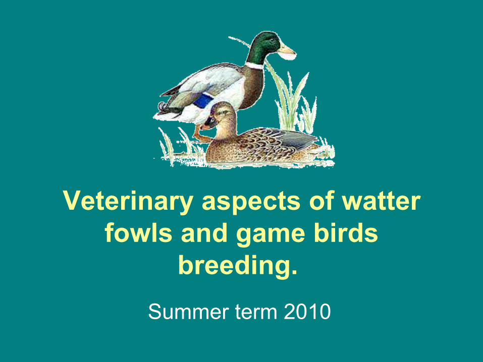
Veterinary aspects of watter fowls and game birds
breeding. Summer term 2010

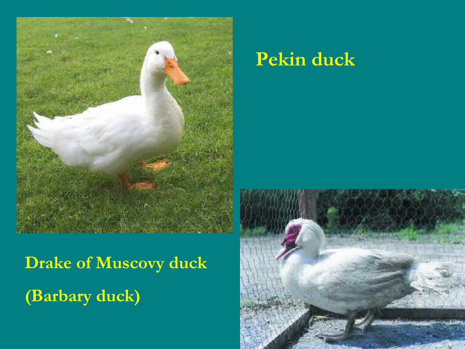
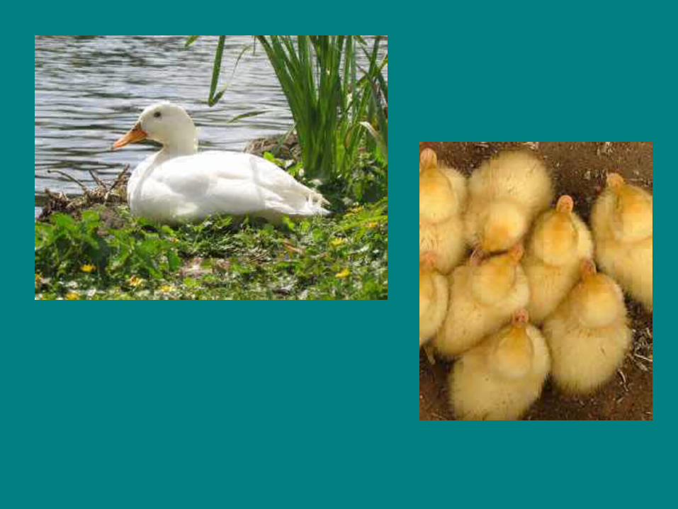
Pekin duck
Drake of Muscovy duck
(Barbary duck)


Disease of watter fowls
Viral diseasesViral diseases• Duck Viral Hepatitis
• Goose Parvovirus (Derzsy's Disease)
• Duck Virus Enteritis, Duck Plague

Disease of watter fowls
Viral disease –new described
• Duck hepatitis B – Avihepadnavirus
• Goose hemorrhagic polyomavirus
• Goose circovirus (GoCV)
• Duck circovirus (DuCV)
Disease of watter fowls
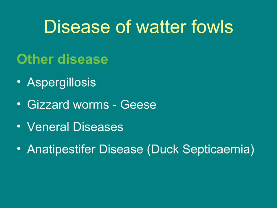
Other disease• Aspergillosis
• Gizzard worms - Geese
• Veneral Diseases
• Anatipestifer Disease (Duck Septicaemia)
Duck Viral HepatitisA viral disease of ducks occurring worldwide. Morbidity is around 100% and mortality 0-95%.
The disease is transmitted by infected ducks and other waterfowl and spreads rapidly, recovered birds carrying the virus for 8 weeks. The infective agent, a Picornavirus may also survive for ten weeks in brooders and five weeks in faeces. A different picornavirus causes a similar condition in North America.

Duck Viral Hepatitis
Signs• Sudden death. • Death in good condition. • Depression. • Fall on side, paddling of legs, arching of back,
rapid deterioration and death, often in opisthotonus.

Duck Viral Hepatitis
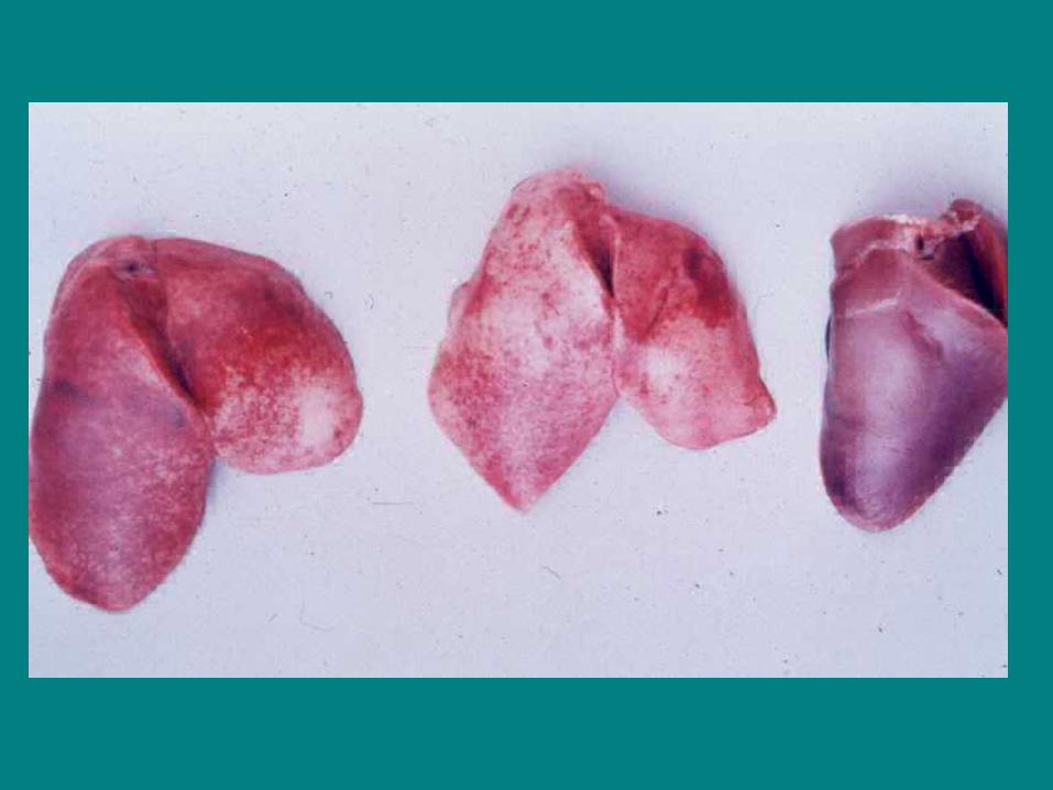
Post-mortem lesions• Liver swollen.
• Punctate/diffuse haemorrhages.
• Kidneys and spleen swollen.
• Microscopically - focal necrosis, bile duct proliferation
and inflammation.


Duck Viral Hepatitis
Diagnosis• History, lesions, SN (serum neutralization test)
serology, isolation in CE (causes stunting of 9 day embryo).
Differentiate from Duck plague (viral enteritis), Duck septicaemia (anatipestifer), coccidiosis, Newcastle disease, Influenza and a 'Type II Variant' hepatitis caused by Astrovirus.
Duck Viral HepatitisTreatment• Antiserum, 0.5 ml serum of recovered birds
given intramuscularly. Prevention• Vaccination and/or antiserum.• Breeder vaccination.

Duck Virus Enteritis, Duck Plague
A herpesvirus infection of ducks and geese diagnosed in the UK in 1972, mostly in ornamental collections, in USA since 1967, also the Netherlands and other countries.
All waterfowl are susceptible and the Barbary duck is more susceptible than the Pekin. The disease follows a very acute course with a morbidity of 5-100% and mortality of 5-100%. Transmission is by infected birds, fomites and arthropods. Recovered birds may carry the virus for a year.

Duck Virus Enteritis, Duck Plague
Signs• Sudden deaths. • Rapidly spreading disease. • Drop egg production. • Photophobia. • Ataxia. • Closed eyes. • Thirst. • Severe diarrhoea, sometimes dysentery. • Dehydration. • Paresis. • Tremor. • Occasionally penile prolapse in the penis in drakes. • Occasionally cyanosis of the bill in the young.
Duck Virus Enteritis, Duck Plague
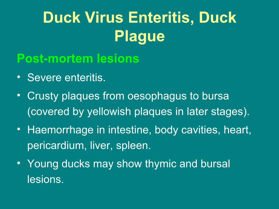
Post-mortem lesions• Severe enteritis. • Crusty plaques from oesophagus to bursa
(covered by yellowish plaques in later stages). • Haemorrhage in intestine, body cavities, heart,
pericardium, liver, spleen. • Young ducks may show thymic and bursal
lesions.

Duck Virus Enteritis, Duck Plague
Diagnosis• Isolation: Duck CAMs 12 day embryos die
in 4 days, HA-, intranuclear inclusions
• Differentiate from Duck hepatitis, oesophagitis (birds on restricted feed), vent gleet, pasteurellosis, coccidiosis.
Duck Virus Enteritis, Duck Plague
Treatment• None, but vaccination in face of outbreak
is of value, probably through interference. Prevention• Isolation from waterfowl, vaccination if
approved by authorities (CE adapted live virus).

Goose Parvovirus (Derzsy's Disease)
Goose Parvovirus is a highly contagious condition of geese and young Muscovy ducks. The younger the bird affected the more acute the condition and the higher the mortality. Losses are negligible in birds over 5 weeks of age.
Derzsy's Disease is caused by a parvovirus distinct from chicken and mammalian parvoviruses. The amount of maternal antibody passed from the breeding birds will affect the severity and timing and severity of the condition in the young birds. Vertical transmission resulting in congenital infection may occur.

Goose Parvovirus (Derzsy's Disease)
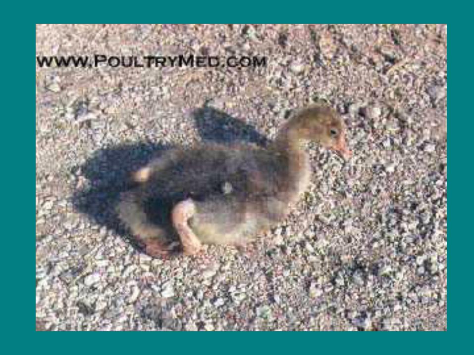
Signs• Prostration and death in acutely affected goslings. • Reduced feed intake. • Excessive water intake. • Swollen eyelids and eye and nasal discharge. • Profuse white diarrhoea. • Membrane covering tongue. • Loss of down. • Reddening of skin.
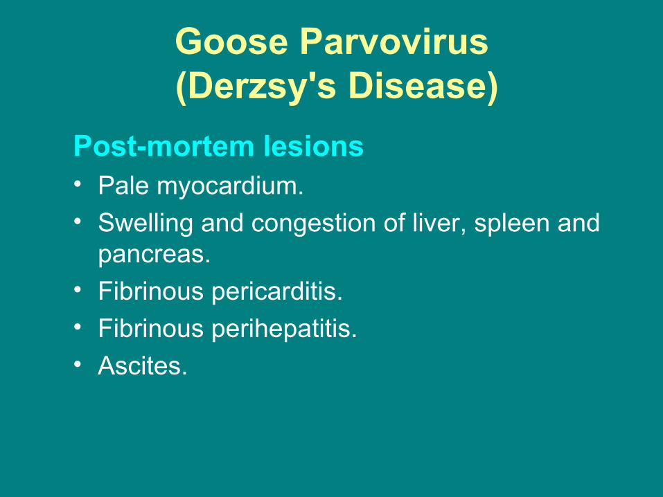
Goose Parvovirus (Derzsy's Disease)
Post-mortem lesions• Pale myocardium. • Swelling and congestion of liver, spleen and
pancreas. • Fibrinous pericarditis. • Fibrinous perihepatitis. • Ascites.

Goose Parvovirus (Derzsy's Disease)
Diagnosis• Signs and lesions in birds of the appropriate age and species. Treatment• No specific treatment. Antimicrobials may be of value in reducing
the effects of secondary bacterial infections. Prevention• Hatching and brooding geese from different parent flocks together
should be avoided. Ideally flocks that have suffered the disease should not be used for breeding as they may become persistent excreters of the infection.
Administration of immune serum has been shown to be effective but may require two doses (day old and around 3 weeks). The preferred approach is to immunise breeding birds with an attenuated live vaccine.

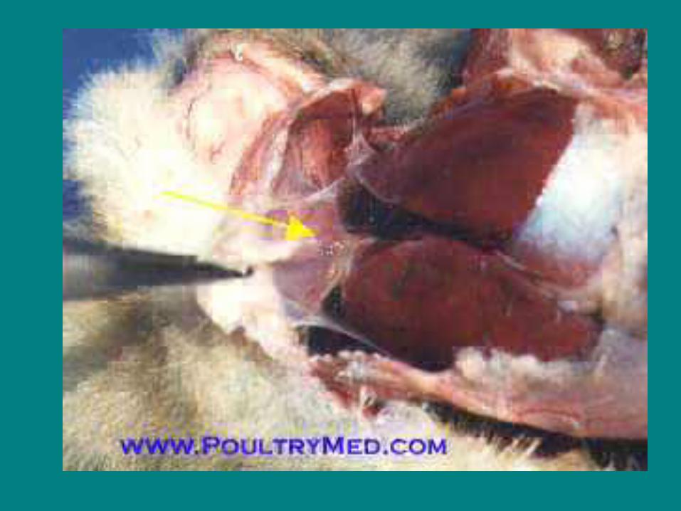
Hemorrhagic nephritis enteritis of geese
• Haemorrhagic nephritis enteritis of geese (HNEG) is a fatal disease of geese aged from 3 to 12 weeks.
• The causative virus, Goose haemorrhagic polyomavirus (GHPV), is a member of the Polyomaviridae family.
• In field cases, morbidity was 30 to 80% and the lethality was nearly 100% in geese 4 to 10 weeks old.
• In these field conditions, death generally followed coma and, in few acute cases, nervous symptoms. Necropsy findings were a subcutaneous oedema, ascites and swelling of the kidneys.

Hemorrhagic nephritis enteritis of geese
• Less frequently, haemorrhagic enteritis was observed in
all areas of the intestine and caeca.
• Older geese, which died after several days of clinical
disease, had visceral gout with urate deposits in the
joints.

Hemorrhagic nephritis enteritis of geese
Symptoms: When suffering from this disease, geese are often
unsteady on their feet, have difficulty getting up and have erratic
movements. These symptoms are accompanied by diarrhoea and
trembling and death usually follows shortly afterwards. The
characteristic lesions are urates and haemorrhaging in the kidneys,
an exaggerated sub-cutaneous swelling and the presence of
intestinal parasites.
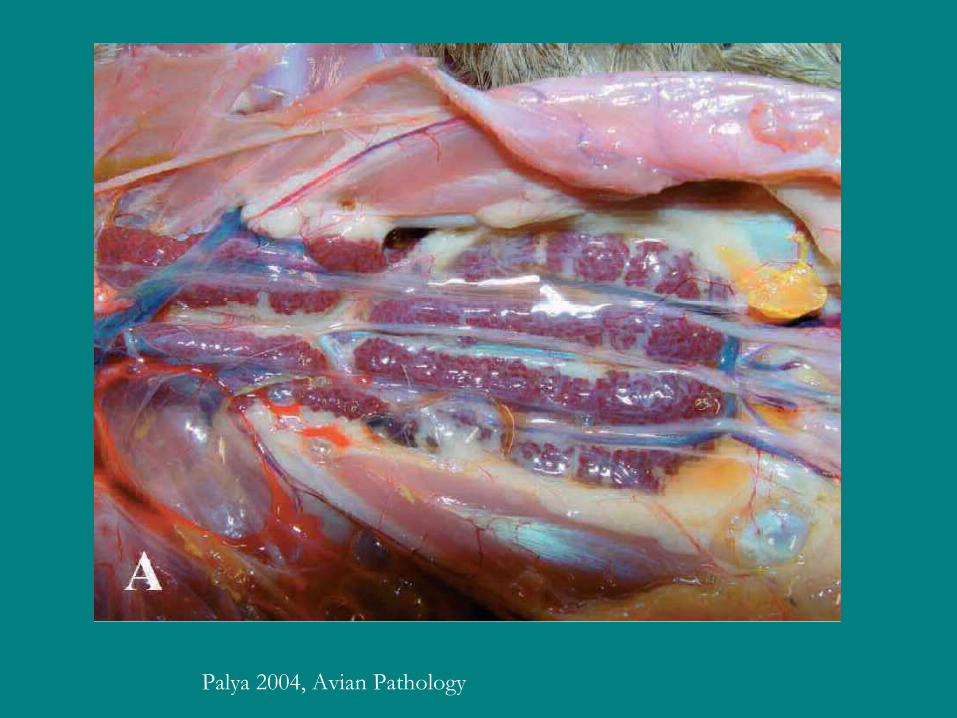
Palya 2004, Avian Pathology

Enteritis
Necrosis in renal tubules
Veneral Diseases
Bacteria, especially Neisseria, Mycoplasma, and
Candida albicans have been associated with a venereal
disease in ganders although it now seems that
Mycoplasma are the primary infective agents.

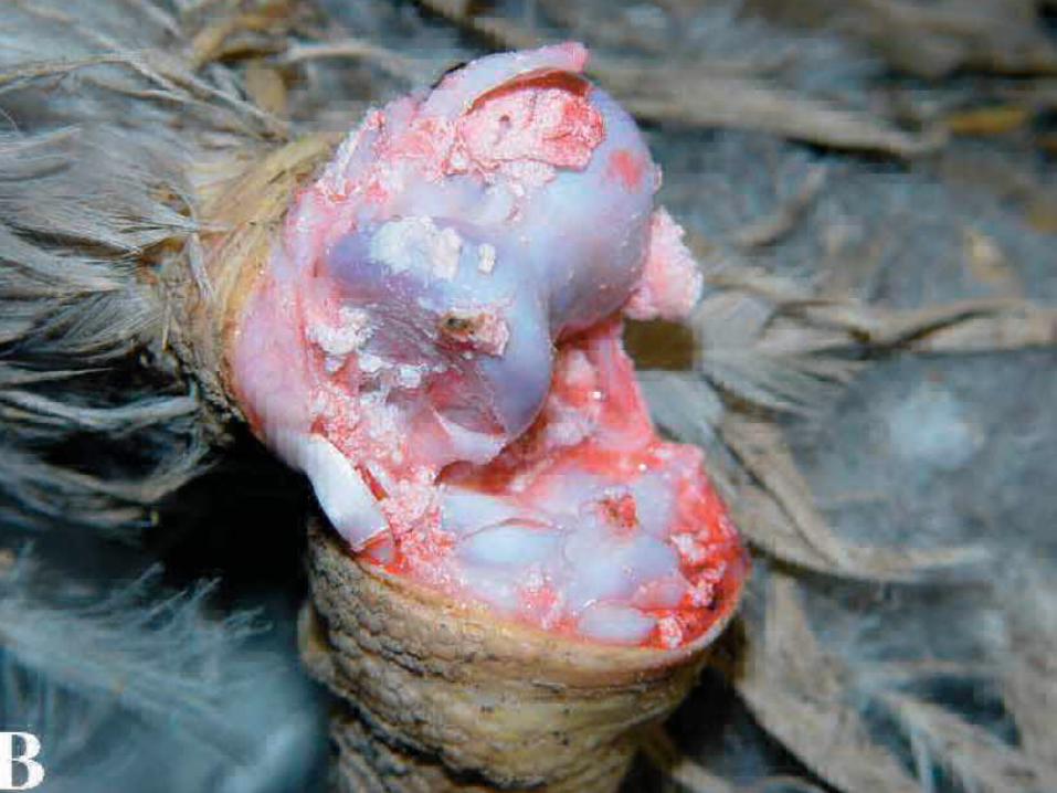
Veneral Diseases
Symptoms: Initially, the base of the phallus becomes
swollen and inflamed with the infection extending to the
cloaca. Later, there is necrosis, ulceration and eventually
considerable scarring, making reproduction impossible.
The disease spreads throughout the flock very rapidly.

Normal appearance of phalus in the ganders


Veneral DiseasesTreatment: The onset of the disease has, in some cases, been associated with a high density of ganders that has led to fighting, resulting in the phallus of some ganders being injured and becoming infected. The infection then spreads through the flock via the females. When infected, the females exhibit symptoms such as airsaculitis, peritonitis, and salpingitis.
The first control measure to take is good management of the breeder flock. Because of the principle involvement of Mycoplasma, some veterinarians view the disease as a component of Mycoplasma infections rather than as a separate disease. Treatment is therefore with antibiotics effective against Mycoplasma such as tylosin, tetracycline, chlortetracycline, linomycin, oxytetracycline, spectinomycin, spinomycin and tiamulin. Sensitivity tests should be conducted to select the appropriate antibiotic.
Anatipestifer DiseaseDuck Septicaemia
An acute or chronic septicaemic disease caused by Riemerella anatipestifer, syn Pasteurella, or Moraxella a. It affects ducks of any age, sometimes turkeys, and may also be isolated from chickens, game birds and wild waterfowl. Mortality is 2-75% in young ducks. Transmission is mainly direct, bird-to-bird, via toenail scratches, especially of the duckling foot, or through respiratory epithelium during respiratory disease. It can also be by faecal contamination of feed, water or the environment where survival of the infectious agent may be prolonged. Adverse environmental conditions and pre-existing disease are predisposing factors
Anatipestifer DiseaseDuck Septicaemia
Signs• Weakness. • Neck tucked in. • Head/neck tremor. • Ataxia. • Disinclined to walk. • Incoordination. • Dyspnoea. • Ocular and/or nasal discharge. • Hyperexcitability

Anatipestifer DiseaseDuck Septicaemia
Post-mortem lesions• Perihepatitis without much smell or liver damage. • Pericarditis. • Airsacculitis. • Enlarged liver and spleen. • Occasionally fibrinous meningitis. • Salpingitis • Purulent synovitis. • Chronic arthritis, sometimes with erosions of the joint
cartilage.

Erosions of the cartilage of the hock joint in a duck with chronic Riemerella anatipestifer infection.

Anatipestifer DiseaseDuck Septicaemia
Diagnosis
Lesions, isolation and identification of organism - blood or chocolate agar in candle jar or 5% CO2.
Differentiate from duck viral enteritis, duck viral hepatitis, fowl cholera, colibacillosis, coccidiosis, chlamydiosis.
Anatipestifer DiseaseDuck Septicaemia
Treatment• Sulphonamides and potentiated sulphonamides are the
products most commonly recommended for drinking water application. Subcutaneous injections of penicillin + dihydrostreptomycin, or streptomycin + dihydrostreptomycin are also highly effective.
Prevention• Good husbandry and hygiene, rigid depopulation and
disinfection, adequate protection, 'hardening off', correct house relative humidity, sulphonamides in feed. Inactivated and attenuated vaccines available in some countries. Autogenous bacterins sometimes used.
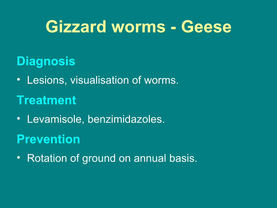
Gizzard worms - Geese
A nematode worm parasite, Amidostomum
anseris, affecting geese and ducks. Worms
develop to L3 in eggs and infection is by the oral
route direct from environment.
Gizzard worms - Geese
Signs
• Depression.
• Loss in condition and weight.
• Slow growth.
Gizzard worms - Geese
Post-mortem lesions
• Ulceration, necrosis and partial sloughing of
gizzard lining, muscular wall may be sacculated
or ruptured.
• Adults are 2-4 cm long and usually bright red.

Gizzard worms - Geese
Diagnosis• Lesions, visualisation of worms.
Treatment• Levamisole, benzimidazoles.
Prevention• Rotation of ground on annual basis.
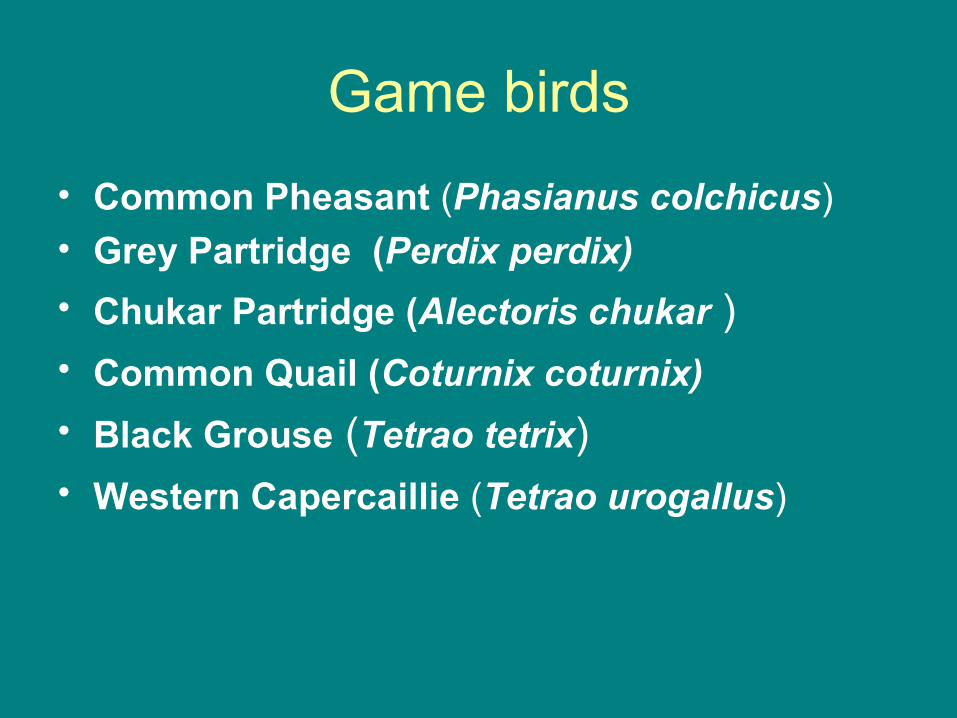
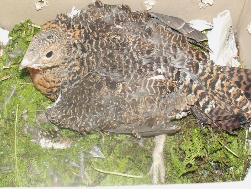
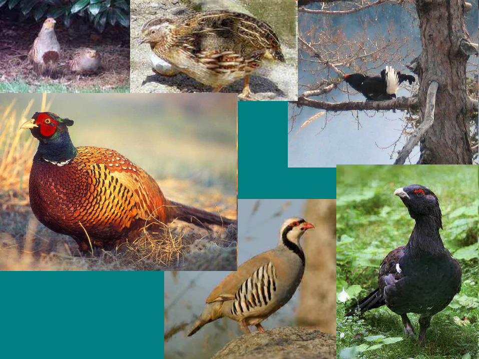
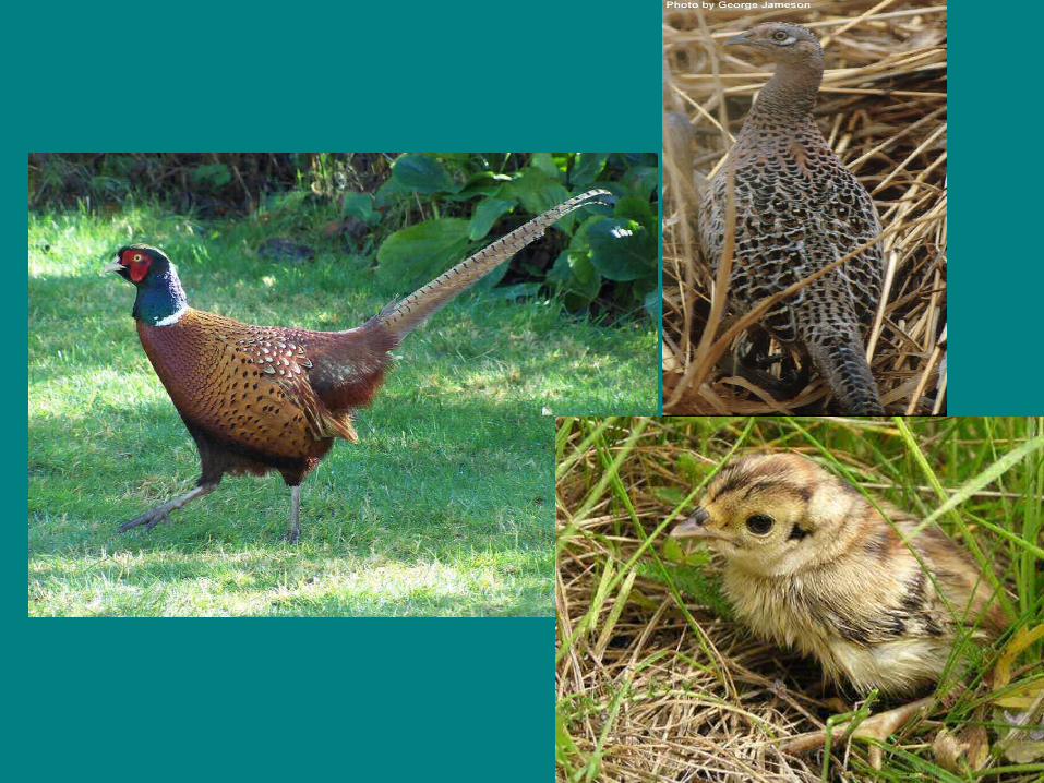
Game birds• Common Pheasant (Phasianus colchicus)• Grey Partridge (Perdix perdix) • Chukar Partridge (Alectoris chukar )• Common Quail (Coturnix coturnix) • Black Grouse (Tetrao tetrix)• Western Capercaillie (Tetrao urogallus)
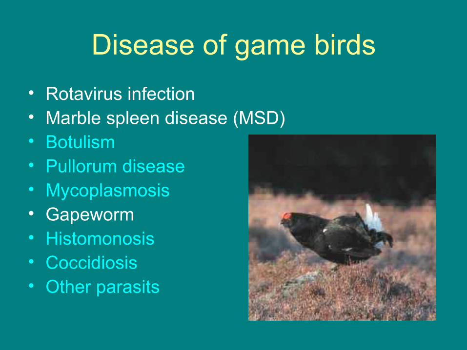
Disease of game birds• Rotavirus infection• Marble spleen disease (MSD) • Botulism• Pullorum disease• Mycoplasmosis• Gapeworm• Histomonosis• Coccidiosis• Other parasits

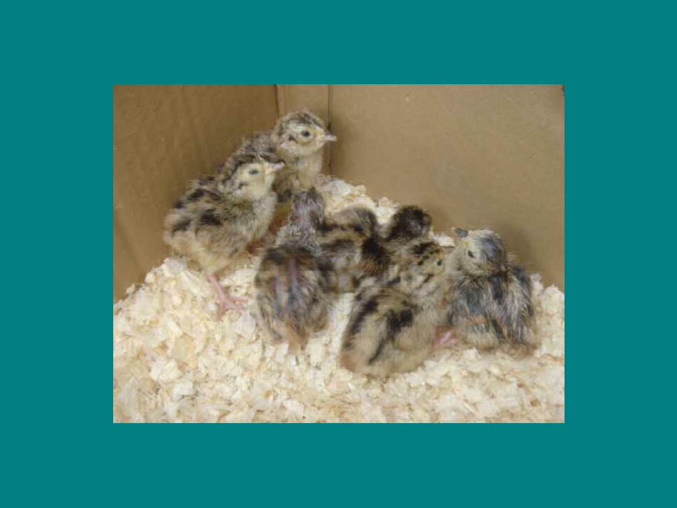
New hatched pheasant chick


Rotavirus infection
A specific infectious agent causing early poult mortality
and marked stunting and unevenness in brooding groups
is rotaviral enteritis.
In the case of rotaviral enteritis mortality rates of 70 per
cent in the first week of life have been reported.
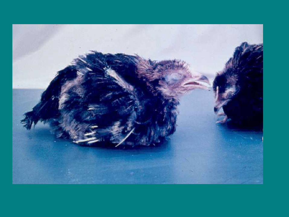
Affected poults are very lethargic and dehydrated, but
often do not show evidence of scouring.
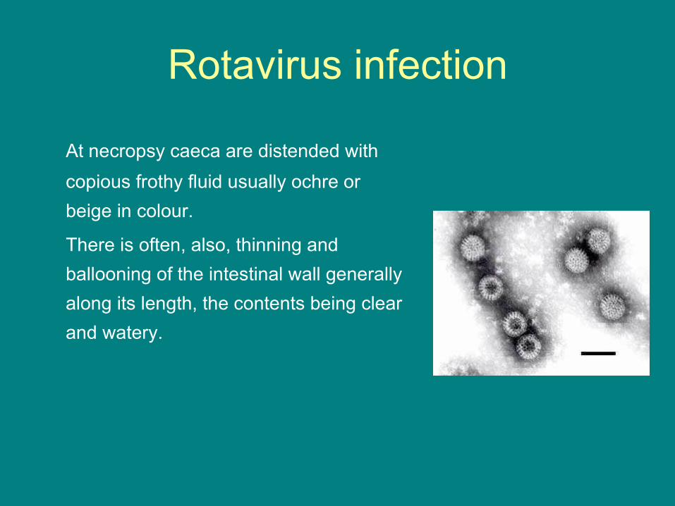
Rotavirus infection
At necropsy caeca are distended with copious frothy fluid usually ochre or beige in colour.
There is often, also, thinning and ballooning of the intestinal wall generally along its length, the contents being clear and watery.
Rotavirus infection
Clinical signs(There is a 2-5 day incubation period). Subclinical (no
signs) to severe diarrhoea may occur. Other signs
include dehydration, poor weight gain, increased
mortality (4-7%), restlessness, litter eating and water
droppings.

Rotavirus infection
Postmortem lesions
Lesions include abnormal amounts of fluid and
gas in the intestinal tract, dehydration, inflamed
vents, vent picking and litter in the gizzard.

Rotavirus infection
Diagnosis:• Diagnosis is by detection of the virus in the
faeces using electron microscope and isolation of the virus in cell culture and staining with specific fluorescent conjugated antisera.
• It simulates coccidiosis, pale bird syndrome, toxic enteritis, astrovirus, coronavirus and enteric bacteria.
Rotavirus infection
Treatment and control:• Prevention• Biosecurity is important. No vaccine is available.
• Treatment• Vitamins, minerals and electrolytes for treating
fluid loss are helpful.

Marble spleen disease (MSD)
Marble spleen disease is an acute respiratory disease of
pheasants characterized by depression, enlarged
mottled spleens, pulmonary congestion, and death.
The etiologic agent is a nonenveloped, icosahedral DNA
virus, 70-90 nm in diameter. It is a member of the family
Adenoviridae and has recently been assigned to the new
genus Siadenovirus .
Marble spleen disease (MSD)
• Pheasants <4 wk of age are resistant to infection due to age-related
resistance or, more commonly, the presence of maternal antibody.
• Marble spleen disease typically affects pheasants 3-8 mo of age.
Onset is acute, with dyspnea, asphyxiation, and sudden death
occurring as a result of pulmonary congestion and edema.
Mortality is commonly 2-3% but can reach 15%. Secondary bacterial
infections as a result of immunosuppression have also been noted.
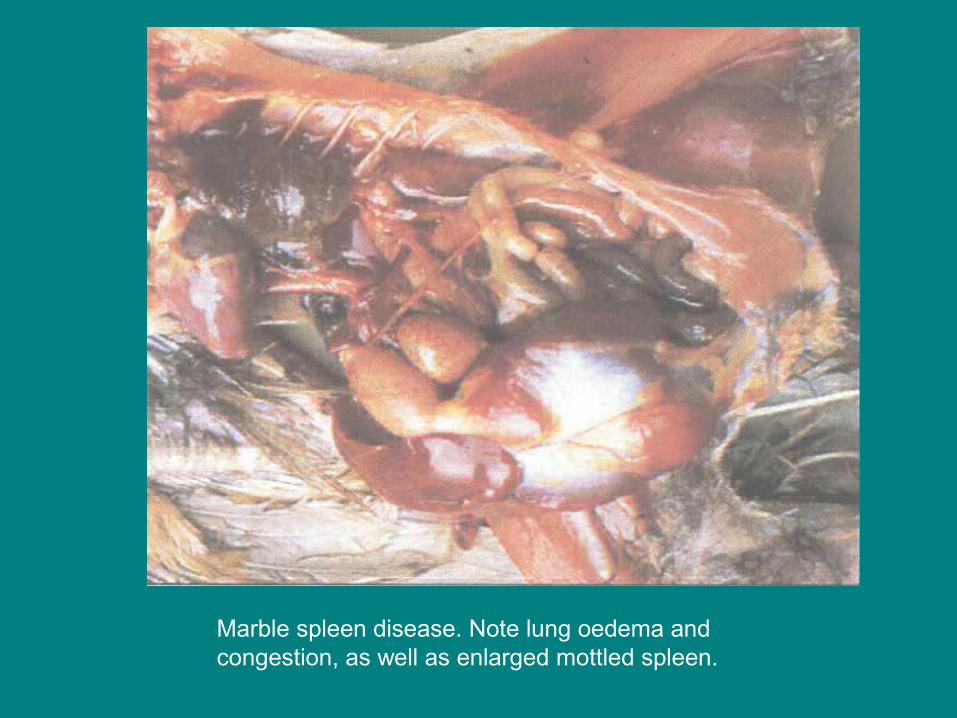
Marble spleen disease. Note lung oedema and congestion, as well as enlarged mottled spleen.
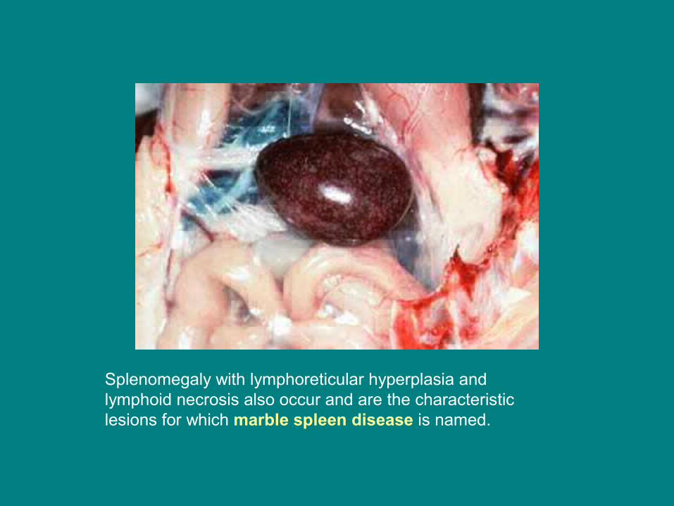
Splenomegaly with lymphoreticular hyperplasia and lymphoid necrosis also occur and are the characteristic lesions for which marble spleen disease is named.

Marble spleen disease (MSD)
On histopathologic evaluation of pheasants with marble spleen disease, flooding of the atria and tertiary bronchi with fibrin and RBC, as well as generalized vascular congestion and focal necrosis, are often seen in the lung.
This response may be anaphylactic in nature, with the lung being considered the target shock organ in the pheasant.
Splenomegaly with lymphoreticular hyperplasia and lymphoid necrosis also occur and are the characteristic lesions for which marble spleen disease is named. Basophilic intranuclear inclusions may be found in a variety of tissues excluding the GI tract with the highest concentration of virus again
being found in the spleen.

Marble spleen disease (MSD)• Diagnosis of virulent outbreaks of marble spleen disease can often
be made based on clinical signs and gross lesions. • Confirmation is by histopathology and the presence of
seroprecipitating virus in the spleen as determined by agar gel
immunodiffusion. • PCR techniques to detect viral DNA in tissue have also been
described and are in regular use in select laboratories.
• In pheasants with acute respiratory disease, differential diagnoses include Newcastle disease, avian influenza, and in the case of birds reared in confinement, gaseous toxins.

Gapeworm
Syngamus trachea, a nematode worm parasite of chickens, turkeys, pheasants, and other game and ornamental birds occurring worldwide. Infection is by the oral route with earthworms, slugs and snails acting as transfer hosts but the life cycle may also be direct, by ingestion of embryonated egg or L3. There is an 18-20 day prepatent period. The condition is seen more commonly in poultry on free range where ground may be contaminated by wild birds e.g. from rookeries.
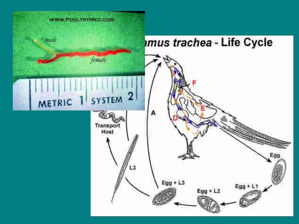
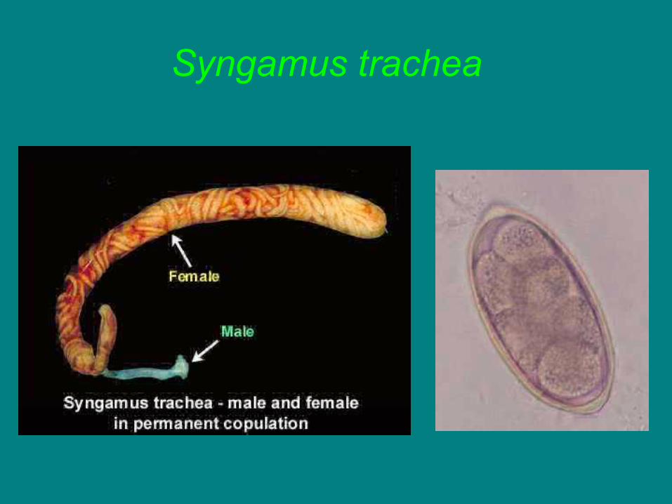
Syngamus trachea


Cannibalism
• Predisposing factors include insufficient feed or
feeder space, high density rearing, excessive
light, too much heat, nutritional deficiencies or
irritation from external parasites.

Cannibalism
Clinical signs:• The following are common forms of
cannibalism seem in commercial poultry operations:– Vent-picking– Feather-pulling– Toe-picking– Head picking
Cannibalism
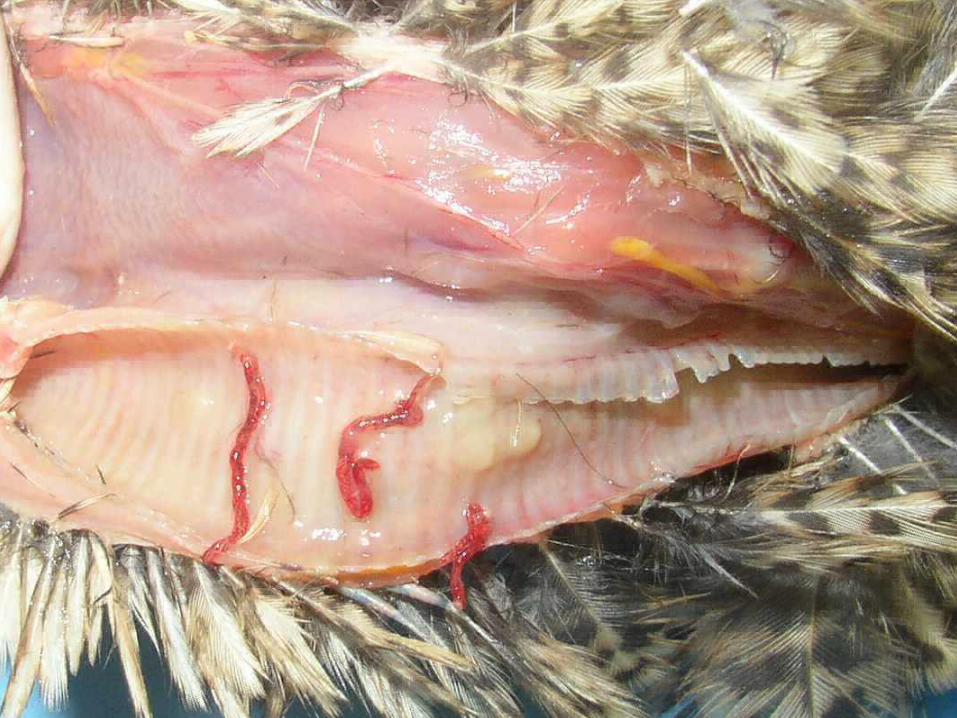
Vent-pickingPicking of the vent or region of the abdomen several
inches below the vent is the most severe form of
cannibalism. This is generally more common in high-
production or overweight pullet flocks. Predisposing
factors are prolapse or tearing of the tissues by passage
of an abnormally large egg. Vent picking can result in
anaemia.

Cannibalism
Feather-pulling
Frequently seen in flocks kept in close confinement
resulting in lack of sufficient exercise. Nutritional
deficiencies may contribute to the problem.

Cannibalism
Toe-picking• Most commonly seen in young birds. Inadequate
feeder space or inability of the chick to find the feed will lead to toe-picking.
Head picking• Follows injuries to the comb or wattles.

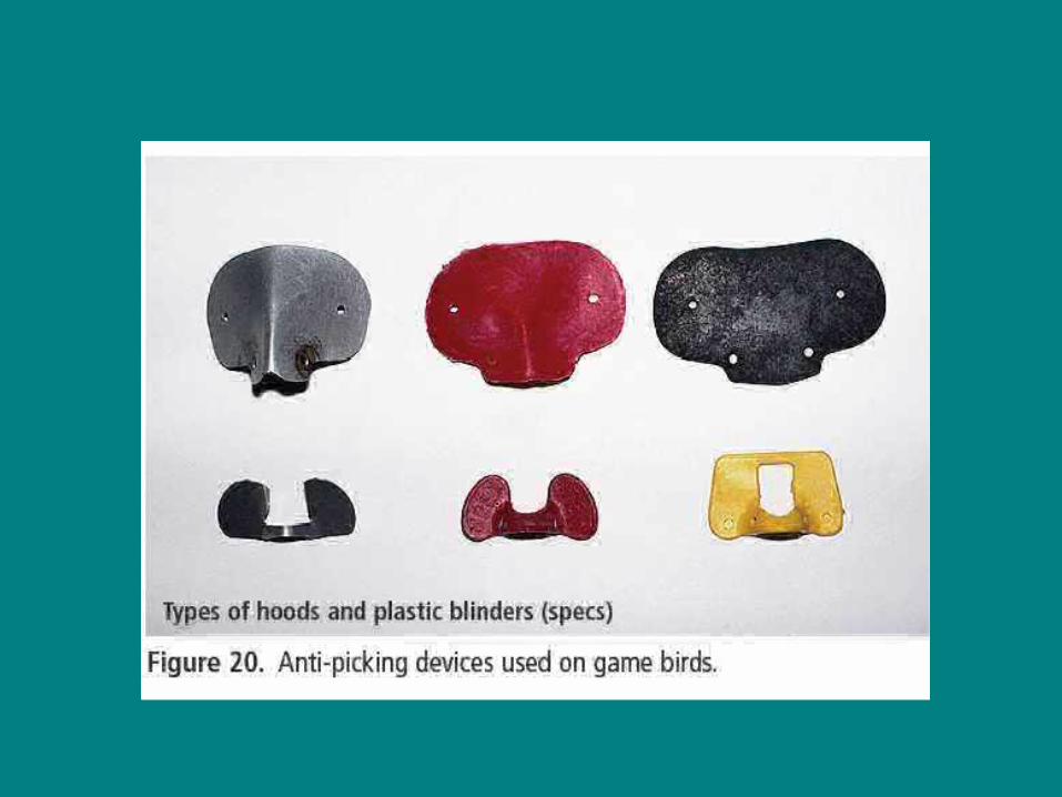
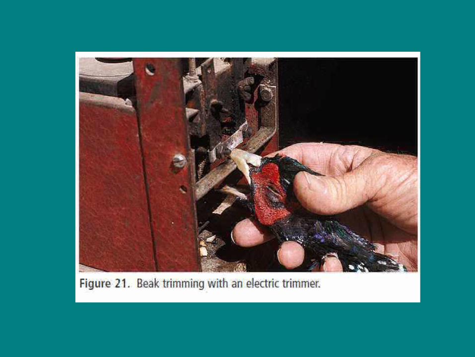
CannibalismDiagnosis:• Injuries seen around the head, vent and feet, or
observation of cannibalistic activity are indicative.• Treatment and control:Prevention• Provide adequate feed and feeding space, reduce bird
density, reduce light, beak trimming, toe and comb trimming in breeders and wattle trimming in cage birds. Coloured light can help to reduce the problem. Give "toys" for the birds to play with like straw, branches etc.