vessel caliber and branch-angle of human coronary...
TRANSCRIPT
572
Vessel Caliber and Branch-Angle of Human CoronaryArtery Branch-Points
GROVER M. HUTCHINS, M.D., MARTIN M. MINER, AND JOHN K. BOITNOTT, M.D.
SUMMARY Measurements were made of parent and branchvessel diameters and of the included angles of branch-points frompostmortem human coronary arteriograms to determine the useful-ness of theoretical equations predicting the relationships betweenparent and branch vessel caliber and between arterial caliber andbranch-angle. The formulas were based on the concept that bloodvessel size and arrangement provided for blood flow with minimumenergy loss. Size relationships between parent vessel and its brancheswere determined for 42 left main and S3 other epicardial coronaryartery branch-points in hearts with angiographically normal arteries.Left main coronary artery branch-points were studied in 68 heartswith various degrees of angiographically defined coronary arterydisease. Measured diameters (D) of parent and branch ves-sels corresponded well to the theoretical formula: (DParent)
3 =(DBr.nch i)3 + (DBr.nch 2 ) 3 + (DBranch 3V • • • •> '•> angiographically
normal coronary arteries. The exponent, on the average, is less withincreasing grades of vascular disease for left main coronary arterybranch-points. Mean area ratio, the sum of the cross-sectional areaof the branches divided by the area of parent vessel, decreased withgreater arteriographic disease. Area ratio varies with changes in therelative calibers of branch vessels. Fifty-seven branch-angles weredetermined by graphic analysis of postmortem biplane coronaryarteriograms. No relationship could be found between branch-angleand vessel caliber. The included angle between branches varied from32° to 124° without respect to relative or absolute vessel calibers. Theresults of these postmortem measurements on human coronary ar-teriogram suggest that coronary artery caliber may adjust to mini-mize energy loss at the branch-point but that branch-angle is deter-mined by other factors. Restudy of arteriograms suggests that branch-angle may be determined by branch vessel destination.
THE CONCEPT of the principle of minimum work asapplied to the function of the circulatory system has beenwell stated by D'Arcy Wentworth Thompson1 in his classicwork, On Growth and Form:
"That this mechanism is the best possible under all thecircumstances of the case, that its work is done with amaximum of efficiency and at a minimum of cost, may notalways lie within our range of quantitative demonstration,but to believe it to be so is part of our common faith in theperfection of Nature's handiwork
The minimum work concept has been applied to atheoretical analysis of the problem of blood flow througharterial branch-points relative to the size of parent andbranch vessels and to branch-angles. By assuming thecirculation to have the steady laminar flow of a Newtonianfluid which obeys the Poiseuille equation, the relationshiphas been derived that the cube of the radius of a vesselshould equal the sum of the cubes of the radii of the vesselsinto which it branches.2 With a similar consideration theangle of vascular branching has been predicted to be afunction of the relative sizes of the parent trunk and itsoffspring.3- * Although it is well known that blood flow doesnot follow the assumptions made, the possible usefulness ofthese principles to a description of deviations from normalvascular geometry which could then be related to vessel walldisease led us to examine their validity for the coronaryarterial tree. Postmortem coronary arteriograms fromhearts with various degrees of vascular disease were studied
From Ihe Department of Pathology, The Johns Hopkins UniversitySchool of Medicine and Hospital, Baltimore. Maryland.
Supported by Grant P17-HL-17655-O1 from the National Institutes ofHealth, U.S. Public Health Service.
Address for reprints: Dr. Grover M. Hutchins, Department of Pathology,The Johns Hopkins Hospital, Baltimore, Maryland 21205.
Received October 20, 1975; accepted for publication February 27, 1976.
to determine the relationship of parent to branch vessel sizeand the branch-angle.
Methods
We reviewed the hearts from 738 patients in the autopsyfiles of The Johns Hopkins Hospital, studied by a standardmethod employing coronary arteriography and fixation indistention.5 Two sets of stereoscopic radiographs, one madeon the intact heart and one on the transverse sections of theheart, were examined to assess the adequacy of the arterio-graphic injection procedure. Radiographs of specimens withadequate filling of both right and left coronary arterial treeswere graded on a scale of 0 to 4+ for severity of coronaryartery disease and for tortuosity of the epicardial branches.Using a scale and hand lens, we measured the diametersof the right, left, left anterior descending, and circumflexcoronary arteries directly from the radiographs. Since thecross-sectional area of a blood vessel increases just proxi-mal to a branch point, 6 care was taken to make measure-ments at representative sites proximal to the zone of increas-ing size. When the left main coronary artery trifurcated theadditional branch also was measured. In many hearts theleft coronary artery could not be measured because ofdistortion or nonfilling of that vessel from ligature place-ment during the injection procedure. No compensation wasmade for the trivial magnification effects caused by thedivergent x-ray beam.
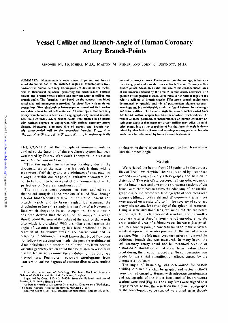
The angle of branching was determined for vesselsdividing into two branches by graphic and vector methodsfrom the radiographs. Hearts with adequate arteriogramsand radiographs of the intact heart and of its transversesections were used (Fig. I). The x-ray films were aligned on alarge viewbox so that the vessels on the biplane radiographsat the branch-point to be studied were lined up as though
by guest on June 19, 2018http://circres.ahajournals.org/
Dow
nloaded from

CORONARY ARTERY BRANCH-POWJS/Hutchins el al. 573
FIGURE I Postmortem coronary arteriograms of the intact heart(below) and the transverse section of the base (above) arranged asfor preparation of orthographic projections of the left maincoronary artery branch-point.
they were orthographic projections at right angles to eachother. To determine the diameters of the parent and branchvessels, we used an optical measuring device with a scalecalibrated to 0.1 mm. A sheet of tracing paper was overlaidand the branch-point area was traced from both radio-graphs. The center line of the parent and each branch vesselwas determined on each tracing and drawn. The point ofintersection of the axes of the vessels was taken as the originof a three-dimensional Cartesian coordinate system. WithT-square and triangles, lines were drawn so that thecoordinates of an arbitrarily chosen point on the axis line of
each vessel, which represented the end of the vector, could bemeasured on the construction. The angle d between twovessels was calculated by considering the coordinates of thepoints to represent scalar values for vectors, A and B,representing the axes of the vessels and using the formula:7
Cos0 =A.B, A y B y AZBZ
Repeat determinations on the same vessel showedreproducibility in the range of 1 or 2 degrees difference.Independent determinations of all three angles around abranch-point produced a sum in the range of 330-362°. Thatthe total of the angles is usually less than 360° is because themajority of epicardial coronary artery branch-points studiedlay on a slightly curved surface rather than in a plane.
Results
In the first review of the 738 coronary angiograms eachusable heart was assigned to a category of severity ofangiographically detectable coronary artery disease on ascale of 0 to 4 + . In 145 hearts with grade 0 there was novascular abnormality seen on the angiograms and at mostonly trivial atherosclerosis recorded from examination ofthe multiple transections of the coronary arteries made at 2-to 3-mm intervals. The 108 hearts in grade 1 had only mildirregularities detectable on the coronary arteriogram, withno lesion exceeding 20% occlusion of the luminal area asconfirmed by gross study. Grade 2, with 99 hearts, hadeither moderately widespread obstructions of up to 50% oflumen area or a single severe or complete occlusion with anotherwise normal angiogram, the angiographic findingsagain being confirmed by the results of direct gross exami-nation. In grade 3, with 115 hearts, and grade 4 with 13hearts, widespread arterial disease was combined with severestenoses or complete vascular occlusion. A total of 258hearts was excluded from the study because the injections orangiograms were incomplete or inadequate.
THE RELATIONSHIP OF THE CALIBERS OF PARENTAND BRANCH VESSELS
The diameter of the left main coronary artery and itsbranches could be determined in 110 hearts. Measurementsof parent and branch vessel diameters were made at 53 otherbranch-points of the epicardial coronary arteries fromhearts with grade 0 coronary disease. The Wang computerwas programmed to solve the equation, (DP a r e n l )" =
(Der.nch l)" + (Deranch 2)" + (DBranch 3)" for fl foreach branch to the nearest thousandth using the measuredvalues of D. The mean, standard deviation, and range of nfor each group are shown in Table 1.
The values of n do not show significant differencesbetween the groups of successive increases in coronaryartery disease. The larger mean exponent for the left maincoronary artery branch-points with no angiographic vesseldisease is partly explained by two unusual cases with large nvalues where thediametersof parent and both branch vesselswere nearly equal. With increasing degrees of vessel diseasethe exponent decreased but the mean diameter of the leftmain coronary artery and the mean heart weight weresimilar in these groups.
by guest on June 19, 2018http://circres.ahajournals.org/
Dow
nloaded from
574 CIRCULATION RESEARCH VOL. 38, No. 6, JUNE 1976
TABLE 1 Exponent n and Area Ratios for Coronary Artery Branch-Points
Group
Left main coronary arteriesGrade 0 AS
Grade 1 AS
Grade 2 AS
Grade 3 and 4 AS
Other epicardial coronaryarteries: Grade 0 AS
No. ofobservations
42
26
25
17
53*
Exponent n
3.2 ±(1.5 -2.8 =t
( 1 . 9 -2.6 ±
(1.3 -2.2 ±
(0.8 -2.7 ±
( 1 7 -
1.614.2)
1.36.2)1.54.7)2.1
10.8)1.3
11.2)
Area ratio
1.28 ±0 .18(0.93 - 1.4)1.18 ± 0.16
(0.79 - 1.49)1.18 ± 0.15
(0.93 - 1.44)1.10 ± 0.39
(0.42 - 1.71)1.13 ± 0.14
(0.88 - 1.77)
DParent(mm)
2.5(1.02.4
(1.92.3
(1.92.4
(1.9
2.8(1.0
± 0.5- 3 . 3 )± 0.4- 3.5)± 0.4- 3.3)± 0.4- 3.3)± 0.9- 5.0)
Heart wt(E)
538 ±(168 -561 ±
(280 -515 ±
(280 -545 ±
(425 -389 ±
(220 -
2171040)
165900)142822)
95710)147750)
Results are given as mean ± SD. Ranges are shown in parentheses.AS - arteriosclerosis.* Made on 17 hearts.
THE AREA RATIO OF PARENT AND BRANCH VESSELSArea ratios for branch-points were calculated from the
formula:
ranch .)2 + (D B r a n c h j)* + (D B r a n c h 3 ) 2 . . .Area ratio = (Dparenc)2
The mean and standard deviation of the values for eachgroup are shown in Table 1. There is a smaller mean arearatio in arteriosclerotic left main coronary artery branch-points than in the normals. The similarity of parent vesseldiameters for these groups suggest that the lower area ratiosobserved with increasing vascular disease are caused bygreater involvement of the branches than of the parent.
THE RELATIONSHIP OF VESSEL CALIBER ANDBRANCH-ANGLE
The angle between parent and branch vessels and/orbetween branch vessels was determined as described abovefor left main and other epicardial coronary artery branch-
LL)_ lCD
X
orrCD
20°
O
90
<O
60
io
30
n
•
•
•t •
( • V*•• •'• <•
9
•
•
• •
•
•
«• • •
* • •
•
•
•
•
•
1.0 1.5 2.0 2.5RATIO OF DLAR6ER BRANCH
3.0
^SMALLER BRANCH
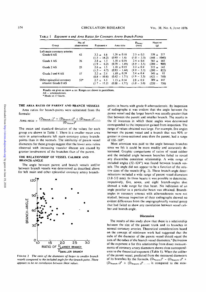
FIGURE 2 The ratio of the diameters of larger to smaller branchvessels compared to the included angle for that branch-point. Thereappears to be no correlation between these values.
points in hearts with grade 0 atherosclerosis. By inspectionof radiographs it was evident that the angle between theparent vessel and the larger branch was usually greater thanthat between the parent and smaller branch. The results inthe 12 instances in which these angles were determinedcorresponded to the impression gained from inspection. Therange of values obtained was large. For example, five anglesbetween the parent vessel and a branch that was 90% orgreater in cross-sectional area than the parent, had a rangeof 130-169°.
Most attention was paid to the angle between branchessince we felt it could be more readily and accurately de-termined. Graphic comparisons of ratios of vessel caliberand the included angle between branches failed to revealany discernible consistent relationship. A wide range ofincluded angles (32-124°) was found between branch ves-sels. The angle did not appear to be a function of the rela-tive sizes of the vessels (Fig. 2). These branch-angle deter-minations included a wide range of parent vessel diameters(1.0-5.0 mm). In three hearts it was possible to determine,respectively, five, seven, and eight branch-angles thatshowed a wide range for that heart. No indication of anangle peculiar to a particular heart was obtained. Branch-angles in coronary arteries with atherosclerosis were notstudied, because inspection of their radiographs showed noevident differences from the angiographically normal groupthat had failed to show any correlation between vessel cali-ber and branch-angle.
Discussion
The results of this study show that there is a relationshipbetween the size of the parent trunk and its branches innormal coronary arteries. Theoretical considerations basedon the concept of minimum work had suggested that thecube of the diameter of the parent vessel should equal thesum of the cubes of the branch vessel diameters.2 Derivationof the exponent n for this relationship from direct measure-ments of coronary artery diameters shows close correspond-ence to the theoretical exponent (Table 1). When the caliberof the parent vessel, predicted from the measured diametersof its branches by the formula, (DP>ren l)
3 = (DBran<:h ,)3 +(DBranch a)3 + (DBranch s)3 • • • . is compared to the mea-
by guest on June 19, 2018http://circres.ahajournals.org/
Dow
nloaded from

CORONARY ARTERY RRAWCH-POINTS/ Hutchins et al. 575
mm
4.0a.
aa 3.0UJ
<rg 2.0UJ
1.0
mm
4.0
a 3.0UJ
rr2.0
UJ
o ASLEFT MAINCORONARY ARTERYBRANCH-POINTS
1.0 2.0 3.0 4.0 mm
PREDICTED Dp
1.0 0 ASOTHER EPICARDIALCORONARY ARTERYBRANCH-POINTS
1.0 2.0 3.0 4.0 mm
PREDICTED Dp
FIGURE 3 Comparison of DParenl
(DP) predicted from the formula:(Dparen,)' = (DBranc)l , ) 3 +(DBrancn ,)3 + (DBranch 3 ) 3 . . . withDparent measured from the arterio-gram. There is close correspond-ence for left main coronary artery{left) and other epicardial coronaryartery branch-points (right). 0 ASis the grade and means no arlerio-sclerotic change was seen.
measured DParen, the correspondence, for angiographicallynormal coronary arteries, is excellent (Fig. 3).
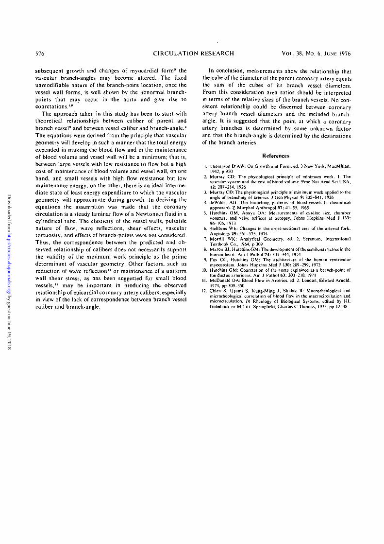
With abnormal vessels the measured DParei l l tends to begreater than the predicted DP a r e n t (Fig. 4). The same trend isseen with the area ratio, the more severe degree of vasculardisease having larger parent vessels relative to branches. Theparent trunks are of similar caliber in the diseased ves-sels and the normals. The decreased area ratio in the leftmain coronary artery branch for more severe vessel diseaseis probably a reflection of the common observation thatvessel lumen narrowing is usually more severe in theproximal left anterior descending and circumflex coronaryarteries than in the left main coronary artery. The meanheart weight for the groups are nearly the same.
The area ratio is not particularly useful for describing thecharacteristics of branch-points since it could be expected tovary according to the relative sizes of the branch vessels. Asshown above, the ratio of the cube of the diameter of theparent vessel to the sum of the cubes of the branch vessels is1. The same relationship between diameters squared, i.e.,the area ratio, will vary according to the sizes of the branchvessels relative to each other. In theory, the area ratio wouldbe 1.26 when the branch vessels are equal and wouldapproach 1.0 if one branch is very small and the other nearlythe size of the parent trunk. In a recent review of the subject,
Stehbens6 also has expressed caution in correlating arearatio determinations with abnormalities of blood flow oratherosclerosis.
No relationship could be discovered between the sizes ofbranch vessels and the angles between them. Our impres-sion, based on measurements and radiographs, was that thelarger of two branches tended to pursue a course closer tothe path of continuation of the parent trunk than did thesmaller branch. Measurements showed a wide variation andconsiderable overlap in this feature. It appears that relativebranch vessel diameter has little to do with determiningcoronary artery branch-angle. Upon reexamining the radio-graphs in the light of this negative finding, we have been leftwith the impression that the destination of the blood vesselmay help determine the angles made with its parent andfellows. For example, the artery to the sinoatrial node has arather constant destination but its point of origin showsconsiderable variation and the angle it makes with theparent trunk seems determined, in part at least, by thecourse it must pursue to reach its termination. A similarconsideration may apply to the bifurcation of the left maincoronary artery into left anterior descending and circumflexbranches. It is possible that the branch-angle of the epicar-dial coronary arteries is determined by relative size whenthese structures first develop embryologically8 but that with
mm
2.0£/)
1.0
0
mm
,5-3.0
2CO<
2.0
ASLEFT MAINCORONARY ARTERYBRANCH-POINTS
1.0 2.0 3.0PREDICTED Dp
mm0
mm
Q°"3.0
2 * ASLEFT MAINCORONARY ARTERYBRANCH-POINTS
2.0
1.0
1.0 2.0 3.0PREDICTED DD
mm0
3AND4* ASLEFT MAINCORONARY ARTERYBRANCH-POINTS
1.0 2.0 3.0PREDICTED Do
mm
FIGURE 4 Comparison of DParenl (Dp) predicted and Dparent measured for the left main coronary artery branch-point in hearts with variousdegrees of arleriographic coronary arteriosclerosis (AS). There is greater scatter and a greater tendency for the measured value to exceedthe predicted with increasing vascular disease. The diagonal lines show the relationship for n = 3; I + , 2+, 3 and 4+ denote grades of AS.
by guest on June 19, 2018http://circres.ahajournals.org/
Dow
nloaded from
576 CIRCULATION RESEARCH VOL. 38, No. 6, JUNE 1976
subsequent growth and changes of myocardial form9 thevascular branch-angles may become altered. The fixedunmodifiable nature of the branch-point location, once thevessel wall forms, is well shown by the abnormal branch-points that may occur in the aorta and give rise tocoarctations.10
The approach taken in this study has been to start withtheoretical relationships between caliber of parent andbranch vessel2 and between vessel caliber and branch-angle.3
The equations were derived from the principle that vasculargeometry will develop in such a manner that the total energyexpended in making the blood flow and in the maintenanceof blood volume and vessel wall will be a minimum; that is,between large vessels with low resistance to flow but a highcost of maintenance of blood volume and vessel wall, on onehand, and small vessels with high flow resistance but lowmaintenance energy, on the other, there is an ideal interme-diate state of least energy expenditure to which the vasculargeometry will approximate during growth. In deriving theequations the assumption was made that the coronarycirculation is a steady laminar flow of a Newtonian fluid in acylindrical tube. The elasticity of the vessel walls, pulsatilenature of flow, wave reflections, shear effects, vasculartortuosity, and effects of branch-points were not considered.Thus, the correspondence between the predicted and ob-served relationship of calibers does not necessarily supportthe validity of the minimum work principle as the primedeterminant of vascular geometry. Other factors, such asreduction of wave reflection" or maintenance of a uniformwall shear stress, as has been suggested for small bloodvessels,12 may be important in producing the observedrelationship of epicardial coronary artery calibers, especiallyin view of the lack of correspondence between branch vesselcaliber and branch-angle.
In conclusion, measurements show the relationship thatthe cube of the diameter of the parent coronary artery equalsthe sum of the cubes of its branch vessel diameters.From this consideration area ratios should be interpretedin terms of the relative sizes of the branch vessels. No con-sistent relationship could be discerned between coronaryartery branch vessel diameters and the included branch-angle. It is suggested that the point at which a coronaryartery branches is determined by some unknown factorand that the branch-angle is determined by the destinationsof the branch arteries.
References
1. Thompson D'AW: On Growth and Form. ed. 3 New York, MacMillan,1942,p 950
2. Murray CD: The physiological principle of minimum work. 1. Thevascular system and the cost of blood volume. Proc Nat Acad Sci USA,12: 207-214, 1926
3. Murray CD: The physiological principle of minimum work applied to theangle of branching of arteries. J Gen Physiol 9: 835-841, 1926
4. deWilde, AG: The branching patterns of blood-vessels (a theoreticalapproach). Z Morphol Anthropol 57: 41-55, 1965
5. Hutchins GM, Anaya OA: Measurements of cardiac size, chambervolumes, and valve orifices at autopsy. Johns Hopkins Med J 133:96-106, 1973
6. Stehbens WE: Changes in the cross-sectional area of the arterial fork.Angiology 25: 561-575, 1974
7. Morrill WK: Analytical Geometry, ed. 2, Scranton, InternationalTextbook Co., 1964, p 309
8. Maron BJ, Hutchins GM: The development of the semilunar valves in thehuman heart. Am J Pathol 74: 331-344, 1974
9. Fox CC, Hutchins GM: The architecture of the human ventricularmyocardium. Johns Hopkins Med J 130: 289-299, 1972
10. Hutchins GM: Coarctation of the aorta explained as a branch-point ofthe ductus arteriosus. Am J Pathol 63: 203-210, 1971
11. McDonald DA: Blood Flow in Arteries, ed. 2. London, Edward Arnold,1974, pp 309-350
12. Chien S, Usami S, Kung-Ming J, Skalak R: Macrorheological andmicrorheological correlation of blood flow in the macrocirculation andmicrocirculation. In Rheology of Biological Systems, edited by HLGabelnick or M Litt. Springfield, Charles C Thomas, 1973, pp 12-48
by guest on June 19, 2018http://circres.ahajournals.org/
Dow
nloaded from

G M Hutchins, M M Miner and J K BoitnottVessel caliber and branch-angle of human coronary artery branch-points.
Print ISSN: 0009-7330. Online ISSN: 1524-4571 Copyright © 1976 American Heart Association, Inc. All rights reserved.is published by the American Heart Association, 7272 Greenville Avenue, Dallas, TX 75231Circulation Research
doi: 10.1161/01.RES.38.6.5721976;38:572-576Circ Res.
http://circres.ahajournals.org/content/38/6/572World Wide Web at:
The online version of this article, along with updated information and services, is located on the
http://circres.ahajournals.org//subscriptions/
is online at: Circulation Research Information about subscribing to Subscriptions:
http://www.lww.com/reprints Information about reprints can be found online at: Reprints:
document. Permissions and Rights Question and Answer about this process is available in the
located, click Request Permissions in the middle column of the Web page under Services. Further informationEditorial Office. Once the online version of the published article for which permission is being requested is
can be obtained via RightsLink, a service of the Copyright Clearance Center, not theCirculation Research Requests for permissions to reproduce figures, tables, or portions of articles originally published inPermissions:
by guest on June 19, 2018http://circres.ahajournals.org/
Dow
nloaded from