venous disease update - sierra veinsierravein.com/education/ppt_venousdisease.pdfprevalence of...
TRANSCRIPT

Venous Disease Update
VN20-52-A 12/04

Prevalence
Abnormal venous anatomy:– 15% of males and 30% of females– Familial tendency– Standing vocation– Associated with pregnancy
(varicose veins)– Leg injury or surgery– Generally progressive
• Often symptomatic– Tired, aching legs– Leg swelling– Phlebitis and thrombosis
VN20-52-A 12/04

Prevalence of Venous Disease
VN20-52-A 12/04
Varicose Veins 20+ million
Swollen Leg 6 million
Skin Changes 1 million
Skin Ulcer 500,000

Saphenofemoral Reflux and Venous Hypertension
Venous insufficiency, or venous reflux, is the impaired return of venous blood from the legs or feet caused by dilated venous vessels in the legs or damaged/absent vein valves
VN20-52-A 12/04

Anatomy Review
• Deep Venous System– Common femoral vein– Femoral vein– Popliteal vein– Gastrocnemius veins– Tibial veins

Anatomy Review Great Saphenous Vein
• Courses from medial ankle to groin
• Joins the common femoral vein proximally at the saphenofemoral junction
GSV

AL – AnterolateralPM – PosteromedialSEP – Superficial external pudendalSE – Superficial epigastricSCI – Superficial circumflex iliac
Anatomy Review SFJ Tributaries

• Courses from the lateral ankle up the posterior calf– Terminates near the popliteal
fossa at the saphenopopliteal junction (SPJ)
• Confluence with the popliteal vein (PV) is variable
– Proximal portion lies between superficial & deep fascial layers
SPJ
SSV
PV
Anatomy Review Small Saphenous Vein

Anatomy Review Perforators
• Connect superficial to deep veins• Locations
– Proximal thigh - Hunterian– Distal thigh - Dodd’s– Knee - Boyd’s– Ankle/Calf - Cockett’s
• Incompetent perforators often source of venous stasis ulcers at medial ankle

Pathophysiology
• Vein Valves– Blood propelled by calf
muscle pump opens the valve in one direction
– Blood moving with gravity closes the normal valve
Valve Open Valve Closed
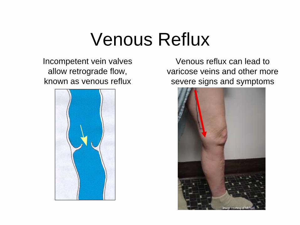
Venous RefluxIncompetent vein valves
allow retrograde flow, known as venous reflux
Venous reflux can lead to varicose veins and other more severe signs and symptoms
Image courtesy of Michael Vasquez, MD

Signs of Venous Insufficiency
Edema Varicose Veins
Skin Changes and
Discoloration
Stasis Ulcers

Ultrasound Diagnostic Study• Must be performed to
determine the sources of reflux.
• Evaluate for venous occlusion or thrombus
• Map the course of the incompetent superficial veins
• Localize sites for treatment with minimally invasive techniques
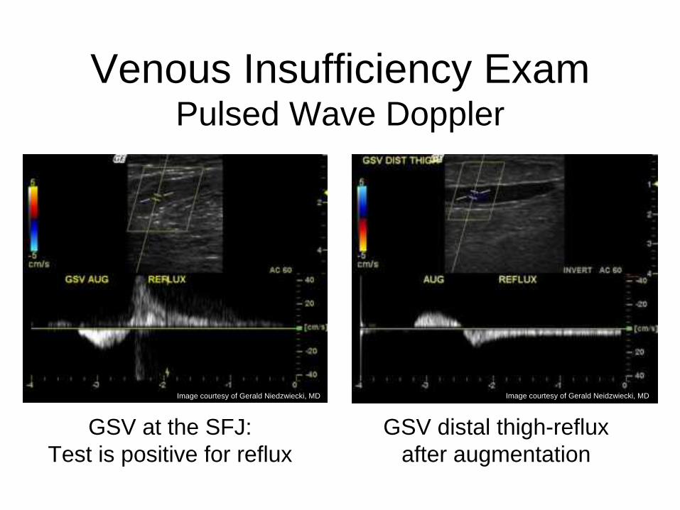
GSV at the SFJ: Test is positive for reflux
Venous Insufficiency Exam Pulsed Wave Doppler
Image courtesy of Gerald Niedzwiecki, MD Image courtesy of Gerald Neidzwiecki, MD
GSV distal thigh-reflux after augmentation

Venous Insufficiency Exam Perforators
Perforator passing through deep fascia

Treatment Options
• Pressure Garment Use• Compression Wraps• Surgical Ligation/Stripping• Chemical Sclerotherapy• Endovenous Ablation RF/Laser• Perforator Ablation (RF)

Pressure Garments
• Class I 20 to 30 mmHg aching, swelling small varicose vein changes
• Class II 30 to 40 mmHg symptomatic Varicose veins, Chronic venous insufficiency, post ulcer
• Class III 40 to 50 mmHg Chronic venous insufficiency, post ulcer, lymphedema
• Class IV 50 to 60 mmHg (same as III)

Compression Wraps
• Multi-Layer Wraps• Una Boot• Unna Sleeve (ACI Medical): cotton sock
that laces up the front for fit• “Circ Aid” ( Circ-Aid corp.): series of
straps that circle the leg with Velcro tension adjustment

Surgical Ligation/Stripping
• Performed in the OR• Historically the “Gold Standard”• Invasive, Prolonged Recovery, Nerve
Injuries• Occasionally the only option for patients

Chemical Sclerotherapy
• Used in smaller reticular veins and spider veins
• Poor results when used in larger veins• Products include: Polidocanol, Hypertonic
Saline, Sotradecol, Glycerine

Endovenous Ablation (RF)
Catheterinserted
inrefluxing vein
Catheter positioned, electrodesdeployed
RF energyheats andcontractsvein wall
Catheter withdrawn,closing vein
Denudedvein
is physicallynarrowed
VN20-52-A2/04

RF Ablation Stylet For Incompetent Perforators
Electrodes

How does Vein Ablation Work?

Collagen Contraction
Controlled heating of the vein wall causes:– Shortening and thickening of
collagen fibrils– Vein lumen diameter
shrinkage– Fibrotic sealing of the vessel
Caprine vein 6 weeks post-treatment
Caprine vein pre-treatment
VN20-52-A 12/04

RF Catheter Positioned at SFJ
CFV
SEV
CFASFJ
CatheterOpened electrodes
Image courtesy of Olivier Pichot, MD
VN20-52-A 12/04
GSV

RF Treated SFJ
Post-TreatmentNo flow in GSV
Images courtesy of Olivier Pichot, M.D.
SFJ
SEV
CFV
VN20-52-A 12/04

Perforator Identified
Perforator (RF) Treatment
Treatment with Stylet

Case Presentations

Venous Stasis Ulcer Perforator Treatment Only (RF)
• 57 y/o male• Ulcer has been present for 4 years• Treated with Compression Wraps & Stockings
VN20-52-A 12/04
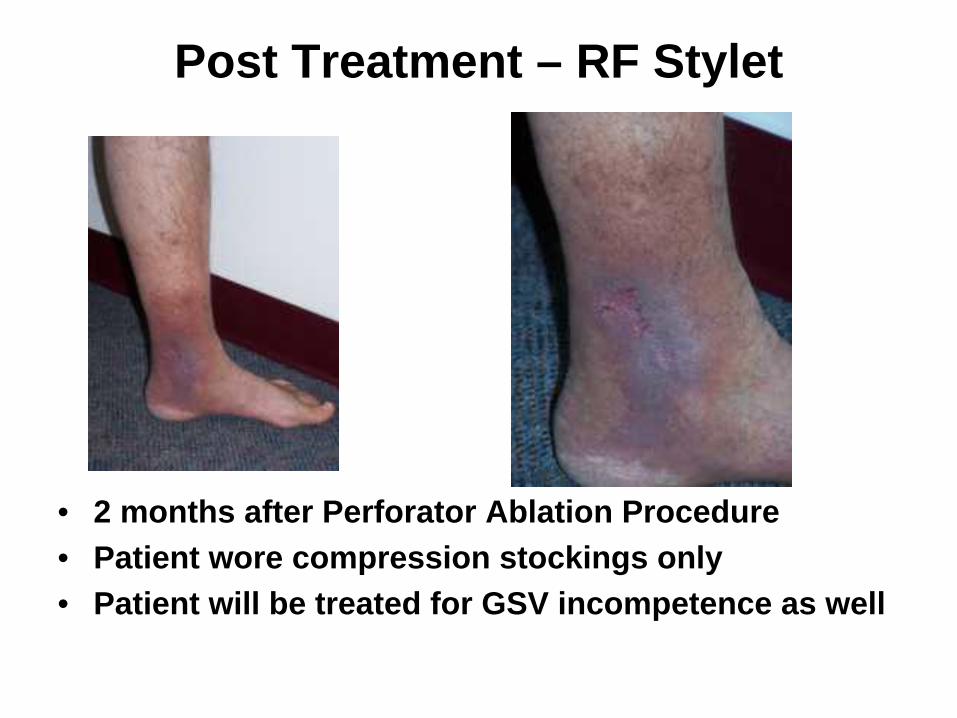
Post Treatment – RF Stylet
• 2 months after Perforator Ablation Procedure• Patient wore compression stockings only• Patient will be treated for GSV incompetence as well

Venous Stasis Ulcer GSV Treatment Only
•35 y/o Male – Venous Stasis Ulcer & Varicose Veins•SFJ & GSV Incompetence discovered•Treated GSV with RF Vein Ablation Procedure•No incompetent perforators found
Pre Procedure Post Procedure – 6wks

Venous Stasis Ulcer SSV Treatment Only
•65 y/o Female – Venous Stasis Ulcer, Varicose Veins, Significant Edema•SSV & Cockett Perforator Incompetence•Treated SSV with RF Vein Ablation Procedure
Pre Procedure (5/25/06)Procedure Performed (6/9/06)
Post Procedure (7/11/06)