vasopressin-induced ventricular arrhythmia filevasopressin-induced ventricular arrhythmia ghanta et...
TRANSCRIPT

31Case Report
Vasopressin-Induced Ventricular ArrhythmiaSoma Sekhar Ghanta1 Sarada Srinivas Parvathaneni1 Gopala Krishna Koduru1 Sudarshan Palaparthi1 Raghuram Palaparthi1 Purna Chandra Rao Kondru1
1 Department of Cardiology, Aayush Hospital, Vijayawada, Andhra Pradesh, India
Address for correspondence Soma Sekhar Ghanta, MD, DM, Department of Cardiology, Aayush Hospital, Vijayawada, Andhra Pradesh, India (e-mail: [email protected]).
Long QT and ventricular tachyarrhythmias can occur due to a number of causes including dyselectrolytemia, drugs, and intracranial lesions, predominantly subarach-noid hemorrhage. Here the authors report a rare case of acquired long QT with R on T ventricular ectopics due to vasopressin in the setting of intracerebral bleed, which reverted on withdrawal of vasopressin.
Abstract
Keywords ► long QT ► vasopressin-induced long QT ► vasopressin-induced ventricular ectopics ►drug-induced long QT
Indian J Cardiovasc Dis Women-WINCARS 2018;3:31–33
THIEME
DOI https://doi.org/ 10.1055/s-0038-1668466.
Copyright ©2018 Women in Cardiology and Related Sciences
IntroductionVasopressin is used to manage anti-diuretic hormone defi-ciency. It has off-label uses and is used in the treatment of vasodilatory shock or gastrointestinal bleeding. Vasopressin is used to treat diabetes insipidus related to low levels of an-tidiuretic hormone.
The most common side effects during treatment with vasopressin are dizziness, angina, abdominal cramps, and water intoxication. The most severe adverse reactions are myocardial infarction and hypersensitivity. Acquired long QT and Torsades de pointes is seen in subarachnoid hemorrhage. Here, we report a case of drug induced QT prolongation and arrhythmia in setting of intracranial bleed which reversed on discontinuation of drug.
Case ReportA 45-year-old man presented to our hospital with acute- onset hemiparesis. Computed tomographic (CT) imaging of the brain revealed intracerebral bleed with mass effect. His bedside cardiac evaluation including 12-lead electro-cardiogram (►Fig. 1a) and transthoracic echocardiography was normal at presentation. On progressive deterioration of neurologic status, the patient underwent emergency craniot-omy with improvement in neurologic status. During postop-erative recovery period, the patient developed hypotension
with normal cardiac function. Other causes of hypotension including sepsis and other sources of bleeding were ruled out and the patient was started on vasopressin. He was noticed to have ventricular arrhythmias manifesting as prolonged repolarization with R on T ventricular ectopics (►Fig. 1b). His neurologic status and serum electrolytes including sodium, potassium, calcium, and magnesium were in normal range, and other causes of QT prolongation including drugs and dyselectrolytemia were ruled out. Vasopressin is discon-tinued with normalization of QT interval and disappearance of ectopics (►Fig. 1d).
DiscussionElectrocardiographic (ECG) changes are commonly seen in neurologic disorders, especially in subarachnoid hemor-rhage (SAH). ECG changes commonly seen in SAH are sinus bradycardia,1 tachycardia, atrial ectopics, repolarization abnormalities seen as ST-T changes, prolonged QT intervals, prominent U waves, R on T ectopics, and life-threatening torsades de pointes. These ECG changes are misinterpreted, causing delay in neurointervention and also inappropriately treated as acute coronary syndrome. Long QT and torsades in setting of neurologic disorders are primarily neural-mediated mechanisms. Factors that influence the development of arrhythmias include cerebral vasospasm,2 hypoxia, electro-lyte imbalance, and sudden increase in intracranial pressure
32 Vasopressin-Induced Ventricular Arrhythmia Ghanta et al.
Indian Journal of Cardiovascular Disease in Women-WINCARS Vol. 3 No. 1/2018
triggering a sympathetic or vagal discharge due to compres-sion of brain structures.
Common causes of long QT and ventricular ectopics in the setting of intracranial bleed in addition to neural mechanisms are dyselectrolytemia and drugs. Ventricular arrhythmias are seen in SAH patients, more so if they have both prolonged QT and hypokalemia.1
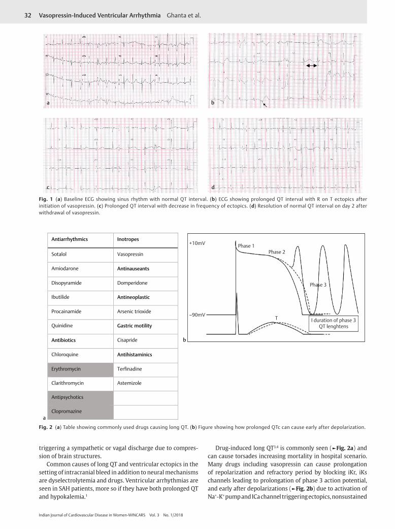
Drug-induced long QT3,4 is commonly seen (►Fig. 2a) and can cause torsades increasing mortality in hospital scenario. Many drugs including vasopressin can cause prolongation of repolarization and refractory period by blocking iKr, iKs channels leading to prolongation of phase 3 action potential, and early after depolarizations (►Fig. 2b) due to activation of Na+-K+ pump and ICa channel triggering ectopics, nonsustained
Antiarrhythmics Inotropes
Sotalol Vasopressin
Amiodarone Antinauseants
Disopyramide Domperidone
Ibutilide Antineoplastic
Procainamide Arsenic trioxide
Quinidine Gastric motility
Antibiotics Cisapride
Chloroquine Antihistaminics
Erythromycin Terfinadine
Clarithromycin Astemizole
Antipsychotics
Clopromazine
I duration of phase 3QT lenghtens
–90mV
+10mV Phase 1Phase 2
Phase 3
T
a
b
Fig. 2 (a) Table showing commonly used drugs causing long QT. (b) Figure showing how prolonged QTc can cause early after depolarization.
a
c
b
d
Fig. 1 (a) Baseline ECG showing sinus rhythm with normal QT interval. (b) ECG showing prolonged QT interval with R on T ectopics after initiation of vasopressin. (c) Prolonged QT interval with decrease in frequency of ectopics. (d) Resolution of normal QT interval on day 2 after withdrawal of vasopressin.

33Vasopressin-Induced Ventricular Arrhythmia Ghanta et al.
Indian Journal of Cardiovascular Disease in Women-WINCARS Vol. 3 No. 1/2018
ventricular tachycardia (VT), and torsades. Some of these patients can be silent carriers of long QT genetic mutations5 who manifest arrhythmia precipitated by drugs or dyselec-trolytemia. Here in our case either long QT or ectopics may not be due to intracranial bleed as initial ECG is normal and abnormality starts after vasopressin infusion,6 and it is mani-festation of drug itself or bradycardia due to baro receptor re-flex by increasing blood pressure causing prolongation of QT and R on T ectopics. Polypharmacy,4 dyselectrolytemia, and bradycardia are the common causes of long QT and ventric-ular tachyarrhythmia in patients with or without silent long QT. Prompt recognition of the event, withdrawal of precipitat-ing factors, and treatment of arrhythmia are required to avoid mortality. Recognizing underlying genetic abnormality and counseling is required for family members.
ConclusionLong QT and ventricular ectopics are commonly seen in patients with intracranial bleed.
Predominantly they occur due to neurogenic mechanisms and dyselectrolytemia. However, drugs that are used
in treatment such as vasopressin can rarely cause QT prolongation precipitating life-threatening ventricular arrhythmias. Prompt recognition and withdrawal of drug are important and lifesaving.
References
1 Di Pasquale G, Pinelli G, Andreoli A, Manini G, Grazi P, Tognetti F. Holter detection of cardiac arrhythmias in intracranial subarach-noid hemorrhage. Am J Cardiol 1987;59(6):596–600
2 Sommargren CE. Electrocardiographic abnormalities in patients with subarachnoid hemorrhage. Am J Crit Care 2002;11(1): 48–56
3 Letsas KP, Efremidis M, Filippatos GS, Sideris AM. Drug-induced long QT syndrome. Hellenic J Cardiol 2007;48(5):296–299
4 Hajiesmaeili M, Afzal G, Sahraei Z. Poly pharmacy-induced long-QT syndrome and torsades de pointes: a case report. J Pharma Care Health Sys 2017;4:174
5 Kallergis EM, Goudis CA, Simantirakis EN, Kochiadakis GE, Vardas PE. Mechanisms, risk factors, and management of acquired long QT syndrome: a comprehensive review. Sci World J 2012;2012(12):212178
6 Yap YG, Camm AJ. Drug induced QT prolongation and torsades de pointes. Heart 2003;89(11):1363–1372