vasculitis and antineutrophil cytoplasmic autoantibodies associated with propylthiouracil therapy
TRANSCRIPT
651
Short report
Vasculitis and antineutrophil cytoplasmicautoantibodies associated with
propylthiouracil therapy
SummaryVasculitis is a rare complication of propylthiouracil therapy.Antineutrophil cytoplasmic antibodies (ANCA) have been
described in association with several vasculitic disorders. We
report detection of ANCA against human neutrophil elastase,proteinase 3, and myeloperoxidase in serum from six patientswho developed evidence of vasculitis during propylthiouraciltreatment of hyperthyroidism. On withdrawal of the drug ANCAconcentrations fell and clinical symptoms resolved completely.
Lancet 1993; 342: 651-52
Autoantibodies against cytoplasmic determinants of
neutrophilic granulocytes (ANCA) are importantdiagnostic markers in systemic vasculitic disorders, such asWegener’s granulomatosis, microscopic polyarteritis,Churg Strauss syndrome, and necrotising and crescenticglomerulonephritis. The target antigens of the most
common ANCA specificities are proteinase 3 (PR3) andmyeloperoxidase (MPO), both located in the azurophilgranules of neutrophils.1 Antibodies against other
constituents of these granules, such as human neutrophilelastase (HNE), have also been detected but are less
common.2-4 Anti-PR3 are associated with Wegener’sgranulomatosis and anti-MPO occur predominantly in
patients with idiopathic necrotising and crescentic
glomerulonephritis, as well as in limited and extra-renalforms of vasculitis. The clinical significance of anti-HNEis unclear, although evidence to date suggests that they arehighly specific for vasculitis.2We describe the specific occurrence of anti-HNE,
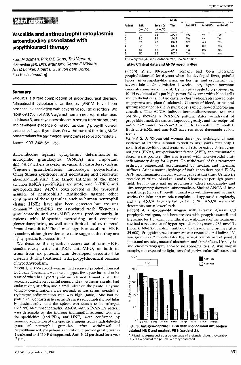
simultaneously with anti-PR3, anti-MPO, or both inserum from six patients who developed vasculitis-likedisorders during treatment with propylthiouracil becauseof hyperthyroidism.Patient 1, a 37-year-old woman, had received prophylthiouracilfor 2 years. Treatment was then stopped for a year but had to beresumed when her hyperthyroidism relapsed. 4 months later thepatient reported fever, painful joints, and a sore throat; she also hadconjunctivitis, scleritis, and a small ulcer on the palate. Thyroidhormone concentrations were normal, as was serum creatinine;erythrocyte sedimentation rate was high (table). She had noprotein, cells, or casts in her urine. A chest radiograph showed hilarlymphadenopathy, and the spleen was shown to be enlarged(12 cm) on ultrasonography. ANCA with a P-ANCA patternwere detectable by the indirect immunofluorescence test andthe specificities (anti-PR3, anti-HNE) were confirmed byimmunoprecipitation of the specific antigens from a radiolabelledlysate of neutrophil granules. After withdrawal of
prophylthiouracil, the patient’s condition improved greatly within4 weeks and anti-HNE disappeared. Anti-PR3 persisted for a year(figure).
ESR=erythrocyte sedimentation rate; Cr=creatinine.
Table: Clinical data and ANCA specificities
Patient 2, an 80-year-old woman, had been receivingprophylthiouracil for 6 years when she developed fever, painfulknees, an erysipelas-like lesion on her leg, and erythema overseveral joints. On admission 4 weeks later, thyroid hormoneconcentrations were normal. Urinalysis revealed no proteinuria,10-15 red blood cells per high-power field, some white blood cellsand epithelial cells, but no casts. A chest radiograph showed onlyemphysema and pleural calcinosis. Cultures of blood, urine, andsputum remained sterile. A skin biopsy sample showed necrotisingvasculitis. The ANCA indirect immunofluorescence test was
positive, showing a P-ANCA pattern. After withdrawal of
propylthiouracil, the patient improved greatly, and the reciprocalindirect immunofluorescence titre fell to 128 within 12 months.Both anti-HNE and anti-PR3 have remained detectable at lowlevels.
Patient 3, A 32-year-old woman developed arthralgia withoutevidence of arthritis in small as well as large joints after only 1
month of propylthiouracil treatment. Tests for extractable nuclearantigens (ENA), anti-perinuclear factor (APF), and rheumatoidfactor were positive. She was treated with non-steroidal anti-inflammatory drugs for 2 years. On withdrawal of this treatmentarthralgia reappeared, accompanied by myalgia and morningstiffness. After a month, hydrops of both knees developed. ENA,APF, and rheumatoid factor were negative at this time. Urinalysisrevealed 15-50 red blood cells and 0-5 leucocytes per high-powerfield, but no casts and no proteinuria. Chest radiography andultrasonography showed no abnormalities. She had ANCA of threespecificities (table). Propylthiouracil was withdrawn and within 6weeks, the joint and muscle complaints disappeared completely,and the ANCA titre started to fall (128). ANCA were still
detectable, but at lower levels.Patient 4, a 45-year-old woman with Graves’ disease and
porphyria variegata, had been treated with propylthiouracil andthyroxine for 1 ’5 years. 8 months after withdrawal of the treatmentshe had a recurrence of hyperthyroidism (thyroxine 285 nmol/L[normal 60-145 nmol/L], antibody to thyroid microsomes titre25 600). Propylthiouracil treatment was restarted, and iodine-131was given too. 2 months later the patient complained of painfuljoints and muscles, mucosal ulceration, and skin defects. Urinalysisand chest radiography showed no abnormalities. A skin biopsysample, not exposed to light, revealed perivascular infiltrates and
Figure: Antigen-capture ELISA with monoclonal antibodiesagainst HNE and against PR3 (patient 1).Antibodies expressed as a percentage of a standard positive control.0-20% =normal range. PTU=propyith!Ourac!!.

652
fibrosis. ANCA antibodies (anti-HNE and anti-MPO) were found.Joint and muscle symptoms had disappeared within 3 weeks ofwithdrawal of propylthiouracil. 2 months later ANCA were stilldetectable, but their titres were lower (256).Patient 5, a 37-year-old woman had a relapse of Graves’ disease 15years after a subtotal thyroidectomy. She was treated with
propylthiouracil and thyroxine. Pain in joints and muscles,morning stiffness, and discrete swelling of the wrists developedwithin a few weeks and persisted for a year. Ultrasonographyrevealed splenomegaly (18 2 cm). Her white blood cell count was3 1 X 10⁹/L and haemoglobin 67 mmol/L. Urine contained 0-5 redblood cells and 5-15 white blood cells per high-power field but noprotein. A chest radiograph showed no abnormalities. Antibodiesto thyroid microsomes were detected (titre 256) and circulatingimmune-complexes were slightly positive. A biopsy sample fromthe nasal mucosa revealed vasculitis, suggesting Wegner’sgranulomatosis, but no granuloma was found. The ANCA titre was2048, and three specificities were found. Withdrawal of
propylthiouracil and thyroxine resolved all symptoms.Patient 6, a 49-year-old woman, with a history of chronic
leucopenia, was admitted to hospital with general malaise,relapsing fever, and weight loss. She had been receivingprophythiouracil for 6 years. Her white blood cell count was2-1 x 10⁹/L and haemoglobin 66 mmol/L. Urinalysis revealedmicroscopic haematuria. The chest radiograph was normal butcomputed tomography of the abdomen showed the spleen wasenlarged. ANCA with a P-ANCA pattern were detected.
Propylthiouracil was withdrawn and the patient was subsequentlytreated with 1311. 5 months later her condition remained
unchanged. We suspected Wegener’s granulomatosis, so initiatedtreatment with prednisone (50 mg daily) and cyclophosphamide(50 mg daily). Clinical symptoms resolved completely.Vasculitis is an uncommon side-effect of propylthiouraciltreatment. 5-8 Although it usually arises within weeks ofinitiation of therapy, it may also develop after many monthsor even years.7 Skin lesions are often of the purpuric type,and necrotic ulceration may occur. The pathogenesis ofpropylthiouracil-induced vasculitis is unknown. Griswoldet all’ suggested that circulating immune complexes mightplay a part, because they detected immunoglobulin andcomplement in the glomeruli and in the walls of dermalvessels by immunofluorescence. The detection of ANCA inassociation with this type of vasculitis suggests other
pathogenetic mechanisms. The six patients described heredeveloped vasculitis-like disorders during propylthiouraciltreatment, associated with the presence of anti-HNE,together with anti-PR3, anti-MPO, or both. We testedseven other patients who received propylthiouracil forhyperthyroidism but did not develop vasculitic
complications; no anti-HNE, anti-PR3, or anti-MPO weredetectable. Moreover, clinical improvement in all patientsand the disappearance or concomitant fall in antibody titreson withdrawal of propylthiouracil implicate the drug in theproduction of these autoantibodies.The fall in anti-HNE (and to a lesser extent anti-PR3 and
anti-MPO) ANCA titres coinciding with clinical
improvement is similar to that observed in other ANCA-positive types of vasculitis. The different rates of
disappearance of anti-HNE suggest that these antibodiesare directed against different antigens rather than againstepitopes shared by HNE and PR3. Binding to PR3 is notinhibited by HNE or vice versa (data not shown). Thedifferences in disappearance rates also suggest differentregulation of the different ANCA specificities. ANCA havebeen implicated in the development of tissue damage in"primary" vasculitis and glomerulonephritis. Possiblemechanisms include activation of primed neutrophils todegranulate and to produce oxygen radicals,9 andinterference by ANCA with control of enzyme activity of
these target antigens, as we have described for anti-PR3 inserum from patients with Wegener’s granulomatosis.10 Wehave tested the anti-HNE and anti-PR3 activity bymeasuring complex formation with a,-antitrypsin, the
major physiological inhibitor of both enzymes. The
strongest inhibition was detected in serum of patient 6, whohad suspected incipient Wegener’s granulomatosis.Complex formation of the relevant antigen with al-antitrypsin was inhibited more strongly by anti-PR3 thanby anti-HNE. Clinical improvement was alwaysaccompanied by a decreasing effect of anti-HNE andanti-PR3 complex formation. These findings suggest thepossibility of a similar mechanism by which anti-HNE andanti-PR3 ANCA might be involved in the pathogenesis ofpropylthiouracil-related and other ANCA-positive formsof vasculitis. However, the true pathogenetic significance ofanti-HNE, as well as of anti-PR3 and anti-MPO, in thesedisorders remains to be established.
We thank Dr W M Wiersinga for control serum samples. This study wassupported by grant no C 88-733 of the Dutch Kidney Foundation.
References
1 Kallenberg CGM, Cohen Tervaert JW, van der Woude FJ,Goldschmeding R, von dem Borne AEGKr, Weening JJ.Autoimmunity to lysosomal enzymes: new clues to vasculitis andglomerulonephritis. Immunol Today 1991; 12: 61-64.
2 Cohen Tervaert JW, Huitema MG, Dolman KM, Goldschmeding R,The TH, Kallenberg CGM. The clinical significance of autoantibodiesto human leucocyte elastase. Cohen Tervaert JW, PhD Thesis, 1990:University of Groningen, Netherlands.
3 Nässberger L, Sjöholm AG, Jonsson H, Sturfelt G, Åkesson A.Autoantibodies against neutrophil cytoplasm components in systemiclupus erythematosus and in hydralazine-induced lupus. Clin ExpImmunol 1990; 81: 380-83.
4 Gallicchio MC, Savige JA. Detecton of anti-myeloperoxidase andanti-elastase antibodies in vasculitides and infections. Clin ExpImmunol 1991; 84: 232-37.
5 Vasily DB, Tyler WB. Propylthiouracil-induced cutaneous vasculitis.JAMA 1980; 243: 458-61.
6 Cooper DS. Antithyroid drugs. N Engl J Med 1984; 311: 1353-62.7 Carrasco MD, Riera C, Clotet B, Grifol M, Foz M. Cutaneous
vasculitis associated with prophylthiouracil therapy. Arch Intern Med1987; 147: 1677.
8 Griswold WR, Mendoza SA, Johnston W, Nichols S. Vasculitisassociated with propylthiouracil: evidence for immune-complexpathogenesis and response to therapy. West J Med 1978; 128: 543-46.
9 Falk RJ, Terrell RS, Charles LA, Jennette JC. Anti-neutrophilcytoplasmic autoantibodies induce neutrophils to degranulate in vitro.Proc Natl Acad Sci USA 1989; 87: 4115-19.
10 Van de Wiel BA, Dolman KM, van der Meer-Gerritsen CE, Hack CE,von dem Borne AEGKr, Goldschmeding R. Interference of Wegener’sgranulomatosis autoantibodies with neutrophil proteinase 3 activity.Clin Exp Immunol 1992; 90: 409-14.
Central Laboratory of the Netherlands Red Cross Blood TransfusionService, and Laboratory for Experimental and Clinical Immunology,University of Amsterdam (K M Dolman PhD, Prof A E G Kr von demBorne MD, R Goldschmeding MD); Department of Nephrology, FreeUniversity Hospital, Amsterdam (R D B Gans MD, Prof AJ M DonkerMD); Department of Internal Medicine, Sint Lucas HospitalAmsterdam (Th J Vervaat MD); Department of Internal Medicine,Westfies Gastluis, Hoom (G Zevenbergen MD); Department ofInternal Medicine, Gool-Noord Hospital, Blaricum (D Maingay MD);Department of Internal Medicine, De Wever Hospital, Hearten (R ENikkels MD); Departments of Hematology (Prof A E G Kr von demBorne MD) and Pathology (R G), Academic Medical Centre,Amsterdam, Netherlands
Correspondence to: Prof A E G Kr von dem Borne, PublicationSecretariat, Central Laboratory of the Netherlands Red Cross BloodTransfusion Service, PO Box 9406, 1006 AK Amsterdam,Netherlands