vascular access in resuscitation -...
TRANSCRIPT
Anesthesiology, V 120 • No 4 1015 April 2014
E STABLISHING vascular access is a critical compo-nent of resuscitation during a cardiac arrest. The 2010
American Heart Association (AHA) Guidelines for Car-diopulmonary Resuscitation (CPR) and Emergency Car-diovascular Care emphasize drug administration during cardiac arrest is of secondary importance to high-quality CPR and that interruptions in CPR should be minimized while obtaining intravenous access.1 Interruptions in CPR decrease coronary perfusion pressure which requires a “re-building” period when chest compressions are resumed.2 Vascular access, however, is still a critical component of resuscitation. A randomized, double-blinded study examin-ing prehospital ventricular fibrillation (VF) demonstrated decreased survival rates from arrest to hospital admission when amiodarone administration was delayed.3 In addi-tion, a swine model has shown that the time from arrest to drug administration is an independent predictor of return of spontaneous circulation.4 Given these data, it is clear that the benefits of early vascular access must be considered in conjunction with the importance of uninterrupted CPR. Intraosseous access can be obtained quickly with minimal or no disruption of CPR. As a result, the AHA has proposed providers establish intraosseous access if an intravenous line is not easily obtainable.1 Similarly, the International Liaison Committee on Resuscitation, as well as the European Resus-citation Counsel, both advocate intraosseous over central venous or endotracheal drug administration if intravenous
access cannot be achieved quickly in an emergency.5 This review aims to examine the literature regarding intraosseous vascular access in the setting of resuscitation, as well as to provide a framework for incorporating the technique into the practice of clinical anesthesia.
Materials and MethodsTo answer the question posed, a systematic review was con-ducted using the PubMed and Ovid Medline databases through August 1, 2013. The primary aim was to determine whether there is a role for intraosseous vascular access in the resuscitation of critically ill patients. Secondary aims were to investigate the evidence regarding clinical use, drug admin-istration, and complications of intraosseous access. The key MeSH terms included: “Infusions, Intraosseous”; “Anesthe-siology”; “Critical Care”; “Tibia”; “Advanced Cardiac Life Support”; “American Heart Association”; “Cardiopulmonary Resuscitation”; “Emergency Medical Services”; “Resuscitation/Methods”; “Infusions, Intravenous”; “Catheterization, Cen-tral Venous”; “Femoral Vein”; “Sternum”; “Out-of-Hospital Cardiac Arrest”; “Blood Transfusion, Autologous”; “Colloids”; “Hetastarch/Administration and Dosage”; “Bone Marrow/Blood supply”; “Compartment Syndromes”; and “Embolism, Fat.” The search was expanded by assessing the reference lists for all retrieved literature. Individual studies were assessed for risk of bias or commercial influence. Only English-language full-text articles published in peer-reviewed journals were
Copyright © 2014, the American Society of Anesthesiologists, Inc. Lippincott Williams & Wilkins. Anesthesiology 2014; 120:1015–31
ABSTRACT
Intraosseous vascular access is a time-tested procedure which has been incorporated into the 2010 American Heart Associa-tion Guidelines for Cardiopulmonary Resuscitation. Intravenous access is often difficult to achieve in shock patients, and central line placement can be time consuming. Intraosseous vascular access, however, can be achieved quickly with minimal disruption of chest compressions. Newer insertion devices are easy to use, making the intraosseous route an attractive alterna-tive for venous access during a resuscitation event. It is critical that anesthesiologists, who are often at the forefront of patient resuscitation, understand how to properly use this potentially life-saving procedure. (Anesthesiology 2014; 120:1015-31)
This article is featured in “This Month in Anesthesiology,” page 1A. Supplemental Digital Content is available for this article. Direct URL citations appear in the printed text and are available in both the HTML and PDF versions of this article. Links to the digital files are provided in the HTML text of this article on the Journal’s Web site (www.anesthesiology.org).
Submitted for publication May 10, 2013. Accepted for publication January 3, 2014. From the Department of Anesthesiology, Penn State Milton S. Hershey Medical Center, Penn State College of Medicine, Hershey, Pennsylvania.
David S. Warner, M.D., Editor
Vascular Access in Resuscitation
Is There a Role for the Intraosseous Route?
Jonathan A. Anson, M.D.
ReView ARTiCle
Anesthesiology 2014; 120:1015-31 1016 Jonathan A. Anson
A Review of Intraosseous Access in Resuscitation
considered. The following inclusion criteria were applied to all studies involved in the clinical use analysis: (1) prospective studies (level of evidence III or higher); (2) focus on insertion success and/or insertion speed; and (3) reporting of complica-tions. Studies were assigned a level of evidence based on Sack-ett criteria (table 1).6 One study was excluded over concerns for commercial bias (investigators received free needles and included a manufacturer in the acknowledgments).7 Another study was excluded as the authors had previously published the same data (original study was used).8 On the basis of these criteria, a total of 18 studies were included.
HistoryThe principles of intraosseous access were first popularized in 1922 by Cecil K. Drinker, M.D. (1887–1956; Professor, Department of Physiology, Harvard Medical School, Boston, Massachusetts), an anatomist who studied hematopoiesis. He postulated that the capillaries of the marrow cavity could be used as an entry point to systemic circulation.9 This idea was revisited in the 1940s by Leandro M. Tocantins, M.D. (1901–1963; Professor, Department of Medicine, Jefferson Medical College, Philadelphia, Pennsylvania), who conducted a series of experiments on rabbits for examining intraosseous infusions. He first modeled a hemorrhagic state by aspirating blood from rabbits. The next day, fresh blood was transfused via an intraosseous line. Six of seven rabbits had a return to baseline hemoglobin level (one died from complications of the original phlebotomy).10 Next, he corrected insulin-induced hypoglycemic seizures in rabbits with intraosseous dextrose infusions.10 In addition, he demonstrated that Congo Red dye injected into the marrow cavity of the tibia reached the heart within 10 s.10 Tocantins also reported a case series of success-ful intraosseous infusions of blood and saline in nine pediatric patients with “impossible” intravenous access.11
The field of anesthesiology first crossed paths with the con-cept of intraosseous infusions thanks to the work by Emanuel Papper, M.D., Ph.D. (1915–2002; Professor, Department of Anesthesiology, Columbia-Presbyterian Medical Center, New York, New York). In a study published in ANESTHESIOLOGY
in 1942, he demonstrated that the circulation time for fluids administered via intravenous and sternal intraosseous routes was nearly identical.12 In a series of seven patients, Papper injected 2% sodium cyanide via the antecubital vein as well as the sternal intraosseous route and measured the cyanide circulation time to the throat, abdomen, and perineum. Ster-nal intraosseous injections had an average time to endpoint of 11.4 s, whereas venous injections had an average time of 15.5 s.12 Papper12 also described the administration of sodium pentothal in a surgical patient and concluded that it is “pos-sible to administer anesthetic drugs ordinarily given by vein into the sternal marrow with the production of anesthesia in therapeutic doses and toxic manifestations in overdose.”
World War II provided an opportunity for wide-spread appli-cation of the intraosseous technique. Hamilton Bailey, F.R.C.S., F.A.C.S. (1894–1961; Emeritus Surgeon, Royal Northern Hospital, London, United Kingdom), noted that the sternal intraosseous route could be effectively used even in black-out conditions.13 He developed a special trocar to prevent the needle from penetrating the back wall of the sternum and injuring the heart. As a result, sternal intraosseous needles were included in emergency medical supply kits during World War II.14 As mili-tary medical personal returned home after the war, the practice of intraosseous infusion was largely forgotten. This can be explained by both the development of better plastic intravenous catheters and the absence of formal paramedics groups. The concept of intraosseous access was “re-discovered” in the early 1980s by James P. Orlowski, M.D. (Department of Pediatrics, Cleveland Clinic Foundation, Cleveland, Ohio). Orlowski, a pediatrician, visited India during a cholera epidemic and observed intraosse-ous infusions saving the lives of severely dehydrated children. He subsequently published an editorial entitled “My kingdom for an intravenous line” which helped lead to the incorporation of intraosseous access into Pediatric Advanced Life Support.15
Anatomy and PhysiologyPeripheral veins can collapse in a state of hemorrhage or dehy-dration. The intraosseous space, however, is a noncollapsible entry point into the systemic circulation. A vast central sinus, composed of distensible endothelium, runs in the middle of the diaphysis. This sinus can distend to accommodate a five-fold increase in volume.16 Blood vessels of the intraosseous space are connected to the systemic circulation by a series of longitudinal Haversian canals containing a small artery and vein. The Haversian canals are linked to a system of Volkmann canals which penetrate the cortex and terminate in connec-tions with the osseous venous drainage. The proximal tibia, a common site of intraosseous insertion, ultimately drains to the popliteal vein. The distal tibia drains to the saphenous vein, whereas the proximal humerus connects with the axillary vein.
The mean blood pressure in the medullary space is approximately 20 to 30 mmHg, or approximately one third of systemic mean pressure.17 Therefore, fluid administra-tion often requires a pressure bag to achieve optimal flow rates. Depending on the infusion characteristics and clinical
Table 1. Levels of Evidence
Level Type of EvidenceGrade of
Recommendation
I Large randomized trials with clear results
A
II Small randomized trials with uncer-tain results
B
III Nonrandomized cohort/case controls
C
IV Nonrandomized historical controls C
V Case series (no controls) C
Levels of evidence assigned to studies as adapted, with permission, from Sackett. Chest 1989; 95(2 suppl):2S–4.6 Adaptations are themselves works protected by copyright. So in order to publish this adaptation, authoriza-tion must be obtained both from the owner of the copyright in the original work and from the owner of copyright in the translation or adaptation.

Anesthesiology 2014; 120:1015-31 1017 Jonathan A. Anson
EDUCATION
scenario, it will likely be necessary to augment the infusion rates of intraosseous lines with a pressure bag or rapid infusion device. Flow rates can also be influenced by hypoxia, hypercarbia, and acidosis (often present during resuscitation) leading to local vasodilation and increased intraosseous blood flow.
Opportunities of the intraosseous Route, insertion, and DevicesInsertion SpeedThe 2010 AHA guidelines state that “it is reasonable for pro-viders to establish intraosseous access if intravenous access is not readily available.”1 This recommendation is in part due to several studies18–22 demonstrating that intraosseous access can be achieved quickly and effectively in a variety of clinical settings. One trial examining 60 dehydrated children (aged from 3 months to 2 yr) found a 5-min success rate of 100% for intraosseous insertion versus just 67% success for periph-eral intravenous catheter placement.18 A prospective study of adult patients (medical and trauma patients in the emer-gency department setting) compared intraosseous cannula-tion with central venous access in patients with “impossible” intravenous access. Intraosseous access was achieved on the first attempt 90% of the time versus just a 60% first-attempt success rate for central line placement.19 In addition, intraos-seous cannulation took significantly less time than central line placement (intraosseous: 2.3 ± 0.8 min vs. central line: 9.9 ± 3.7 min; P < 0.001).19 A prospective simulation study examining intraosseous insertion in the prehospital setting found that access could be established in less than 1 min 84.8% of the time, even in an ambulance traveling 35 mph speed with sudden starting and stopping.20
Establishing peripheral venous access in a prehospital set-ting can be challenging. Prehospital success rates vary from 43 to 91%.18,21,23,24 A retrospective chart review of 641 adult patients with attempted intravenous catheter placement in a moving ambulance found a success rate of just 80%.23 Intraosseous cannulation has been shown to be rapid and effective specifically in the setting of prehospital cardiac arrest. In a randomized trial of 182 patients receiving vas-cular access for nontraumatic cardiac arrest, tibial intraos-seous access was achieved on the first attempt 91% of the time as compared with just a 43% first-attempt success rate for peripheral venous access.21 In another trial, emergency medicine residents treating cardiac arrest in a high-fidelity simulator placed intraosseous lines significantly faster than central lines (intraosseous: 49.0 s vs. central: 194.6 s).22 When considered collectively, these studies indicate that intraosseous access can be achieved faster and with fewer attempts in critical situations.
Obtaining access quickly in cardiac arrest can have a sub-stantial impact on outcome. In a prospective study, 30 swine
in VF were randomized to receive epinephrine (intravenous or intraosseous) or placebo.25 To simulate realistic scenarios of successful vascular access, intraosseous epinephrine was administered 1 min after onset of CPR, whereas intrave-nous epinephrine was administered after an 8-min delay. At equivalent doses, early intraosseous epinephrine admin-istration resulted in a shorter time to return of spontaneous circulation, decreased total defibrillation energy, and better 24-h survival than delayed intravenous epinephrine.25 These results are consistent with another swine model of VF which concluded that early intraosseous epinephrine resulted in decreased time to return of spontaneous circulation, faster termination of VF, and better 20-min survival.26
Historically, resuscitation drugs have been administered via an endotracheal tube in instances where intravenous access cannot be obtained. However, resuscitation drugs administered via the trachea have lower peak plasma con-centrations compared with the peak plasma concentrations of the same drugs given intravenously.27 Therefore, the 2010 AHA guidelines stipulate that endotracheal administration of resuscitation drugs should only be considered if attempts at both intravenous and intraosseous access have failed.1
Infection RiskIt is difficult to directly compare the infectious risk of central venous catheters and intraosseous lines placed during a resus-citation event as there are no head-to-head studies in this setting. In most instances, central lines are left in place for extended periods of time, whereas intraosseous lines gener-ally serve as a short-term means of vascular access. The infec-tion risks of these routes independently have been described. A recent meta-analysis of central venous catheters found no significant difference in the risk of catheter-related blood-stream infections between the femoral and internal jugular sites (risk ratio, 1.35; 95% CI, 0.84–2.19; P = 0.2; I = 0%).28 Despite these findings, the 2013 Joint Commission National Patient Safety Goals state that providers should “NOT insert catheters into the femoral veins unless other sites are unavail-able.”* Central venous access during code situations is often obtained with suboptimal sterile technique and without the use of proper barrier precautions. In a retrospective review of adult trauma patients (emergency department, operating room, and intensive care settings), 25 of 35 (71%) diagnosed central line–associated blood stream infections occurred in patients with known breaches in sterile technique.29
The use of intraosseous access in an emergency setting allows clinicians to avoid placing femoral lines and obviates the potential for improper barrier precautions and less than ideal sterile technique. A meta-analysis examining 30 studies and 4,270 patients concluded that there was a 0.6% incidence rate of osteomyelitis attributed to intraosseous cannulation.30 Most infections occurred during prolonged infusions or in situ-ations of concurrent bacteremia at the time of insertion.30 This dated meta-analysis was conducted before the advent of inser-tion-assist devices and looked only at manual needle insertion.
* The Joint Commission: National Patient Safety Goals effective January 1, 2013. Available at: http://www.jointcommission.org/assets/1/18/NPSG_Chapter_Jan2013_HAP.pdf. Accessed May 1, 2013.
Anesthesiology 2014; 120:1015-31 1018 Jonathan A. Anson
A Review of Intraosseous Access in Resuscitation
A more recent prospective, randomized trial involving the EZ-IO® (Vidacare Corporation, San Antonio, TX) and Bone Injection Gun (BIG®; Waismed, Houston, TX) intraosseous insertion devices in adult patients was conducted in an emer-gency department resuscitation setting. In this study, zero infec-tions were reported in 40 patients receiving intraosseous lines with one of the two devices.31 Similarly, two prospective studies of 60 (adult) and 30 (25 adult and 5 pediatric cardiac arrest patients) intraosseous insertions using the EZ-IO® device, both reported no cases of infection.32,33 Larger studies, including direct comparisons of central and intraosseous lines placed dur-ing resuscitation, represent an area of future research.
Drug DeliveryIntraosseous access is equivalent to intravenous access in terms of functionality and drug delivery. This was demonstrated by Papper12 in 1942 and has subsequently been verified in other studies. Orlowski used a canine model to examine peak effect and serum concentrations of commonly used emergency drugs. He demonstrated equivalency of the intraosseous route to peripheral and central venous drug administration for epi-nephrine, sodium bicarbonate, calcium chloride, hydroxy-ethyl starch, and normal saline.34 A prospective, randomized, crossover pharmacokinetic study was conducted to compare the bioequivalence of morphine administered by intraosseous and intravenous routes in adult patients with cancer (nonre-suscitation setting). Each patient had both an intravenous and intraosseous line and was randomized to receive 5 mg of morphine via one route, followed by 5 mg of morphine by the other route 24 h later. No statistically significant differences were observed between the intravenous and intraosseous routes in calculated pharmacokinetic data including peak concentra-tion and time to peak concentration.35 Another study used a swine model of VF to demonstrate intraosseous epinephrine administered during CPR is rapidly transported to the central circulation and results in a dose-dependent increase in mean arterial blood pressure.36 More recently, the pharmacokinet-ics of intraosseous drug delivery has been compared with central venous drug delivery. A “double dye tracer technique” was used in a swine cardiac arrest model to compare simulta-neous epinephrine injections in the sternum and tibia. Peak plasma concentrations were achieved faster with the sternal route than the tibia route (sternal: 53 ± 11 s vs. tibia: 107 ± 27 s; P = 0.03).37 The time to peak blood concentration was simi-lar for both routes (sternal: 97 ± 17 s vs. central: 70 ± 12 s; P = 0.17).37 The authors concluded that intraosseous adminis-tration of medications through both the sternum and tibia are effective during CPR in anesthetized swine, but the sternal route results in faster uptake.37 As per the 2010 AHA guidelines, all Advanced Cardiac Life Support medications are administered at the same doses regardless of route.1 A summary of available clinical data showing intraosseous medication dosing in adult and pediatric patients is presented in table 2.1,11,30,34,35,38–59
Volume ResuscitationBoth the intraosseous and intravenous routes also offer equiv-alent delivery of resuscitative fluid. The safety and efficacy of intraosseous packed erythrocyte transfusion are well docu-mented. A prospective study (swine model) demonstrated that radiolabeled erythrocytes administered via the intraosse-ous route were rapidly delivered to systemic circulation (30 s to 1 min).60 The safety of intraosseous blood transfusion was shown in a randomized, controlled, blinded swine study. Phle-botomized animals received a transfusion by either an intrave-nous catheter or an intraosseous line. In both groups, blood pressure returned to baseline values within 15 min, and labo-ratory studies assessing for disseminated intravascular coagula-tion were negative.61 In addition, there was no evidence of fat embolism or inflammation on pathologic examination of the lungs or kidneys in the intraosseous group.61 Crystalloids and colloids have also been effectively administered through the intraosseous route. An analysis of hydroxyethyl starch phar-macokinetics demonstrated no significant difference between intravenous and intraosseous administration in hypovolemic swine.62 Crystalloid infusion via the intraosseous route has been demonstrated to be as effective as the central or periph-eral route in treating hemorrhagic shock in a swine model.63
Diagnostic StudiesThe intraosseous medullary space can also serve as a source of blood for laboratory analysis. The initial aspirate after intraosseous line placement can be used for routine labora-tory tests after wasting 2 ml of the marrow/blood mixture.64 In a study involving human volunteers, blood samples were drawn simultaneously from both a peripheral vein and the intraosseous space. Analysis revealed a significant correlation between venous and intraosseous samples for hemoglobin, hematocrit, glucose, blood urea nitrogen, and creatinine. Carbon dioxide and platelet measurements may be lower in intraosseous samples, whereas the leukocyte count may be higher.64 Blood gas measurements from intraosseous blood are “intermediate” between arterial and venous blood gases, suggesting intraosseous samples correspond with arterialized capillary blood samples.65 Intraosseous blood samples can also be used to obtain a reliable type and cross. A prospec-tive study comparing simultaneous intraosseous and venous blood draws in humans found no difference in the accuracy of ABO and Rh typing.66 Laboratory values from an intraos-seous line may not be accurate after a sustained infusion.52 Given these data, it is evident that blood samples drawn immediately after intraosseous cannulation can provide accu-rate laboratory and blood bank data to aid in resuscitation.
Cost EffectivenessA multicenter, observational study compared the costs of central venous catheter insertion with the cost of intraos-seous insertion in unstable patients presenting to the emergency department. A total of 105 patients received intraosseous access (85% were “medical” patients and 53%
Anesthesiology 2014; 120:1015-31 1019 Jonathan A. Anson
EDUCATION
presented with cardiac or respiratory arrest), and costs were compared with published central line data. Cost savings of intraosseous placement over of central venous access were found to be $195 per procedure.67 However, this study has limitations that must be considered. It focused only on ini-tial insertion costs in the emergency department and did not address issues such as daily central line maintenance costs or the percentage of patients in the intraosseous group who eventually received central lines during their hospital admis-sion. The percentage of patients who later require central access has not been studied and must be determined before true conclusions of “overall” cost effectiveness can be made.
Insertion SitesEarly pioneers of intraosseous access tended to focus on the sternum as the preferred insertion site. The sternum offers easy accessibility and close proximity to the central venous circulation via the mammary veins. Time to peak blood con-centration of epinephrine injected through a sternal intraos-seous needle is similar to epinephrine injected through a central line (intraosseous: 97 ± 17 s vs. central: 70 ± 12 s) in a swine model of cardiac arrest.37 In the same study, the sternal intraosseous site achieved peak arterial epinephrine concen-trations significantly faster than the tibial intraosseous site (sternum: 53 ± 11 s vs. tibial: 107 ± 27 s; P = 0.3).37 There
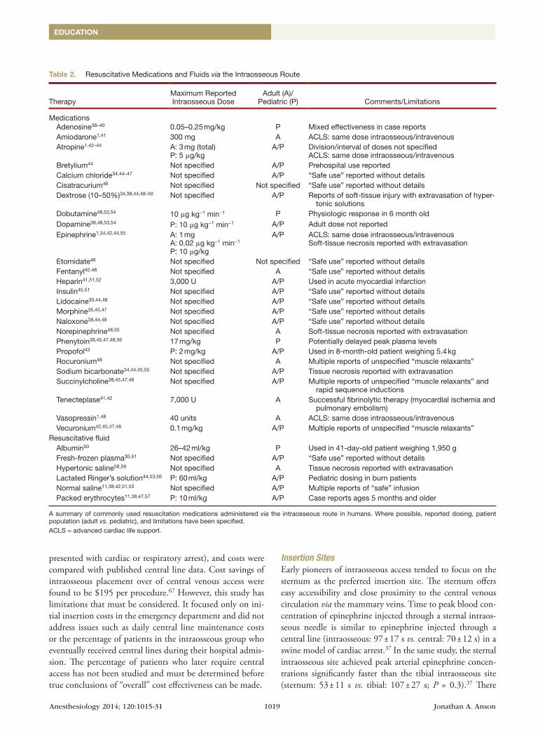
Table 2. Resuscitative Medications and Fluids via the Intraosseous Route
TherapyMaximum Reported Intraosseous Dose
Adult (A)/ Pediatric (P) Comments/Limitations
Medications Adenosine38–40 0.05–0.25 mg/kg P Mixed effectiveness in case reports Amiodarone1,41 300 mg A ACLS: same dose intraosseous/intravenous Atropine1,42–44 A: 3 mg (total)
P: 5 μg/kgA/P Division/interval of doses not specified
ACLS: same dose intraosseous/intravenous Bretylium44 Not specified A/P Prehospital use reported Calcium chloride34,44–47 Not specified A/P “Safe use” reported without details Cisatracurium46 Not specified Not specified “Safe use” reported without details Dextrose (10–50%)34,38,44,48–50 Not specified A/P Reports of soft-tissue injury with extravasation of hyper-
tonic solutions Dobutamine48,53,54 10 μg kg−1 min−1 P Physiologic response in 6 month old Dopamine38,48,53,54 P: 10 μg kg−1 min−1 A/P Adult dose not reported Epinephrine1,34,42,44,55 A: 1 mg
A: 0.02 μg kg−1 min−1
P: 10 μg/kg
A/P ACLS: same dose intraosseous/intravenousSoft-tissue necrosis reported with extravasation
Etomidate46 Not specified Not specified “Safe use” reported without details Fentanyl42,48 Not specified A “Safe use” reported without details Heparin41,51,52 3,000 U A/P Used in acute myocardial infarction Insulin45,51 Not specified A/P “Safe use” reported without details Lidocaine30,44,48 Not specified A/P “Safe use” reported without details Morphine35,45,47 Not specified A/P “Safe use” reported without details Naloxone38,44,48 Not specified A/P “Safe use” reported without details Norepinephrine48,55 Not specified A Soft-tissue necrosis reported with extravasation Phenytoin38,45,47,48,56 17 mg/kg P Potentially delayed peak plasma levels Propofol43 P: 2 mg/kg A/P Used in 8-month-old patient weighing 5.4 kg Rocuronium48 Not specified A Multiple reports of unspecified “muscle relaxants” Sodium bicarbonate34,44,45,55 Not specified A/P Tissue necrosis reported with extravasation Succinylcholine38,45,47,48 Not specified A/P Multiple reports of unspecified “muscle relaxants” and
rapid sequence inductions Tenecteplase41,42 7,000 U A Successful fibrinolytic therapy (myocardial ischemia and
pulmonary embolism) Vasopressin1,48 40 units A ACLS: same dose intraosseous/intravenous Vecuronium42,45,47,48 0.1 mg/kg A/P Multiple reports of unspecified “muscle relaxants”Resuscitative fluid Albumin50 26–42 ml/kg P Used in 41-day-old patient weighing 1,950 g Fresh-frozen plasma30,51 Not specified A/P “Safe use” reported without details Hypertonic saline58,59 Not specified A Tissue necrosis reported with extravasation Lactated Ringer’s solution44,53,56 P: 60 ml/kg A/P Pediatric dosing in burn patients Normal saline11,38,42,51,53 Not specified A/P Multiple reports of “safe” infusion Packed erythrocytes11,38,47,57 P: 10 ml/kg A/P Case reports ages 5 months and older
A summary of commonly used resuscitation medications administered via the intraosseous route in humans. Where possible, reported dosing, patient population (adult vs. pediatric), and limitations have been specified.ACLS = advanced cardiac life support.

Anesthesiology 2014; 120:1015-31 1020 Jonathan A. Anson
A Review of Intraosseous Access in Resuscitation
are, however, several disadvantages of the sternal site. Chest compressions must be briefly interrupted during insertion. It also carries the risk of inadvertently puncturing the heart or great vessels. Pediatric patients are more susceptible to injury from sternal intraosseous insertion due to the proximity to the great vessels and the small size of the marrow cavity (with subsequent poor flow). As a result, the FAST1® (Pyng Medi-cal Corporation, Richmond, British Columbia, Canada) ster-nal insertion device is not approved for patients less than 12 yr of age.
Although the sternal site is of important historical sig-nificance, most providers favor the proximal tibial site. In a survey of emergency-room physicians, 84% selected the proximal tibia as their preferred insertion site. Just 10% of physicians surveyed preferred the humerus and another 10% chose the medial malleolus.68 Although the sample size of this survey was small, it is consistent with newer intraos-seous studies supporting the proximal tibia as a safe, easily accessible site. A prospective study of 182 patients compared proximal tibia and humeral intraosseous insertion sites head-to-head. The proximal tibia group had a higher first-attempt success rate (tibia: 91% vs. humerus: 51%) and faster inser-tion time (tibia: 4.6 min vs. humerus: 7.0 min) than the humeral group.21 In newborns, the needle should be inserted 10-mm distal to the anterior tibial tuberosity and aimed in a slight posterior and inferior direction to avoid damaging the growth plate.69 In children and adults, the needle insertion site is 2 cm below the tibial tuberosity and 1 cm medially on the tibial plateau (fig. 1) (see video, Supplemental Digi-tal Content 1, http://links.lww.com/ALN/B32, which is a guide to intraosseous insertion).48
For adults or skeletally mature adolescents, the proxi-mal humerus is another potential intraosseous site. The patient is positioned with their arm adducted and inter-nally rotated (placing the patient’s hand on their abdo-men facilitates proper positioning). The acromion process is then palpated and the greater tubercle of the humerus is located 2 cm distal to this point (fig. 2) (see video, Supplemental Digital Content 1, http://links.lww.com/ALN/B32, which is a guide to intraosseous insertion). The humeral site has a lower first-attempt success rate com-pared with the tibia and it has a higher rate of needle dis-lodgement.21 This can delay medication administration during cardiac arrest and may lead to more complications from fluid extravasation.21
Although there are practical disadvantages such as nee-dle dislodgement with the humeral intraosseous site, it may offer the benefit of higher flow rates. Flow rates of fluid through EZ-IO® needles placed in the humerus, tibia, and femur of swine were compared in a prospective interven-tional study. The humerus had a statistically significant (P < 0.001) higher flow rate (213 ml/min) com-pared with that of the tibia (103 ml/min) or femur (138 ml/min) when saline was infused via a pres-sure bag.70 Human studies comparing flow rates of
the humerus and tibia offer mixed results. A study of 10 human volunteers demonstrated a significantly higher mean flow rate at the humeral site (humerus: 5,093 ± 2,632 ml/h vs. tibia 1,048 ± 831 ml/h) with a pressurized infusion.71 However, a prospective observa-tional study of 24 critically ill patients (emergency depart-ment setting) comparing humeral and tibial EZ-IO® flow
Fig. 1. Identification of proximal tibia insertion site. Repro-duced, with permission, from Vidacare Corporation, San An-tonio, Texas.
Fig. 2. Identification of proximal humerus insertion site 2 cm distal to the acromion process. Reproduced, with permission, from Vidacare Corporation, San Antonio, Texas.
Anesthesiology 2014; 120:1015-31 1021 Jonathan A. Anson
EDUCATION
rates demonstrated no statistically significant difference between sites (humerus: 153 ml/min vs. tibia: 165 ml/min). Both sites in this study had significantly faster flow rates with a pressurized infusion bag than with gravity drip.72 On the basis of these small swine and human studies, the humeral site may offer higher flow rates than the tibia, but trials with larger sample sizes are needed to make a conclusive determination. For comparison, a prospective study of human volunteers showed a mean infusion rate of 35.6 ml/min via an 18-gauge intravenous catheter (gravity drip).73 Higher intravenous flow rates (18 gauge: 205 ml/min; 16 gauge: 412 ml/min) have been demonstrated using a Rapid Infusion System (Haemonet-ics Corp., Braintree, MA).74
Insertion DevicesManual Needles. Manually inserted intraosseous needles have evolved significantly since the early experiments in the 1920s. Several manufacturers now produce inexpensive needles with specialized handles specifically designed for intraosseous use (fig. 3). Insertion techniques are similar for all of the manual needle types. The needle is oriented per-pendicular to the entry site and pressure is applied in con-junction with a twisting motion until a “loss of resistance” is felt as the needle enters the marrow cavity (see video, Supple-mental Digital Content 1, http://links.lww.com/ALN/B32, which is a guide to intraosseous insertion).
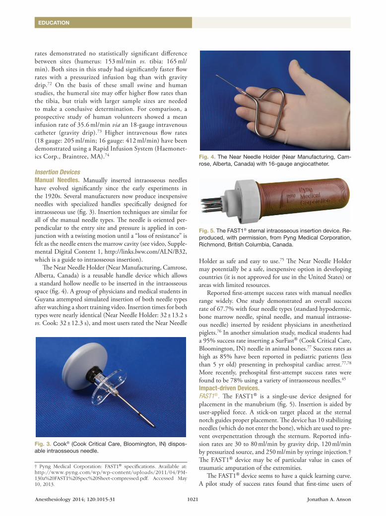
The Near Needle Holder (Near Manufacturing, Camrose, Alberta, Canada) is a reusable handle device which allows a standard hollow needle to be inserted in the intraosseous space (fig. 4). A group of physicians and medical students in Guyana attempted simulated insertion of both needle types after watching a short training video. Insertion times for both types were nearly identical (Near Needle Holder: 32 ± 13.2 s vs. Cook: 32 ± 12.3 s), and most users rated the Near Needle
Holder as safe and easy to use.75 The Near Needle Holder may potentially be a safe, inexpensive option in developing countries (it is not approved for use in the United States) or areas with limited resources.
Reported first-attempt success rates with manual needles range widely. One study demonstrated an overall success rate of 67.7% with four needle types (standard hypodermic, bone marrow needle, spinal needle, and manual intraosse-ous needle) inserted by resident physicians in anesthetized piglets.76 In another simulation study, medical students had a 95% success rate inserting a SurFast® (Cook Critical Care, Bloomington, IN) needle in animal bones.77 Success rates as high as 85% have been reported in pediatric patients (less than 5 yr old) presenting in prehospital cardiac arrest.77,78 More recently, prehospital first-attempt success rates were found to be 78% using a variety of intraosseous needles.45
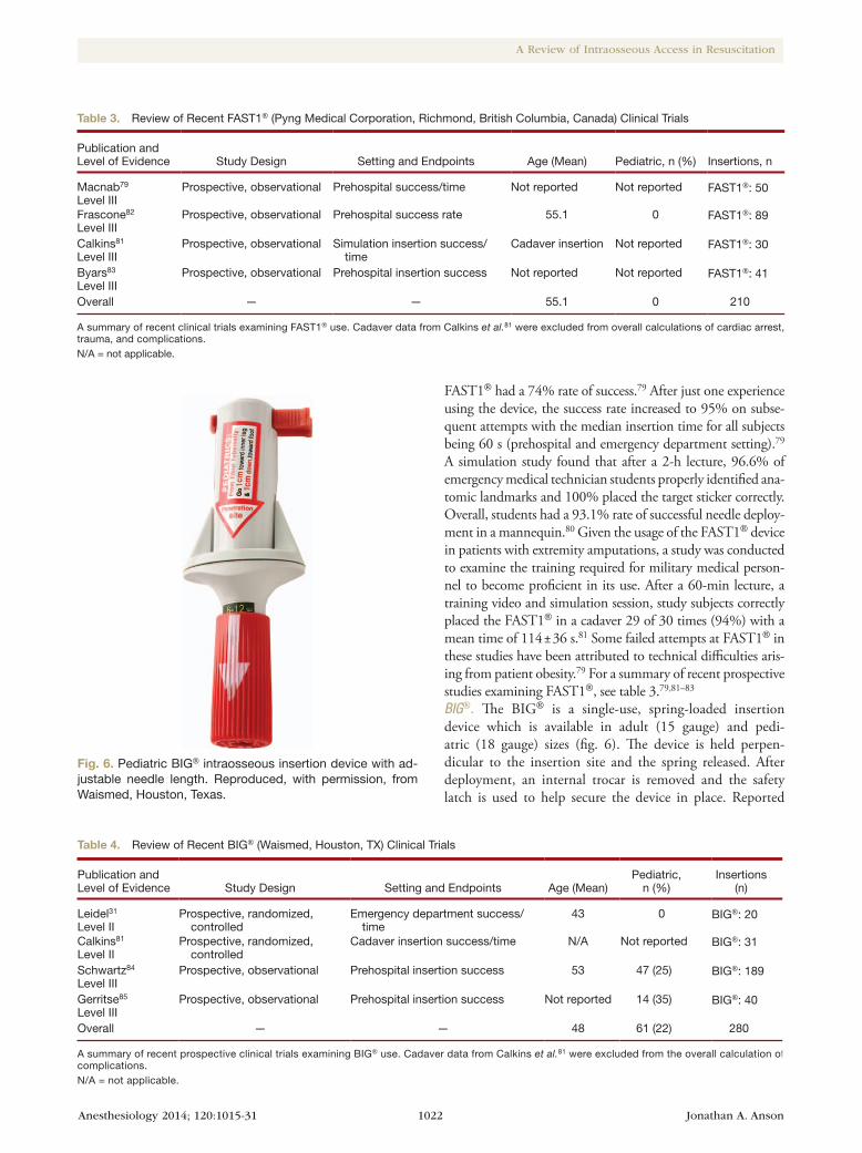
Impact-driven Devices. FAST1®. The FAST1® is a single-use device designed for placement in the manubrium (fig. 5). Insertion is aided by user-applied force. A stick-on target placed at the sternal notch guides proper placement. The device has 10 stabilizing needles (which do not enter the bone), which are used to pre-vent overpenetration through the sternum. Reported infu-sion rates are 30 to 80 ml/min by gravity drip, 120 ml/min by pressurized source, and 250 ml/min by syringe injection.† The FAST1® device may be of particular value in cases of traumatic amputation of the extremities.
The FAST1® device seems to have a quick learning curve. A pilot study of success rates found that first-time users of
Fig. 3. Cook® (Cook Critical Care, Bloomington, IN) dispos-able intraosseous needle.
† Pyng Medical Corporation: FAST1® specifications. Available at: http://www.pyng.com/wp/wp-content/uploads/2011/04/PM-130a%20FAST1%20Spec%20Sheet-compressed.pdf. Accessed May 10, 2013.
Fig. 4. The Near Needle Holder (Near Manufacturing, Cam-rose, Alberta, Canada) with 16-gauge angiocatheter.
Fig. 5. The FAST1® sternal intraosseous insertion device. Re-produced, with permission, from Pyng Medical Corporation, Richmond, British Columbia, Canada.
Anesthesiology 2014; 120:1015-31 1022 Jonathan A. Anson
A Review of Intraosseous Access in Resuscitation
FAST1® had a 74% rate of success.79 After just one experience using the device, the success rate increased to 95% on subse-quent attempts with the median insertion time for all subjects being 60 s (prehospital and emergency department setting).79 A simulation study found that after a 2-h lecture, 96.6% of emergency medical technician students properly identified ana-tomic landmarks and 100% placed the target sticker correctly. Overall, students had a 93.1% rate of successful needle deploy-ment in a mannequin.80 Given the usage of the FAST1® device in patients with extremity amputations, a study was conducted to examine the training required for military medical person-nel to become proficient in its use. After a 60-min lecture, a training video and simulation session, study subjects correctly placed the FAST1® in a cadaver 29 of 30 times (94%) with a mean time of 114 ± 36 s.81 Some failed attempts at FAST1® in these studies have been attributed to technical difficulties aris-ing from patient obesity.79 For a summary of recent prospective studies examining FAST1®, see table 3.79,81–83
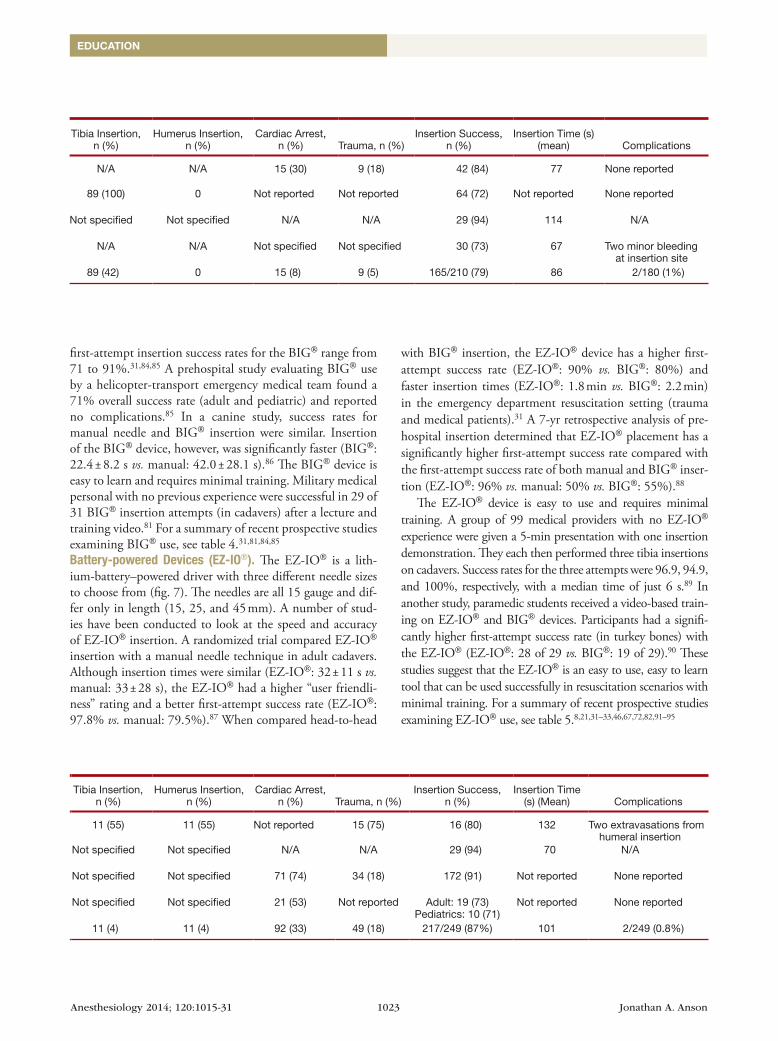
BIG®. The BIG® is a single-use, spring-loaded insertion device which is available in adult (15 gauge) and pedi-atric (18 gauge) sizes (fig. 6). The device is held perpen-dicular to the insertion site and the spring released. After deployment, an internal trocar is removed and the safety latch is used to help secure the device in place. Reported
Table 3. Review of Recent FAST1® (Pyng Medical Corporation, Richmond, British Columbia, Canada) Clinical Trials
Publication and Level of Evidence Study Design Setting and Endpoints Age (Mean) Pediatric, n (%) Insertions, n
Tibia Insertion, n (%)
Humerus Insertion, n (%)
Cardiac Arrest, n (%) Trauma, n (%)
Insertion Success, n (%)
Insertion Time (s) (mean) Complications
Macnab79
Level IIIProspective, observational Prehospital success/time Not reported Not reported FAST1®: 50 N/A N/A 15 (30) 9 (18) 42 (84) 77 None reported
Frascone82
Level IIIProspective, observational Prehospital success rate 55.1 0 FAST1®: 89 89 (100) 0 Not reported Not reported 64 (72) Not reported None reported
Calkins81
Level IIIProspective, observational Simulation insertion success/
timeCadaver insertion Not reported FAST1®: 30 Not specified Not specified N/A N/A 29 (94) 114 N/A
Byars83
Level IIIProspective, observational Prehospital insertion success Not reported Not reported FAST1®: 41 N/A N/A Not specified Not specified 30 (73) 67 Two minor bleeding
at insertion siteOverall — — 55.1 0 210 89 (42) 0 15 (8) 9 (5) 165/210 (79) 86 2/180 (1%)
A summary of recent clinical trials examining FAST1® use. Cadaver data from Calkins et al.81 were excluded from overall calculations of cardiac arrest, trauma, and complications.N/A = not applicable.
Fig. 6. Pediatric BIG® intraosseous insertion device with ad-justable needle length. Reproduced, with permission, from Waismed, Houston, Texas.
Table 4. Review of Recent BIG® (Waismed, Houston, TX) Clinical Trials
Publication and Level of Evidence Study Design Setting and Endpoints Age (Mean)
Pediatric, n (%)
Insertions (n)
Tibia Insertion, n (%)
Humerus Insertion, n (%)
Cardiac Arrest, n (%) Trauma, n (%)
Insertion Success, n (%)
Insertion Time (s) (Mean) Complications
Leidel31
Level IIProspective, randomized,
controlledEmergency department success/
time43 0 BIG®: 20 11 (55) 11 (55) Not reported 15 (75) 16 (80) 132 Two extravasations from
humeral insertionCalkins81
Level IIProspective, randomized,
controlledCadaver insertion success/time N/A Not reported BIG®: 31 Not specified Not specified N/A N/A 29 (94) 70 N/A
Schwartz84
Level IIIProspective, observational Prehospital insertion success 53 47 (25) BIG®: 189 Not specified Not specified 71 (74) 34 (18) 172 (91) Not reported None reported
Gerritse85
Level IIIProspective, observational Prehospital insertion success Not reported 14 (35) BIG®: 40 Not specified Not specified 21 (53) Not reported Adult: 19 (73)
Pediatrics: 10 (71)Not reported None reported
Overall — — 48 61 (22) 280 11 (4) 11 (4) 92 (33) 49 (18) 217/249 (87%) 101 2/249 (0.8%)
A summary of recent prospective clinical trials examining BIG® use. Cadaver data from Calkins et al.81 were excluded from the overall calculation of complications.N/A = not applicable.
Anesthesiology 2014; 120:1015-31 1023 Jonathan A. Anson
EDUCATION
first-attempt insertion success rates for the BIG® range from 71 to 91%.31,84,85 A prehospital study evaluating BIG® use by a helicopter-transport emergency medical team found a 71% overall success rate (adult and pediatric) and reported no complications.85 In a canine study, success rates for manual needle and BIG® insertion were similar. Insertion of the BIG® device, however, was significantly faster (BIG®: 22.4 ± 8.2 s vs. manual: 42.0 ± 28.1 s).86 The BIG® device is easy to learn and requires minimal training. Military medical personal with no previous experience were successful in 29 of 31 BIG® insertion attempts (in cadavers) after a lecture and training video.81 For a summary of recent prospective studies examining BIG® use, see table 4.31,81,84,85
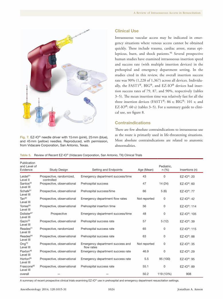
Battery-powered Devices (EZ-IO®). The EZ-IO® is a lith-ium-battery–powered driver with three different needle sizes to choose from (fig. 7). The needles are all 15 gauge and dif-fer only in length (15, 25, and 45 mm). A number of stud-ies have been conducted to look at the speed and accuracy of EZ-IO® insertion. A randomized trial compared EZ-IO® insertion with a manual needle technique in adult cadavers. Although insertion times were similar (EZ-IO®: 32 ± 11 s vs. manual: 33 ± 28 s), the EZ-IO® had a higher “user friendli-ness” rating and a better first-attempt success rate (EZ-IO®: 97.8% vs. manual: 79.5%).87 When compared head-to-head
with BIG® insertion, the EZ-IO® device has a higher first-attempt success rate (EZ-IO®: 90% vs. BIG®: 80%) and faster insertion times (EZ-IO®: 1.8 min vs. BIG®: 2.2 min) in the emergency department resuscitation setting (trauma and medical patients).31 A 7-yr retrospective analysis of pre-hospital insertion determined that EZ-IO® placement has a significantly higher first-attempt success rate compared with the first-attempt success rate of both manual and BIG® inser-tion (EZ-IO®: 96% vs. manual: 50% vs. BIG®: 55%).88
The EZ-IO® device is easy to use and requires minimal training. A group of 99 medical providers with no EZ-IO® experience were given a 5-min presentation with one insertion demonstration. They each then performed three tibia insertions on cadavers. Success rates for the three attempts were 96.9, 94.9, and 100%, respectively, with a median time of just 6 s.89 In another study, paramedic students received a video-based train-ing on EZ-IO® and BIG® devices. Participants had a signifi-cantly higher first-attempt success rate (in turkey bones) with the EZ-IO® (EZ-IO®: 28 of 29 vs. BIG®: 19 of 29).90 These studies suggest that the EZ-IO® is an easy to use, easy to learn tool that can be used successfully in resuscitation scenarios with minimal training. For a summary of recent prospective studies examining EZ-IO® use, see table 5.8,21,31–33,46,67,72,82,91–95
Table 3. Review of Recent FAST1® (Pyng Medical Corporation, Richmond, British Columbia, Canada) Clinical Trials
Publication and Level of Evidence Study Design Setting and Endpoints Age (Mean) Pediatric, n (%) Insertions, n
Tibia Insertion, n (%)
Humerus Insertion, n (%)
Cardiac Arrest, n (%) Trauma, n (%)
Insertion Success, n (%)
Insertion Time (s) (mean) Complications
Macnab79
Level IIIProspective, observational Prehospital success/time Not reported Not reported FAST1®: 50 N/A N/A 15 (30) 9 (18) 42 (84) 77 None reported
Frascone82
Level IIIProspective, observational Prehospital success rate 55.1 0 FAST1®: 89 89 (100) 0 Not reported Not reported 64 (72) Not reported None reported
Calkins81
Level IIIProspective, observational Simulation insertion success/
timeCadaver insertion Not reported FAST1®: 30 Not specified Not specified N/A N/A 29 (94) 114 N/A
Byars83
Level IIIProspective, observational Prehospital insertion success Not reported Not reported FAST1®: 41 N/A N/A Not specified Not specified 30 (73) 67 Two minor bleeding
at insertion siteOverall — — 55.1 0 210 89 (42) 0 15 (8) 9 (5) 165/210 (79) 86 2/180 (1%)
A summary of recent clinical trials examining FAST1® use. Cadaver data from Calkins et al.81 were excluded from overall calculations of cardiac arrest, trauma, and complications.N/A = not applicable.
Table 4. Review of Recent BIG® (Waismed, Houston, TX) Clinical Trials
Publication and Level of Evidence Study Design Setting and Endpoints Age (Mean)
Pediatric, n (%)
Insertions (n)
Tibia Insertion, n (%)
Humerus Insertion, n (%)
Cardiac Arrest, n (%) Trauma, n (%)
Insertion Success, n (%)
Insertion Time (s) (Mean) Complications
Leidel31
Level IIProspective, randomized,
controlledEmergency department success/
time43 0 BIG®: 20 11 (55) 11 (55) Not reported 15 (75) 16 (80) 132 Two extravasations from
humeral insertionCalkins81
Level IIProspective, randomized,
controlledCadaver insertion success/time N/A Not reported BIG®: 31 Not specified Not specified N/A N/A 29 (94) 70 N/A
Schwartz84
Level IIIProspective, observational Prehospital insertion success 53 47 (25) BIG®: 189 Not specified Not specified 71 (74) 34 (18) 172 (91) Not reported None reported
Gerritse85
Level IIIProspective, observational Prehospital insertion success Not reported 14 (35) BIG®: 40 Not specified Not specified 21 (53) Not reported Adult: 19 (73)
Pediatrics: 10 (71)Not reported None reported
Overall — — 48 61 (22) 280 11 (4) 11 (4) 92 (33) 49 (18) 217/249 (87%) 101 2/249 (0.8%)
A summary of recent prospective clinical trials examining BIG® use. Cadaver data from Calkins et al.81 were excluded from the overall calculation of complications.N/A = not applicable.
Anesthesiology 2014; 120:1015-31 1024 Jonathan A. Anson
A Review of Intraosseous Access in Resuscitation
Clinical Use
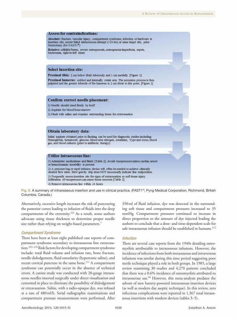
Intraosseous vascular access may be indicated in emer-gency situations where venous access cannot be obtained quickly. These include trauma, cardiac arrest, status epi-lepticus, burn, and shock patients.96 Several prospective human studies have examined intraosseous insertion speed and success rate (with multiple insertion devices) in the prehospital and emergency department setting. In the studies cited in this review, the overall insertion success rate was 90% (1,228 of 1,367) across all devices. Individu-ally, the FAST1®, BIG®, and EZ-IO® devices had inser-tion success rates of 79, 87, and 90%, respectively (tables 3–5). The mean insertion time was relatively fast for all the three insertion devices (FAST1®: 86 s; BIG®: 101 s; and EZ-IO®: 60 s) (tables 3–5). For a summary guide to clini-cal use, see figure 8.
Contraindications
There are few absolute contraindications to intraosseous use as the route is primarily used in life-threatening situations. Most absolute contraindications are related to anatomic abnormalities.
Fig. 7. EZ-IO® needle driver with 15 mm (pink), 25 mm (blue), and 45 mm (yellow) needles. Reproduced, with permission, from Vidacare Corporation, San Antonio, Texas.
Table 5. Review of Recent EZ-IO® (Vidacare Corporation, San Antonio, TX) Clinical Trials
Publication and Level of Evidence Study Design Setting and Endpoints Age (Mean)
Pediatric, n (%) Insertions (n)
Tibia (T), n (%)
Humerus (H), n (%)
Cardiac Arrest, n (%)
Trauma, n (%)
Insertion Success, n (%)
Insertion Time (s) (Mean) Complications
Leidel31
Level IIProspective, randomized,
controlledEmergency department success/time 43 0 EZ-IO®: 20 9 (45) 11 (55) Not reported 14 (70) 18 (90) 108 Two extravasations (humerus)
Santos32
Level IIIProspective, observational Prehospital success 47 14 (24) EZ-IO®: 60 51 (98) 1 (2) 43 (74) 15 (26) 54 (90) Not reported None reported
Schalk91
Level IIIProspective, observational Prehospital success/time 66 5 (6) EZ-IO®: 77 77 (10) 0 41 (53) 15 (19) 75 (97) Not reported None reported
Tan92
Level IIIProspective, observational Emergency department flow rates Not reported 0 EZ-IO®: 42 42 (100) 0 Not reported 21 (50) 39 (93) Not reported None reported
Torres93
Level IIIProspective, observational Prehospital insertion time 56 0 EZ-IO®: 114 85 (75) 12 (11) 64 (67) 29 (27) 114 (100) <30 None reported
Dolister67
Level IIIProspective Emergency department success/time 48 0 EZ-IO®: 105 Not specified Not specified 55 (53) Not reported 99 (94) 103.6 One compartment syndrome
Gazin33
Level IIIProspective, observational Prehospital success rate 57 5 (12) EZ-IO®: 39 Not specified Not specified 30 (76) Not reported First: 33 (84)
Second: 38 (97)Not reported None reported
Reades21
Level IIIProspective, randomized Prehospital success rate 65 0 EZ-IO®: 115 64 (35) 51 (28) 115 (100) None T: 58 (91)
H: 26 (51)Not reported 5 (20%) humerus
dislodgementReades94
Level IIIProspective, observational Prehospital success rate 63 0 EZ-IO®: 88 58 (66) 30 (34) 88 (100) None T: 52 (90)
H: 18 (60)Not reported Six humerus and three tibia
needle dislodgementsOng72
Level IIIProspective, observational Emergency department success and
flow ratesNot reported 0 EZ-IO®: 35 24 (69) 11 (31) Not reported 8 (23) 35 (100) All 35 <20 s None reported
Paxton46
Level IIIProspective, observational Emergency department success rate 46.9 0 EZ-IO®: 29 None 29 (100) 2 (7) 12 (40) 24 (80) 90 None reported
Horton95
Level IIIProspective, observational Emergency department success rate 5.5 95 (100) EZ-IO®: 95 Not reported Not reported Not reported 30 (31) 89 (94) 77% in <10 s One dislodgement
One extravasationFrascone82
Level IIIProspective, observational Prehospital success rate 55.1 0 EZ-IO®: 89 89 (100) 0 Not reported Not reported 78 (87) Not reported None reported
overall — — 50.2 119 (13%) 908 499 (55) 145 (16) 438 (46) 144 (16) 817/908 (90%) 60 19/908 (2.1%)
A summary of recent prospective clinical trials examining EZ-IO® use in prehospital and emergency department resuscitation settings.

Anesthesiology 2014; 120:1015-31 1025 Jonathan A. Anson
EDUCATION
Absolute Contraindications79,84,91,97‡,§
1. Fracture in target bone (risk of fluid extravasation)2. Compartment syndrome in target extremity3. Vascular injury in target extremity4. Acute infection at insertion site5. Previous orthopedic surgery with hardware at inser-
tions site6. Recent failed intraosseous attempt in same extremity
(within 24–48 h)7. Inability to identify landmarks8. History of sternotomy (for FAST1®)9. Sternal thickness less than 6.5 mm (for FAST1®)
Relative Contraindications79,84,97–99
1. Cellulitis or burns of target extremity2. Osseous abnormalities such as osteogenesis imperfect or
severe osteoporosis
3. Right-to-left intracardiac shunts (fat or bone marrow cerebral embolic risk)
4. Sepsis or bacteremia5. Inferior vena cava injury
ComplicationsA total of 1,367 intraosseous insertions were reported in the studies cited in this review (908 EZ-IO®; 249 BIG®; 210 FAST1®). These insertions were associated with 23 reported complications for an overall complication rate of 1.6%. Of these 23 complications, 12 can be considered “minor” (10 needle dislodgements and 2 reports of minor bleeding at site). Excluding these minor complications, the overall complication rate for studies cited in this review was 0.80% (tables 3–5). A recent retrospective cohort study involving 291 pediatric patients with intraosseous lines placed in a variety of settings found zero associated complications.100 The most commonly reported complication is extravasation of fluids. Reported extravasation rates vary widely, ranging from 1 to 22%.101 Risk factors include: incorrect needle placement, multiple punctures in the same bone, and incorrect needle length.101 Osseous punctures can take 12 to 48 h to clot; therefore, subsequent intraosseous placement in the same bone should be avoided during that period. Inadequate needle length can lead to higher rates of dislodgement and extravasation.
Table 5. Review of Recent EZ-IO® (Vidacare Corporation, San Antonio, TX) Clinical Trials
Publication and Level of Evidence Study Design Setting and Endpoints Age (Mean)
Pediatric, n (%) Insertions (n)
Tibia (T), n (%)
Humerus (H), n (%)
Cardiac Arrest, n (%)
Trauma, n (%)
Insertion Success, n (%)
Insertion Time (s) (Mean) Complications
Leidel31
Level IIProspective, randomized,
controlledEmergency department success/time 43 0 EZ-IO®: 20 9 (45) 11 (55) Not reported 14 (70) 18 (90) 108 Two extravasations (humerus)
Santos32
Level IIIProspective, observational Prehospital success 47 14 (24) EZ-IO®: 60 51 (98) 1 (2) 43 (74) 15 (26) 54 (90) Not reported None reported
Schalk91
Level IIIProspective, observational Prehospital success/time 66 5 (6) EZ-IO®: 77 77 (10) 0 41 (53) 15 (19) 75 (97) Not reported None reported
Tan92
Level IIIProspective, observational Emergency department flow rates Not reported 0 EZ-IO®: 42 42 (100) 0 Not reported 21 (50) 39 (93) Not reported None reported
Torres93
Level IIIProspective, observational Prehospital insertion time 56 0 EZ-IO®: 114 85 (75) 12 (11) 64 (67) 29 (27) 114 (100) <30 None reported
Dolister67
Level IIIProspective Emergency department success/time 48 0 EZ-IO®: 105 Not specified Not specified 55 (53) Not reported 99 (94) 103.6 One compartment syndrome
Gazin33
Level IIIProspective, observational Prehospital success rate 57 5 (12) EZ-IO®: 39 Not specified Not specified 30 (76) Not reported First: 33 (84)
Second: 38 (97)Not reported None reported
Reades21
Level IIIProspective, randomized Prehospital success rate 65 0 EZ-IO®: 115 64 (35) 51 (28) 115 (100) None T: 58 (91)
H: 26 (51)Not reported 5 (20%) humerus
dislodgementReades94
Level IIIProspective, observational Prehospital success rate 63 0 EZ-IO®: 88 58 (66) 30 (34) 88 (100) None T: 52 (90)
H: 18 (60)Not reported Six humerus and three tibia
needle dislodgementsOng72
Level IIIProspective, observational Emergency department success and
flow ratesNot reported 0 EZ-IO®: 35 24 (69) 11 (31) Not reported 8 (23) 35 (100) All 35 <20 s None reported
Paxton46
Level IIIProspective, observational Emergency department success rate 46.9 0 EZ-IO®: 29 None 29 (100) 2 (7) 12 (40) 24 (80) 90 None reported
Horton95
Level IIIProspective, observational Emergency department success rate 5.5 95 (100) EZ-IO®: 95 Not reported Not reported Not reported 30 (31) 89 (94) 77% in <10 s One dislodgement
One extravasationFrascone82
Level IIIProspective, observational Prehospital success rate 55.1 0 EZ-IO®: 89 89 (100) 0 Not reported Not reported 78 (87) Not reported None reported
overall — — 50.2 119 (13%) 908 499 (55) 145 (16) 438 (46) 144 (16) 817/908 (90%) 60 19/908 (2.1%)
A summary of recent prospective clinical trials examining EZ-IO® use in prehospital and emergency department resuscitation settings.
‡ Vidacare Corporation: Who needs an IO device? Available at: http://www.vidacare.com/EZ-IO/Clinical-Applications-Who- needs-an-IO-device.aspx. Accessed May 10, 2013.
§ Pyng Medical Corporation: FAST1® Protocol Guide. Available at: http://www.pyng.com/products/fast1/clinical-and-technical-information/protocol/?pi=51. Accessed May 10, 2013.
Anesthesiology 2014; 120:1015-31 1026 Jonathan A. Anson
A Review of Intraosseous Access in Resuscitation
Alternatively, excessive length increases the risk of puncturing the posterior cortex leading to infusion of fluids into the deep compartments of the extremity.102 As a result, some authors advocate using tissue thickness to determine proper needle size rather than relying on weight-based parameters.7
Compartment SyndromeThere have been at least eight published case reports of com-partment syndrome secondary to intraosseous line extravasa-tion.103–110 Risk factors for developing compartment syndrome include: total fluid volume and infusion rate, bone fracture, needle dislodgement, fluid osmolarity (hypertonic saline), and recent cortical puncture in the same bone.111 A compartment syndrome can potentially occur in the absence of technical errors. A canine study was conducted with 20-gauge intraos-seous needles inserted surgically under direct visualization and cemented in place to eliminate the possibility of dislodgement or extravasation. Saline, with a radio-opaque dye, was infused at a rate of 480 ml/h. Serial radiographic examinations and compartment pressure measurements were performed. After
350 ml of fluid infusion, dye was detected in the surround-ing soft tissue and compartment pressures increased to 35 mmHg. Compartment pressures continued to increase in direct proportion to the amount of dye injected leading the authors to conclude that a dose- and time-dependent scale for safe intraosseous infusion should be established in humans.112
InfectionThere are several case reports from the 1940s detailing osteo-myelitis attributable to intraosseous infusions. However, the incidence of infections from both intraosseous and intravenous infusions was similar during this time period suggesting poor sterile technique played a role in both groups. In 1985, a large review examining 30 studies and 4,270 patients concluded that there was a 0.6% incidence of osteomyelitis attributed to intraosseous use.30 However, this meta-analysis predates the advent of new battery-powered intraosseous insertion devices (as well as modern day aseptic technique). In this review, zero infectious complications were reported in 1,367 total intraos-seous insertions with modern devices (tables 3–5).
Fig. 8. A summary of intraosseous insertion and use in clinical practice. (FAST1®; Pyng Medical Corporation, Richmond, British Columbia, Canada.)
Anesthesiology 2014; 120:1015-31 1027 Jonathan A. Anson
EDUCATION
Embolic ComplicationsFat or bone marrow embolism is another potential complica-tion of intraosseous therapy. Even small increases in intraosseous pressure can lead to fat embolism.113 Levels of radioactivity in the lungs were measured after injection of Triolein-131I–labeled fat into the tibia of rabbits. After 2 to 5 h, 44.8% of the injected radioactive substance was present in the lungs on histologic examination.113 More recently, Orlowski et al.98 demonstrated that bone marrow and fat emboli in the lungs (mean, 0.91 emboli per square millimeter lung) were present in 89 to 100% of dogs after 4 h of intraosseous infusion. In addition, they demonstrated an average of 0.23 and 0.71 emboli per square millimeter lung, respectively, in pulmonary autopsy specimens of two children who received intraosseous infusions during resuscitation attempts. The incidence of fat embolism does not seem to be related to the rate of intraosseous infusion.114
The incidence of fat embolism after CPR with concurrent intraosseous infusion has been studied in a piglet model of hypoxic cardiac arrest. There was no statistically significant difference in the quantity of pulmonary emboli between the intraosseous and intravenous resuscitated groups.115 These results correlate with recent human findings. Autopsies con-ducted on 50 decedents showed a pulmonary fat emboli rate of 76% in patients who received CPR without an intraos-seous line.116 These collective data suggest that patients undergoing CPR are at risk for pulmonary fat emboli with or without the presence of an intraosseous infusion.
Interestingly, despite the high percentage of fat and mar-row emboli occurring with intraosseous infusions, there does not seem to be a detrimental clinical correlation. Despite an 89 to 100% incidence of emboli in his experiments, Orlowski et al.98 found no significant alterations in PaO2 and no evidence of intrapulmonary shunting. There is at least a theoretical risk of cerebral emboli if right-to-left intracardiac shunts are present.98 There is one case report of death from fat embolism after intraos-seous phlebography to examine inferior vena cava obstruction in a patient with reticulum-cell sarcoma.117 To date, however, there are no case reports of death or significant morbidity from marrow or fat emboli after resuscitation with an intraosseous infusion. Despite the near-universal occurrence of emboli, intraosseous infusions seem safe to use during resuscitation.
Bone InjuryThere is a theoretical potential for both acute and long-term osseous injury related to intraosseous infusions. Bilateral tibia fractures were reported in a 3-month-old septic patient after unsuccessful intraosseous attempts.118 Iatrogenic fracture has also been documented after aggressive intraosseous placement with “considerable force” during an unsuccessful resuscitation of a 2-yr-old trauma patient.119 Overall, reports of fracture or acute boney injury attributable to intraosseous insertion are rare. Pig models have been used to demonstrate no long-term effects on bone marrow after intraosseous drug administration. Animals received sodium bicarbonate, epinephrine, and dopa-mine in one extremity while another extremity served as the
control. Bone marrow examination revealed normal cellular differentiation in all groups.120 In a similar study, experimental and control legs were harvested 6 months after intraosseous infusion. No differences in bone growth, degree of epiphy-seal closure, or radiographic properties were observed between groups.121 The rate or osmolality of intraosseous infusion does not appear to have an influence on long-term histologic changes of the marrow space in humans.122
Data from more recent human studies support the findings of these pig models. A prospective radiographic analysis of pedi-atric patients with tibial intraosseous infusions placed in emer-gency situations was conducted. After a mean follow-up period of 29.2 months, there was no statistically significant difference (in a variety of radiographic measurements) between the punc-tured and control legs.123 Similarly, a small study (prospective, observer-blinded) found no difference in tibial length 1 yr after intraosseous infusion.124 Given the rarity of iatrogenic fractures attributed to intraosseous cannulation and the lack of evidence showing adverse long-term bone growth effects, the intraosse-ous route seems to be low risk in terms of osseous complications.
Current limitationsThe available intraosseous literature has some limitations that must be considered. Most data come from prehospital or emergency department insertion. In this setting, intraosse-ous access is often used only after intravenous attempts have failed. As such, it is difficult to conduct large, randomized, clinical trials because the patients studied are already “self-selected” as difficult access patients. Therefore, we are left with primarily prospective observational studies. However, the findings of level III evidence were generally consistent in this review, allowing for a higher grade of recommendation.
Anesthesiologists frequently respond to in-hospital cardiac arrest situations, and literature specifically in this setting is scant. Head-to-head in-hospital studies comparing central and intraosseous access in terms of insertion speed and accu-racy are lacking. There are no studies directly comparing the infection risks of the two routes when these lines are inserted during cardiac arrest. Furthermore, there are no studies com-paring mortality data in cardiac arrest patients resuscitated with either central or intraosseous access.
Finally, long-term follow-up studies on the safety of intraosseous infusions are absent, particularly with newer insertion devices. Most of the recent literature tends to focus on speed and success of insertion. Therefore, we are left to rely on a few case reports and animal studies when consider-ing the risk of delayed complications.
ConclusionIntraosseous cannulation is a time-tested procedure that will play a role in the resuscitation of patients in the future. Intra-venous access is often difficult to achieve in shock patients and central line placement can be time consuming. This lit-erature review has demonstrated that intraosseous vascular
Anesthesiology 2014; 120:1015-31 1028 Jonathan A. Anson
A Review of Intraosseous Access in Resuscitation
access can be achieved quickly and accurately in emergency situations. Given the efficiency of insertion combined with a favorable complication profile, there is clearly a role for intraosseous vascular access in the resuscitation of critically ill patients. Therefore, anesthesiologists should become familiar with intraosseous insertion techniques and understand how to properly use this potentially life-saving procedure. In the 1940s, Dr. Papper played an important early role in advanc-ing the field of intraosseous infusions. Today, anesthesiolo-gists have the opportunity to follow Dr. Papper’s footsteps and be at the forefront of the intraosseous resurgence as we adopt this technique in our clinical practice.
AcknowledgmentsSupport was provided solely from institutional and/or de-partmental sources.
Competing InterestsThe author declares no competing interests.
CorrespondenceAddress correspondence to Dr. Anson: Department of An-esthesiology, Penn State Milton S. Hershey Medical Center, Penn State College of Medicine, 500 University Drive, Mail Code H187, P.O. Box 850, Hershey, Pennsylvania 17033-0850. [email protected]. Information on purchasing re-prints may be found at www.anesthesiology.org or on the masthead page at the beginning of this issue. ANESTHESIOLO-
Gy’s articles are made freely accessible to all readers, for per-sonal use only, 6 months from the cover date of the issue.
References 1. Neumar RW, Otto CW, Link MS, Kronick SL, Shuster M, Callaway
CW, Kudenchuk PJ, Ornato JP, McNally B, Silvers SM, Passman RS, White RD, Hess EP, Tang W, Davis D, Sinz E, Morrison LJ: Part 8: Adult advanced cardiovascular life support: 2010 American Heart Association Guidelines for Cardiopulmonary Resuscitation and Emergency Cardiovascular Care. Circulation 2010; 122(18 suppl 3):S729–67
2. Kern KB, Hilwig RW, Berg RA, Sanders AB, Ewy GA: Importance of continuous chest compressions during car-diopulmonary resuscitation: Improved outcome during a simulated single lay-rescuer scenario. Circulation 2002; 105:645–9
3. Kudenchuk PJ, Cobb LA, Copass MK, Cummins RO, Doherty AM, Fahrenbruch CE, Hallstrom AP, Murray WA, Olsufka M, Walsh T: Amiodarone for resuscitation after out-of-hospital cardiac arrest due to ventricular fibrillation. N Engl J Med 1999; 341:871–8
4. Rittenberger JC, Menegazzi JJ, Callaway CW: Association of delay to first intervention with return of spontaneous circulation in a swine model of cardiac arrest. Resuscitation 2007; 73:154–60
5. Nolan JP, Soar J, Zideman DA, Biarent D, Bossaert LL, Deakin C, Koster RW, Wyllie J, Böttiger B; ERC Guidelines Writing Group: European Resuscitation Council Guidelines for Resuscitation 2010 Section 1. Executive summary. Resuscitation 2010; 81:1219–76
6. Sackett DL: Rules of evidence and clinical recommendations on the use of antithrombotic agents. Chest 1989; 95(2 suppl):2S–4
7. Frascone RJ, Jensen J, Wewerka SS, Salzman JG: Use of the pediatric EZ-IO needle by emergency medical services pro-viders. Pediatr Emerg Care 2009; 25:329–32
8. Ngo AS, Oh JJ, Chen Y, Yong D, Ong ME: Intraosseous vascu-lar access in adults using the EZ-IO in an emergency depart-ment. Int J Emerg Med 2009; 2:155–60
9. Drinker CK, Drinker KR, Lund CC: The circulation in the mammalian bone marrow. Am J Physiol 1922; 62:1–92
10. Tocantins L: Rapid absorption of substances injected into the bone marrow. Proc Soc Exp Biol Med 1940; 45:292–6
11. Tocantins L, O’Neill J, Jones H: Infusion of blood and other fluids via the bone marrow: Applilcation in pediatrics. JAMA 1941; 117:1229–34
12. Papper EM: The Bone marrow route for injecting fluids via the bone marrow. ANESTHESIOLOGY 1942; 3:307–13
13. Bailey H: Bone-marrow as a site for the receprtion of infu-sions, trans-fusion and anesthetic agents. Br Med J 1944; 2:181–2
14. Dubick MA, Holcomb JB: A review of intraosseous vascular access: Current status and military application. Mil Med 2000; 165:552–9
15. Orlowski JP: My kingdom for an intravenous line. Am J Dis Child 1984; 138:803
16. Laroche M: Intraosseous circulation from physiology to dis-ease. Joint Bone Spine 2002; 69:262–9
17. Tøndevold E, Eriksen J, Jansen E: Observations on long bone medullary pressure in relation to mean arterial blood pressure in the anaesthetized dog. Acta Orthop Scand 1979; 50:527–31
18. Banerjee S, Singhi SC, Singh S, Singh M: The intraosseous route is a suitable alternative to intravenous route for fluid resuscitation in severely dehydrated children. Indian Pediatr 1994; 31:1511–20
19. Leidel BA, Kirchhoff C, Bogner V, Stegmaier J, Mutschler W, Kanz KG, Braunstein V: Is the intraosseous access route fast and efficacious compared to conventional central venous catheterization in adult patients under resuscitation in the emergency department? A prospective observational pilot study. Patient Saf Surg 2009; 3:24
20. Fuchs S, LaCovey D, Paris P: A prehospital model of intraos-seous infusion. Ann Emerg Med 1991; 20:371–4
21. Reades R, Studnek JR, Vandeventer S, Garrett J: Intraosseous versus intravenous vascular access during out-of-hospital cardiac arrest: A randomized controlled trial. Ann Emerg Med 2011; 58:509–16
22. Reiter DA, Strother CG, Weingart SD: The quality of car-diopulmonary resuscitation using supraglottic airways and intraosseous devices: A simulation trial. Resuscitation 2013; 84:93–7
23. Slovis CM, Herr EW, Londorf D, Little TD, Alexander BR, Guthmann RJ: Success rates for initiation of intravenous therapy en route by prehospital care providers. Am J Emerg Med 1990; 8:305–7
24. Jones SE, Nesper TP, Alcouloumre E: Prehospital intravenous line placement: A prospective study. Ann Emerg Med 1989; 18:244–6
25. Zuercher M, Kern KB, Indik JH, Loedl M, Hilwig RW, Ummenhofer W, Berg RA, Ewy GA: Epinephrine improves 24-hour survival in a swine model of prolonged ventricular fibrillation demonstrating that early intraosseous is superior to delayed intravenous admin-istration. Anesth Analg 2011; 112:884–90
26. Mader TJ, Kellogg AR, Walterscheid JK, Lodding CC, Sherman LD: A randomized comparison of cardiocerebral and cardio-pulmonary resuscitation using a swine model of prolonged ventricular fibrillation. Resuscitation 2010; 81:596–602
27. Niemann JT, Stratton SJ, Cruz B, Lewis RJ: Endotracheal drug administration during out-of-hospital resuscitation: Where are the survivors? Resuscitation 2002; 53:153–7
28. Marik PE, Flemmer M, Harrison W: The risk of catheter-related bloodstream infection with femoral venous catheters as compared to subclavian and internal jugular venous cath-eters: A systematic review of the literature and meta-analysis. Crit Care Med 2012; 40:2479–85
Anesthesiology 2014; 120:1015-31 1029 Jonathan A. Anson
EDUCATION
29. Smith JW, Egger M, Franklin G, Harbrecht B, Richardson JD: Central line-associated blood stream infection in the critically ill trauma patient. Am Surg 2011; 77:1038–42
30. Rosetti VA, Thompson BM, Miller J, Mateer JR, Aprahamian C: Intraosseous infusion: An alternative route of pediatric intra-vascular access. Ann Emerg Med 1985; 14:885–8
31. Leidel BA, Kirchhoff C, Braunstein V, Bogner V, Biberthaler P, Kanz KG: Comparison of two intraosseous access devices in adult patients under resuscitation in the emergency department: A prospective, randomized study. Resuscitation 2010; 81:994–9
32. Santos D, Carron PN, Yersin B, Pasquier M: EZ-IO(®) intraos-seous device implementation in a pre-hospital emergency service: A prospective study and review of the literature. Resuscitation 2013; 84:440–5
33. Gazin N, Auger H, Jabre P, Jaulin C, Lecarpentier E, Bertrand C, Margenet A, Combes X: Efficacy and safety of the EZ-IO™ intraosseous device: Out-of-hospital implementa-tion of a management algorithm for difficult vascular access. Resuscitation 2011; 82:126–9
34. Orlowski JP, Porembka DT, Gallagher JM, Lockrem JD, VanLente F: Comparison study of intraosseous, central intra-venous, and peripheral intravenous infusions of emergency drugs. Am J Dis Child 1990; 144:112–7
35. Von Hoff DD, Kuhn JG, Burris HA III, Miller LJ: Does intraos-seous equal intravenous? A pharmacokinetic study. Am J Emerg Med 2008; 26:31–8
36. Spivey WH, Crespo SG, Fuhs LR, Schoffstall JM: Plasma cat-echolamine levels after intraosseous epinephrine administra-tion in a cardiac arrest model. Ann Emerg Med 1992; 21:127–31
37. Hoskins SL, do Nascimento P Jr, Lima RM, Espana-Tenorio JM, Kramer GC: Pharmacokinetics of intraosseous and cen-tral venous drug delivery during cardiopulmonary resuscita-tion. Resuscitation 2012; 83:107–12
38. Spivey WH: Intraosseous infusions. J Pediatr 1987; 111:639–43 39. Friedman FD: Intraosseous adenosine for the termination of
supraventricular tachycardia in an infant. Ann Emerg Med 1996; 28:356–8
40. Goodman IS, Lu CJ: Intraosseous infusion is unreliable for adenosine delivery in the treatment of supraventricular tachycardia. Pediatr Emerg Care 2012; 28:47–8
41. Ruiz-Hornillos PJ, Martínez-Cámara F, Elizondo M, Jiménez-Fraile JA, Del Mar Alonso-Sánchez M, Galán D, García-Rubira JC, Macaya C, Ibanez B: Systemic fibrinolysis through intraos-seous vascular access in ST-segment elevation myocardial infarction. Ann Emerg Med 2011; 57:572–4
42. Valdés M, Araujo P, de Andrés C, Sastre E, Martin T: Intraosseous administration of thrombolysis in out-of-hospital massive pul-monary thromboembolism. Emerg Med J 2010; 27:641–4
43. Joseph G, Tobias JD: The use of intraosseous infusions in the operating room. J Clin Anesth 2008; 20:469–73
44. Glaeser PW, Hellmich TR, Szewczuga D, Losek JD, Smith DS: Five-year experience in prehospital intraosseous infusions in children and adults. Ann Emerg Med 1993; 22:1119–24
45. Fiorito BA, Mirza F, Doran TM, Oberle AN, Cruz EC, Wendtland CL, Abd-Allah SA: Intraosseous access in the set-ting of pediatric critical care transport. Pediatr Crit Care Med 2005; 6:50–3
46. Paxton JH, Knuth TE, Klausner HA: Proximal humerus intraosseous infusion: A preferred emergency venous access. J Trauma 2009; 67:606–11
47. Guy J, Haley K, Zuspan SJ: Use of intraosseous infusion in the pediatric trauma patient. J Pediatr Surg 1993; 28:158–61
48. Buck ML, Wiggins BS, Sesler JM: Intraosseous drug adminis-tration in children and adults during cardiopulmonary resus-citation. Ann Pharmacother 2007; 41:1679–86
49. Brunette DD, Fischer R: Intravascular access in pediatric car-diac arrest. Am J Emerg Med 1988; 6:577–9
50. Kelsall AW: Resuscitation with intraosseous lines in neonatal units. Arch Dis Child 1993; 68(3 Spec No):324–5
51. Tarrow AB, Turkel H, Thompson MS: Infusions via the bone marrow and biopsy of the bone and bone marrow. ANESTHESIOLOGY 1952; 13:501–9
52. Johnson L, Kissoon N, Fiallos M, Abdelmoneim T, Murphy S: Use of intraosseous blood to assess blood chemistries and hemoglobin during cardiopulmonary resuscitation with drug infusions. Crit Care Med 1999; 27:1147–52
53. Goldstein B, Doody D, Briggs S: Emergency intraosseous infusion in severely burned children. Pediatr Emerg Care 1990; 6:195–7
54. Berg RA: Emergency infusion of catecholamines into bone marrow. Am J Dis Child 1984; 138:810–1
55. Christensen DW, Vernon DD, Banner W Jr, Dean JM: Skin necrosis complicating intraosseous infusion. Pediatr Emerg Care 1991; 7:289–90
56. Walsh-Kelly CM, Berens RJ, Glaeser PW, Losek JD: Intraosseous infusion of phenytoin. Am J Emerg Med 1986; 4:523–4
57. Weiser G, Poppa E, Katz Y, Bahouth H, Shavit I: Intraosseous blood transfusion in infants with traumatic hemorrhagic shock. Am J Emerg Med 2013; 31:640.e3–4
58. Dubick MA, Kramer GC: Hypertonic saline dextran (HSD) and intraosseous vascular access for the treatment of haem-orrhagic hypotension in the far-forward combat arena. Ann Acad Med Singapore 1997; 26:64–9
59. Chávez-Negrete A, Majluf Cruz S, Frati Munari A, Perches A, Argüero R: Treatment of hemorrhagic shock with intraos-seous or intravenous infusion of hypertonic saline dextran solution. Eur Surg Res 1991; 23:123–9
60. Bell MC, Olshaker JS, Brown CK, McNamee GA Jr, Fauver GM: Intraosseous transfusion in an anesthetized swine model using 51Cr-labeled autologous red blood cells. J Trauma 1991; 31:1487–9
61. Plewa MC, King RW, Fenn-Buderer N, Gretzinger K, Renuart D, Cruz R: Hematologic safety of intraosseous blood transfu-sion in a swine model of pediatric hemorrhagic hypovole-mia. Acad Emerg Med 1995; 2:799–809
62. Kentner R, Haas T, Gervais H, Hiller B, Dick W: Pharmacokinetics and pharmacodynamics of hydroxyethyl starch in hypovolemic pigs; a comparison of peripheral and intraosseous infusion. Resuscitation 1999; 40:37–44
63. Neufeld JD, Marx JA, Moore EE, Light AI: Comparison of intraosseous, central, and peripheral routes of crystalloid infusion for resuscitation of hemorrhagic shock in a swine model. J Trauma 1993; 34:422–8
64. Miller LJ, Philbeck TE, Montez D, Spadaccini CJ: A new study of intraosseous blood for laboratory analysis. Arch Pathol Lab Med 2010; 134:1253–60
65. Orlowski JP, Porembka DT, Gallagher JM, Van Lente F: The bone marrow as a source of laboratory studies. Ann Emerg Med 1989; 18:1348–51
66. Brickman KR, Krupp K, Rega P, Alexander J, Guinness M: Typing and screening of blood from intraosseous access. Ann Emerg Med 1992; 21:414–7
67. Dolister M, Miller S, Borron S, Truemper E, Shah M, Lanford MR, Philbeck TE: Intraosseous vascular access is safe, effec-tive and costs less than central venous catheters for patients in the hospital setting. J Vasc Access 2013; 14:216–24
68. Molin R, Hallas P, Brabrand M, Schmidt TA: Current use of intraosseous infusion in Danish emergency departments: A cross-sectional study. Scand J Trauma Resusc Emerg Med 2010; 18:37
69. Boon JM, Gorry DL, Meiring JH: Finding an ideal site for intraosseous infusion of the tibia: An anatomical study. Clin Anat 2003; 16:15–8
70. Lairet J, Bebarta V, Lairet K, Kacprowicz R, Lawler C, Pitotti R, Bush A, King J: A comparison of proximal tibia, distal femur, and proximal humerus infusion rates using the EZ-IO intraosseous device on the adult swine (Sus scrofa) model. Prehosp Emerg Care 2013; 17:280–4
Anesthesiology 2014; 120:1015-31 1030 Jonathan A. Anson
A Review of Intraosseous Access in Resuscitation
71. Miller L, Philbeck T, Montez D, Puga T: 467: A two-phase study of fluid administration measurement during intraosse-ous infusion. Ann Emerg Med 2010; 56:S151
72. Ong ME, Chan YH, Oh JJ, Ngo AS: An observational, prospec-tive study comparing tibial and humeral intraosseous access using the EZ-IO. Am J Emerg Med 2009; 27:8–15
73. Li SF, Cole M, Forest R, Chilstrom M, Reinersman E, Jones MP, Zinzuwadia S, King S, Yadav K: Are 2 smaller intrave-nous catheters as good as 1 larger intravenous catheter? Am J Emerg Med 2010; 28:724–7
74. Barcelona SL, Vilich F, Coté CJ: A comparison of flow rates and warming capabilities of the Level 1 and Rapid Infusion System with various-size intravenous catheters. Anesth Analg 2003; 97:358–63
75. Kalechstein S, Permual A, Cameron BM, Pemberton J, Hollaar G, Duffy D, Cameron BH: Evaluation of a new pediatric intraosseous needle insertion device for low-resource set-tings. J Pediatr Surg 2012; 47:974–9
76. Wagner MB, McCabe JB: A comparison of four techniques to establish intraosseous infusion. Pediatr Emerg Care 1988; 4:87–91
77. Jun H, Haruyama AZ, Chang KS, Yamamoto LG: Comparison of a new screw-tipped intraosseous needle versus a standard bone marrow aspiration needle for infusion. Am J Emerg Med 2000; 18:135–9
78. Miner WF, Corneli HM, Bolte RG, Lehnhof D, Clawson JJ: Prehospital use of intraosseous infusion by paramedics. Pediatr Emerg Care 1989; 5:5–7
79. Macnab A, Christenson J, Findlay J, Horwood B, Johnson D, Jones L, Phillips K, Pollack C Jr, Robinson DJ, Rumball C, Stair T, Tiffany B, Whelan M: A new system for sternal intraosseous infusion in adults. Prehosp Emerg Care 2000; 4:173–7
80. Miller DD, Guimond G, Hostler DP, Platt T, Wang HE: Feasibility of sternal intraosseous access by emergency medi-cal technician students. Prehosp Emerg Care 2005; 9:73–8
81. Calkins MD, Fitzgerald G, Bentley TB, Burris D: Intraosseous infusion devices: A comparison for potential use in special operations. J Trauma 2000; 48:1068–74
82. Frascone RJ, Jensen JP, Kaye K, Salzman JG: Consecutive field trials using two different intraosseous devices. Prehosp Emerg Care 2007; 11:164–71
83. Byars DV, Tsuchitani SN, Erwin E, Anglemyer B, Eastman J: Evaluation of success rate and access time for an adult ster-nal intraosseous device deployed in the prehospital setting. Prehosp Disaster Med 2011; 26:127–9
84. Schwartz D, Amir L, Dichter R, Figenberg Z: The use of a powered device for intraosseous drug and fluid administra-tion in a national EMS: A 4-year experience. J Trauma 2008; 64:650–4; discussion 654–5
85. Gerritse BM, Scheffer GJ, Draaisma JM: Prehospital intraos-seus access with the bone injection gun by a helicopter-trans-ported emergency medical team. J Trauma 2009; 66:1739–41
86. Olsen D, Packer BE, Perrett J, Balentine H, Andrews GA: Evaluation of the bone injection gun as a method for intraos-seous cannula placement for fluid therapy in adult dogs. Vet Surg 2002; 31:533–40
87. Brenner T, Bernhard M, Helm M, Doll S, Völkl A, Ganion N, Friedmann C, Sikinger M, Knapp J, Martin E, Gries A: Comparison of two intraosseous infusion systems for adult emergency medical use. Resuscitation 2008; 78:314–9
88. Sunde GA, Heradstveit BE, Vikenes BH, Heltne JK: Emergency intraosseous access in a helicopter emergency medical ser-vice: A retrospective study. Scand J Trauma Resusc Emerg Med 2010; 18:52
89. Levitan RM, Bortle CD, Snyder TA, Nitsch DA, Pisaturo JT, Butler KH: Use of a battery-operated needle driver for intraosseous access by novice users: Skill acquisition with cadavers. Ann Emerg Med 2009; 54:692–4
90. Shavit I, Hoffmann Y, Galbraith R, Waisman Y: Comparison of two mechanical intraosseous infusion devices: A pilot, randomized crossover trial. Resuscitation 2009; 80:1029–33
91. Schalk R, Schweigkofler U, Lotz G, Zacharowski K, Latasch L, Byhahn C: Efficacy of the EZ-IO needle driver for out-of-hos-pital intraosseous access—A preliminary, observational, multi-center study. Scand J Trauma Resusc Emerg Med 2011; 19:65
92. Tan BK, Chong S, Koh ZX, Ong ME: EZ-IO in the ED: An observational, prospective study comparing flow rates with proximal and distal tibia intraosseous access in adults. Am J Emerg Med 2012; 30:1602–6
93. Torres F, Galán MD, Alonso Mdel M, Suárez R, Camacho C, Almagro V: Intraosseous access EZ-IO in a prehospital emergency service. J Emerg Nurs 2013; 39:511–4
94. Reades R, Studnek JR, Garrett JS, Vandeventer S, Blackwell T: Comparison of first-attempt success between tibial and humeral intraosseous insertions during out-of-hospital car-diac arrest. Prehosp Emerg Care 2011; 15:278–81
95. Horton MA, Beamer C: Powered intraosseous insertion pro-vides safe and effective vascular access for pediatric emer-gency patients. Pediatr Emerg Care 2008; 24:347–50
96. Luck RP, Haines C, Mull CC: Intraosseous access. J Emerg Med 2010; 39:468–75
97. LaRocco BG, Wang HE: Intraosseous infusion. Prehosp Emerg Care 2003; 7:280–5
98. Orlowski JP, Julius CJ, Petras RE, Porembka DT, Gallagher JM: The safety of intraosseous infusions: Risks of fat and bone marrow emboli to the lungs. Ann Emerg Med 1989; 18:1062–7
99. Orlowski JP: Emergency alternatives to intravenous access. Intraosseous, intratracheal, sublingual, and other-site drug administration. Pediatr Clin North Am 1994; 41:1183–99
100. Hansen M, Meckler G, Spiro D, Newgard C: Intraosseous line use, complications, and outcomes among a population-based cohort of children presenting to California hospitals. Pediatr Emerg Care 2011; 27:928–32
101. Paxton JH: Intraosseous vascular access: A review. J Trauma 2012; 14:195–32
102. LaSpada J, Kissoon N, Melker R, Murphy S, Miller G, Peterson R: Extravasation rates and complications of intraosseous needles during gravity and pressure infusion. Crit Care Med 1995; 23:2023–8
103. Atanda A Jr, Statter MB: Compartment syndrome of the leg after intraosseous infusion: Guidelines for prevention, early detection, and treatment. Am J Orthop (Belle Mead NJ) 2008; 37:E198–200
104. Moen TC, Sarwark JF: Compartment syndrome following intraosseous infusion. Orthopedics 2008; 31:815
105. Galpin RD, Kronick JB, Willis RB, Frewen TC: Bilateral lower extremity compartment syndromes secondary to intraosse-ous fluid resuscitation. J Pediatr Orthop 1991; 11:773–6
106. Gayle M, Kissoon N: A case of compartment syndrome follow-ing intraosseous infusions. Pediatr Emerg Care 1994; 10:378
107. Moscati R, Moore GP: Compartment syndrome with resul-tant amputation following intraosseous infusion. Am J Emerg Med 1990; 8:470–1
108. Ribeiro JA, Price CT, Knapp DR Jr: Compartment syndrome of the lower extremity after intraosseous infusion of fluid. A report of two cases. J Bone Joint Surg Am 1993; 75:430–3
109. Vidal R, Kissoon N, Gayle M: Compartment syndrome fol-lowing intraosseous infusion. Pediatrics 1993; 91:1201–2
110. Wright R, Reynolds SL, Nachtsheim B: Compartment syn-drome secondary to prolonged intraosseous infusion. Pediatr Emerg Care 1994; 10:157–9
111. Alam HB, Punzalan CM, Koustova E, Bowyer MW, Rhee P: Hypertonic saline: Intraosseous infusion causes myonecro-sis in a dehydrated swine model of uncontrolled hemor-rhagic shock. J Trauma 2002; 52:18–25

Anesthesiology 2014; 120:1015-31 1031 Jonathan A. Anson
EDUCATION
112. Günal I, Köse N, Gürer D: Compartment syndrome after intraosseous infusion: An experimental study in dogs. J Pediatr Surg 1996; 31:1491–3
113. Whitenack SH, Hausberger FX: Intravasation of fat from the bone marrow cavity. Am J Pathol 1971; 65:335–45
114. Hasan MY, Kissoon N, Khan TM, Saldajeno V, Goldstein J, Murphy SP: Intraosseous infusion and pulmonary fat embo-lism. Pediatr Crit Care Med 2001; 2:133–8
115. Fiallos M, Kissoon N, Abdelmoneim T, Johnson L, Murphy S, Lu L, Masood S, Idris A: Fat embolism with the use of intraosseous infusion during cardiopulmonary resuscita-tion. Am J Med Sci 1997; 314:73–9
116. Eriksson EA, Pellegrini DC, Vanderkolk WE, Minshall CT, Fakhry SM, Cohle SD: Incidence of pulmonary fat embolism at autopsy: An undiagnosed epidemic. J Trauma 2011; 71:312–5
117. Thomas ML, Tighe JR: Death from fat embolism as a compli-cation of intraosseous phlebography. Lancet 1973; 2:1415–6
118. La Fleche FR, Slepin MJ, Vargas J, Milzman DP: Iatrogenic bilateral tibial fractures after intraosseous infusion attempts in a 3-month-old infant. Ann Emerg Med 1989; 18:1099–101
119. Bowley DM, Loveland J, Pitcher GJ: Tibial fracture as a com-plication of intraosseous infusion during pediatric resusci-tation. J Trauma 2003; 55:786–7
120. Pollack CV Jr, Pender ES, Woodall BN, Tubbs RC, Iyer RV, Miller HW: Long-term local effects of intraosseous infusion on tibial bone marrow in the weanling pig model. Am J Emerg Med 1992; 10:27–31
121. Woodall BN, Pender ES, Pollack CV Jr, Miller H, Tubbs RC, Andrew ME: Intraosseous infusion of resuscitative fluids and drugs: Long-term effect on linear bone growth in pigs. South Med J 1992; 85:820–4
122. Brickman KR, Rega P, Schoolfield L, Harkins K, Weisbrode SE, Reynolds G: Investigation of bone developmental and histopathologic changes from intraosseous infusion. Ann Emerg Med 1996; 28:430–5
123. Claudet I, Baunin C, Laporte-Turpin E, Marcoux MO, Grouteau E, Cahuzac JP: Long-term effects on tibial growth after intraosseous infusion: A prospective, radiographic analysis. Pediatr Emerg Care 2003; 19:397–401
124. Fiser RT, Walker WM, Seibert JJ, McCarthy R, Fiser DH: Tibial length following intraosseous infusion: A prospective, radiographic analysis. Pediatr Emerg Care 1997; 13:186–8
Anesthesiology ReFleCtions FRoM the WooD liBRARy-MUseUM
Hasbrouck’s Advertising “Proposal” for “Use of Nitrous Oxide”
Eventually notorious as the dentist-anesthetist for the secret shipboard surgery in 1893 to treat U.S. President Grover Cleveland’s oral cancer, Dr. Ferdinand Hasbrouck was one of many professionals who shared use of this same stock illustration (above), an image copyrighted by A. B. Frenzel in 1881. Sadly, Dr. Hasbrouck failed to center his personal stamping (lower right), which advertised that teeth were “extracted without pain by the use of Nitrous Oxide Gas, a specialty.” Depicting a broom-wielding lady menacing a young man proposing marriage to her rival or relative, this trade card is part of the WLM’s Ben Z. Swanson Collection. (Copyright © the American Society of Anesthesiologists, Inc.)
George S. Bause, M.D., M.P.H., Honorary Curator, ASA’s Wood Library-Museum of Anesthesiology, Park Ridge, Illinois, and Clinical Associate Professor, Case Western Reserve University, Cleveland, Ohio. [email protected].