van t hoff analysis - reed...
TRANSCRIPT

van’t Hoff Analysis
0
0.1
0.2
0.3
0.4
0.5
0.6
0.7
0.8
0.9
1
270 275 280 285 290 295 300 305 310 315 320 325 330
T (K)
Nor
mal
ized
sig
nal
Kunfold = 0.18
Kunfold = 2.3
Kunfold = 5.6
Kunfold = 1.0
Kunfold = 0.43

van’t Hoff Plot
T (K) Kunfold 1/T (K-1) ln(K) 290 0.18 .00345 -1.7 295 0.43 .00339 -.83 300 1 .00333 0 305 2.3 .00328 +.84 310 5.6 .00323 +1.7
ln(K) = -15700(1/T)+ 52
∆H˚ = -slope*R = +31 kcal/mol ∆S˚ = intercept*R = +100 cal/molK

Equilibrium Denaturation
slope = “m” (α non-polar SA)
intercept = ∆Gunfold H2O
∆G(urea) = -0.83[urea] + ∆G(H2O)
-3
-2
-1
0
1
2
3
0 1 2 3 4 5 6
[urea] (M)
∆G
unfo
ldin
g0
0.2
0.4
0.6
0.8
1
1.2
0 1 2 3 4 5 6
[urea]
Sig
na
l (n
orm
aliz
ed
)
Measure equilibrium constants at each concentration of denaturant, which are used to get ∆Gunfold at each conc.
∆Gunfold = ∆Gunfold - m[urea] [urea] H2O

Protein G - B1 domain
Value of the Hydrophobic Core in Protein Stability
Guessing at Enthalpy Assume each H-bond in 2˚ Structure is 0.5 kcal/mol more energetic than w/ water. 50 H-bond provides ∆Hunfold = +25 kcal/mol stability Guessing at Entropy Assume gain of 9 conformations/residue in unfolding: ∆Sunfold = Rln956 res. = 240 cal/mol•K -T∆Sunfold = -72 kcal/mol Actual ∆Hunfold = +64 kcal/mol ∆Sunfold = +148 cal/mol•K (-T∆S = -42 kcal/mol) vdW contribute bulk of enthalpic stabilization H-phobic effect counters unfavorable entropy of folded protein.
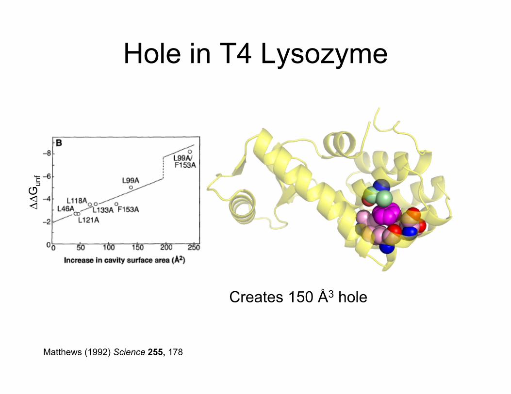
Hole in T4 Lysozyme
Leu99 to Ala Mutant Creates 150 Å3 hole
Matthews (1992) Science 255, 178
∆∆G
unf

Environment Plays A Role in Energetics of H-bonding
Nat. Mol. Struct. Biol., 16, 684 (2009)
PIN1

Environment Plays A Role in Energetics of H-bonding
Nat. Mol. Struct. Biol., 16, 684 (2009)

Replacing a Charged Triad

Disulfide in RNase Sa
C
N
∆Gu = 6.1 kcal/mol + disulfide ∆Gu = 0.3 kcal/mol - disulfide
Cys7-Cy96

Gene 32 Protein: A Structural Zn2+
∆Hunfold = + 139 kcal/mol +Zn2+ ∆Hunfold = + 84 kcal/mol -Zn2+
Biochemistry. 1988 Jul 12;27(14):5240-5.

Distribution of 4˚ Structures

α4 Tetramers
C4 Tetramer D2 Tetramer

α6 Hexamers
C6 Hexamer D3 Hexamer

Hemoglobin - α2β2 Tetramer

Heterotrimeric G Protein

Functionally Distinct Domains
Mammalian Fatty Acid Synthase

Domain Motion - MntR Manganese Transport Regulator
Thanks Wendy Breyer, Kayce Spear

Domain Motion - MntR Manganese Transport Regulator
Thanks Wendy Breyer, Kayce Spear

Tobacco Mosaic Virus