valvular regurgitation: can we do better than...
TRANSCRIPT

Valvular Regurgitation: Can We Do Better Than Colour
Doppler?
A/Prof David Prior
St Vincent’s Hospital
Melbourne Sports Cardiology
Valvular Regurgitation
• Valve regurgitation volume loads the ventricles
• Chronic volume loading may lead to ventricular dysfunction
• Irreversible ventricular dysfunction may precede the development of symptoms
• ie You may miss the boat if you wait for symptoms
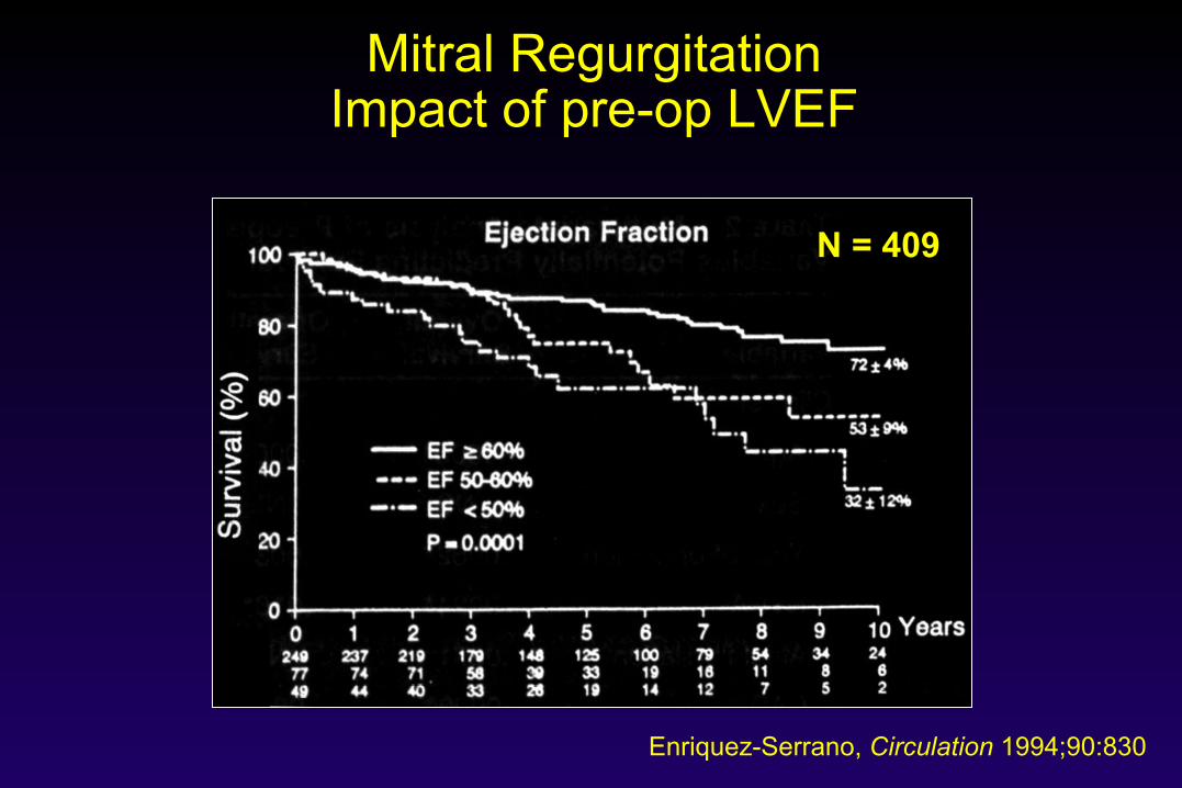
Mitral Regurgitation Impact of pre-op LVEF
Enriquez-Serrano, Circulation 1994;90:830
N = 409
Key Clinical Decisions
• Is the mitral regurgitation clinically significant? – How severe?
• Is the patient symptomatic? • Is ventricular function affected? • If regurgitation is severe, but the patient is
asymptomatic – when is the right time for surgery?
• If regurgitation is not severe – how do we monitor this in the future?

Optimal timing for surgery
Reversible LV Dysfunction
Irreversible LV Dysfunction
Time (Years)
Hyperdynamic, dilating LV
Atrial Fibrillation, Pulm H/T
Symptoms
“Normal” EF, dilated LV Poor EF, dilated LV
Dis
ease
Pro
gres
sion
Too Early Too Late

When to operate?
Risks
Benefits
Mortality < 1% Morbidity < 5% Failed repair ?
Improve Exercise Prevent LV failure Maintain NSR
Echo Assessment of Regurgitation
• Assessing the mechanism of regurgitation
• Determining the severity of regurgitation:
– qualitative and quantitative
• Assessing the hemodynamic consequences of regurgitation
– LV size and function, LA size, PA pressure

Echo Assessment of Regurgitation
• Assessing the mechanism of regurgitation
• Determining the severity of regurgitation:
– qualitative and quantitative
• Assessing the hemodynamic consequences of regurgitation
– LV size and function, LA size, PA pressure

Who Has The Most Regurgitation?

It’s The Same Patient
Low gain High scale
Low scale High gain
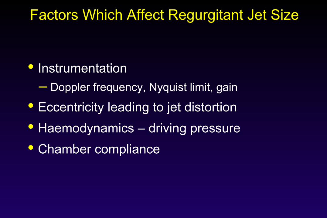
Factors Which Affect Regurgitant Jet Size
• Instrumentation – Doppler frequency, Nyquist limit, gain
• Eccentricity leading to jet distortion • Haemodynamics – driving pressure • Chamber compliance
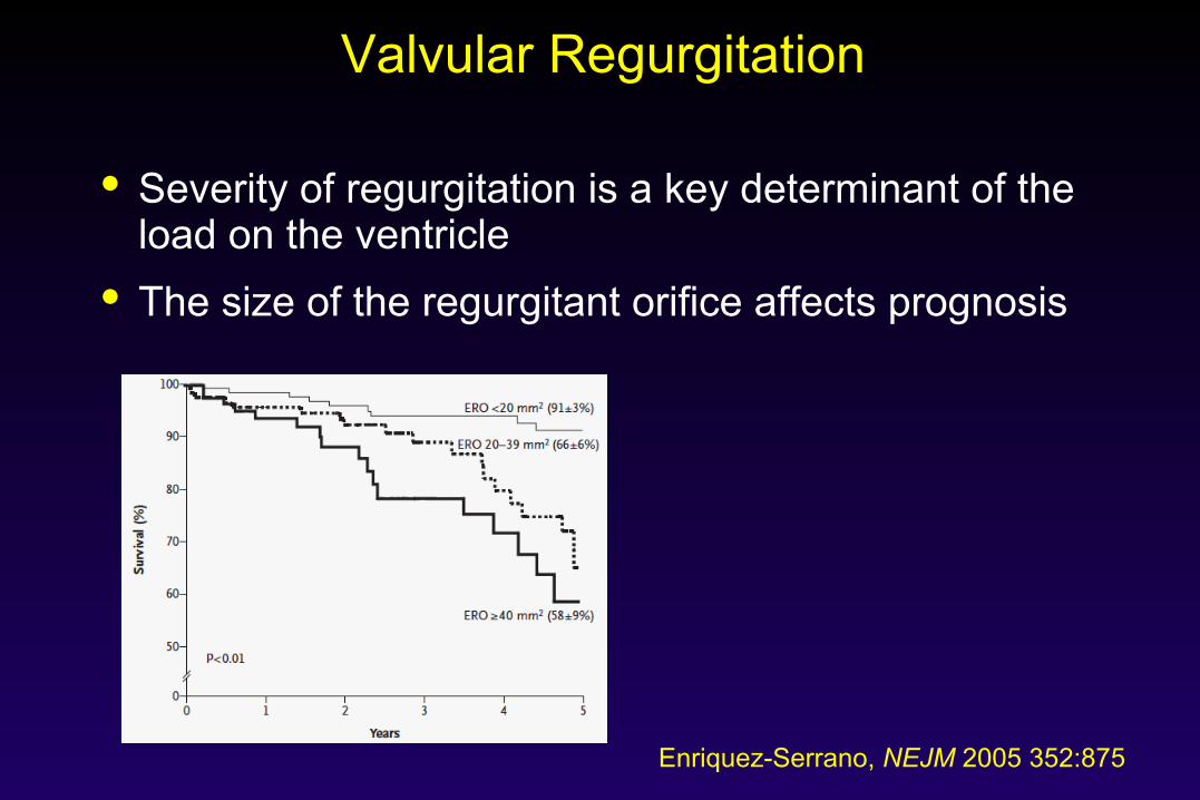
Valvular Regurgitation
• Severity of regurgitation is a key determinant of the load on the ventricle
• The size of the regurgitant orifice affects prognosis
Enriquez-Serrano, NEJM 2005 352:875

Quantitative Measures Of Valve Regurgitation
• Vena Contracta Size – 2d – 3d
• Regurgitant Orifice Area – PISA – Volumetric Flow

Key Quantitative Parameters
• Regurgitant Volume – the volume of blood which flow
backwards through the leaky valve
• Regurgitation Fraction – the percentage of the total
stroke volume which flow backwards
• (Effective) Regurgitant Orifice Area – the effective area of the leak
RV = 40 ml RF = 40%

Quantitative Assessment Of The Mitral Valve

Vena Contracta Width
Lancellotti, EHJ-CVI 2013 14:611

Vena Contracta Width
• Remains valid with eccentric jets
• Can be technically challenging
• Problematic with multiple jets
• ?Benefit with 3-D vena contracta
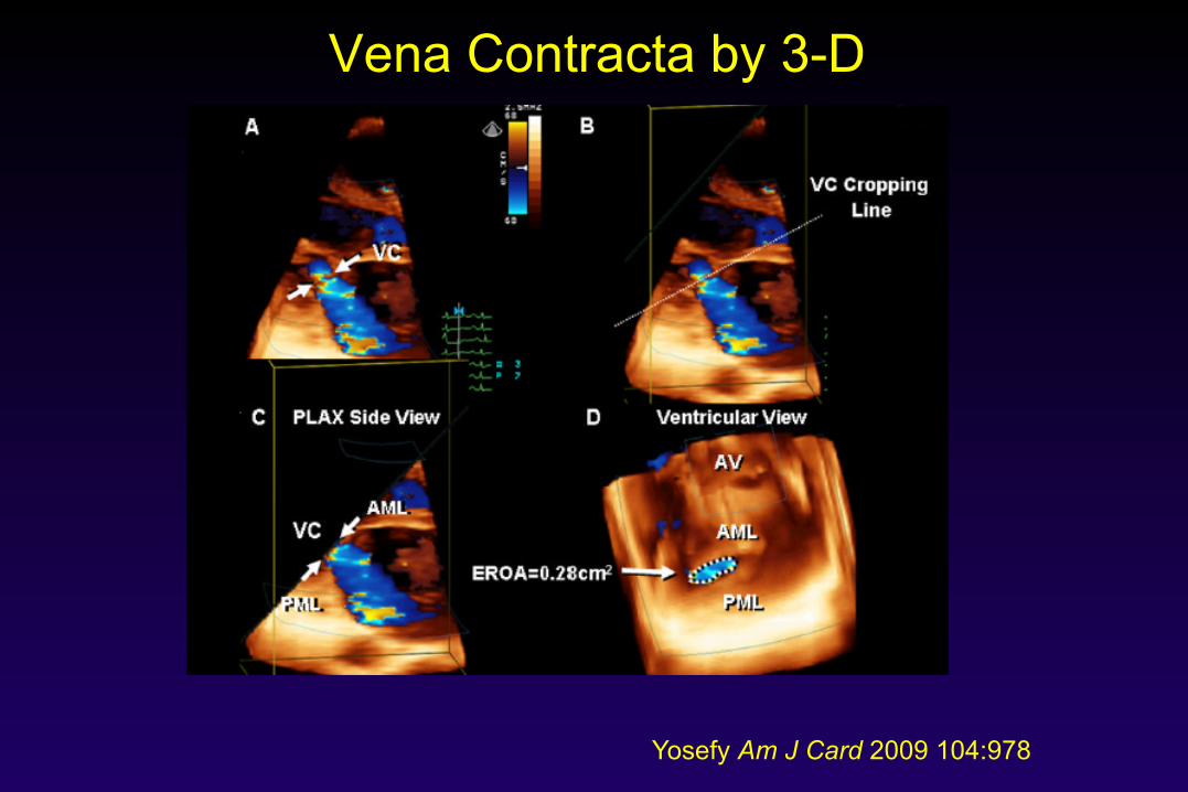
Vena Contracta by 3-D
Yosefy Am J Card 2009 104:978
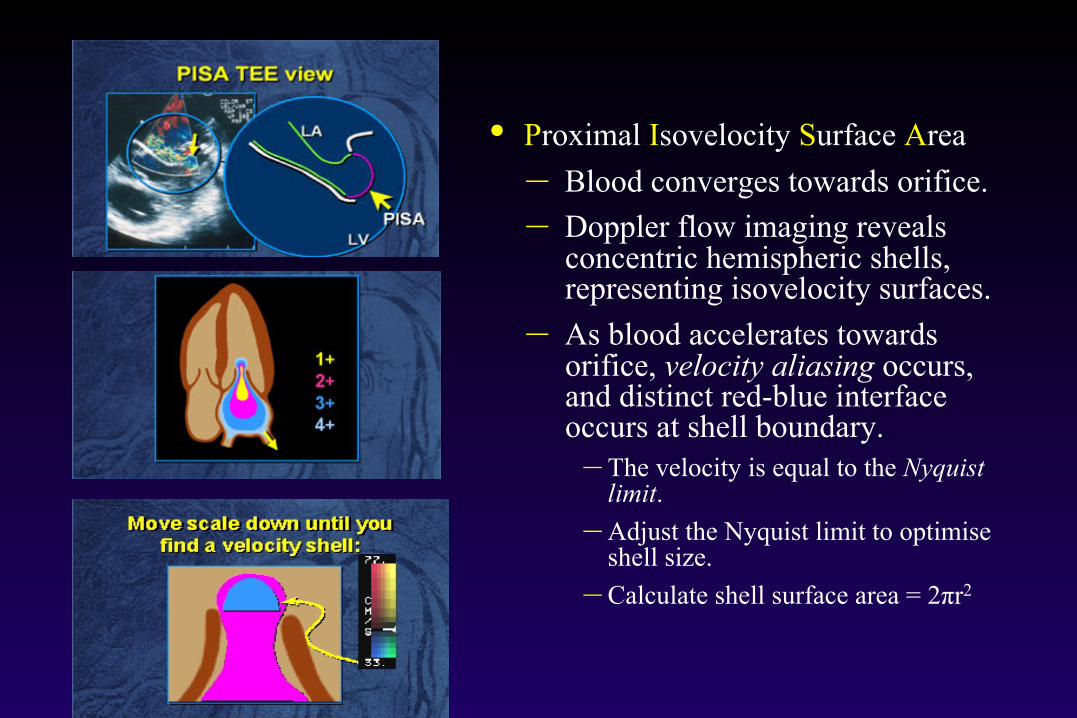
• Proximal Isovelocity Surface Area – Blood converges towards orifice. – Doppler flow imaging reveals
concentric hemispheric shells, representing isovelocity surfaces.
– As blood accelerates towards orifice, velocity aliasing occurs, and distinct red-blue interface occurs at shell boundary.
– The velocity is equal to the Nyquist limit.
– Adjust the Nyquist limit to optimise shell size.
– Calculate shell surface area = 2πr2
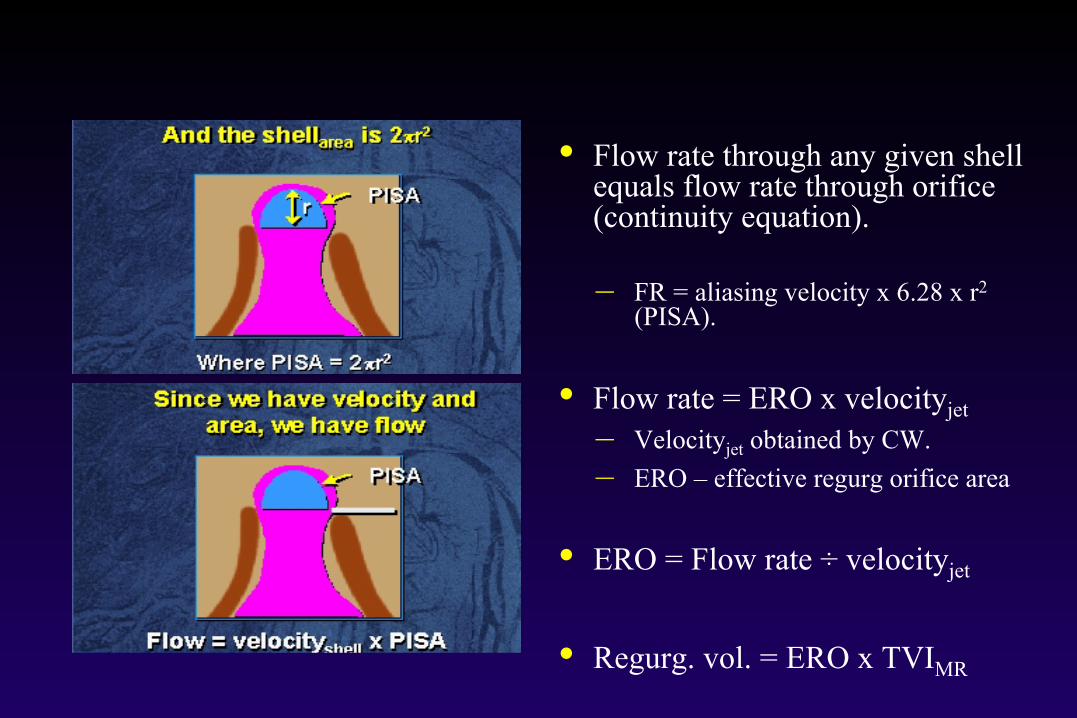
• Flow rate through any given shell equals flow rate through orifice (continuity equation).
– FR = aliasing velocity x 6.28 x r2
(PISA).
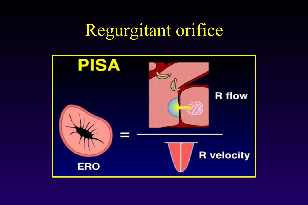
• Flow rate = ERO x velocityjet – Velocityjet obtained by CW. – ERO – effective regurg orifice area
• ERO = Flow rate ÷ velocityjet
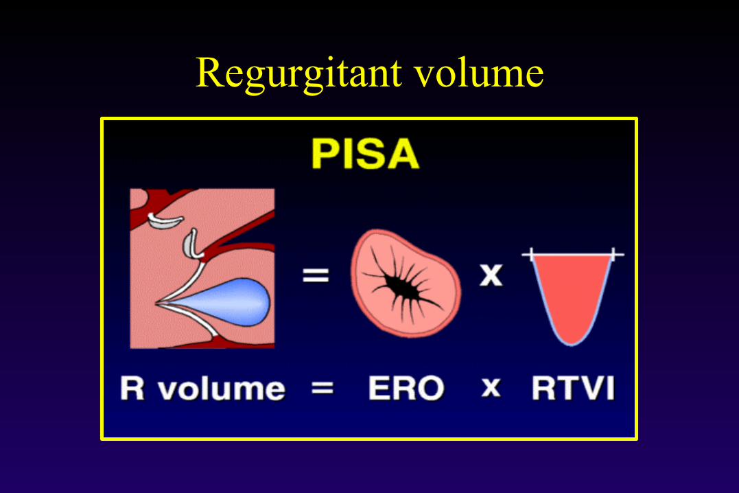
• Regurg. vol. = ERO x TVIMR
Regurgitant orifice
Regurgitant volume
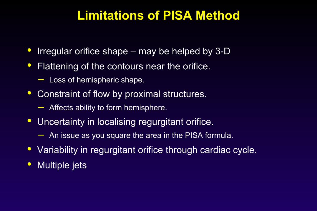
Limitations of PISA Method
• Irregular orifice shape – may be helped by 3-D • Flattening of the contours near the orifice.
– Loss of hemispheric shape.
• Constraint of flow by proximal structures. – Affects ability to form hemisphere.
• Uncertainty in localising regurgitant orifice. – An issue as you square the area in the PISA formula.
• Variability in regurgitant orifice through cardiac cycle. • Multiple jets

Sources of Error with PISA Contour Flattening Near the Orifice
Contour velocity: va Orifice velocity: v0
Conventional PISA Q = 2πr2va
Flow underestimated by va/v0 Ensure the hemisphere is large enough to minimize this

PFC LA
LV
LA
LV
Flail Leaflet
Flail Leaflet Wall Constraint
Sources of Error with PISA Proximal Flow Constraint by Surrounding Structures
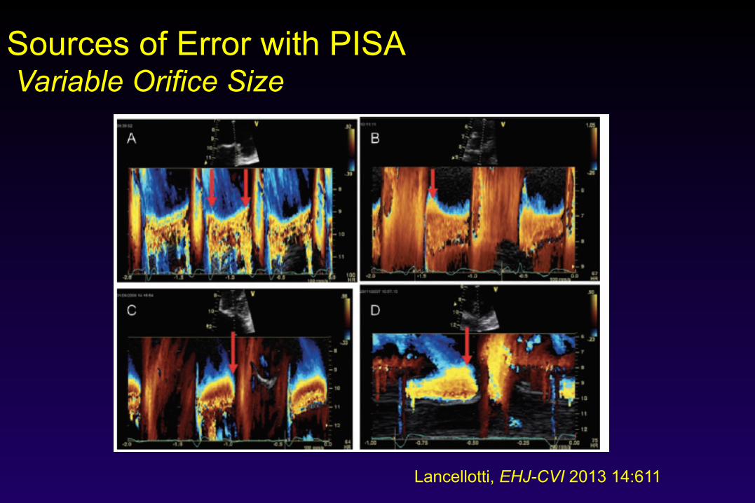
Sources of Error with PISA Variable Orifice Size
Lancellotti, EHJ-CVI 2013 14:611

Measurement of Mitral ROA Simplified PISA Formula
40
40
• Assume LV-LA Δp is 100 mmHg • Set aliasing velocity to 40 cm/sec • Then ROA = r2/2
r = 8 mm
ROA = 82/2 = 32 mm2
LA
LV
MV
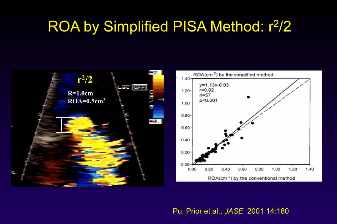
ROA by Simplified PISA Method: r2/2
r2/2 R=1.0cm ROA=0.5cm2
Pu, Prior et al., JASE 2001 14:180
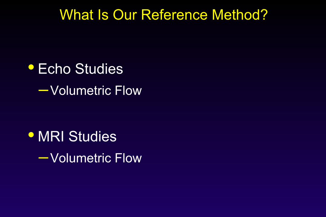
What Is Our Reference Method?
• Echo Studies – Volumetric Flow
• MRI Studies – Volumetric Flow

Quantitative Assessment of MR - Volumetric Flow
• Measure SV in 2 regions, one of which includes the regurgitant volume.
• Difference b/n these two SVs is the regurgitant volume through the valve.
– Area of the LVOT x VTI – Mitral annular area x VTI
Or – LV stroke volume
– LVEDV-LVESV (3-d or Simpson’s biplane)
• Regurg. flow rate (ml/s), fraction (%), orifice area. LVOT - Beware of AR
Mitral annulus
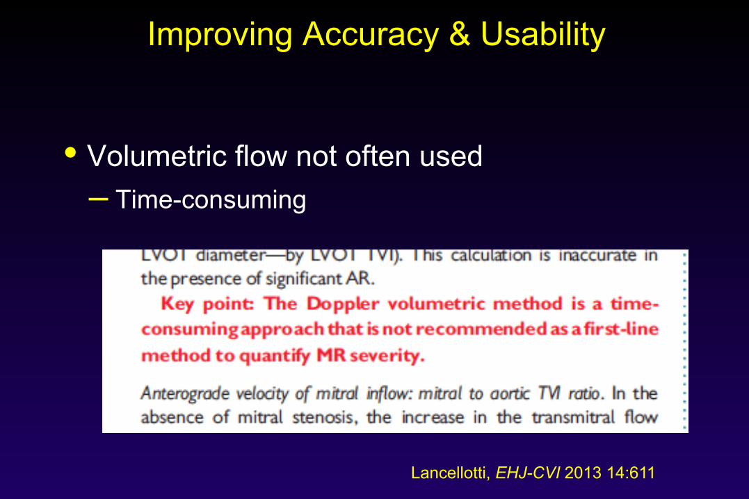
Improving Accuracy & Usability
• Volumetric flow not often used – Time-consuming
Lancellotti, EHJ-CVI 2013 14:611

Simultaneous MV and LVOT flow - Real Time Colour Flow Doppler
Thavendiranathan, JASE 2012 25:1
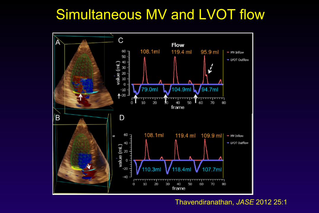
Simultaneous MV and LVOT flow
Thavendiranathan, JASE 2012 25:1
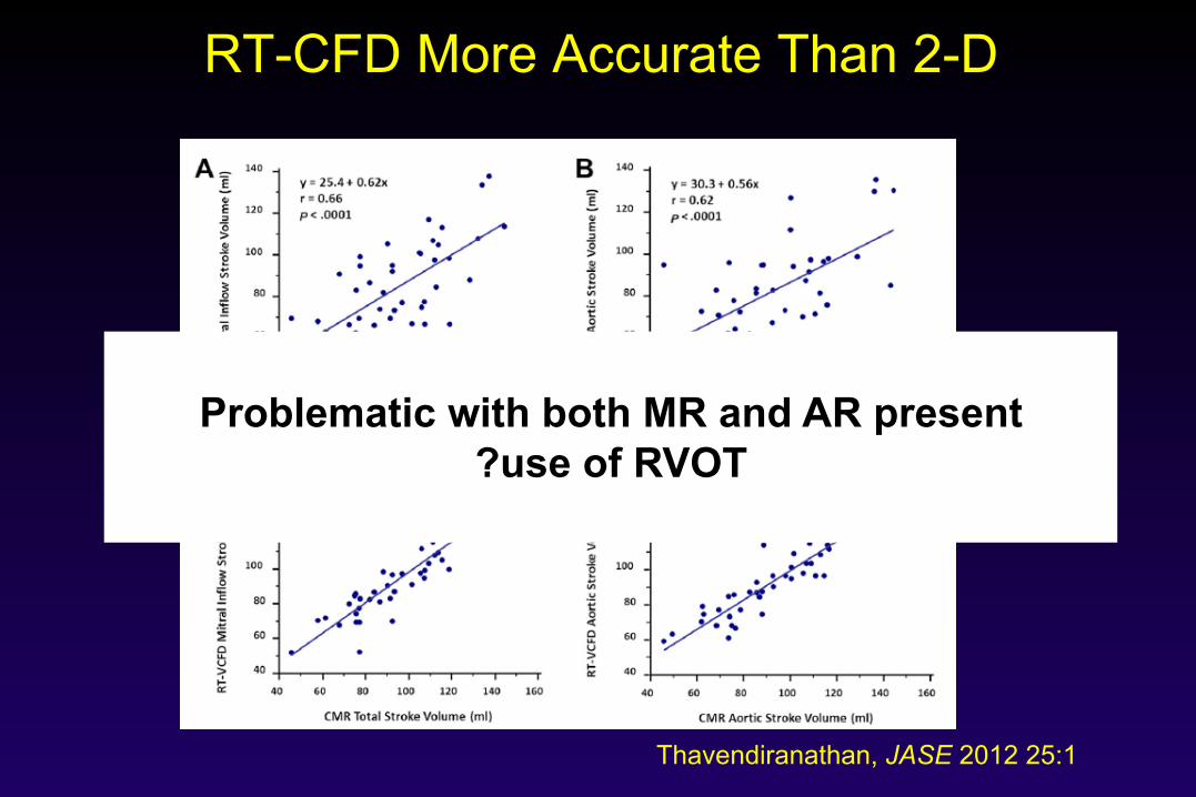
RT-CFD More Accurate Than 2-D
Thavendiranathan, JASE 2012 25:1
Problematic with both MR and AR present
?use of RVOT

Use All The Available Information
• Pulmonary vein flow • Mitral inflow characteristics • CW of the MR jet
– Signal intensity – Shape of the signal

Systolic flow reversal in pulmonary veins
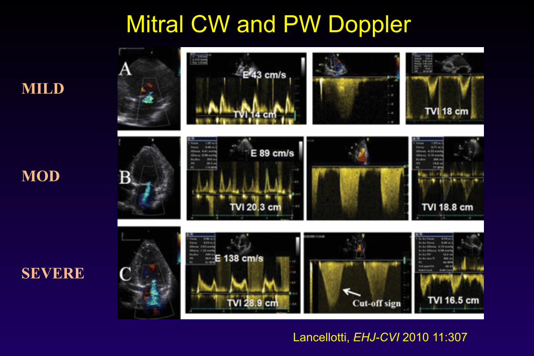
Mitral CW and PW Doppler
Lancellotti, EHJ-CVI 2010 11:307
MILD
MOD
SEVERE

Key Cut-off Values for MR
Mild Moderate Severe Mod Mod Severe
VC width < 0.3 0.3 – 0.69 ≥ 0.7 R Vol (ml/beat) < 30 30 - 44 45 - 59 ≥ 60 R Fract (%) < 30 30 - 39 40 – 49 ≥ 50 EROA (cm2) < 0.2 0.20 – 0.29 0.30 – 0.39 ≥ 0.4
Zoghbi, JASE 2003 16:777

Aortic Regurgitation
• JET AREA AND JET LENGTH ARE NOT WELL CORRELATED WITH SEVERITY
• Quantification can be more difficult
Aortic Regurgitation
• JET AREA AND JET LENGTH ARE NOT WELL CORRELATED WITH SEVERITY
• Quantification can be more difficult

Use of PISA in Aortic Regurgitation
Lancellotti, EHJ-CVI 2010 11:223

Use of PISA in Aortic Regurgitation
Tribouilloy, JACC 1998 32:1032
Underestimation with tented valves

Volumetric Flow in Aortic Regurgitation

Additional Parameters
• AR pressure half-time • Diastolic flow reversal
– Upper descending aorta – Abdominal aorta
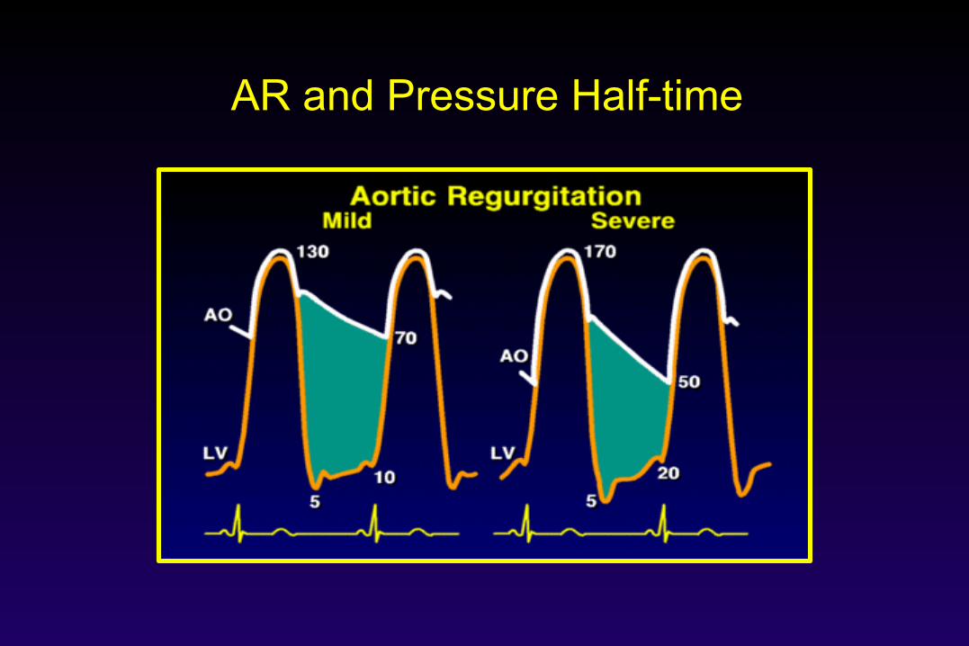
AR and Pressure Half-time
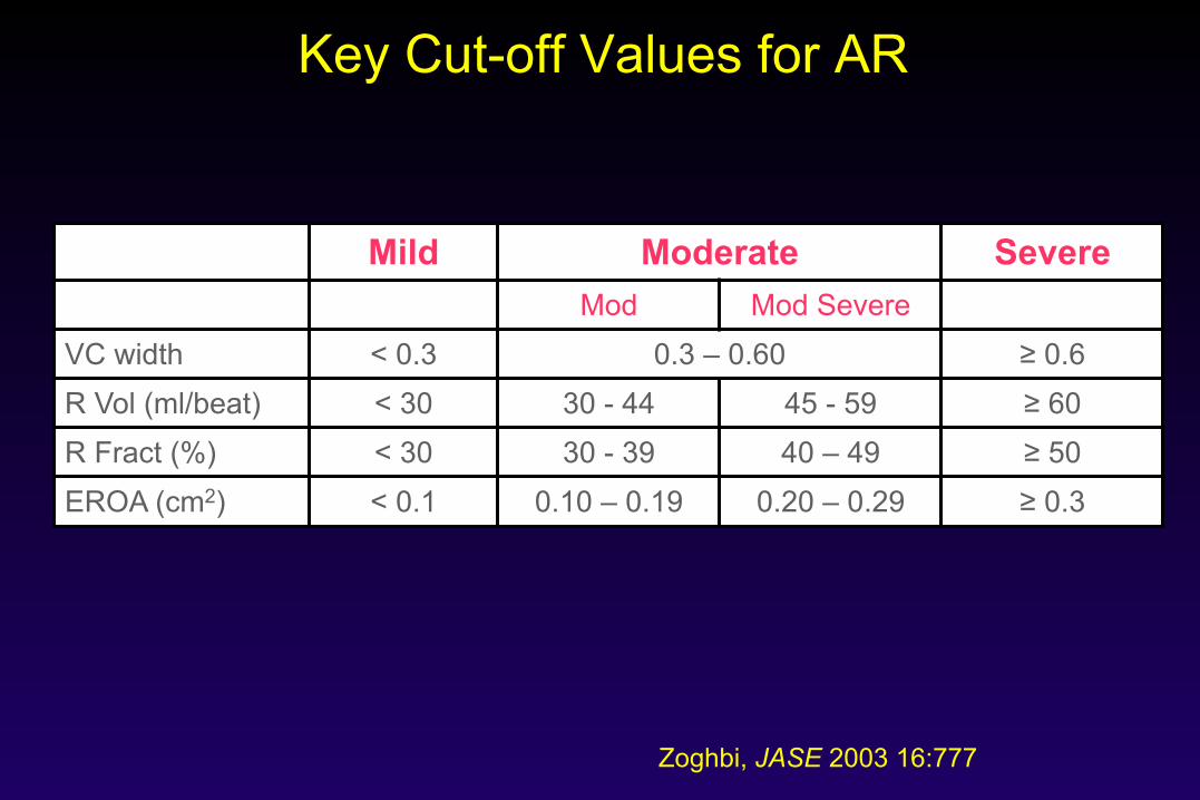
Key Cut-off Values for AR
Mild Moderate Severe Mod Mod Severe
VC width < 0.3 0.3 – 0.60 ≥ 0.6 R Vol (ml/beat) < 30 30 - 44 45 - 59 ≥ 60 R Fract (%) < 30 30 - 39 40 – 49 ≥ 50 EROA (cm2) < 0.1 0.10 – 0.19 0.20 – 0.29 ≥ 0.3
Zoghbi, JASE 2003 16:777

Tricuspid Regurgitation
• Many parallels with MR • Vena contracta width can be used • PISA can be used
– an EROA ≥ 40 mm2 indicates severe TR – R Vol > 45 ml suggests severe TR
• Other parameters suggesting severe TR – systolic flow reversal in the hepatic veins – V wave cut-off sign
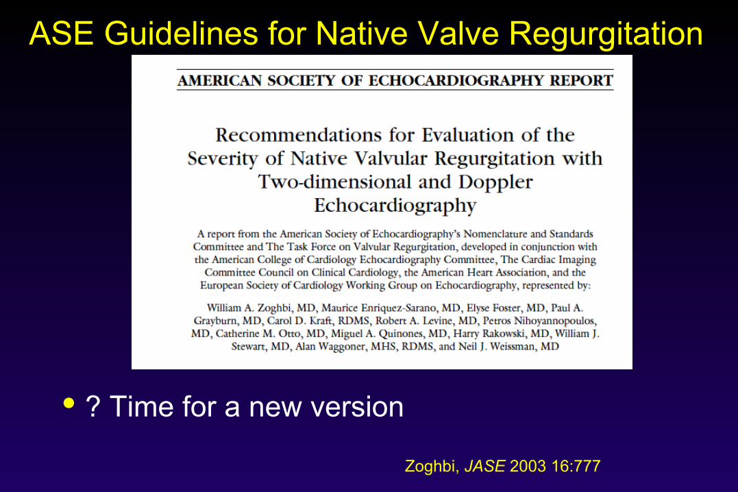
ASE Guidelines for Native Valve Regurgitation 2003
Zoghbi, JASE 2003 16:777
• ? Time for a new version

EACVI Guidelines 2013
Estimation of the severity of valvular regurgitation: recommendations 1. The colour flow area of the regurgitant jet is not recommended to quantify the severity of valvular regurgitation. 2. Both VC measurement and the PISA method are recommended to evaluate the severity of regurgitation when feasible. 3. Adjunctive parameters should be used when there is discordance between the quantified degree of regurgitation and the clinical context.
Lancellotti, EHJ-CVI 2013 14:611

Summary
• Accurate assessment of valvular regurgitation is important for clinical decision making
• Colour flow jet area is NOT recommended • Quantitative measures are preferable • PISA continues to be useful in selected cases • Real-time 3D colour flow Doppler may become
a method of choice for future quantification