vaginal leiomyoma ina dog
TRANSCRIPT

CASE REPORT
Vaginal Leiomyoma in a Dog
T.B. KANG AND D.L. HOLMBERG
Department of Veterinary Anesthesiology, Radiology and Surgery, WesternCollege of Veterinary Medicine, University of Saskatchewan, Saskatoon, Sas-katchewan 57N 0 WO
SummaryThe surgical treatment of vaginal
leiomyoma in a seven year old BlueHeeler bitch is described. A smooth,12 cm diameter submucosal, intralum-inal, firm mass was found on vaginalexamination. It appeared to arise fromthe left ventral vaginal wall, cranial tothe clitoris but caudal to the cervix.There was no history of urinary prob-lems and the dog was normal in allother aspects. The treatment was sur-gical excision of the mass via an episi-otomy. Histological examinationindicated a leiomyoma. The differen-tial diagnoses, possible etiologies andcontrol or prevention of the conditionby ovariohysterectomy are alsodiscussed.ResumeLeiomyome vaginal, chez une chienne
Cet article decrit l'excision chirurgi-cale d'un leiomyome vaginal, chez unechienne "Blue Heeler" agee de septans. L'examen du vagin revela qu'ilarborait, sur sa partie ventrale gauche,entre le clitoris et le col uterin, unemasse ferme et lisse qui mesuraitenviron 12 cm de diametre. L'anam-nese ne rapportait pas de problemesurinaires et la chienne etait par ailleurstout a fait normale. Le traitement con-sista a effectuer l'excision chirurgicalede la masse, en ayant recours a uneepisiotomie. L'histopathologie decette masse revela qu'il s'agissait d'unleiomyome. Les auteurs commententles diagnostics differentiels, les etiolo-gies possibles et le contr6le ou laprevention d'un tel neoplasme parl'ovario-hysterectomie.IntroductionTumors of the female reproductive
tract are divided into two categories:
those arising from the ovaries andthose that are derived from the tubulargenitalia (1,2,3). It is important to dis-tinguish whether these neoplasms arebenign or malignant and to differen-tiate them from other conditions suchas hyperplasia, granulation tissue orabscessation. Treatment and progno-sis vary considerably between thesediseases.Neoplasms of the female tubular
genitalia account for 3% of all caninetumors; of these 85-90% occur in thevagina and vulva (1). Tumors of mes-enchymal origin: leiomyomas, fibro-leiomyomas and fibromas occur mostcommonly (1). Leiomyosarcomas,lipomas, mastocytomas, adenocarci-nomas, squamous cell carcinomas andtransmissible venereal tumors occurmuch less frequently (1,3,4).Leiomyoma is a tumor of smooth
muscle cells that may arise in anyorgan with a connective tissue or mes-enchymal component (1,10). Leiomy-omas have been found in many organsincluding: the trachea (11), gastroin-testinal system (12,13,14,15,16), uri-nary tract (12,17,18), prostate (12) andfemale reproductive tract (1,2,5,19,20).This tumor has also been reported in awide variety of animal species rangingfrom the guinea pig to the Indian ele-phant (1,5,14,15,16,18,21,22,23).Leiomyomas are common in the
canine female reproductive tract andaccount for 2.4% of all canine neo-plasms (1,2,16,19,20). About 85% ofleiomyomas occurring in the repro-ductive tract of the bitch arise from thevagina, vestibule and vulva (1,2,4,9).The incidence of leiomyomas is high-est between five and 16 years of age(1,19). The Boxer breed may compriseup to 16% of dogs showing reproduc-
tive tract leiomyomas (1,5,19). Theincidence of vaginal leiomyomas inmultiparous bitches is considered to beincreased by some authors (1,2), yetother statistics reveal no significantdifference in the rate of occurrencebetween multiparous and nulliparousanimals (19).Leiomyomas of the reproductive
tract in the bitch are frequently asso-ciated with estrogen secreting tumorsor ovarian follicular cysts. Cysticendometrial hyperplasia, mammaryhyperplasia and/or neoplasia mayalso be concurrently found (1,2,6,19,20). The role of estrogens in theetiology of leiomyomas is unclear.Experimentally, administration ofcontinuous low levels of estrogens toguinea pigs will stimulate the spon-taneous formation of leiomyomas aswell as cause hyperplasia of endomet-rial and mammary tissue (20,21).There has also been speculation thatthe transition from hyperplastic toneoplastic tissue may constitute stagesin reproductive tract tumorogenesis inthe female, with estrogens acting as thestimulating factor. Estrogens areknown to be carcinogenic, while theprogestational phase of estrus inhibitstumor formation (20,21).
Vaginal leiomyomas may be singleor multiple, intraluminal or extralum-inal. The tumor is usually round oroval, well defined and encapsulated.The size and consistency may varydepending upon duration of growth,becoming firmer due to an increase inconnective tissue. Large intraluminaltumors may protrude through thevulva, while extraluminal tumors tendto cause perineal swelling. At this stagethe tumor may impinge upon the ure-thra causing the animal to show signs
Reprint requests should be addressed to Dr. D.L. Holmberg.
Can Vet J 1983; 24: 258-260258

of dysuria, constipation and tenesmus.Leiomyomas must be differentiated
from leiomyosarcomas by histologicalexamination. Malignancy is character-ized by cellular pleomorphism, anincrease in mitotic activity and multi-nucleation. The cells are bizarre inappearance and may range from small,round forms with hyperchromic nucleito large multinucleated types. Necrosismay be evident grossly and micro-scopically (10, 12,19,20).The prognosis for vaginal leiomy-
omas is good as they are benign. Lei-omyosarcomas may carry a goodprognosis provided that no metastasishas occurred. The prognosis is consid-erably worse if other organs areinvolved (1). Metastasis to the sacral,internal iliac and superficial inguinallymph nodes have been reported (1).In the following case of a vaginal leio-myoma in a dog, the diagnostic proce-dures, treatment and pathologicalfindings will be discussed.
History and Clinical FindingsA seven year old Blue Heeler bitch,
was referred to the Small AnimalClinic at the Western College of Vete-rinary Medicine because of perinealswelling and an internal mass adjacentto the vulva. The duration ofthe prob-lem was not known and the animal hadnot been showing signs of discomfort.The dog's appetite and thirst werenormal and she had no difficulty uri-nating or defecating. The breeding his-tory of the dog and characteristics ofher estrous cycle were not known.The dog was in good physical condi-
tion, alert and responsive. There wasgross distortion of the perineal areacaused by a swelling between the anusand the vulva. The skin and tissueoverlaying the mass were intact andappeared normal. Vaginal examina-tion resulted in finding a 12 cm diame-ter round, smooth, firm mass arisingfrom the region of the left ventral vag-inal wall, cranial to the clitoris butcaudal to the cervix. The mass wassubmucosal and extended to within3 cm of the vulvar opening. The vagi-nal mucosa overlying the mass wasintact and the extent of the attachmentof the mass to the vaginal wall couldnot be fully evaluated during thisexamination.
TreatmentAfter anesthesia and surgical prepa-
ration the dog was positioned in aperineal stand with the tail flexed cran-ially over the spine. An episiotomy wasperformed at the 11 o'clock positionrelative to the dorsal commissure ofthe vulva. The mass was easily visual-ized and it was noted that the vaginawas extremely dilated. The mass wasfound to be a smooth, oval, submuc-osal growth that measured approxi-mately 12 cm in diameter (Figure 1). Itwas firm to the touch and had a welldefined stalk (5 by I cm base) attachedto the left ventral vaginal wall. Theurethral orifice was close to the pedicleof the mass and a size ten french Foleycatheter was placed in the urethra toaid in defining and avoiding trauma tothis structure (9). The pedicle of themass was transected in a line parallelto the urethra (Figure 2). The growthwas excised and portions were sentimmediately to pathology for impres-sion smears, cytology and frozen sec-tion; other portions were fixed in 10%formalin.
Closure of the excision site wasachieved by approximating themucosa of the vaginal floor as well asoccluding the submucosal dead space
via a submucosal interrupted simplesuture pattern of 3-0 chronic catgut.'The episiotomy site was closedroutinely.
ResultsThe dog recovered uneventfully
from anesthesia and did not developpostoperative complications such asurine retention or incontinence. Shewas sent home the following day, ontreatment with oral antibiotics for thenext five days.
Results of the impression smearsand cytological examination of the ex-cised mass were only a few exfoliatedspindle cells. Frozen section of themass led to a preliminary diagnosis offibroma. However, histopathologicallythe mass had a connective tissue coat-ing covered by a thin layer of stratifiedepithelium. The tumor cells were ofsmooth muscle origin, lying in parallelbundles with collagen fibers interposed.
Final diagnosis of a leiomyoma wasmade and the owner was given a goodprognosis and the recommendationthat the dog should be spayed to pre-vent regrowth of the tumor. Thirteenmonths after the surgery regrowth orproblems associated with the tumor orits removal have not occurred.
FIGURE 1. Vaginal leiomyoma delivered through the vulva of a dog following an episiotomy.
'Davis and Geck, Cyanamid of Canada Inc., Montreal, Quebec.
259
DiscussionAs seen in this case, local treatment
of vaginal leiomyomas primarilyinvolves surgical excision of the mass.If few discrete metastatic foci are pre-sent, these may also be removed.Because most tumors arise from thevestibule or the smooth muscle wall ofthe vagina, they are usually removedper vulva. An episiotomy may benecessary for larger tumors (1,7,9).Radiation therapy may be consideredif surgical removal of the tumorand/or its metastatic foci is not possi-ble (1,2,19).
latrogenic damage to the urethra oraccidental injury to other perinealstructures are possible surgical com-plications. Urethral catheterizationwill greatly assist in avoiding damageto this structure. Postoperative infec-tion and/or scar contracture can also
result in urethral obstruction. Whilesome authors believe that excision ofthe vaginal leiomyoma is curative (5),the condition will usually recur due tohormonal (i.e. estrogenic) influence.Thus prevention and control of thedisease is best achieved by ovariohys-terectomy (1,2,7,8,19).
References1. SUSANECK sJ. Tumors of the female repro-
ductive tract. In: Oncology notes. Fort Col-lins: Comparative Oncology Unit, Colo-rado State University, 1981.
2. BARRETT RE, THEILEN GH. Neoplasms of thecanine and feline reproductive tracts. In:Kirk RW, ed. Current veterinary therapyVI. Philadelphia, London, Toronto: W.B.Saunders Co., 1977: 1263-1267.
3. BOSTOCK DE, OWEN LN. A colour atlas ofneoplasia in the cat, dog and horse. Lon-don, United Kingdom: Wolfe Medical Pub-lications Ltd., 1975: 62-79.
~~~~~-t '4
~~~~~.i.~~~~~~~~~~.
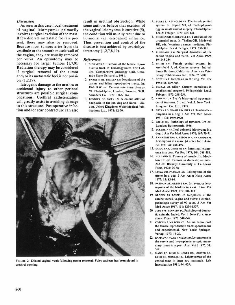
FIGURE 2. Dilated vaginal vault following tumor removal. Foley catheter has been placed inurethral opening.
4. BURKE TJ, REYNOLDS HA. The female genitalsystem. In: Bojrab MJ, ed. Pathophysiol-ogy in small animal surgery. Philadelphia:Lea & Febiger, 1979: 425-441.
5. THEILEN GH, MADEWELL BR. Tumours of theurogenital tract. In: Theilen GH, MadewellBR, eds. Veterinary cancer medicine. Phi-ladelphia: Lea & Febiger, 1979: 357-381.
6. FURNEAUX RW. Surgical disorders of thecanine vagina and vulva. Vet Anim 1979;19: 245-254.
7. SMITH KW. Female genital system. In:Archibald J, ed. Canine surgery. 2nd ed.Santa Barbara, California: American Vete-rinary Publications Inc., 1974: 751-782.
8. COTCHIN E. Neoplasia in the dog. Vet Rec1954; 66: 879-888.
9. BOJRAB MJ, editor. Current techniques insmall animal surgery I. Philadelphia: Lea &Febiger, 1975: 248-254.
10. ASHLEY DJB. Evan's histological appearan-ces of tumours. 3rd ed, Vol. 1. New York:Longman Co. Ltd., 1978.
11. BRYAN RD, FRAME RW, KIER AB. Tracheal lei-omyoma in a dog. J Am Vet Med Assoc1981; 178: 1969-1970.
12. WILLIS RA. Pathology of tumours. 3rd ed.London: Butterworth, 1966.
13. ECKERLIN RH. Ileal polypoid leiomyoma in adog. J Am Vet Med Assoc 1976; 167: 70-7 1.
14. RAMAKRISHNA K, REDDY MV, MAHANDER M.
Leiomyoma in a mare. (A note). Ind J AnimSci 1971; 41: 498-499.
15. SAIDU SNA, CHINEME CN. Intestinal leiomy-oma in a cow. Vet Rec 1979; 104: 388-389.
16. HULLAND Ti. Tumors of muscle, In: Moul-ton JE, ed. Tumors in domestic animals.2nd ed. Berkely: University of CaliforniaPress, 1978: 75-88.
17. LISKA WD, PATNAIK AK. Leiomyoma of theureter in a dog. J Am Anim Hosp Assoc1977; 13: 83-84.
18. PATNAIK AK, GREENE RW. Intravenous leio-myoma of the bladder in a cat. J Am VetMed Assoc 1979; 175: 381-383.
19. BRODEY RS, ROSZEL JF. Neoplasms of thecanine uterus, vagina and vulva: a clinico-pathologic survey of 90 cases. J Am VetMed Assoc 1967; 151: 1294-1307.
20. JUBB KVF, KENNEDY PC. Pathology ofdomes-tic animals. 2nd ed, Vol. 1. New York: Aca-demic Press, 1970: 548-549.
21. COTCHIN E, MARCHANTJ. Animal tumours ofthe female reproductive tract: spontaneousand experimental. New York: Springer-Verlag, 1977: 16-20.
22. RAMADAN RO, EL HASSAN AM. Leiomyoma inthe cervix and hyperplastic ectopic mam-mary tissue in a goat. Aust Vet J 1975; 51:362.
23. MANN PC, BUSH M, JONES DM, GRINER LA,KUEH GR, MONTALI RJ. Leiomyomas of thegenital tract in large zoo mammals. LabInvestigation 1981; 44: 40A.
260