v2 i2 15 - pt. deendayal upadhyay memorial health sciences and...
TRANSCRIPT

(I)
Jou
rnal
ISSN 2348 - 4195
CHHATTISGARH JOURNAL OF HEALTH SCIENCESAn official publication of Ayush and Health Sciences University, Chhattisgarh
PatronDr. G. B. Gupta Vice Chancellor
Executive Editorial BoardDr. K. L. Tiwari– Registrar Ayush & Health Sciences University
Dr. N. Gandhi – Dean Faculty Medical
Dr. Anil G. Ghom – Dean Faculty, Dental
Mrs. Abhilekha Biswal –Dean Faculty Nursing
Dr. D. Katariya – Dean Faculty Ayurvedic
Dr. A. R. Rudrajwar –Dean Faculty Homoeopathy
Associate EditorsDr. Raghavendra Shetty
Dr. Divya Sahu
Dr. A. K. Chandrakar Dr. S. Pawar
Dr. A K Vishwakarma Dr. Rajendra Prasad
Dr. Tripti NagariaDr. O. P. Khandelwal
Dr. Rajendra K. DubeyDr. Sanjay N
Mrs. Sreelata Pillai Dr. Anand Sharma
Dr. Deepesh K. GuptaDr. S. R. Inchulkar Dr. Vineeta Gupta
Dr. R.P. Gupta Mrs. Uma Shendey Mrs. Preetha Sunil
Ms. Bhuneshwari Sahare Dr. Rohit Rajput
Editorial Board

REVIEW ARTICLE
Intensity modulated radiotherapy in head and neck cancer : A review
J ....... aideep Sur, Rachita Jain, Latha.S, Fatima Khan, Fiza Khan, Divya Chaurasia 01
Diagnosis & management of the pathological temporomandibular joint
M.S. Senthil kumar, Senthil kumar S., Deepesh Gupta, N.Vidyasankari .......06
ORIGINAL ARTICLE
Sex determination using dental pulp in permanent & deciduous dentition
N.Mohan, Sukriti Kumar, Jayashree Mohan .......10
Evaluation of pentraxin-3 inflammatory Marker level in generalized chronic periodontitis
before and after mechanical therapy (scaling and root lanning)
Kokila G, Renuka Devi R, Vineeta Gupta .......15
Study of serum phosphate levels and risk of infection in hemodialysis patients
P. Gupta, S. Verma, P. Dubey .......22
Edentulousness , prosthetic status and prosthetic need ofinstitutionalized elderly people
in old age homes of Chhattisgarh
R. K. Dubey, P. Shetty, D. K. Gupta, S. Pandey .......25
Trends in epidemiology of oral cancer in central part of India in Madhya Pradesh : An
institutional study
Vanita Rathod, Chandan Rathod .......32
Assessment of dental aesthetic index among school children of Bilaspur, Chhattisgarh : A pilot study R S Makkad, Madhu Pandey, S Hamdani, V. Agrawal, M Motlani , Gunjan Agrawal .......37
CASE REPORT
Neurofibroma of spindle cell origin, a diagnostic dilemma to general dentist
Swapnil Moghe, Ajay Kumar Pillai, Vineeta Gupta, Geeta Mishra .......42
TMJ ankylosis associated with odontogenic keratocyst of mandibular ramus : A rare case report
Biju Pappachan, R K Dubey, Manish Raghani, Raghav Agrawal .......45
Wilckodontics demystified : A case report
Sumit Gandhi, Lokesh Advani, Javed Sodawala, G. Anita, Srinias T.S., Parul Agrawal .......48
Volume 2 issue 2
CONTENTS
(ii)
CHHATTISGARH JOURNAL OF HEALTH SCIENCESAn official publication of Ayush and Health Sciences University, Chhattisgarh
ISSN 2348-4195
It is my proud privilege to present you the “Chhattisgarh Journal Of Health Sciences” that reflects voice of medicine professionals in Chhattisgarh. As, an Editor, I humbly accept the responsibilities entrusted to me and assure you that I will do my best to prove worthy of it. I vow that I will do everything to uphold the standard of our quarterly bulletin. All the advances in medicine field are meaningless if the masses do not have the access to healthcare facilities and get the benefit of these advances. To move forward with this vision, it is wise to look backward with a perception not to blame ourselves or our predecessors but to learn from history and plan for the future. You have precious skill & abilities to make a lot many lives in the community happier.I invite your valuable articles, suggestions, write-ups, views, book reviews, achievement & classified advertisement to make the journal adequately interactive and interesting one.Our quarterly bulletin is a complete scientific publication for the benefit of our members.Once again thanks to all for motivation and co-operation.
Anil G Ghom(Editor-in-Chief)e-mail: [email protected]@gmail.com
Editorial
(iii)
E
dit
or
ial


Ayush & Health Sciences University of Chhattisgarh
1
REVIEW
1 2 3 4 5 6Jaideep Sur , Rachita Jain , Latha.S , Fatima Khan , Fiza Khan , Divya Chaurasia1. Jaideep Sur, Associate Professor, Department of Oral Medicine & Radiology, RCDSR, Bhilai (C.G.)2. Rachita Jain, Post Graduate Student, Department of Oral Medicine & Radiology, RCDSR, Bhilai (C.G.)3. Latha.S, Professor & HOD, Department of Oral Medicine & Radiology, RCDSR, Bhilai (C.G.)4. Fatima Khan, Senior Lecturer, Department of Oral Medicine & Radiology, RCDSR, Bhilai (C.G.)5. Fiza Khan, Post Graduate Student, Department of Oral Medicine & Radiology, RCDSR, Bhilai (C.G.)6. Divya Chaurasia, Post Graduate Student, Department of Oral Medicine & Radiology, RCDSR, Bhilai (C.G.)
Corresponding Author :Dr. Jaideep SurDept of Oral Medicine & Radiology, RCDSR, Bhilai (C.G.)Email: Mobile No: 93029 [email protected],
ABSTRACT:Radiation therapy is a principal modality in the treatment of head and neck cancer. Its capabilities have steadily progressed with the increase in clinical knowledge and technological development. Intensity-modulated radiotherapy (IMRT) concept had been described back in 1978, but it was not until the 90's that it was applied in practice, following improvement and development of computer equipment. A big step forward was made in the past decade by constructing a device with Multi Leaf Collimators. IMRT appears to be clinically justifiable for cancers in the nasopharynx, sinonasal region, parotid gland, tonsil, buccal mucosa, gingiva, and thyroid. IMRT may also be useful in the re-treatment of previously irradiated head and neck cancers, due to its ability to spare adjacent normal tissues with acceptable target dose uniformity. IMRT represents a significant advance in conformal radiotherapy. In particular, it allows the delivery of dose distributions with concave isodose profiles such that radiosensitive normal tissue close to, or even within a concavity of, a tumour may be spared from radiation injury. Key words: IMRT, head and neck cancer, Multi Leaf Collimators.
INTRODUCTIONOverall 57.5% of global head and neck cancers occur in Asia, especially in India. The greatest challenge for
1
radiation therapy or any cancer therapy is to attain the highest probability of cure with the least morbidity. The simplest way in theory to increase this therapeutic ratio with radiation is to encompass all cancer cells with sufficient doses of radiation during each fraction, while simultaneously sparing surrounding normal tissues. In practice, however, we have been hampered by our abilities to both identify the cancer cells and target them with radiation. Over the past decade, enormous progress has been made on both fronts. Technical improvements in the application of X-rays, computed tomography scans, magnetic resonance imaging with and without spectroscopy, ultrasound, PET scans, and electronic portal imaging—and our understanding of
their limitations— have greatly improved our ability to identify tumors.2
In 1960, Professor Shinji Takahashi developed a method of conformation radiotherapy that used multileaf collimators. In 1967, a 6-MV linear accelerator at the Aichi Cancer Center became the first in Japan to be equipped with a multileaf collimator. This unit was used in conformational radiotherapy for various types of cancers. In the 1980s, rotation radiography devices used for conformational radiotherapy were replaced by CT devices. Moreover, conformational radiotherapy evolved into intensity-modulated radiation therapy (IMRT), which gained widespread use in the 1990s.
3
Three-dimensional (3D), or CT-based, planning was a major advance because it took into account axial
REV
IEW
Intensity modulated radiotherapy in head and neck cancer : A review
Chhattisgarh Journal of Health Sciences (Vol-2, Issue-2: July-Dec. 2014)ISSN 2348 - 4195

2
anatomy and complex tissue contours such as the hourglass shape of the neck and shoulders. While 3D planning allowed for accurate dose calculations to such irregular shapes, we were still limited in the corrections we could make. As its name implies, intensity-modulated radiation allows us to modulate the intensity of each radiation beam, so each field may have one or many areas of high intensity radiation and any number of lower intensity areas within the same field, thus allowing for greater control of the dose
2distribution with the target. Two opposing beams of single intensities, represented by the yellow arrows, create a single-dose distribution through a nasopharynx tumor (GTV in red, CTV in purple) and normal tissue alike in two-dimensional radiotherapy, whereas IMRT creates a highly sculpted dose distribution with relative sparing of the brain, brainstem, and parotid glands by delivering beams of
2different intensities as shown in figure 1.
Advanced treatment planning software has furthered our ability to modulate radiation dose. Instead of the clinician choosing every beam angle and weighting, computer optimization techniques can now help determine the distribution of beam intensities across a treatment volume, which often include a non-intuitive distribution of “beamlets,” or 1 cm2 areas of isointensity. IMRT for head and neck tumors refers to a
2,4
new approach that aims at increasing the radiation dose gradient between the target tissues and the surrounding normal tissues at risk, thus offering the prospect of increasing the locoregional control probability while decreasing the complication rate.
5
PRODUCTION OF INTENSITY MODULATED BEAMSTechniques for generation of intensity modulated beams:Metal compensators: A specifically manufactured metallic compensator is milled or moulded so that a variable thickness of the absorber is presented before the radiation beam.6Multiple segments per field: Each treatment field is divided into several smaller segments or subfields,
which are delivered sequentially (the “step and shoot'' method). Each segment shape is defined by a MLC or by shaped blocks. Addition of several segments produces an IMB.
5
Dynamic MLC (dMLC): Modulation of beam intensity by pairs of moving MLC leaves characterizes this technique (also known as the ̀ `sliding window'' technique).
6
Tomotherapy: Tomotherapy descr ibes IMRT techniques that irradiate the target slice by slice. The NOMOS Corporation developed the first commercially available tomotherapy machine, the multivane intensity modulating collimator (MIMiC), which is in use in several centre.5 This device attaches to the head of the linear accelerator (LINAC), which arcs about the craniocaudal axis of the patient.6,7
ADVANTAGES OF IMRT:IMRT has attracted wide spread interest because of its dosimetric and potential clinical advantages.8 Numerous dosimetry studies on linear accelerator based IMRT treatments of different anatomical sites
FIGURE 1: Beam Delivery in radiotherapy A: Two dimensional Radiotherapy B: IMRT
Chhattisgarh Journal of Health Sciences (Vol-2, Issue-2: July-Dec. 2014)
IMRT in head and neck cancer
3
have been reported, and all of them show that IMRT can have definite dosimetry advantages over 2D and conventional 3DCRT treatments. Whether the
9,10
dosimetric advantages of IMRT can be realized clinically would depend on a number of factors, including (a) the accuracy in localisation and delineation of the tumour and the adjacent critical tissue structures, (b) understanding of the optimum relationship between dose and response for the individual tumour, and (c) delivery of the prescription doses according to the treatment plans.
IMRT's high conformity with dose facilitates escalation of dose and better protection of normal tissue structures. These features make it particularly 11,12
suitable for the treatment of diseases that involve high rates of local recurrence and toxicity and complications related to treatment.
13,14
Radiotherapy plays an important role in head and neck tumor treatment because of the cosmetic and functional preservation that becomes possible. IMRT 15
significantly improves broad aspects of health related quality of life in head and neck cancer survivors. 16
It highly reduces parotid irradiation and thus reduces post radiotherapy xerostomia. With the advent of
17
IMRT and its capability to treat multiple targets simultaneously to different doses, a new accelerated fractionation scheme is introduced. It is known as simultaneous modulated accelerated radiation therapy (SMART) boost. SMART boost can be applied to
18
various sites including head and neck, brain and prostate. The principle is to treat two different targets with different fraction sizes to different total doses.19
According to a study done by Beadle et al on 3172 patients with head and neck cancers IMRT treated patients experienced significant improvements in cause specific survival (CSS) compared with patients treated with non-IMRT techniques. IMRT improves the overall survival rate in patients.
20
CLINICAL APPLICATIONS OF IMRT IN HEAD AND NECK CANCER:The management of head and neck cancer in recent years has involved increasingly complex, combined-modality programs, as well as the integration of new diagnostic and therapeutic technologies. That head and neck cancer is the most complex “organ site” for treatment decision making is not an overstatement,
and supports a best practices model of multidisciplinary team involvement. Intensity-modulated radiotherapy (IMRT) has been widely adopted as a standard technology for head and neck cancer. IMRT therefore offers a significant advance in conformal therapy, by improving conformality and reducing radiation dose to radiosensitive normal tissues close to the tumour even if they lie within a concavity in the planning target volume (PTV).
6
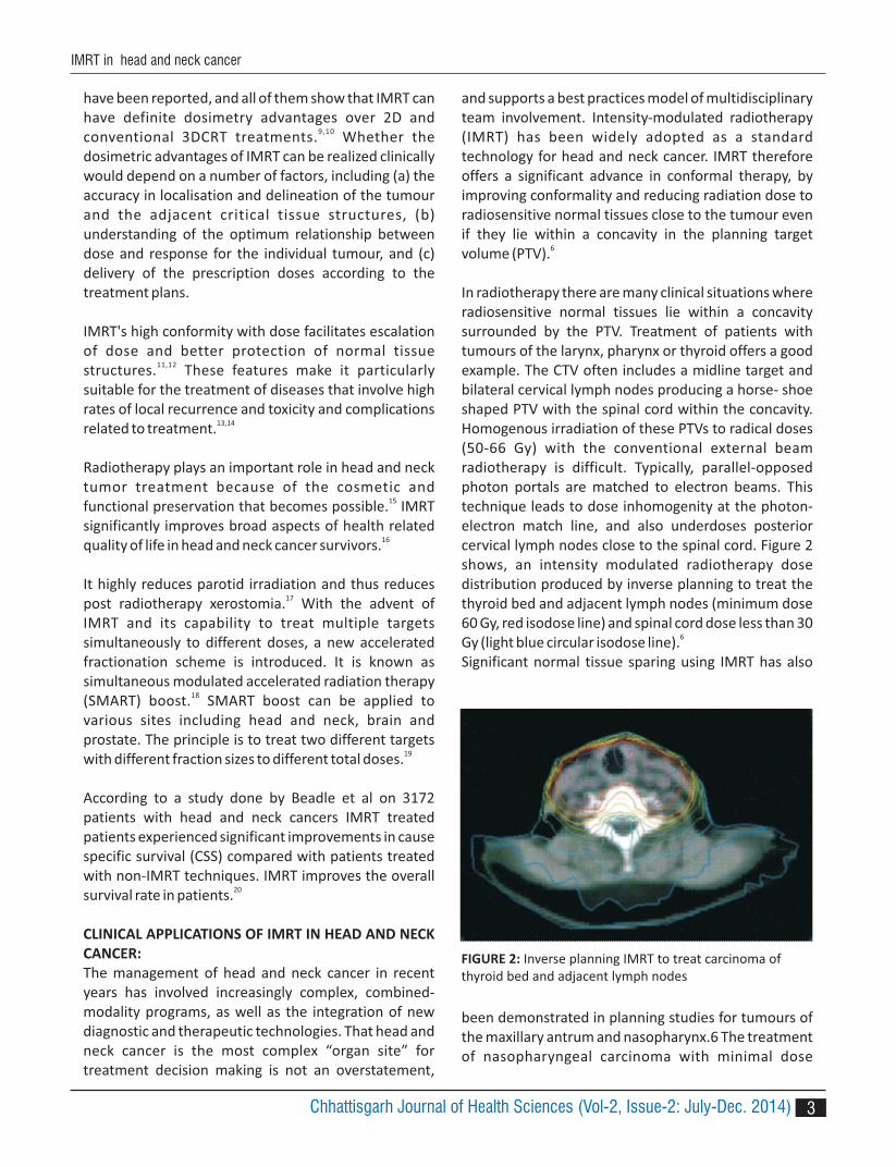
In radiotherapy there are many clinical situations where radiosensitive normal tissues lie within a concavity surrounded by the PTV. Treatment of patients with tumours of the larynx, pharynx or thyroid offers a good example. The CTV often includes a midline target and bilateral cervical lymph nodes producing a horse- shoe shaped PTV with the spinal cord within the concavity. Homogenous irradiation of these PTVs to radical doses (50-66 Gy) with the conventional external beam radiotherapy is difficult. Typically, parallel-opposed photon portals are matched to electron beams. This technique leads to dose inhomogenity at the photon-electron match line, and also underdoses posterior cervical lymph nodes close to the spinal cord. Figure 2 shows, an intensity modulated radiotherapy dose distribution produced by inverse planning to treat the thyroid bed and adjacent lymph nodes (minimum dose 60 Gy, red isodose line) and spinal cord dose less than 30 Gy (light blue circular isodose line).6
Significant normal tissue sparing using IMRT has also
been demonstrated in planning studies for tumours of the maxillary antrum and nasopharynx.6 The treatment of nasopharyngeal carcinoma with minimal dose
FIGURE 2: Inverse planning IMRT to treat carcinoma ofthyroid bed and adjacent lymph nodes
Chhattisgarh Journal of Health Sciences (Vol-2, Issue-2: July-Dec. 2014)
IMRT in head and neck cancer

Complex dose distributions can be delivered that avoid a number of radiosensitive normal tissues close to a t u m o u r. Fo r exa m p l e , i n t h e t re at m e nt o f nasopharyngeal cancer, large parallel-opposed lateral portals are used to encompass macroscopic disease and sites of occult metastases. With this technique parotid glands, spinal cord and brainstem are inevitably included in the irradiated volume although these structures do not need to be included in the target volume. . By defining concavities in the PTV, IMRT can
11
produce a dose distribution that reduces the radiation dose to these organs and this promises a significant reduction in treatment morbidity. IMRT could be used for the whole duration of a radiotherapy treatment, or
simply as a boost after more conventional treatment. The appropriateness of these two approaches is likely to depend on the tolerance doses of surrounding radiosensitive normal tissues. IMRT also reduces
6,12,14
parotid dose to less than 15Gy in treatment of nasopharyngeal carcinoma as shown in figure 4.6,11
Issues in clinical application of IMRT includes, increased risk of a marginal miss because of intrafraction target movements, accurate determination of the target volume and the geometry of the organs at risk (OAR) is difficult. Another issue is the high cost, which limits 21,22
the large scale implementation of IMRT.23
CONCLUSION:Head and neck sites have always been among the most challenging, complex and time consuming to plan because of their complex anatomy. IMRT is designed to deliver more dose to the cancer and less to surrounding healthy tissues. This allows for less normal tissue toxicity, which maintains the patient's quality of life and also improves survival rate. Excellent disease control can be achieved by IMRT with minimum complications like xerostomia, mucositis, dysphagia. The future of head and neck radiotherapy lies in optimally using targeted therapy (IMRT) in order to maximize the therapeutic ratio with minimal morbidity.
delivery to parotid gland bilaterally (26 Gy) and sparing of optic structure with a minimum dose of 30 Gy in a
patient with sinonasal carcinoma can be achieved using IMRT6 as shown in figure3.
FIGURE 3: IMRT in A: Nasopharynx cancer, B: Sinonasal cancer.
B
FIGURE 4: Inverse planning in carcinoma of nasopharynx.
A
4 Chhattisgarh Journal of Health Sciences (Vol-2, Issue-2: July-Dec. 2014)
IMRT in head and neck cancer

5
REFERENCES:1. Kulkarni RM. Head and Neck Cancer Burden in India. International Journal of Head and Neck Surgery 2013;4:29-35.2. Bucci MK, Bewan A, Roach M. Advances in radiation therapy: Conventional to 3D, to IMRT, to 4D, and beyond. CA Cancer J Clin 2005;55:117-34.3. Doi K, Morita K, Sakuma S, Takahashi M. Shinji Takahashi, M.D. (1912–1985): pioneer in early development toward CT and IMRT. Radiol Phys Technol 2012;5:1–4.4. Bourhis J AC, Pignon JP. Update of MACH-NC (Meta- analysis of chemotherapy in head and neck cancer) database focussed on concomitant chemotherapy. J Clin Oncol 2004;22:5505.5. Vincent G, Awilfried N. Intensity modulated radiotherapy for head and neck carcinoma. The Oncologist 2007;12:555-64.6. Nutting C. Intensity modulated radiation therapy: a clinical review. The British Journal of Radiology 2000;73: 459-69.7. Woo SY, Sanders M, Grant W, Butler EB. Does the ``Peacock'' have anything to do with radiotherapy? Int J Radiat Oncol Biol Phys 1994;29:213-14.8. Lanceford M, Hunt CA. IMRT For Head And Neck Cancer, A Practical Guide to Intensity-Modulated Radiation Therapy. Madison,Wis: Medical Physics Pub,c2003. 191-216.9. Kam MK, Chau RM, Suen J. Intensity-modulated radiotherapy in nasopharyngeal carc inoma: Dosimetric advantage over conventional plans and feasibility of dose escalation. Int J Radiat Oncol Biol Phys 2003;56:145-57.10. Hunt MA, Zelefsky MJ, Wolden S. Treatment planning and delivery of intensity-modulated radiation therapy for primary nasopharynx cancer. Int J Radiat Oncol Biol Phys 2001;49:623-32.11. Wu Q, Manning M, Schmidt RU. The potential for sparing of parotids and escalation of biologically effective dose with intensity-modulated radiation treatments of head and neck cancers: a treatment d e s i g n st u d y. I nt J R a d i at O n co l B i o l P hys 2000;46:195-205.12. Nutting CM, Rowbottom CG, Cosgrove VP. Optimisation of radiotherapy for carcinoma of the parotid gland: A comparison of conventional, three- dimensional conformal, and intensitymodulated techniques. Radiother Oncol 2001;60:163-72.13. Pirzkall A, Carol M, Lohr F, Höss A, Wannenmacher M, Debus J. Comparison of intensity modulated ra d i o t h e ra p y w i t h co nve nt i o n a l co nfo r m a l
radiotherapy for complex-shaped tumors. Int J Radiat Oncol Biol Phys 2000;48:1371-80.14. KY Cheung. Intensity modulated radiotherapy: advantages, limitations and future developments Biomed Imaging Interv J 2006;2:1-19.15. Obinata K, Nakamura M, Carrozzo M, Macleod L, C a r r A , S h i ra i S . C h a n ge s i n p a ro t i d g l a n d morphology and function in patients treated with i n t e n s i t y - m o d u l a t e d r a d i o t h e r a p y f o r nasopharyngeal and oropharyngeal tumors. Oral Radiol 2014;30:135–41.16. Leung S, Lee T, Chien C. Health-related Quality of life in 640 head and neck cancer survivors after radiotherapy using EORTC, QLQ-C30 and QLQ-H & N35 questionnaires. BMC Cancer 2011;11:128-38.17. Anand AK, Jain J, Negi PS, Chaudhoory AR, Sinha SN, Choudhury PS. Can dose reduction to one parotid gland prevent xerostomia? A feasibility study for locally advanced head and neck cancer patients
treated with intensity modulated radiotherapy. C l i n -Oncol 2006;18:497-504.18. Butler EB, Teh BS, Grant WH et al. SMART (Simultaneous Modulated Radiation Therapy) boost-a new accelerated fractionation schedule for the treatment of head and neck cancer with intensity modulated radiotherapy. Int J Radiat Oncol Biol Phys 1999;45:21-32.19. Teh BS, Woo SY, Butler EB. Intensity Modulated Radiation Therapy ( IMRT): A New Promising Technology in Radiation Oncology. The Oncologist 1999;4:433-42.20. Beadle BM, Liao KP, Elting LS, , Ang KK, Garden AS, Guadagnolo BA. Improved survival using in h e a d and neck cancer: a SEER-Medicare analysis . Cancer 2014;20:702-10. 21. Mendenhall WM, Amdur RJ, Palta JR. Intensity- m o d u l a t e d r a d i o t h e r a p y i n t h e s t a n d a r d management of head and neck cancer: promises a n d pitfalls. J Clin Oncol 2006;24:2618-23.22. Sankaralingam M, Glegg M, Smith S, James A, R izwanul lah M. Quant itat ive comparison of volumetric modulated arc therapy and intensity modulated radiotherapy plan quality in sino nasal cancer. J Med Phys 2012;37:8-13.23. Verbakel WF, Cuijpers JP, Hoffmans D, Bieker M, S l o t m a n B J , S e n a n S . V o l u m e t r i c intensity-modulated arc therapy vs. conventional I M R T i n h e a d a n d n e c k c a n c e r : A comparative planning and dosimetric study. Int J Radiat Oncol Biol Phys 2009;74:252-9.
IMRT in head and neck cancer
Chhattisgarh Journal of Health Sciences (Vol-2, Issue-2: July-Dec. 2014)

Ayush & Health Sciences University of Chhattisgarh
6
ABSTRACT
Temporomandibular joint (TMJ) is a unique joint for the practitioner not by its anatomy and function but by the
complexity in the diagnosis and treatment. It is widely accepted that palliative and conservative therapy is the best
treatment choice. This discussion is about the importance of documenting all the clinical signs, symptoms and
findings that are not so common to the internal derangement of the temporomandibular joint. Pathological
temporomandibular joint requires simple biochemical and radiological investigations in addition to an altered
medical and occlusal therapy as discussed here.
INTRODUCTION
Temporomandibular joint (TMJ) pain involves the joint
and the muscles of mastication. The pain involves the
lateral face region that radiates to the neck and the ear
region. Patients usually visit or treated by other
specialities before being referred to a dental
pract it ioner. Most patients can identify the
predisposing factor that leads to the pain in their
questionnaire or during examination. The predisposing
factors usually are trauma, sports injury, dietary habits,
chewing pattern, prolonged dental treatment etc.
Dental practitioners first priority is to identify the
occlusal harmony of the mouth.
Any missing tooth or dental filling or prosthesis leads to
the habit of chewing on one side which is identified by
the timing of the clicking noise in the TMJ or irregular
movement of the joint as whole. Severe pain will be
experienced by the individuals who try to change their
chewing pattern. These are patients who broadly fall
under the category of internal derangement of the TMJ1,
2. These patients are usually treated conservatively with
soft diet, stabilization splints, occlusal rehabilitation
and modification in the chewing pattern. Patients who
understand that medical and surgical management is of
no use usually accept life style changes and respond
well to treatment.
Patient without occlusal disharmony fall under the
broad category of myofacial group with or without
etiological factors . These patients are usually treated 3
with occlusal splints to relieve the pressure on the disc
and analgesics for a short period of time. Both
categories of patients need long term follow up. A
modification in the regular protocol is indicated if it
involves psycho social factors, with opinion from other
specialities.
The third category of patients includes inflammatory
TMJ who exhibits the same clinical signs and symptoms
but do not respond for the regular treatment protocol.
Failure to identify the etiological and clinical factors will
worsen the disease.
DISEASE DIAGNOSING AND IMAGING
Much has been written and documented about the
management of diagnosis and treatment of the joint. A
basic understanding of the anatomy, diagnosis,
classification and the routine protocol in the
management of the Temporomandibular disorders is
1 2 3 4M.S. Senthil kumar , Senthil kumar S. , Deepesh K Gupta , N.Vidyasankari1. Associate Professor, Department of Oral and Maxillofacial Surgery, SRK Dental College, Coimbatore (TN)2. Professor, Department of Restorative dentistry, JKK Nataraja Dental College, Salem (TN)3. Reader, Department of Oral and Maxillofacial Prosthodontics, Govt. Dental College, Raipur (CG)4. Reader, Department of Oral and Maxillofacial Prosthodontics, K.S.R Dental College, Salem, (TN)
Correspondence Author :Dr. M.S.Senthil kumar, Sri Ramakrishna Dental Colloge, Coimbatore (TN)Contact Number – 09443505060
Diagnosis and management of the pathological temporomandibular joint
REVIEW
Chhattisgarh Journal of Health Sciences (Vol-2, Issue-2: July-Dec. 2014)
REV
IEW
ISSN 2348 - 4195
7
needed to treat a case successfully. Various protocols
have been suggested from the diagnosis and treatment
point of TMJ. Pain on palpating in relation to the
temporal and massetric region, intra oral palpation of
the coronoid region which is usually very tender is a
single indication of the non harmonious muscle
movements.
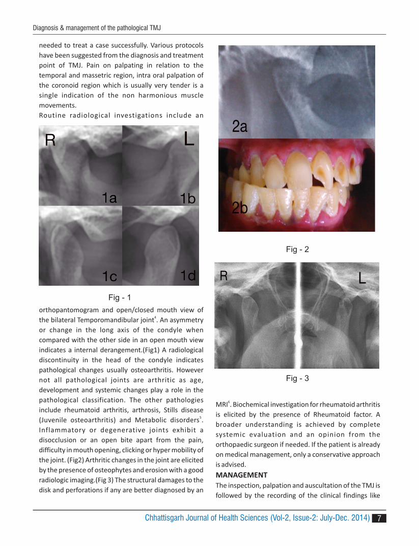
Routine radiological investigations include an
orthopantomogram and open/closed mouth view of
the bilateral Temporomandibular joint . An asymmetry 4
or change in the long axis of the condyle when
compared with the other side in an open mouth view
indicates a internal derangement.(Fig1) A radiological
discontinuity in the head of the condyle indicates
pathological changes usually osteoarthritis. However
not all pathological joints are arthritic as age,
development and systemic changes play a role in the
pathological classification. The other pathologies
include rheumatoid arthritis, arthrosis, Stills disease
(Juvenile osteoarthritis) and Metabolic disorders .5
Inflammatory or degenerative joints exhibit a
disocclusion or an open bite apart from the pain,
difficulty in mouth opening, clicking or hyper mobility of
the joint. (Fig2) Arthritic changes in the joint are elicited
by the presence of osteophytes and erosion with a good
radiologic imaging.(Fig 3) The structural damages to the
disk and perforations if any are better diagnosed by an
MRI . Biochemical investigation for rheumatoid arthritis 6
is elicited by the presence of Rheumatoid factor. A
broader understanding is achieved by complete
systemic evaluation and an opinion from the
orthopaedic surgeon if needed. If the patient is already
on medical management, only a conservative approach
is advised.
MANAGEMENT
The inspection, palpation and auscultation of the TMJ is
followed by the recording of the clinical findings like
Fig - 1
Fig - 2
Fig - 3
Chhattisgarh Journal of Health Sciences (Vol-2, Issue-2: July-Dec. 2014)
Diagnosis & management of the pathological TMJ

8
maximum mouth opening, relationship of the dental
midline (upper maxillary incisor midline) to the facial
midline on opening and closing. Deviation, dislocation
and deflection of the mandible (with reference to the
dental midline) on opening and closing, timing of the
clicking on opening and closing are to be documented.
As these finding play an important role in evaluating the
prognosis. The authors follow the below mentioned
protocol in their line of management.
Initial/Pain management:
1. Ice pack on the affected side
2. Soft diet
3. Analgesics and anti depressants
After pain reduces :
4. Mouth opening exercises- regular opening and
closing
5. Conscious bilateral chewing on both sides
(practiced with mirror in front)
6. Conventional Occlusal splint- to relieve stress at the
TMJ- to be worn at nights, travel, watching TV
Patients with reduced mouth opening :
7. Warm fomentation bilaterally on the lateral side of
the face, temporal region and neck regions.
8. Physiotherapy- ultra shortwave diathermy or TENS
9. Low level laser therapy
Further management:
10. Occlusal rehabilitation to aid in bilateral chewing-
replacement of missing teeth, extraction of supra
erupted maxillary wisdom teeth, to check
functional occlusion if all teeth are present.
Pathological management:
11. Tricyclic antidepressants
12. Glucosamine and chondroitin sulphate
13. Steroids
14. Patients who do not respond to this protocol are
further evaluated with MRI and minimally invasive
surgical therapy.
Evaluation of the occlusal splint and the treatment as a
whole can be assessed by the timing of the clicking in
the opening and closing movements. The deviation of
the mandible from the dental midline reduces. Anterior
repositioning appliance has also been documented of
good use if properly made . Medical management is not 7
a long term option, it should be reduced or
discontinued as needed. As mentioned earlier the
treatment protocol will not be effective if appropriate
care is not given to the occlusal rehabilitation. Often
patients do not report to the dentist after their acute
phase subsides. Hence the need for occlusal harmony
should be stressed during every visit.
DISCUSSION
Pain and pathology of TMJ is multi factorial which
makes the diagnosis and treatment more complex. It
requires a multi disciplinary approach . Occlusal 8
rehabilitation should be the prime target for the dental
practitioner as various studies has pointed out. One
should also understand occlusal splint therapy is a
supportive splint therapy. During occlusal rehabilitation
the endodontic procedures should not be for long
duration and the prosthodontic aim should be focused
on achieving good functional occlusion. The supportive
occlusal splints can be hard, soft or functional as
needed.
The role of surgery is always indicated for those patients
w i t h l i m i t e d o r n o m o u t h o p e n i n g a t a l l .
S imple invas ive surg ica l therapies l ike TMJ
arthrocentesis should be considered before an open 9
surgery . Conservative and palliative management 10
seems to provide better and long term results with less
or no morbidity.
CONCLUSION
Fo l lowing the bas ic protoco l w i th min imal
investigations and occlusal therapies which are aimed
at patient education and long term follow should be the
g o a l s i n t h e t r e a t m e n t o f p a t h o l o g i c a l
Temporomandibular joints.
REFERENCES
1. Dworkin SF, LeResche L. Research diagmostic
criteria for temporomandibular disorders: review,
criteria, examinations and specifications, critique. J
Craniomandib Disord. 1992;6:301–355
2. Wilkes CH. Internal derangements of the
temporomandibular joint: pathological variations.
Arch Otolaryngol Head Neck Surg. 1989; 115:
469–477
3. In: de Leeuw R editors. Orofacial pain: guidelines
for asssessment, diagnosis, and management. 4th
Chhattisgarh Journal of Health Sciences (Vol-2, Issue-2: July-Dec. 2014)
Diagnosis & management of the pathological TMJ

ed.. Chicago: Quintessence Publishing; 2008
4. Inclination of the temporomandibular joint
eminence and anterior disc displacement. Int J of
Oral Maxillofac Surg.1989;18:229-232
5. Gynther GW, Holmlund AB, Reinholt FP, Lindblad S.
Temporomandibular joint involvement in
generalized osteoarthritis and rheumatoid arthritis:
a c l i n i ca l , a r t h ro s co p i c , h i s to l o g i c , a n d
immunohistochemical study. Int J Oral Maxillofac
Surg. 1997;26:10–16
6. L.M.J. Helenius, P. Tervahartiala, I. Helenius, J. Al-
Sukhun, et al. Clinical, radiographic and MRI
findings of the temporomandibular joint in patients
with different rheumatic diseases International
Journal of Ora l & Maxi l lofac ia l Surgery.
2006;35:11:983-989
7. Roger A. Solow. Customized anterior guidance for
occlusal devices: Classification and rationale.The
Journal of Prosthetic Dentistry, 2013;110:4:259-
263
8. Epidemiology, Diagnosis, and Treatment of
Temporomandibular DisordersReview Article.
Dental Clinics of North America 2013;57: 465-479
9. F.A. Al-Belasy, M.F. Dolwick. Arthrocentesis for the
treatment of temporomandibular joint closed lock:
a review article. International Journal of Oral &
Maxillofacial Surgery 2007;36:773-782
10. Dolwick MF, Dimitroulis G. Is there a role for
temporomandibular joint surgery. Br J Oral
Maxillofac Surg. 1994;32:307–313
Diagnosis & management of the pathological TMJ
9Chhattisgarh Journal of Health Sciences (Vol-2, Issue-2: July-Dec. 2014)
10
1 2 3N.Mohan , Sukriti Kumar , Jayashree Mohan1. Professor & HOD, Department of Oral Medicine & Radiology, VMS Dental college, Salem (TN)2. Post Graduate Student, Dept. of Oral medicine & Radiology, VMS Dental college, Salem (TN)3. Professor & HOD, Department of Prosthodontics, VMS Dental college, Salem (TN)
Corresponding Author:Dr. N. Mohan, Professor, Department of Oral Medicine & Radiology, VMS Dental college, Salem (TN)Email : [email protected], Mobile: 09843082608
ABSTRACT
Objective : This study was carried out to determine the reliability of sex determination from tooth pulp tissue.
Methods : This study was carried on 30 teeth samples. Out of which 15 were permanent and 15 were deciduous
teeth. (8 male teeth and 7 female teeth in each group) which were indicated for extraction advised for orthodontic
treatment, Retained deciduous& Periodontally compromised tooth. Teeth was extracted and pulp taken out after
access opening was transferred in to fixative solution for 24 hours. the pulp cells were stained with harris's
hemotoxylin and eosin stains which was examined under oil immersion lens of light microscope to study the barr
body.
Results : Study of sex determination with tooth pulp proved to be reliable for deciduous teeth when association of
barr body and sex of permanent and deciduous teeth were tested. And also the overall statistical analysis of sex wise
estimation of barr bodies involving both deciduous and permanent teeth showed significant results for female
group.
Conclusion : The Barr body test is shown to be a reliable, simple, and cost-effective technique for sex identification.
Keywords : Barr bodies, sex determination, tooth pulp tissue,odontology
INTRODUCTION:
Forensic odontology can be defined in many ways . The 1
Federation Dentaire Internationale (FDI) defines
forensic odontology as that branch of dentistry which,
in the interest of justice, deals with the proper handling
and examination of dental evidence and with the
proper evaluation and presentation of dental findings.
According to the American Society of Forensic
Odontology, forensic odontology is by definition, the
application of dental science to the law, i.e. the use of
dental evidence in the interest of justice.Human
identification is one of the major fields of study and
research in forensic science because it deals with the
human body and aims at establishing human identity 2
Tooth enamel is the hardest tissue in the body, and the
teeth remain intact after death, thus making them
useful for forensic identification of sex with respect to
morphological characteristics (Haga, 1959; Gonda,
1959;Garm, 1964) and soft tissues (Das et al., 2004) 2
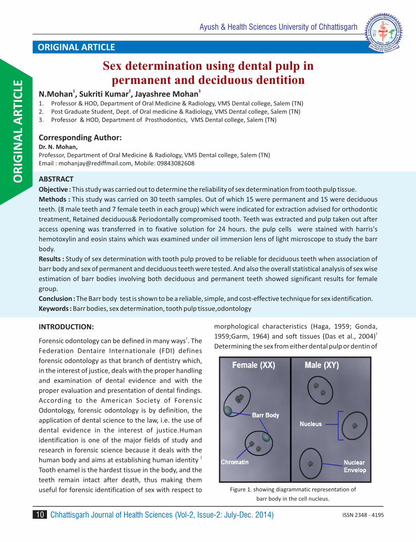
Determining the sex from either dental pulp or dentin of
Figure 1. showing diagrammatic representation of
barr body in the cell nucleus.
Sex determination using dental pulp in permanent and deciduous dentition
OR
IGIN
AL
AR
TIC
LEAyush & Health Sciences University of Chhattisgarh
ORIGINAL ARTICLE
Chhattisgarh Journal of Health Sciences (Vol-2, Issue-2: July-Dec. 2014) ISSN 2348 - 4195

11
tooth can also provide criminal investigators with useful
intelligence and can aid the identification of missing
persons and disaster victims. Forensic odontology is
useful in identification of age and sex of patients Sex of 3
the individual can be determined by using X and Y
chromosomes in the cells which are inactive. X
chromatin in its inactivated form is present as a mass
against the nuclear membrane in females is known as
Barr body as it was first named by Barr and Bertem
(1949)(Fig 1). These Barr bodies are present in 40% of
females who are considered as chromatin positive and
absent in males who are considered as chromatin
negative.
MATERIAL AND METHODS:
A total of 30 teeth were collected and were grouped in
to permanent and deciduous. Each group comprised
of 15 teeth . Out of 15 teeth (7 males and 8 females)
were selected out of patients who came for treatment
in vinayaka missions sankarachariyar dental college
salem .Eligibility criteria included was extractions
advised for orthodontic treatment. Retained
deciduous.,Periodontally compromised tooth, age
criteria:-up to 45 years .and those Teeth with dental
caries,Grossly destructed teeth, Non vital tooth were
excluded from the study.
An ethical committee clearance was taken and
informed consent was obtained .Either the patients or
their guardians, if they are minors, were informed
about the objectives of the investigation.
The teeth were removed by conventional technique,
washed with sterile water to remove residual blood,
The pulp was conventionally obtained through the
normal access cavity on the occlusal surface of the
teeth; dental pulp tissues were obtained using
standardized K-files(Fig 5). The pulp tissue was then
transferred to the dry and clean conical centrifuge tubes
containing 5 ml. of fixative (3 Methanol: 1 Glacial acetic
acid) and left as such for about half an hour to 24 hours
for the fixation of the pulp cells. It was then crushed /
teased with the glass rod sufficiently to isolate the pulp
cells. A suspension thus obtained was centrifuged for 10
Figure 5. Pictures showing material and method involved in the study
Sex Determination Using Dental Pulp
Chhattisgarh Journal of Health Sciences (Vol-2, Issue-2: July-Dec. 2014)

Sex Determination Using Dental Pulp
12
minutes at 1000 rpm. The supernatant was discarded,
leaving behind the pellet in the centrifuge tube. 5ml of
fresh fixative was then added to re-suspend the pellet
and the process was repeated thrice till a clear
suspension of the pulp cells was obtained.
Thin smears were prepared on chilled microscope slides
of 1 mm thickness by the air drying method i.e. by
dropping 2 –3 drops of the above suspension on the
slide from a distance of inches to get a homogenous
population of cells. Two smears were made from each
suspension of the specimen; one slide was stained with
Harris's Hemotoxylin and Eosin stain to study the Barr
bodies 3
RESULTS:
When association of barr body and sex of permanent
and deciduous teeth were tested with chi square test
and compared it was observed that p-value was highly
significant in deciduous (Table 2) and significant in
permanent teeth(Table1). Hence,suggesting study to be
more reliable for the deciduous teeth.
Overall statistical analysis of sex wise estimation of barr
bodies involving both deciduous and permanent teeth
(Table3) showed p-value 0.001 which was highly
significant at 1% therefore more percentage of barr
bodies was observed in female group.
And finally, the association between the type of teeth
and presence of barr bodies were tested with chi square
test , Table 4 showed that the p-value is less than 0.5
hence the result is significant at 5%,Therefore it is
concluded that there is significant association found
between the type of teeth and barr bodies.It was
observed from the study that deciduous teeth is
showing more percentage towards barr bodies than
permanent teeth. Estimation of barr bodies in
deciduous teeth were more significant when compared
to permanent teeth
Table 1 . Association between sex and Barr Bodies - Permanent Teeth
Sex
Barr bodies
Total Chi
square p Positive Negative
N % N %
Male 8 100.00 8 4.28 0.038*
Female 3 42.86 4 57.14 7
Total 3 20.00 12 80.00 15
* Significant at 5 %
Table 2 . Association between sex and Barr Bodies - Deciduous Teeth
Sex
Barr bodies
Total Chi
square p Positive Negative
N % N %
Male 1 14.29 6 85.71 7 11.43 0.001**
Female 8 100.00 - - 8
Total 9 60.00 6 40.00 15
** Significant at 1 % (Highly Significant)
Chhattisgarh Journal of Health Sciences (Vol-2, Issue-2: July-Dec. 2014)
13
DISCUSSION:
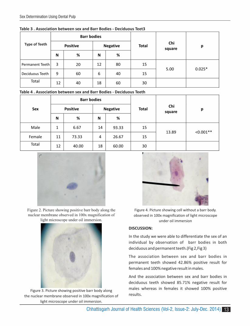
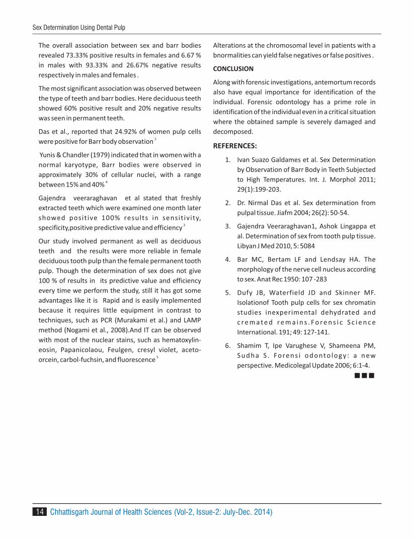
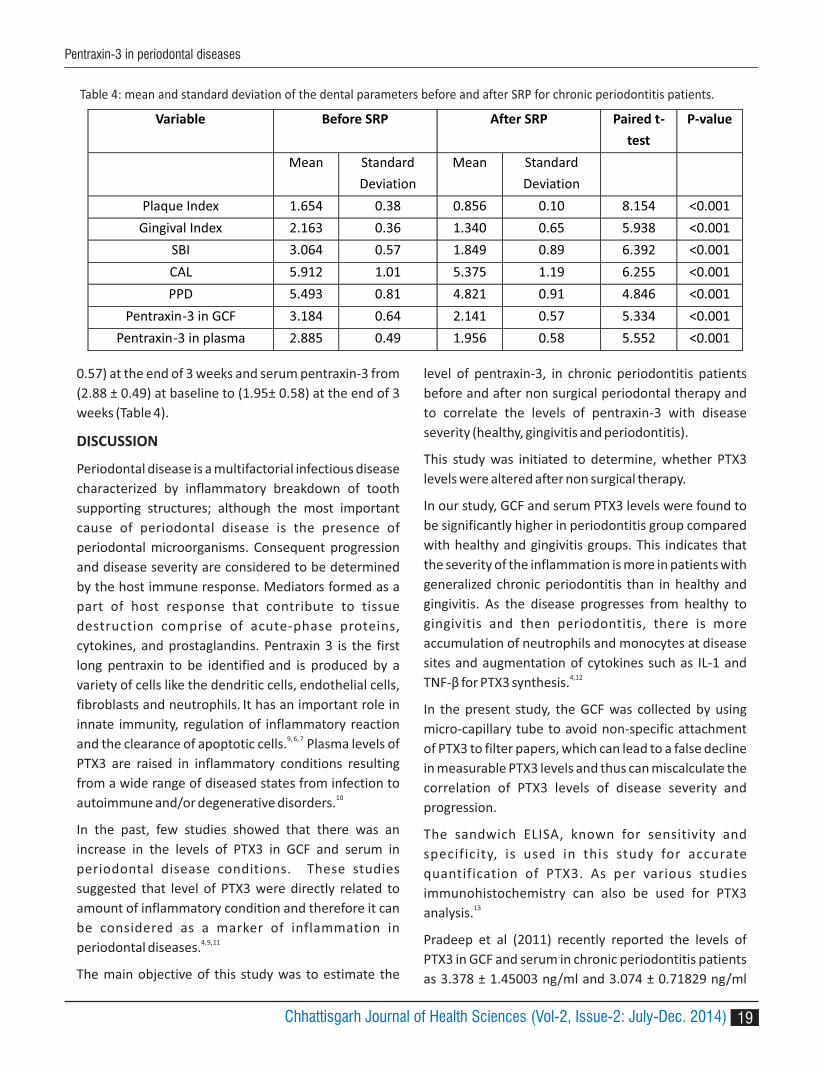
In the study we were able to differentiate the sex of an
individual by observation of barr bodies in both
deciduous and permanent teeth.(Fig 2,Fig 3)
The association between sex and barr bodies in
permanent teeth showed 42.86% positive result for
females and 100% negative result in males.
And the association between sex and barr bodies in
deciduous teeth showed 85.71% negative result for
males whereas in females it showed 100% positive
results. Figure 3. Picture showing positive barr body along
the nuclear membrane observed in 100x magnification of
light microscope under oil immersion.
Figure 2. Picture showing positive barr body along the nuclear membrane observed in 100x magnification of
light microscope under oil immersion.
Figure 4. Picture showing cell without a barr body.
observed in 100x magnification of light microscope
under oil immersion
Table 4 . Association between sex and Barr Bodies - Deciduous Teeth
Sex
Barr bodies
Total Chi
square p Positive Negative
N % N %
Male 1 6.67 14 93.33 15 13.89 <0.001**
Female 11 73.33 4 26.67 15
Total 12 40.00 18 60.00 30
Type of Teeth
Table 3 . Association between sex and Barr Bodies - Deciduous Teet3
Barr bodies
Total Chi
square p Positive Negative
N % N %
Permanent Teeth 3 20 12 80 15 5.00 0.025*
9 60 6 40 15
Total 12 40 18 60 30
Deciduous Teeth
Sex Determination Using Dental Pulp
Chhattisgarh Journal of Health Sciences (Vol-2, Issue-2: July-Dec. 2014)
Sex Determination Using Dental Pulp
14
The overall association between sex and barr bodies
revealed 73.33% positive results in females and 6.67 %
in males with 93.33% and 26.67% negative results
respectively in males and females .
The most significant association was observed between
the type of teeth and barr bodies. Here deciduous teeth
showed 60% positive result and 20% negative results
was seen in permanent teeth.
Das et al., reported that 24.92% of women pulp cells
were positive for Barr body observation 2
Yunis & Chandler (1979) indicated that in women with a
normal karyotype, Barr bodies were observed in
approximately 30% of cellular nuclei, with a range
between 15% and 40% 4
Gajendra veeraraghavan et al stated that freshly
extracted teeth which were examined one month later
showed posit ive 100% results in sensit iv ity,
specificity,positive predictive value and efficiency 3
Our study involved permanent as well as deciduous
teeth and the results were more reliable in female
deciduous tooth pulp than the female permanent tooth
pulp. Though the determination of sex does not give
100 % of results in its predictive value and efficiency
every time we perform the study, still it has got some
advantages like it is Rapid and is easily implemented
because it requires little equipment in contrast to
techniques, such as PCR (Murakami et al.) and LAMP
method (Nogami et al., 2008).And IT can be observed
with most of the nuclear stains, such as hematoxylin-
eosin, Papanicolaou, Feulgen, cresyl violet, aceto-
orcein, carbol-fuchsin, and fluorescence 5
Alterations at the chromosomal level in patients with a
bnormalities can yield false negatives or false positives .
CONCLUSION
Along with forensic investigations, antemortum records
also have equal importance for identification of the
individual. Forensic odontology has a prime role in
identification of the individual even in a critical situation
where the obtained sample is severely damaged and
decomposed.
REFERENCES:
1. Ivan Suazo Galdames et al. Sex Determination
by Observation of Barr Body in Teeth Subjected
to High Temperatures. Int. J. Morphol 2011;
29(1):199-203.
2. Dr. Nirmal Das et al. Sex determination from
pulpal tissue. Jiafm 2004; 26(2): 50-54.
3. Gajendra Veeraraghavan1, Ashok Lingappa et
al. Determination of sex from tooth pulp tissue.
Libyan J Med 2010, 5: 5084
4. Bar MC, Bertam LF and Lendsay HA. The
morphology of the nerve cell nucleus according
to sex. Anat Rec 1950: 107 -283
5. Dufy JB, Waterfield JD and Skinner MF.
Isolationof Tooth pulp cells for sex chromatin
studies inexperimental dehydrated and
c r e m a t e d r e m a i n s . F o r e n s i c S c i e n c e
International. 191; 49: 127-141.
6. Shamim T, Ipe Varughese V, Shameena PM,
Su d h a S . Fo ren s i o d o nto lo gy : a n ew
perspective. Medicolegal Update 2006; 6:1-4.
Chhattisgarh Journal of Health Sciences (Vol-2, Issue-2: July-Dec. 2014)

15
1 2 3Kokila G , Renuka Devi R , Vineeta Gupta1. Lecturer, Department of Periodontics, KSR Institute of Dental Science and Research, Tiruchengode, (TN) India
2. Reader, Department of Periodontics, KSR Institute of Dental Science and Research, Tiruchengode, (TN) India
3. Reader, Department of Periodontics , Government Dental College Raipur (C.G)
Corresponding Author :Dr. G. Kokila
Lecturer, Department of Periodontics, KSR Institute of Dental Science and Research Tiruchengode – 637215,
email: [email protected]
ABSTRACT :
Background : Pentraxins are acute phase proteins which belong to a family of evolutionarily conserved proteins,
considered as markers of inflammation. Pentraxin 3 (PTX3) is a prototype of the long pentraxin group. It is suggested
to play an important role in innate immunity, regulation of inflammation and clearance of apoptotic cells. Hence this
study was planned and designed to estimate the level of pentraxin-3 in chronic periodontitis before and after non
surgical periodontal therapy and correlate its level with disease severity (healthy, gingivitis and periodontitis).
Materials and methods : A total of 45 individuals both males and females of age group (23-50yrs) were included in
the study and they were divided into three groups of 15 in each. Control group A (group I, = 15) healthy, Control group
B (group II = 15) gingivitis, Test group (group III =15) generalized chronic periodontitis.3weeks after intervention
(scaling and root lanning), the 15 subjects from Group III were categorized as fourth group (Group IV). GCF and
plasma samples obtained from each subjects were quantified for pentraxin-3 using sandwich enzyme linked
immunosorbent assay (ELISA) technique.
Statistical analysis : Chi-square test, ANOVA, ANCOVA, Spearman correlation coefficient and Paired t test were used
for statistical analysis of this study. P value of less than 0.05 was considered to be statistically significant.
Results : The mean GCFPTX3 concentration increased from healthy to gingivitis groups and then from gingivitis to
periodontitis groups (1.402 ng/ml <2.299 ng/ml <3.184 ng/ml). Similarly the mean plasma PTX3 concentration was
highest in periodontitis group (2.885ng/ml) followed by the gingivitis group (2.118 ng/ml) and lowest in the healthy
group (0.983 ng/ml). The mean differences between the groups were also statistically significant (p<0.001). The GCF
and plasma PTX3 concentrations in chronic periodontitis decreased (2.14 ± 0.57, 1.95 ± 0.58) after treatment.
Conclusion : Pentraxin3 level increases in GCF and plasma, from periodontal health to the diseased condition, as well
as there is distinct decrease in the level after periodontal therapy. This data indicates that pentraxin-3 plays a key role
in periodontal disease and could be considered as a biomarker in periodontal disease progression.
Key Words : Chronic periodontitis, Enzyme linked immunoabsorbent assay, Gingival crevicular fluid, Pentraxin-3.
Evaluation of pentraxin-3 inflammatory Marker level in generalized chronic periodontitis before and after mechanical
therapy (scaling and root planing)
OR
IGIN
AL
AR
TIC
LE
Ayush & Health Sciences University of Chhattisgarh
ORIGINAL ARTICLE
Chhattisgarh Journal of Health Sciences (Vol-2, Issue-2: July-Dec. 2014)ISSN 2348 - 4195

16
INTRODUCTION
Chronic periodontitis is an infectious disease of bacteria
characterized by the inflammatory breakdown of tooth
supporting structures including hard and soft tissues.
The initiation and progression of periodontal disease is
caused by interaction between periodontal pathogens
and host immune system. The periodontal pathogens
contains a number of potential virulence factors like
antigens, lipopolysaccharide and heat shock proteins,
which trigger the local and systemic immune and
inflammatory response. The local inflammatory
response stimulates hepatocytes and the other cells
including neutrophils, monocytes, macrophages,
vascular endothelial cells, fibroblast and smooth
muscle cells to release various acute phase proteins
(APR).1, 2, 3
Pentraxins, a super family of acute phase proteins are
identified as biomarkers in inflammatory conditions. It
has an important role in the innate immune system.
Pentraxins are divided into two groups based on
primary structure of the subunit: short pentraxin (C-
reactive protein and serum amyloid protein), long
pentraxins (pentraxin-3 [PTX3] and PTX4), and several
neuronal pentraxins. 4, 5
Pentraxin-3 is identified as first member of the long
pentraxin super family. It is produced by macrophages
and other cell types in response to IL-1 , tumour β
necrosis factor-alpha [TNF- ] and microbial α
components including lipopolysaccharides.6, 7
Measurement of pentraxin-3 in GCF or plasma may help
in the identification of a subset of patients who are at a
higher risk for destructive disease or those who are
undergoing the process of periodontal breakdown.8
To date, this is the second study to examine the
effect of nonsurgical periodontal therapy on GCF and
serum level of pentraxin-3. The aim of study was to
estimate the level of pentraxin-3 in GCF and blood
before and after non surgical periodontal therapy in
chronic periodontitis and correlate its levels with
disease severity.
MATERIALS AND METHODS
The study protocol was analyzed and approved by the
Institutional Ethical Review Board. Written and verbal
informed consent was obtained from the subjects
participating in the study. A total of 45 subjects (20
males and 25 females) were participated in the study.
Inclusion criteria:
1. Age group 23 to 55yrs
2. At least 20 natural teeth
3. Good general health without any history of systemic
disease
Exclusion criteria:
1. Any autoimmune disease or other systemic
diseases that could change the course of
periodontal disease.
2. Subjects having history of smoking or any form of
tobacco use previously
3. Use of a medication like antibiotic drugs or anti
inflammatory drugs in the past 3 weeks
4. History of periodontal therapy in the past 6 months
5. Pregnant/ lactating women
6. Unwillingness to join in the study.
Each subject underwent full mouth periodontal probing
and charting, along with digital OPG.
The subjects were divided into 3 groups of 15 each
based on scores of plaque index (Sillness and Loe 1964),
gingival index (Loe and Sillness 1963), sulcus bleeding
index (Muhlemann 1971), probing depth (PD), clinical
attachment level (CAL) and radiographic evidence of
bone loss. Control group (Group 1 = clinically healthy
[n=15 (no bleeding on probing, gingival index = 0
probing depth 3mm, CAL=0, radiographically no bone ≤
loss). Control group (Group 2 = gingivitis [n=15
(clinically, signs of gingival inflammation and bleeding
on probing present, gingival index >1, probing depth ≤
3mm and no CAL or radiographic bone loss). Test group
(Group 3= generalized chronic periodontitis [n=15
(clinically, signs of gingival inflammation and bleeding
on probing present, gingival index >1, probing depth ≥
5mm, CAL 3mm and radiographic evidence of ≥
bone loss). Group 4 = Group 3 patients [n=15
(generalized chronic periodontitis) receiving non
surgical therapy (scaling and root planning) are
converted into Group4.
Chhattisgarh Journal of Health Sciences (Vol-2, Issue-2: July-Dec. 2014)
Pentraxin-3 in periodontal diseases

17
EXAMINATION METHOD
The clinical and radiographic examinations, group
allocations were performed by single examiner for all
patients. Samples were collected from predetermined
sites in each patient on the following day by same
examiner. This was carried out to avoid the
contamination of GCF with blood associated with the
probing of inflamed sites. Only one site per subject was
selected as sampling site in group 2 (gingivitis) and
group 3 (generalized chronic periodontitis) whereas, in
group 1 (healthy), many sites were sampled. Probing
depth and clinical attachment levels were measured by
using a William graduated periodontal probe.
GCF Collection
After drying the selected area, supragingival plaque was
removed by using the Gracey curettes. Care was taken
not to touch the gingival margins after which the area
was isolated with cotton rolls to prevent saliva
contamination. GCF was collected by gently placing the
microcapillary tube at the entrance of the gingival
sulcus. A standardized volume of 1μl was collected in
each group by ibration on black using the cal
colour–coded 1- to 5-μL calibrated volumetric
microcapillary tubes. Maximum of 10-15 minutes were
allotted for each sample. If GCF is not expressed within
the allotted time, that sites were excluded. This was
done to ensure atraumatism. The micropipettes that
were suspected to be contaminated with blood or saliva
were also excluded. Collected GCF samples were
immediately transferred to airtight plastic vials and
were diluted with phosphate buffer saline up to 100μl 0and immediately transferred and stored at -70 C until
assayed.
Blood Collection
2ml of blood was collected from anticubital fossa by
venipuncture using a graduated syringe with 20 gauge
needle. The collected blood was transferred to a test
tube containing EDTA. Immediately the collected
samples were sent to laboratory for processing. Plasma
was separated within 30 minutes from collected blood
by centrifuging at 1000 x g for 15 minutes and
immediately transferred to a plastic vial and stored at -
70 C until assayed.◦
Non surgical periodontal therapy
The patient in group 3 received non surgical periodontal
therapy (scaling and root planning) within 1 or 2 visits
according to patient needs. Oral hygiene instructions
were given which included, tooth brushing techniques
and the use of dental floss. If patient reported any
sensitivity, instruction was given to use desensitizing
tooth paste (Thermoseal). The patients were recalled
after 21 days for recording of plaque index, gingival
index, sulcus bleeding index, probing depth and CAL.
GCF and blood samples were collected and sent to
laboratory and stored at -70 C ◦
Assay Procedure
The pentraxin-3 levels in collected GCF and blood
sample were assayed using an enzyme-linked
immunosorbent assay (ELISA KIT). The assay employed
the quantitative sandwich enzyme immunoassay
technique. Antibody specific for PTX3 was pre-coated
onto a microplate and antigen samples to be tested was
added into the wells and any PTX3 present was bound
by the immobilized antibody. After removing any
unbound substances, a biotin-conjugated antibody
specific for PTX3 was added to the wells. After washing,
avidin conjugated Horseradish Peroxidase (HRP) was
added to the wells. Following a wash to remove any
unbound avidin-enzyme reagent, a substrate solution
was added to the wells and color developed in
proportion to the amount of PTX3 bound in the initial
step. Once pentraxin-3 binds with antibody completely,
the color development stops and the intensity of the
color was measured. The absorbance of each well is
read on an ELISA reader using 450 nm as the primary
wavelength. The concentration of PTX3 in the tested
samples was estimated using the standard curve.4
Statistical Analysis
Statistical analysis was done using software program.
One way ANOVA was carried out to compare the mean
clinical parameters between the groups. In addition,
Chi-square test was done to assess sex distribution of
subjects in groups. Pearson's correlation coefficient has
been applied to find out the relationship between the
pentraxin-3 level and the selected variables. Paired t
test was used to analyze mean and standard deviation
Chhattisgarh Journal of Health Sciences (Vol-2, Issue-2: July-Dec. 2014)
Pentraxin-3 in periodontal diseases

18
of clinical parameters and pentraxin-3 levels before and
after SRP for group III patients. P <0.05 was considered
to be statistically significant.
RESULTS
Descriptive statistics of baseline parameters of the
study population are shown in Table1.
The results of the present study indicated that the mean
PTX3 concentration in GCF was highest in group III
(3.184ng/ml).The mean GCFPTX3 concentration
increased from healthy to gingivitis groups and then
from gingivitis to periodontitis groups (1.402 ng/ml
<2.299 ng/ml <3.184 ng/ml) (Table 2). Similarly the
mean plasma PTX3 concentration was highest in
periodontitis group (2.885ng/ml) followed by the
gingivitis group (2.118 ng/ml) and lowest in the healthy
group (0.983 ng/ml). The mean differences between
the groups were also statistically significant (p<0.001)
(Table 3).
Paired t test showed a statistically very highly significant
reduction of clinical parameters and CF and plasma G
PTX3 levels of group III after SRP. The clinical parameters
has been reduced from (1.65 ± 38, 2.16 ± 0.36, 3.06±
0.57, 5.91± 1.01 and 5.49 ± 0.81) at baseline to (0.85
±0.10, 1.34 ±0.64, 1.84 ± 0.89, 5.37 ±1.19 and 4.82 ±
0.91) at the end of 3 weeks (Table 4).
Result of group III revealed that there was a very highly
statistically significant (p < 0.001) reduction of GCF
pentraxin-3 from (3.18 ± 0.64) at baseline to (2.14 ±
Table 1: Descriptive statistics of baseline parameters in the study population
PI (Mean±SD) GI (Mean±SD) SBI (Mean±SD) CALMean±SD)
PPDMean±SD)
Group I 0.338 ± 0.11 0.216 0.226 1.402 ± 0.29 0.938± 0.29
Group II 0.675 ±0.21 0.964 1.224 2.299± 0.36 2.118± 0.41
Group III 1.654 ±0.38 2.162 3.064 3.184± 0.64 2.885± 0.49
Table 2: Descriptive statistics of pentraxin-3 inflammatory marker level in GCF
Pentraxin-3
in GCF
Mean Standard
deviation
Minimum Maximum ANOVA- X² - value
P-value Scheffe’s
multiple
comparison
test result
Group I 1.402 0.29 0.87 1.87 56.76 <0.001 GI<GII<GIII
Group II 2.299 0.36 1.54 2.83
Group III 3.184 0.64 1.98 4.25
Table 3: Descriptive statistics of pentraxin-3 inflammatory marker level in plasma
Pentraxin-3
in plasma
Mean Standard
deviation
Minimum Maximum ANOVA- X²- value
P-
value
Scheffe’s
multiple
comparison
test result
Group I 0.938 0.29 0.32 1.56 84.90 <0.001 GI <GII <GIII
Group II 2.118 0.41 1.35 3.00
Group III 2.885 0.49 1.63 3.56
Pentraxin-3 in periodontal diseases
Chhattisgarh Journal of Health Sciences (Vol-2, Issue-2: July-Dec. 2014)
19
0.57) at the end of 3 weeks and serum pentraxin-3 from
(2.88 ± 0.49) at baseline to (1.95± 0.58) at the end of 3
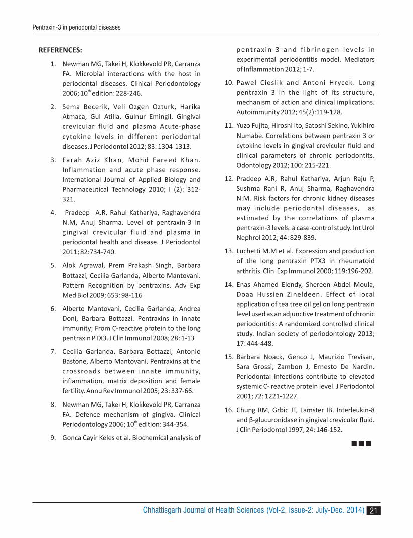
weeks (Table 4).
DISCUSSION
Periodontal disease is a multifactorial infectious disease
characterized by inflammatory breakdown of tooth
supporting structures; although the most important
cause of periodontal disease is the presence of
periodontal microorganisms. Consequent progression
and disease severity are considered to be determined
by the host immune response. Mediators formed as a
part of host response that contribute to tissue
destruction comprise of acute-phase proteins,
cytokines, and prostaglandins. Pentraxin 3 is the first
long pentraxin to be identified and is produced by a
variety of cells like the dendritic cells, endothelial cells,
fibroblasts and neutrophils. It has an important role in
innate immunity, regulation of inflammatory reaction
and the clearance of apoptotic cells. Plasma levels of 9, 6, 7
PTX3 are raised in inflammatory conditions resulting
from a wide range of diseased states from infection to
autoimmune and/or degenerative disorders.10
In the past, few studies showed that there was an
increase in the levels of PTX3 in GCF and serum in
periodontal disease conditions. These studies
suggested that level of PTX3 were directly related to
amount of inflammatory condition and therefore it can
be considered as a marker of inflammation in
periodontal diseases.4, 9, 11
The main objective of this study was to estimate the
level of pentraxin-3, in chronic periodontitis patients
before and after non surgical periodontal therapy and
to correlate the levels of pentraxin-3 with disease
severity (healthy, gingivitis and periodontitis).
This study was initiated to determine, whether PTX3
levels were altered after non surgical therapy.
In our study, GCF and serum PTX3 levels were found to
be significantly higher in periodontitis group compared
with healthy and gingivitis groups. This indicates that
the severity of the inflammation is more in patients with
generalized chronic periodontitis than in healthy and
gingivitis. As the disease progresses from healthy to
gingivitis and then periodontitis, there is more
accumulation of neutrophils and monocytes at disease
sites and augmentation of cytokines such as IL-1 and 4,12
TNF-β for PTX3 synthesis.
In the present study, the GCF was collected by using
micro-capillary tube to avoid non-specific attachment
of PTX3 to filter papers, which can lead to a false decline
in measurable PTX3 levels and thus can miscalculate the
correlation of PTX3 levels of disease severity and
progression.
The sandwich ELISA, known for sensitivity and
specificity, is used in this study for accurate
quantification of PTX3. As per various studies
immunohistochemistry can also be used for PTX3
analysis.13
Pradeep et al (2011) recently reported the levels of
PTX3 in GCF and serum in chronic periodontitis patients
as 3.378 ± 1.45003 ng/ml and 3.074 ± 0.71829 ng/ml
Table 4: mean and standard deviation of the dental parameters before and after SRP for chronic periodontitis patients.
Variable Before SRP After SRP Paired t-
test
P-value
Mean Standard
Deviation
Mean Standard
Deviation
Plaque Index 1.654 0.38 0.856 0.10 8.154 <0.001
Gingival Index 2.163 0.36 1.340 0.65 5.938 <0.001
SBI 3.064 0.57 1.849 0.89 6.392 <0.001
CAL 5.912 1.01 5.375 1.19 6.255 <0.001
PPD 5.493 0.81 4.821 0.91 4.846 <0.001
Pentraxin-3 in GCF 3.184 0.64 2.141 0.57 5.334 <0.001
Pentraxin-3 in plasma 2.885 0.49 1.956 0.58 5.552 <0.001
Chhattisgarh Journal of Health Sciences (Vol-2, Issue-2: July-Dec. 2014)
Pentraxin-3 in periodontal diseases

20
respectively using the ELISA technique. The deepest
probing sites were used for sample collection. In our 4
study, the samples were obtained from sites with
deepest probing depth and PTX3 values in the GCF and
serum from patients with chronic periodontitis were
estimated at 3.184 ± 0.64 ng/ml and 2.885 ± 0.49 ng/ml
respectively.
In our study, PTX3 concentrations positively correlate
with clinical parameters in the periodontitis group. The
positive correlation between clinical parameters and
PTX3 levels can be attributed to the production of
cytokines at tissue injury sites. Neutrophils appear early
at sites of infection and injury. They represent a
reservoir of pre-stored PTX3 that are ready for rapid
release. These specific granules of PTX3 are released
from neutrophils in response to inflammatory signals.4
A study by Yuzo Fugita et al in 2012 reported GCF PTX3
levels to be significantly higher in patients with
periodontal disease site (0.64 ± 0.39 ng/ml) than
periodontal healthy sites (0.06 ± 0.10ng/ml) in patients
with chronic periodontitis. A strong positive correlation
was also observed between mean gingival index, pocket
depth, bleeding on probing, GCF levels and PTX3
levels. The GCF PTX3 level in the above study is almost [11]
6 times lower than the level obtained in our study
(3.184±0.64ng/ml).
The result of the present study revealed statistically
significant (p < 0.001) increase in the mean
concentration of PTX3 in GCF as the diseases
progressed from healthy (1.402 ± 0.29) to gingivitis
(2.299 ± 0.36) to periodontitis (3.184 ± 0.64). The results
were in accordance with the result of a study done by
Yuzo Fugita et al (2012) which showed that the mean
concentration of PTX3 was significantly higher (p < 0.01)
in diseased sites (0.64 ± 0.39) as compared to healthy
sites (0.06 ± 0.10).
Enas Ahmed Elgand et al (2013) study was conducted to
evaluate the effectiveness of SRP (Group I) and SRP with
adjunct treatment of tea tree oil (Group II) on clinical
parameters and level of pentraxin-3 in chronic
periodontitis. Serum samples were collected to
measure the serum PTX3 levels by using ELISA. This
study showed statistically significant reduction in
clinical parameters PTX3 levels in group II compared
with group I.14
The patients after non surgical periodontal therapy
(scaling and root planning) showed reduced GCF and
serum PTX3 levels and clinical parameters. Serum PTX3
was reduced from 2.885 ± 0.49 to 1.956 ± 0.5 at the end
of 3 weeks and GCF PTX3 also reduced from 3.184 ± 0.64
to 2.141 ± 0.57.
Mean pentraxin 3 values in comparison were analysed
before and after non surgical therapy in GCF and plasma
by using paired t test. Mean values shows statistically (p
< 0.001) significant differences. The result of our study
revealed that there was a highly statistically significant
(p < 0.001) reduction of clinical parameters and GCF
PTX3 levels in patient with chronic periodontitis after
nonsurgical therapy.
Clinical improvement after periodontal therapy was
associated with significant reduction in PTX3 in GCF and
plasma. Non surgical therapy (Scaling and root
planning) controls the local bacterial infection and leads
to minimum influx of PMN into GCF and reduces PTX3
expression in GCF. At the same time it decreases the
entry of bacteria into systemic circulation, thus
reducing PTX3 expression in serum.15, 16
To date, only one study by Enas Ahmed Elgend et al
(2013) showed effect of non-surgical therapy on
pentraxin-3 level in GCF samples of patients with
periodontal diseases.14
Limitations of our study were, Gingivitis patients did not
receive any SRP, because the aim of our study was to
check the impact of SRP on inflammatory marker PTX3
in GCF and plasma samples and for that the most
destructive periodontal disease was selected to obtain
better results. Other systemic inflammatory markers
were not analysed.
CONCLUSION
Quantitative sandwich enzyme immunoassay
techniques revealed the GCF and plasma PTX3 levels are
higher in pat ients with general ized chronic
periodontitis than healthy patients and those with
gingivitis. After non surgical therapy the PTX3 levels
reduced in both GCF and plasma. PTX3 concentration
was elevated with increasing severity of periodontal
d iseases and decreases with lower level of
inflammatory conditions.
Chhattisgarh Journal of Health Sciences (Vol-2, Issue-2: July-Dec. 2014)
Pentraxin-3 in periodontal diseases
21
REFERENCES:
1. Newman MG, Takei H, Klokkevold PR, Carranza
FA. Microbial interactions with the host in
periodontal diseases. Clinical Periodontology
2006; 10 edition: 228-246.th
2. Sema Becerik, Veli Ozgen Ozturk, Harika
Atmaca, Gul Atilla, Gulnur Emingil. Gingival
crevicular fluid and plasma Acute-phase
cytokine levels in different periodontal
diseases. J Periodontol 2012; 83: 1304-1313.
3. Farah Az iz Khan, Mohd Fareed Khan.
Inflammation and acute phase response.
International Journal of Applied Biology and
Pharmaceutical Technology 2010; I (2): 312-
321.
4. Pradeep A.R, Rahul Kathariya, Raghavendra
N.M, Anuj Sharma. Level of pentraxin-3 in
gingival crevicular fluid and plasma in
periodontal health and disease. J Periodontol
2011; 82:734-740.
5. Alok Agrawal, Prem Prakash Singh, Barbara
Bottazzi, Cecilia Garlanda, Alberto Mantovani.
Pattern Recognition by pentraxins. Adv Exp
Med Biol 2009; 653: 98-116
6. Alberto Mantovani, Cecilia Garlanda, Andrea
Doni, Barbara Bottazzi. Pentraxins in innate
immunity; From C-reactive protein to the long
pentraxin PTX3. J Clin Immunol 2008; 28: 1-13
7. Cecilia Garlanda, Barbara Bottazzi, Antonio
Bastone, Alberto Mantovani. Pentraxins at the
crossroads between innate immunity,
inflammation, matrix deposition and female
fertility. Annu Rev Immunol 2005; 23: 337-66.
8. Newman MG, Takei H, Klokkevold PR, Carranza
FA. Defence mechanism of gingiva. Clinical
Periodontology 2006; 10 edition: 344-354. th
9. Gonca Cayir Keles et al. Biochemical analysis of
p entrax in - 3 an d f ib r in o gen leve l s in
experimental periodontitis model. Mediators
of Inflammation 2012; 1-7.
10. Pawel Cieslik and Antoni Hrycek. Long
pentraxin 3 in the light of its structure,
mechanism of action and clinical implications.
Autoimmunity 2012; 45(2):119-128.
11. Yuzo Fujita, Hiroshi Ito, Satoshi Sekino, Yukihiro
Numabe. Correlations between pentraxin 3 or
cytokine levels in gingival crevicular fluid and
clinical parameters of chronic periodontits.
Odontology 2012; 100: 215-221.
12. Pradeep A.R, Rahul Kathariya, Arjun Raju P,
Sushma Rani R, Anuj Sharma, Raghavendra
N.M. Risk factors for chronic kidney diseases
may include periodontal diseases, as
estimated by the correlations of plasma
pentraxin-3 levels: a case-control study. Int Urol
Nephrol 2012; 44: 829-839.
13. Luchetti M.M et al. Expression and production
of the long pentraxin PTX3 in rheumatoid
arthritis. Clin Exp Immunol 2000; 119:196-202.
14. Enas Ahamed Elendy, Shereen Abdel Moula,
Doaa Hussien Zineldeen. Effect of local
application of tea tree oil gel on long pentraxin
level used as an adjunctive treatment of chronic
periodontitis: A randomized controlled clinical
study. Indian society of periodontology 2013;
17: 444-448.
15. Barbara Noack, Genco J, Maurizio Trevisan,
Sara Grossi, Zambon J, Ernesto De Nardin.
Periodontal infections contribute to elevated
systemic C- reactive protein level. J Periodontol
2001; 72: 1221-1227.
16. Chung RM, Grbic JT, Lamster IB. Interleukin-8
and β-glucuronidase in gingival crevicular fluid.
J Clin Periodontol 1997; 24: 146-152.
Chhattisgarh Journal of Health Sciences (Vol-2, Issue-2: July-Dec. 2014)
Pentraxin-3 in periodontal diseases

OR
IGIN
AL
AR
TIC
LE 1 2 3P. Gupta , S. Verma , P. Dubey1 Associate Professor, Nephrology Unit, Department of Medicine, Pt.J.N.M. Medical College & Dr. B.R.A.M. Hospital, Raipur2 Associate Professor, Department of Medicine, Pt.J.N.M. Medical College & Dr. B.R.A.M. Hospital, Raipur3. PG Student, Department of Medicine, Pt.J.N.M. Medical College & Dr. B.R.A.M. Hospital, Raipur
Corresponding Author: Dr. P. GuptaAssociate Professor, Department of Medicine, Pt.J.N.M. Medical College & Dr. B.R.A.M. Hospital, Raipur, Email : [email protected], Mob: 9009200001
ABSTRACT
Objectives: Hyperphosphatemia is highly prevalent in dialysis patients and may be associated with immune
dysfunction. The association of serum phosphate level with infection remains largely unexamined.
Material and method: A study group contain total of 100 patients, out of which 15 patients blood culture and central
venous catheter tip culture was positive. All Patients were underwent investigation in form of renal function test, c
reactive protein level, serum phosphorus, blood culture,urine culture, central line tip culture.
Results: Out of 15 patients of renal failure on hemodialysis with sepsis none had serum phosphate level less than 3.5
mg /dl, 4 (26.67%) had serum phosphorus level between 3.5 – 5.5 mg/dl and 11 (73.33%) patients had serum
phosphorus level > 5.5 mg/dl. Infections of any type were more frequent among patients with high phosphate levels
at baseline, relative to normal. Male sex, advanced age, diabetes, anemia, hypoalbuminemia were found to be risk
factors for infections.Gram positive cocci (Staphylococcus aureus) was the most common organism found in blood of
80% patients of renal failure on haemodialysis with sepsis. Incidence of sepsis was high with femoral vein (66.67%)
usage and prolonged hemodialysis (more than 21 days). Serum Phosphorus level was high in 73.33% patients and
CRP was raised in all 15 patients with sepsis. Most of the patients were euthyroid and their lipid profile was normal.
Conclusions: High phosphate levels may be associated with increased risk for infection, contributing further to the
rationale for aggressive management of hyperphosphatemia in dialysis patients.
INTRODUCTION
Hyperphosphatemia is highly prevalent in dialysis
patients and may be associated with immune 1
dysfunction . The association of serum phosphate level
with infection remains largely unexamined. Disorders of
bone mineral metabolism, including hypo- and
hyperphosphatemia, have been shown to be associated
with increased risk for all-cause and cardiovascular 2-5
mortality and morbidity in dialysis patients . The risk
for infectious morbidity and mortality has also been
shown to be increased in patients with increased 6
phosphate levels .
Hyperphosphatemia could be associated with the risk
for infection in dialysis patients through other
mechanisms. Phosphate may act purely as a surrogate
for the uremic state, which has also been associated 8
with immune dysfunction . 7Yoon et al. showed that hyperphosphatemia was
directly associated with diminished populations of
naive and central memory T lymphocytes. This
observation may in part contribute to the acquired
impaired immune response of this population, leading
to an increased risk for infection.
MATERIAL METHOD
This study was conducted in Department of Medicine,
Dr. B. R. A. M. Hospital Raipur (C.G.) from 2013 to 2014.
100 Indoor patients of both sexes who were diagnosed
Study of serum phosphate levels and risk of infection in hemodialysis patients
22
Ayush & Health Sciences University of Chhattisgarh
ORIGINAL ARTICLE
Chhattisgarh Journal of Health Sciences (Vol-2, Issue-2: July-Dec. 2014) ISSN 2348 - 4195

23
as a case of renal failure that include both Acute Kidney
Injury and Chronic Kidney Disease on basis of clinical
history, examination, biochemical markers and who
were advised for hemodialysis were included in the
study. The criteria used for AKI in the study was RIFLE
criteria and CKD is diagnosed by KDOQI guidelines.
A im of our study to f ind of assoc iat ion of
hyperphosphatemia in hemodialysis patients and its
relation with sepsis.
All patients will undergo complete clinical examination
including pulse, blood pressure, general examination,
systemic examination including Local examination at
catheter site. Following investigations were done in all
the patients whom included: Haematological test (Hb%,
TLC, DLC, Platelet count) , RFT (S.creatinine, Blood urea,
S.electrolyte), CRP, Serum Phosphorus, LFT (SGOT,
SGPT, S.Bilurubin ,S. Total Protein, S. Albumin, Alkaline
phospatase), TFT (T3, T4, and TSH) , Viral markers
(HBsAg, HCV, HIV), Blood Culture,Central Line Tip
Culture , Urine Routine/Microscopy, CXR P/A view, USG
Abdomen and KUB.
RESULTS
Total 100 patients were taken for our study, 15 patients
have signs and symptoms of sepsis and their blood
culture was positive.
Out of these 15 patients, 11 (73.33%) patients have
raised serum phosphorus level. In this study group were
63.33 % males and 36.67% were females patients
suffering from catheter related infection in the form of
fever with chills and rigors, redness and induration over
the site of catheter insertion and their blood culture
was positive. Mean age in our study was 41.78 ± 13.61
year. 30% patients were diabetic. Among these 11
patients 27.28% patients has mild anemia (Hb 9 – 11
gm%), 36.36% has moderate anemia( Hb 7 - 9 gm%) and
36.36% (Hb < 7gm%) has severe anemia. Mean Hb was
8.18 ± 1.91 m %. Hypoalbuminemia found in 55.55%.
Hypothyroidism found in 18.19% patients.
63.37% patients have femoral catheter and 36.63%
patients have internal jugular catheter. None has
subclavian catheter. Mean duration of dialysis was
15.86 ± 7.19 days. Most common organism found was S.
aureus.
CRP was high in all these patients. All patients have
creatinine level more than 6 mg/dl.
On statistical analysis of above observation, data found
significant. (P value < 0.005) suggest strong association
of serum phosphorus level and infection.
Fig showing serum Phosphorus level in Renal Failure
patients on hemodialysis
DISCUSSION
Phosphorus is essential for life. As phosphate, it is a
component of , , , and also the DNA RNA ATP
phospholipids that form all cell membranes.
In addition to being essential for the structural stability
of bones and teeth, cell membranes (phospholipids),
and nucleic acid molecules, phosphorus plays an
important role in metabolic activity such as
carbohydrate and energy metabolism that inherently
depends on the capacity to phosphorylate intermediate
metabolites and to store energy released during
oxidation in high-energy phosphate bonds such as ATP
or phosphocreatine. Phosphorus is an integral
component of 2,3-DPG, a compound that regulates
oxygen release from hemoglobin and therefore is
critical for oxygen delivery to tissues. Inorganic
phosphorus (phosphate, PO , or Pi) is also an important 4
buffer in the body. Quantification of phosphate levels is
useful for diagnosis and management of bone,
parathyroid, and renal disease, as well as various other
disorders. Refferance range is Age 18 years or older -
Serum phosphate levels and infection risk in hemodialysis patients
Chhattisgarh Journal of Health Sciences (Vol-2, Issue-2: July-Dec. 2014)

24
112.5-4.5 mg/dL(Walter Gruenberg 2014)
Laura C Plantinga et al (10) conducted a prospective
cohort study found that high levels of phosphate i.e >
5.5 mg/dl in 37.3% early in the course of dialysis were
associated with increased risk for subsequent infection.
This association was not explained by evidence of
secondary hyperparathyroidism or uremia as a result of
poor dialysis, suggesting that phosphate may be an
independent risk factor for infection. They Found sepsis
is associated with high level of phosphate level.
CONCLUSISON
High levels of phosphate early in the course of dialysis
were associated with increased risk for subsequent
infection. Thus phosphorus can be use as a significant
marker of infection in dialysis patients, And More
aggressive management of hyperphosphatemia in
dialysis patients could result in decreased infectious
morbidity among dialysis patients.
REFERENCES
1. National Kidney Foundation: Kidney Disease
Outcomes Quality Initiative (K/DOQI).Accessed
December 15, 2007
2. Block GA, Hulbert-Shearon TE, Levin NW, Port
FK: Association of serum phosphorus and
calcium x phosphate
3. product with mortality risk in chronic
hemodialysis patients: a national study. Am J
Kidney Dis 31: 607–617, 1998.
4. 3. Block GA, Klassen PS, Lazarus JM, Ofsthun N,
Lowrie EG, Chertow GM: Mineral metabolism,
mortality, and
5. morbidity in maintenance hemodialysis. J Am
Soc Nephrol 15: 2208–2218, 2004.
6. Melamed ML, Eustace JA, Plantinga L, Jaar BG,
Fink NE, Coresh J, Klag MJ, Powe NR: Changes in
serum calcium, phosphate, and PTH and the
risk of death in incident dialysis patients: A
l o n g i t u d i n a l s t u d y. K i d n e y I n t 7 0 :
351–357,2006.
7. Ganesh SK, Stack AG, Levin NW, Hulbert-
Shearon T, Port FK: Association of elevated
serum PO(4), Ca x PO(4) product, and
parathyroid hormone with cardiac mortality
risk in chronic hemodialysis patients. J Am Soc
Nephrol 12:2131–2138, 2001.
8. Lange LG, Hartman M, Sobel BE: Oxygen at
physiological concentrations: A potential,
paradoxical mediator of reperfusion injury to
mitochondria induced by phosphate. J Clin
Invest 73: 1046–1052, 1984.
9. Yoon JW, Gollapudi S, Pahl MV, Vaziri ND: Naive
and central memory T-cell lymphopenia in end-
stage renal disease. Kidney Int 70: 371–376,
2006.
10. Laura C. Plantinga, Nancy E. Fink, Michal L.
Melamed, William A. Briggs, Neil R. Powe, and
Bernard G. Jaar CJASN Clin J Am Soc Nephrol.
Sep 2008; 3(5): 1398–1406.
11. Walter Gruenberg, DrMedVet, MS, PhD,
DECAR, DECBHM April 2014
Chhattisgarh Journal of Health Sciences (Vol-2, Issue-2: July-Dec. 2014)
Serum phosphate levels and infection risk in hemodialysis patients

25
1 2 3 4R. K. Dubey , P. Shetty , D. K. Gupta , S. Pandey1. Professor, Department of Prosthodontics, Government Dental College, Raipur (C.G.)2. Professor, Department of Prosthodontics, TIDS, Bilaspur 3. Associate Professor, Department of Prosthodontics, GDC, Raipur4. Lecturer, Department of Prosthodontics, TIDS, Bilaspur
Corresponding Author :Dr. R. K. Dubey
Professor, Department of Prosthodontics, Government Dental College, Raipur (C.G.)Mob. No.- 9229927756, E-mail ID – [email protected]
ABSTRACT: Objectives: A descriptive cross-sectional study was conducted among institutionalized geriatric individuals in cities
of Chhattisgarh to assess their oral health status primarily concerned with prosthetic status and needs that would
aid in formulating plan for oral health service programs.
Materials and methods: The oral examination of the study subjects was carried out using Basic Oral Health Surveys
WHO 1997 guidelines.
Results: A total of 125 individuals were included in the study out of which 68 were males and 57 were females.
11(8.8%) study participants had some prosthesis at the time of examination, whereas 119 (95.2%) were in need of
prosthesis. 51(40.8%) people, with all or more than 20 teeth missing/root stumps, had intense prosthetic need to
restore oral function and consequently the general health. 48(39.2%) residents had need of U/L RPD followed by
need of U/L CD among 37(29.6%) persons. 15.2%(19) people requires combination of RPD and CD in upper and
lower dental arch while 11.2%(14)of residents had need of either FPD or combination of RPD and FPD.
Conclusions: The prosthetic status of the institutionalized geriatric individuals in cities of Chhattisgarh is poor with
higher unfulfilled prosthetic needs. A planed strategy is needed to address this problem of elderly people.
INTRODUCTION:Though aging is an inevitable natural phenomenon, the advancement in medical discoveries and improving socio economic condition has created possible environment of enhanced lifespan throughout the world . The consequences strengthen the expectation 1
and reflected in literature that there will be 1.2 billion elderly peoples worldwide by 2025 and will reach to the mark of 2 billion by 2050 out of which 80% will belong to developing nation . Mission of the health professional
1, 2
is not merely to increase the life span but also perhaps more importantly to make the later life more productive and enjoyable .3
The joint family system and traditional Indian society have been instrumental in safe guarding the social and economic securities of older peoples in country.
However, the rapid change in social scenario and emerging trend of nuclear family set up in India, the elderly people are likely to be exposed physical, emotional and financial insecurities in years to come . 4
Government of India has adopted 'National policy on senior citizens 2011 to help such elderly peoples and '
number of programs are being efficiently implemented by various state governments to provide shelter and support to the elderly peoples.The loss of teeth is an end product of oral disease and reflects the attitudes of the patients, the dentists in a society, the availability and accessibility of dental care as well as the prevailing philosophies of care . The lower 5
socioeconomic condition, cultural misbelieves, unfavorable environmental and demographic situation may further aggravate the causative factors of tooth
Edentulousness , prosthetic status and prosthetic need ofinstitutionalized elderly people in old age homes of Chhattisgarh
OR
IGIN
AL
AR
TIC
LE
Ayush & Health Sciences University of Chhattisgarh
ORIGINAL ARTICLE
Chhattisgarh Journal of Health Sciences (Vol-2, Issue-2: July-Dec. 2014)ISSN 2348 - 4195

26
loss.The barriers to oral health care like, impaired mobility that impedes access to oral health care, financial hardship following retirement, the cost or perceived cost of dental treatment, together with poor attitudes to oral health, may further exaggerated the
6edentulousness in institutionalized elderly people . Improved oral health maintains nutritional status of geriatric person and consequently improves their self confidence, ability to contribute their possible services to society and active happy social contacts. Numerous Old age Homes are delivering their sincere help to such elderly with credible support of state government across the whole Chhattisgarh. Oral health care needs of these elderly living in such care facilities has been least addressed till date.In order to promote oral health and formulating a plan fo r a n o ra l h e a l t h c a r e p r o g ra m fo r s u c h institutionalized elderly, we need to acquire the baseline information regarding their oral health status, prosthetic status and prosthetic needs. As of today no data available for state of Chhattisgarh. Hence, an effort was performed to collect this baseline information.
MATERIAL AND METHOD:The present study was conducted among elderly peoples residing in old age home of Chhattisgarh with prior written permission of concerned authority and informed verbal &written consent of all individual examined. The examiners were trained and intra-examiner calibration showed a good agreement statically. All the available residents of 5 old age homes of Chhattisgarh had interviewed for their socio-demographic factors like age, sex, education, habits. Clinical examination of dentition status and treatment needs has been performed using a mouth mirror and a Community Periodontal Index Probe in proper light. The assessment of dentition status and treatment needs
were the recording of the number of teeth lost by dental caries/periodontal diseases, number of existing prostheses (if any) and a detailed prosthodontic treatment requirement (Complete removable dental prosthesis, Partial removable dental prosthesis, Fixed dental prosthesis) based on the clinical assessment of the operator and patient's acceptability of the type of treatment. Primary aim was to record all information advocated in 'WHO oral health status assessment form' approved for such investigation in 1997. But, due to practical problems like diminished co-operation for detail examination in older age and inadequate seating arrangement for comfortable periodontal examination with probe, all information as per WHO Performa 1997 had not been recorded. Only status of dentition (decayed, missing, and level of abrasion and attrition) and superficial periodontal health observation (gingival recession, periodontal condition) had been performed. Prosthodontic status and needs were recorded as per WHO oral health status assessment form 1997.The extract of observation was tabulated. Simple statistical analysis was done to draw prosthodontic treatment requirements and prosthetic status in residents of old age homes.
RESULTS:Total 125 residents were examined from 5 old age home of Chhattisgarh (two from Raipur, one from Durg, Rajnandgoan and Bilaspur). Few residents from each center were not examined due to unavailability on date of examination, not interested for examination or their much compromised general health. Among the examined people, 68(54.4%) and 57 (45.6%) were male and female respectively (Table -1). 52residents (41.6%) were of age group 70-80 yrs followed by 48(38.4%) of 60-70 yrs and 20% (25 residents) were above the age of 80 yrs (Table- 2).
Table-1. Total number of residents examined = 125 Male subjects = 68 (54.4%) Female subjects = 57 (45.6%)
Table-2. Distribution of subjects by age
Age (years) Number of subjects Percentage60-69 48 38.470-79 52 41.6> 80 25 20.0Total 125 100.0
Chhattisgarh Journal of Health Sciences (Vol-2, Issue-2: July-Dec. 2014)
Prosthetic status and prosthetic need of institutionalized elderly of CG

27
Only 09(7.2%) of the examined residents had fully intact dentition. Among 116(92.8%) of the subjects having 01 or more than 01 teeth loss, 40(32%) person had lost 20 or more than 20 teeth in both arches (Table-3). 17(13.6%) people were fully edentulous. When root
stumps were also added with missing teeth it was found that only 7(5.6%) had no tooth mortality. Large number of people (40.8%) had 20 or more than 20 missing teeth and root stump (Table-4).
Table-3. Subjects by Number of missing teeth and sex
No. of missing teeth Male Female Total
0 04(5.88%) 05(8.77%) 09(7.2%)
01-09 25(36.76%) 24(42.1%) 49(39.2%)
10 -19 16(23.53%) 11(19.2%) 27(21.6%)
?20 14(20.58%) 09 (15.7%) 23 (18.4%)
Fully Edentulous 09(13.23%) O8 (14.03%) 17(13.6%)
Total 68(100%) 57(100%) 125(100%)
Table-4. Subjects by Sex and Tooth mortality in form of missing and root stump
Prosthetic need were evaluated on the basis of missing teeth, root stumps, tooth/teeth to be extracted due to mobility or gross mutilation of coronal part due to caries, abrasion or attrition . It was observed that 119(95.2%) residents had need of some kinds of dental prosthesis. The overall prosthetic needs in females (96.4%) were little higher than males (94.1%) but statical ly
insignificant (table-5). Need for dental prosthesis was slightly higher in lower arch (92.0%) compared to upper arch (87.2%) both among male as well as female subjects but statistically insignificant. Need of multiunit prosthesis in upper [50(40.0%)] or lower arches [52(41.6%)] of the subjects surveyed was highest followed by full prosthesis in upper [45(36.0%)] or lower arches [48(38.4%)] (table-6).
Table -5.Correlation between overall prosthetic need and sex
The surveyed male subjects had highest need of multiunit prosthesis [44.11% in Upper & lower arches respectively] followed by full dental prosthesis in both upper (32.35%) and lower arches (33.82%). Whereas the female subjects
experienced highest need of full dental prosthesis [U (40.35%) & L (43.85%)] followed by multiunit prosthesis in upper (35.01%) or lower arches (38.59%) [table-6].
Chhattisgarh Journal of Health Sciences (Vol-2, Issue-2: July-Dec. 2014)
No. of missing teeth + root stump Male Female Total 0 04(5.88%) 03(5.2%) 07(5.6%)01-09 18(26.47%) 22(38.59%) 40(32.0%)10-19 19(27.94%) 08(14.03%) 27(21.6%)?20 27(39.7%) 24(42.1%) 51(40.8%)Total 68(100%) 57(100%) 125(100%)
Male Female Total n % n % n % Prosthetic needs 64 94.1 55 96.4 119 95.2Total 68 100.0 57 100.0 125 100.0 P-value > 0.1
2 X = 0.539
Prosthetic status and prosthetic need of institutionalized elderly of CG
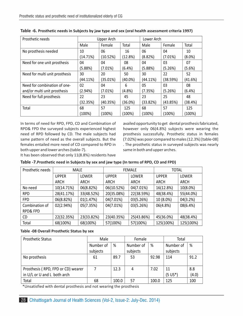
28
In terms of need for RPD, FPD, CD and Combination of RPD& FPD the surveyed subjects experienced highest need of RPD followed by CD. The male subjects had same pattern of need as the overall subjects. But the females entailed more need of CD compared to RPD in both upper and lower arches (table-7).It has been observed that only 11(8.8%) residents have
availed opportunity to get dental prosthesis fabricated, however only 06(4.8%) subjects were wearing the prosthesis successfully. Prosthetic status in females (7.02%) was poor compared to males (12.3%) [table-08] . The prosthetic status in surveyed subjects was nearly same in both and upper arches.
Table -6. Prosthetic needs in Subjects by jaw type and sex (oral health assessment criteria 1997)
Prosthetic needs Upper Arch Lower Arch
Male Female Total Male Female Total
No prosthesis needed 10 (14.71%)
06 (10.52%)
16 (12.8%)
06 (8.82%)
04 (7.01%)
10 (8.0%)
Need for one unit prosthesis
04 (5.88%)
04 (7.01%)
08 (6.4%)
04 (5.88%)
03 (5.26%)
07 (5.6%)
Need for multi unit prosthesis 30 (44.11%)
20 (35.01%)
50 (40.0%)
30 (44.11%)
22 (38.59%)
52 (41.6%)
Need for combination of one-and/or multi unit prosthesis
02 (2.94%)
04 (7.01%)
6 (4.8%)
05 (7.35%)
03 (5.26%)
08 (6.4%)
Need for full prosthesis 22 (32.35%)
23 (40.35%)
45 (36.0%)
23 (33.82%)
25 (43.85%)
48 (38.4%)
Total 68 (100%)
57 (100%)
125 (100%)
68 (100%)
57 (100%)
125 (100%)
Table -7.Prosthetic need in Subjects by sex and jaw type (In terms of RPD, CD and FPD)
Prosthetic needs MALE FEMALE TOTAL
UPPER ARCH
LOWER ARCH
UPPER ARCH
LOWER ARCH
UPPER ARCH
LOWER ARCH
No need 10(14.71%) 06(8.82%) 06(10.52%) 04(7.01%) 16(12.8%) 10(8.0%)
RPD 28(41.17%) 33(48.52%) 20(35.08%) 22(38.59%) 48(38.4%) 55(44.0%)
FPD 06(8.82%) 01(1.47%) 04(7.01%) 03(5.26%) 10 (8.0%) 04(3.2%)
Combination of RPD& FPD
02(2.94%) 05(7.35%) 04(7.01%) 03(5.26%) 06(4.8%) 08(6.4%)
CD 22(32.35%) 23(33.82%) 23(40.35%) 25(43.86%) 45(36.0%) 48(38.4%)
Total 68(100%) 68(100%) 57(100%) 57(100%) 125(100%) 125(100%)
Table -08 Overall Prosthetic Status by sex
Prosthetic Status Male Female Total
Number of subjects
% Number of subjects
% Number of subjects
%
No prosthesis
61 89.7 53 92.98 114 91.2
Prosthesis ( RPD, FPD or CD) wearer in U/L or U and L both arch
7 12.3 4 7.02 11 (5 US*)
8.8 (4.0)
Total 68 100.0 57 100.0 125 100
*Unsatisfied with dental prosthesis and not wearing the prosthesis
Chhattisgarh Journal of Health Sciences (Vol-2, Issue-2: July-Dec. 2014)
Prosthetic status and prosthetic need of institutionalized elderly of CG

29
DISCUSSION-119(95.2%) people residing in old age homes of Raipur, Bilaspur, Durg and Rajnandgoan (Chhattisgarh) had one or more than one teeth missing / root stumps and they had need of dental prosthesis. This result is a little higher but nearly accordance with similar studies
7conducted by and Suryakant C. S. Chaware et.al. 8
Deogade et.al. among subjects of old age homes of Nasik (MH) and Jabalpur(MP) respectively. But it is
9-11much higher than some like studies carried out in Indian places reporting prosthetic needs within range of
12-1670-80%. Almost all the current studies from places of world other than India reported edentulousness more than 70% in elderly peoples. Reason for higher prosthetic needs among institutionalized elderly may ascribe to old age and factors associated with old age such as reduced salivary flow rate, quality and quantity, lowered immunity and the reduced ability of the body
17to repair itself . Several other factors such as multiple chronic diseases, intake of several medications and their side effects, psychological factors such as depression and isolation (because of gradual loss of spouse and friends and feeling of being unwanted by family members), feeling of low self worth owing to loss of earning power and social recognition which leads to
18, 19 poor oral hygiene health , may result into higher edentulousness and prosthetic needs.
51(40.8%) of the surveyed people had all or more than 20 teeth missing/root stumps out of which 17(13.7%) subjects were fully edentulous. The prevalence of edentulism apparently looks lower than the report of
o World Oral Health 2003 i.e.19% in Indian population
but it appeared much higher in terms of need of full dental prosthesis in our study. The reason might be the involvement of the socio economically disadvantaged elderly subjects who were deficient to avail the already scant dental facilities for extraction of the mobile / grossly decayed teeth. The loss of more than 20 teeth badly affects oral function and consequently the general health of these people. Thus, they have intense call for restoration with dental prosthesis as early as possible.
The examined men had little lower overall dental prosthetic need compared to women. The women experienced more need of full dental prosthesis compared to men whereas the men were in more need of multiunit prosthesis compared to women. These
8findings are supported in an analogous study performed. However results are contrast to the similar
7,10,11 studies conducted at other places in India. The social & economical dependency since the beginning and much higher illiteracy in the women surveyed in this study may be one reason for such results. The post menopausal osteoporotic changes in the women may also be a contributory factor.
The prosthetic status observed among residents of old age homes of Chhattisgarh is very poor. Only 11(8.8%) persons had availed the facility of dental prosthetic treatment out of which 5(4.0%) were unsatisfied with their prosthesis and not using the prosthesis. Thus, merely 4.8% (6) of the examined old age home dwellers had successful dental prosthesis. The prosthetic status in females was more worrying compared to males.
Table -09 Prosthetic Status by sex and jaw type
Prosthetic status
MALE FEMALE TOTAL
UPPER ARCH
LOWER ARCH
UPPER ARCH
LOWER ARCH
UPPER ARCH
LOWER ARCH
No prosthesis 61(89.7%) 61(89.7%) 54(94.3%) 53(92.9%) 115(92.0%) 114(91.2%)
Bridge/ Crown 00(0.0%) 00(0.0%) 00(0.0%) 00(0.0%) 00(0.0%) 00(0.0%)
>one Bridge 00(0.0%) 00(0.0%) 00(0.0%) 00(0.0%) 00(0.0%) 00(0.0%)
Partial denture 00(0.0%) 00(0.0%) 01(.17%) 02(.34%) 01(0.8%) 02(1.6%)
Both Bridges & Partial Denture
00(0.0%) 00(0.0%) 00(0.0%) 00(0.0%) 00(0.0%) 00(0.0%)
Full mouth Removable denture
7(32.35%) 7(33.82%) 2(40.35%) 2(43.86%) 09( 05US*) (7.2%)
09( 05US*) (7.2%)
Total 68(100%) 68(100%) 57(100%) 57(100%) 125(100%) 125(100%)
*Unsatisfied with dental prosthesis and not wearing the prosthesis
Chhattisgarh Journal of Health Sciences (Vol-2, Issue-2: July-Dec. 2014)
Prosthetic status and prosthetic need of institutionalized elderly of CG

30
Comparable outcome were observed by R P Shenoy 10 11et.al. (12%) , A Srivastava et.al. (~11.5%) and V Bansal
9et.al.(13.8%) in institutionalized elderly. A little better
prosthetic status( 30%) was noticed in like studies by S ≥7 8 20Chaware et.al. ,S C Deogade et.al . and V Thakare et.al.
All the like mentioned studies were reported poor prosthetic status in females compared to males as recognized in our study. The poor prosthetic status among subjects of present study may be due to the fact that institutionalized elderly underuse the available dental facilities due to lack of awareness, financial constraint, lack of interest, reduced mobility and components of dental care like poor access to services and higher costs of dental care. The dependency on their counterpart (males), higher level of illiteracy and lack of self earning in our society may further worsen the prosthetic status in females.
CONCLUSION:The findings of this survey demonstrate a high unmet need for prosthetic care among the population of old age homes of Raipur, Bilaspur, Durg and Rajnandgoan. Most of the institutionalized elderly also requires extraction. The Study demonistrates that most of the residents have lack of knowledge as well as priority for
oral health. Thus it is suggested to initiate immediate p r e v e n t i v e m e a s u r e p r o g r a m s t o r e d u c e edentulousness and provide oral health care & rehabilitation facilities to these residents with help of state government, non-government organization, nearby dental institution and private institutions.
Acknowledgment; Sincere thanks to Department of Social and Family welfare, Chhattisgarh for the permission to perform the study, especially Rajesh Tiwari, Deputy Director and Mr. M. L.Pandey, Joint Director for their kind support throughout the survey. We would also like to thanks all participants who have contributed to the completion of this study.
REFERENCES;1. United Nations Population Division. World
population prospects: 332 the 2002 revision, New York, 2003. http://www.un.org/esa/ 333 population/publications/wpp2002/WPP2002-HIGHLIGHTSrev1. 334 PDF.
2. World Health Organization (2002) Active ageing: a policy 344framework. WHO, Geneva
3. Goel P, Singh K, Kaur A, Verma M . Oral health care for 339 elderly: identifying the needs and feasible strategies for service 340 provision. Indian J Dent Res2006; 17:11–21
4. N a t i o n a l p o l i c y o n o l d e r persons.www.social justice.nic. in/hindi 342/pdf/npopcomplete.pdf. Accessed on 24.05.2014
5. Burt BA and Eklund SA. Tooth loss. Dentistry, Denta l Pract i ce and the Community. W.B.Saunder Company. Philadelphia. 5th edition: 203-211
6. Zarb GA, Bolender CL. Prosthodontic th
treatment for edentulous patients. 12 ed. St. Louis: Mosby, 2004:6–23.
7. Chaware S, Ghodpage SL, Sinha M, Chauhan V, Thakare V. Prosthetic Status and Prosthetic Needs among Institutionalized Geriatric Individuals in Nashik City,Maharashtra: A Descriptive Study. J Contemp Dent Pract 2011;12 (3):192-195.
8. Suryakant C. Deogade, S. Vinay, S. Naidu Dental Prosthetic Status and Prosthetic Needs of Institutionalised Elderly Population in Oldage Homes of Jabalpur City, Madhya Pradesh, India. J Indian Prosthodont Soc.2013 .
9. Bansal, GM Sogi, KL Veeresha Assessment of oral health status and treatment needs of elders associated with elders' homes of Ambala division, Haryana, India Indian J Dent Res 2010;21:244-7
10. R. P. Shenoy and V. Hegde Dental Prosthetic S t a t u s a n d P r o s t h e t i c N e e d o f t h e Institutionalized Elderly Living in Geriatric Homes in Mangalore: A Pilot Study ISRN Dent. 2011;
11. A. Shrivastav, A. Bhambal, V. Reddy,M. Jain Dental prosthetic status and needs of the residents of geriatric homes in Madhya
Pradesh, India J. Int Oral Health 2011;3(4):9-14
12. Bonakdarchian M, Ghorbanipour R, Majdzadeh F, Hojati T. Prevalence of edentulism among adults aged 35 years and over and associated factors in Yasooj (Iran). Journal of Isfahan Dental School 2011; 7(1):101-4
13. Mamai-Homata E, Margaritis V, Koletsi-Kounari H, Oulis C, Polychronopoulou A, Topitsoglou V. Tooth loss and oral rehabilitation in Greek
Chhattisgarh Journal of Health Sciences (Vol-2, Issue-2: July-Dec. 2014)
Prosthetic status and prosthetic need of institutionalized elderly of CG
31
middle-aged adults and senior citizens. Int J Prosthodont. 2012;25(2):173-9.
14. Nadia Khalifa, Patrick F. Allen, Neamat H. Abu-bakr, and Manar E. Abdel-Rahman4. Factors associated with tooth loss and prosthodontic status among Sudanese adults Journal of Oral Science2012; 54(4):303-312.
15. Adrienne Nickles, Dr. Sheila Vandenbush et. al. Results from a 2010 Oral Health Screening and Needs Assessment of Michigan Residents and Managers of Alternative Long-Term Care F a c i l i t i e s . http//www.michigon.gov./senior_smile_report_final_050311_355 assesed on Accessed on 24.05.2014
16. Kethy Phipps, Nicole Laws et. al . The Commonwealth's High-Risk Senior Population; Results and Recommendations from 2009 Statewide Oral Health Assessment in
M a s s a c h u s e t t s . www.mossgov/eohhs/doc/../senior-oral-health-assessment-report pdf
17. Navazesh M. Dry mouth: aging and oral health. Compend Contin Educ Dent2002;23(10):41–48
18. Ganguli M, Dube S, Johnston JM, Pandav R,Chandra V, Dodge HH. Depressive symptoms,cognitive impairment and functional impairment in a rural elderly population in India: a Hindi version of the geriatric depression scale (GDS-H). Int J Geriatr Psychiatry 1999; 14: 807–820.
19. Shah N. Geriatric oral health issues in India. Int Dent J 2001; 51: 212–218.
20. Thakare V, Ajith Krishnan CG. Periodontal status, prosthetic status and prosthetic needs among institutionalized geriatric individuals in Vadodara City, Gujarat—A descriptive study. J Ind Asso Public Health Dentistry 2010(15):153-57.
Chhattisgarh Journal of Health Sciences (Vol-2, Issue-2: July-Dec. 2014)
Prosthetic status and prosthetic need of institutionalized elderly of CG

32
1 2Vanita Rathod , Chandan Rathod1. Professor, Department of Oral Pathology, Rungta College of Dental Sciences & Research Centre, Bhilai (CG)2. Lecturer, Department of Prosthodontics, Rungta College of Dental Sciences & Research Centre, Bhilai (CG)
Corresponding Author :Dr. Vanita RathodProfessor, Department of Oral Pathology, Rungta College of Dental Sciences & Research Centre, Bhilai (CG)E: mail: [email protected]
ABSTRACT
Objective: The purpose of the study was to identify trends in incidence rates of oral squamous cell carcinoma (OSCC)
at specific anatomic sites or within specific age or sex groups in the central Madhya Pradesh population.
Materials and Methods: The study covers the period of January 2007 through July 2011. OSCC cases were
retrospectively analyzed for site, age, gender, habits and histopathological grading. And the findings were
formulated to chart the trends in Madhya Pradesh.
Results: The study revealed a male to female ratio of 2:1 with the largest number of OSCCs developing in the peak
age of 46-55 years. Overall, the most common site was the alveolar mucosa and buccal mucosa followed by tongue,
palatal mucosa, floor of mouth, retro molar area. Smokeless tobacco habit was more prevalent than smoking
tobacco in both men as well as women. Smokeless tobacco in the form of gutkha is more prevalent in this region.
Conclusion: OSCC is significant cause of mortality and morbidity worldwide with an incidence rate that varies widely
by geographic location. Even within one geographic location, the incidence varies among group categorized by age,
sex, site or habits.
Key words : epidemiology, oral squamous cell carcinoma, trends.
INTRODUCTION
Squamous cell carcinoma is the most common
malignant neoplasm of the oral cavity and represents
about 90% of all oral malignancies. Oral squamous cell 1
carcinoma (OSCC) is significant cause of morbidity and
mortality worldwide with an incidence rate that varies
widely by geographic location. In India, oral cancer 2
represent a major health problem constituting up to
40% of all cancer in males and the third most prevalent
in females. Even within one geographic location, the
incidence varies among groups categorized by age, sex,
or race. Recent publications have highlighted 1,2
variations in oral cancer trends by geographic area are
vital for many reasons including understanding the
extent of the problem , determining which groups
within the population are at highest and lowest risk, and
relating the burden of oral cancer, consequently it helps
in evaluating the allocation of resources for research,
prevention, treatment and support services Despite 3,4
several diagnostic and therapeutic advances, the
overall incidence and mortality associated with OSCC
are rising. The current estimates of age-standardized
incidence and mortality is and 3.1/100,000 and
2.9/100,000 in men and women respectively. 5
Trends in epidemiology of oral cancer in central part of India in Madhya Pradesh : An institutional study
Ayush & Health Sciences University of Chhattisgarh
ORIGINAL ARTICLE
Chhattisgarh Journal of Health Sciences (Vol-2, Issue-2: July-Dec. 2014)
OR
IGIN
AL
AR
TIC
LE
ISSN 2348 - 4195

Table 1: OSCC trends in central part India in M.P. population according to age, sex, site and habits.
SEX
male
female SITE
BM
AM
PM
LM
FM
RA HABITS
SL
ST
NH
25-35 5 7 4 4 7 4 1
36-45 5 11 5 7 11 3 2
46-55 20 10 13 11 2 1 2 1 20 6 4
56-65 10 4 4 2 - 2 1 1 9 3 2
66-75 4 3 3 3 1 1 1 5 2
76-85 - 2 1 1 1 1
86-95 1 2 1 Total 45 37 30 30 2 4 4 3 53 20 9
Sites S: BM-Buccal mucosa, AM-Alveolar mucosa, PM-Palatal mucosa, LM- Labial mucosa, FM- Floor of mouth, RA-
Retro molar area.
Habits : SL- Smokeless Tobacco (gutakha & quid chewing), ST- Smoking tobacco and NH- No habits .
33
Studies reported on the incidence and pattern of OSCCs
from various parts of the world is 4.7./100000.
However, very few studies have reported on the
incidence and trends of OSCCs in Madhya Pradesh
(M.P.) population. The purpose of this retrospective
study was to identify trends in the number of cases or
incidence rates of OSCCs at specific anatomic sites or
within specific age or sex groups in the Madhya Pradesh
population.
MATERIALS AND METHODS
82 Histologically proven in cases of OSCCs verified in the
oral pathology and microbiology from January 2007 to
July 2011 were extracted from the archives of Hitkarini
dental college Jabalpur. The anatomic sites included in
the study were alveolar mucosa, buccal mucosa, floor
of mouth, retro molar area, tongue and hard palate. As
the pathophysiologic and epidemiologic behavior of the
lip cancer is believed to be substantially different from
the oral cavity sites, cancers originating in the lip were
not included in this study. Charts were made listing the
age, sex, site, habits and histopathology grading of
eighty OSCC patients. A comprehensive analysis was
done on the data collected and the results were
formulated.
RESULTS:
Of the 82 OSCC patients, males represented a higher
proportion (66%) of squamous cell carcinomas than
females (33%) {Diagram 1}. Larger numbers of cases
were seen to develop in 46-56 years followed by 36-45
and 25-35 years. Over all alveolar mucosa and buccal
mucosa were the most common sites involved 42.5%,
and 37.5% respectively. While the floor of the mouth,
retromolar areas showed least incidence in this region
of India (3.7%) {Diagram 2}. The study also revealed that
larger number of patients had the smokeless tobacco in
the form of gutkha and quid chewing than the bidi and
cigarette smoking habit. {Diagram 3}
Chhattisgarh Journal of Health Sciences (Vol-2, Issue-2: July-Dec. 2014)
Trends in epidemiology of oral cancer in central part of India

34
Diag. 1: OSCC in 82 patients (Gender with respect to age)
Diag. 2: OSCC in 82 patients (Sites with respect to age)
Diag. 3: OSCC in 82 patients (Habits with respect to age)
DISCUSSION:
The incidence of OSCC seems to be increasing and is
global health problem with increasing incidence and
mortality rates. Around 3,00,000 patients are annually
estimated to have oral cancer worldwide. OSCC is 4,7,8
known to show geographic variation with respect to
the age, site, sex and habits of the population. The 1,2,4,8,9
present study revealed a male to female ratio of 2:1
with the largest number of OSCCs developing in peak
age of 46-55 years. This is consistent with an earlier
report by Mehrotra and coworkers confirming that 8
oral cancer in Northern India was a disease of the
middle aged men. An epidemiologic study on palatal
changes in reverse smokers conducted in Andhra
Pradesh (Southern India) by Mehta et al. showed a 10
predominance of females in the middle age group (35-
54years).
Regarding the site of preference for intra-oral SCC, our
study showed some degree of variation from most of
the studies conducted at Spain, Canada, Scandianavia
and some part of India. A retrospective study 11-13
conducted by S. manuel and co-workers, in 2003, at 14
the regional Cancer (RCC), Thiruvananthapuram
Kerala analyzed one of the largest series of young
patients under the age of 45 years having SCC of the
oral tongue.
In the present study, the alveolar mucosa and buccal
mucosa were the most frequent involved sites (41 and
37.5% respectively) while the floor of mouth was the
least commonly involved site (3.75%). These regional
difference may be attributed to the exclusive use of
chewing tobacco in the India subcontinent compared
to smoking in the west. SCC of buccal mucosa is one 12-14
of the most common cancers along the geographical
belt extending from central to south east asia because
of practice of chewing pan a combination of tobacco,
nut and lime. In contrast, the lateral tongue and floor 15
of mouth are the more commonly involved site in the
West. The anterior 2/3 of the tongue is commonly 11-13 rd
involved in India, while the posterior lateral border
and ventral surfaces are frequently involved in the
United State.(8
In 1969,the result of first epidemiologic survey of
Chhattisgarh Journal of Health Sciences (Vol-2, Issue-2: July-Dec. 2014)
Trends in epidemiology of oral cancer in central part of India
35
palatal changes in reverse smokers in the Srikakulam
district of Andra Pradesh in India was reported by
Mehta FS et al, who later emphasized that the palatal 10
changes seen in reverse smokers exhibited greater
clinical variation than the leukokeratosis nicotina palate
known from the Western countries. Earlier, OSCC was
thought to be a disease primarily of the elderly. Some 1,2
recent studies conducted in united states, South East of
England, Spain and Scandinavia have , however, shown
that the incidences of oral cancer are increasingly being
reported in the young (<40 years of age) also
particularly younger male patients. Our study 13,14,16,17
finds increasing number of OSCC cases being recorded
in the 4 and 5 decades of life. This may be related to th th
the habits like tobacco and alcohol.
Men represented a higher proportion of OSCCs than
women simulating the trends in many recent
publications. Some studies show the opposite 3,6,11,18
trend with the increased incidence among women,
which may be due to the changing social habit in the
high socioeconomics groups or cultural habits of some
rural area of India. Interestingly, 3.75% of the patient 10,16
were not associated with any habits like tobacco
smoking or chewing in our study, Probably attributed to
other etiological factors of OSCCs like certain viruses
(such as human papilloma virus), low consumption of
fruits and vegetables, genetic predisposition ,etc. 16
Gutkha chewing or Pan chewing were the most
prevalent habits recorded in the study. The incidence
was highest at mucosal sites with prolonged contact
with carcinogens. There has been strong evidence that
smokless tobacco can cause oral cancer and
precancerous oral lesions like leukoplakia. smokeless 8
tobacco is thought to induce cancer in regions where it
is held in direct contact, such as the cheek or gum. The 8
clinicopathological profile of Indian oral cancers shows
significant differences from oral cancer in several
developed countries of world, including the USA, UK,
France and Japan, where it is associated with tobacco
smoking with or without alcohol consumption 19
CONCLUSSION:
As useful clinical information on the trends of OSCCs
among mid of the central part of India in Madhya
Pradesh population is limited, this retrospective study
was undertaken to present a compressive data on the
trends of OSCC in M.P. population. Different levels of
tobacco and alcohol exposure, diet, socio economic
circumstances factors in the diff age, gender and sites
are the causative factors in the difference seen in the
incidence rates of OSCC in various populations globally.
Because of the magnitude of the oral cancer problem
and trends reported serious thought should be given to
plans for prevention and early detection of
premalignant and malignant oral diseases in central
part of India in M.P. race, ethnicity and age cannot be
altered; however, lifestyle behavior such as use of
tobacco and alcohol are amenable to change and
increased intake of fruits and vegetables must be
addressed. The dental profession has a well deserved
reputation for preventing other oral diseases. Now is
time to focus on the prevention and early detection of
oral cancer.
REFERENCES:
1. Lawoyin JO, Lawoyin DO,Aderinokun G. Intra-
Oral squamous cell carcinoma in Ibadan: a
review of 90 cases. Afr J Med Sci 1997; 26:187-
8.
2. Howell RE, Wright BA, Dewar R. Trends in oral
cancer in Nova scotia from 1983 to 1997. Oral
Surg Oral Med Oral Pathol Oral Radiol Endod
2003; 95:205-12.
3. Schantz SP, Yu Gp, Head and Neck cancer
incidence trends in young American, 1973-
1997, with aa special analysis for tongue cancer.
Arch Otolaryangol Head Neck SURG 2002 ;
128:268-74.
4. Rautava J, Luukkaa M, Heikinheimo K, Happone
RP. Squamous cell carcinomas arising from
different types of oral epithelia differ in their
tumour and patient characteristics and
survival. Oral Oncol 2007; 43:911-9.
5. Carvalho AL, Singh B, Spiro RH, Kowalski LP,
Shah JP. Cancer of the oral cavity: a comparison
between institutions in a developing and a
developed nation, Head Neck 2004; 26:31-8.
Chhattisgarh Journal of Health Sciences (Vol-2, Issue-2: July-Dec. 2014)
Trends in epidemiology of oral cancer in central part of India
36
6. Llewelly3n CD, Linklater K,Bell J, Johnson NW,
Warnakulasuriya KA. Squamous cell carcinoma
of the oral cavity in the patients aged 45 years
and under: a discriptiveanalysis of 116 cases
diagnosed in the south east of England from
1990 to 1997. Oral Oncol 2003; 39:106-14.
7. Funk GF, Karnell LH, Robinos RA, Zhen WK,
Trask DK, Hoff iman HT. Presentat ion,
treatment, and outcome of oral cavity: a
national cancer data base report. Head Neck
2002; 24:165-80.
8. Mehrotra R, Singh MK, Pandya S, Singh M. The
use of an oral brush biopsy without computer-
assisted analysis in the oral lesions. A study of
94 patients. Oral Surg Oral Pathol Oral Radiol
Endod 2008; 106:204-53.
9. Shiboski CH, Shiboski SC, Silverman S Jr.Ttrends
in oral cancer rates in the united states, 1973-
1996. Community Dent Oral Epidemiol 2000;
28:249-56.
10. Mehta FS, Jalnawalla PN, Daftary DK,Gupta
PC,Pindborg JJ. Reverse smoking in Andhra
Pradesh, India: variability of clinical and
histologic appearance of palate changes. Int J
Oral Surg 1997; 6:75-83.
11. Martin-Granizo R, Rodriguez-Campo F, Naval l,
Diaz Gonzalez FJ. Squamous cell carcinoma of
the oral cavity in patients younger than 40
years. Otolaryngyol Head Neck Surg 1997;
117:268-75.
12. Gorsky M, Epstein JB, Oakley C, Le ND, Hay J,
Stevenson- Moore P. Carcinoma o f tongue : a
series analysis of clinical presentation, risk
factors, staging, and outcome. Oral Surg Oral
Med Oral Pathol Oral Radiol Endod 2004;
98:546-52.
13. Annertz K, Anderson H, Biorklund A, Moller T,
Kantola S, Mork J, et al. incidence and survival
oral squamous cell carcinoma of the tongue in
Scandinavia, with spcial reference to young
adults. Int J Cancer 2004; 101:95-9.
14. Manuel S, Raghavan SK, Pandey M, Sebastian P.
Survival in patients under 45 tears with
squmaous cell carcinoma of the tongue. Int J
Oral Maxillofac Surg 2003; 32:167-73.
15. Diaz EM Jr, Holsinger FC, Zuniga ER, Robert DB,
Sorensen DM. Squamous cell carcinoma of the
buccal mucosa: one institution's experience
with 119 previously untreated patients. Head
Neck 2003; 25:267-73.
16. Silverman S Jr. demographic and occurrence of
oral and pharyngeal cancers. The outcomes,
the trends, the challenge. J AM Dent Assoc
2001; 132:57-11.
17. Rodriguez T Altieri A, Chatenoud L, GallusS,et
al. risk factors for oral and pharyngeal cancer in
young adults. Oral Oncol 2004; 40:207-13.
18. Shiboski CH, Schmidt BL, Jordan RC. Tongue and
tonsil carcinoma increasing trends in the U.S.
population ages 20-44years. Cancer 2005;
103:1843-9.
19. Jane C, Nerurkar AV, Shirsat NV, Deshpande RB,
Amrapurkar AD, Karjodkar FR. Incresed
surviving expression in high grade oral
squamous cell carcinoma: a study in Indian
tobacco chewers. J Oral Pathol Med 2006;
35:595-601.
Chhattisgarh Journal of Health Sciences (Vol-2, Issue-2: July-Dec. 2014)
Trends in epidemiology of oral cancer in central part of India

37
INTRODUCTION
Dento-facial appearance has a lot to do with the 1way the people are perceived in the society.
People equate good dental appearance with 2success in many aspects. Social interactions that
have a negative effect on self-image, career
advancement and a peer group acceptance have
been associated with an unacceptable dental 3
appearance. The prevalence of malocclusion
varies from country to country and among different 1
races. The reasons to develop malocclusion could
be genetic or environmental and/or combination
of both the factors along with various local factors
such as adverse oral habits, tooth anomalies, form
and developmental posit ion of teeth can cause
malocclusion. Orthodontics has traditionally 4focussed on children and adolescents. There is an
increases concern for dental appearance during 2adolescents to early childhood has been observed.
Malocclusions are 3rd in the ranking of priorities
among the problems of dental public health
worldwide, surpassed only by dental cavity and 5
periodontal diseases. The benefits of taking
orthodontic treatment are to prevention of tissue
damage and correction of aesthetic component, 2
improve the physical function . A variety of indices
have been developed to assist professionals in
categorizing malocclusion according to the
1 2 3 4 5 6R S Makkad , Madhu Pandey ,S Hamdani , V. Agrawal , M Motlani , Gunjan Agrawal1. Lecturer, Department of Oral Medicine & Radiology, New Horizon Dental College and Research Institute, Bilaspur, (CG)2. Lecturer, Department of Orthodontics, Rungta Dental College and Hospital, Bhilai, (CG)3. Post Graduate, Department of Orthodontics, Rungta Dental College and Hospital, Bhilai, (CG)4. Lecturer, Department of Oral and Maxillofacial Surgery, Maitri Dental College, Anjora, Durg (CG)5. Lecturer, Department of Endodontics, Chhattisgarh Dental College and Research Institute, Rajnandgaon (CG)6. Lecturer, Department Oral and Maxillofacial Surgery, (CG)Pt. Jawaharlal Nehru Medical College, Raipur
Corresponding Author: Dr. Ramanpal Singh Makkad Lecturer, Department Oral Medicine & Radiology, New Horizon Dental College and Research Institute, Bilaspur, (CG)Mobile no- 090986 99300 email: [email protected]
ABSTRACT
Objectives-This study is to know the prevalence of malocclusion and orthodontic treatment needs among 12-15yr
old school children of Bilaspur.
Materials and Methods-A total of 351 study subjects were selected based on convenience sampling and
examination was carried out under natural light and data was recorded using WHO Proforma 1997. The collected
data was subjected to statistical analysis using SPSS16.
Results-Out of the 351 children examined, 46.2% were boys & 53.8% were girls and their mean age was 13.89yrs.
One and two segment crowding was seen in 24.5% & 11.4% respectively. Normal molar relation was seen in 80.3% of
children. Definite, severe and very severe or handicapping malocclusion was seen in 9.7%, 4.3% & 3.4% of children
respectively. There is no statistically significant difference in malocclusion status between boys and girls.
Conclusion- needs.Only 4.3% and 3.4% of children required highly desirable and mandatory orthodontic treatment
KEYWORDS- Malocclusion, Dental Aesthetic Index, OrthodonticTreatment needs.
Assessment of dental aesthetic index among school children of Bilaspur, Chhattisgarh : A pilot study
OR
IGIN
AL
AR
TIC
LE
Ayush & Health Sciences University of Chhattisgarh
ORIGINAL ARTICLE
Chhattisgarh Journal of Health Sciences (Vol-2, Issue-2: July-Dec. 2014)ISSN 2348 - 4195

TABLE 1. AGE WISE DISTRIBUTION OF STUDY POPULATION
AGE FREQUENCY PERCENTAGE
12 13 3.7
13 95 27.1
14 133 37.9
15 110 31.3
TOTAL 351 100
6treatment needs . Dental Aesthetic Index (DAI)
introduced by Cons et al(1986), which links clinical
and aesthetic components. It was developed
originally based on North American Caucasian 7
sample. The World Health Organizat ion
concerning to acknowledge the real malocclusions
conditions in different countries, adopted it as a
cross cultural index and advocated it in the 4th
Edition of the Manual of Basic Oral Health Survey,
so there would be a suitable instrument to gather
epidemiological data collection and assessment of 5,7-9
orthodontic treatment needs . DAI is proven to
be reliable, valid, versatile, simple and easily 7,9
applied index . Most of the malocclusion can be
corrected if detected early by correctional 1methods. This study was intended to evaluate the
prevalence of malocclusion, its severity and the
orthodontic treatment needs using DAI, among 12-
15yr old school children of Bilaspur, Chattishgarh.
MATERIALS AND METHODS:
The present study was conducted among 12-15yr
old school children of Bilaspur, Chattishgarh. The
schools were selected based on convenience
sampling. A total of 351 school children of both
sexes were selected for the study based on
convenience sampling. Approval was obtained
from the concerned authorities before the start of
the study. All examinations were performed at
schools while children were seated on chair under
normal illumination. The examiners were trained
and intra-examiner calibration was done. Kappa
statistics showed a good agreement. Sufficient
number of autoclaved instruments was taken to
the examination site. The WHO Proforma (1997)
was used to assess the malocclusion. Data
collected was coded, processed and subjected to
statistical analysis using SPSS version16.
RESULTS
The study population consisted of about 351
school children aged 12-15years in Bilaspur city,
out of which 46.2% were males and 53.8% were
females (Table 1). Table 2 shows the distribution of
DAI components. Out of 351 school children, 24.5%
had one segment crowding and 11.4% had two
segments crowding. One and two segment spacing
was seen in 8.5% and 1.7% school children
respectively. Diastema of 1-3mm was seen among
5.7% of the study subjects. Largest maxillary
irregularity of 0, 1-3 and >3mm was seen among
80.9%, 17.1% and 2% of school chi ldren
respectively. Largest mandibular irregularity of 0,
1-3 and >3mm was seen among 72.1%, 27.6% and
0.3% of school children respectively. Maxillary
over-jet of 0-3mm is considered normal and was
seen among 76.4% of school children and >3mm
was seen among 23.6%of school children.
Mandibular overjet of 0-3mm was among 99.4% of
school children and 0.6% of them had >3mm of
overjet. Open bite of >3mm was seen among 0.9%
of study subjects. Molar relation was normal
among 80.3% of school children whereas half cusp
and full cusp molar relation was seen among 14.8%
38 Chhattisgarh Journal of Health Sciences (Vol-2, Issue-2: July-Dec. 2014)
Assessment of dental aesthetic index

TABLE 2: DISTRIBUTION OF DAI COMPONENT
TABLE 3: DISTRIBUTION OF THE SUBJECTS ACCORDING TO DAI SCORES,
SEVERITY OF MALOCCLUSION, TREATMENT NEEDS AND GENDER (P=3.946).
DAI COMPONENTS PERCENTAGE (%)
CROWDING 0 64.1
0NE SEGMENT 24.5
TWO SEGMENT 11.4
SPACING 0 89.7
0NE SEGMENT 8.5
TWO SEGMENT 1.7
DIASTEMA 0 94.3
1-3 5.7
LARGEST MAXILLARY 0 80.9
IRREGULARITY(mm) 0-3 17.1
>3 2
LARGEST MANDIBULAR 0 72.1
IRREGULARITY(mm) 0-3 27.6
>3 0.3
MAXILLARY OVERJET (mm) 0-3 76.4
>3 23.6
MANDIBULAR OVERJET(mm) 0 99.4
>3 0.6
OPEN BITE(mm) 0 99.1
>3 0.9
MOLAR RELATION NORMAL 80.3
HALF CUSP 14.8
FULL CUSP 4.8
DAI SCORE Severity Of Treatment MALE (%) FEMALE (%) TOTAL (%) Malocclusion Indicated<25 No/ minor No/slight 84 81.5 82.6 Malocclusion Treatment 26-30 Definite Elective 8 11.1 9.7 Malocclusion 31-35 Severe Highly 3.1 5.3 4.3 Malocclusion Desirable>35 Very severe or Mandatory 4.9 2.1 3.4 handicapping malocclusion TOTAL 100 100 100
ASSESSMENT OF DENTAL AESTHETIC INDEX
39Chhattisgarh Journal of Health Sciences (Vol-2, Issue-2: July-Dec. 2014)
Assessment of dental aesthetic index

and 4.8% of school children. There was no
statistically significant difference between the DAI
scores and the gender. Table 3 shows the
distribution of according to DAI score, severity of
malocclusion, treatment indicated and gender.
4.3% and 3.4% of the study subjects had severe and
very severe malocclusion respectively and required
highly desirable and mandatory orthodontic
treatment needs.
DISCUSSION
Many epidemiological studies have been
conducted worldwide utilizing various indices for
quantifying the extent of malocclusion. Crowding 1
of incisal segment affects half of all children in
mixed dentitions and it worsens in adolescent
years as the permanent teeth erupt and continues
to increases as the age progresses. In the current 2
study, 35.9% of the study population had incisal
crowding. The results of the current study are in
correlation with the study conducted by
Shivakumar et al and in contrast with a study 2
conducted by Bhardwaj et al . Both the upper and 1
lower incisal segments were examined for spacing.
In the present study, 10.2% had incisal segment
spacing either in one or both the arches which was
in correlation with the study conducted by Artenio
Jose Isper Garbin et al . Diastema>1mm was seen 5
among 5.7% of school children and this result was
in correlation to the study conducted by Artenio
Jose IsperGarbin et al . Irregularity may occur with 5
or without crowding. In the current study, 19.1% of
the children had maxillary anterior irregularity of
>1mm, and the results are in correlation with the
study conducted by Shivakumar et al and Artenio 2
Jose IsperGarbin et al . 27.9% had mandibular 5
anterior irregularity >1mm and the result were in
contrast with the study conducted by Bhardwaj et
al , DS Rwakatema et al , B. Eduardo and F.M 1 8
Carlos . 9
In the present study, maxillary overjet of >3mm
was seen in 23.6% and it was similar to the study
conducted by B. Eduardo and F.M Carlos and 9
Bhardwaj et al and in contrast to a study 1
conducted by Matilda Mtaya et al . 10
Mandibular overjet of >3mm was seen in 0.6% of
school children and it was in correlation with
studies conducted by Shivakumar et al , DS 2
Rwakatema et al , Bhardwaj et al and Artenio Jose 8 1
IsperGarbin et al .5
An anterior openbite of >3mm was seen in 0.9% of
school children which was similar to studies
conducted by Bhardwaj et al and B. Eduardo and 1
F.M Carlos . Normal molar relation was seen in 9
80.3% of the school children and which was similar
to the study conducted by Bhardwaj et al and was 1
in contrast with the study conducted by Artenio
Jose IsperGarbin et al . Definite malocclusion was 5
seen in 9.7% of the school children, severe
malocclusion was seen in 4.3% of school children
and very severe or handicapping malocclusion was
seen in 3.4% of children. Similar results were found
in the study conducted by Vijaya Hedge and
RekhaShenoy , Bhardwaj et al and Shivakumar et 11 1
al , whereas it was in contrast with the study 2
conducted by B. Eduardo and F.M Carlos and D.S 9
Rwakatema et al . 8
CONCLUSION
Thus the present study concluded that out of 351
study subjects, 4.3% and 3.4% of school children
required highly desirable and mandatory type of
orthodontic treatment needs respectively. The
information from this study forms a part of the
basis not only for further research, but also for
planning orthodontic care.
40 Chhattisgarh Journal of Health Sciences (Vol-2, Issue-2: July-Dec. 2014)
Assessment of dental aesthetic index

REFERENCES
1. VK Bhardwaj, KL Veeresha and KR Sharma.
Prevalence of malocclusion and orthodontic
needs among 16 and 17year old school going
children in Shimla city, Himachal Pradesh.
Indian Journal of Dental Research 2011;22(4):
556-560.
2. Shivakumar KM, Chandu GN, Subba Reddy VV,
e t a l . P r e v a l e n c e o f m a l o c c l u s i o n a n d
orthodontic treatment needs among middle a n d
high school children of Davangere city, India
b y D e n t a l A e s t h e t i c I n d e x . J I n d i a
SocPedodPrev Dent 2009; 27:211-218.
3. H. Nihal, B. Guvenc and U. Ersin.Dental
Aesthetic Index scores and perception of
personal dental appearance among Turkish
university students. European Journal of
Orthodontics 2009; 31: 168-173.
4. B.A Carlos, M.C Jose-Maria, M.P David, et al.
Orthodontic treatment need in Spanish young
adult population. Med Oral Patol Oral Cir Bucal
2012; 17(4):638-643.
5. I.G Artenio Jose , P.P Paulo Cesar, S.G CleaAdas,
et al. Malocclusion prevalence and comparison
between the Angle classification and the
Dental Aesthetic Index in scholars in the
interior of Sao Paulo state- Brazil. Dental Press J
Orthod 2010; 15(4):94-102.
6. Poonacha KS, Deshpande SD, Shigli AL. Dental
Aesthet i c Index , app l i cab i l i ty in Ind ian
population: a retrospective study. J Indian
PedodPrev Debt 2010; 28: 13-17.
7. B. Venkatesh, Gopu H. Assessment of
Orthodontic treatment needs according to
Dental Aesthetic Index. Journal of Dental
Sciences and Research 2011; 2(2):9-13.
8. D.S Rwakatema, P.M. Ng'ang'a and A.M.
Kemoli. Orthodontic treatment needs among
12-15 year olds in Moshi, Tanzania. East African
Medical Journal 2007; 84(5): 226-232.
9. B. Eduardo and F.M Carlos. Orthdontic
treatment need in Peruvian young adults
evaluated through Dental Aesthetic Index.
Angle Orthodontist 2006; 76(3): 417- 421.
10. M Matilda, B. Pongsri and A. Anne Nordrehaug.
Prevalence of malocclusion and its relationship
with socio-demographic factors, dental caries
and oral hygiene in 12 to 14 year old Tanzanian
s c h o o l c h i l d r e n . E u r o p e a n J o u r n a l o f
Orthodontics 2009; 31: 467-476.
11. H. Vijaya and S. Rekha.Dentition status,
treatment needs and malocclusion status
a m o n g 1 5 y e a r o l d s c h o o l c h i l d r e n o f
Mangalore- a pilot study. JIDA 2010; 4 (12): 568-
569.
Assessment of dental aesthetic index
41Chhattisgarh Journal of Health Sciences (Vol-2, Issue-2: July-Dec. 2014)

42
CA
SE R
EPO
RT
Ayush & Health Sciences University of Chhattisgarh
CASE REPORT
ABSTRACT
Neurofibroma is a benign tumor of neural origin derived from peripheral nerve sheath. Nerve sheath tumors are
extremely rate. There is no sex predilection and average age of occurrence is 28 years. In the present case it was 22 yr.
old patient . The surgical removal of tumor mass was done under GA and histological confirmation was done.
Key Words: Neurofibroma, Spindle cell, Tumor
1 2 3 4Swapnil Moghe , Ajay Kumar Pillai , Vineeta Gupta , Geeta Mishra1. Reader, Department of MaxilloFacial Surgery, Peoples Dental Academy, Bhopal (MP)2. Reader, Department of MaxilloFacial Surgery, Peoples Dental Academy, Bhopal (MP)3. Reader, Department of Periodontics, Govt. Dental College, Raipur (CG)4. Assistant Professor, Department of Dentistry, Govt.Medical College. Rewa (MP)
Corresponding author : Dr. Ajay Kumar Pillai
Reader, Department of Maxillofacial Surgery, Peoples Dental Academy, Bhopal (MP)Contact No: 98932 60776, Email : [email protected]
INTRODUCTION:
Neurofibromas arise from a mixture of cell types
including Schwann cells and perineural fibroblasts. They
may occur as solitary lesions or in association with
neurofibromatosis. Although most commonly reported
in soft tissues, neurofibromas do occur in bone. And
very few cases have been reported in association with
the inferior alveolar nerve. We report a case of neuro-
fibroma of spindle cell origin associated with the
inferior alveolar nerve in a 22 year old man. Pain or
paresthesia may result from lesions of the inferior
alveolar nerve. Patients presents with cortical
expansion. Intra-osseous lesions may produce a well
demarcated or poorly defined unilocular or multilocular
radiolucency. Adjacent soft tissue neurofibromas may
produce cortical erosion. Solitary neurofibromas and
those found in association with neurofibromatosis
share the same microscopic features . The tumor is 1
composed of spindle-shaped cells with fusiform or
wavy nuclei in a delicate connective tissue matrix. It is
not encapsulated and may blend with the adjacent
connective tissues. The normally recommended
treatment of solitary lesions following biopsy is
localized excision.
CASE PRESENTATION
The patient reported a slow growing lesion in lower left
mandibular region extending from canine to 2 molar nd
region. A 22-year-old man presented to the department
of the Oral and Maxillofacial Surgery with a 1 month
history symptom of paraesthesia of lower left side of
mandible extending from corner of mouth to angle of
mandible. His medical history was unremarkable and
there was no history of gum-related disease or trauma
to the maxillofacial complex. Mobility of teeth was
absent, also, a history of numbness of the lower lip since
2 months with no history of extraoral swelling was
significant. An intraoral examination revealed a
obliteration of muco-buccal fold of about 3 × 1 cm in the
left lower canine- molar region with no signs of
ulceration (Figure 1). On palpation, the swelling was
firm in consistency with underlying bone from left
canine to molar region. No neck nodes were palpable
and the cranial examination was normal. Funelling &
widening of inferior alveolar canal was evident on OPG.
(Figure 2)
Neurofibroma of spindle cell origin, a diagnostic dilemma to general dentist
Chhattisgarh Journal of Health Sciences (Vol-2, Issue-2: July-Dec. 2014) ISSN 2348 - 4195
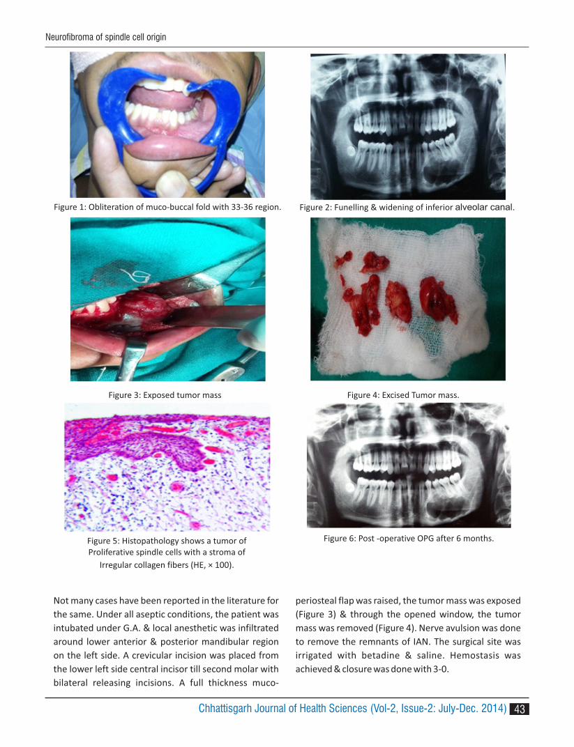
Figure 3: Exposed tumor mass Figure 4: Excised Tumor mass.
Figure 5: Histopathology shows a tumor of Proliferative spindle cells with a stroma of
Irregular collagen fibers (HE, × 100).
Figure 6: Post -operative OPG after 6 months.
Not many cases have been reported in the literature for
the same. Under all aseptic conditions, the patient was
intubated under G.A. & local anesthetic was infiltrated
around lower anterior & posterior mandibular region
on the left side. A crevicular incision was placed from
the lower left side central incisor till second molar with
bilateral releasing incisions. A full thickness muco-
periosteal flap was raised, the tumor mass was exposed
(Figure 3) & through the opened window, the tumor
mass was removed (Figure 4). Nerve avulsion was done
to remove the remnants of IAN. The surgical site was
irrigated with betadine & saline. Hemostasis was
achieved & closure was done with 3-0.
Figure 1: Obliteration of muco-buccal fold with 33-36 region. Figure 2: Funelling & widening of inferior alveolar canal.
43Chhattisgarh Journal of Health Sciences (Vol-2, Issue-2: July-Dec. 2014)
Neurofibroma of spindle cell origin

Investigations
Orthopantamogram revealed an osteolytic scalloped
lesion extending from the lower left canine region to the
second molar. The lesion also shows erosion of buccal
cortical plate.
Microscopically the tumor is composed of an irregular
pattern of proliferative spindle cells (Figure 5). The
stroma is composed of collagen fibers and mucoid
masses. Small axons all over the tumoral tissue are
demonstrated with silver staining.
Treatment & follow up:
The patient was followed up for 6 months (Figure 6).The
postoperative OPG showed good healing with no signs
of recurrence at the surgical site.
DISCUSSION:
Neurofibroma (NF) is a benign tumor of neural origin
derived from the peripheral nerve sheath that may
have variable histology. Nerve sheath tumors located in
the jaw are extremely rare, having published only a few
cases of central neurofibroma of the mandible. There is
no sex predilection and average age of occurrence is 28
years. In our case it was 22 years old man.
Ninety percent of the neurofibromas are associated
with neurofibromatosis type 1, so the physical 2
examination and family history should be elicited to
exclude the disease. In this case, there were no clinical
signs or family history suggestive of neurofibromatosis.
The lesion was a solitary one.
Hubner and Lewis developed an animal model to 3
investigate the causative factors in the development of
the lesion. They reported that the peripheral nerve
section resulted in the formation of an expanded
connective tissue cap at the end of the proximal
segment. Nerve fibers attempting to re-establish
continuity with the distal segment penetrated into and
beyond the cap, becoming tangled and entrapped in the
soft tissue. But in our case, there was no history of
trauma.
In the mandible, the lesions most commonly arise from
the mandibular nerve with accompanying pain and par
aesthesia. In such cases the radiograph shows flaring of
mandibular foramen, the so called “blunderbuss'
foramen or fusiform enlargement of the mandibular
canal, as was seen in our case .4
Histologically the tumor is composed of spindle cells
a r r a n g e d i n b u n d l e s w i t h i n c o l l a g e n a n d
mucopolysaccharides matrix that makes the tumor soft
& even gelatinous. The nerve fibres are within the
lesion. The tri-chrome stains like Mallory's or Masson's
may be useful in identifying collagen. Alcian blue stain is
helpful in staining perineural mucin which is not present
in scar tissue.
The lesion should be differentiated with schwanomma
(Antoni A and Antoni B areas) and perineuroma (pattern
similar to onion bulbs), as proposed by Ide .5
The solitary intraosseous neurofibroma may be the first
manifestation of neurofibromatosis. It is important to
put patient on regular follow-up & correlating clinically
& radiographically, since recurrence and malignant
changes have been reported .6
REFERENCES:
1. Zachariades N, Mezitis M, Vairaktaris E,
Triantafyllou D, Skoura- Kafoussia C, Konsolaki-
Agouridaki E, Hadjiolou E, Papavassiliou D:
Benign neurogenic tumors of the oral cavity. Int
J Oral Maxillofac Surg 1987, 16:70-76.
2. Sharma P, Narwal A, Rana AS, Kumar S.
Intraosseous neurofibroma of maxilla in a child.
J Indian Soc Pedod Prev Dent. 2009; 27: 62-4.
3. HUB E . Amputat ion neuromas: The i r
development and prevention. Archives of
Surgery 1920; 1(1):85.
4. Rajendran R, Sivapada Sundaram B. Benign and
malignant tumors of the oral cavity. Shafer,
Hine, Lavy, editors Shafer's Text book of Oral
Pathology India: Elsevier2009:120-7.
5. Ide F, Shimoyama T, Horie N, Kusama K.
C o m p a r a t i v e u l t r a s t r u c t u r a l a n d
immunohistochemical study of perineurioma
and neurofibroma of the oral mucosa. Oral
Oncol. 2004; 40: 948-53.
6. Mori H, Kakuta S, Yamaguchi A, Nagumo M.
Solitary intraosseous neurofibroma of the
maxilla: report of a case. J Oral Maxillofac Surg.
1993; 51:688-90.
Neurofibroma of spindle cell origin
44 Chhattisgarh Journal of Health Sciences (Vol-2, Issue-2: July-Dec. 2014)

45
1 2 3 4Biju Pappachan , R K Dubey , Manish Raghani , Raghav Agrawal1. Professor, Dept of Oral And Maxillofacial Surgery Govt Dental College, Raipur, Chhattisgarh,India.2. Professor, Dept Of Prosthodontics, Govt Dental College Raipur3. Lecturer, Govt Dental College, Raipur, Chhattisgarh, India.4. Private Practioner, Raipur, Chhattisgarh, India.
Corresponding Author :Biju Pappachan572/10-47, Lane No-6, New Shanti Nagar, Raipur Chhattisgarh E MAIL- [email protected], Tel- 00918109006001
ABSTRACT
A range of disorder affects temporomandibular joint(TMJ) and structures associated to limit its motion. If the restriction is because of fusion in TMJ, it may be complete or incomplete. We present here a case of incomplete fusion of TMJ where complete restriction in mobility was noted following appearance of odontogenic keratocyst in the ramus. The treatment here was a gap arthroplasty which included the pathology in the block of bone which was resected.
Key-words: Temporomandibular Joint Ankylosis, Odontogenic Keratocyst
INTRODUCTION
Temporomandibular joint (TMJ) ankylosis is a disorder
that leads to a restriction of the mouth opening from
partial reduction to complete immobility of the jaw.
Ankylosis is most commonly associated with trauma
(31% to 98%), local or systemic infection (10% to 49%),
or systemic disease(10%). Infection is most commonly 1-4
secondary to spread from otitis media or mastoiditis,
but may also result from hematogenous spread,
including tuberculosis, gonorrhea, and scarlet fever.
Systemic causes of TMJ ankylosis include ankylosing
spondylitis, rheumatoid arthritis, and psoriasis.5,6
TMJ ankylosis may be classified by a combination of
location (intra- or extra- articular), type of tissue
involved (boney, fibrous, or fibro-osseous), and extent
of fusion (complete or incomplete). Literature classifies
ankylosis as true and false.Any condition that gives rise
to osseous or fibrous adhesion between the surfaces of
the TMJ is true ankylosis. False ankylosis results from
pathological condition not directly related to the joint. 7
This case report presents a unique case of fibrous
Temporomandibular joint ankylosis associated with
odontogenic keratocyst.
CASE REPORT
A 48 years old male patient reported to the department
of Oral and Maxillofacial Surgery with chief complaint of
Inability to open the mouth since –2 years, Pain and
swelling over left mandibular posterior region since – 2
years.
Patient was apparently all right 2 years back when he
noticed swelling over left mandibular posterior region
(Angle and Ramus) accompanied with pain. Pain was
dull in nature and intermittent. The swelling was initially
small but gradually increased in size. Mouth opening
was around one and half finger width, which gradually
reduced to nil.
Extra- orally – small swelling was present over left
mandibular angle and ramus region. Deep antegonial
notch present over left angle of mandible. Chin was
retruded with slight deviation towards left. TMJ
movements were not palpable in left side with slight
movement of right side. Intra-orally – Mouth opening
was NIL.
OPG shows unilocular radiolucency over left ramus
region involving the coronoid process ( Fig-1). Lower
left third molar was displaced upwards and inverted
TMJ ankylosis associated with odontogenic keratocyst of mandibular ramus : A rare case report
CA
SE R
EPO
RT
Ayush & Health Sciences University of Chhattisgarh
CASE REPORT
Chhattisgarh Journal of Health Sciences (Vol-2, Issue-2: July-Dec. 2014)ISSN 2348 - 4195

inside the radiolucent cavity. Second molar displaced
and involved in the radiolucent cavity. Left TMJ region,
joint space was partially obliterated with incomplete
fusion; condylar demarcation was not clear in left side.
The length of ramus and body were comparable both
sides. Aspiration with wide bore needle was negative.A
provisional diagnosis of left sided TMJ ankylosis
a s s o c i a te d w i t h O d o n to g e n i c Ke ra to c yst /
Ameloblastoma was made. Histopathology confirmed
Odontogenic keratocyst.
Patient was planned for excision of the lesion which
consequently would create gap arthroplasty.
Under G.A. the left Temporomandibular Joint was
approached through Risdon incision. The lesion and the
ankylotic mass was resected in a block (Fig-2).
DISCUSSION:
TMJ ankylosis commonly presents as facial asymmetry,
chin deviation to the affected side, elongation and
flatness on the non affected side with roundness and
fullness on the affected side when observed from a
frontal view. A bony thickening is often felt in the
preauricular area of the affected TMJ. Mandibular
morphology is severely influenced in terms of size, and
shape with marked antegonial notch, enlarged
coronoid process, reduced vertical ramus height on the
affected side, and flattened mandibular body and
ramus on the non affected side. The ankylosed
mandibular condyle can be hyperplastic with irregular 8
contours and absent joint spaces.
This case of our clinically had features similar to TMJ
ankylosis, but further investigation suggested that the
features were secondary to an underlying pathology
which is dreaded for it notorious presentation and high
rate of recurrence.
This case is unique because there was only a minimum
restriction before the pathology appeared. With
expansion of the pathology in the bone the boney
interfaces moved towards each other increasing the
degree of ankylosis. This later progressed to full
hypomobility with the appearance of odontogenic
keratocyst. The pathology here itself may have not
primarily caused ankylosis but was the reason for
complete hypomobility of the joint.
This case is again unique because there was only a
minimum restriction before the pathology appeared.
This later progressed to full hypomobility with the
appearance of odontogenic keratocyst. The pathology
here itself may have not primarily caused ankylosis but
was the reason for complete hypomobility of the joint.
All cysts in the angle of the mandible with extension into
the ascending ramus, or completely located in the
ramus, should be treated as potentially aggressive 9cysts .
Many operative techniques have been described in the
literature. The most frequently reported operations
include gap arthroplasty,interpositional arthroplasty, 10and excision and joint reconstruction. Gap
arthroplasty has fallen out of favour because of
p o te nt i a l c h a n c e o f re a n k y l o s i s a n d o t h e r 5,10,11
disadvantages and complications. However in
specific cases like this it is useful as a larger block of
fig-1 OPG showing ankylosed mass withodontogenic keratocyst of ramus mandibularis
fig-2 showing the osteo-arthrectomy withcystic lesion within the resected mass
46 Chhattisgarh Journal of Health Sciences (Vol-2, Issue-2: July-Dec. 2014)
TMJ ankylosis associated with odontogenic keratocyst
bone is removed and chances of reankylosis is none.This
particular case was unique in the sense that, the bone
required to be removed for creating the gap in itself
contained an aggressive cyst.
Recently distraction osteogenesis has been used
successfully to reconstruct ramus and condylar portions 12-15. of mandible. In this case patient refused for any
reconstructive procedure.
CONCLUSION
In such cases where multiple pathologic findings are
observed, a well planned treatment is to be performed
following all basic principles of treatment. A group of
findings in same patient does not rule out possibility of
syndrome or the reason of one or more incidental
findings can be because of the effect of the present
pathology. Proper case study, advanced investigations
and basic principle of surgery with periodic follow up
are key to manage such pathologies.
REFERENCES:
1. MM Chidzonga. Temporomandibular joint
ankylosis: review of thirty-two cases. British Journal
of Oral and Maxillofacial Surgery 1999;37: 123–126
2. Guralnick WC, Kaban LB: Surgical treatment of
mandibular hypomobility. J Oral Surg 1976; 34:343-
45
3. Topazian RG. Etiology of ankylosis of the TMJ:
Analysis of 44 cases. J Oral Surg Anesth Hosp Dent
Serv1964; 22:227-31
4.. Sawhney CP. Bony ankylosis of the TMJ: Follow up
of 70 patients treated with arthroplasty and acrylic
spacer interposition. Plast Reconstr Surg1986;
77:29-33
5. Moorthy AP, Finch LD: Interpositional arthroplasty
for ankylosis of the temporomandibular joint. Oral
Surg 1983;55:45-47
6. K Su-Gwan: Treatment oftemporomandibular
joint ankylosiswith temporalis muscle and fascia f l a p .
International journal of oral and maxillofacial
surgery2001; 30: 189–193
7. M Jagannathan: Temporomandibular joint
ankylosis. Indian journal of plastic surgery2009; 42(2):
187–188.
8. Belmiro Cavalcanti Do Egito V, Ricardo V, Bessa-
N o g u e i r a , R a f a e l V C , T r e a t m e n t o f
Temporomandibu lar Jo int Anky los i s by gap
Arthroplasty. Med Oral Patol Oral Cir Bucal
2006;11:E 66-69
9. Paul J.W. Stoelinga :The management of aggressive
cysts of the jaws. J Maxillofac.oral surg2012
11(1):2-12-16.
10. Topazian RG: Comparison of gap and interposition
arthroplasty in the treatment of TMJ ankylosis. J O r a l
Surg 1966; 24:405-08.
11. Hili G, Kaneda T, Oka T: Indication and appreciation
of operative procedures for mandibular ankylosis.
Int J Oral Surg 1978;7:333-36
12. A Roychoudhury, H Parkash, A Trikha -. Functional
r e s t o r a t i o n b y g a p a r t h r o p l a s t y i n t e m
poromandibular joint ankylosis: a report of 50 cases.
Oral Surgery, Oral Medicine, Oral pathology 1999;
87: 166–169
13. Stucki-McCormick SU: Reconstruction of the
mandibular condyle using transport distraction
osteogenesis. J Craniofac Surg1997;8:48-51
14. Dean A, Alamillos F: Mandibular distraction in
temporomandibular joint ankylosis. Plast Reconstr
Surg1999; 104:2021-26
15. Piero C, Alessandro A, Giorgio S, et al: Combined
surgical therapy of temporomandibular joint
ankylosis and secondary deformity using intraoral
distraction. J Craniofac Surg2002 13:401-5
47Chhattisgarh Journal of Health Sciences (Vol-2, Issue-2: July-Dec. 2014)
TMJ ankylosis associated with odontogenic keratocyst

CA
SE R
EPO
RT
Ayush & Health Sciences University of Chhattisgarh
CASE REPORT
INTRODUCTION
The number of adult patients seeking orthodontic
treatment has been increasing in the recent years.
There are several psychological, biological and clinical
differences between the orthodontic treatment of
adults and adolescents. Adults have more specific
objectives and concerns related to facial and dental
aesthetics, the type of orthodontic appliance and the
duration of treatment. Growth is an almost insignificant
factor in adults compared to children, and there is
increasing chance that hyalinization will occur during
treatment. In addition, cell mobilization and conversion
of collagen fibers is much slower in adults than in
children. Finally, adult patients are more prone to
periodontal complications since their teeth are
confined in non-flexible alveolar bone .All these factors 1
make adult orthodontic treatment a challenging
therapeutic modality in dentistry, which necessitates
the need for an improvised concepts and procedures for
the purpose of creating a functional dentition in a
healthy periodontal environment.
Corticotomy assisted orthodontic treatment and -Periodontally Assisted Osteogenic Orthodontics (PAOO)
opened doors and offered solutions to many limitations
in the orthodontic treatment of adults, that included
increased anchorage control,reduced treatment
duration and lesser chances of relapse.2
This paper highlights a case report of an adult patient
who was treated with Cort icotomy Assisted
Orthodontic Treatment.
CASE REPORT
A 19 year old adult male patient reported to the OPD of
Department of Orthodontics, Rungta College Of Dental
Sciences & Research with a chief complaint of forwardly
placed upper and lower front teeth and spacing
between the teeth.
Pretreatment Evaluation. Extra oral examination
revealed that patient had a convex profile with
protrusive and potentially competent lips & reduced
Nasolabial angle (Fig.1).
Intra oral examination revealed a Angle's Class II molar
1 2 3 4 5 6Sumit Gandhi , Lokesh Advani , Javed Sodawala , G. Anita , Srinias T.S. , Parul Agrawal1 Reader, Department of Orthodontics, Rungta College of Dental Sciences and Research.2. Post Graduate student, Department of Orthodontics, Rungta College of Dental Sciences and Research.3. Reader, Department of Orthodontics, Rungta College of Dental Sciences and Research.4. Proffessor and H.O.D, Department of Orthodontics, Rungta College of Dental Sciences and Research.5. Reader, Department of Periodontics, Rungta College of Dental Sciences and Research.6. Post Graduate student, Department of Periodontics, Rungta College of Dental Sciences and Research.
Corresponding author :Dr. Sumit GandhiDept. of Orthodontics & Dentofacial Orthopedics, Rungta College of Dental Sciences and ResearchKohka kurud Road, Bhilai- 490023Contact no.- 9826992112, Email id.- [email protected]
ABSTRACT:
This paper illustrates the combined nonextraction orthodontic treatment with the corticotomy technique in an
adult patient with severely spaced arches to accelerate tooth movement and shorten the treatment time. Initial
fixed orthodontic appliances were bonded and three months later corticotomy procedure in the maxilla and
mandible was performed. The space closure was performed in 10 weeks with elastics (sliding mechanics).
Key words: Corticotomy, Adult orthodontics, PAOO
Wilckodontics demystified : A case report
48 Chhattisgarh Journal of Health Sciences (Vol-2, Issue-2: July-Dec. 2014) ISSN 2348 - 4195
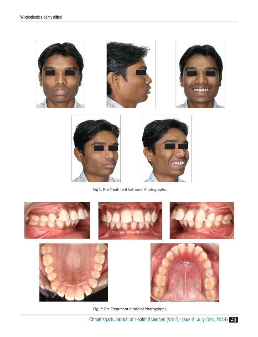
Wilckodontics demystified
Fig 1: Pre Treatment Extraoral Photographs.
Fig. 2: Pre Treatment Intraoral Photographs.
49Chhattisgarh Journal of Health Sciences (Vol-2, Issue-2: July-Dec. 2014)

and Class II canine relationship with 8 mm overjet and
1 mm overbite. Maxillary arch had spacing of 10 mm
and mandibular arch had spacing of 6.5 mm with
severely proclined upper anterior teeth & lower
anterior teeth (Fig.2). Upper midline was deviated to
the left side of facial midline by 2mm.
Orthopantogram of the patient revealed normal hard
tissue structures and absence of pathology (Fig.3).
Pre Treatment Lateral Cephalogram (Fig.4) and
Cephalometric measurements showed a skeletal class II
pattern, hypodivergent mandibular plane with
proclined upper and lower incisors.(Table 1)
Treatment Plan. As the patient only desired the space
closure within a shorter duration of time, a
nonextraction orthodontic treatment plan with fixed
appliances (Pre adjusted edgewise MBT .018 slot) along
with the corticotomy procedure in both the arches was
decided.
Treatment Progress. 018 MBT fixed appliance (Ormco)
was bonded and .014 inch NiTi arch wire was inserted
for initial leveling and alignment, which was followed by
.018NiTi, .016 X .022 NiTi & .017 X .025 SS.
Surgical Procedure. Corticotomy technique as 1described by Wilcko was performed by the
periodontist (fig.5). After administering the proper local
anesthetic dose, a full thickness flap was reflected
sharply facially, from canine to canine in the maxilla and
between the central incisors in the mandibular arch.
The flap was released with a sulcular incision and with
papillary preservation technique. No vertical releasing 3
incisions were used. Cuts in the alveolus that penetrate
the entire thickness of the cortical plate and penetrate
just barely into the medullary bone were performed 4,5,6
buccally & lingualy around the teeth in both arches.
Vertical decortication cuts were made between the
roots of the teeth and they were stopped 2-3mm shy of
the alveolar crest. Horizontal cuts were used to connect
the vertical cuts along with perforations in the cortical
Fig 3 Pre Treatment Panoramic Radiograph.
Fig. 4: Pre Treatment Lateral Cephalogram.
SNA 870
SNB 820
ANB 50
Wits 5mm
FMPA 130
UI-NA 330, 11 mm
LI-NB 420, 11 mm
IMPA 1250
Nasolabial angle 900
S LINE UL
LL
6 mm ahead
7 mm ahead
Table 1: Cephalometric values (Pre treatment)
50 Chhattisgarh Journal of Health Sciences (Vol-2, Issue-2: July-Dec. 2014)
Wilckodontics demystified

(a) (b) (c)
(d) (e)
(f)
Fig.5: Corticotomy in maxillary and mandibular arch.
(g) (h) (i)
(j) (k)
plate. Flaps were repositioned to their presurgical
positions and sutured with interrupted loop sutures.
The sutures were removed after 7 days from the
procedure. The patient was kept under antibiotic
regimen for 5 days following the surgery.
After removal of sutures, 0.017 × 0.025-inch SS
Fig. 6: Comparison of Pre Treatment and Post space closure Profile photographs
51Chhattisgarh Journal of Health Sciences (Vol-2, Issue-2: July-Dec. 2014)
Wilckodontics demystified

archwires with power arms were inserted in both the
arches. Short clear E chain (Rabbit Force, Libral) was
used for the enmass retraction of the anteriors & 150 g
force was applied. Post space closure profile
photographs showed reduced convexity and
competent lips (fig.6). After the active treatment of 10
Fig.7: Extraoral photographs (Post space closure)
weeks, space closure was achieved. (fig 7)
DISCUSSION
The corticotomy was planned as an adjunct to the
treatment in this case to achieve a better anchorage
control.5
Another reason why corticotomy was planned was the
desire of the patient to get the treatment finished in a
shorter duration. The chances of relapse in patients 6
with spacing is reduced with corticotomy as the
procedure require incisional cuts that are extended to
the bone level that sever the periodontal fibres in
particular the transeptal fibres.7
The space closure in this patient was achieved in 10
weeks, which is significantly less than the normal
duration which is (6 months) required by conventional
orthodontic treatment. 8
Intraoral photographs showed that arches were well
aligned and spacing have been closed (fig.8).
52 Chhattisgarh Journal of Health Sciences (Vol-2, Issue-2: July-Dec. 2014)
Wilckodontics demystified

PARAMETERS PRE-TREATMENT POST SPACE CLOSURE
SNA 870 860
SNB 820 840
ANB 50 40
WITS 5 mm 4 mm
FMPA 130 120
U1-NA 330,11 mm 240,6.5 mm
L1-NB 420,11 mm 380,7 mm
IMPA 1250 1180
Nasolabial Angle 900 990
S Line UL
LL
6 mm ahead
7 mm ahead
4 mm ahead
3 mm ahead
Table 2: Comparison of Pre treatment and Post space closure cephalometric values.
Fig.8: Intraoral photographs (Post space closure)
Pretreatment and post space closure values when
compared showed that there was a reduction in the
proclination of anteriors in the maxillary and
mandibular arches (Table 2).
CONCLUSION
Corticotomy Assisted Orthodontic Treatment is a
promising technique that has many applications in the
orthodontic treatment of adults because it helps to
overcome many of the current limitations of this
treatment including lengthy duration, potential for
periodontal complications, lack of growth and the
limited envelope of tooth movement.
REFERENCES
1. Murphy KG, Wilcko MT, Wilcko WM, Fergusson WJ.
Periodontal Accelerated Osteogenic Orthodontics:
A Description Of The Surgical Technique. J Oral
Maxillo Surg 2009; 67:2160-2166.
2. Aljhani AS, Zawai KH Nonextraction Treatment of
Severe Crowding with the Aid of Corticotomy-
53Chhattisgarh Journal of Health Sciences (Vol-2, Issue-2: July-Dec. 2014)
Wilckodontics demystified

Assisted Orthodontics. Case Reports In Dentistry
2012;2012:694527(1-8).
3. Thakur A. Halwai H,Corticotomy Assisted
Orthodontic Treatment Journal of Universal College
of Medical Sciences 2013;1(1):1-6
4. Goyal A et al. Periodontally accelerated osteogenic
orthodontics (PAOO) - A review. J Clin Exp Dent
2012;4(5):292-6.
5. Bhat SG, Singh V. PAOO technique for the
b i m a x i l l a r y p r o t r u s i o n : P e r i o - o r t h o
interrelationship. Journal of Indian Society of
Periodontology 2012;16 (4):584-87
6. Karanth S ,Ramesh A ,Thomas B, John AM.
Periodontally accelerated osteogenic orthodontics:
Review on a surgical technique and a case report.
J o u r n a l o f I n t e r d i s c i p l i n a r y D e n t i s t r y
2012;2(3):179-185.
7. Ali H, Ahmed A. Corticotomy-Assisted Orthodontic
Treatment: Review. The Open Dentistry Journal
2010;4: 159-164.
8. Fischer TJ. Orthodontic Treatment Acceleration
with Corticotomy-assisted Exposure of Palatally
Impacted Canines. Angle Orthodontist 2007; 77(3 ):
417-420.
54 Chhattisgarh Journal of Health Sciences (Vol-2, Issue-2: July-Dec. 2014)
Wilckodontics demystified