uwe mueller · 2019-10-04 · sample and detector. beamline characteristics: • support all...
TRANSCRIPT

Life Science at the First Ultimate Storage Ring MAX IV, Lund, Sweden
Uwe Mueller

MAX IV – A dream for a long time came true
Architects 3D model

MAX IV
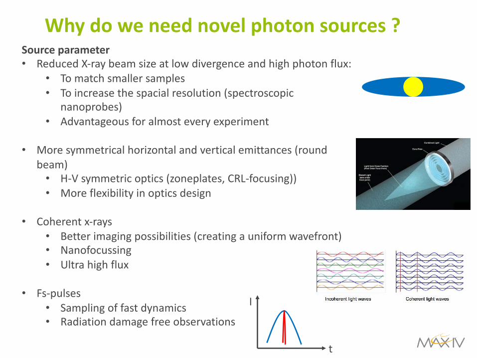
Why do we need novel photon sources ?Source parameter• Reduced X-ray beam size at low divergence and high photon flux:
• To match smaller samples• To increase the spacial resolution (spectroscopic
nanoprobes)• Advantageous for almost every experiment
• More symmetrical horizontal and vertical emittances (round beam)• H-V symmetric optics (zoneplates, CRL-focusing))• More flexibility in optics design
• Coherent x-rays• Better imaging possibilities (creating a uniform wavefront)• Nanofocussing• Ultra high flux
• Fs-pulses• Sampling of fast dynamics• Radiation damage free observations
t
I
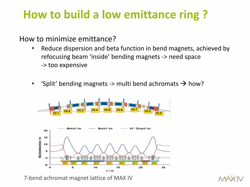
How to build a low emittance ring ?
How to minimize emittance?• Reduce dispersion and beta function in bend magnets, achieved by
refocusing beam ‘inside’ bending magnets -> need space -> too expensive
• ‘Split’ bending magnets -> multi bend achromats à how?
7-bend achromat magnet lattice of MAX IV
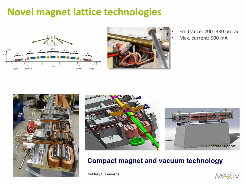
Novel magnet lattice technologies
Compact magnet and vacuum technology
Courtesy S. Leemans
• Emittance: 200 -330 pmrad• Max. current: 500 mAUnit
Cells

New storage rings in the future
R. Bartolini
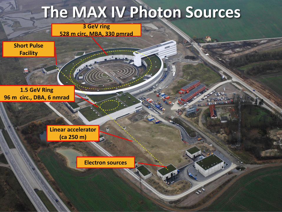
Electron sources
3 GeV ring528 m circ, MBA, 330 pmrad
Linear accelerator (ca 250 m)
1.5 GeV Ring 96 m circ., DBA, 6 nmrad
The MAX IV Photon Sources
Short Pulse Facility
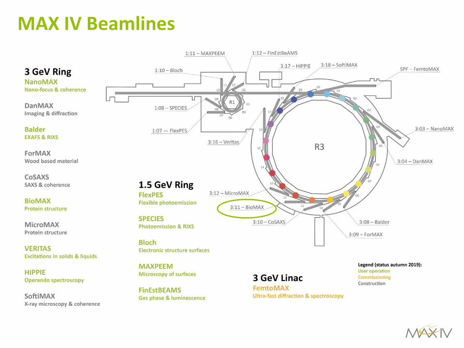
MAX IV Beamlines
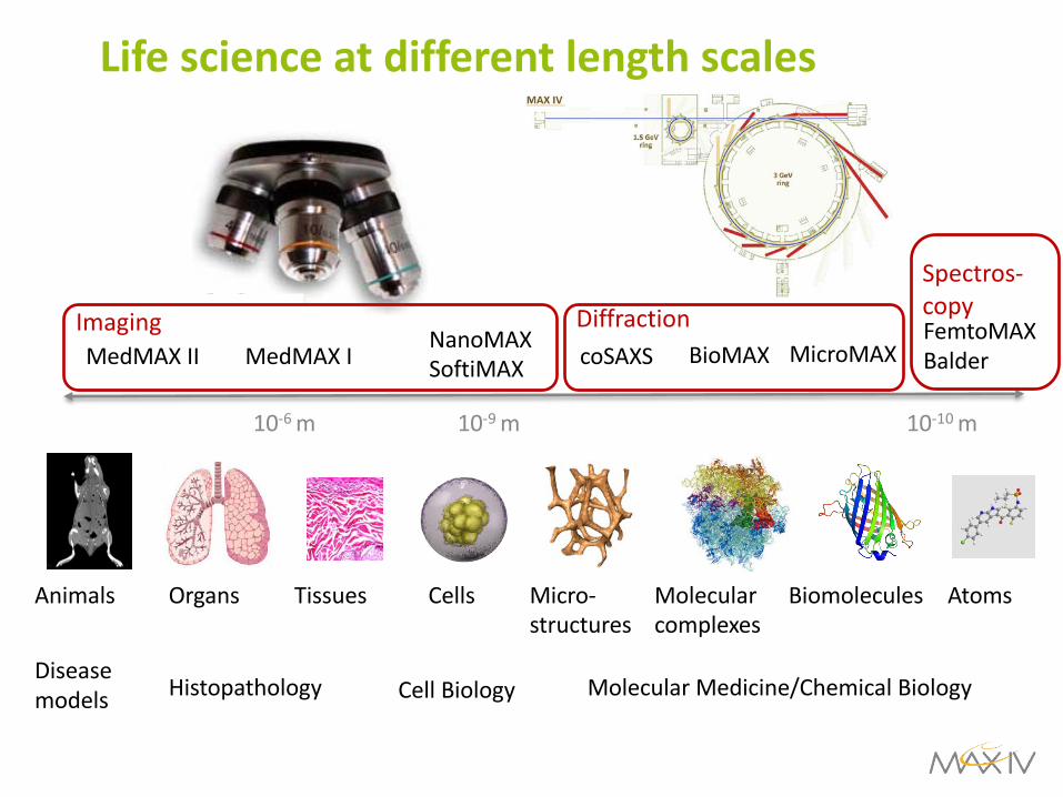
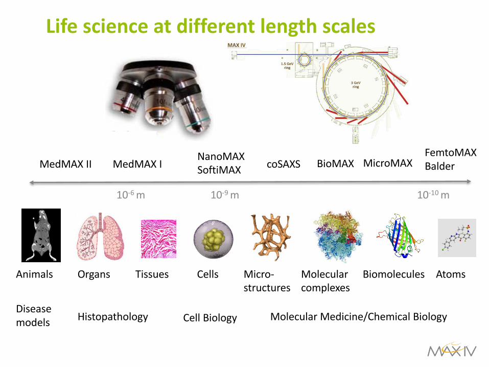
Life science at different length scales
CellsTissues BiomoleculesOrgans
Cell BiologyHistopathology Molecular Medicine/Chemical BiologyDisease models
Animals AtomsMicro-structures
Molecular complexes
NanoMAXSoftiMAXMedMAX I BioMAXMedMAX II MicroMAX
10-6 m 10-9 m 10-10 m
coSAXSFemtoMAXBalder
Imaging Diffraction
Spectros-copy

Robot
Flow through Static BioSAXS
Dynamic BioSAXSConformational changes
100-1000Hz readout
Detector
THz radiation
CoSAXS-FemtoMAXLübeck University-EMBL/Ham.
Raman
SAXSIncomming X-ray beam
Fluorescence in
Fluorescence out
UV-VIS out
UV-VIS in
20°45°
90°
225°
340°
Multiple probe BioSAXS-SURF
CoSAXS-LU-SWING (Soleil)
coSAXS-BioSAXS
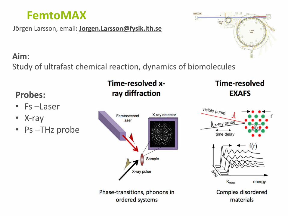
FemtoMAXJörgen Larsson, email: [email protected]
Aim:Study of ultrafast chemical reaction, dynamics of biomolecules
Probes:• Fs –Laser• X-ray• Ps –THz probe
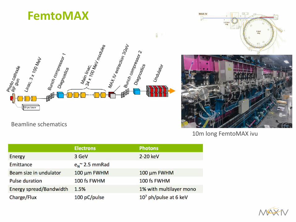
FemtoMAX
Beamline schematics10m long FemtoMAX ivu
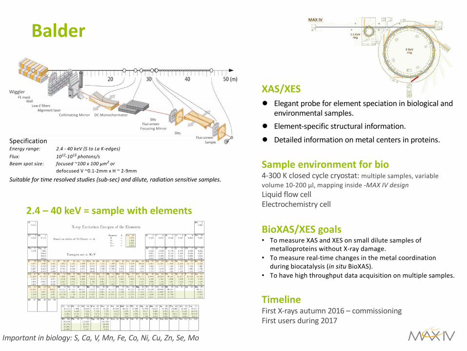
Balder
Wiggler
FE mask
Wall
Low-E filters
Alignment laser
Collimating Mirror DC Monochormator
Slits
Fluo screen
Focusing Mirror
Slits
Fluo screen
SampleSpecification
Energy range: 2.4 - 40 keV (S to La K-edges)
Flux: 1012-1013 photons/s Beam spot size: focused ~100 x 100 µm2 or
defocused V ~0.1-2mm x H ~ 2-9mm
Suitable for time resolved studies (sub-sec) and dilute, radiation sensitive samples.
2.4 – 40 keV = sample with elements
Important in biology: S, Ca, V, Mn, Fe, Co, Ni, Cu, Zn, Se, Mo
XAS/XES● Elegant probe for element speciation in biological and
environmental samples.
● Element-specific structural information.
● Detailed information on metal centers in proteins.
Sample environment for bio 4-300 K closed cycle cryostat: multiple samples, variable
volume 10-200 µl, mapping inside -MAX IV designLiquid flow cell
Electrochemistry cell
BioXAS/XES goals• To measure XAS and XES on small dilute samples of
metalloproteins without X-ray damage.
• To measure real-time changes in the metal coordination
during biocatalysis (in situ BioXAS).
• To have high throughput data acquisition on multiple samples.
TimelineFirst X-rays autumn 2016 – commissioning
First users during 2017
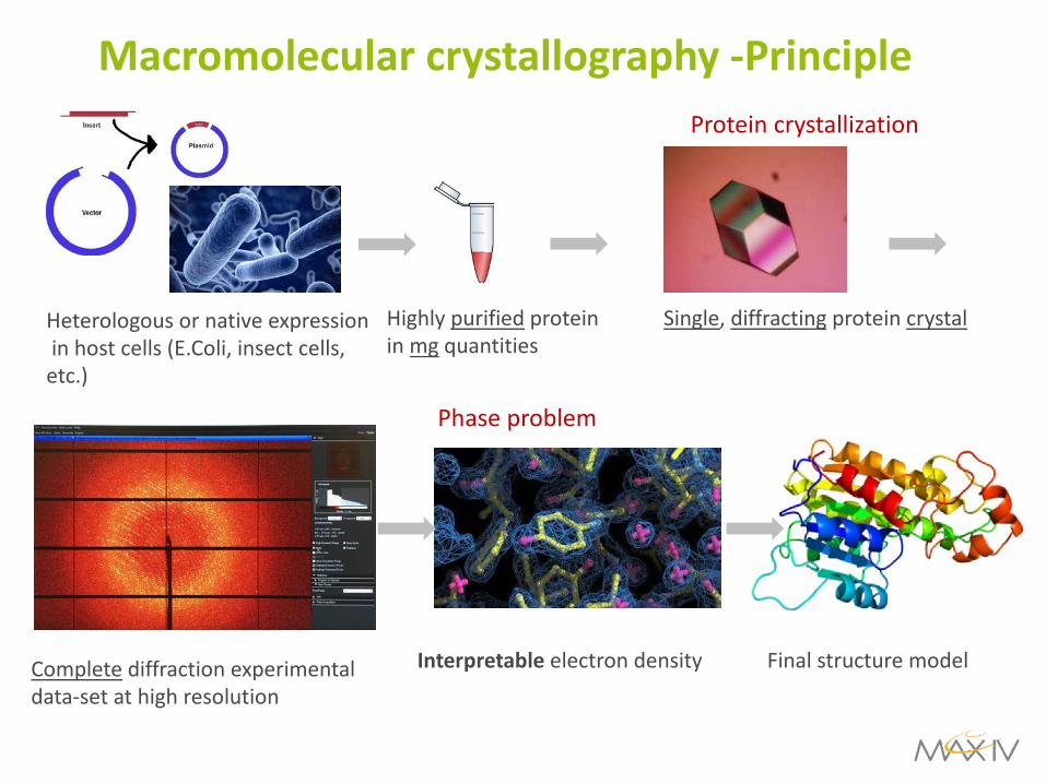
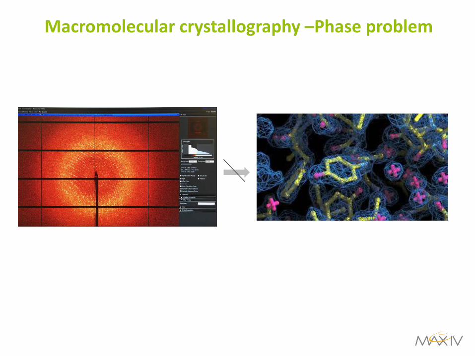
Macromolecular crystallography -Principle
Heterologous or native expressionin host cells (E.Coli, insect cells,etc.)
Highly purified proteinin mg quantities
Single, diffracting protein crystal
Complete diffraction experimentaldata-set at high resolution
Interpretable electron density Final structure model
Protein crystallization
Phase problem
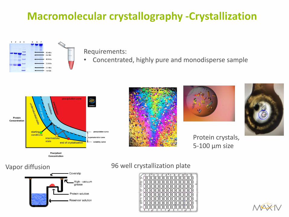
Macromolecular crystallography -Crystallization
Requirements: • Concentrated, highly pure and monodisperse sample
Vapor diffusion 96 well crystallization plate
Protein crystals, 5-100 µm size
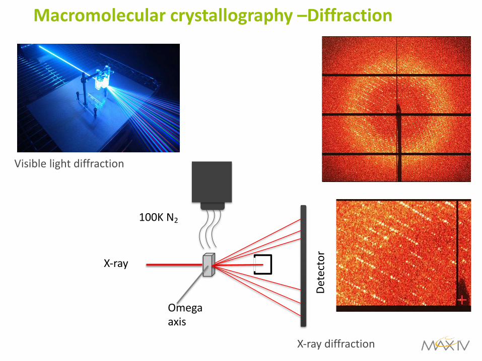
Macromolecular crystallography –Diffraction
100K N2
X-ray
Omegaaxis
Dete
ctor
Visible light diffraction
X-ray diffraction
Macromolecular crystallography –Phase problem
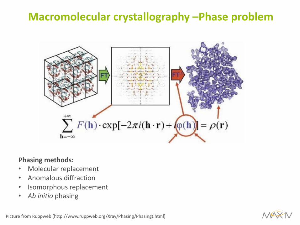
Macromolecular crystallography –Phase problem
Picture from Ruppweb (http://www.ruppweb.org/Xray/Phasing/Phasingt.html)
Phasing methods:• Molecular replacement• Anomalous diffraction• Isomorphous replacement• Ab initio phasing
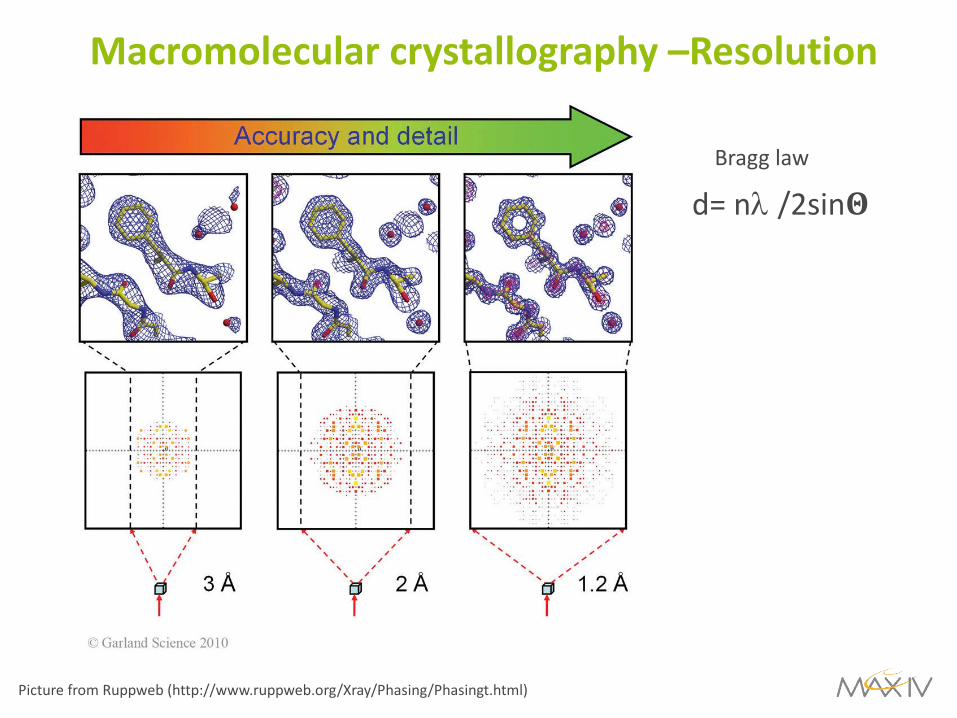
Macromolecular crystallography –Resolution
d= nl /2sin!Bragg law
Picture from Ruppweb (http://www.ruppweb.org/Xray/Phasing/Phasingt.html)
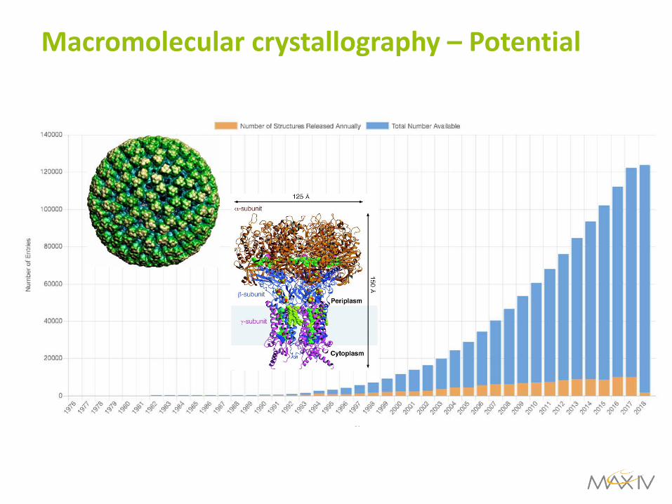
Macromolecular crystallography – Potential

Macromolecular crystallography –Potential
NMR
Cryo-EM

Macromolecular crystallography
CellsTissues BiomoleculesOrgans
Cell BiologyHistopathology Molecular Medicine/Chemical BiologyDisease models
Animals AtomsMicro-structures
Molecular complexes
NanoMAXSoftiMAXMedMAX I BioMAXMedMAX II MicroMAX
10-6 m 10-9 m 10-10 m
coSAXSFemtoMAXBalder

BioMAX Overview
Schematic of BioMAX optics design with undulator, monochromator, focusing optics, sample and detector.
Beamline characteristics:• Support all relevant techniques for MX• Beam characteristics:
• Small focus (20 x 5 µm2 hxv FWHM)• Low divergence (0.1 x 0.1 mrad2)• High flux (2x 10¹³ phot/s x 0.1% bw)
• MD3 Microdiffractometer• Large energy range (5-25 keV)• Short data collection times (2-30 ms /frame)• High throughput environment
2017-2019: 71 proposals, 506 users, 5860 samples,9495 datasets

BioMAX Undulator
In-vacuum undulator
5 10 15 20 25100
101
102
103
104
105
106
Photon Energy [keV]
Flux
[Cou
nts]
measurementsimulation
3 mA ring current, courtesy of H. Tarawneh
1.0
0.8
0.6
0.4
0.2
0.0
No
rm
alliz
ed
ph
oto
n f
lux
27.427.227.026.826.6
Photon Energy[keV]
Measured at 149 mA
e0 = 0 pmrad, s
p=0
sp=7.7 10
-4
, e0 = 330 pmrad
BioMAX undulator @ 15th
harmonic
Flux in 10x10 µrad2
rect. aperture
calcs (Spectra),no phase errors)
10% coupling
Data by T. Ursby and D.Olsson
149 mA ring current

BioMAX experimental station
Experimental environment
• MD3 micro-diffractometer
• Eiger 16M hybrid pixel
detector
• ISARA sample changer
• Operations center
• Sample preparation lab
• BioLab
BioMAX experimental station

BioMAX Commissioning
20 deg/sec frame rotation
Complete data collection within 4 sec., Cubic Insulin (R. Lizatovic)
Exp. Conditions:• Exposure time: 10 msec• Omega increment: 0.2 deg• Omega range: 80 deg• Images 400• Total time dc.: 4 sec
Final table from CORRECT.LP
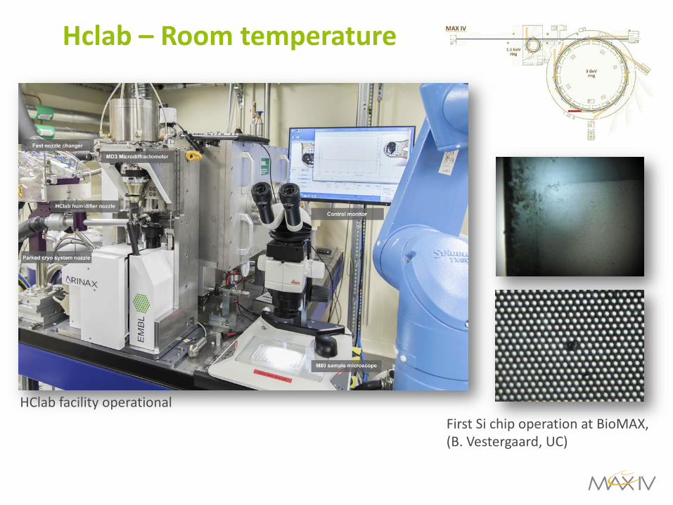
Hclab – Room temperature
HClab facility operationalFirst Si chip operation at BioMAX,(B. Vestergaard, UC)

X-ray detector
Eiger 16M hybrid pixel detector:• Sensitive area: 311.2 × 327.8 mm2
• 4150 × 4371 = 18’139’650 pixels• Pixel size 75 x75 µm2
• Scan speed:• 133 Hz (16M)• 750 (4M)
Challenges• HDF5/Nexus file format• Data streaming for real time analysis • Large data volumes
• Av. dataset size 10-20 Gb (15 sec)• Data storage & processing for users
Eiger 16M hybrid pixel detector
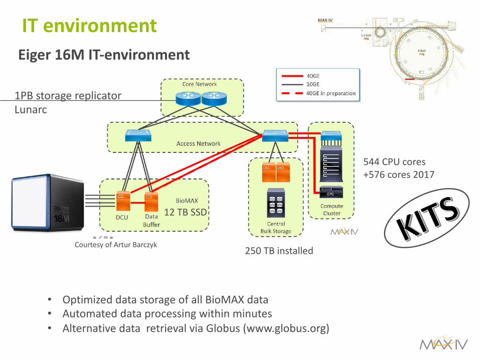
IT environmentEiger 16M IT-environment
Courtesy of Artur Barczyk
12 TB SSD
250 TB installed
544 CPU cores
+576 cores 2017
1PB storage replicator
Lunarc
• Optimized data storage of all BioMAX data
• Automated data processing within minutes
• Alternative data retrieval via Globus (www.globus.org)

MAX IV 80 m2 BioLAB
BioMAX operations and sample lab
BioMAX operations and sample lab
Lab facilities
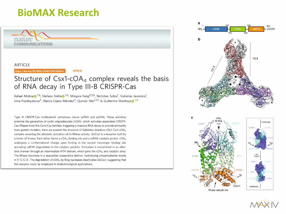
BioMAX Research

Scientific results
BioMAX Research

BioMAX Research

Life science at different length scales
CellsTissues BiomoleculesOrgans
Cell BiologyHistopathology Molecular Medicine/Chemical BiologyDisease models
Animals AtomsMicro-structures
Molecular complexes
NanoMAXSoftiMAXMedMAX I BioMAXMedMAX II MicroMAX
10-6 m 10-9 m 10-10 m
coSAXSFemtoMAXBalder

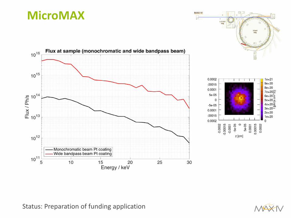
MicroMAX
Plans for the second MX beamline
• Microfocus beamline
• 1 x 0.7 µm2 beam at sample
• Photon flux 1013 - 1015phot/sec
• Traditional setup (goniometry, sample environment)
• Exploratory setup (serial crystallography, fixed target single shot)
• Optimal source for most demanding projects (large complexes, membrane proteins)
Status: funded by Novo Nordisk Foundation
Thomas Ursby: email: [email protected]
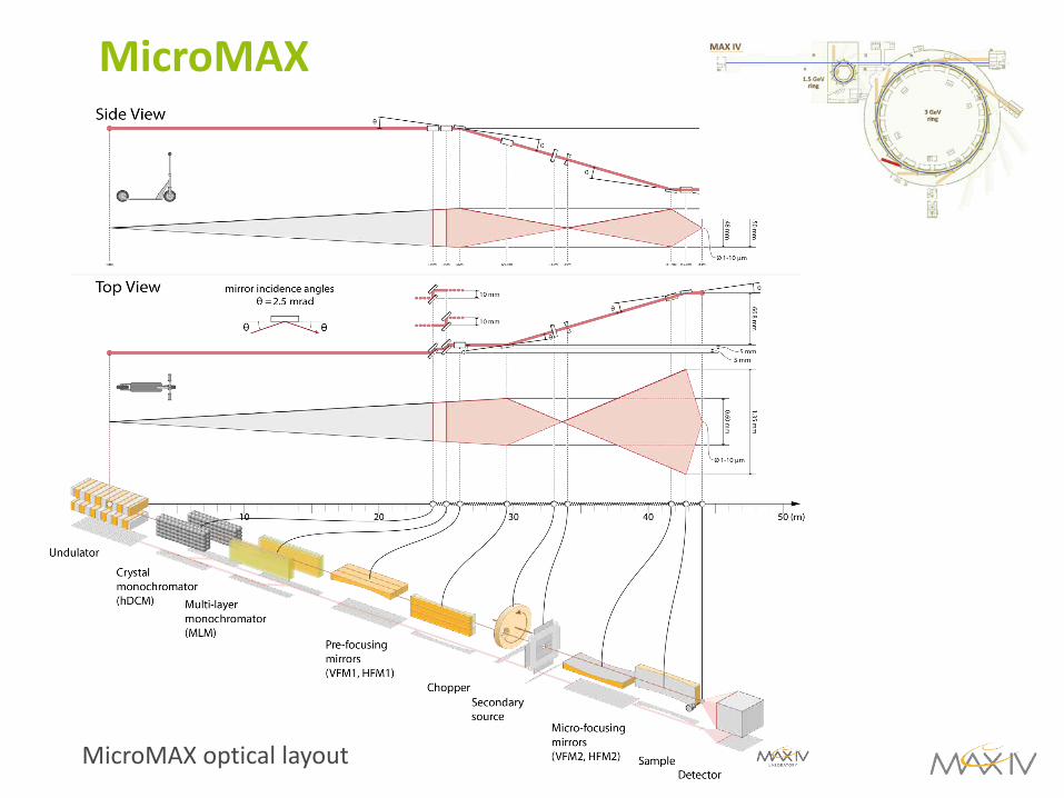
MicroMAX
MicroMAX optical layout
MicroMAX
Status: Preparation of funding application

MicroMAX
Novel sample delivery for micro-crystallography
Nogy et al., Gati et al.
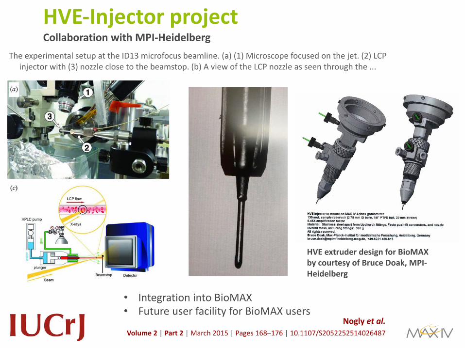
HVE-Injector project The experimental setup at the ID13 microfocus beamline. (a) (1) Microscope focused on the jet. (2) LCP
injector with (3) nozzle close to the beamstop. (b) A view of the LCP nozzle as seen through the ...
Nogly et al.Volume 2 | Part 2 | March 2015 | Pages 168–176 | 10.1107/S2052252514026487
HVE extruder design for BioMAX by courtesy of Bruce Doak, MPI-Heidelberg
• Integration into BioMAX• Future user facility for BioMAX users
Collaboration with MPI-Heidelberg
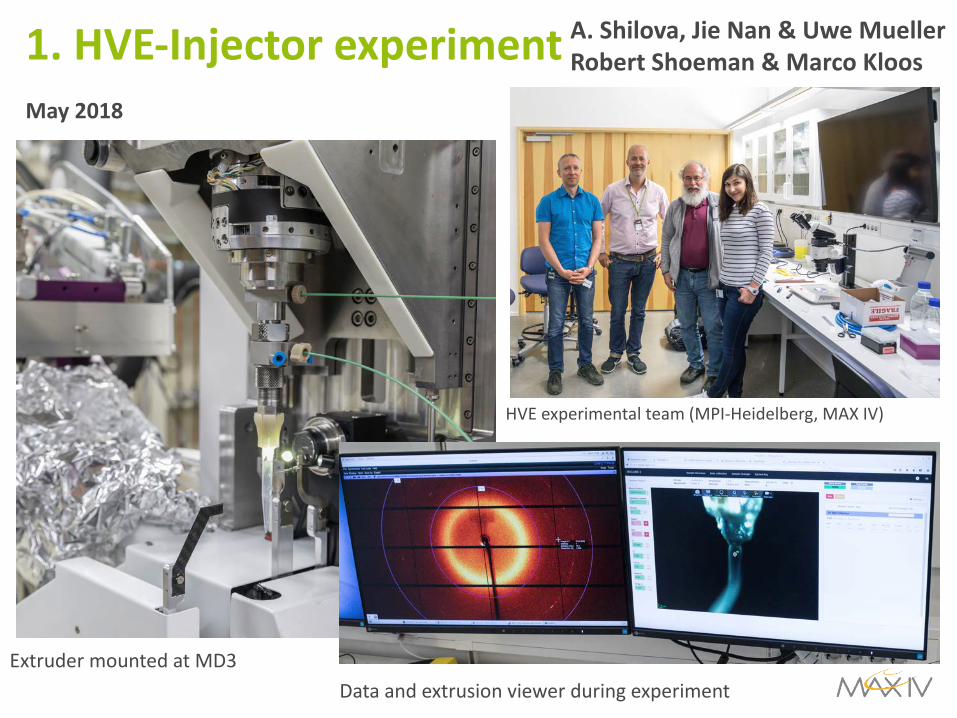
1. HVE-Injector experiment
New project scientistAnastasia Shilova
May 2018
HVE experimental team (MPI-Heidelberg, MAX IV)
Extruder mounted at MD3Data and extrusion viewer during experiment
A. Shilova, Jie Nan & Uwe MuellerRobert Shoeman & Marco Kloos
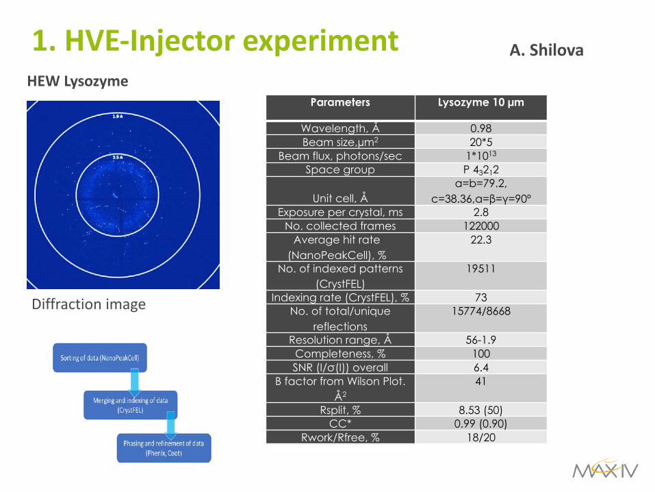
HEW Lysozyme
1. HVE-Injector experiment
Diffraction image
Parameters Lysozyme 10 µm
Wavelength, Å 0.98Beam size,µm2 20*5
Beam flux, photons/sec 1*1013
Space group P 43212
Unit cell, Åa=b=79.2,
c=38.36,α=β=γ=90ºExposure per crystal, ms 2.8
No. collected frames 122000Average hit rate
(NanoPeakCell), %22.3
No. of indexed patterns (CrystFEL)
19511
Indexing rate (CrystFEL), % 73No. of total/unique
reflections15774/8668
Resolution range, Å 56-1.9Completeness, % 100SNR (I/σ(I)) overall 6.4
Β factor from Wilson Plot. Å2
41
Rsplit, % 8.53 (50)CC* 0.99 (0.90)
Rwork/Rfree, % 18/20
A. Shilova

2. HVE experiment4.-7. June 2018
• 3 proteins injected• 10 TB+ of data• Data processing on-going
MPI HVE injector
ASU – LCP injector
Bränden, Neutze team at BioMAX
A. Shilova, J. Nan & U. MuellerBränden & Neutze group

Cytochrome c oxidase
Experiment was performed with user groups from Gothenburg University (Richard Neutze group and Gisela Branden)
Parameters BCO
Wavelength, Å 0.98Beam size,µm2 20*5
Beam flux, photons/sec 1*1013
Space group C2
Unit cell, Å
a=145.17 b=100.15, c=96.64,α=γ=90º,
β=126.64Exposure per crystal, ms 2.8
No. collected frames 253766Average hit rate
(NanoPeakCell), %3.5
No. of indexed patterns (CrystFEL)
6513
Indexing rate (CrystFEL), % 68No. of total/unique
reflections27514/18454
Resolution range, Å 75.94-3.5Completeness, % 100SNR (I/σ(I)) overall 2.7
Β factor from Wilson Plot. Å2
68
Rsplit, % 34.02 (50)CC* 0.90 (0.88)
Rwork/Rfree, % 32/36
A. Shilova
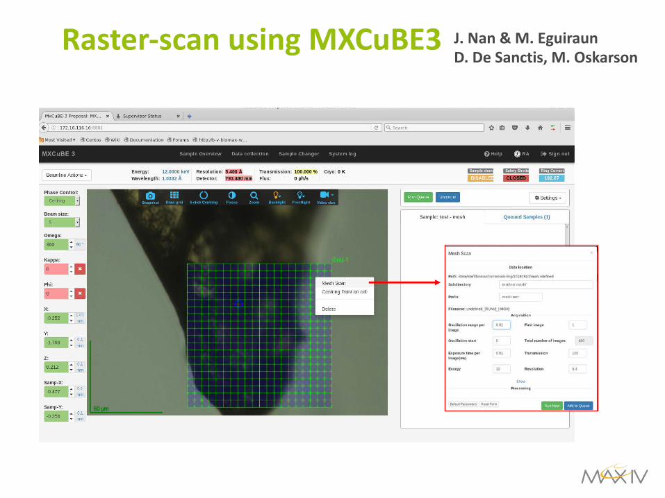
Raster-scan using MXCuBE3 J. Nan & M. EguiraunD. De Sanctis, M. Oskarson
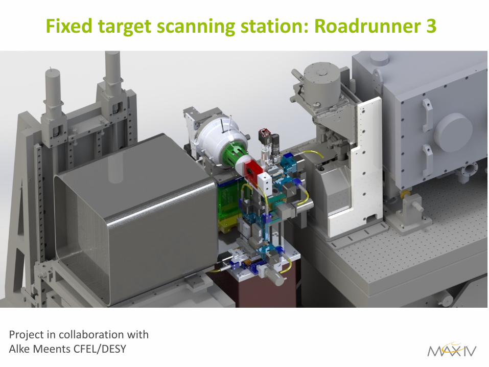
Fixed target scanning station: Roadrunner 3
Roadrunner 3 at BioMAX, CAD view
Project in collaboration with Alke Meents CFEL/DESY

AcknowledgementThe MX group
• All BioMAX users
From left to right:• Oskar Aurelius• Vladimir Talibov• Laila Benz• Thomas Ursby• Gustavo Lima• Ana Gonzalez• Uwe Mueller• Vahid Haghighat• Mikel Eguiraun (KITS)• Jie Nan• Johan Unge• Mirko Milas• Monika Bjelcic• Elmir Jagudin
Life science at different length scales
CellsTissues BiomoleculesOrgans
Cell BiologyHistopathology Molecular Medicine/Chemical BiologyDisease models
Animals AtomsMicro-structures
Molecular complexes
NanoMAXSoftiMAXMedMAX I BioMAXMedMAX II MicroMAX
10-6 m 10-9 m 10-10 m
coSAXSFemtoMAXBalder