uvgi for cooling coil disinfection, air treatment, and ...uvgi for cooling coil disinfection, air...
TRANSCRIPT
UVGI for Cooling Coil Disinfection, Air Treatment, and
Hospital Infection Control
Report Prepared by
Dr. Wladyslaw J. Kowalski, PE
for
American Air & Water, Inc.
12 Gibson Drive, Hilton Head Island, SC 29926 Phone: 843-785-8699 * 888-378-4892 * Fax * 843-785-2064
www.americanairandwater.com
January 24, 2011
Revision 1.06

AAWI UVGI Disinfection
2
TABLE OF CONTENTS
1. Executive Summary .................................................................. 3
2. Introduction and Background .................................................... 4
3. Microbial Disinfection Model ..................................................... 8
4. Cooling Coil Disinfection .......................................................... 11
5. Performance of Cooling Coil Disinfection Systems................... 15
6. Economics of Cooling Coil Disinfection Systems ..................... 17
7. Guidelines for Cooling Coil Disinfection Systems ..................... 21
8. Hospital Air & Surface Disinfection Systems............................. 23
8.1 Hospital-Acquired (Nosocomial) Infections ............................... 23
8.2 Surgical Site Infections (SSIs) and Overhead UV Systems ...... 25
8.3 Hospital Air & Surface Disinfection Systems............................. 27
9. Hotel & Residential Air Disinfection Systems............................ 29
10. Electric Utilities and UV Air Disinfection.................................... 34
11. References and Bibliography ................................................... 35
Appendix A: Microbial Rate Constants....................................... 55
Appendix B: Common Indoor Bacteria....................................... 67
Appendix C: Common Indoor Fungi .......................................... 68
Appendix D: Health Care Facility Cost Estimates .................... 69
Appendix E: Excerpts from articles on UVC effectiveness ............. 71

AAWI UVGI Disinfection
3
1. Executive Summary This report reviews the history and current literature on ultraviolet germicidal irradiation (UVGI) systems used for air and surface disinfection applications. Three of the most promising applications are addressed in this report:
1) Cooling Coil Disinfection 2) Hospital Infection Control 3) Hotel & Residential Air Disinfection
Cooling coil disinfection has proved to be a most economical application
and can produce a payback in terms of energy savings of about two years or less. These savings result from reduced coil cleaning maintenance costs and energy savings due to improved heat transfer and reduced pressure losses from airflow through the coils. The use of UVGI on cooling coils tends to restore them to original design conditions and will maintain them in a clean state for as long as the UVGI system is operated. This report summarizes the available information on laboratory and field testing of such installations. Information on the energy savings and payback period of cooling coil irradiation are provided along with an example of the computation of a typical payback period. Draft guidelines on cooling coil irradiation systems from the International Ultraviolet Association are reviewed. Applications in hospital or other health care facilities include cooling coil cleaning, medical equipment disinfection, whole-room disinfection in unoccupied areas, Overhead surgical site disinfection, and air disinfection in operating rooms, procedure rooms, delivery rooms, isolation rooms, patient wards, and general areas. Hospital-acquired (nosocomial) infections have continued to present a challenge to health care facilities, and cause an undue number of fatal infections annually in the US, with concomitant economic costs. UVGI may be able to reduce many types of nosocomial infections, including surgical site infections (SSIs), and is especially effective against airborne pathogens. This paper explores some of the various applications for air and surface disinfection that may assist hospitals in reducing nosocomial infection rates. Hotels may obtain significant economic benefits both from the use of UVGI to keep cooling coils clean and from improvements in air quality, which will provide a better environment for both guests and employees. The kinds of problems encountered in large hotels that are amenable to UVGI solutions are addressed in this report, and data is summarized from some previous applications. Residential applications are similar to hotels and these can include homes, apartments, and dormitories.

AAWI UVGI Disinfection
4
2. Introduction and Background The effects of ultraviolet light (UV) on microorganisms were first noted in the late 1800s and the first scientific study of UV is attributed to Downes and Blunt (1877). Baker (1948) indicates that the first mention of the use of ultraviolet light for water disinfection was in 1877. In 1909/1910 the first water disinfection system was operated at Marseilles, France, but it wasn’t until 1916 that the first UV system for water disinfection was tested in the US, at Henderson, KY (AWWA 1971).
The first attempts to scientifically quantify the effects of UV irradiation of microorganisms were published in the 1920s. Bedford (1927) and Gates (1929) were among the first to study bacteria and establish UV dosages necessary for disinfection. The earliest study that demonstrated fungicidal action of UV was published by Fulton and Coblenz (1929). The first studies on irradiating viruses appear to have been those published by Sturm et al (1932). In the 1930s a flurry of research and applications demonstrated the feasibility of applying UV light systems in hospitals (Wells and Wells 1936, Wells 1938, Hart and Sanger 1939, Robertson et al 1939, Kraissl et al 1940, Overholt and Betts 1940). The first Upper Air UV systems appear to have been installed around 1938 (Wells 1938).
In the 1940s the first detailed studies of UV air disinfection were published along with basic guidelines for applying UV in ventilation systems (Rentschler and Nagy 1940, Sharp 1940, Wells 1940, Buchbinder and Phelps 1941, DelMundo and McKhann 1941, Henle et al 1942, Luckiesh and Holladay 1942, Sommer and Stokes 1942, Hollaender 1943). The first attempts to apply UV systems to schools and barracks to control respiratory infections occurred shortly thereafter (Wells et al 1942, Wells 1943, Schneiter et al 1944, Wheeler et al 1945, Higgons and Hyde 1947, Perkins et al 1947). Several early attempts were made to develop sizing methods and engineering guidelines for UV applications (Luckiesh and Holladay 1942a, Luckiesh 1945, Luckiesh 1946). Table 1 summarizes many of the seminal developments in UV history. By 1950 it had been established that UV irradiation was effective at disinfecting both air and surfaces, and engineering applications were being developed. General Electric catalogs detailed many UV applications including various methods of installing UV lamps inside ducts and air conditioners (Buttolph and Haynes 1950, GE 1950). At this time it was not generally known that mold growth on cooling coils could cause respiratory problems. In 1954 it was demonstrated by Harstad et al (1954) that installation of UV lamps in air conditioners would reduce airborne contamination. It was further noted in this published study that microorganisms were impinging upon and collecting on internal AHU surfaces. Although it was understood that UVGI could be used to control microbial growth inside air handling equipment and on cooling coils, little or no attention would be focused on this issue for nearly twenty years. In 1957, Riley and associates completed a demonstration of how a UV air disinfection could be used to control the spread of tuberculosis (TB) I hospital wards (Riley et al 1957). It wasn’t until 1994 that the Centers for Disease Control (CDC) began to acknowledged that UV could be effective for controlling TB, and

AAWI UVGI Disinfection
5
this was not in response to the numerous published studies, but because the growing worldwide TB epidemic had resisted control by traditional methods (CDC 2005). Although UVGI systems had been in use in hospitals since 1936, it wasn’t until 2003 that the CDC formally acknowledged that UV systems were effective and could be used in hospitals with their conditional blessing – UV Upper Room and in-duct systems could only be used to supplement other air cleaning systems (CDC 2003).
Bacterial growth on cooling coils had been recognized as a potential health problem as early as 1958 (Walter 1969). The first evidence that air cooling equipment could actually cause respiratory infections was presented by Anderson (1959) when an air cooling apparatus was found to be contaminated with microbial growth. This very same concern had been raised in hospital environments since about 1944 but the possibility of growth of bacteria on air-conditioning cooling coils wasn't conclusively demonstrated until 1964 (Cole et al 1964). The growth of microbes on other equipment like filters and dust inside air-conditioning ducts was first demonstrated by Whyte (1968). The fact that microbes growing in air handling equipment could be disseminated by ventilation systems and cause respiratory infections became widely recognized in the late 1960s and early 1970s in both the medical and engineering fields (Banaszak et al 1970, Schicht 1972, Zeterberg 1973). It was widely known at this time that microbial growth could occur anywhere that air came into contact with moisture (Gunderman 1980, Ager and Tickner 1983, Spendlove and Fannin 1983). The first UVGI system designed specifically for disinfecting the surfaces of air handling equipment, including humidifier water and filters, was detailed by Grun and Pitz (1974). Luciano (1977) published a book detailing many applications of UVGI, including health care applications in which the UV lamps are specifically placed upstream of the cooling coils and downstream of the filters, which has become the primary location for both air disinfection and cooling coil irradiation systems. In 1985 Phillips published a design guide in which the first definitive description of applications of UV lamps for the control of microbial growth on cooling coils were presented (Philips 1985). This design guide, "Germicidal Lamps and Applications" provides details of how to locate lamps at specific distances from cooling coils or walls, and referred to installations that were already in operation at the time. Apparently, Europeans had been using such systems prior to 1985 but no publications exist to document such applications. In January of 1996 the first UVGI system in the U.S. designed for controlling microbial growth on cooling coils was installed by Public Service of Omaha (PSO) in Tulsa. Tom McKain of PSO reports that the idea of irradiating their fouled cooling coils came both from Dr. Richard Shaughnessy of Tulsa University (TU), and from a European professor who suggested the idea at a recent conference (Kowalski 2005). PSO hired Steril-Aire to implement the system, which was found to be highly effective after studies by TU researchers. A patent for cooling coil irradiation was filed later by Steril-Aire. Table 1 summarizes the critical events described above and a number of other major events in the history of UVGI technology development and applications.

AAWI UVGI Disinfection
6
Year Event Reference1870s Bactericidal Effects of UV light discovered Downes & Blunt 18771877 First demonstration of UV water disinfection Baker 19481903 UV spectrum near 250 nm found to be germicidal Lorch 19871904 First quartz lamp for UV developed Lorch 19871906 UV first used to disinfect drinking water von Recklinghausen 19141909 First European applications for UV water disinfection AWWA 19711916 First USA applications of UV for water disinfection AWWA 19711921 UV photoreactivity with TiO2 first demonstrated Renz 19211925 UV photodegradation of materials first demonstrated Luckiesh & Taylor 19251927 Bactericidal action of UV first quantified Bedford 1927, Gates 19291929 Fungicidal action of UV first quantified Fulton & Coblentz 19291932 Virucidal action of UV first quantified Sturn et al 19321936 First Overhead UV system in hospitals Hart 1936, Wells&Wells 19361936 UV photoreactivation phenomena first identified Prat 19361937 First Upper Air application in schools Wells 19381938 First flourescent gas discharge UV lamp Whitby & Scheible 20041940 UV first applied to air conditioning systems Rentschler & Nagy 19401942 First UV air disinfection sizing guidelines Luckiesh & Holladay 19421942 Upper & Lower UV applied to Army/Navy barracks Wells 19421949 UV doses for disinfecting various surface molds quantified Luckiesh 19491950 First catalog sizing methods Buttolph & Haynes 19501954 First air conditioner application Harstad et al 19541954 Faulty British study concludes UV is ineffective MRC 19541957 Riley proves effectiveness of UV for TB control Riley et al 19571974 First microbial growth control systems Grun & Pitz 1974
1977 Luciano specifies locating UV lamps between filters and cooling coils in air handling units
Luciano 1977
1985 Cooling coil UV systems in use in European breweries Philips 19851994 CDC acknowledges UV effectiveness for TB control CDC 20051996 First cooling coil irradiation system in US Kowalski 20051997 First UV light emitting diodes at 265 nm (LEDs) Guha & Bojarczuk 19981999 WHO recommends UVGI for TB control WHO 19992000 US Army recommends UVGI for disease isolation USACE 20002003 CDC formally sanctions UVGI use in hospitals CDC 20032003 FEMA sanctions UVGI as a biodefense option for buildings FEMA 20032003 In-duct UVGI system demonstrates lower illness symptoms Menzies et al 20032003 ASHRAE forms Air Treatment Group Martin et al 20082005 GSA specifies UV for cooling coil disinfection in buildings GSA 2005
2007 Long-term hospital study demonstrates significant reductions in surgical site infections w/ overhead UV
Ritter et al 2007
2009 US Army addresses UVGI for medical facilities USACE 2009 2009 ASHRAE issues Position Statement on airborne disease ASHRAE 2009
Table 1: Chronology of Critical Events in UV History

AAWI UVGI Disinfection
7
The topic of UV air disinfection is currently being addressed in detail by the American Society of Heating, Refrigerating and Air-Conditioning Engineers (ASHRAE) who have released a position paper on the subject of airborne infectious diseases as they relate to ventilation systems (ASHRAE 2009). ASHRAE’s position as stated in the paper is as follows:
• Many infectious diseases are transmitted through inhalation of
airborne infectious particles termed droplet nuclei • Airborne infectious particles can be disseminated through buildings
including ventilation systems • Airborne infectious disease transmission can be reduced using
dilution ventilation, specific in-room flow regimes, room pressure differentials, personalized and source capture ventilation, filtration, and UVGI.
ASHRAE further explains that some diseases are transmitted through the
airborne route when the mean aerodynamic diameter of a droplet or particle is less than 20 microns. These particles may be generated by coughing or sneezing and to a lesser extent by talking. These particles may remain airborne for hours and be transported over great distances, and their distribution may be impacted by HVAC system operation. Increasing the amount of airflow to an indoor environment can dilute the concentrations of infectious particles and so lower the infection risk. Filtration and ultraviolet germicidal irradiation can also be used for engineering control. Three UVGI strategies are discussed by ASHRAE, in-duct air disinfection, upper room UVGI, and whole-room disinfection. Further research is needed, ASHRAE notes, to determine the efficacy of these air disinfection technologies and how these controls can be applied in buildings to reduce the risk of airborne disease transmission.

AAWI UVGI Disinfection
8
3. Microbial Disinfection Model A microbial population subject to UV exposure will tend to decay exponentially over time. The survival fraction at any time t after exposure can be defined by the following single stage exponential decay equation:
kteS −= (1)
where k = UV rate constant, cm2/µJ
Figure 1 illustrates the exponential decay curve on a logarithmic scale with various values of k. The slope of the logarithmic decay curve (the slope of the line in Figure 1) is called the rate constant. The rate constant will determine how fast the population decreases under exposure. The value of the rate constant depends on both the species and the UV irradiance.
0.001
0.01
0.1
1
0 2 4 6 8 10
Time, sec
Surv
ival
Fra
ctio
n
k = 1
k = .5
k = 0.2
Figure 1: Survival curves for various rate constants.
The rate constant determines how fast the microbial population decays under the influence of UV. The UV irradiation may vary in intensity. The variation of irradiance is accounted for by a multiplier designated I. The classic exponential decay equation is then written as:
kIteS −= (2)
In the form shown in equation (2), the rate constant k is known as the standard rate constant and it represents the susceptibility of the species for unit intensity only. In general, k is unique to each species. Often the quantity ‘It’ is combined into a single term called the dose. The dose can therefore be defined as:
D = It (3)

AAWI UVGI Disinfection
9
When the dose is defined as in equation (3), the exponential decay
equation is simply written as:
kDeS −= (4) Sometimes a microbial population under UV exposure behaves as if it is
two separate populations – one that succumbs rapidly and another that resists the factor. This effect has often been referred to as tailing or as nonlogarithmic survivor curves (Fujikawa and Itoh 1996, Moats et el 1971). Under these conditions the result is a two-stage decay curve. The two-stage curve is treated mathematically as if it were two distinct and separate populations that are simply added together. Each population has a unique rate constant, denoted by k1 and k2. The fraction of the population that is resistant is denoted by f, while the complementary fraction is denoted by (1-f), as follows:
DkDk feefS 21)1( −− +−= (5) Figure 2 shows a survival curve fitted to equation (7.12) based on UVGI data for Streptococcus pyogenes. The curve was fitted by splitting equation (5) into two halves and fitting them individually to the split data set. The intercept of the second stage provided the population fraction.
0.01
0.1
1
0 1000 2000 3000 4000
Time, s
Surv
ival
frac
tion
Streptococcus pyogenesLidwell 1949
Figure 2: Survival of Streptococcus pyogenes under UVGI exposure. Two stage curve fitted to data from Lidwell (1949).

AAWI UVGI Disinfection
10
Data on two stage decay curves is limited and most of the available data for UVGI disinfection is for single stage curves only. The single stage rate constant is appropriate for use provided there is either no second stage or when the UV dose being applied does not result in the survival curve extending into the second stage region. This is certainly the case for most air and water disinfection systems. It may not be the case for many surface disinfection systems, but surface disinfection often involves extended exposure times (i.e. 1-24 hours or more) in which the applied dose often produces complete sterilization, as in medical equipment disinfection and cooling coil disinfection systems.
When a UV rate constant is used for design purposes, it should be applied only within the limits of the original test data. That is, the rate constant should not be extrapolated beyond the original test conditions. In general, a rate constant can be safely defined within the limits of a dose described by a D90 value. A D90 is the dose that produces 90% disinfection (or 10% survival). The D90 is defined as follows:
kkD )1.0ln()90.01ln(
90−
=−−
= (6)
Related terms that are sometimes used include the D99 (99% inactivation
or 1% survival), D99.9 (99.9% inactivation or 0.1% survival), and D99.99 (99.99% inactivation or 0.01% survival). A term that is less commonly used in the literature is the D37 value, which is often a misnomer since it is sometimes defined as the dose that produces 37% inactivation and sometimes defined as the dose that produces 37% survival. All D values other than D90 should be used with caution since the second stage make become manifest near the 1% survival level. In many cases D99.9 and D99.99 values are extrapolations from the first stage and may not be valid. The source document from which the UV rate constant is culled should be consulted to ensure that the UV dose or survival range is supported by the data. A summary of microbial rate constants is provided in Appendix A along with an associated D90 value, which should be considered an upper limit below which the rate constant is valid. Most of these rate constants will be valid up to a D99 value, but beyond that caution should be exercised.
Shoulders may also exist in the survival curve, but for viruses and bacteria these shoulders are often negligible. For fungal and bacterial spores, however, the shoulders may distort any predictions made with the rate constant. Even when shoulders are present, the D90 remains a reliable and absolute indicator of the UV susceptibility.

AAWI UVGI Disinfection
11
4. Cooling Coil Disinfection Model In typical cooling coil disinfection systems, a UV lamp, or array of UV lamps, is positioned so as to irradiate a coil surface. In the example shown in Figure 3, UV lamps are positioned so as to irradiate both the upstream and downstream sides of a cooling coil. Often, it is not possible to position lamps on both sides of a coil like this and only one side is irradiated. Lamps are often positioned in a crossflow arrangement in which the axis of the lamp runs perpendicular to the fins of the coil. The orientation of the lamp is not necessarily critical and lamps may be positioned horizontally, vertically, or at any angle relative to the coil surface. Lamp position will impact the irradiance levels at the coil surface but adjusting the total wattage, number of lamps, reflectivity, and other factors can compensate for less than optimum positioning of the lamp.
Figure 3: Air handling unit with UV lamps irradiating both upstream and downstream sides of the cooling coil.
When a single lamp is positioned with its axis parallel to the coil surface, the irradiance at any point on the coil surface can be determined using the view factor model of the lamp as a cylinder, as detailed by Kowalski et al (2000). Computer algorithms for this view factor model have been provided by Kowalski (2001 & 2003). The view factor model has been demonstrated to provide fairly accurate agreement with actual lamp irradiance measurements. Alternate lamp models have been proposed by others but there is either limited agreement with lamp data or a lack of quantitative data on the models (IESNA 2000, Krasnochub 2005). The view factor model can be used to generate irradiance profiles and contours such as those shown in Figure 4 and Figure 5, in which a single cylindrical lamp irradiates a rectangular cooling coil surface. The peak irradiance can be seen as a blunt outline of the cylindrical lamp.

AAWI UVGI Disinfection
12
1
6
11
16
21
26
31
36
41
46
S1S8
S15S22
S29S36
S43S50
y axis
x axis
18000-1900017000-1800016000-1700015000-1600014000-1500013000-1400012000-1300011000-1200010000-110009000-100008000-90007000-80006000-70005000-60004000-50003000-40002000-30001000-20000-1000
Irradiance, µW/cm2
Figure 4: Example of irradiance profile on a cooling coil surface (x-y axes) from a single UV lamp located a short distance away.
1
4
7
10
13
16
19
22
25
28
31
34
37
40
43
46
49
S1
S4
S7
S10
S13
S16
S19
S22
S25
S28
S31
S34
S37
S40
S43
S46
S49
y axis
x axis
18000-1900017000-1800016000-1700015000-1600014000-1500013000-1400012000-1300011000-1200010000-110009000-100008000-90007000-80006000-70005000-60004000-50003000-40002000-30001000-20000-1000
Irradiance, µW/cm2
Figure 5: Irradiance contour on the cooling coil face (x-y axes) from the example in Figure 4.

AAWI UVGI Disinfection
13
Placing a UV lamp in front of a cooling coil entrance or exit plane will
produce an irradiance contour on the leading edges or exit edges of the coil fins similar to that of Figure 5. Only the front surface (or back surface) irradiance levels can be predicted with certainty because determination of the irradiance within the cooling coil fins is an exceedingly complex problem that involves a limited field view factor and the reflective characteristics of the fins and coil tubes. At present, predictions of the surface irradiance must suffice as an indicator of the adequacy of UV exposure levels, but photometer measurements can also be used to confirm irradiance levels upstream and downstream. The ultimate confirmation of the adequacy of UV irradiance levels can only be obtained via surface sampling for spores. An alternative indicator of the effectiveness of UVGI may be coil performance, since the elimination of surface contamination should theoretically restore cooling coil performance to original design values.
Under UV exposure, the disinfection of cooling coil surfaces follows the basic mathematical decay models detailed in the previous section. Because the exposure times are extended in these types of surface disinfection systems, it is appropriate to use the two-stage decay equation to define the disinfection rates. The reason is that if a second stage does exist (i.e. for any mold or bacterial spore) it will likely become the only remaining stage after relatively brief initial exposure period. That is, the first stage will show rapid decay, after which only the second stage remains. Since the second stage becomes dominant in the long run, it is a better predictor than the single stage rate constant. However, few second stage rate constants are known with any certainty and predictive methods generally rely on theoretical values.
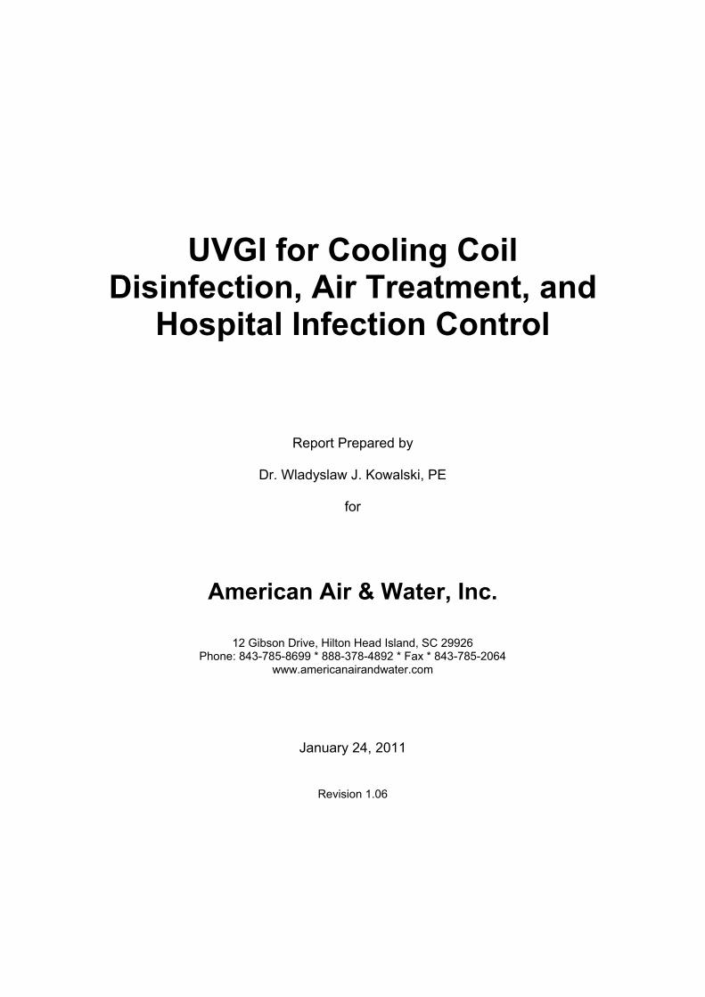
Figure 6 shows an example of a two stage decay curve for Aspergillus niger spores compared with predictions from a single stage model. The single stage model (in red) shows a log-linear decrease in microbial population over time, while the two-stage model (in blue) shows a second stage (a tail) becoming dominant after about 1000 seconds. It is clear that after extended exposure the single stage model will grossly overpredict the survival rate of the spores. This two stage behavior is typical for most spores under prolonged exposure and indicates the need to use a two stage model when evaluating cooling coil surface disinfection. Data for the single stage is based on IESNA (2000) while the two stage curve is based on laboratory data from UVDI (2000).
AAWI UVGI Disinfection
14
0.001
0.01
0.1
1
0 1000 2000 3000 4000 5000 6000
Time, s
Surv
ival
Fra
ctio
n
Single stage model
Two-stage model
Figure 6: Comparison of a single stage model vs. a two stage model of the inactivation of Aspergillus spores under UV exposure of 50 µW/cm2. The single stage model will underestimate the required dose for sterilization.
AAWI UVGI Disinfection
15
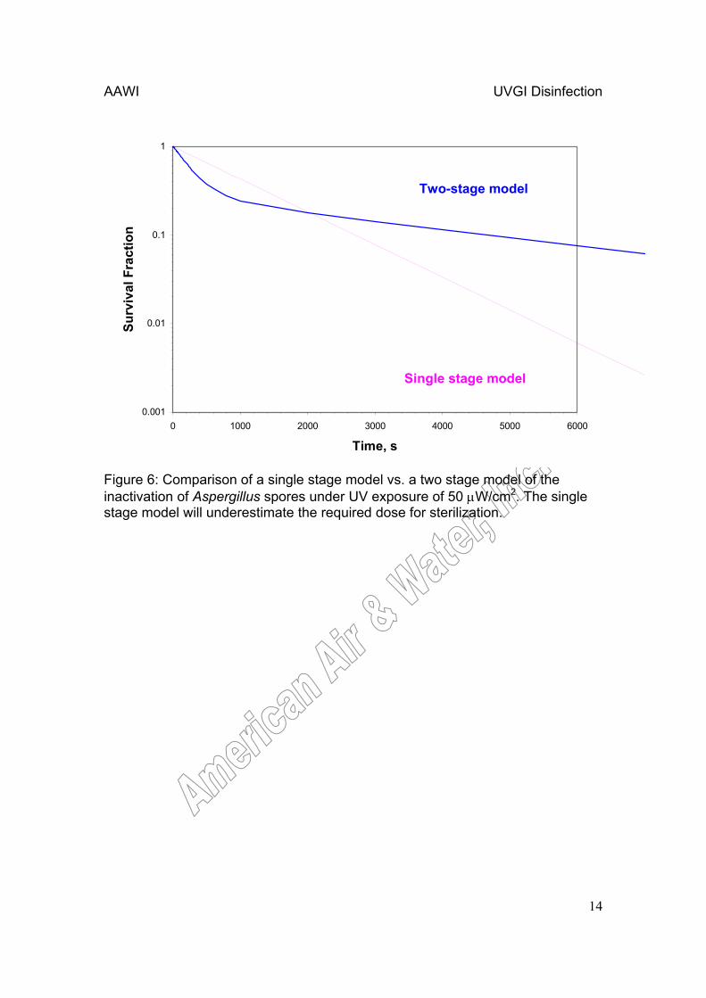
5. Performance of Cooling Coil Disinfection Systems Although studies on the inactivation of mold spores and inhibition of mold growth by UV abound in the literature, information on the actual disinfection of cooling coils remains limited and reports of successful disinfection are primarily anecdotal, although some formal studies are underway (EPRI 2004, Shaughnessy et al 1999). There is, however, no reason to believe that the anecdotal reports are not accurate, and the indications are that disinfection of cooling coils with UV is so effective that payback periods of about 2-4 years are possible. That is, the cleaning of the coils under UV exposure proceeds so rapidly that fouled coils are restored to pristine condition and save energy and maintenance costs so effectively that the retrofit of a UV coil cleaning system pays for itself in about 2-4 years. Theoretically, continuous exposure of cooling coil surfaces to UV should result in eradication of virtually all surface contamination within a few hours or days, depending on the irradiance levels. That is, any contamination on the exposed surface of the coils (entrance or exit respectively) should be sterilized rapidly. Figure 7 shows a system for which surface samples taken by the author indicated virtual sterilization of the leading edges after two weeks of operation.
Figure 7: A UVGI system installed in front (upstream) of a cooling coil that sterilized the front face of the coil after two weeks of operation.

AAWI UVGI Disinfection
16
Contamination on the internal surfaces of the cooling coil fins should also
be sterilized over time, but it is difficult to predict how much time this might require. It does appear that, based on anecdotal field reports, that a few weeks or months is all that is required to restore coils to original design operating conditions, suggesting that internal coil contamination is sterilized in these time periods.

AAWI UVGI Disinfection
17
6. Economics of Cooling Coil Disinfection Systems
The economic savings that can result from the installation of a UV cooling coil disinfection system can be estimated by comparing the operating costs after installation with the operating costs before installation, minus the cost of installing and operating the UV system. Ideally, operating data would be drawn from field test results, but this necessitates installing such a system first. Little published data is available for installed systems but anecdotal evidence suggests that UV disinfection systems are fully capable of restoring a fouled cooling coil to approximately the original design operating conditions. The cost savings will then depend on how much coil fouling has occurred and how far the system capacity has been diminished in comparison with the original design conditions. Table 2 shows the basic costs and the basic savings of UV cooling coil disinfection systems.
Costs SavingsFirst Cost of installation Fan energy savingsOperating Cost of UVGI Cooling energy savingsMaintenance costs of UVGI Maintenance savings
Table 2: Costs vs. Savings of Cooling Coil Disinfection
The first cost of the UVGI system will always be known, as will the operating and maintenance costs, which consist of electrical energy consumption and lamp replacement. The heat added to the system by the lamps is generally negligible and can be ignored, and furthermore, in cold climates the heat becomes a credit but this will also be ignored. The energy savings will result from two effects, the first being the reduced pressure drop through the coils once the fouling is removed, and the second being the increased rate of heat transfer from the coils when the fouling film is gone. Both of these can be significant, as can the reduction or elimination of maintenance on the cooling coils. Since the coils will be maintained in a clean condition, there is likely to be no requirement for periodic cleaning of the coils. In fact, since the UV system will maintain the coils in pristine condition, the lifetime of the coil will likely be extended well beyond the normal lifespan of unirradiated cooling coils, but this aspect of the savings will be difficult to quantify until field data is accumulated from installations. In order to estimate cost savings, it is necessary to assume that 1) the cooling coil is fouled, which is usually true if a system is being considered, and 2) the fouling will be completely eliminated and the coils restored to design condition, which is reportedly the usual case. Alternatively, a UV system may be installed on a brand new cooling coil, in which case the savings would have to be estimated based on the projected rate of fouling. The cost savings in dollars of a UV cooling coil disinfection system can be written as:
( ) ( ) ( ) uvuvuvCFCFCF MCOCFCMMCECEFEFESavings −−−−+−+−= (1)

AAWI UVGI Disinfection
18
where FEF = Fan Energy cost, Fouled ($) FEC = Fan Energy cost, Clean ($) CEF = Cooling Energy cost, Fouled ($) CEC = Cooling Energy cost, Clean ($) MF = Maintenance cost, Fouled ($) MC = Maintenance cost, Clean ($) FCuv = First Cost of UV ($) OCuv = Operating Cost of UV ($) MCuv = Maintenance Cost of UV ($)
The fan energy in kW is computed as follows:
( ) 7355.075.075.06350
⋅⋅
⋅=
CFMdPFE (2)
where dP = pressure drop, in.w.g. CFM = airflow, cfm 0.7355 = conversion factor from BHP to kW 0.75 = typical motor efficiency 0.75 = typical fan efficiency 6350 = conversion factor (in.w.g-cfm) to BHP The fan energy savings is then the fan energy in the operating condition
(fouled coils) minus the fan energy under design conditions.
The cooling energy savings in kW is computed as follows:
⋅=
COPCLCE
3412 (3)
where CL = the capacity loss due to fouling, Btuh COP = Coefficient of Performance 3412 = conversion from Btuh to kW The COP can be computed as the seasonal energy efficiency ratio
(SEER) divided by 3.412. The typical value for the SEER is about 9 or 10, and the respective COP would be about 2.64 – 2.93.
The maintenance cost Before, MB, can vary and depends on local facility procedures. Cooling coil maintenance is typically a few hours of labor a year, and may vary from a few hundred to a few thousand dollars. A reasonable estimate for small cooling coil units might be about MB = $500 a year. Presumably, there will be no maintenance cost After, or MA = 0.
The first cost of the UV system, FCuv, will be established at the beginning of any project and no estimates can be provided. The operating cost, OCuv, of the UV system is simply the electrical energy consumed by the lamp and ballast. The energy cost can be written as follows:

AAWI UVGI Disinfection
19
cPWOC
10008760⋅
= (4)
where W = total watts of power consumed by lamp fixture Pc = power charge (typically 0.08 – 0.1 $/kWh) 8760 = hours of operation per year (continuous assumed) 1000 = conversion from kW to W The maintenance cost of the UV system consists of the annual
replacement of the UV lamps, which is simply the cost per lamp times the number of lamps. This cannot be estimated in advance and will depend on the particular project.
Application of the above equations can be demonstrated through an example of a typical cooling coil disinfection system. Consider a system with the following parameters:
• Airflow, cfm - 48,500 CFM • Cooling Coil leaving air temperature: 52 degrees F. • Cooling Coil pressure drop, 0.75 in.w.g. • UV wattage – 552W UVC output. • UV lamp fixture first cost - $3,528 per total number of fixtures per
coil. • UV lamp installation labor cost, $1000. • UV lamp replacement bulb cost - $1,800 annually. • Annual hours of cooling – approximately 4,500 hours per year. • Cost per kWh, $0.09. • COP = 4.1 (typical for chilled water system) • Cooling Load (design), 1,500,000 Btuh (assumed)
Keikavousi (2004) reports that a 27 year old system retrofitted with UV
had a reduction in fan static pressure from 1.8 iwg to 0.7 iwg. The fan energy in our example above assumes only a fouled condition of 0.9 iwg, reducing to 0.75 after UV installation. The fan energy under design conditions is:
( ) 49.77355.075.075.06350
500,4875.0=⋅
⋅⋅
=FE kW
The fan energy under fouled conditions (assumed 0.9 in.w.g.) is:
( ) 988.87355.075.075.06350
500,489.0=⋅
⋅⋅
=FE kW
The fan energy savings (Fouled-Clean) is:

AAWI UVGI Disinfection
20
( ) 60709.0450049.7988.8 =⋅−=−CFFE $ The cooling energy (design operating conditions) is:
2.1071.43412
000,500,1=
⋅=CE kW
Assuming a 20% loss due to fouling, the energy savings would be:
( ) 868509.045002.10720.0 =⋅⋅⋅=−CFCE $ The operating costs are:
43509.01000
8760552=
⋅=OC $
The total savings can then be summed up as follows, assuming $1000 in
maintenance savings:
( ) ( ) ( ) 45291800435352810008685607 =−−−++=Savings $
The payback (PB) period can be approximated by dividing the initial cost by the annual savings as follows:
8.045293528
==PB years
Some examples of estimates of the savings that might be accrued from
the use of cooling coil disinfection systems in health care facilities are provided in Appendix D and Appendix E. In Appendix D summaries for six facilities are provided showing inpatient and outpatient occupancies, number of clinical procedure rooms and number of procedures performed. Appendix E provides estimated costs for cooling coil disinfection systems, in-duct UV systems, and operating room UV systems, along with estimated savings based on assumptions regarding nosocomial infection rates and operating costs. Although the available data on nosocomial infection rates due to airborne infections is not specific enough to isolate the true savings that might be anticipated, the ball-park figures provided in Appendix E clearly show the potential savings are great and that payback periods computed from these estimates would be in the range of 1-2 years or less, similar to the payback periods demonstrated previously.

AAWI UVGI Disinfection
21
7. Guidelines for Cooling Coil Disinfection Systems Several guidelines have been recently introduced, or are in preparation that address the use of UV for either cooling coil disinfection or air disinfection (GSA 2003, NIOSH 2005, IUVA 2005). Based on the literature, including draft guidelines from IUVA (2005), and the analysis previously presented, certain basic design guidelines can be summarized. These are as follows:
• Guidelines for Cooling Coil Disinfection • Minimum Filtration: MERV 6 • Recommended Filtration: MERV 8-11 • Maximum air velocity of between 400-500 fpm • Maximum air temperature between 40oF-110oF • Maximum ballast operating temperature of 40oC or 50oC (104oF or 122oF)
depending on ballast • Lamp placement: upstream, downstream, or both sides of coils • Lamp distance from coil face: 1-4 feet (30-120 cm) • Exposed Coil Surface:
o Recommended coil average irradiance: 50-500 µW/cm2 o Minimum coil irradiance: 50 µW/cm2 o Minimum coil irradiance in any corner or side: 10 µW/cm2
• Opposite Coil Surface (if unexposed) o Recommended coil average irradiance: 50-100 µW/cm2 o Minimum coil average irradiance: 10 µW/cm2 o Minimum coil irradiance in any corner or side: 1 µW/cm2
The above recommendations are preliminary (per IUVA 2005) and should
not be considered to be strict requirements as these matters are still under study. In addition to the above guidelines, it is recommended that UV lamp ballasts be placed externally if possible, or, if placed internally, be shielded from any heat sources. All electrical wiring should be in accordance with UL/ETL requirements. Alarms or disconnect switches should be included to disengage the UV lamps if an access door is opened. Warning signs should be placed in the vicinity and proper training given to maintenance personnel. UV lamps should be handled with care and used lamps disposed of in accordance with regulations regarding mercury content.
As verification of coil disinfection, surface sampling for fungi and/or bacteria could be performed before UV lamp installation, and then follow-up testing could be performed about 2 weeks or any time later. Major reductions in coil contaminants would suggest effective disinfection while the absence of all fungal contamination would indicate complete sterilization. As an alternative to microbiological testing, coil performance could be monitored over time to verify that the cooling coil heat transfer and pressure drop characteristics are being returned to design conditions, a process that requires an unknown amount of time, but for highly fouled coils it may require weeks or months.

AAWI UVGI Disinfection
22
8. Hospital Air & Surface Disinfection Systems There are four primary applications for UVGI in hospitals – 1) Overhead UV systems for control of surgical site infections (SSIs), 2) UV Air Disinfection systems for control of airborne nosocomial (hospital-acquired) infections (i.e. TB isolation rooms), 3) Medical Equipment Disinfection, and 4) Cooling Coil Disinfection. The latter subject has been addressed in the previous section and is not re-addressed here. Equipment disinfection systems such as UV cabinets are in wide use in health care settings but this subject is not addressed here in favor of the first two applications, which are topical subjects of much current interest.
Although UVGI systems have been in use in hospitals since the very beginnings of UV technology, they have never been formally recognized and accepted by governing agencies until very recently. There continues to be a lack of formal acknowledgement on the part of the responsible health authorities and a shortage of guidelines, information, and training for health professionals in the application of UVGI to hospital infection control. Some recent guidelines have acknowledged the potential effectiveness of UVGI for the control of nosocomial infections (CDC 2003, ASHRAE 2003). The most dramatic success of UVGI in the health care industry has been in the area of surgical site infection control, although cooling coil systems are poised to make major inroads because of their favorable economics.
The use of UVGI to clean cooling coils in hospital ventilation systems has the same economic benefits as those addressed in the previous sections of this report. However, the removal of mold and bacterial slime from cooling coils is much more important in the hospital environment which should be kept cleaner than the average commercial office building. That is, environmental bacteria and mold spores may pose hazards to patients in hospitals, especially those with impaired immune systems or burn victims. Microbial levels on surfaces and in the air need to be lower in hospitals due to the higher risks of nosocomial infection. The disinfection of cooling coils is one way to reduce microbial loading in hospitals. In theory, the requisite levels of air filtration should keep microbial contamination from accumulating on cooling coils, which are typically downstream of the filters, but casual inspections of hospital cooling coils and ductwork typically show levels of biocontamination above and beyond what would be expected with such high efficiency filters installed. The reasons for the apparent penetration of the filters are unclear but may be due to poorly fitting filters, leaking filters “bypass factor”, or the performance of filter change-outs while the ventilation system continues to operate. The installation of a UVGI cooling coil disinfection system should resolve such problems and contribute to net reductions of biocontamination in hospitals. The use of dual purpose UVGI systems that both clean coils and disinfect air, should also enhance the reductions of microbial loading.

AAWI UVGI Disinfection
23
8.1 Hospital-Acquired (Nosocomial) Infections Nosocomial infections include many diverse diseases, the sources and etiology of which are uncertain at present. Table 3 summarizes nosocomial agents that have the potential to transmit by the airborne route. The majority of nosocomial agents are potentially airborne, although most of the actual transmission is probably through direct contact (Kowalski 2005). The degree to which a cooling coil disinfection system will decrease nosocomial infections is probably quite limited, although the cost savings alone should justify such systems in any health care facility. The degree to which air and surface disinfection systems can reduce nosocomial infections is much more quantifiable, and a number of studies have been performed in this regard.
There are some aspects of the savings that could be expected in health care facilities that cannot be generalized or quantified exactly, such as the reduction in worker illness (nosocomial worker illnesses are an ongoing problem in health care facilities for which rates and costs are unknown at present), and possible reductions in insurance costs once such air cleaning systems are installed. The estimated costs in these examples, including the labor costs and energy costs, are based on assumptions and should not be construed to represent the actual costs for any specific installation, which need to be independently determined. The geographic area can also impact the energy costs through climate. One related report, that addresses air cleaning and bioterrorism, is a guideline from FEMA on the subject of insurance costs that may also be used to address reductions of naturally occurring diseases and possible savings from UVGI (FEMA 2003). UVGI may also allow for the use of filters with lower pressure drops (ACEEE 2005).

AAWI UVGI Disinfection
24
AIRBORNE PATHOGEN TYPE PRIMARY INFECTION CAUSED Annual AnnualCases Fatalities
Varicella-zoster virus C chickenpox common 250Streptococcus pyogenes C scarlet fever, pharyngitis 213,962 -Streptococcus pneumoniae C lobar pneumonia, sinusitis, meningitis 500,000 50000Staphylococcus aureus E staphylococcal pneumonia, opportunistic 2,750 -Serratia marcescens E bacteremia, endocarditis, pneumonia. 479 -SARS virus C Severe Acute Respiratory Syndrome (10) (?)Rubella virus C rubella (German measles) 3,000 noneRhizopus stolonifer NC zygomycosis, allergic reactions rare -Respiratory Syncytial Virus C pneumonia, bronchiolitis common rarePseudomonas aeruginosa NC pneumonia 2,626 -Pneumocystis carinii NC pneumocystosis rare rareParainfluenza virus C flu, colds, croup, pneumonia common -Nocardia brasiliensis NC nocardiosis uncommon -Nocardia asteroides NC nocardiosis uncommon rareMycobacterium tuberculosis C tuberculosis, TB 20,000 -Mucor plumbeus NC mucormycosis, rhinitis rare rareMoraxella E otitis media, opportunistic rare 0Measles virus C measles (rubeola) 500,000 rareLegionella pneumophila NC Legionnaire's Disease, opportunistic 1,163 10Klebsiella pneumoniae E opportunistic, pneumonia 1,488 -Influenza A virus C flu, secondary pneumonia 2,000,000 20000Histoplasma capsulatum NC histoplasmosis, fever, malaise common -Haemophilus parainfluenzae E conjunctivitis, pneumonia, meningitis common -Haemophilus influenzae C meningitis, pneumonia, endocarditis 1,162 -Cryptococcus neoformans NC cryptococcosis, cryptococcal meningitis high rareCorynebacterium diphtheriae C diphtheria, toxin produced. 10 -Coccidioides immitis NC coccidioidomycosis, valley fever uncommon -Chlamydia pneumoniae C pneumonia, bronchitis, pharyngitis uncommon -Cardiobacterium E opportunistic infections, endocarditis rare -Burkholderia pseudomallei NC meliodosis, opportunistic rare rareBurkholderia mallei NC Glanders, fever, opportunistic - noneBordetella pertussis C whooping cough 6,564 15Blastomyces dermatitidis NC blastomycosis, Gilchrist's Disease rare -Aspergillus NC aspergillosis, alveolitis, asthma uncommon -Alcaligenes E opportunistic rare rareAcinetobacter E opportunistic/septic, meningitis 147 -
Abbreviations: C = Communicable, NC = Noncommunicable, E=Endogenous.
Table 3: Nosocomial Agents with Airborne Transmission Potential

AAWI UVGI Disinfection
25
8.2 Surgical Site Infections (SSIs) and Overhead UV Systems The aerobiology of operating rooms (ORs) is primarily a function the microbial flora of the occupants, with common skin microbes like Staphylococcus and Streptococcus, and some intestinal flora like Escherichia coli and Enterobacter, being major contributors. Environmental contaminants like Pseudomonas aeruginosa and Bacillus subtilis also show up in ORs. Direct contact is generally regarded as being the primary mode of contamination of surgical sites, but airborne transmission may be a major factor. Airborne transport can result in contamination of equipment and surfaces in the OR, which may result in direct contact infections. Figure 8 shows the major microbial causes of SSIs ranked by their frequency of occurrence and proportioned by their relative size.
0.1
1
10
100
% o
f Ann
ual S
urgi
cal S
ite In
fect
ions
MRSACoag- Staphylococcus
Enterococcus
E. coli
Pseudomonas aeruginosa
Enterobacter
Candida albicans
Klebsiella pneumoniae
Streptococcus spp.
Proteus mirabilisBacteroides fragilis
Group D Streptococcus
Gram+ aerobes
AcinetobacterCitrobacter
Gram+ anaerobes
Group B Streptococcus
Candida spp.
Enterobacteriacae aerobes
Klebsiella spp.
Non-enterobacteriaceae aerobes
Serratia
Copyright 2009 American Air & Water, Inc. Figure 8: Major SSI infectious agents ranked by occurrence and proportioned by relative size.
In one study of airborne microbial contamination by Holcatova et al (1993) in the OR and ICUs of a surgery clinic, airborne bacterial concentrations were measured to be 150-250 cfu/m3. The most frequently isolated microbes included Staphylococcus epidermis, S. haemolyticus, Pseudomonas spp., Enterococcus spp., Enterobacter, Micrococcus, Corynebacteria, and Streptococcus faecalis. The airborne microorganisms most frequently found in the OR include Staphylococcus epidermis and S. aureus (MRSA) (Nelson et al 1973). Streptococcus pyogenes has been found in somet 15% of preoperative throat swabs from patients (Dubuc et al 1973). The total number of personnel in an OR

AAWI UVGI Disinfection
26
directly influences the total concentration of airborne bacteria (Hart 1938, Hambreaus et al 1977, Mangram et al 1999, Duvlis and Drescher 1980). OR personnel may shed from 3,000-50,000 microorganisms per minute depending on activity and the effectiveness of protective clothing. Conversation among personnel can increase the bacterial load of the air, and contaminated face masks (measured postoperatively) occur in about 9-10% of surgeons and nurses (Ritter 1984). Aerosolized blood pathogens can present hazards to surgeons that face masks may not protect them against. Aerosols in the respirable size range (less than 5 microns) containing blood can be generated in an operating room during surgery with common surgical power tools (Kowalski 2006).
Previous studies have shown UV installations in operating rooms to be effective in reducing surgical site infections. The first UVGI systems developed and installed were overhead UV systems for ORs and were intended to reduce the rate of surgical site infections (SSIs). SSIs cause undue costs through repeat procedures and unnecessary fatalities. From the first applications, these systems proved highly effective. Overhead UV systems have been in continuous use in some operating rooms since at least 1937 and some of these are still operating today (Hart and Sanger 1939). Reductions in post-operative infection rates of about 24-44% have been reported (Goldner and Allen 1973). Duke University reduced the SSI rate by over 80% and has continued to use overhead UVGI systems to maintain a low level of orthopedic infections (Lowell et al 1980). Table 4 summarizes the SSI reductions that have been demonstrated by previous Overhead UV installations.
Before After Net %Duke University Hospital SSI 5% 1% 4% 80% Kraissl et al 1940
Duke University Hospital Hip arthroplasty infection
5% 0.5% 5% 90% Lowell et al 1980
NE Deaconess Hospital SSI 15% 6.53% 8.5% 56% Overholt and Betts 1940
Infant & Children's Hospital, Boston
SSI 12.5% 2.7% 9.8% 78% Del Mundo & McKhann 1941
Watson Clinic, FL Mediastinitis 1.4% 0.23% 1.2% 84% Brown et al 1996St. Francis Hospital SSI 1.77% 0.57% 1.2% 68% Ritter et al 2007
76%
Table 3: Results of Hospital Field Trials of Overhead UV Systems
AVERAGE REDUCTION
Overhead Surgical
Site UVGI
Infection Cases Decrease
Surgical Site
Infections
Type System Location Infection Reference

AAWI UVGI Disinfection
27
8.3 UVGI Air Disinfection for Controlling Nosocomial Infections Airborne microbes exist in all indoor environments, but hospitals require air that is cleaner than found in normal indoor environments because of the potential for transmission of respiratory and other infections, and because of the presence of patients who may be more vulnerable to infections. In spite of this, hospitals generally do not attempt to control or monitor their airborne microbial levels beyond a basic adherence to existing guidelines for ventilation systems. Standards set by ASHRAE (1999 & 2003) and AIA (2001) for ventilation rates and filter use are assumed to provide clean, disinfected air to all areas of the hospital, but this is not the case. Indoor airborne levels in hospitals have been measured and although they tend to be lower than those in commercial office buildings and residential environments, they tend to be higher than what would be expected for health care facilities.
The aerobiology of open areas in hospital facilities does not differ greatly different from levels we might find in ordinary occupied buildings. In a study of mold spores in the air of a hospital ward, Tormo et al (2002) found twenty-two different types of spores, with total concentrations of 175-1396 spores/m3. The most frequently isolated were Cladosporium, Ustilago and various basidiospores. Considering the filtration requirements specified by ASHRAE (1999), virtually no spores should get through the filters at all. Tighe and Warden (1995) found airborne levels of bacteria to be about 104 cfu/m3 in patient rooms and about 207 cfu/m3 in general areas. Andrade and Brown (2003) found levels of bacteria as high as 160 cfu/m3 in general areas.
A growing list of reports and clinical studies have addressed the effectiveness of UVGI systems for air disinfection in health care facilities. Upper room UVGI systems have been used at The New England Deaconess Hospital, The Infant and Children’s Hospital in Boston, The Cradle in Evanston, and St. Luke’s Hospital in New York for the control of respiratory infections, which decreased by a net average of 50% (Overholt and Betts 1940, Del Mundo and McKhann 1941, Sauer et al 1942, Higgons and Hyde 1947). The Home for Hebrew Infants in New York successfully brought a halt to a Varicella epidemic using UVGI (Wells 1955). Table 4 summarizes the reductions in nosocomial infections that have resulted from installation of UV systems for air disinfection, which have primarily been Upper Room UVGI systems.

AAWI UVGI Disinfection
28
Before After Net %
The Cradle, EvanstonRespiratory infection 14.5% 4.6% 9.9% 68% Sauer et al 1942
St. Luke's Hospital, NY Respiratory infection
10.0% 6.6% 3.4% 33% Higgons & Hyde 1947
Livermore, CA Veteran's Hospital
Influenza epidemic
19.0% 2.0% 17.0% 89% McLean 1961
North Central Bronx Hospital
TB conversions among staff
2.5% 1% 2% 60% EPRI 1997
Home for Hebrew Infants, NY
Varicella epidemic
97% 0% 97% 100% Wells 1955
70%
System Location Infection Reference
Table 5: Results of Hospital Field Trials of UV Air Disinfection Systems
Airborne Infections
AVERAGE REDUCTION
Upper Room UVGI
Infection Cases DecreaseType

AAWI UVGI Disinfection
29
9. Hotel & Residential Air Disinfection Systems Hotels, apartments, private residential homes, and dormitories are all types of living facilities where individuals or families spend much of their time. In single family residences, families regularly exchange bacterial and viral infections, and are also subject to allergens that include mold spores and indoor fungal contamination. Families with children are especially subject to regular infections, including colds and flus that are routinely brought home from school by the youngest children. The need for protection against airborne pathogens has not yet been formally recognized by any agencies and not only do few homes have any type of air cleaning devices (i.e. filtration) but many homes have no ventilation at all, other than natural ventilation or air conditioners. All such living environments can benefit from air cleaning systems, including reductions in illnesses symptoms, allergies, and asthma symptoms (Bernstein et al 2006, Menzies et al 2003).
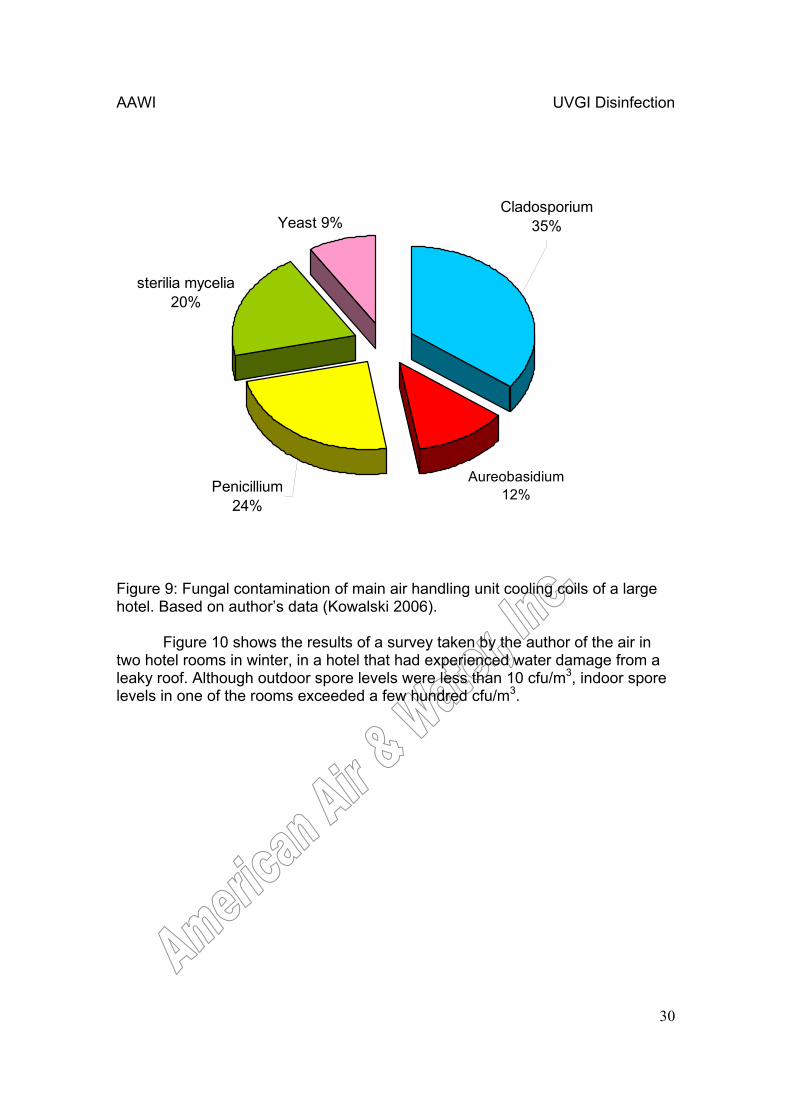
Hotels and dormitories are often unique residential environments in that the living quarters are typically small and do not always have direct supply air, only air conditioning or heaters under occupant control. Hotels will typically have one or more central air handling units providing air to the lobbies, hallways, restaurants, and other large areas. This supply air is intended to infiltrate into the individual hotel rooms. Sometimes the individual air conditioning units may have individual outside air dampers. Although the central air handling units in hotels may have medium-to-low efficiency filters, the room air conditioners rarely have anything more than a simple dust filter fabric. As a result, these air conditioners tend to accumulate spores over time. With the presence of condensation, these spores may even amplify and lead to air quality problems in rooms. Regular maintenance of room air conditioners normally involves removing the unit and cleaning with an acid or fungicide once a year. In a 200-400 unit hotel where only one or two units can be cleaned per day, this means that about half the units will have months of accumulation during the times of the year they need it most – summer and fall. As a result, hotel patrons often discover that when they turn on the air conditioner it produces a somewhat unpleasant odor. Figure 9 shows the results of samples taken from a hotel central air handling unit cooling coil.
AAWI UVGI Disinfection
30
Aureobasidium 12%
sterilia mycelia 20%
Yeast 9%Cladosporium
35%
Penicillium 24%
Figure 9: Fungal contamination of main air handling unit cooling coils of a large hotel. Based on author’s data (Kowalski 2006). Figure 10 shows the results of a survey taken by the author of the air in two hotel rooms in winter, in a hotel that had experienced water damage from a leaky roof. Although outdoor spore levels were less than 10 cfu/m3, indoor spore levels in one of the rooms exceeded a few hundred cfu/m3.

AAWI UVGI Disinfection
31
Cladosporium52%
Aureobasidium6%
Penicillium20%
Yeast6%
Aspergillus1%
Fusarium1%
sterilia mycelia11%
Pithomyces3%
Alternaria0.3%
Figure 10: Airborne fungal spores in a hotel that had water damage. Based on average of two rooms from author’s data (Kowalski 2006). A UVGI lamp was installed in one room in the stand-alone A/C unit over the coils. Airborne fungal spores were measured before installation and after two weeks of operation. Figure 11 shows the results, in which airborne levels dropped significantly. Although there was no filter, other than a dust filter, on the A/C unit, this modification appeared to greatly reduce fungal spore levels in the room. UVGI has a very limited effect on fungal spores, which tend to be resistant to UV exposure, but the constant recirculation of the room air through the unit produces a ‘chronic dosing, effect – that is, if a 1% kill rate is produced by a single pass, then after several hundred passes the total kill rate will approach 99%.

AAWI UVGI Disinfection
32
1 2 3 4
After
Before0
100
200
300
400
500
Airb
orne
cfu
/m3
Air Sample
Figure 11: Airborne levels of fungi in a hotel room before and two weeks after UVGI installation. Based on author’s data (Kowalski 2006). One of the problems most frequently encountered in hotels is that the local room A/C units tend to accumulate mold spores on the coils and the dust filter. Condensation on the coils can then produce mold growth, which manifests itself as a foul smell when the A/C unit is turned on. These A/C units are typically subject to cleaning by maintenance personnel approximately once a year. Maintenance programs will often cycle through all the units in the hotel by removing them individually, cleaning them with steam or chemicals, and then reinstalling them. Such maintenance programs are not necessarily tied to the seasons, but may operate continuously throughout the year. What this means is that statistically up to one half of all A/C units will not have been cleaned in over six months, and if these six months are in the mold season (Spring through Fall) then up to one half the rooms may have moldy odors when the units are turned on. The odds are that many guests will experience moldy odors when they turn on the A/C units. Changes in maintenance programs may be one way to address the problem.
A more cost-effective way to control mold growth on local room A/C units, and also on central A/C units, is to install UVGI lamps around the cooling coils, provided there is sufficient space. This is relatively easy to accomplish for larger central air handling units with cooling coils, but can be problematic for local room A/C units due to the lack of space around the coils. Stand-alone A/C units

AAWI UVGI Disinfection
33
located in walls and overhead may have very limited space and UV lamps can be installed with appropriate reflectors to ensure coil exposure. Window A/C units may have no space to install UV lamps and might have to be modified to create such space, or else replaced with newer A/C units that can accommodate UV lamps.

AAWI UVGI Disinfection
34
10. Electric Utilities and UV Air Disinfection The use of UV for air disinfection has long seen applications in the electric utilities, with the primary interest being energy savings from cooling coil disinfection systems. In fact, the first US installation of a UV cooling coil disinfection system was at an electric utility, Public Service of Omaha (PSO), who installed UV lamps in their air handling units in 1996 in an attempt to control mold growth around the cooling coils and drains pans. The system proved to be so effective in reducing odors and suppressing mold growth that all 28 air handling units were designated for the retrofit (Scheir 2000).
In 1996, the Central and Southwest Corporation (CSW), a holding company that owns America’s second largest utility system and many electrical subsidiaries in Texas, Ohio, Oklahoma, Missouri, Louisiana, and Arkansas, began installing the first of 170 UV lamps in its building’s air handling units (ELP 2000). By eliminating microbial growth as well as maintenance costs, the increase in cooling efficiency allowed the company to reduce its summertime energy consumption and save an estimated $58,000 in the first year. They were able to switch to operating only three of their four 300 ton chillers, and saved an estimated 300 kW of power. There was a significant improvement in coil heat transfer efficiency and a reduction in airside pressure drop, which further resulted in a lowering of fan RPM -- thanks to the fortunate fact that they had variable frequency drives (VFD). That produced an estimated 28% reduction in total air conditioning system usage. Uncounted costs include the four-times-a-year coil cleaning maintenance program that is no longer needed since the coils stay permanently clean, and the fact that the change-out of the cooling coils has been pushed back indefinitely. The system energy savings has been particularly welcome to the utility due to costly, record-breaking heat waves in the South.
Utilities have also taken an interest in UV as a cost-effective method of solving health care problems. Consolidated Edison Company in New York is funding research to combat the spread of drug-resistant TB in the cities by installing Upper Room UVGI lamps in homeless shelters (Wald 1994). As part of a six-year plan, the effectiveness of the UV lamps will be monitored and studied by Dr. Philip Brickner of St. Vincent’s Hospital and Dr. Ed Nardell (currently with the Harvard School of Public Health). The Electric Power Research Institute (EPRI) also has an interest in controlling TB with UV lights and in 1993 their Community Environmental Center funded the installation Upper Room fixtures in the VA Medical Center in Memphis (EPRI 1996). The Memphis VA hospital found that the UV installations provided cost-effective protection against airborne pathogens. New York Central Bronx Hospital installed Upper Room UVGI systems in 1995 to successfully control TB and nosocomial infections, in a project that was supported by The New York Power Authority (NYPA), who provide electricity to all the NYC Health and Hospitals Corporation facilities (EPRI 1997).

AAWI UVGI Disinfection
35
11. References & Bibliography Aaronson, S. A. (1970). "Effect of ultraviolet irradiation on the survival of simian virus 40 functions
in human and mouse cells." J Virol 6(4), 393-399. Abraham, G. (1979). "The effect of ultraviolet radiation on the primary transcription of Influenza
virus messenger RNAs." Virol 97, 177-182. Abshire, R. L., and Dunton, H. (1981). "Resistance of selected strains of Pseudomonas
aeruginosa to low-intensity ultraviolet radiation." Appl Envir Microb 41(6), 1419-1423. ACGIH (2004). "Threshold Limit Values and Biological Exposure Indices." , American Conference
of Governmental Industrial Hygienists, Cincinnati, OH. Aerotech (2001). IAQ Sampling Guide. Aerotech Laboratories, Phoenix. Ager, B. P., and Tickner, J. A. (1983). "The control of microbiological hazards associated with air-
conditioning and ventilation systems." Ann Occup Hyg 27(4), 341-358. Ahearn, D. G., Simmons, R. B., Switzer, K. F., Ajello, L., and Pierson, D. L. (1991). "Colonization
by Cladosporium spp. of painted metal surfaces associated with heating and air conditioning systems." J Ind Microb 8, 277-280.
Ahne, W. (1982). "Comparative studies of the stability of 4 fish-pathogenic viruses (VHSV, PFR, SVCV, IPNV)." Zentbl Vetmed Reihe B 29, 457-476.
AIA (2001). Guidelines for construction and equipment of hospital and medical facilities Mechanical Standards A. I. o. Architects, ed., Washington
AIHA (2001). "Nonionizing Radiation Guide Series, Ultraviolet Radiation." ISBN 0-932627-08-8, American Industrial Hygiene Association, Akron, OH.
Albrecht, T. (1974). "Multiplicity reactivation of human cytomegalovirus inactivated by ultra-violet light." Biochim Biophys Acta 905, 227-230.
Allegra, L., Blasi, F., Tarsia, P., Arosio, C., Fagetti, L., and Gazzano, M. (1997). "A novel device for the prevention of airborne infections." J Clinical Microb 35(7), 1918-1919.
Allen, E. G., Bovarnick, M. R., and Snyder, J. C. (1954). "The effect of irradiation with ultraviolet light on various properties of typhus rickettsiae." J Bact 67, 718-723.
Alpen, E. L. (1990). Radiation Biophysics. Prentice-Hall, Englewood. Alper, T., Cramp, W., Haig, D., and Clarke, M. (1967). "Does the scrapie agent replicate without
nucleic acid?" Nature (London) 214, 764-766. Alper, T. (1979). Cellular Radiobiology. Cambridge University Press, Cambridge. Anellis, A., Grecz, N., and Berkowitz, D. (1965). "Survival of Clostridium botulinum spores." Appl/
Microbiol 13(3), 397-401. Antopol, S. C., and Ellner, P. D. (1979). "Susceptibility of Legionella pneumophila to ultraviolet
radiation." Appl & Environ Microb 38(2), 347-348. ASHRAE (1999). "ASHRAE Standard 52.2-1999." , Atlanta. ASHRAE (1999). Chapter 7: Health Care Facilities ASHRAE Handbook of Applications ASHRAE,
ed., American Society of Heating, Refrigerating and Air Conditioning Engineers, Atlanta, 7.1-7.13.
ASHRAE (1999). "Standard 90.1-1999: Energy Standard for Building Except Low-Rise Residential Building." , American Society of Heating, Refrigeration and Air-Conditioning Engineers, Atlanta.
ASHRAE (2003). HVAC Design Manual for Hospitals and Clinics. American Society of Heating, Ventilating, and Air Conditioning Engineers, Atlanta.
ASHRAE (2009). “ASHRAE Position Document on Airborne Infectious Diseases.” American Society of Heating, Refrigerating, and Air-Conditioning Engineers, Atlanta.
Asthana, A., and Tuveson, R. W. (1992). "Effects of UV and phototoxins on selected fungal pathogens of citrus." Int J Plant Sci 153(3), 442-452.
AWWA (1971). Water Quality and Treatment. I. The American Water Works Association McGraw-Hill, New York.
Balch, W. M., Vaughn, J., Novotny, J., Drapeau, D., Vaillancourt, R., Lapierre, J., and Ashe, A. (2000). "Light scattering by viral suspensions." Limnol Oceanogr 45(2), 492-498.
Ballester, N., and Malley, J. (2004). "Sequential disinfection of Adenovirus Type 2 with UV-chlorine-chloramine." J Amer Wat Works Assoc 96(10), 97-102.

AAWI UVGI Disinfection
36
Banrud, H., and Moan, J. (1999). "The use of short wave ultraviolet radiation for disinfection in operating rooms." Tidsskrift for den Norske Laegeforening 119(18), 2670-2673.
Barnhart, B. J., and Cox, S. H. (1970). "Recovery of Haemophilus influenzae from ultraviolet and X-ray damage." Photochem Photobiol 11, 147-162.
Batch, L., Sculz, C., and Linden, K. (2004). "Evaluating water quality effects on UV disinfection of MS2 coliphage." J Amer Wat Works Assoc 96(7), 75-87.
Battigelli, D., Sobsey, M., and Lobe, D. (1993). "The inactivation of hepatitis A virus and other model viruses by UV irradiation." Wat Sci Technol 27, 339.
Bay, P. H. S., and Reichman, M. E. (1979). "UV inactivation of the biological activity of defective interfering particles generated by Vesicular Stomatitis virus." J Virol 32(3), 876-884.
Becker, M. M., and Wang, Z. (1989). "Origin of ultraviolet damage in DNA." J Mol Biol 210, 429-438.
Bedford, T. H. B. (1927). "The nature of the action of ultra-violet light on micro-organisms." Brit J Exp Path 8, 437-441.
Beebe, J. M. (1959). "Stability of disseminated aerosols of Pastuerella tularensis subjected to simulated solar radiations at various humidities." Journal of Bacteriology 78, 18-24.
Beggs, C. B., Kerr, K. G., Donelly, J. K., Sleigh, P. A., Mara, D. D., and Cairns, G. (2000). "An engineering approach to the control of Mycobacterium tuberculosis and other airborne pathogens: a UK hospital based pilot study." Transactions of the Royal Society of Tropical Medicine and Hygiene 94, 141-146.
Beggs, C. B., and Sleigh, P. A. (2002). "A quantitative method for evaluating the germicidal effects of upper room UV lights." J Aerosol Sci 33, 1681-1699.
Begum, M., Hocking, A., and Miskelly, D. (2009). "Inactivation of food spoilage fungi by ultraviolet (UVC) irradiation." Int J Food Microbiol 129, 74-77.
Bellinger-Kawahara, C., Cleaver, J., Diener, T., and Prusiner, S. (1987). "Purified scrapie prions resist inactivation by UV irradiation." J Virol 61(1), 159-166.
Benoit, T. G., Wilson, G. R., Bull, D. L., and Aronson, A. I. (1990). "Plasmid-associated sensitivity of Bacillus thuringensis to UV light." Appl & Environ Microbiol 56(8), 2282-2286.
Benzer, S. (1952). "Resistance to ultraviolet light as an index to the reproduction of bacteriophage." J Bact 63, 59-72.
Bishop, J. M., Quintrell, N., and Koch, G. (1967). "Poliovirus double-stranded RNA: Inactivation by ultraviolet light." J Mol Biol 24, 125-128.
Bister, K., Varmus, H. E., Stavnezer, E., Hunter, E., and Vogt, P. K. (1977). "Biological and biochemical studies on the inactivation of Avian Oncoviruses by ultraviolet irradiation." Virol(689-704)
Blatchley, E. F. (1997). "Numerical modelling of UV intensity: Application to collimated-beam reactors and continuous-flow systems." Wat Res 31(9), 2205-2218.
Bockstahler, L. E., Lytle, C. D., Stafford, J. E., and Haynes, K. F. (1976). "Ultraviolet enhanced reactivation of a human virus: Effect of delayed infection." Mutat Res 35, 189-198.
Bockstahler (1977). "Radiation enhanced reactivation of nuclear replicating mammalian viruses." Photochem Photobiol 25(5), 477-482.
Bohrerova, Z., and Linden, K. G. (2006). "Assessment of DNA damage and repair in Mycobacterium terrae after exposure to UV irradiation." J Appl Microbiol 101(5), 995-1001.
Bohrerova, Z., Shemer, H., Lantis, R., Impellitteri, C., and Linden, K. (2008). "Comparative disinfection efficiency of pulsed and continuous-wave UV irradiation technologies." Wat Res 42, 2975-2982.
Bolton, J. R. (2001). Ultraviolet Applications Handbook. Bolton Photosciences, Inc., Ayr, Ontario, Canada.
Bolton, J. R., and Cotton, C. A. (2001). The Ultraviolet Disinfection Handbook. American Water Works Association, Ayr, Ontario, Canada.
Boss, M. J., and Day, D. W. (2001). Air Sampling and Industrial Hygiene Engineering. Lewis Publishers, Boca Raton.
Bossart, W., Nuss, D. L., and Paoletti, E. (1978). "Effect of UV irradiation on the expression of Vaccinia virus gene products synthesized in a cell-free system coupling transcription and translation." J Virol 26(3), 673-680.

AAWI UVGI Disinfection
37
Bourre, F., Benoit, A., and Sarasin, A. (1989). "Respective Roles of Pyrimidine Dimer and Pyrimidine (6-4) Pyrimidone Photoproducts in UV Mutagenesis of Simian Virus 40 DNA in Mammalian Cells." J Virol 63(11), 4520-4524.
Bradley, D., Burdett, G. J., Griffiths, W. D., and Lyons, C. P. (1992). "Design and performance of size selective microbiological samplers." Journal of Aerosol Science 23(S1), s659-s662.
Bukhari, Z., Abrams, F., and LeChevallier, M. (2004). "Using ultraviolet light for disinfection of finished water." Wat Sci Technol 50(1), 173-178.
Burroughs, H. E. B. (1998). "Improved filtration in residential environments." ASHRAE J 40(6), 47-51.
Butkus, M. A., Labare, M. P., Starke, J. A., Moon, K., and Talbot, M. (2004). "Use of aqueous silver to enhance inactivation of coliphage MS-2 by UV disinfection." Appl Environ Microbiol 70(5), 2848-2853.
Butler, R. C., Lund, V., and Carlson, D. A. (1987). "Susceptibility of Campylobacter jejuni andYersinia enterolitica to UV radiation." Zbl Vet Med B 29, 129-136.
Caballero, S., Abad, F., Loisy, F., LeGuyader, F., Cohen, J., Pinto, R., and Bosch, A. (2004). "Rotavirus virus-like particles as surrogates in environmental persistence and inactivation studies." Appl Environ Microbiol 70(7), 3904-3909.
Campbell, A., and Wallis, P. (2002). "The effects of UV irradiation on human-derived Giardia lamblia cysts." Wat Res 36(4), 963-969.
Carlson, H. J. (1975). "Germicidal lamp inactivation of poliovirus." Am J Publ Health 32, 1256-1262.
Casarett, A. P. (1968). Radiation Biology. Prentice-Hall, Englewood. CDC (2003). "Guidelines for Environmental Infection Control in Health-Care Facilities." MMWR
52(RR-10), 1-48. CDC (2005). Guidelines for Preventing the Transmission of Mycobacterium tuberculosis in
Health-Care Facilities. Centers for Disease Control, Atlanta, GA. CDC (2006). Prevention and Control of Tuberculosis in Correctional and Detention Facilities:
Recommendations from CDC Federal Register CDC, ed., US Govt. Printing Office, Washington
Chang, J. C. H., Ossoff, S. F., Lobe, D. C., Dorfman, M. H., Dumais, C. M., Qualls, R. G., and Johnson, J. D. (1985). "UV inactivation of pathogenic and indicator microorganisms." Appl & Environ Microbiol 49(6), 1361-1365.
Chevrefils, G., Caron, E., Wright, H., Sakamoto, G., Payment, P., Barbeau, B., and Cairns, B. (2006). "UV dose required to achieve incremental log inactivation of bacteria, protozoa and viruses." IUVA News 8(1), 38-45.
Chick, E. W., A.B. Hudnell, J., and Sharp, D. G. (1963). "Ultraviolet sensitivity of fungi associated with mycotic keratitis and other mycoses." Sabouviad 2(4), 195-200.
CIE (2003). "Ultraviolet Air Disinfection." CIE 155:2003, International Commission on Illumination, Vienna, Austria.
Coggle, J. E. (1971). Biological effects of radiation. Wykeham Publ., London. Collier, L. H., McClean, D., and Vallet, L. (1955). "The antigenicity of ultra-violet irradiated
vaccinia virus." J Hyg 53(4), 513-534. Collins, F. M. (1971). "Relative susceptibility of acid-fast and non-acid fast bacteria to ultraviolet
light." Appl Microbiol 21, 411-413. Cornelis, J. J., and Rommelaere, J. (1982). "Direct and indirect effects of ultraviolet light on the
mutagenesis of parvovirus H-1 in human cells." EMBO J 1(6), 693-699. Corporation, C. (1999). Selection Guide: Ultraviolet Germicidal Lamp. Cox, C. S. (1987). The Aerobiological Pathway of Microorganisms. John Wiley & Sons, New
York. Craik, S., Weldon, D., Finch, G., Bolton, J., and Belosevic, M. (2001). "Inactivation of
Cryptosporidium oocysts using medium- and low-pressure ultraviolet radiation." Wat Res 35(1387-1398)
Danner, K., and Mayr, A. (1979). "In vitro studies on Borna virus. II. Properties of the virus." Arch Virol 61, 261-271.
Darken, M. A., and Swift, M. E. (1962). "Effects of ultraviolet-absorbing compounds on spore germination and cultural variation in microorganisms." Applied Microbiology 11, 154-156.

AAWI UVGI Disinfection
38
Darnell, M. E. R., Subbarao, K., Feinstone, S. M., and Taylor, D. R. (2004). "Inactivation of the coronavirus that induces severe acute respiratory syndrome, SARS-CoV." J Virol Meth 121, 85-91.
David, C. N. (1964). "UV inactivation and thymine dimerization in bacteriophage phiX." Z. Verberbungsl. 95, 318-325.
David, H. L., Jones, W. D., and Newman, C. M. (1971). "Ultraviolet light inactivation and photoreactivation in the mycobacteria." Infect and Immun 4, 318-319.
David, H. L. (1973). "Response of mycobacteria to ultraviolet radiation." Am Rev Resp Dis 108, 1175-1184.
Davidovich, I. A., and Kishchenko, G. P. (1991). "The shape of the survival curves in the inactivation of viruses." Mol Gen, Microb & Virol 6, 13-16.
de Roda Husman, A. M., Bijkerk, P., Lodder, W., Berg, H. v. d., Pribil, W., Cabaj, A., Gehringer, P., Sommer, R., and Duizer, E. (2004). "Calicivirus Inactivation by Nonionizing (253.7-Nanometer-Wavelength [UV]) and Ionizing (Gamma) Radiation." Appl Environ Microbiol 70(9), 5089-5093.
DeFendi, V., and Jensen, F. (1967). "Oncogenicity by DNA tumor viruses." Science 157, 703-705.
DeGiorgi, C. F., Fernandez, R. O., and Pizarro, R. A. (1996). "Ultraviolet-B lethal damage on Pseudomonas aeruginosa." Current Microb 33, 141-146.
Deguchi, K., Yamaguchi, S., and Ishida, H. (2005). "UV-disinfection reactor validation by computational fluid dynamics and relation to biodosimetry and actinometry." J Wat Technol Environ Technol 3(1), 77-84.
Deshmukh, D., and Pomeroy, B. (1968). "Ultraviolet inactivation and photoreactivation of avian viruses." Sci J Minnesota Agricult Exp Station Paper No. 6744
Dietz, P., Bohm, R., and Strauch, D. (1980). "Investigations on disinfection and sterilization of surfaces by ultraviolet radiation." Zbl Bakt Mikrobiol Hyg 171(2-3), 158-167.
DiStefano, R., Burgio, G., Ammatuna, P., Sinatra, A., and Chiarini, A. (1976). "Thermal and ultraviolet inactivation of plaque purified measles virus clones." G Batteriol Virol Immunol 69, 3-11.
Dolman, P. J., and Dobrogowski, M. J. (1989). "Contact lens disinfection by ultraviolet light." Am J Opthalmol 108(6), 665-669.
Dring, G. J., Ellar, D. J., and Gould, G. W. (1985). Fundamental and Applied Aspects of Bacterial Spores. Academic Press, London.
Drobnik, J., and Krekulova, A. (1969). "UV sensitivity of germinating culture of B. cereus." Folia Microbiol (Praha) 14, 104-111.
Dubunin, N. P., Zasukhina, G. D., Nesmashnova, V. A., and Lvova, G. N. (1975). "Spontaneous and Induced Mutagenesis in Western Equine Encephalomyelitis Virus in Chick Embryo Cells with Different Repair Activity." Proc Nat Acad Sci 72(1), 386-388.
Duizer, E., Bijkerk, P., B. Rockx, A, d., Twisk, F., and Koopmans, M. (2004). "Inactivation fo Caliciviruses." Appl Environ Microbiol 70(8), 4538-4543.
Dulbecco, R. (1952). "A critical test of the recombination theory of multiplicity reactivation." J Bact 63, 199-207.
Dulbecco, R., and Vogt, M. (1955). "Biological properties of Poliomyelitis viruses as studied by the plaque technique." Ann N Y Acd Sci 61(4), 790-800.
Dumyahn, T., and First, M. (1999). "Characterization of ultraviolet upper room air disinfection devices." Am Ind Hyg Assoc J 60(2), 219-227.
Durance, C. S., Hoffman, R., Andrews, R. C., and Brown, M. (2005). "Applications of Ultraviolet Light for Inactivation of Adenovirus." , University of Toronto Department of Civil Engineering
Eisenstark, A. (1989). "Bacterial genes involved in response to near-ultraviolet radiation." Advances in Genetics 26, 99-147.
El-Adhami, W., Daly, S., and Stewart, P. R. (1994). "Biochemical studies on the lethal effects of solar and artificial ultraviolet radiation on Staphylococcus aureus." Arch Microbiol 161, 82-87.
Elasri, M. O., and Miller, R. V. (1999). "Response of a biofilm bacterial community to UV Radiation." Appl & Environ Microbiol 65(5), 2025-2031.

AAWI UVGI Disinfection
39
ELP (2000). "How UV-C Lamps Saved One Company $58,000." Electric Light & Power 78(2) Ensor, D. S., Hanley, J. T., and Sparks, L. E. (1991). Particle-size-dependent efficiency of air
cleaners IAQ '91 , Healthy Buildings/IAQ '91, Washington EPA (2006). "Biological Inactivation Efficiency by HVAC In-Duct Ultraviolet Light Systems: Steril-
Aire, Inc." EPA 600/R-06/052, U.S. Environmental Protection Agency EPRI (1996). "UVGI for Infection Control in Hospitals." TA-106887, Electric Power Research
Institute, Palo Alto, CA. EPRI (1997). "UVGI for TB Infection Control in a Hospital." TA-107885, Electric Power Research
Institute, Palo Alto, CA. Erhardt, L., and C.P.Steinmetz (1977). Radiation, Light, and Illumination. tx Camarillo
Reproduction Center, Camarillo. FEMA (2003). "Reference Manual to Mitigate Potential Terrorist Attacks Against Buildings."
FEMA 426, Federal Emergency Management Agency http://www.fema.gov/fima/rmsp426.shtm.
FEMA (2003). "Primer for Design of Commercial Buildings to Mitigate Terrorist Attacks." FEMA 427, Federal Emergency Management Agency http://www.fema.gov/fima/rmsp427.shtm.
FEMA (2003). "Primer to Design Safe School Projects in Case of Terrorist Attacks." FEMA 428, Federal Emergency Management Agency http://www.fema.gov/fima/rmsp428.shtm.
FEMA (2003). "Insurance, Finance, and Regulation Primer for Terrorism Risk Management in Buildings." FEMA 429, Federal Emergency Management Agency http://www.fema.gov/fima/rmsp429.shtm.
Fernandez, R. O., and Pizarro, R. A. (1996). "Lethal effect induced in Pseudomonas aeruginosa exposed to ultraviolet-A radiation." Photochem & Photobiol 64(2), 334-339.
Field, A. A. (1973). "Operating theater air conditioning." HPAC October, 91-93. First, M. W., Nardell, E. A., Chaisson, W., and Riley, R. (1999). "Guidelines for the application of
upper-room ultraviolet germicidal irradiation for preventing transmission of airborne contagion - Part II: Design and operational guidance." ASHRAE J 105, 869-876.
Fletcher, L. A., Noakes, C. J., Beggs, C. B., Sleigh, P. A., and Kerr, K. G. (2003). "The ultraviolet susceptibility of aerosolised microorganisms and the role of photoreactivation." 2nd International Conference fo the IUVA, Vienna
Fletcher, L. (2004). "The influence of relative humidity on the UV susceptibility of airborne gram negative bacteria." IUVA News 6(1), 12-19.
Fluke, D. J., and Pollard, E. C. (1949). "Ultraviolet action spectrum of T1 bacteriophage." Science 110, 274-275.
Foarde, K. K., Hanley, J. T., Ensor, D. S., and Roessler, P. (1999). "Development of a method for measuring single-pass bioaerosol removal efficiencies of a room air cleaner." Aerosol Sci & Technol 30, 223-234.
Foarde, K. K., Myers, E. A., Hanley, J. T., Ensor, D. S., and Roessler, P. F. (1999a). "Methodology to perform clean air delivery rate type determinations with microbiological aerosols." Aerosol Sci & Technol 30, 235-245.
Foarde, K. K., Hanley, J. T., and Veeck, A. C. (2000). "Efficacy of antimicrobial filter treatments." ASHRAE J Dec, 52-58.
Fraenkel-Conrat, H., and Wagner, R. R. (1981). Comprehensive Virology. Plenum Press, New York.
Freeman, A. G., Schweikart, K. M., and Larcom, L. L. (1987). "Effect of ultraviolet radiation on the Bacillus subtilis phages SPO2c12, SPP1, and phi 29 and their DNAs." Mut Res 184(3), 187-196.
Frerichs, G. N., Tweedle, A., Starkey, W. G., and Richards, R. H. (2000). "Temperature, pH, and electrolyte sensitivity, and heat, UV and disinfectant inactivation of sea bass (Dicentrarchus labrax) neuropathy nodavirus." Aquaculture 185, 13-24.
Fuerst, C. R. (1960). "Inactivation of bacterial viruses by physical means." Annals of the New York Academy of Sciences 82, 684-691.
Fujikawa, H., and Itoh, T. (1996). "Tailing of thermal inactivation curve of Aspergillus niger spores." Appl Microb 62(10), 3745-3749.
Fulton, H. R., and Coblentz, W. W. (1929). "The fungicidal action of ultraviolet radiation." J Agric Res 38, 159.

AAWI UVGI Disinfection
40
Furuhashi, M., Nakamura, H., Ueda, I., and Kozuka, M. (1987?). "Ultraviolet irradiation of air sterilization under dynamic airflow." unk, 2.7-2.10.
Furuse, K., and Watanabe, I. (1971). "Effects of ultraviolet light (UV) irradiation on RNA phage in H2O and in D2O." Virol 46, 171-172.
Futter, B. V., and Richardson, G. (1967). "Inactivation of bacterial spores by visible radiation." J Appl Bact 30(2), 347-353.
Galasso, G. J., and Sharp, D. G. (1965). "Effect of particle aggregation on the survival of irradiated Vaccinia virus." J Bact 90(4), 1138-1142.
Gates, F. L. (1929). "A study of the bactericidal action of ultraviolet light." J Gen Physiol 13, 231-260.
Gates, F. L. (1934). "Results of irradiating Staphylococcus aureus bacteriophage with monochromatic ultraviolet light." J Exp Med 60, 179-188.
Gerba, C., Gramos DM, Nwachuku N. (2002). "Comparative inactivation of enteroviruses and adenovirus 2 by UV light." Appl Environ Microbiol 68(10), 5167-5169.
Germaine, G. R., and Murrell, W. G. (1973). "Effect of dipicolinic acid on the ultraviolet radiation resistance of Bacillus cereus spores." Photochem & Photobiol 17, 145-154.
Giese, N., and Darby, J. (2000). "Sensitivity of microorganisms to different wavelengths of UV light: Implications on modeling of medium pressure UV systems." Wat Res 34(16), 4007-4013.
Gillis, H. L. (1974). Photoreactivation and ultraviolet inactivation of Mycobacteria in air, MS Thesis , Georgia Technical University, Atlanta.
Gilpin, R. W. (1984). Laboratory and Field Applications of UV Light Disinfection on Six Species of Legionella and Other Bacteria in Water Legionella: Proceedings of the 2nd International Symposium C. Thornsberry, ed., American Society for Microbiology, Washington
Gilpin, R. D., Dillon, S. B., Keyser, P., Androkites, A., Berube, M., Carpendale, N., Skorina, J., Hurley, J., and Kaplan, A. M. (1985). "Disinfection of circulating water systems by ultraviolet light and halogenation." Water Res 19(7), 839-848.
Glassner, A. S. (1989). "An Introduction to Ray Tracing." Academic Press. London. Glaze, W. H., Payton, G. R., Huang, F. Y., Burleson, J. L., and Jones, P. C. (1980). "Oxidation of
water supply refractory species by ozone with ultraviolet radiation." 600, U.S. EPA Golde, A., Latarjet, R., and Vigier, P. (1961). "Isotypical interference in vitro by Rous virus
inactivated by ultraviolet rays." C R Acad Sci (Paris) 253, 2782-2784. Goldstein, M. A., and Tauraso, N. M. (1970). "Effect of formalin,B-propiolactone, merthiolate, and
ultraviolet light upon Influenza virus infectivity, chicken cell agglutination, hemagglutination, and antigenicity." Appl Microb 19(2), 290-294.
Green, C. F., Favidson, C. S., Scarpino, P. V., and S, G., Gibbs (2004). "Disinfection of selected Aspergillus spp. using ultraviolet irradiation." Can J Microbiol 50(3), 221-224.
Griego, V. M., and Spence, K. D. (1978). "Inactivation of Bacillus thuringiensis spores by ultraviolet and visible light." Appl Environ Microbiol 35(5), 906-910.
Griffiths, W. D., S.L.Upton and D.Mark (1993). "An investigation into the collection efficiency & bioefficiencies of a number of aerosol samplers." Journal of Aerosol Science 24(S1), s541-s542.
Gritz, D. C., Lee, T. Y., McDonnell, P. J., Shih, K., and Baron, N. (1990). "Ultraviolet radiation for the sterilization of contact lenses." CLAO J 16(4), 294-298.
Grun, L., and Pitz, N. (1974). "U.V. radiators in humidifying units and air channels of air conditioning systems in hospitals." Zbl Bakt Hyg B159, 50-60.
GSA (2003). The Facilities Standards for the Public Buildings Service. Public Buildings Service of the General Services Administration, Washington. http://hydra.gsa.gov/pbs/p100/.
Guha, S., and Bojarczuk, N. (1998). "Ultraviolet and violet GaN light emitting diodes on silicon." Appl Phys Lett 72, 415.
Guillemain, B., Mamoun, R., Astier, T., and Duplan, J. (1981). "Mechanisms of early and late polykaryocytosis induced by the Bovine Leukaemia virus." J Gen Virol 57, 227-231.
Gurol, M. D., and Vatista, R. (1987). "Oxidation of phenolic compounds by ozone and ozone + UV radiation." Water Res 21, 895.
Gurzadyan, G. G., Nikogosyan, D. N., Kryukov, P. G., Letokhov, V. S., Balmukhanov, T. S., Belogurov, A. A., and Zavilgelskij, G. B. (1981). "Mechanism of high power picosecond

AAWI UVGI Disinfection
41
laser UV inactivation of viruses and bacterial plasmids." Photochem Photobiol 33, 835-838.
Hanley, J. T., D.D.Smith and D.S.Ensor (1995). "A fractional aerosol filtration efficiency test method for ventilation air cleaners." ASHRAE Transactions 101(1), 97.
Harm, W. (1961). "Gene-controlled reactivation of ultraviolet-irradiated bacteriophage." J Cell Comp Physiol Suppl 58(1), 169.
Harm, W. (1968). "Effects of dose fractionation on ultraviolet survival of Escherichia coli." Photochem & Photobiol 7, 73-86.
Harm, W. (1980). Biological effects of ultraviolet radiation. Cambridge University Press, New York.
Harper, G. J. (1961). "Airborne micro-organisms : Survival tests with four viruses." Journal of Hygiene 59, 479-486.
Harris, G. D., Adams, V. D., Sorenson, D. L., and Curtis, M. S. (1987). "Ultraviolet inactivation of selected bacteria and viruses with photoreactivation of the bacteria." Water Res 21(6), 687-692.
Harris, M. G., Fluss, L., Lem, A., and Leong, H. (1993). "Ultraviolet disinfection of contact lenses." Optom Vis Sci 70(10), 839-842.
Harstad, J. B., H.M.Decker, and A.G.Wedum (1954). "Use of ultraviolet irradiation in a room air conditioner for removal of bacteria." American Industrial Hygiene Association Journal 2, 148-151.
Havelaar, A. H. (1987). "Virus, bacteriophages and water purification." Vet Q 9(4), 356-360. Hayes, S. L., White, K. M., and Rodgers, M. R. (2006). "Assessment of the effectiveness of low-
pressure UV light for inactivation of Helicobacter pylori." Appl Environ Microbiol 72(5), 3763-3765.
Hedrick, R., McDowell, T., Marty, G., Mukkatira, K., Antonio, D., Andree, K., Bukhari, Z., and Clancy, T. (2000). "Ultraviolet irradiation inactivates the waterborne infective stages of Myxobolus cerebralis: A treatment for hatchery water supplies." Dis Aquatic Org 42(1), 53-59.
Helentjaris, T., and Ehrenfeld, E. (1977). "Inhibition of host cell protein synthesis by UV-inactivated poliovirus." J Virol 21(1), 259-267.
Henderson, E., Heston, L., Grogan, E., and Miller, G. (1978). "Radiobiological inactivation of Epstein-Barr virus." J Virol 25(1), 51-59.
Hercik, F. (1937). "Action of ultraviolet light on spores and vegetative forms of B. megatherium sp." J Gen Physiol 20(4), 589-594.
Hernandez, M., Miller, S. L., Landfear, D. W., and Macher, J. M. (1999). Aerosol Sci & Technol 30, 145-160.
HICPAC (2003). "Guidelines for Environmental Infection Control in Health-Care Facilities." MMWR 52(RR-10), 1-48.
Hijnen, W. A. M., Beerendonk, E. F., and Medema, G. J. (2006). "Inactivation credit of UV irradiation for viruses, bacteria and protozoan (oo)cysts in water: A review." Wat Res 40, 3-22.
Hill, W. F., Hamblet, F. E., Benton, W. H., and Akin, E. W. (1970). "Ultraviolet devitalization of eight selected enteric viruses in estuarine water." Appl Microb 19(5), 805-812.
Hirai, K., Maeda, F., and Watanabe, Y. (1977). "Expression of early virus functions in Human Cytomegalovirus infected HEL cells: Effect of ultraviolet light-irradiation of the virus." J Gen Virol 38, 121-133.
Hofemeister, J., and Bohme, H. (1975). "DNA repair in Proteus mirabilis. III. Survival, dimer excision, and UV reactivation in comparison with Escherichia coli K12." Mol Gen Genetic 141(2), 147-161.
Holah, J. T., Rogers, S. J., Holder, J., Hall, K. E., Taylor, J., and Brown, K. L. (1995). "The evaluation of air disinfection systems." R&D Report No. 13, Campden & Chorleywood Food Research Association, Gloucestershire.
Hollaender, A. (1943). "Effect of long ultraviolet and short visible radiation (3500 to 4900) on Escherichia coli." J Bact 46, 531-541.
Hollaender, A., and Oliphant, J. W. (1944). "The inactivating effect of monochromatic ultraviolet radiation on influenza virus." J Bact 48(4), 447-454.

AAWI UVGI Disinfection
42
Hollaender, A. (1955). Radiation Biology, Volume II: Ultraviolet and Related Radiations. McGraw-Hill, New York.
Horneck, G., Bucker, H., and Reitz, G. (1985). Bacillus subtilis spores on Spacelab I: Response to solar UV radiation in free space Fundamental and Applied Aspects of Bacterial Spores G. J. Dring, D. J. Ellar, and G. W. Gould, eds., Academic Press, London, 241-249.
Hotz, G., Mauser, R., and Walser, R. (1971). "Infectious DNA from coliphage T1. 3. The occurrence of single-strand breaks in stored, thermally-treated and UV-irradiated molecules." Int J Radiat Biol Relat Stud Phys Chem Med 19, 519-536.
Hoyer, O. (2000). "The status of UV technology in Europe." IUVA News 2, 22-27. Huber, T. W., Reddick, R. A., and Kubica, G. P. (1970). "Germicidal effect of ultraviolet irradiation
on paper contaminated with mycobacteria." Appl Microbiol 19, 383-384. Huffman, D., Gennccaro, A., Rose, J., and Dussert, B. (2002). "Low- and medium-pressure UV
inactivation of microsporidia Encephalitozoon intestinalis." Wat Res 36(12), 3161-3164. IES (1981). Lighting Handbook Application Volume. Illumination Engineering Society, IESNA (2000). Lighting Handbook: Reference & Application IESNA HB-9-2000. Illumination
Engineering Society of North America, New York. IRPA (1985). "IRPA Guidelines on Limits of Exposure to Ultraviolet Radiation of Wavelengths
Between 180 nm and 400 nm (Incoherent Optical Radiation)." Health Physics 49, 331-340.
Ishida, H., Nahara, Y., Tamamoto, M., and Hamada, T. (1991). "The fungicidal effect of ultraviolet light on impression materials." J Prosthet Dent 65(4), 532-535.
IUVA (2005). "Guideline for the Design and Installation of UVGI Cooling Coil Surface Disinfection Systems." IUVA-G04A-2005, International Ultraviolet Association, Ayr, Ontario, Canada.
IUVA (2005). "General Guideline for UVGI Air and Surface Disinfection Systems." IUVA-G01A-2005, International Ultraviolet Association, Ayr, Ontario, Canada.
IUVA (2005). "Guideline for the Testing and Commissioning of UVGI In-Duct Air Treatment Systems." IUVA-S01A-2005, International Ultraviolet Association, Ayr, Ontario, Canada.
IUVA (2005). "Draft Standard for Laboratory Testing of UVGI Air and Surface Rate Constants." IUVA-S06A-2005, International Ultraviolet Association, Ayr, Ontario, Canada.
IUVA (2005a). "Guideline for Design and Installation of UVGI Air Disinfection Systems in New Building Construction." IUVA-G02A-2005, International Ultraviolet Association, Ayr, Ontario, Canada.
IUVA (2005b). "Guideline for Design and Installation of UVGI In-Duct Air Disinfection Systems." IUVA-G03A-2005, International Ultraviolet Association, Ayr, Ontario, Canada.
Jagger (1956). "Action spectra of light-restoration in Escherichia coli B/r." Ann Inst Pasteur 91(6), 858-873.
Jagger, J. (1967). Ultraviolet Photobiology. Prentice-Hall, Inc., Englewood Cliffs. Jagger, J., Takebe, H., and Snow, J. M. (1970). "Photoreactivation of killing in Streptomyces:
Action spectra and kinetic studies." Photochem Photobiol 12, 185-196. Jensen, M. M. (1964). "Inactivation of airborne viruses by ultraviolet irradiation." Applied
Microbiology 12(5), 418-420. Jensen, M. (1967). "Bacteriophage aerosol challenge of installed air contamination control
systems." Appl Microbiol 15(6), 1447-1449. Jensen, P. A., and Schafer, M. P. (1998). Chapter J: Sampling and Characterization of
Bioaerosols NIOSH Manual of Analytical Methods, NIOSH Publication 94-113 M. E. Cassinelli and P. F. O'Connor, eds., National Institute for Occupational Safety and Health, Atlanta, 82-112.
Jepson, J. D. (1973). "Disinfection of water supplies by ultraviolet radiation." Wat Treat Exam 22, 175-193.
Johnson, D. L., Pearce, T. A., and Esmen, N. A. (1999). "The effect of phosphate buffer on aerosol size distribution of nebulized Bacillus subtilis and Pseudomonas fluorescens bacteria." Aerosol Sci & Technol 30, 202-210.
Johnson, A., Linden, K., Ciociola, K., DeLeon, R., Widmer, G., and Rochelle, P. (2005). "UV inactivation of Cryptosporidium hominis as measured in cell culture." Appl Environ Microbiol 71(5), 2800-2802.
Kamat, A. S., and Pradhan, D. S. (1991). "Influence of cellular differentiation on ultraviolet

AAWI UVGI Disinfection
43
induced DNA damage and its repair mechanisms in B. cereus." Indian J of Biochemistry and Biophysics 28, 83-92.
Kariwa, H., Fujii, N., and Takashima, I. (2004). "Inactivation of SARS coronavirus by means of povidone-iodine, physical conditions, and chemical reagents." Jpn J Vet Res 52(3), 105-112.
Kassanis (1965). "Inactivation of a strain of Tobacco necrosis virus and the RNA isolated from it, by ultraviolet radiation of differnt wavelengths." Photochem Photobiol 4, 209-214.
Ke, Q., Craik, S., El-Din, M., and Bolton, J. (2009). "Development of a protocol for the determination of the ultraviolet sensitivity of microorganisms suspended in air." Aerosol Sci Technol 43(4), 284-289.
Keller, L. C., Thompson, T. L., and Macy, R. B. (1982). "UV light-induced survival response in a highly radiation-resistant isolate of the Moraxella-Acinetobacter group." Appl & Environ Microb 43(2), 424-429.
Kelloff, G., Aaronson, S. A., and Gilden, R. V. (1970). "Inactivation of Murine Sarcoma and Leukemia viruses by ultra-violet irradiation." Virol 42, 1133-1135.
Kelner, A. (1949). "Effect of visible light on the recovery of Streptomyces griseus conidia from ultraviolet irradiation injury." Proc Nat Acad Sci 35(2), 73-79.
Kethley, T. W. (1973). "Feasibility Study of Germicidal UV Lamps for Air Disinfection in Simulated Patient Care Rooms." , American Public Health Association, San Francisco, CA.
Kim, T., Silva, J. L., and Chen, T. C. (2002). "Effects of UV irradiation on selected pathogens in peptone water and on stainless steel and chicken meat." J Food Prot 65(7), 1142-1145.
Klein, B., Filon, A. R., vanZeeland, A. A., and vanderEb, A. J. (1994). "Survival of UV-irradiated vaccinia virus in normal and xeroderma pigmentosum fibroblasts; evidence for repair of UV-damaged viral DNA." Mutat Res 307(1), 25-32.
Knudson, G. B. (1985). "Photoreactivation of UV-irradiated Legionella pneumophila and other Legionella species." Appl & Environ Microbiol 49(4), 975-980.
Knudson, G. B. (1986). "Photoreactivation of ultraviolet-irradiated, plasmid-bearing, and plasmid-free strains of Bacillus anthracis." Appl & Environ Microbiol 52(3), 444-449.
Ko, G., First, M. W., and Burge, H. A. (2000). "Influence of relative humidity on particle size and UV sensitivity of Serratia marcescens and Mycobacterium bovis BCG aerosols." Tuber Lung Dis 80(4/5), 217-228.
Ko, G., Cromenas, T. L., and Sobsey, M. D. (2005). "UV inactivation of adenovirus type 41 measured by cell culture mRNA RT-PCR." Wat Res 39, 3643-3649.
Koller, L. R. (1939). "Bactericidal effects of ultraviolet radiation produced by low pressure mercury vapor lamps." J Appl Phys 10, 624.
Koller, L. R. (1965). Ultraviolet Radiation. John Wiley & Sons, New York. Kowalski, W. J. (1997). Technologies for controlling respiratory disease transmission in indoor
environments: Theoretical performance and economics. M.S., The Pennsylvania State University
Kowalski, W. J., Bahnfleth, W. P., and Whittam, T. S. (1998). "Bactericidal effects of high airborne ozone concentrations on Escherichia coli and Staphylococcus aureus." Ozone Science & Engineering 20(3), 205-221. http://www.bio.psu.edu/people/faculty/whittam/research/ozone.html.
Kowalski, W., and Bahnfleth, W. P. (1998). "Airborne respiratory diseases and technologies for control of microbes." HPAC 70(6), 34-48.
Kowalski, W. J., W. P. Bahnfleth, T. S. Whittam (1999). "Filtration of Airborne Microorganisms: Modeling and prediction." ASHRAE Transactions 105(2), 4-17. http://www.engr.psu.edu/ae/wjk/fom.html.
Kowalski, W. J. (2000). "Shedding Light on Moldy Ductwork." Home Energy 17(3), 6. Kowalski, W. J., and Bahnfleth, W. P. (2000). "Effective UVGI system design through improved
modeling." ASHRAE Transactions 106(2), 4-15. Kowalski, W. J., Bahnfleth, W. P., Witham, D. L., Severin, B. F., and Whittam, T. S. (2000).
"Mathematical modeling of UVGI for air disinfection." Quantitative Microbiology 2(3), 249-270.
Kowalski, W. J., and Bahnfleth, W. P. (2000). "UVGI Design Basics for Air and Surface Disinfection." HPAC 72(1), 100-110.

AAWI UVGI Disinfection
44
Kowalski, W. J. (2001). Design and optimization of UVGI air disinfection systems PhD, The Pennsylvania State University, State College.
Kowalski, W. J., and Dunn, C. E. (2002). "Current Trends in UVGI Air and Surface Disinfection." INvironment Professional 8(6), 4-6.
Kowalski, W. J. (2003). Immune Building Systems Technology. McGraw-Hill, New York. Kowalski, W. J. (2006). Aerobiological Engineering Handbook: A Guide to Airborne Disease
Control Technologies. McGraw-Hill, New York. Kowalski, W. J. (2009). Ultraviolet Germicidal Irradiation Handbook: UVGI for Air and Surface
Disinfection. Springer, New York. Kowalski, W., Bahnfleth, W., and Hernandez, M. (2009). "A Genomic Model for the Prediction of
Ultraviolet Inactivation Rate Constants for RNA and DNA Viruses." IUVA, Boston, MA Kowalski, W., Bahnfleth, W., and Hernandez, M. (2009). "A Genomic Model for Predicting the
Ultraviolet Susceptibility of Viruses." IUVA News 11(2), 15-28. Kuluncsics, Z., Perdiz, D., Brulay, E., Muel, B., and Sage, E. (1999). "Wavelength dependence of
ultraviolet-induced DNA damage distribution: Involvement of direct or indirect mechanisms and possible artefacts." J Photochem & Photobiol 49(1), 71-80.
Kumar, R. (1981). "Effect of glass filtered solar radiation & of 2,4 dinitrophenol on growth of bacillus cereus & on its survival after far-UV irradiation." Indian J of Experimental Biology 19(April), 345-348.
Kundsin, R. B. (1966). "Characterization of Mycoplasma aerosols as to viability, particle size, and lethality of ultraviolet radiation." J Bacteriol 91(3), 942-944.
Kundsin, R. B. (1968). "Aerosols of Mycoplasmas, L forms, and bacteria: Comparison of particle size, viability, and lethality of ultraviolet radiation." Applied Microbiology 16(1), 143-146.
Kundsin, R. B. (1988). Architectural design and indoor microbial pollution. Oxford Press, Lai, K. M., Burge, H., and First, M. W. (2004). "Size and UV germicidal irradiation susceptibility of
Serratia marcescens when aerosolized from different suspending media." Appl Environ Microbiol 70(4), 2021-2027.
Lamola, A. (1973). "Photochemistry and structure in nucleic acids." Pure Appl Chem 34(2), 281-303.
Latarjet, R., Cramer, R., and Montagnier, L. (1967). "Inactivation, by UV-, X-, and gamma-radiations, of the infecting and transforming capacities of polyoma virus." Virol 33, 104-111.
Lazarova, V., and Savoye, P. (2004). "Technical and sanitary aspect of wastewater disinfection by ultraviolet irradiation for landscape irrigation." Wat Sci Technol 50(2), 203-209.
Lee, J. E., Zoh, K. D., and Ko, G. P. (2008). "Inactivation and UV disinfection of Murine Norovirus with TiO2 under various environmental conditions." Appl Environ Microbiol 74(7), 2111-2117.
Levetin, E., Shaughnessy, R., Rogers, C. A., and Scheir, R. (2001). "Effectiveness of germicidal UV radiation for reducing fungal contamination within air-handling units." Applied & Environ Microbiol 67(8), 3712-3715.
Levinson, W., and Rubin, R. (1966). "Radiation studies of avian tumor viruses and of Newcastle disease virus." Virol 28, 533-542.
Li, D., Craik, S., Smith, D., and Belosevic, M. (2007). "Comparison of levels of inactivation of two isolates of Giardia lamblia cysts by UV light." Appl Environ Microbiol 73(7), 2218-2223.
Lidwell, O. M., and Lowbury, E. J. (1950). "The survival of bacteria in dust." Annual Review of Microbiology 14, 38-43.
Liltved, H., Hektoen, H., and Efraimsen, H. (1995). "Inactivation of bacterial and fish pathogens by ozonation or UV irradiation in water of different salinity." Aquacult Eng 14, 107-122.
Liltved, H., and Landfald, B. (1996). "Influence of liquid holding recovery and photoreactivation on survival of ultraviolet-irradiated fish pathogenic bacteria." Wat Res 30(5), 1109-1114.
Lin, W.-H., and Li, C. S. (1999). "Evaluation of impingement and filtration methods for yeast bioaerosol sampling." Aerosol Sci & Technol 30, 119-126.
Linden, K. G., Thurston, J., Schaefer, R., and Malley, J. P. (2007). "Enhanced UV inactivation of Adenoviruses under polychromatic UV lamps." Appl Environ Microbiol 73(23), 7571-7574.
Lindsey, J. L. (1997). Applied Illumination Engineering. The Fairmont Press, Inc., Lilburn. Linscomb, M. (1994). "AIDS clinic HVAC system limits spread of TB." HPAC February

AAWI UVGI Disinfection
45
Litonski, B. (1974). "Air conditioning as decontamination unit for the air of operating theatres." Zbl Bakt Hyg B159, 244-271.
Little, J. S., Kishimoto, R. A., and Canonico, P. G. (1980). "In vitro studies of interaction of rickettsia and macrophages: Effect of ultraviolet light on Coxiella burnetti inactivation and macrophage enzymes." Infect Immun 27(3), 837-841.
Lojo, M. M. (1995). "Thymine auxotrophy is associated with increased UV sensitivity in Escherichia coli and Bacillus subtilis." Mutation Research 347, 25-30.
Love, T. J. (1968). Radiative Heat Transfer. Charles A. Merrill, Columbus. Lovinger, G. G., Ling, H. P., Gilden, R. V., and Hatanaka, M. (1975). "Effect of UV light on RNA
directed DNA polymerase activity of murine oncornaviruses." J Virol 15, 1273. Luciano, J. R. (1977). Air Contamination Control in Hospitals. Plenum Press, New York. Lucio-Forster, A., Bowman, D. D., Lucio-Martinez, B., Labare, M. P., and Butkus, M. A. (2006).
"Inactivation of the Avian Influenza virus (H5N2) in typical domestic wastewater and drinking water treatment systems." Environ Eng Sci 23(6), 897-903.
Luckiesh, M., and Holladay, L. L. (1942). "Tests and data on disinfection of air with germicidal lamps." General Electric Review 45(4), 223-231.
Luckiesh, M., and Holladay, L. L. (1942a). "Designing installations of germicidal lamps for occupied rooms." General Electric Review 45(6), 343-349.
Luckiesh, M. (1945). "Disinfection with germicidal lamps: Control -- I." Electrical World Sep.29, 72-73.
Luckiesh, M. (1945). "Disinfection with germicidal lamps: Air -- II." Electrical World Oct.13, 109-111.
Luckiesh, M. (1945). "Disinfection with germicidal lamps: Water -- III." Electrical World Oct.27, 90-91.
Luckiesh, M. (1946). Applications of Germicidal, Erythemal and Infrared Energy. D. Van Nostrand Co., New York.
Luckiesh, M., Taylor, A. H., and Knowles, T. (1947). "Killing air-borne microorganisms with germicidal energy." Journal of the Franklin Institute Oct., 267-290.
Luckiesh, M., Taylor, A. H., Knowles, T., and Leppelmeier, E. T. (1949). "Inactivation of molds by germicidal ultraviolet energy." Journal of the Franklin Institute 248(4), 311-325.
Lytle, C. D. (1971). "Host-cell reactivation in mammalian cells. 1. Survival of ultra-violet-irradiated herpes virus in different cell-lines." Int J Radiat Biol Relat Stud Phys Chem Med 19(4), 329-337.
Lytle, C. D., and Sagripanti, J. L. (2005). "Predicted Inactivation of Viruses of Relevance to Biodefense by Solar Radiation." J Virol 79(22), 14244-14252.
Macher, J. M., Alevantis, L. E., Chang, Y. L., and Liu, K. S. (1992). "Effect of ultraviolet germicidal lamps on airborne microorganisms in an outpatient waiting room." Appl Occup Environ Hyg 7(8), 505-513.
Malley, J. P., Ballester, N. A., Margolin, A. B., Linden, K. G., Mofidi, A., Bolton, J. R., Crozes, G., Laine, J. M., and Janex, M. L. (2004). Inactivation of pathogens with innovative UV technologies. American Research Foundation & American Water Works Association,
Mamane-Gravetz, H., Linden, K. G., Cabaj, A., and Sommer, R. (2005). "Spectral sensitivity of Bacillus subtilis spores and MS2 coliphage for validation testing of ultraviolet reactors for water disinfection." Environ Sci Technol 39, 7845-7852.
Marquenie, D., Lammertyn, J., Geeraerd, A., Soontjens, C., VanImpe, J., Nicolai, B., and Michiels, C. (2002). "Inactivation of conidia of Botrytis cinerea and Monilinia fructigena using UV-C and heat treatment." Int J Food Microbiol 74, 27-35.
Marshall, M., Hayes, S., Moffett, J., Sterling, C., and Nicholson, W. (2003). "Comparison of UV inactivation of three Encephalitozoon species with that of spores of two DNA repair-deficient Bacillus subtilis biodosimetry strains." Appl Environ Microbiol 69(1), 683-685.
Martin, J. P., Aubertin, A. M., and Kirn, A. (1982). "Expression of Frog Virus 3 early genes after ultraviolet irradiation." Virol 122, 402-410.
Martin, E., Reinhardt, R., Baum, L., Becker, M., Shaffer, J., and Kokjohn, T. (2000). "The effects of ultraviolet radiation on the moderate halophile Halomonas elongata and the extreme halophile Halobacterium salinarum." Can J Microbiol 46(2), 180-187.
Mason, J. M., and Setlow, P. (1986). "Essential role of small, acid-soluble spore proteins in

AAWI UVGI Disinfection
46
resistance of Bacillus subtilis spores to UV light." J of Bact 167(1), 174-178. Matallana-Surget, S., Meador, J., Joux, F., and Douki, T. (2008). "Effect of the GC content of
DNA on the distribution of UVB-induced bipyrimidine photoproducts." Photochem Photobiol Sci 7, 794-801.
Maya, C., Beltran, N., Jimenez, B., and Bonilla, P. (2003). "Evaluation of the UV disinfection process in bacteria and amphizoic ameoba inactivation." Wat Sci Technol 3(4), 285-291.
McCarthy, C., and Schaefer, J. (1974). "Response of Mycobactrium avium to ultraviolet irradiation." Appl Environ Microbiol 28(1), 151-153.
McClain, M. E., and Spendlove, R. S. (1966). "Multiplicity reactivation of Reovirus particles after exposure to ultraviolet light." J Bact 92(5), 1422-1429.
McDevitt, J. J., Lai, K. M., Rudnick, S. N., Houseman, E. A., First, M. W., and Milton, D. K. (2007). "Characterization of UVC Light Sensitivity of Vaccinia Virus." Appl Environ Microbiol 73(18), 5760-5766.
Meistrich, M., Lamola, A. A., and Gabbay, E. (1970). "Sensitized photoinactivation of bacteriophage T4." Photochem Photobiol 11(3), 169-178.
Memarzadeh, F., and Jiang, J. (2000). "Methodology for minimizing risk from airborne organisms in hospital isolation rooms." ASHRAE Transactions 106(2)
Meng, Q. S., and Gerba, C. P. (1996). "Comparative inactivation of enteric Adenoviruses, Poliovirus and coliphages by ultraviolet irradiation." Wat Res 30(11), 2665-2668.
Menzies, D., Popa, J., Hanley, J. A., Rand, T., and Milton, D. K. (2003). "Effect of ultraviolet germicidal lights installed in office ventilation systems on workers' health and wellbeing: double-blind multiple crossover trials." The Lancet 362(November 29), 1785-1791.
Miller, R. L., and Plageman, P. G. W. (1974). "Effect of Ultraviolet Light on Mengovirus: Formation of Uracil Dimers, Instability and Degradation of Capsid, and Covalent Linkage of Protein to Viral RNA." J Virol 13(3), 729-739.
Miller, S. L., and Macher, J. M. (2000). "Evaluation of a methodology for quantifying the effect of room air ultraviolet germicidal irradiation on airborne bacteria." Aerosol Sci & Tech 33, 274-295.
Miocevic, I., Smith, J., Owens, L., and Speare, R. (1993). "Ultraviolet sterilization of model viruses important to finfish aquaculture in Australia." Aust Vet J 70(1), 25-27.
Mitscherlich, E., and Marth, E. H. (1984). Microbial Survival in the Environment. Springer-Verlag, Berlin.
Modest, M. F. (1993). Radiative Heat Transfer. McGraw-Hill, New York. Mongold, J. (1992). "DNA repair and the evolution of transformation in Haemophilus influenzae."
Genetics 132, 893-898. Montz, W. E. (2000). "Contamination control in hospitals." Engineered Systems 17(6), 68-72. Mooers, A., and Holmes, E. (2000). "The evolution of base composition and phylogenetic
interference." Trends Ecol Evol 15, 365-369. Morita, S., Namikoshi, A., Hirata, T., Oguma, K., Katayama, H., Ohgaki, D., Motoyama, N., and
Fujiwara, M. (2002). "Efficacy of UV irradiation in inactivating C. parvum oocysts." Appl Environ Microbiol 68(11), 5387-5393.
Morrissey, R. F., and Phillips, G. B. (1993). Sterilization Technology. Van Nostrand Reinhold, New York.
MRC (1954). "Air Disinfection with Ultra-violet Irradiation; Its Effect on Illness among School-children by the Air Hygiene Committee." 283, Medical Research Council, Her Majesty's Stationary Office, London.
Munakata, N., and Rupert, C. S. (1972). "Genetically controlled removal of "spore photoproduct" from deoxyribonucleic acid of ultraviolet-irradiated Bacillus subtilis spores." J Bact 111(1), 192-198.
Munakata, N., and Rupert, C. S. (1975). "Effects of DNA-polymerase-defective and recombination-deficient mutations on the ultraviolet sensitivity of Bacillus subtilis spores." Mutation Res 27, 157-169.
Munakata, N., Saito, M., and Hieda, K. (1991). "Inactivation action spectra of Bacillus subtilis spores in extended ultraviolet wavelengths (50-300 nm) obtained with synchrotron radiation." Photochem & Photobiol 54(5), 761-768.
Murdoch, J. B. (1985). Illumination Engineering. Macmillan, New York.
AAWI UVGI Disinfection
47
Nagy, R. (1964). "Application and measurement of ultraviolet radiation." AIHA J 25, 274-281. Nakamura, H. (1987). "Sterilization efficacy of ultraviolet irradiation on microbial aerosols under
dynamic airflow by experimental air conditioning systems." Bull Tokyo Med Dent Univ 34(2), 25-40.
Nardell, E. A. (1988). Chapter 12: Ultraviolet air disinfection to control tuberculosis Architectural Design and Indoor Microbial Pollution R. B. Kundsin, ed., Oxford University Press, New York, 296-308.
Nardell, E. A., Keegan, J., Cheney, S. A., and Etkind, S. C. (1991). "Airborne infection: Theoretical limits of protection acheivable by building ventilation." Am Rev Resp Dis 144, 302-306.
NCBI (2009). "Entrez Genome." National Center for Biotechnology Information. NEHC (1992). "Ultraviolet Radiation Guide." Technical Manual NEHC-TM92-5, Navy
Environmental Health Center, Bureau of Medicine and Surgery, Norfolk, VA. Neidle, S. (1999). Oxford Handbook of Nucleic Acid Structure. Oxford University Press, New
York. Nicas, M., and Miller, S. L. (1999). "A multi-zone model evaluation of the efficacy of upper-room
air ultraviolet germicidal irradiation." Appl & Environ Occup Hyg J 14, 317-328. Nieuwstad, T., and Havelaar, A. (1994). "The kinetics of batch ultraviolet inactivation of
bacteriophage MS2 and microbiological calibration of an ultraviolet pilot plant." J Environ Sci Health A29(9), 1992-2007.
Ninomura, P., and Bartley, J. (2001). "New ventilation guidelines for health-care facilities." ASHRAE J 43(6), 2933.
NIOSH (1972). "Occupational Exposure to Ultraviolet Radiation." HSM 73-110009, National Institute for Occupational Safety and Health, Cincinnati, OH.
NIOSH (2002). "Guidance for Protecting Building Environments from Airborne Chemical, Biological, or Radiological Attacks." DHHS (NIOSH) Pub. No. 2002-139, Dept. of Health and Human Services, CDC, National Institute for Occupational Safety and Health, Cincinnati, OH.
NIOSH (2003). "Guidance for Filtration and Air-cleaning Systems to Protect Building Environments." DHHS (NIOSH) Pub. No. 2003-136, Dept. of Health and Human Services, CDC, National Institute for Occupational Safety and Health, Cincinnati, OH.
Nomura, S., Bassin, R. H., Turner, W., Haapala, D. K., and Fischinger, P. J. (1972). "Ultraviolet inactivation of Maloney Leukaemia Virus: Relative target size required for virus replication and rescue of 'defective' Murine Sarcoma virus." J Gen Virol 14, 213-217.
Nozu, K., and Ohnishi, T. (1977). "Ultraviolet sensitivity of Vibrio parahaemolyticus, a causative bacterium of food poisoning." Photochem Photobiol 26(5), 483-486.
Nuanualsuwan, S., Mariam, T., Himathongkham, S., and Cliver, D. O. (2002). "Ultraviolet inactivation of Feline Calicivirus, Human Enteric Viruses, and coliphages." Photochem Photobiol 76(4), 406-410.
Nuanualsuwan, S., and Cliver, D. O. (2003). "Infectivity of RNA from Inactivated Proteins." Appl Environ Microbiol 69(3), 1629-1632.
Nwachuku, N., Gerba, C. P., Oswald, A., and Mashadi, F. D. (2005). "Comparative Inactivation of Adenovirus Serotypes by UV Light Disinfection." Appl Environ Microbiol 71(9), 5633-5636.
O'Hara, P. J., and Gordon, M. P. (1980). "Ultraviolet inactivation of the midi variant of QBeta RNA: the sites of UV-induced replication inhibition." Photochem Photobiol 31, 47-54.
Oguma, K., Katayama, H., Mitani, H., Morita, S., Hirata, T., and Ohgaki, S. (2001). "Determination of pyrimidine dimers in Escherichia coli and Cryptosporidium parvum during UV light inactivation, photoreactivation, and dark repair." Appl Environ Microbiol 67(10), 4630-4637.
Oguma, K., Katayama, H., and Ohgaki, S. (2004). "Photoreactivation of Legionella pneumophila after inactivation by low- or medium-pressure ultraviolet lamps." Wat Res 38(11), 2757-2763.
Oppenheimer, J., Hoagland, J., Laine, J.-M., Jacangelo, J., and Bhamrah, A. (1993). "Microbial inactivation and characterization of toxicity and by-products occurring in reclaimed wastewater disinfected with UV radiation." Conference on Planning, Design and

AAWI UVGI Disinfection
48
Operation of Effluent Disinfection Systems, Whippany, NJ Oppenheimer, J. A., Jacangelo, J. G., Laine, J.-M., and Hoagland, J. E. (1997). "Testing the
equivalency of ultraviolet light and chlorine for disinfection of wastewater to reclamation standards." Wat Environ Res 69(1), 14-24.
Otaki, M., Okuda, A., Tajima, K., Iwasaki, T., Kinoshita, S., and Ohgaki, S. (2003). "Inactivation differences of microorganisms by low pressure UV and pulsed xenon lamps." Wat Sci Technol 47(3), 185-190.
Owada, M., Ihara, S., and Toyoshima, K. (1976). "Ultraviolet inactivation of Avian Sarcoma viruses: Biological and Biochemical analysis." Virol 69, 710-718.
Oye, A. K., and Rimstad, R. (2001). "Inactivation of infectious salmon anaemia virus, viral hemorrhagic septicaemia virus and infectious pancreatic necrosis virus in water using UVC irradiation." Dis Aquatic Organ 48, 1-5.
Pannkoke, T. (1973). "Modern clean room concepts." HPAC October, 63-70. Patrick, M. H. (1977). "Studies on thymine-derived UV photoproducts in DNA - I. Formation and
biological role of pyrimidine adducts in DNA." Photochem Photobiol 25(4), 357-372. Peak, M. J., and Peak, J. G. (1978). "Action spectra for the ultraviolet and visible light inactivation
of phage T7: Effect of host-cell reactivation." Radiat Res 76, 325-330. Peccia, J., and Hernandez, M. (2001). "Photoreactivation in Airborne Mycobacterium
parafortuitum." Appl and Environ Microbiol 67, 4225-4232. Peccia, J., Werth, H. M., Miller, S., and Hernandez, M. (2001). "Effects of relative humidity on the
ultraviolet induced inactivation of airborne bacteria." Aerosol Sci & Technol 35, 728-740. Peccia, J., and Hernandez, M. (2002). "UV-induced inactivation rates for airborne Mycobacterium
bovis BCG." J Occup Environ Hyg 1(7), 430-435. Philips (1985). UVGI Catalog and Design Guide. Catalog No. U.D.C. 628.9, Netherlands. Phillips, G. B., and Novak, F. E. (1955). "Applications of germicidal ultraviolet in infectious
disease laboratories." Appl Microb 4, 95-96. Pizzarello, D. J., and Witcofski, R. L. (1982). Medical Radiation Biology. Lea & Febiger,
Philadelphia. Poduska, R. A., and Hershey, D. (1972). "Model for virus inactivation by chlorination." J Wat
Pollut Control Fed 44(5), 738-745. Pollard, E. C. (1960). "Theory of the physical means of the inactivation of viruses." Annals of the
New York Academy of Sciences 82, 654-660. Potapchenko, N. G., Illyashenko, V. V., and Savlak, O. S. (1995). "Inactivation of drinking water
microorganisms by UV-radiation." Mikrobiologichnyi Zhurnal 57(1), 85-91. Powell, W. F. (1959). "Radiosensitivity as an index of herpes simplex virus development." Virol
9(1-19) Prahl, S. (2009). "Mie Scattering Calculator." Oregon Medical Laser Center.
http://omlc.ogi.edu/mie_calc.html. Prat, S. (1936). "Strahlung und antagonistische wirkungen." Protoplasma 26, 113-149. Prengle, H. W. J. (1983). "Experimental rate constants and reactor conditions for the destruction
of micropollutants and trihalomethane precursors by ozone with ultraviolet radiation." Environ Sci Technol 17, 743.
Qualls, R. G., and Johnson, J. D. (1983). "Bioassay and dose measurement in UV disinfection." Appl Microb 45(3), 872-877.
Qualls, R. G., and Johnson, J. D. (1985). "Modeling and efficiency of ultraviolet disinfection systems." Water Res 19(8), 1039-1046.
Quek, P. H., and Hu, J. (2008). "Indicators for photoreactivation and dark repair studies following ultraviolet disinfection." J Ind Microbiol Biotechnol 35(6), 533-541.
Rahn, R. O., Xu, P., and Miller, S. L. (1999). "Dosimetry of room-air germicidal (254 nm) radiation using spherical actinometry." Photochem and Photobiol 70(3), 314-318.
Rainbow, A. J., and Mak, S. (1970). "Functional Heterogeneity of Virions in Human Adenovirus Types 2 and 12." J Vir 5, 188-193.
Rainbow, A. J., and Mak, S. (1973). "DNA damage and biological function of human adenovirus after U.V. irradiation." Int J Radiat Biol 24(1), 59-72.
Ramsay, I. A., Niedziela, J.-C., and Ogden, I. D. (2000). "The synergistic effect of excimer and low-pressure mercury lamps on the disinfection of flowing water." J of Food Protection

AAWI UVGI Disinfection
49
63(11), 1529-1533. Rastogi, V. K., Wallace, L., and Smith, L. S. (2007). "Disinfection of Acinetobacter baumanii-
contaminated surfaces relevant to medical treatment facilities with ultraviolet C light." Mil Med 172(11), 1166-1169.
Rauth, A. M. (1965). "The physical state of viral nucleic acid and the sensitivity of viruses to ultraviolet light." Biophysical Journal 5, 257-273.
Rea, M. S. (1990). Lighting Handbook Reference & Application, 8th Edition. Illuminating Engineering Society of North America, New York.
Rentschler, H. C., and Nagy, R. (1940). "Advantages of bactericidal ultraviolet radiation in air conditioning systems." HPAC 12, 127-130.
Rentschler, H. C., Nagy, R., and Mouromseff, G. (1941). "Bactericidal effect of ultraviolet radiation." J Bacteriol 42, 745-774.
Rentschler, H. C., and Nagy, R. (1942). "Bactericidal action of ultraviolet radiation on air-borne microorganisms." J Bacteriol 44, 85-94.
Reponen, T., Willeke, K., Ulevicius, V., Reponen, A., and Grinshpun, S. (1996). "Effect of relative humidity on the aerodynamic diameter and respiratory deposition of fungal spores." Atmos Environ 30, 3967-3974.
Rice, E. W., and Hoff, J. C. (1999). "Inactivation of Giardia lamblia cysts by ultraviolet irradiation." Appl & Environ Microbiol 42(3), 546-547.
Riley, R. L., and O'Grady, F. (1961). Airborne Infection. The Macmillan Company, New York. Riley, R. L., and Kaufman, J. E. (1972). "Effect of relative humidity on the inactivation of airborne
Serratia marcescens by ultraviolet radiation." Applied Microbiology 23(6), 1113-1120. Riley, R. L. (1972). "The ecology of indoor atmospheres: Airborne infection in hospitals." J Chron
Dis 25, 421-423. Riley, R. L., Knight, M., and Middlebrook, G. (1976). "Ultraviolet susceptibility of BCG and virulent
tubercle bacilli." Am Rev Resp Dis 113, 413-418. Riley, R. L., and Nardell, E. A. (1989). "Clearing the air: The theory and application of ultraviolet
disinfection." Am Rev Resp Dis 139, 1286-1294. Ritter, M., Olberding, E., and Malinzak, R. (2007). "Ultraviolet Lighting During Orthopaedic
Surgery and the Rate of Infection." J Bone Joint Surg 89, 1935-1940. Rohsenow, W. M., and Hartnett, J. P. (1973). Handbook of Heat Transfer. McGraw-Hill, New
York. Rommelaere, J., Vos, J.-M., Cornelis, J. J., and Ward, D. C. (1981). "UV-enhanced reactivation of
Minute-Virus-of-Mice: Stimulation of a late step in the viral life cycle." Photochem Photobiol 33, 845-854.
Ronto, G., Gaspar, S., and Berces, A. (1992). "Phages T7 in biological UV dose measurement." Photochem Photobiol 12, 285-294.
Ross, L. J. N., Wildy, P., and Cameron, K. R. (1971). "Formation of small plaques by Herpes viruses irradiated with ultraviolet light." Virol 45, 808-812.
Rubin, H., and Temin, H. (1959). "A radiological study of cell-virus interaction in the Rous sarcoma." Virol 7, 75.
Russell, A. D. (1982). The destruction of bacterial spores. Academic Press, New York. Ryan, D., and Rainbow, A. (1986). "Comparative studies of host-cell reactivation, cellular capacity
and enahnced reactivation of herpes simplex virus in normal, xeroderma pigmentosum and Cockayne syndrome fibroblasts." Mut Res 166, 99-111.
Sako, H., and Sorimachi, M. (1985). "Susceptibility of fish pathogenic viruses, bacteria and fungus to ultraviolet radiation and the disinfectant effect of U.V.-ozone water sterilise on the pathogens in water." Bull Nat Res Inst Aquacult 8, 51-58.
Samad, S. A., Bhattacharya, S. C., and Chatterjee, S. N. (1987). "Ultraviolet inactivation and photoreactivation of the cholera phage 'Kappa'." Radiat Environ Biophjys 26, 295-300.
Sanz, E. N., Davila, I. S., Balao, J. A. A., and Alonso, J. M. Q. (2007). "Modelling of reactivation after UV disinfection: Effect of UV-C dose on subsequent photoreactivation and dark repair." Wat Res 41, 3141-3151.
Sarasin, A. R., and Hanawalt, P. C. (1978). "Carcinogens enhance survival of UV-irradiated simian virus 40 in treated monkey kidney cells: Induction of a recovery pathway?" Proc Natl Acad Sci 75(1), 356-350.

AAWI UVGI Disinfection
50
Scheir, R., and Fencl, F. B. (1996). "Using UVC Technology to Enhance IAQ." HPAC February Scheir, R. (2000). "Electric utility solves IAQ problem with UVC electrical energy." HPAC
Engineering 69(5), 28-29,85,87. Schoenborn, H. W. (1956). "Protection against lethal damage induced by ultraviolet radiation." J
Eukaryotic Microbiol 3(2), 97-99. Schoenen, D., Kolch, A., and Gebel, J. (1993). "Influence of geometrical parameters in different
irradiation vessels on UV disinfection rate." Zbl Hyg Umweltmed 194(3), 313-320. Scholes, C. P., Hutchinson, F., and Hales, H. B. (1967). "Ultraviolet-induced damage to DNA
independent of molecular weight." J Mol Biol 24, 471-474. Seagal-Maurer, S., and Kalkut, G. E. (1994). "Environmental control of tuberculosis: Continuing
controversy." Clinical Infectious Diseases 19, 299-308. Seemayer, N. H. (1973). "Analysis of minimal functions of Simian virus 40." J Virol 12(6), 1265-
1271. Sellers, M. I., Nakamura, R., and Tokunaga, T. (1970). "The effects of ultraviolet irradiation on
Mycobacteriophages and their infectious DNAs." J Gen Virol 7(3), 233-247. Selsky, C., Weichselbaum, R., and Little, J. B. (1978). Defective host-cell reactivation of UV-
irradiated Herpes Simplex virus by Bllom's Syndrome skin fibroblasts DNA Repair Mechanisms P. C. Hanawalt, E. C. Friedberg, and C. F. Cox, eds., Academic Press, New York
Setlow, R., and Boyce, R. (1960). "The ultraviolet light inactivation of phiX174 bacteriophage at different wavelengths and ph's." Biophys J 1, 29-41.
Setlow, J. K., and Duggan, D. E. (1964). "The resistance of Micrococcus radiodurans to ultraviolet radiation. I. Ultraviolet-induced lesions in the cell's DNA." Biochim Biophys Acta 87, 664-668.
Setlow, J. K. (1966). "Photoreactivation." Radiat Res Suppl 6, 141-155. Setlow, R. B., and Carrier, W. L. (1966). "Pyrimidine dimers in ultraviolet-irradiated DNA's." J Mol
Biol 17, 237-254. Setlow, J., and Boling, M. (1972). "Bacteriophage of Haemophilus influenzae - II. Repair of
ultraviolet-irradiated phage DNA and the capacity of irradiated cells to make phage." J Mol Biol 63, 349-362.
Severin, B. F., Suidan, M. T., and Englebrecht, R. S. (1983). "Kinetic modeling of U.V. disinfection of water." Water Res 17(11), 1669-1678.
Severin, B. F., Suidan, M. T., and Englebrecht, R. S. (1984). "Mixing effects in UV disinfection." J Water Pollution Control Federation 56(7), 881-888.
Severin, B. F. (1986). "Ultraviolet disinfection for municipal wastewater." Chemical Engineering Progress 81, 37-44.
Severin, B. F., and Roessler, P. F. (1998). "Resolving UV photometer outputs with modeled intensity profiles." Wat Res 32(5), 1718-1724.
Shafaat, H., and Ponce, A. (2006). "Applications of a rapid endospore viability assay for monitoring UV inactivation and characterizing arctic ice cores." Appl Environ Microbiol 72(10), 6808-6814.
Shama, G. (1992). "Inactivation of Escherichia coli by ultraviolet light and hydrogen peroxide in a thin film contactor." Letters in Appl Microb 15, 259-260.
Shama, G. (1992). "Ultraviolet irradiation apparatus for disinfecting liquids of high ultraviolet absorptivities." Letters in Appl Microb 15, 69-72.
Shanley, J. D. (1982). "Ultraviolet irradiation of Murine Cytomegalovirus." J Gen Virol 63, 251-254.
Sharp, D. G. (1938). "A quantitative method of determining the lethal effect of ultraviolet light on bacteria suspended in air." J Bact 35, 589-599.
Sharp, G. (1939). "The lethal action of short ultraviolet rays on several common pathogenic bacteria." J Bact 37, 447-459.
Sharp, G. (1940). "The effects of ultraviolet light on bacteria suspended in air." J Bact 38, 535-547.
Shaughnessy, R., Levetin, E., and Rogers, C. (1999). "The effects of UV-C on biological contamination of AHUs in a commercial office building: Preliminary results." Indoor Environment '99, 195-202.
AAWI UVGI Disinfection
51
Shimizu, A., Shimizu, N., Tanaka, A., Jinno-Oue, A., Roy, B., Shinagawa, M., Ishikawa, O., and Hoshino, H. (2004). "Human T-cell leukaemia virus type 1 is highly sensitive to UV-C light." J Gen Virol 85, 2397-2406.
Shin, G., Linden, K., Arrowood, M., and Sobsey, M. (2001). "Low-pressure inactivation and DNA repair potential of Cryptosporidium parvum oocysts." Appl Environ Microbiol 67(7), 3029-3032.
Shin, G., Linden, K. G., and Sobsey, M. D. (2005). "Low pressure ultraviolet inactivation of pathogenic enteric viruses and bacteriophages." J Environ Eng Sci 4(Supp 1), S7-S11.
Shin, G. (2008). "Inactivation of Mycobacterium avium complex by UV irradiation." Appl Environ Microbiol 74(22), 7067-7069.
Simonet, J., and Gantzer, C. (2006). "Inactivation and genome degradation of poliovirus 1 and F-specific RNA phages and degradation of their genomes by UV irradiation at 254 nanometers." Appl Environ Microbiol 72(12), 7671-7677.
Smirnov, Y., Kapitulez, S., and Kaverin, N. (1992). "Effects of UV-irradiation upon Venezuelan equine encephalomyelitis virus." Virus Res 22(2), 151-158.
Smith, K. C., and Hanawalt, P. C. (1969). Molecular Photobiology: Inactivation and Recovery. Academic Press, New York.
Sommer, R., Weber, G., Cabaj, A., Wekerle, J., Keck, G., and Schauberger, G. (1989). "UV-inactivation of microorganisms in water." Zentralbl Hyg Umweltmed 189(3), 214-224.
Sommer, R., Cabaj, A., Schoenen, D., Gebel, J., Kolsch, E., and Havalaar, A. H. (1995). "Comparison of three laboratory devices for UV-inactivation of microorganisms." Wat Sci Technol 31, 147-156.
Sommer, R., Haider, T., Cabaj, A., Pribil, W., and Lhotsky, M. (1998). "Time dose reciprocity in UV disinfection of water." Wat Sci Technol 38(12), 145-150.
Sommer, R., Cabaj, A., Sandu, T., and Lhotsky, M. (1999). "Measurement of UV radiation using suspensions of microorganisms." J Photochem Photobiol 53(1-3), 1-5.
Sommer, R., Pribil, W., Appelt, S., Gehringer, P., Eschweiler, H., Leth, H., Cabaj, A., and Haider, T. (2001). "Inactivation of bacteriophages in water by means of non-ionizing (UV 253.7nm) and ionizing (gamma) radiation: A comparative approach." Wat Res 35(13), 3109-3116.
Sparrow, E. M., and Cess, R. D. (1997). Radiation Heat Transfer. Brooks/Cole Publ., Belmont. Spendlove, J. C., and Fannin, K. F. (1983). "Source, significance, and control of indoor microbial
aerosols: Human health aspects." Public Health Reports 98(3), 229-244. Steril-Aire, I. (2000). "UVC lights save on energy while cleaning coils." HPAC Engineering 72(1),
131-132. Stull, H., and Gazdar, A. (1976). "Stability of Rauscher Leukemia Virus under certain laboratory
conditions." Proc Soc Exp Biol Med 152, 554-556. Sturm, E., Gates, F. L., and Murphy, J. B. (1932). "Properties of the causative agent of a chicken
tumor. II. The inactivation of the tumor-producing agent by monochromatic ultra-violet light." J Exp Med 55, 441-444.
Suidan, M. T., and Severin, B. F. (1986). "Light intensity models for annular UV disinfection reactors." AIChE Journal 32(11), 1902-1909.
Summer, W. (1962). Ultra-violet and infra-red engineering. Interscience Publishers, New York. Sussman, A. F., and Halvorson, H. O. (1966). Spores: Their Dormancy and Germination. Harper
& Row, New York. Sylvania (1981). "Sylvania Engineering Bulletin 0-342, Germicidal and Short-Wave Ultraviolet
Radiation." , GTE Products Corp. Takahashi, N. (1990). "Ozonation of several organic compounds having low molecular weight
under ultraviolet irradiation." Ozone Science & Engineering 12, 1-17. Tamm, I., and Fluke, D. J. (1950). "The effect of monochromatic ultraviolet radiation on the
infectivity and hemagglutinating ability of the influenza virus type A strain PR-8." J Bact 59, 449-461.
Taylor, A. R. (1960). "Effects of nonionizing radiations of animal viruses." Annals of the New York Academy of Sciences 82, 670-683.
Templeton, M. R., Andrews, R. C., and Hofmann, R. (2006). "Impact of iron particles in groundwater on the UV inactivation of bacteriophages MS2 and T4." J Appl Microbiol

AAWI UVGI Disinfection
52
101(3), 732-741. Templeton, M., Antonakaki, M., and Rogers, M. (2009). "UV dose-response of Acinetobacter
baumanii in water." Environ Sci Eng 26(3), 697-701. Thompson, S. S., Jackson, J. L., Suva-Castillo, M., Yanko, W. A., Jack, Z. E., Chen, C. L.,
Williams, F. P., and Schnurr, D. P. (2003). "Detection of infectious human adenovirus in tertiary-treated and ultraviolet-disinfected wastewater." Water Environ Res 75(2), 163-170.
Thornsberry, C., Balows, A., Feeley, J., and Jakubowski, W. (1984). "Legionella: Proceedings of the 2nd International Symposium." , Atlanta
Thurman, R., and Gerba, C. (1989). "The molecular mechanisms of copper and silver ion disinfection of bacteria and viruses." CRC Crit Rev Environ Control 18, 295-315.
Thurston-Enriquez, J. A., Haas, C. N., Jacangelo, J., Riley, K., and Gerba, C. P. (2003). "Inactivation of Feline calicivirus and Adenovirus Type 40 by UV radiation." Appl Environ Microbiol 69(1), 577-582.
Tosa, K., and Hirata, T. (1998). "Photoreactivation of Salmonella following UV disinfection." Proc IAWQ 19th Biennial Int Conf. 10, Health-Related Water Microbiology
Tothova, L., and Frankova, E. (2001). The fungal spores elimination in drinking water by UV radiation Environmental Health Risk D. Fajzieva and C. A. Brebbia, eds., WIT Press, Southampton
Tree, J., Adams, M., and Lees, D. (1997). "Virus inactivation during disinfection of wastewater by chlorination and UV irradiation and the efficacy of F+ bacteriophage as a 'viral indicator'." Wat Sci Technol 35(11-12), 227-232.
Tree, J., Adams, M., and Lees, D. (2005). "Disinfection of Feline calicivirus (a surrogate for Norovirus) in wastewaters." J Appl Microbiol 98, 155-162.
Tseng, C.-C., and Li, C.-S. (2005). "Inactivation of virus-containing aerosols by ultraviolet germicidal irradiation." Aerosol Sci Technol 39(1136-1142)
Tyrrell, R. M., Moss, S. H., and Davies, D. J. G. (1972). "The variation in UV sensitivity of four K12 strains of Escherichia coli as a function of their stage of growth." Mutat Res 16, 1-12.
Unrau, P., Wheatcroft, R., Cox, B., and Olive, T. (1973). "The formation of pyrimidine dimers in the DNA of fungi and bacteria." Biochim Biophys Acta 312, 626-632.
USACE (2000). "Guidelines on the Design and Operation of HVAC Systems in Disease Isolation Areas." TG252, U.S. Army Center for Health Promotion and Preventive Medicine
USACE (2009). “Unified Facilities Critera UFC 4-510-01. Design: Medical Military Facilities.” US Army Corps of Engineers.
UVDI (1999). "Report on Lamp Photosensor Data for UV Lamps." , Ultraviolet Devices, Inc., Valencia, CA.
UVDI (2001). "Report on Survival Data for A. niger and R. nigricans Under UVGI exposure." , Ultraviolet Devices, Inc., Valencia, CA.
van der Eb, A. J., and Cohen, J. A. (1967). "The effect of UV-irradiation on the plaque-forming ability of single- and double-stranded polyoma virus DNA." Biochem Biophys Res Comm 28(2), 284-293.
VanOsdell, D., and Foarde, K. (2002). "Defining the Effectiveness of UV Lamps Installed in Circulating Air Ductwork." ARTI-21CR/610-40030-01, Air-Conditioning and Refrigeration Technology Institute, Arlington, VA.
Vincent, J. H. (1995). Aerosol Science for Industrial Hygienists. Pergamon, New York. Von Sonntag, C. (1986). "Disinfection by free radicals and UV-radiation." Water Supply 4, 11-18. vonBrodrotti, H. S., and Mahnel, H. (1982). "Comparative studies on susceptibility of viruses to
ultraviolet rays." Zbl Vet Med B 29, 129-136. Vos, J. M., Cornelis, J. J., Limbosch, S., Zampetti-Bosseler, F., and Rommelare, J. (1981). "UV-
irradiation of related mouse hybrid cells: Similar increase in capacity to replicate intact Minute-Virus-of-Mice but differential enhancment of survival of UV-irradiated virus." Mutat Res 83, 171-178.
Wald, M. (1994). "Ultraviolet Faces Revival to Fight TB." New York Times(Tuesday, January 4th) Walker, C. M., and Ko, G. (2007). "Effect of ultraviolet germicidal irradiation on viral aerosols."
Environ Sci Technol 41(15), 5460-5465. Walter, C. W. (1969). "Ventilation and air conditioning as bacteriologic engineering."

AAWI UVGI Disinfection
53
Anesthesiology 31, 186-192. Wang, Y., and Casadevall, A. (1994). "Decreased susceptibility of melanized Cryptococcus
neoformans to UV light." Appl Microb 60(10), 3864-3866. Wang, C.-H., Tschen, S.-Y., and Flehmig, B. (1995). "Antigenicity of hepatitis A virus after ultra-
violet irradiation." Vaccine 13(9), 835-840. Wasserman, F. (1962). "The inactivation of Adenoviruses by ultraviolet irradiation and nitrous
acid." Virol 17, 335-341. Watson, H. E. (1908). "A note on the variation of the rate of disinfection with change in the
concentration of disinfectant." J Hyg 8, 536. Webb, S. J. (1965). Bound Water in Biological Integrity. Charles C. Thomas, Springfield, IL. Webb, S. J., and Tai, C. C. (1970). "Differential, lethal and mutagenic action of 254 nm and 320-
400 nm radiation on semi-dried bacteria." Photochem Photobiol 12, 119-143. Weidenmann, A., Fischer, B., Straub, U., Wang, C.-H., Flehmig, B., and Schoenen, D. (1993).
"Disinfection of Hepatitis A virus and MS-2 coliphage in water by ultraviolet irradiation: Comparison of UV-susceptibility." Wat Sci Technol 27(3-4), 335-338.
Weigle, J. J. (1953). "Induction of mutations in a bacterial virus." Proc Natl Acad Sci USA 39, 628. Weinberger, S., Evenchick, Z., and Hertman, I. (1983). "Postincision steps of photoproduct
removal in a mutant of Bacillus cereus 569 that produces UV-sensitive spores." J of Bacteriol 156(2), 909-913.
Weinberger, S., Evenchick, Z., and Hertman, I. (1984). "Transitory UV resistance during germination of UV-sensitive spores produced by a mutant of Bacillus cereus 569." Photochem & Photobiol 39(6), 775-780.
Weisova, H., Vinter, V., and Starka, J. (1966). "Heat and UV-resistance of spores of Bacillus cereus produced endotrophically in the presence of b-2-thienylalanine." Praha, Acad Sci Bohem 11(5), 387-391.
Weiss, M., and Horzinek, M. C. (1986). "Resistance of Berne virus to physical and chemical treatment." Vet Microbiol 11, 41-49.
Wells, W. F. (1940). "Bactericidal irradiation of air, physical factors." J Franklin Inst 229, 347-372. Wells, W. F. (1955). Airborne Contagion. A. o. t. N. A. o. Sciences New York Academy of
Sciences, New York. Westinghouse (1982). "Booklet A-8968, Westinghouse Lighting Handbook." , Westinghouse
Electric Corp., Lamp Div. Willmon, T. L., Hollaender, A., and Langmuir, A. D. (1948). "Studies of the control of acute
respiratory diseases among naval recruits." Am J Hyg 48, 227-232. Wilson, B., Roessler, P., vanDellen, E., Abbaszadegan, M., and Gerba, C. (1992). "Coliphage
MS-2 as a UV water disinfection efficacy test surrogate for bacterial and viral pathogens." Proceedings of the AWWA Wat Qual Technol Conf, Denver, CO
Winkler, U., Johns, H. E., and Kellenberger, E. (1962). "Comparative study of some properties of bacteriophage T4D irradiated with monochromatic ultraviolet light." Virol 18, 343-358.
Wolff, M. H., and Schneweis, K. E. (1973). "UV inactivation of herpes simplex viruses, types 1 and 2." Zentralbl Bakteriol 223(4), 470-477.
Woods, J. E. (1980). "The animal Enclosure -- A microenvironment." Lab Animal Sci 30(2), 407. Woods, J. E., Grimsrud, D. T., and Boschi, N. (1997). Healthy Buildings / IAQ '97. ASHRAE,
Washington, DC. Xu, P., Peccia, J., Fabian, P., Martyny, J. W., Fennelly, K. P., Hernandez, M., and Miller, S. L.
(2003). "Efficacy of ultraviolet germicidal irradiation of upper-room air in inactivating airborne bacterial spores and mycobacteria in full-scale studies." Atmos Environ 37, 405-419.
Yamamoto, H., Urakami, I., Nakano, K., Ikedo, M., and Yabuuchi, E. (1987). "Effects of Flonlizer, ultraviolet sterilizer, on Legionella species inhabiting cooling tower water." Microbiol Immunol 31(8), 745-752.
Yarus (1964). "The U.V.-resistance of double-stranded phiX174 DNA." J Mol Biol 8, 614-615. Yaun, B. R., and Summer, S. S. (2002). Efficacy of Ultraviolet Treatments for the Inhibition of
Pathogens on the Surface of Fresh Fruits and Vegetables , Virginia Polytechnic Institute and State University, Blacksburg, VA.
Yaun, B., Eifert, S. S. J., and Marcy, J. (2003). "Response of Salmonella and E. coli O157:H7 to

AAWI UVGI Disinfection
54
UV energy." J Food Prot 66(6), 1071-1073. Yoshikura, H. (1971). "Ultraviolet inactivation of murine leukemia and sarcoma viruses." Int J
Cancer 7, 131-140. Yoshikura, H. (1989). "Thermostability of Human Immunodeficiency virus (HIV-1) in a Liquid
Matrix is far higher than that of an ecotropic Murine Leukemia virus." Jpn J Cancer Res 80, 1-5.
Yoshimizu, M., Yoshinaka, T., Hatori, S., and Kasai, H. (2005). "Survivability of fish pathogenic viruses in environmental water, and inactivation of fish viruses." Bull Fish Res Agen Suppl 2, 47-54.
Zahl, P. A., Koller, L. R., and Haskins, C. P. (1939). "The effects of ultraviolet radiation on spores of the fungus Aspergillus niger." J Gen Physiol 16, 221-235.
Zavadova, Z., Gresland, L., and Rosenbergova, M. (1968). "Inactivation of single- and double-stranded ribonucleic acid of encephalomyocarditis virus by ultraviolet light." Acta Virol 12, 515-522.
Zavadova, Z. (1971). "Host-cell repair of vaccinia virus and of double stranded RNA of encephalomyocarditis virus." Nature (London) New Biol 233, 123.
Zavadova, Z., and Libikova, H. (1975). "Comparison of the sensitivity to ultraviolet irradiation of reovirus 3 and some viruses of the Kemerovo group." Acta Virol 19, 88-90.
Zelle, M. R., and Hollaender, A. (1955). Radiation Biology Volume II. McGraw-Hill, New York. Zemke, V., and Schoenen, D. (1989). "UV disinfecting experiments with E. coli and actinometric
determination of the irradiation intensity." Zbl Hyg Umweltmed 188(3-4), 380-384. Zemke, V., Podgorsek, L., and Schoenen, D. (1990). "Ultraviolet disinfection of drinking water. 1.
Communication: Inactivation of E. coli and coliform bacteria." Zentralbl Hyg Umweltmed 190(1-2), 51-61.
Zimmer, J. L., and Slawson, R. M. (2002). "Potential repair of Escherichia coli following exposure to UV radiation from both medium- and low-pressure UV sources used in drinking water treatment." Appl Environ Microbiol 68(7), 3293-3299.
Zimmer, J., Slawson, R., and Huck, P. (2003). "Inactivation and potential repair of C. parvum following low- and medium-pressure ultraviolet radiation." Wat Res 37(14), 3517-3523.

AAWI UVGI Disinfection
55
Microbe Group Type D90 D99 Media RH Dia. Base Pairs SourceJ/m2 J/m2 % µm kb
Acinetobacter baumannii Bacteria Veg 18 36 S - 1.225 3598 Rastogi 2007Acinetobacter baumannii Bacteria Veg 12 24 W - 1.225 3598 Templeton 2009Aeromonas Bacteria Veg 11 23 W Wat 2.098 4740 Sako 1985Aeromonas hydrophila Bacteria Veg 16 33 W Wat 2.098 4740 Liltved 1996B. atrophaeus (B. globigii) Bacteria Sp 144 288 Air (Lo RH) 1.12 4140 EPA 2006B. atrophaeus spores Bacteria Sp 1323 2647 W Wat 1.12 4140 Shafaat 2006 Bacillus anthracis spores Bacteria Sp 411 822 W Wat 1.118 5220 Nicholson 2003Bacillus anthracis spores Bacteria Sp 45 90 S - 1.118 5220 Sharp 1939Bacillus anthracis spores Bacteria Sp 743 1486 S - 1.118 5220 Knudson 1986Bacillus cereus spores Bacteria Sp 267 534 S - 1.118 5700 Weinberger 1984Bacillus cereus spores Bacteria Sp 210 419 S - 1.118 5700 Weisova 1966Bacillus cereus spores Bacteria Sp 116 233 S - 1.118 5700 Germaine 1973Bacillus cereus spores Bacteria Sp 408 817 S - 1.118 5700 Benoit 1990Bacillus megatherium Bacteria Sp 273 546 S - 1.12 4600 Hercik 1937Bacillus megatherium Bacteria Veg 113 226 S - - 4600 Hercik 1937Bacillus pumilis spores Bacteria Sp 50 100 W Wat - - New combe 2005Bacillus subtilis Bacteria Veg 25 50 W Wat - 4210 Lojo 1995Bacillus subtilis Bacteria Veg 14 27 Air (Lo RH) - 4210 Nakamura 1987Bacillus subtilis spores Bacteria Sp 250 501 W Wat 1.12 4140 Nicholson 2003Bacillus subtilis spores Bacteria Sp 161 322 W Wat 1.12 4140 Hoyer 2000Bacillus subtilis spores Bacteria Sp 116 232 W Wat 1.12 4140 Sommer 1989Bacillus subtilis spores Bacteria Sp 220 440 W Wat 1.12 4140 Sommer 1998Bacillus subtilis spores Bacteria Sp 199 399 W Wat 1.12 4140 Sommer 1999Bacillus subtilis spores Bacteria Sp 77 154 W Wat 1.12 4140 Qualls 1983Bacillus subtilis spores Bacteria Sp 155 309 W Wat 1.12 4140 Mamane-Gravetz 2005Bacillus subtilis spores Bacteria Sp 89 178 W Wat 1.12 4140 Horneck 1985Bacillus subtilis spores Bacteria Sp 200 400 W Wat 1.12 4140 Chang 1985Bacillus subtilis spores Bacteria Sp 80 160 W Wat 1.12 4140 DeGuchi 2005Bacillus subtilis spores Bacteria Sp 94 187 S - 1.12 4140 Rentschler 1941Bacillus subtilis spores Bacteria Sp 68 137 S - 1.12 4140 Munakata 1975Bacillus subtilis spores Bacteria Sp 89 177 Air Hi RH 1.12 4140 Peccia 2001aBacillus subtilis spores Bacteria Sp 85 171 Air Lo RH 1.12 4140 Peccia 2001aBacillus subtilis spores Bacteria Sp 149 297 Air Lo RH 1.12 4140 Ke 2009Bacillus thuringiensis Bacteria Sp 2303 4605 W Wat - - Griego 1978Burkholderia cenocepacia Bacteria Veg 58 116 W Wat 0.707 7270 Abshire 1981Burkholderia cepacia Bacteria Veg 11 22 Air Lo RH 0.77 7700 Fletcher 2004Burkholderia cepacia Bacteria Veg 22 44 Air Hi RH 0.77 7700 Fletcher 2004Campylobacter jejuni Bacteria Veg 11 22 W Wat 2.12 1641 Wilson 1992Campylobacter jejuni Bacteria Veg 29 58 W Wat 2.12 1641 Butler 1987Citrobacter diversus Bacteria Veg 32 64 W Wat 1.2 - Giese 2000Citrobacter freundii Bacteria Veg 42 84 W Wat 1.2 - Zemke 1990Citrobacter freundii Bacteria Veg 46 92 W Wat 1.2 - Giese 2000Clostridium perfringens Bacteria Veg 38 77 W Wat 5 3031 Hijnen 2006Clostridium perfringens Bacteria Veg 135 271 - - 5 3031 Jepson 1973Clostridium tetani Bacteria Veg 49 98 - - 5 2790 Jepson 1973Corynebacterium diphtheriae Bacteria Veg 33 66 S - 0.698 2480 Sharp 1939Coxiella burnetii Bacteria Veg 15 30 W Wat 0.283 2030 Little 1980Deinococcus radiodurans Bacteria Veg 365 731 W Wat - 3280 Setlow 1964Enterobacter cloacae Bacteria Veg 64 128 W Wat 1.414 - Zemke 1990Escherichia coli Bacteria Veg 21 42 W Wat 0.5 5490 Zelle 1955Escherichia coli Bacteria Veg 53 107 W Wat 0.5 5490 Tyrrell 1972Escherichia coli Bacteria Veg 20 40 W Wat 0.5 5490 Oguma 2001Escherichia coli Bacteria Veg 47 93 W Wat 0.5 5490 Kim 2002Escherichia coli Bacteria Veg 43 87 W Wat 0.5 5490 Hofemeister 1975Escherichia coli Bacteria Veg 13 26 W Wat 0.5 5490 Harris 1987Escherichia coli Bacteria Veg 20 40 W Wat 0.5 5490 Harm 1968Escherichia coli Bacteria Veg 24 48 W Wat 0.5 5490 David 1973Escherichia coli Bacteria Veg 81 163 W Wat 0.5 5490 Abshire 1981Escherichia coli Bacteria Veg 25 49 S - 0.5 5490 Sharp 1939Escherichia coli Bacteria Veg 19 38 S Hi RH 0.5 5490 Rentschler 1942Escherichia coli Bacteria Veg 12 24 S Lo RH 0.5 5490 Rentschler 1942Escherichia coli Bacteria Veg 25 50 S - 0.5 5490 Rentschler 1941
APPENDIX A: UV Rate Constants for Pathogens and Allergens

AAWI UVGI Disinfection
56
Microbe Group Type D90 D99 Media RH Dia. Base Pairs SourceJ/m2 J/m2 % µm kb
Escherichia coli Bacteria Veg 20 39 S - 0.5 5490 Quek 2008Escherichia coli Bacteria Veg 34 69 S - 0.5 5490 Kim 2002Escherichia coli Bacteria Veg 55 110 S - 0.5 5490 Hollaender 1955Escherichia coli Bacteria Veg 8 16 S - 0.5 5490 Collins 1971Escherichia coli Bacteria Veg 3 6 Air Lo RH 0.5 5490 Webb 1970Escherichia coli Bacteria Veg 11 21 Air Hi RH 0.5 5490 Webb 1970Escherichia coli Bacteria Veg 11 21 Air Hi RH 0.5 5490 Rentschler 1942Escherichia coli Bacteria Veg 13 25 Air Lo RH 0.5 5490 Rentschler 1942Escherichia coli Bacteria Veg 15 29 Air Lo RH 0.5 5490 Luckiesh 1949Escherichia coli Bacteria Veg 2 5 Air Lo RH 0.5 5490 Koller 1939Escherichia coli Bacteria Veg 11 22 Air Hi RH 0.5 5490 Koller 1939Francisella tularensis Bacteria Veg 256 512 Air Lo RH 0.2 1890 Beebe 1959Francisella tularensis Bacteria Veg 288 576 Air Hi RH 0.2 1890 Beebe 1959Haemophilus influenzae Bacteria Veg 38 77 S - 0.285 1910 Mongold 1992Haemophilus influenzae Rd Bacteria Veg 13 26 W Wat 0.285 1910 Barnhart 1970Halobacterium sp. NRC-1 Bacteria Veg 25 50 S - - 2571 Crow ley 2006Halobacterium salinarum Bacteria Veg 68 136 - - - - Martin 2000Halomonas elongata Bacteria Veg 13 25 - - - - Martin 2000Helicobacter pylori Bacteria Veg 33 67 W Wat 2.1 1780 Hayes 2006Klebsiella pneumoniae Bacteria Veg 42 84 W Wat 0.671 5315 Zemke 1990Klebsiella pneumoniae Bacteria Veg 68 136 W Wat 0.671 5315 Giese 2000Klebsiella terrigena Bacteria Veg 33 66 W Wat - - Wilson 1992Legionella dumoffi Bacteria Veg 24 48 S - 0.52 3400 Knudson 1985Legionella bozemanii Bacteria Veg 19 26 W Wat 0.52 3400 Yamamoto 1987Legionella bozemanii Bacteria Veg 15 30 S - 0.52 3400 Knudson 1985Legionella gormanii Bacteria Veg 26 52 S - 0.52 3400 Knudson 1985Legionella jordanis Bacteria Veg 11 22 S - 0.52 3400 Knudson 1985Legionella longbeach Bacteria Veg 11 22 S - 0.52 3400 Knudson 1985Legionella micdadei Bacteria Veg 15 30 S - 0.52 3400 Knudson 1985Legionella oakridgensis Bacteria Veg 22 44 S - 0.52 3400 Knudson 1985Legionella pneumophila Bacteria Veg 13 26 W Wat 0.52 3400 Yamamoto 1987Legionella pneumophila Bacteria Veg 12 24 W Wat 0.52 3400 Gilpin 1985Legionella pneumophila Bacteria Veg 9 19 W Wat 0.52 3400 Antopol 1979Legionella pneumophila Bacteria Veg 5 10 S - 0.52 3400 Knudson 1985Legionella pneumophila Bacteria Veg 25 51 W Wat 0.52 3400 Wilson 1992Legionella pneumophila Bacteria Veg 16 32 W Wat 0.52 3400 Oguma 2004 (LP)Legionella pneumophila Bacteria Veg 19 38 W Wat 0.52 3400 Oguma 2004 (MP)Legionella wadsworthii Bacteria Veg 4 8 S - 0.52 3400 Knudson 1985Listeria monocytogenes Bacteria Veg 73 145 W Wat 0.707 3130 Kim 2002Listeria monocytogenes Bacteria Veg 156 311 S - 0.707 3130 Kim 2002Listeria monocytogenes Bacteria Veg 10 20 S - 0.707 3130 Collins 1971Micrococcus candidus Bacteria Veg 61 121 S - 1.2 4050 Hollaender 1955Micrococcus piltonensis Bacteria Veg 81 162 S - 2.2 - Rentschler 1941Micrococcus sphaeroides Bacteria Veg 100 200 S - 1.2 4050 Rentschler 1941Moraxella Bacteria Veg 10965 20933 W Wat 1.225 1940 Keller 1982Mycobacterium avium-intra. Bacteria Veg 84 168 W Wat 1.118 5470 David 1973Mycobacterium avium Bacteria Veg 60 120 W Wat 1.118 5470 Shin 2008Mycobacterium avium Bacteria Veg 35 70 W Wat 1.118 5470 McCarthy 1974Mycobacterium bovis BCG Bacteria Veg 22 44 S - 0.637 4340 Collins 1971Mycobacterium bovis BCG Bacteria Veg 10 19 Air 50 0.637 4340 Riley 1976Mycobacterium bovis BCG Bacteria Veg 12 24 Air - 0.637 4340 Peccia 2002Mycobacterium bovis BCG Bacteria Veg 19 38 Air Lo RH 0.637 4340 Ko 2000Mycobacterium bovis BCG Bacteria Veg 33 66 Air Hi RH 0.637 4340 Ko 2000Mycobacterium flaviscens Bacteria Veg 120 240 W Wat 0.637 - David 1973Mycobacterium fortuitum Bacteria Veg 68 136 W Wat 0.637 5000 David 1973Mycobacterium fortuitum Bacteria Veg 96 192 W Wat 0.637 5000 David 1971Mycobacterium kansasii Bacteria Veg 80 160 W Wat 0.637 4345 David 1973Mycobacterium marinum Bacteria Veg 76 152 W Wat 0.637 6485 David 1973Mycobacterium marinum Bacteria Veg 743 1486 W Wat 0.637 6485 David 1971Mycobacterium parafortuitum Bacteria Veg 13 26 Air 50 0.637 - Peccia 2001Mycobacterium parafortuitum Bacteria Veg 46 92 Air 95 0.637 - Peccia 2001Mycobacterium parafortuitum Bacteria Veg 19 38 Air 50 0.637 - Xu 2003
APPENDIX A: UV Rate Constants for Pathogens and Allergens

AAWI UVGI Disinfection
57
Microbe Group Type D90 D99 Media RH Dia. Base Pairs SourceJ/m2 J/m2 % µm kb
Mycobacterium phlei Bacteria Veg 76 152 W Wat 0.637 6000 David 1973Mycobacterium phlei Bacteria Veg 63 126 Air 50 0.637 6000 Riley 1976Mycobacterium phlei Bacteria Veg 23 46 Air 50 0.637 6000 Kethley 1973Mycobacterium phlei Bacteria Veg 16 33 Air 50 0.637 6000 Gillis 1974Mycobacterium smegmatis Bacteria Veg 108 216 W Wat 0.637 6980 David 1973Mycobacterium smegmatis Bacteria Veg 1047 2093 W Wat 0.637 6980 David 1971Mycobacterium smegmatis Bacteria Veg 68 135 W Wat 0.637 6980 Boshoff 2003Mycobacterium smegmatis Bacteria Veg 12 24 Air 50 0.637 6980 Gillis 1974Mycobacterium terrae Bacteria Veg 50 100 W Wat 0.637 - Bohrerova 2006Mycobacterium tuberculosis Bacteria Veg 28 56 W Wat 0.637 4400 David 1973Mycobacterium tuberculosis Bacteria Veg 77 154 W Wat 0.637 4400 David 1971Mycobacterium tuberculosis Bacteria Veg 74 149 W Wat 0.637 4400 Boshoff 2003Mycobacterium tuberculosis Bacteria Veg 11 22 S - 0.637 4400 Collins 1971Mycobacterium tuberculosis Bacteria Veg 5 10 Air 50 0.637 4400 Riley 1976Mycoplasma arthritidis Bacteria Veg 7 15 S - 0.177 816 Furness 1977Mycoplasma fermentans Bacteria Veg 9 18 S - 0.177 816 Furness 1977Mycoplasma hominis Bacteria Veg 7 14 S - 0.177 816 Furness 1977Mycoplasma Orale type 1 Bacteria Veg 11 21 S - 0.177 816 Furness 1977Mycoplasma Orale type 2 Bacteria Veg 6 12 S - 0.177 816 Furness 1977Mycoplasma pneumoniae Bacteria Veg 8 17 S - 0.177 816 Furness 1977Mycoplasma salivarium Bacteria Veg 11 22 S - 0.177 816 Furness 1977Neisseria catarrhalis Bacteria Veg 44 88 S - 0.177 816 Rentschler 1941Nocardia asteroides Bacteria Veg 280 560 S - 1.118 6021 Chick 1963Phytomonas tumefaciens Bacteria Veg 44 88 S - - - Rentschler 1941Proteus mirabilis Bacteria Veg 8 16 W Wat 0.494 4063 Hofemeister 1975Proteus vulgaris Bacteria Veg 30 60 S - 0.291 3462 Rentschler 1941Pseudomonas aeruginosa Bacteria Veg 10 20 W Wat 0.494 5900 Gilpin 1985Pseudomonas aeruginosa Bacteria Veg 172 344 W Wat 0.494 5900 Dolman 1989Pseudomonas aeruginosa Bacteria Veg 36 70 W Wat 0.494 5900 Abshire 1981Pseudomonas aeruginosa Bacteria Veg 55 110 W Wat 0.494 5900 Zelle 1955Pseudomonas aeruginosa Bacteria Veg 55 110 S - 0.494 5900 Hollaender 1955Pseudomonas aeruginosa Bacteria Veg 22 44 S - 0.494 5900 Elasri 1999Pseudomonas aeruginosa Bacteria Veg 10 19 S - 0.494 5900 Collins 1971Pseudomonas aeruginosa Bacteria Veg 4 8 Air (Lo RH) 0.494 5900 Sharp 1940Pseudomonas diminuta Bacteria Veg 96 193 W Wat 0.5 - Abshire 1981Pseudomonas fluorescens Bacteria Veg 35 70 S - 0.5 6438 Rentschler 1941Pseudomonas fluorescens Bacteria Veg 3 10 Air 50 0.5 6438 vanOsdell 2002Pseudomonas maltophilia Bacteria Veg 70 140 W Wat 0.5 - Abshire 1981Pseudomonas putrefaciens Bacteria Veg 87 173 W Wat 0.5 - Abshire 1981Rickettsia prowazekii Bacteria Veg 13 26 W Wat 0.6 1110 Allen 1954Salmonella spp. Bacteria Veg 11 22 W Wat 0.8 4746 Yaun 2003Salmonella anatum Bacteria Veg 60 120 W Wat 0.8 - Tosa 1998Salmonella derby Bacteria Veg 36 72 W Wat 0.8 - Tosa 1998Salmonella enteritidis Bacteria Veg 10 21 S - 0.8 4746 Collins 1971Salmonella enteritidis Bacteria Veg 33 66 W Wat 0.8 4746 Tosa 1998Salmonella infantis Bacteria Veg 20 40 W Wat 0.8 - Tosa 1998Salmonella typhi Bacteria Veg 21 43 W Wat 0.806 4791 Zelle 1955Salmonella typhi Bacteria Veg 30 60 W Wat 0.806 4791 Chang 1985Salmonella typhi Bacteria Veg 21 43 S - 0.806 4791 Sharp 1939Salmonella typhi Bacteria Veg 9 18 W Wat 0.806 4791 Wilson 1992Salmonella typhimurium Bacteria Veg 295 590 W Wat 0.8 4950 Kim 2002Salmonella typhimurium Bacteria Veg 18 36 W Wat 0.8 - Tosa 1998Sarcina lutea Bacteria Veg 197 394 S - 1.48 - Rentschler 1941Serratia indica Bacteria Veg 209 419 Air 42-51 0.632 - Harstad 1954Serratia marcescens Bacteria Veg 22 44 W Wat 0.632 5114 Zelle 1955Serratia marcescens Bacteria Veg 105 210 W Wat 0.632 5114 Harris 1993Serratia marcescens Bacteria Veg 22 44 S - 0.632 5114 Sharp 1939Serratia marcescens Bacteria Veg 22 44 S - 0.632 5114 Rentschler 1941Serratia marcescens Bacteria Veg 8 17 S - 0.632 5114 Hollaender 1955Serratia marcescens Bacteria Veg 10 21 S - 0.632 5114 Collins 1971Serratia marcescens Bacteria Veg 2 5 Air Lo RH 0.632 5114 Fletcher 2003Serratia marcescens Bacteria Veg 24 48 Air Hi RH 0.632 5114 Fletcher 2003
APPENDIX A: UV Rate Constants for Pathogens and Allergens

AAWI UVGI Disinfection
58
Microbe Group Type D90 D99 Media RH Dia. Base Pairs SourceJ/m2 J/m2 % µm kb
Serratia marcescens Bacteria Veg 4 8 Air 22-33 0.632 5114 Ko 2000Serratia marcescens Bacteria Veg 115 230 Air Hi RH 0.632 5114 Ko 2000Serratia marcescens Bacteria Veg 5 10 Air (Lo RH) 0.632 5114 Sharp 1940Serratia marcescens Bacteria Veg 20 41 Air (Lo RH) 0.632 5114 Nakamura 1987Serratia marcescens Bacteria Veg 33 66 Air 95 0.632 5114 Peccia 2001Serratia marcescens Bacteria Veg 3 5 Air 68 0.632 5114 Lai 2004Serratia marcescens Bacteria Veg 3 11 Air 50 0.632 5114 vanOsdell 2002Serratia marcescens Bacteria Veg 5 10 Air 50 0.632 5114 Peccia 2001Serratia marcescens Bacteria Veg 1 2 Air 36 0.632 5114 Lai 2004Shigella dysenteriae Bacteria Veg 18 35 W Wat 0.801 4369 Wilson 1992Shigella paradysenteriae Bacteria Veg 17 34 S - 0.801 - Sharp 1939Shigella sonnei Bacteria Veg 18 37 W Wat 0.801 - Chang 1985Spirillum rubrum Bacteria Veg 44 88 S - - - Rentschler 1941Staphylococcus albus Bacteria Veg 18 37 S - 1.06 2900 Sharp 1939Staphylococcus albus Bacteria Veg 33 66 S - 1.06 2900 Rentschler 1941Staphylococcus albus (1) Bacteria Veg 23 46 Air (Lo RH) 1.06 2900 Rentschler 1942Staphylococcus albus (2) Bacteria Veg 52 105 Air (Lo RH) 1.06 2900 Rentschler 1942Staphylococcus aureus Bacteria Veg 52 105 W Wat 0.866 2800 Dolman 1989Staphylococcus aureus Bacteria Veg 27 54 W Wat 0.866 2800 Chang 1985Staphylococcus aureus Bacteria Veg 56 111 W Wat 0.866 2800 Abshire 1981Staphylococcus aureus Bacteria Veg 30 60 S - 0.866 2800 Sturm 1932Staphylococcus aureus Bacteria Veg 50 99 S - 0.866 2800 Hollaender 1955Staphylococcus aureus Bacteria Veg 66 132 S - 0.866 2800 Gates 1934Staphylococcus aureus Bacteria Veg 26 52 S - 0.866 2800 Sharp 1939Staphylococcus aureus Bacteria Veg 37 74 S - 0.866 2800 Luckiesh 1949Staphylococcus aureus Bacteria Veg 19 39 S - 0.866 2800 Gates 1929Staphylococcus aureus Bacteria Veg 20 41 Air (Lo RH) 0.866 2800 Nakamura 1987Staphylococcus aureus Bacteria Veg 7 13 Air (Lo RH) 0.866 2800 Sharp 1940Staphylococcus aureus Bacteria Veg 2 5 Air - 0.866 2800 Luckiesh 1949Staphylococcus aureus Bacteria Veg 2 5 Air (Lo RH) 0.866 2800 Luckiesh 1946Staphylococcus epidermis Bacteria Veg 161 321 W Wat 0.866 2640 Harris 1993Staphylococcus epidermis Bacteria Veg 14 28 Air 50 0.866 2640 vanOsdell 2002Staphylococcus epidermis Bacteria Veg 29 576 Air 85 0.866 2640 vanOsdell 2002Staphylococcus epidermis Bacteria Veg 20 41 Air (Lo RH) 0.866 2640 Nakamura 1987Staphylococcus epidermis Bacteria Veg 22 44 Air (Lo RH) 0.866 2640 Furuhashi 1989Streptococcus agalactiae Bacteria Veg 5 11 Air - 0.707 2127 Luckiesh 1949Streptococcus faecalis Bacteria Veg 55 50 W Wat 0.707 - Chang 1985Streptococcus faecalis Bacteria Veg 195 390 W Wat 0.707 - Sanz 2007Streptococcus faecalis Bacteria Veg 31 61 W Wat 0.707 - Harris 1987Streptococcus faecalis Bacteria Veg 120 240 W Wat 0.707 - Abshire 1981Streptococcus faecium Bacteria Veg 45 90 W Wat 0.632 5114 Martiny 1988Streptococcus haemolyticus Bacteria Veg 22 43 S - 0.707 2680 Sharp 1939Streptococcus lactis Bacteria Veg 62 123 S - 0.707 - Rentschler 1941Streptococcus pneumoniae Bacteria Veg 468 936 S - 0.707 - Gritz 1990Streptococcus pyogenes Bacteria Veg 4 75 S - 0.894 1900 Lidw ell 1950Streptococcus pyogenes Bacteria Veg 1 3 Air - 0.894 1900 Luckiesh 1949Streptococcus viridans Bacteria Veg 20 40 S - 0.707 - Sharp 1939Streptomyces coelicolor Bacteria Veg 60 120 W Wat - 8667 Jagger 1970Streptomyces griseus Bacteria Veg 129 259 W Wat - 8545 Kelner 1949Streptomyces griseus Bacteria Veg 60 120 W Wat - 8545 Jagger 1970Vibrio cholerae Bacteria Veg 17 34 W Wat 2.12 4148 Wilson 1992Vibrio ordalii Bacteria Veg 18 37 W Wat 2.12 - Sako 1985Vibrio parahaemolyticus Bacteria Veg 8 15 W Wat 2.12 5165 Nozu 1977Yersinia enterocolitica Bacteria Veg 15 30 W Wat 0.707 4615 Butler 1987Yersinia enterocolitica Bacteria Veg 28 57 W Wat 0.707 4615 Carlson 1975Yersinia enterocolitica Bacteria Veg 11 23 W Wat 0.707 4615 Butler 1987Yersinia enterocolitica Bacteria Veg 13 27 W Wat 0.707 4615 Wilson 1992
APPENDIX A: UV Rate Constants for Pathogens and Allergens

AAWI UVGI Disinfection
59
Microbe Group Type D90 D99 Media RH Dia. Base Pairs SourceJ/m2 J/m2 % µm kb
Adenovirus Virus dsDNA 34 68 Air Hi RH 0.079 35.937 Walker 2007Adenovirus Virus dsDNA 59 118 Air Lo RH 0.079 35.937 Walker 2007Adenovirus Virus dsDNA 42 84 Air 50 0.079 36.001 Jensen 1964Adenovirus Virus dsDNA 903 1806 W Wat 0.079 36.001 Wasserman 1962Adenovirus type 1 Virus dsDNA 299 598 W Wat 0.079 36.001 Battiggelli 1993Adenovirus type 1 Virus dsDNA 350 700 W Wat 0.079 36.001 Nw achuku 2005Adenovirus type 2 Virus dsDNA 400 800 S - 0.079 35.937 Day 1974Adenovirus type 2 Virus dsDNA 640 1279 W Wat 0.079 35.937 Rainbow 1970Adenovirus type 2 Virus dsDNA 490 980 W Wat 0.079 36.001 Rainbow 1973Adenovirus type 2 Virus dsDNA 533 1066 W Wat 0.079 35.937 Linden 2007 (LP lamp)Adenovirus type 2 Virus dsDNA 150 299 W Wat 0.079 35.937 Linden 2007 (MP lamp)Adenovirus type 2 Virus dsDNA 300 600 W Wat 0.079 35.937 Shin 2005Adenovirus type 2 Virus dsDNA 400 800 W Wat 0.079 35.937 Gerba 2002Adenovirus type 2 Virus dsDNA 276 552 W Wat 0.079 35.937 Ballester 2004Adenovirus type 4 Virus dsDNA 921 1842 W Wat 0.079 35.937 Nw achuku 2005Adenovirus type 15 Virus dsDNA 396 793 W Wat 0.079 35.937 Thompson 2003Adenovirus type 40 Virus dsDNA 300 600 S - 0.069 36.001 Meng 1996Adenovirus type 40 Virus dsDNA 546 1091 W Wat 0.069 36.001 Thurston-Enriquez 2003Adenovirus type 41 Virus dsDNA 240 472 S - 0.069 36.001 Meng 1996Adenovirus type 41 Virus dsDNA 425 850 W Wat 0.069 36.001 Malley 2004Adenovirus type 41 Virus dsDNA 555 1110 W Wat 0.069 36.001 Ko 2005Adenovirus type 41 Virus dsDNA 600 1199 W Wat 0.069 36.001 Durance 2005Adenovirus type 5 Virus dsDNA 400 800 W Wat 0.084 35.938 Durance 2005Adenovirus type 5 Virus dsDNA 541 1081 W Wat 0.084 36.598 Wang 2004Adenovirus type 5 Virus dsDNA 720 1439 W Wat 0.084 35.598 Nw achuku 2005Adenovirus type 6 Virus dsDNA 390 780 W Wat 0.079 35.937 Nw achuku 2005Adenovirus type 6 Virus dsDNA 400 800 W Wat 0.079 35.937 Battiggelli 1993Avian Influenza virus Virus ssRNA 22 43 W Wat 0.09 - Lucio-Forster 2006Avian Influenza virus Virus ssRNA 30 60 W Wat 0.098 - Deshmukh 1968Avian Leukosis virus (RSA) Virus ssRNA 631 1262 W Wat 0.107 7.286 Levinson 1966Avian Sarcoma virus Virus ssDNA 155 309 W Wat 0.098 7 Ow ada 1976Avian Sarcoma virus Virus ssDNA 381 762 W Wat 0.098 7 Bister 1977Bacteriophage MS2 phage ssRNA 26 96 Air Hi RH 0.02 3.569 Walker 2007Bacteriophage MS2 phage ssRNA 61 121 Air Lo RH 0.02 3.569 Walker 2007Bacteriophage MS2 phage ssRNA 3 6 Air Lo RH 0.02 3.569 Tseng 2005Bacteriophage MS2 phage ssRNA 4 7 Air Hi RH 0.02 3.569 Tseng 2005Bacteriophage MS2 phage ssRNA 135 269 W Wat 0.02 3.569 Tree 1997Bacteriophage MS2 phage ssRNA 427 854 W Wat 0.02 3.569 Sommer 2001Bacteriophage MS2 phage ssRNA 193 387 W Wat 0.02 3.569 Sommer 1998Bacteriophage MS2 phage ssRNA 419 837 W Wat 0.02 3.569 Mamane-Gravetz 2005Bacteriophage MS2 phage ssRNA 368 737 W Wat 0.02 3.569 Templeton 2006Bacteriophage MS2 phage ssRNA 295 590 W Wat 0.02 3.569 Ko 2005Bacteriophage MS2 phage ssRNA 40 80 W Wat 0.02 3.569 Weidenmann 1993Bacteriophage MS2 phage ssRNA 173 346 W Wat 0.02 3.569 Wilson 1992Bacteriophage MS2 phage ssRNA 275 550 W Wat 0.02 3.569 Thurston-Enriquez 2003Bacteriophage MS2 phage ssRNA 217 434 W Wat 0.02 3.569 Batch 2004Bacteriophage MS2 phage ssRNA 250 501 W Wat 0.02 3.569 Battiggelli 1993Bacteriophage MS2 phage ssRNA 217 434 W Wat 0.02 3.569 Simonet 2006Bacteriophage MS2 phage ssRNA 217 433 W Wat 0.02 3.569 deRodaHusman 2004Bacteriophage MS2 phage ssRNA 213 426 W Wat 0.02 3.569 Butkus 2004Bacteriophage MS2 phage ssRNA 187 374 W Wat 0.02 3.569 Oppenheimer 1997Bacteriophage MS2 phage ssRNA 169 339 W Wat 0.02 3.569 Nuanualsuw an 2002Bacteriophage MS2 phage ssRNA 164 328 W Wat 0.02 3.569 Rauth 1965Bacteriophage MS2 phage ssRNA 150 299 W Wat 0.02 3.569 Shin 2005Bacteriophage MS2 phage ssRNA 140 281 W Wat 0.02 3.569 Meng 1996Bacteriophage MS2 phage ssRNA 198 397 W Wat 0.02 3.569 Nieuw stad 1994Bacteriophage MS2 phage ssRNA 228 456 W Wat 0.02 3.569 Lazarova 2004Bacteriophage MS2 phage ssRNA 245 490 W Wat 0.02 3.569 Thompson 2003
APPENDIX A: UV Rate Constants for Pathogens and Allergens

AAWI UVGI Disinfection
60
Microbe Group Type D90 D99 Media RH Dia. Base Pairs SourceJ/m2 J/m2 % µm kb
Berne virus Virus ssRNA 13 25 W Wat 0.13 20 Weiss 1986BLV Virus ssRNA 1799 3598 W Wat 0.1 8.419 Shimizu 2004BLV Virus ssRNA 221 443 W Wat 0.1 8.419 Guillemain 1981Borna virus Virus ssRNA 79 158 W Wat 0.09 8.91 Danner 1979Bovine Calicivirus Virus ssDNA 95 190 W Wat 0.02 7.45 Malley 2004Bovine Parvovirus Virus ssDNA 35 70 W - 0.02 5.517 vonBrodorotti 1982Canine Calicivirus Virus ssRNA 67 133 W Wat 0.037 8.513 deRodaHusman 2004Canine hepatic Adenovirus Virus dsDNA 265 530 W Wat 0.08 36.5 vonBrodorotti 1982Coronavirus Virus ssRNA 3 12 Air 50 0.113 30.738 Walker 2007Coronavirus Virus ssRNA 7 14 W Wat 0.113 30.738 Weiss 1986Coronavirus (SARS) Virus ssRNA 226 461 W Wat 0.113 29.751 Kariw a 2004Coronavirus (SARS) Virus ssRNA 3046 6091 W Wat 0.113 29.751 Darnell 2004Coxsackievirus Virus ssRNA 21 41 Air 60 0.027 7.413 Jensen 1964Coxsackievirus Virus ssRNA 128 230 W Wat 0.027 7.413 Hill 1970Coxsackievirus Virus ssRNA 86 172 W Wat 0.027 7.413 Havelaar 1987Coxsackievirus B3 Virus ssRNA 80 160 W Wat 0.027 7.413 Gerba 2002Coxsackievirus B4 Virus ssRNA 60 120 W Wat 0.027 7.413 Shin 2005Coxsackievirus B5 Virus ssRNA 95 190 W Wat 0.027 7.413 Gerba 2002Coxsackievirus B5 Virus ssRNA 72 145 W Wat 0.027 7.413 Battigelli 1993Echovirus (Parechovirus) Virus ssRNA 106 210 W Wat 0.024 7.354 Hill 1970Echovirus 1 Virus ssRNA 80 160 W Wat 0.024 7.354 Gerba 2002Echovirus 2 Virus ssRNA 70 140 W Wat 0.024 7.354 Gerba 2002Encephalomyocarditis virus Virus ssRNA 50 99 W Wat 0.025 7.835 Ross 1971Encephalomyocarditis virus Virus ssRNA 52 103 W Wat 0.025 7.835 Rauth 1965Encephalomyocarditis virus Virus ssRNA 65 130 W Wat 0.025 7.835 Zavadova 1968Epstein-Barr virus (EBV) Virus ssDNA 162 324 W Wat - - Henderson 1978Feline Calicivirus (FeCV) Virus ssRNA 434 869 W Wat 0.034 7.683 Nuanualsuw an 2002Feline Calicivirus (FeCV) Virus ssRNA 80 160 W Wat 0.034 7.683 Thurston-Enriquez 2003Feline Calicivirus (FeCV) Virus ssRNA 40 80 W Wat 0.034 7.683 deRodaHusman 2004Feline Calicivirus (FeCV) Virus ssRNA 44 87 W Wat 0.034 7.683 Tree 2005Friend Murine Leukemia Virus ssRNA 320 640 W Wat 0.094 8.323 Yoshikura 1971Hepatitis A virus Virus dsDNA 40 80 W Wat 0.027 7.478 Battigelli 1993Hepatitis A virus Virus dsDNA 45 90 W Wat 0.027 7.478 Wang 2004Hepatitis A virus Virus dsDNA 50 100 W Wat 0.027 7.478 Weidenmann 1993Hepatitis A virus Virus dsDNA 92 184 W Wat 0.027 7.478 Wang 1995Hepatitis A virus Virus dsDNA 98 197 W Wat 0.027 7.478 Wilson 1992Hepatitis A virus Virus dsDNA 307 614 W Wat 0.027 7.478 Nuanualsuw an 2002Herpes simplex virus (HRE) Virus dsDNA 40 80 W Wat 0.18 152.261 Pow ell 1959Herpes simplex virus Type 1 Virus dsDNA 71 141 W Wat 0.184 152.261 Bockstahler 1976Herpes simplex virus Type 1 Virus dsDNA 110 220 W Wat 0.184 152.261 Selsky 1978Herpes simplex virus Type 1 Virus dsDNA 25 49 W Wat 0.184 152.261 Lytle 1971Herpes Simplex virus Type 1 Virus dsDNA 35 70 W Wat 0.184 152.261 Ross 1971Herpes Simplex virus Type 1 Virus dsDNA 21 42 W Wat 0.184 152.261 Albrecht 1974Herpes Simplex virus Type 1 Virus dsDNA 41 81 W Wat 0.184 152.261 Henderson 1978Herpes Simplex virus Type 2 Virus dsDNA 40 80 W Wat 0.173 154.746 Wolff 1973Herpes Simplex virus Type 2 Virus dsDNA 41 82 W Wat 0.173 154.746 Ross 1971Herpes Simplex virus Type 2 Virus dsDNA 75 150 W Wat 0.173 154.746 Ryan 1986Herpes Simplex virus Type 2 Virus dsDNA 20 39 W Wat 0.173 154.746 Albrecht 1974HIV-1 Virus ssRNA 280 560 W Wat 0.125 9.181 Yoshikura 1989HTLV-1 Virus ssRNA 20 40 W Wat 0.102 8.507 Shimizu 2004Human Cytomegalovirus Virus dsDNA 658 1316 S - 0.1 - Hirai 1977Human Cytomegalovirus Virus dsDNA 50 100 S - 0.1 - Albrecht 1974Influenza A virus Virus ssRNA 19 39 Air 68 0.098 13.498 Jensen 1964Influenza A virus Virus ssRNA 20 39 W Wat 0.098 13.498 Ross 1971Influenza A virus Virus ssRNA 48 96 W Wat 0.098 13.498 Hollaender 1944Influenza A virus Virus ssRNA 17 33 W Wat 0.098 13.498 Abraham 1979Kilham Rat Virus (parvovirus) Virus ssDNA 30 60 W Wat 0.022 5 Proctor 1972
APPENDIX A: UV Rate Constants for Pathogens and Allergens

AAWI UVGI Disinfection
61
Microbe Group Type D90 D99 Media RH Dia. Base Pairs SourceJ/m2 J/m2 % µm kb
Measles virus Virus ssRNA 22 44 W Wat 0.329 15.894 DiStefano 1976Mengovirus Virus dsRNA 162 324 W Wat - 6.1 Miller 1974Minute Virus of Mice (MVM) Virus ssDNA 28 56 W Wat 0.022 5.081 Vos 1981Minute Virus of Mice (MVM) Virus ssDNA 17 34 W Wat 0.022 5.081 Rommelaere 1981Murine Cytomegalovirus Virus dsDNA 46 92 W Wat 0.104 230.278 Shanley 1982Moloney Murine Leukemia v. Virus ssRNA 115 230 W Wat 0.094 8.332 Nomura 1972Moloney Murine Leukemia v. Virus ssRNA 370 740 W Wat 0.094 8.332 Guillemain 1981Moloney Murine Leukemia v. Virus ssRNA 280 560 W Wat 0.094 8.332 Yoshikura 1989Murine Norovirus (MNV) Virus ssRNA 76 151 W Wat 0.032 7.382 Lee 2008Murine sarcoma virus Virus ssRNA 237 475 W Wat 0.12 5.833 Nomura 1972Murine sarcoma virus Virus ssRNA 144 288 W Wat 0.12 5.833 Kelloff 1970Murine sarcoma virus Virus ssRNA 299 598 W Wat 0.12 5.833 Yoshikura 1971Newcastle Disease Virus Virus ssRNA 8 17 W Wat 0.212 15.186 vonBrodorotti 1982Newcastle Disease Virus Virus ssRNA 45 90 W Wat 0.212 15.186 Levinson 1966Newcastle Disease Virus Virus ssRNA 16 32 S - 0.212 15.186 Rubin 1959Parvovirus H-1 Virus ssDNA 25 50 W Wat 0.022 6.194 Cornellis 1982Poliovirus Virus dsRNA 44 88 S - 0.0248 7.44 Ma 1994Poliovirus type 1 Virus dsRNA 41 82 S - 0.0248 7.44 Meng 1996Poliovirus Virus dsRNA 71 142 W Wat 0.0248 7.44 Helentjaris 1977Poliovirus Virus dsRNA 75 150 W Wat 0.0248 7.44 Shin 2005Poliovirus Virus dsRNA 95 190 W Wat 0.0248 7.44 Bishop 1967Poliovirus Virus dsRNA 52 103 W Wat 0.0248 7.44 Dulbecco 1955Poliovirus type 1 Virus dsRNA 67 133 W Wat 0.0248 7.44 Chang 1985Poliovirus type 1 Virus dsRNA 72 144 W Wat 0.0248 7.44 Wilson 1992Poliovirus type 1 Virus dsRNA 96 192 W Wat 0.0248 7.44 Wetz 1982Poliovirus type 1 Virus dsRNA 100 200 W Wat 0.0248 7.44 Thompson 2003Poliovirus type 1 Virus dsRNA 125 250 W Wat 0.0248 7.44 Oppenheimer 1997Poliovirus type 1 Virus dsRNA 224 447 W Wat 0.0248 7.44 Nuanualsuw an 2003Poliovirus type 1 Virus dsRNA 240 480 W Wat 0.0248 7.44 Nuanualsuw an 2002Poliovirus type 1 Virus dsRNA 111 221 W Wat 0.0248 7.44 Hill 1970Poliovirus type 1 Virus dsRNA 77 154 W Wat 0.0248 7.44 Harris 1987 Poliovirus type 1 Virus dsRNA 80 160 W Wat 0.0248 7.44 Gerba 2002Poliovirus type 1 Virus dsRNA 83 167 W Wat 0.0248 7.44 Simonet 2006Poliovirus type 1 Virus dsRNA 57 115 W Wat 0.0248 7.44 Tree 2005Poliovirus type 2 Virus dsRNA 121 241 W Wat 0.0248 7.44 Hill 1970Poliovirus type 3 Virus dsRNA 103 206 W Wat 0.0248 7.44 Hill 1970Polyomavirus Virus dsDNA 480 959 W Wat 0.0424 5 vander Eb 1967Polyomavirus Virus dsDNA 640 1279 W Wat 0.0424 5 Defendi 1967Polyomavirus Virus dsDNA 696 1391 W Wat 0.0424 5 Rauth 1965Polyomavirus Virus dsDNA 501 1001 W Wat 0.0424 5 Latarjet 1967Polyomavirus (ssDNA) Virus ssDNA 120 240 W Wat 0.045 5 vander Eb 1967Porcine Parvovirus (PPV) Virus ssDNA 23 45 W Wat 0.021 6.194 Wang 2004Pseudorabies (PRV) Virus dsDNA 34 68 W Wat 0.194 - Ross 1971Rabies virus (env) Virus ssRNA 10 21 W Wat 0.07 11.932 Weiss 1986Rauscher Murine Leukemia v. Virus ssRNA 157 313 W Wat 0.094 8.282 Kelloff 1970Rauscher Murine Leukemia v. Virus ssRNA 480 959 W Wat 0.094 8.282 Lovinger 1975Rauscher Murine Leukemia v. Virus ssRNA 959 1919 S - 0.094 8.282 Stull 1976Reovirus Virus dsRNA 175 350 W Wat 0.075 11 Hill 1970Reovirus Virus dsRNA 186 371 W Wat 0.075 11 Wang 2004Reovirus Virus dsRNA 69 137 W Wat 0.075 11 vonBrodorotti 1982Reovirus Virus dsRNA 245 490 W Wat 0.075 11 Shaw 1973Reovirus Virus dsRNA 121 241 W Wat 0.075 11 Rauth 1965Reovirus Virus dsRNA 270 540 W Wat 0.075 11 McClain 1966Reovirus Virus dsRNA 174 349 W Wat 0.075 11 Hill 1970Reovirus type 1 Virus dsRNA 153 305 W Wat 0.075 11 Harris 1987Reovirus 3 Virus dsRNA 334 667 W Wat 0.075 11 Zavadova 1975
APPENDIX A: UV Rate Constants for Pathogens and Allergens

AAWI UVGI Disinfection
62
Microbe Group Type D90 D99 Media RH Dia. Base Pairs SourceJ/m2 J/m2 % µm kb
Rotavirus Virus dsRNA 200 400 W Wat 0.07 - Caballero 2004Rotavirus SA11 Virus dsRNA 89 177 W Wat 0.07 - Wilson 1992Rotavirus SA11 Virus dsRNA 75 150 W Wat 0.07 - Meng 1987Rotavirus SA11 Virus dsRNA 105 210 W Wat 0.07 - Battigelli 1993Rotavirus SA11 Virus dsRNA 100 200 W Wat 0.07 - Chang 1985Rotavirus SA11 Virus dsRNA 84 168 W Wat 0.07 - Sommer 1989Rous Sarcoma virus (RSV) Virus ssRNA 720 1439 W Wat 0.127 9.392 Levinson 1966Rous Sarcoma virus (RSV) Virus ssRNA 240 480 W Wat 0.127 9.392 Golde 1961Rous Sarcoma virus (RSV) Virus ssRNA 200 400 S - 0.127 9.392 Rubin 1959Semliki forest virus Virus ssRNA 25 50 W Wat 0.061 11.442 Weiss 1986Simian virus 40 Virus dsDNA 2503 5006 W Wat 0.045 5.243 Bourre 1989Simian virus 40 Virus dsDNA 1599 3198 W Wat 0.045 5.243 Seemayer 1973Simian virus 40 Virus dsDNA 1439 2878 W Wat 0.045 5.243 Cornellis 1981Simian virus 40 Virus dsDNA 1245 2489 W Wat 0.045 5.243 Bockstahler 1977Simian virus 40 Virus dsDNA 886 1771 W Wat 0.045 5.243 Defendi 1967Simian virus 40 Virus dsDNA 650 1301 W Wat 0.045 5.243 Sarasin 1978Simian virus 40 Virus dsDNA 443 886 W Wat 0.045 5.243 Aaronson 1970Simian virus 40 Virus dsDNA 23 46 W Wat 0.045 5.243 Cornellis 1982Simian virus 40 Virus dsDNA 17 35 W Wat 0.045 5.243 Wang 2004Sindbis virus Virus ssRNA 22 44 Air 62 0.075 11.703 Jensen 1964Sindbis virus Virus ssRNA 60 119 W Wat 0.075 11.703 vonBrodorotti 1982Sindbis virus Virus ssRNA 113 227 W Wat 0.075 11.703 Wang 2004Sindbis virus Virus ssRNA 50 100 W Wat 0.075 11.703 Zavadova 1975Vaccinia virus Virus dsDNA 1 2 Air 60 0.307 195.815 McDevitt 2007Vaccinia virus Virus dsDNA 15 30 Air 65 0.307 195.815 Jensen 1964Vaccinia virus Virus dsDNA 7 13 W Wat 0.307 195.815 Galasso 1965Vaccinia virus Virus dsDNA 14 28 W Wat 0.307 195.815 Bossart 1978Vaccinia virus Virus dsDNA 14 29 W Wat 0.307 195.815 Ross 1971Vaccinia virus Virus dsDNA 18 36 W Wat 0.307 195.815 Klein 1994Vaccinia virus Virus dsDNA 22 44 W Wat 0.307 195.815 Zavadova 1971Vaccinia virus Virus dsDNA 28 56 W Wat 0.307 195.815 Rauth 1965Vaccinia virus Virus dsDNA 715 1430 W Wat 0.307 195.815 Davidovich 1991Vaccinia virus Virus dsDNA 677 1354 W Wat 0.307 195.815 Collier 1955VEE Virus ssRNA 55 110 W Wat 0.065 11.444 Smirnov 1992Vesicular Stomatitis virus Virus ssRNA 13 25 W Wat 0.104 11.161 Rauth 1965Vesicular Stomatitis virus Virus ssRNA 12 24 W Wat 0.104 11.161 Helentjaris 1977Vesicular Stomatitis virus Virus ssRNA 100 200 W Wat 0.104 11.161 Bay 1979Vesicular Stomatitis virus Virus ssRNA 6 12 W Wat 0.104 11.161 Shimizu 2004WEE Virus ssRNA 54 107 W Wat 0.07 11.484 Dubinin 1975
NOTES for Appendix A Type: Sp = Spore, Veg = Vegetative, VegY = Vegetative yeastD90: UV Dose for 90% inactivation (10% survival)D99: UV Dose for 99% inactivation (1% survival)Media: A = Air, S = Surface, W = WaterRH = Relative HumidityDia.: Logmean diameter in microns, including envelope for viruses if anyMP: Medium Pressure UV lamp, LP: Low Pressure UV lamp
APPENDIX A: UV Rate Constants for Pathogens and Allergens

AAWI UVGI Disinfection
63
Microbe Group Type D90 D99 Media RH Dia. Base Pairs SourceJ/m2 J/m2 % µm kb
Aspergillus amstelodami Fungi Sp 700 1400 W Wat 3.354 35900 Jepson 1973Aspergillus amstelodami Fungi Sp 258 516 S - 3.354 35900 Luckiesh 1949Aspergillus amstelodami Fungi Sp 669 1339 Air 67 3.354 35900 Luckiesh 1949Aspergillus flavus Fungi Sp 349 698 S - 4.24 35900 Green 2004Aspergillus flavus Fungi Sp 600 1200 - - 4.24 35900 Nagy 1964Aspergillus flavus Fungi Sp 853 1706 W Wat 4.24 35900 Begum 2009Aspergillus fumigatus Fungi Sp 535 1071 S - 4.24 35900 Green 2004Aspergillus fumigatus Fungi Veg 560 1120 S - 24.5 35900 Chick 1963Aspergillus fumigatus Fungi Sp 2240 4480 S - 2.64 35900 Chick 1963Aspergillus glaucus Fungi Sp 440 880 - - 3.354 35900 Nagy 1964Aspergillus niger Fungi Sp 1771 3542 S Lo RH 3.354 35900 Zahl 1939Aspergillus niger Fungi Sp 1439 2878 S - 3.354 35900 Fulton 1929Aspergillus niger Fungi Veg 4480 8960 S - 3.354 35900 Chick 1963Aspergillus niger Fungi Sp 1000 2000 W Wat 3.354 35900 Jepson 1973Aspergillus niger Fungi Sp 315 1316 S - 3.354 35900 Kow alski 2001Aspergillus niger Fungi Sp 1387 2774 S - 3.354 35900 Luckiesh 1949Aspergillus niger Fungi Sp 750 1193 S - 3.354 35900 Gritz 1990Aspergillus niger Fungi Sp 4480 8960 S - 3.354 35900 Chick 1963Aspergillus niger Fungi Sp 3984 7967 Air 55 3.354 35900 Luckiesh 1949Aspergillus niger Fungi Sp 1320 2640 - - 3.354 35900 Nagy 1964Aspergillus niger Fungi Sp 1681 3361 W Wat 3.354 35900 Begum 2009Aspergillus versicolor Fungi Sp 384 768 Air 85 3.354 35900 vanOsdell 2002Aspergillus versicolor Fungi Sp 768 1535 Air 55 3.354 35900 vanOsdell 2002Aspergillus versicolor Fungi Sp 139 277 Air 50 3.354 35900 vanOsdell 2002Aspergillus versicolor Fungi Veg 96 192 Air (Lo RH) 3.354 35900 Nakamura 1987Blastomyces dermatitidis Fungi VegY 140 280 S - 11.000 23000 Chick 1963Botrytis cinerea Fungi Sp 250 501 S - 11.180 42660 Marquenie 2002Candida albicans Fungi VegY 230 419 W Wat 4.899 20000 Dolman 1989Candida albicans Fungi VegY 447 894 W Wat 4.899 20000 Abshire 1981Candida albicans Fungi VegY 750 1131 S - 4.899 20000 Gritz 1990Candida albicans Fungi VegY 280 560 S - 4.899 20000 Chick 1963Candida parapsilosis Fungi VegY 98 195 W Wat - - Severin 1983Cladosporium herbarum Fungi Sp 500 100 W Wat 8.062 36000 Jepson 1973Cladosporium herbarum Fungi Sp 189 377 S - 8.062 36000 Luckiesh 1949Cladosporium herbarum Fungi Sp 622 1245 Air 53 8.062 36000 Luckiesh 1949Cladosporium trichoides Fungi Veg 560 1120 S - 8.062 36000 Chick 1963Cladosporium trichoides Fungi Sp 1120 2240 S - 8.062 36000 Chick 1963C. sphaerospermum Fungi Sp 1439 2193 Air 50 8.062 36000 vanOsdell 2002Cladosporium wernecki Fungi Sp 4480 8960 S - 8.062 36000 Chick 1963Cladosporium wernecki Fungi Veg 560 1120 S - 8.062 36000 Chick 1963Cryptococcus neoformans Fungi Sp 138 276 S - 4.899 23000 Wang 1994Cryptococcus neoformans Fungi VegY 280 560 S - 4.899 23000 Chick 1963Curvularia lunata Fungi Veg 560 1120 S - 17.100 29700 Chick 1963Eurotium rubrum Fungi Sp 434 867 W Wat 5.612 - Begum 2009Fusarium oxysporum Fungi Sp 260 324 W Wat 11.225 43000 Asthana 1992Fusarium solani Fungi Sp 313 627 W Wat 11.225 43000 Asthana 1992Fusarium spp. Fungi Sp 560 1120 S - 11.225 43000 Chick 1963Fusarium spp. Fungi Veg 1120 2240 S - 34.300 43000 Chick 1963Histoplasma capsulatum Fungi Veg 140 280 S - 2.550 23000 Chick 1963Monilinia fructigena Fungi Sp 167 334 S - 10.300 - Marquenie 2002Mucor mucedo Fungi Sp 600 1200 W Wat 7.071 39000 Jepson 1973Mucor mucedo Fungi Sp 180 360 S - 7.071 39000 Luckiesh 1949Mucor mucedo Fungi Sp 577 1154 Air 63 7.071 39000 Luckiesh 1949Mucor racemosus Fungi Sp 170 340 - - 7.071 39000 Nagy 1964Mucor spp. Fungi Sp 140 280 S - 7.071 39000 Chick 1963Mucor spp. Fungi Veg 280 560 S - 31.600 39000 Chick 1963Penicillium chrysogenum Fungi Sp 400 800 W Wat 3.262 34000 Jepson 1973Penicillium chrysogenum Fungi Sp 148 295 S - 3.262 34000 Luckiesh 1949Penicillium chrysogenum Fungi Sp 1645 2558 Air 50 3.262 34000 vanOsdell 2002Penicillium chrysogenum Fungi Sp 531 1061 Air 41 3.262 34000 Luckiesh 1949Penicillium corylophilium Fungi Sp 381 762 W Wat 3.262 34000 Begum 2009Penicillium digitatum Fungi Sp 321 641 W Wat 3.262 34000 Asthana 1992
APPENDIX A: UV Rate Constants for Pathogens and Allergens

AAWI UVGI Disinfection
64
Microbe Group Type D90 D99 Media RH Dia. Base Pairs SourceJ/m2 J/m2 % µm kb
Penicillium digitatum Fungi Sp 440 880 - - 3.262 34000 Nagy 1964Penicillium expansum Fungi Sp 130 260 - - 3.262 34000 Nagy 1964Penicillium italicum Fungi Sp 321 404 W Wat 3.262 34000 Asthana 1992Penicillium roquefortii Fungi Sp 130 260 - - 3.262 34000 Nagy 1964Penicillium spp. Fungi Sp 2240 4480 S - 3.262 34000 Chick 1963Penicillium spp. Fungi Veg 280 560 S - 8.800 34000 Chick 1963Rhizopus nigricans Fungi Sp 3000 6000 W Wat 6.928 54178 Jepson 1973Rhizopus nigricans Fungi Sp 267 535 Air 62 6.928 54178 Luckiesh 1949Rhizopus nigricans Fungi Sp 1110 2220 - - 6.928 54178 Nagy 1964Rhizopus nigricans Fungi Sp 173 346 S - 6.928 54178 Kow alski 2001Rhizopus oryzae Fungi Sp 4480 8960 S - 6.928 - Chick 1963Rhodotorula spp. Fungi VegY 1120 2240 S - 5.900 - Chick 1963Saccharomyces spp. Fungi VegY 44 88 - - - - Nagy 1964Saccharomyces ellipsoideus Fungi VegY 33 66 - - - - Nagy 1964Scopulariopsis brevicaulis Fungi Sp 650 250 W Wat 5.916 - Jepson 1973Scopulariopsis brevicaulis Fungi Sp 226 451 S - 5.916 - Luckiesh 1949Scopulariopsis brevicaulis Fungi Sp 2890 1339 Air 79 5.916 - Luckiesh 1949Sporotrichum schenkii Fungi VegY 280 560 S - 5.500 - Chick 1963Stachybotrys chartarum Fungi Sp 5575 11151 S - 5.623 - Green 2005Torula bergeri Fungi Veg 4480 8960 S - 40 - Chick 1963Torula sphaerica Fungi VegY 23 46 Air 65 40 - Luckiesh 1949Torula sphaerica Fungi VegY 78 157 S - 40 - Luckiesh 1949Trichophyton rubrum Fungi Veg 560 1120 S - 4.899 - Chick 1963Trichophyton rubrum Fungi Sp 560 1120 S - 4.899 - Chick 1963Ustilago zeae Fungi VegY 1120 2240 S - 5.916 20500 Chick 1963Ustilago zeae Fungi Sp 35 70 - - 5.916 20500 Sussman 1966Yeast Fungi VegY 40 80 W Wat - - Jepson 1973Acanthameoba Rhizopod - 999 219 W Wat - - Maya 2003Acanthameoba castellani Rhizopod - 992 1985 S - - - Gritz 1990Algae Algae - 1000 2000 - - - - Summer 1962 Algae, blue-green Algae - 450 900 - - - - Jepson 1973Cryptosporidium hominis Protoz - 30 59 - - - - Johnson 2005Cryptosporidium parvum Protoz - 7 15 W Wat 3 - Oguma 2001Cryptosporidium parvum Protoz - 20 40 W Wat 3 - Zimmer 2003Cryptosporidium parvum Protoz - 10 20 W Wat 3 - Shin 2001Cryptosporidium parvum Protoz - 50 100 W Wat 3 - Craik 2001Cryptosporidium parvum Protoz - 10 20 W Wat 3 - Bukhari 2004Cryptosporidium parvum Protoz - 5 10 W Wat 3 - Morita 2002Encephalitozoon intestinalis Protoz - 29 59 W Wat - - ``````Encephalitozoon intestinalis Protoz - 15 30 W Wat - - Huffman 2002Encephalitozoon cuniculi Protoz - 43 87 W Wat - - Marshall 2003Encephalitozoon hellem Protoz - 80 160 W Wat - - Marshall 2003Giardia lamblia cysts Protoz - 50 100 W Wat - - Campbell 2002Giardia lamblia cysts Protoz - 3 5 W Wat - - Shin 2005Giardia lamblia cysts Protoz - 20 40 W Wat - - Li 2007Giardia muris cysts Protoz - 10 20 W Wat - - Craik 2001Giardia muris cysts Protoz - 7 13 W Wat - - Hayes 2000Protozoa Protoz - 80 160 - - - - Jepson 1973Protozoa Protoz - 240 480 - - - - Summer 1962Prions (scrapie) Prion - 24315 48629 - - - - Bellinger-Kaw ahara 1987Prions (scrapie) Prion - 55618 111236 - - - - Alper 1967
APPENDIX A: UV Rate Constants for Pathogens and Allergens

AAWI UVGI Disinfection
65
Appendix B: Common Indoor Bacteria PATHOGEN GROUP TYPE DISEASE GROUP BIOSAFETY LEVEL
Acinetobacter Bacteria Gram− Endogenous Risk Group 2Actinomyces israelii Bacteria Gram+ Endogenous Risk Group 2Aeromonas Bacteria Gram− Non-communicable Risk Group 2Alcaligenes Bacteria Gram− Endogenous Risk Group 2Bacteroides fragilis Bacteria Gram− Endogenous Risk Group 2Bordetella pertussis Bacteria Gram− Communicable Risk Group 2Brucella Bacteria Gram− Non-communicable Risk Group 2-3Burkholderia cepacia Bacteria Gram− Non-communicable Risk Group 1Burkholderia mallei Bacteria Gram− Non-communicable Risk Group 3Burkholderia pseudomallei Bacteria Gram− Non-communicable Risk Group 2-3Cardiobacterium Bacteria Gram− Endogenous Risk Group 2Chlamydia pneumoniae Bacteria Gram− Communicable Risk Group 2Chlamydophila psittaci Bacteria Gram− Non-communicable Risk Group 2-3Clostridium botulinum Bacteria Gram+ Non-communicable Risk Group 2-4Clostridium perfringens Bacteria Gram+ Non-communicable Risk Group 2Corynebacterium diphtheriae Bacteria Gram+ Communicable Risk Group 2Enterobacter cloacae Bacteria Gram− Endogenous Risk Group 1Enterococcus Bacteria Gram+ Non-communicable Risk Group 1-2Enterococcus faecalis Bacteria Gram+ Endogenous Risk Group 1Francisella tularensis Bacteria Gram− Non-communicable Risk Group 2-3Haemophilus influenzae Bacteria Gram− Communicable Risk Group 2Haemophilus parainfluenzae Bacteria Gram− Endogenous Risk Group 2Klebsiella pneumoniae Bacteria Gram− Endogenous Risk Group 2Legionella pneumophila Bacteria Gram− Non-communicable Risk Group 2Listeria monocytogenes Bacteria Gram+ Non-communicable Moraxella Bacteria Gram− Endogenous Risk Group 2Mycobacterium avium Bacteria Gram+ Non-communicable Risk Group 3Mycobacterium kansasii Bacteria Gram+ Non-communicable Risk Group 2Mycobacterium tuberculosis Bacteria Gram+ (acid fast) Communicable Risk Group 2-3Mycoplasma pneumoniae Bacteria no wall Endogenous Risk Group 2Neisseria meningitidis Bacteria Gram− Endogenous Risk Group 2Proteus mirabilis Bacteria Gram− Endogenous Risk Group 2Pseudomonas aeruginosa Bacteria Gram− Non-communicable Risk Group 1Rickettsia prowazeki Bacteria Gram- Vector-borne Risk Group 2-3Rickettsia rickettsii Bacteria Gram- Vector-borne Risk Group 2-3Salmonella typhi Bacteria Gram- Food-borne Risk Group 2Serratia marcescens Bacteria Gram− Endogenous Risk Group 1Shigella Bacteria Gram- Food-borne Risk Group 2Staphylococcus aureus Bacteria Gram+ Endogenous Risk Group 2Staphylococcus epidermis Bacteria Gram+ Endogenous Risk Group 1Streptococcus pneumoniae Bacteria Gram+ Communicable Risk Group 2Streptococcus pyogenes Bacteria Gram+ Communicable Risk Group 2Vibrio cholerae Bacteria Gram- Food-borne Risk Group 2Yersinia pestis Bacteria Gram− Communicable Risk Group 2-3Coxiella burnetii Bacteria / Rickettsiae Gram− Non-communicable Risk Group 2-3Bacillus anthracis Bacterial Spore Gram+ Non-communicable Risk Group 2-3Micromonospora faeni Bacterial Spore Micromonosporaceae Non-communicable -Nocardia asteroides Bacterial Spore Nocardiaceae Non-communicable Risk Group 2Nocardia brasiliensis Bacterial Spore Nocardiaceae Non-communicable Risk Group 2Saccharopolyspora rectivirgula Bacterial Spore Micromonosporaceae Non-communicable Risk Group 2Thermoactinomyces sacchari Bacterial Spore Micromonosporaceae Non-communicable Risk Group 2Thermoactinomyces vulgaris Bacterial Spore Micromonosporaceae Non-communicable Risk Group 1Thermomonospora viridis Bacterial Spore Micromonosporaceae Non-communicable Risk Group 1

AAWI UVGI Disinfection
66
Appendix C: Common Indoor Fungi PATHOGEN GROUP PHYLUM DISEASE GROUP BIOSAFETY LEVEL
Absidia Fungal Spore Zygomycetes Non-communicable Risk Group 2Acremonium Fungal Spore Hyphomycetes Non-communicable Risk Group 1-2Alternaria alternata Fungal Spore Hyphomycetes Non-communicable Risk Group 1Arthrinium phaeospermum Fungal Spore Hyphomycetes Non-communicable Risk Group 2Aspergillus Fungal Spore Hyphomycetes Non-communicable Risk Group 2Aureobasidium pullulans Fungal Spore Hyphomycetes Non-communicable Risk Group 1Blastomyces dermatitidis Fungal Spore Hyphomycetes Non-communicable Risk Group 2-3Botrytis cinerea Fungal Spore Hyphomycetes Non-communicable Risk Group 3Candida Fungal Spore Hyphomycetes Endogenous Risk Group 1Chaetomium globosum Fungal Spore Ascomycetes Non-communicable Risk Group 1Cladosporium Fungal Spore Hyphomycetes Non-communicable Risk Group 1Coccidioides immitis Fungal Spore Hyphomycetes Non-communicable Risk Group 3Cryptostroma corticale Fungal Spore Hyphomycetes Non-communicable Risk Group 2Curvularia Fungal Spore Hyphomycetes Non-communicable Risk Group 1Drechslera Fungal Spore Hyphomycetes Non-communicable Risk Group 2Emericella nidulans Fungal Spore Ascomycetes Non-communicable Risk Group 1Epicoccum nigrum Fungal Spore Ascomycetes Non-communicable Risk Group 1Eurotium Fungal Spore Ascomycetes Non-communicable Risk Group 1Exophiala Fungal Spore Hyphomycetes Non-communicable Risk Group 2Fusarium Fungal Spore Hyphomycetes Non-communicable Risk Group 1Helminthosporium Fungal Spore Hyphomycetes Non-communicable Risk Group 1Histoplasma capsulatum Fungal Spore Ascomycetes Non-communicable Risk Group 3Mucor plumbeus Fungal Spore Zygomycetes Non-communicable Risk Group 1Paecilomyces variotii Fungal Spore Hyphomycetes Non-communicable Risk Group 1Paracoccidioides Fungal Spore Hyphomycetes Non-communicable Risk Group 2Penicillium Fungal Spore Hyphomycetes Non-communicable Risk Group 1Phialophora Fungal Spore Hyphomycetes Non-communicable Risk Group 2Phoma Fungal Spore Coelomycetes Non-communicable Risk Group 1Pneumocystis carinii Fungal Spore Protozoal Communicable Risk Group 1Rhizomucor pusillus Fungal Spore Zygomycetes Non-communicable Risk Group 1Rhizopus stolonifer Fungal Spore Zygomycetes Non-communicable Risk Group 2Rhodoturula Fungal Spore Blastomycetes Non-communicable -Scopulariopsis Fungal Spore Hyphomycetes Non-communicable Risk Group 2Sporothrix schenckii Fungal Spore Hyphomycetes Non-communicable Risk Group 2Stachybotrys chartarum Fungal Spore Hyphomycetes Non-communicable Risk Group 1-2Trichoderma Fungal Spore Hyphomycetes Non-communicable Risk Group 1Trichophyton Fungal Spore Hyphomycetes Non-communicable Ulocladium Fungal Spore Hyphomycetes Non-communicable Risk Group 1Ustilago Fungal Spore Basidiomycetes Non-communicable Risk Group 1Verticillium Fungal Spore Hyphomycetes Non-communicable Wallemia sebi Fungal Spore Hyphomycetes Non-communicable Risk Group 1Cryptococcus neoformans Fungal Yeast Hyphomycetes Non-communicable Risk Group 2Trichosporon cutaneum Fungi/Yeast Basidiomycetes Non-communicable

AAWI UVGI Disinfection
67
Category Facility 1 Facility 2 Facility 3 Facility 4 Facility 5 Facility 6General Facility
Age 21 years 13 years 31 years 40 years 63 years 27 years
Area 193,200 SF 353,000 SF 294,619 SF 134,988 SF 1,000,000 SF 364,400 SF
No. Licensed Beds 149 247 228 108 345 401
Conditions Good Good Poor Adequate Poor Fair
Inpatient
No. Staffed Beds 172 187 204 74 303 340
Private 38 110 0 10 0 0
Semi-private 84 57 0 44 0 401
Ward 7 7pr.+ 13 NICU 204 20 345 0
Total Admissions 6,864 7,890 6,088 2,637 12,154 21,246
Inpatient Days 30,880 44,513 31,553 15,287 87,354 99,503
Average Daily Census 83 122 149 39 239 273
Percent Occupancy 57 65 73 52 79 80
Routine Beds 108 131 108 54 176 331
Special Care 22 56 22 10 169 46
Nursery 26 44 26 0 0 42
Outpatient
No. of Exam Rooms 57 124 12 38 190 62
Clinic Visits 110,232 119,399 118,234 61,181 257,795 98,897
Visits per exam room 1,934 1,592 9,852 1,610 1,357 1,595
List of Clinics 13 7 12 23 13 2
ER Visits 57,390 41,822 76,875 42,287 95,951 81,890
ER Treatment Rooms 16 7 26 10 80 20
Facility Needs Space Space Space Space Space Space, toilets
Area 13,205 SF 36,000 SF 51,000 SF 6,715 SF 53,540 SF
Visits per Room 3,587 5,975 2,956 4,229 1,194 4,095
Clinical
No. Operating Rooms 4 10 5 4 16 12
O.R. Cases 2,424 3,068 4,780 2,202 11,189 8,959
Cases per O.R. 606 307 956 550 699 747
No. Delivery Rooms 3 3 3 0 0 9
Live Births 948 1,638 1,603 0 0 3,579
Births per Room 316 546 534 0 0 398
No. X-Ray Rooms 9 13 8 6 37 12
X-Ray Equpment See attached See attached See attached See attached See attached See attached
Estimated # of air handlers 23 23 17 23 200 17
Facility Adequacy Adequate Adequate Inadequate Adequate Inadequate Adequate
Major Needs Labor & Delivery, NICULabor &
Delivery, NICU, MRI
ER, Clinic, Parking, Storage
New FacilitySpace, Privacy,
CT Scanner
Appendix D: Health Care Facility Cost Estimates -- Part 1

AAWI UVGI Disinfection
68
Category Facility 1 Facility 2 Facility 3 Facility 4 Facility 5 Facility 6 TOTALSESTIMATED COSTS
Coil Irradiation 38,318.00$ 63,864.00$ 54,603.00$ 34,486.00$ 178,819.00$ 64,928.00$ 435,018.00$ Duct Treatment 30,959.00$ 56,565.00$ 47,210.00$ 21,631.00$ 160,242.00$ 58,392.00$ 374,999.00$ Operating Room 56,000.00$ 140,000.00$ 70,000.00$ 56,000.00$ 224,000.00$ 168,000.00$ 714,000.00$
TOTAL COSTS 125,277.00$ 260,429.00$ 171,813.00$ 112,117.00$ 563,061.00$ 291,320.00$ 1,524,017.00$ Annual Cost 20,044.32$ 41,668.64$ 27,490.08$ 17,938.72$ 90,089.76$ 46,611.20$ 243,842.72$
ANNUAL MAINTENANCE
ESTIMATED SAVINGS
PATIENTS
Nosocomial Infections 154,400.00$ 222,565.00$ 157,765.00$ 76,435.00$ 436,770.00$ 497,515.00$ 1,545,450.00$ Operating Infections 12,120.00$ 15,340.00$ 23,900.00$ 11,010.00$ 55,945.00$ 44,795.00$ 163,110.00$ Clinic Infections 220,464.00$ 238,798.00$ 236,468.00$ 122,362.00$ 515,590.00$ 197,794.00$ 1,531,476.00$ STAFF -$ Nosocomial Infections 3% reduction* 3% reduction* 3% reduction* 3% reduction* 3% reduction* 3% reduction* -$ Health Care 20% reduction* 20% reduction* 20% reduction* 20% reduction* 20% reduction* 20% reduction* -$ Illness Absentee rate 20% reduction* 20% reduction* 20% reduction* 20% reduction* 20% reduction* 20% reduction* -$ COIL & DUCT CLEANING 1,725.00$ 1,725.00$ 1,275.00$ 1,725.00$ 15,000.00$ 1,275.00$ 22,725.00$ ENERGY SAVINGS 10 - 14%* 10 - 14%* 10 - 14%* 10 - 14%* 10 - 14%* 10 - 14%* -$ EXTENDED LIFE OF UNITS 10%* 10%* 10%* 10%* 10%* 10%* -$
-$ TOTAL SAVINGS YEAR 1 388,709.00$ 478,428.00$ 419,408.00$ 211,532.00$ 1,023,305.00$ 741,379.00$ 3,262,761.00$ TOTAL SAVINGS YEAR 2 388,709.00$ 478,428.00$ 419,408.00$ 211,532.00$ 1,023,305.00$ 741,379.00$ 1,708,560.00$ TOTAL SAVINGS YEAR 3 388,709.00$ 478,428.00$ 419,408.00$ 211,532.00$ 1,023,305.00$ 741,379.00$ 1,708,560.00$ LIFETIME SAVINGS 10 YRS 3,887,090.00$ 4,784,280.00$ 4,194,080.00$ 2,115,320.00$ 10,233,050.00$ 7,413,790.00$ 32,627,610.00$
Appendix D: Health Care Facility Cost Estimates -- Part 2
Note: Above estimates are based on SSI rate of 3% (NNIS 2000) and an assumed nosocomial infection rate of 0.5% for airborne pathogens.
*(Savings may vary based on local utility rates, age of equipment, and actual measured before and after results).

AAWI UVGI Disinfection
69
APPENDIX E: Reports from articles and new sources on UVC effectiveness for improving IAQ and realizing savings from reduced energy and maintenance costs A recent laboratory study has shown that ultraviolet (UV) light can effectively kill the Severe Acute Respiratory Syndrome (SARS) virus, according to FP Technologies. The company, an engineering firm that uses UV radiation to sterilize air and surfaces, designed the SARS testing. Tests were performed at ZeptoMetrix Inc., a biotechnology lab.1 -------------------------------
Florida Hospital has been installing high-output ultraviolet C-band (UVC) lights in its air-handling units (AHUs), and found that this has reduced or, in some cases, eliminated coil-cleaning programs. The lights also offer IAQ and infection control benefits.
The air handler was essentially returned to its original performance specifications. The coil and drain pan areas have maintained their clean condition, and eliminating the necessity for routine cleaning. 2 ---------------------------------
Exposing cooling coils to UVC will eventually kill all mold, and keep the drain pan clean, and keeping the coil clean will increase equipment efficiency, up to the design rating of the equipment.” 3
--------------------------------- PSO installed UV lights in 1996-97 to eliminate a persistent mold and IAQ problem in the majority of its HVAC systems. The firm found that, by bathing the coil and drain pan areas from the downstream side was able to eradicate the microbial growth and its related problems. The lights eliminated most of the customary coil cleaning maintenance. It translated into a big energy consumption reduction.” 4 --------------------------------- Placing a UV light close to the air conditioning coil can prevent microbes from breeding in this typically moist area, keeping the coil clean and preventing that yeasty odor that accompanies the growth of these microorganisms. 5
--------------------------------------
UVC light can penetrate the cellular wall of a microbe and damage DNA. UVC renders bacteria and spores unable to spread. Application of UVC at a distance of 12 inches for 15 minutes resulted in a 74 percent spore count reduction as compared to the control sample. 6
----------------------------------
UVC lights can significantly reduce annual cleaning of evaporator coils and condenser coils and can significantly minimize the maintenance staff’s exposure to a variety of chemicals. 7
-----------------------------------
Sickness among office workers in industrialized countries could be reduced by using ultraviolet lamps to kill germs in ventilation systems. Ultraviolet germicidal irradiation, or UVGI, is sometimes used in hospital ventilation systems to disinfect the air but is rarely incorporated into office or other building ducts. In a study published this week in The Lancet medical journal, Canadian scientists found that the technique reduced overall worker sickness by about 20%, including a 40% drop in breathing problems. The cost of UVGI installations could prove cost-effective compared with the yearly losses from absenteeism. 8
-----------------------------------

AAWI UVGI Disinfection
70
------------------------------------- In one example, after a few weeks of UVC operation, static pressure over the coil decreased from 1.8 in.wg to just 0.7 in. wg. Air velocity over the coil more than doubled, from 230 fpm to 520 fpm. The coil and drain pan areas had no visible evidence of mold. The air exit wet bulb temperature decreased significantly, from 57° F (before UVC) to 53° (with UVC). It was estimated a total of $4,867 in savings accrued for this one unit. This hospital is saving approximately 15% in HVAC energy costs. Results from this and other studies indicate that just a one-micron buildup of dirt or debris on coil surfaces can lead to a 15% loss in efficiency. The ability of UVC to inactivate all types of bacteria and viruses is well documented. And, by destroying microbes trapped on cooling coils or in air filters, UVC light may increase the service life of these components and may facilitate safer changeout. 9 1 Testing Shows UV Effective At Killing SARS Virus /08/16/2004 2 Retrofits Boost IAQ, Save Energy /Barb Checket-Hanks / Service, Maintenance and Troubleshooting Editor /08/16/2004 3 Mechanical Inspectors Learn From IAQ Pro /by John R. Hall / Business Management Editor /09/24/2004 4 UV technology sheds light on IAQ problems. Ultraviolet lights clear the air, reduce costs. /07/01/2000 5 Contractor puts UV lighting system to the test. /by Ed Bas / Special Editor. /08/03/2000 6 UVC Light: A Tool For Fighting Airborne Contaminants. /by John R. Hall / Business Management Editor. /07/17/2002 7 IAQ Where We Work, Shop, And Save. /by Barb Checket-Hanks / Service/Maintenance and Troubleshooting Editor. /03/14/2003 8 UV Lamps could reduce worker sickness. LONDON (AP) /11/28/2003 9 UVC: Florida Hospital Puts HVAC Maintenance Under A New Light. Firouz Keikavousi - a mechanical engineer in charge of facilities management for Florida Hospital. /02/24/2004