uveitis - school of medicine · uveitis diagnosis & management introductory lecture series part...
TRANSCRIPT

Uveitis
Diagnosis & Management Introductory Lecture Series
Part I
Armando L. Oliver, MD
Assistant Professor UPR
Ocular Immunology & Uveitis Specialist
Vitreoretinal Surgeon

Definitions
• Uvea:
– Iris
– Cilliary Body: Pars Plicatta, Pars Plana
– Choroid
• The term “Uveitis” generally describes
ocular inflammation that is somehow
related to these ocular structures.
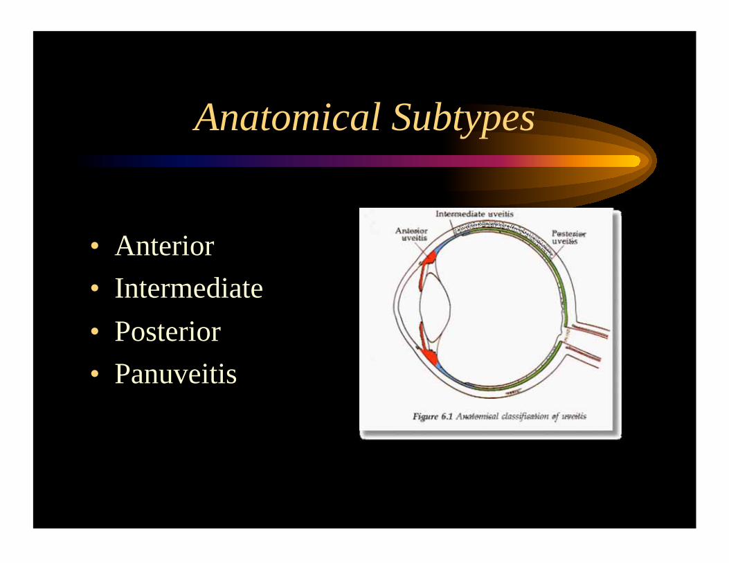
Anatomical Subtypes
• Anterior
• Intermediate
• Posterior
• Panuveitis

History
• Pain, Redness & Photophobia
– Acute vs. Chronic
• Decreased Visual Acuity – CME, Cataract, Media Opacities, etc...
• Flashes & Floaters – Posterior & Intermediate Uveitis Respectively.
History
• Past Medical History
– DM, HTN, Osteoporosis, Lipids, etc…
– Rheumatological Diseases
– Infectious Diseases, STDs

Review of Systems
• Oriented to the differential diagnosis of the
particular uveitic syndrome present.
– Ex. Intermediate Uveitis…
• DDx: MS, Sarcoidosis, Lyme Disease, Luetic…
• Ask for neurologic symptoms, etc…
– If you are not sure ask for questions on all
systems on a systematic fashion...

Physical Examination
• Cells
– Occur due to the breakdown of the blood-ocular
barrier.
– Grading systems allow to assess the adequacy
of treatment and presence of persistent
inflammation.

Grading Systems: A/C Cells
3mm x 1mm slit beam:
0 = Quiet
1 - 4 = trace
5 - 10 = 1+
11- 20 = 2+
21- 50 = 3+
> 50 = 4+
Avoid...
– Rare
– 1 to 2+

Grading systems: Flare
No Flare = 0
Faint = 1+
Iris and Lens Clear = 2+
Iris and Lens Hazy = 3+
Fibrin & Plastic Aq. = 4+
Note: There is no such thing as trace flare…!

Grading Systems: Vitreous Cells
3mm x 1mm slit beam: 0 = Quiet
1 - 10 = 1+
11- 20 = 2+
21- 50 = 3+
> 50 = 4+
Note: There is no such thing as trace vitreous cells...!

Grading Systems: Vitreous Flare/Haze
NFL clearly visible = 0
ON & vessels clear. Hazy NFL = 1+
Hazy ON & vessels = 2+
O N visible = 3+
ON not visible = 4+

Treatment
• Steroids
– Topical
– Periocular (Sub-Tenon’s)
– Intraocular
• Steroid Sparing Agents (Immunomodulation)
– Anti-Metabolic: Methotrexate, CellCept, Azathioprine
– Anti T-Cell: Cyclosporin-A
– Akylating: cyclophosphamide, Chlorambucil

Topical Steroids
• Prednisolone Acetate 1% (Pred Forte)
– Maximun Dose is q1hr
– Side Effects:
• PSC Cataracts
• Glaucoma (takes 3 weeks to ensue)
Topical Steroids
• Rimexolone Acetate 1% (Vexol)
– Almost as good as Pred Forte
– Less likely to cause Ocular Hypertension

Periocular Steroids
• Posterior Sub-Tenon’s Kenalog
– 40mg Dose
– Use 25 G Needle Only
– Half Life 6 weeks

Posterior Sub-Tenon’s Kenalog
• Good for induction treatment when systemic
steroids are better avoided
• Ie. Patients with uncontrolled DM
– Good for CME
• Irrevedsible: • Avoid if OTN is a concern
• Avoid if Viral Etiology is being Considered
• Avoid if patient has chronic disease

Intraocular Kenalog
• Dose is 4MG
• Higher doses can be achieved if concentrated steroid is
used
• Good for CME
• Rarely Needed
• Irrevedsible:
• Avoid if OTN is a concern
• Avoid if Viral Etiology is being Considered
• Avoid if patient has chronic disease

Prednisone
• Start 1mg/kg po qd.
• Do not exceed 60 mg/
day!
• Always prescribe
along with Calcium
supplementation!

Prednisone…
• Weight & arterial blood pressure on every visit…
• Monthly basic chemistry.
• Bi-annual serum lipids.
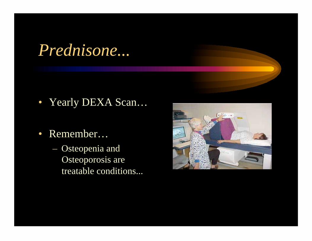
Prednisone...
• Yearly DEXA Scan…
• Remember…
– Osteopenia and
Osteoporosis are
treatable conditions...

The “Standard” Prednisone Taper
Always on a weekly basis: First by ten:
60, 50, 40…
Then by 5:
35, 30, 25, 20…
Then by 2.5:
17.5, 15, 12.5, 10, 7.5, 5, 2.5, stop
• F/U on a monthly basis or sooner if a threshold is reached!

Steroid Sparing Agents
• Very Strong:
– Akylating agents: cyclophosphamide, chlorambucil.
• Moderate to strong:
– CellCept, Imuran.
• Mild:
– Cyclosporine, Methotrexate
– Probably as effective as prednisone itself.


Case # 1
• 57 y/o woman presents with a 2 day history
of pain, redness and photophobia OD.
• Had a similar episode 2 years ago OS which
was treated with a “white drop”, her
symptoms resolved without sequelae.
• 1ry ophthalmologist did a Rheumatoid
factor and a ANA test, both were negative.

Case #1
OD: Va sc: 20/30
Ta: 16
A/C: 3+ C, 1+ Flare
Lens: Clear
Vitreous: Anterior +1 C
Retina: Flat, no CME
OS: A/C: D/Q Focal posterior Synechia at 2
o’clock, trace PSC.

Case # 1
• What is the ocular diagnosis?
• What is the most likely etiology?
• What questions would you ask her?
• What other work-up should be ordered?
• Did the 1ry ophthalmologist order the correct
work-up?
• How would you treat this patient?
• What complications may she develop?

Case # 1
• On further inquiry the patient had history of large joint, episodic, asymmetric, non-deforming polyarthritis which
she ascribed to an old sports injury. She also had history of a heel spur, and recurrent cystitis with negative cultures.
– What is the diagnosis now?
• The patient was treated successfully with q1 hr Pred Forte.
Within 2 weeks she was quiet. She was then tapered off
over 6-8 wks. She recovered 20/20 VA. • She was HLA-B27 positive.

Acute Anterior Uveitis ...
• Acute, Unilateral, Recurrent, Alternating,
Anterior Uveitis…
– If Acute Unilateral: ~50% HLA-B27 (+)
– Acute, Unilateral, Recurrent: ~70% HLA-B27 (+), if
Alternating >80%.
– 50 % of HLA B27 (+) anterior Uveitis have an
underlying Seronegative Spondyloarthropathy.
– The review of Systems Should be directed towards
diagnosing Ankylosing Spondilytis, Reiters, Psoriatic
Arthritis, IBD associated AS, etc…

Trivia Question!
• What is the most common cause of hypopion
associated anterior Uveitis?
• HLA-B27
• Remember HLA-B27 uveitis is far more common
than Beçet’s Disease...
• Beçet’s causes hypopion uveitis in a quiet eye,
HLA-B27 eyes are generally angry!

Case # 2
• 33 y/o AA woman. 7mo h/o progressive VA loss. 5mo ago
she noticed slight pain 3/10 on a sunny afternoon, but her
eyes were never “really” red.
• She then went to a local ophthalmologist who prescribed a
“milky drop” which he told her to take every 2 hours for a
week, this made her eyes feel better, he then told her to
taper down the drops over 2wks because “she could get a
cataract”. She now comes comes for a second opinion.
• She said she was tested for “lupus and syphilis” and the
tests were normal.
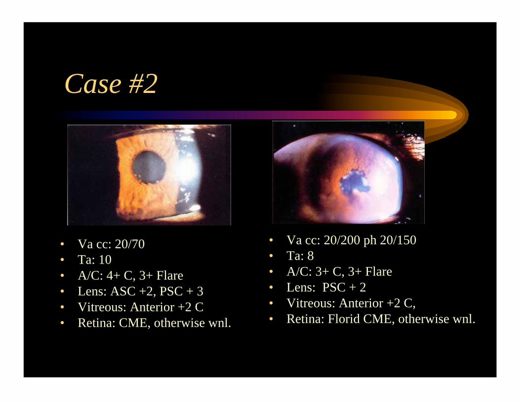
Case #2
• Va cc: 20/70
• Ta: 10
• A/C: 4+ C, 3+ Flare
• Lens: ASC +2, PSC + 3
• Vitreous: Anterior +2 C
• Retina: CME, otherwise wnl.
• Va cc: 20/200 ph 20/150
• Ta: 8
• A/C: 3+ C, 3+ Flare
• Lens: PSC + 2
• Vitreous: Anterior +2 C,
• Retina: Florid CME, otherwise wnl.

Case #2
• What is the ocular diagnosis?
• What are the most likely etiologies?
• What questions would you ask her?
• What other work-up should be ordered?
• How would you treat this patient now?

Case #2
• You called the previous
ophthalmologist and received a
fax with a (-) RPR and (-)
ANA.
• On further questioning she
admits to a “rash” which has
not gone away (see Figure)…,
otherwise ROS is (-).
• You order an FTA-Abs, a CXR
and start Pred Forte (16,16) &
AS 1%.
Case #2
• She comes in 1wk for F/U. She still has 2+ cells
bilaterally, her cc Va is now 20/50 OD and 20/125 OS.
CXR & FTA were (-). The dermatology consult was (+)
for non-caseating granulomata. Her rheumatologist wants
to start her on Plaquenil 200 po bid.
• How should you manage her ophthalmological treatment?
• What advice should you give regarding starting Plaquenil
at this juncture?

Case #2
• She comes for her F/U after being on PF q1hr for one
month. Her Va is unchanged, she still has trace cells OD
and 1+ cells OS.
• The Plaquenil was held and the dermatologist opted for a
topical treatment.
• How would you manage the patient now?
• Any supplements, monitoring?

Case # 2
• You treated her with 60mg oral prednisone, she was
absolutely quiet after 4 wks, her Va was now 20/30 OD
and 20/70 OS. You then started a “standard” taper and
followed her on a monthly basis.
• On the second month, she comes at a dose of 17.5mgs of
prednisone, she is asymptomatic but you notice 1+ cells
OU. Her VA is now 20/40 OD and 20/100 OS.
• How would you manage her now?

Case # 2
• You upped the prednisone back to 60mgs and started
CellCept at 1g bid. She becomes quiet at a week, you start
to taper the prednisone and are able to taper her all the way
to 10mgs in 3 months without a flare-up.
• What labs should you be getting at this juncture and at
what frequency?
• What are the most common side effects of CellCept?
• How should you manage her now?

Case # 2
• She comes for a routine monthly F/U. She is now
at 5mgs of prednisone daily. VA is 20/40 OU, she
is quiet, no CME, there is PSC +2 OU. She
complains of muscle stiffness and pains. Her AST
is 55 (slightly elevated).
• What is the cause of her stiffness?
• How should you manage her now?

Case # 2
• It is a year after her initial visit with you, she is off the
prednisone, and remains on CellCept 1g bid. LFTs are
normal.
• She complains of glare impairing her driving at night, there
is no CME, her BCVA is 20/50, J8 OU, she has bilateral
PSC +3.
• What precautions should you have for her phaco?
• How would you manage her medications?

The “Standard” Prednisone Taper
Always on a weekly basis: First by ten:
60, 50, 40…
Then by 5:
35, 30, 25, 20…
Then by 2.5:
17.5, 15, 12.5, 10, 7.5, 5, 2.5, stop
• F/U on a monthly basis or sooner if a threshold is reached!

A Word about Uveitis Workup
• Syphilis and Sarcoidosis
– Both Syndromes can Imitate almost any type of
Uveitis
– All Patients must have:
• RPR
• FTA-Abs: 20% of Syphilitic Uveitis is RPR (-) and
FTA-Abs (+)
• CXR: 87.5% Sensitive in detecting Sarcoidosis.
– All a other tests should be dictated by the
PMHx and ROS

Conclusion
• Acute Uveitis Should be treated aggressively to avoid structural
complications and should be expected to resolve in 6-8 weeks.
• Chronic Uveitis requires treatment “forever”. You only taper down to
find thresholds and if these are found treatment should be held above
the threshold in order to prevent complications from structural
complications.
• Always remember to seek infectious causes, use common sense:
– Unilateral follicular conjunctivitis with uveitis => HSV
– Syphilitic uveitis is not treated adequately until the patient receives
IV treatment.