uveitic glaucoma
DESCRIPTION
UVEITIC GLAUCOMA. TARIQ ALASBALI, MD. PDS PXF HEMOLYTIC PHACOLYTIC INFLAMMATORY ↑ EVP. PUPILLARY BLOCK PLATEAU IRIS. POAG JUVENILE. PUSHING. PHACOMOROHIC ECTOPIA LENTIS APHAKIC PSEUDOPHAKIC INFLAMMATORY PS AQ MISSDIRECTION SUBRETINAL MASS. AXIAL SHALLOWING - PowerPoint PPT PresentationTRANSCRIPT
UVEITIC GLAUCOMAUVEITIC GLAUCOMA
TARIQ ALASBALI, MDTARIQ ALASBALI, MD..

POAGJUVENILE
PDSPXFHEMOLYTICPHACOLYTICINFLAMMATORY ↑ EVP
PUPILLARY BLOCK
PLATEAU IRIS
AXIAL SHALLOWING•CHOROIDAL EFFUSION•CHOROIDAL HE•AQ MISSDIRECTION•ROP•PHPV•CHOROIDAL MASS
PHACOMOROHICECTOPIA LENTISAPHAKICPSEUDOPHAKICINFLAMMATORY PSAQ MISSDIRECTIONSUBRETINAL MASS
PUSHING
PULLING
PERIPH SHALLOWINGIRIS CYSTCB CYSTCB SWELLING SULFA PRPCB INFLAMMATIONCB TUMOUR
PAS FORMATIONNVGINFLAMMATIONTRAUMATUMOUR RELATED
MEMBRANEGROWTHICEEPITH DOWNGROWTHFIBROUS INGROWTH

Uveitis Classifications
Anatomic Classification
Duration Classification
Uveitis Classifications
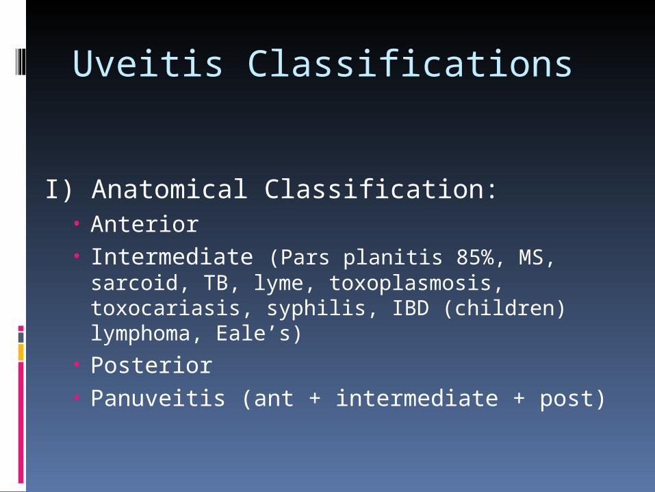
I) Anatomical Classification:• Anterior• Intermediate (Pars planitis 85%, MS, sarcoid,
TB, lyme, toxoplasmosis, toxocariasis, syphilis, IBD (children) lymphoma, Eale’s)
• Posterior• Panuveitis (ant + intermediate + post)
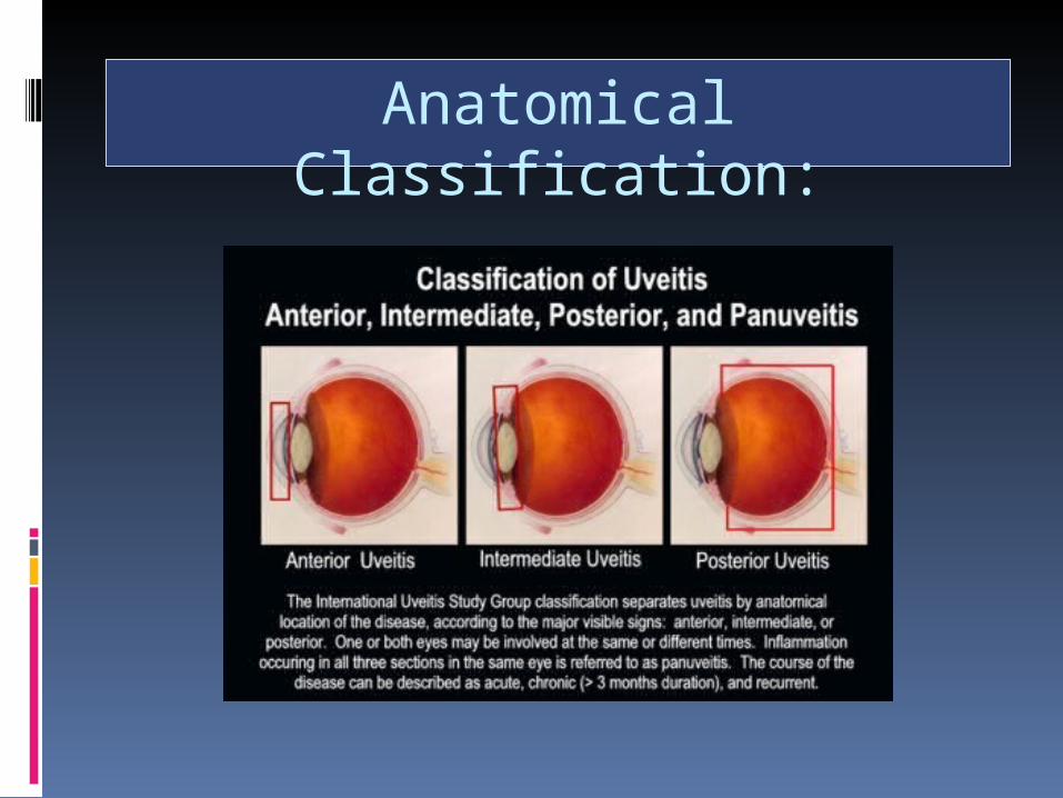
Anatomical Classification:
Q1: Which form of ocular inflammation (anatomical classification)
most frequently produces an IOP elevation?
A1: Anterior uveitis (chronic >>> acute)
Even glaucomas that result from other types of ocular inflammation are usually the consequence of secondary involvement of the anterior uveal tract.

Q2. What are typical IOPs in anterior uveitis?
A2. LOW secondary to CB shut down +/- increased uveoscleral outflow
Q2. What are typical IOPs in anterior uveitis?
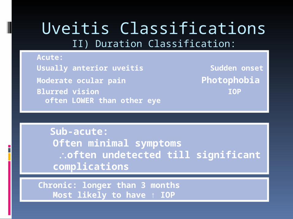
Uveitis ClassificationsII) Duration Classification:
Acute: Usually anterior uveitis Sudden onset
Moderate ocular pain PhotophobiaBlurred vision IOP often
LOWER than other eye
Sub-acute: Often minimal symptoms often undetected till significant complications
Chronic: longer than 3 monthsMost likely to have ↑ IOP
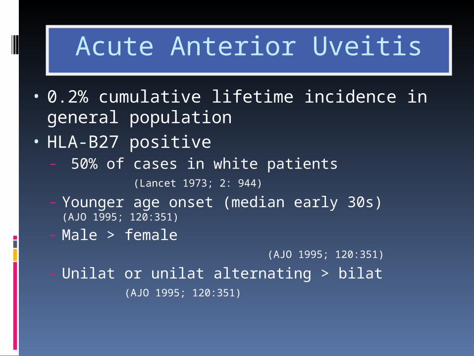
Acute Anterior Uveitis
• 0.2% cumulative lifetime incidence in general population
• HLA-B27 positive – 50% of cases in white patients
(Lancet 1973; 2: 944)
– Younger age onset (median early 30s) (AJO 1995; 120:351)
– Male > female (AJO 1995; 120:351)
– Unilat or unilat alternating > bilat (AJO 1995; 120:351)

BEHÇET’S DISEASE
Idiopathic multisystem disease
More common in men
Occurs in 3rd - 4th decade
Associated with HLA-B5

Incidance: 1/100, 000 prevalence in USA 670/100,000 in Japan
BD is most prevalent (and more virulent) in the Mediterranean region, Middle East, and Far East, with an estimated prevalence of 1 case per 10,000 persons
BEHÇET’S DISEASE

Unknown Various bacteria and viruses
suggested No good evidence to suggest
any of them Tumour necrosis factor (TNF)
thought to be important
BEHÇET’S DISEASEAetiology
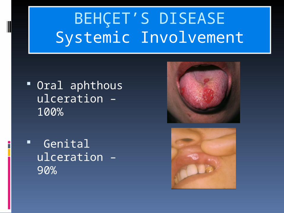
Oral aphthous ulceration – 100%
Genital ulceration – 90%
BEHÇET’S DISEASESystemic Involvement
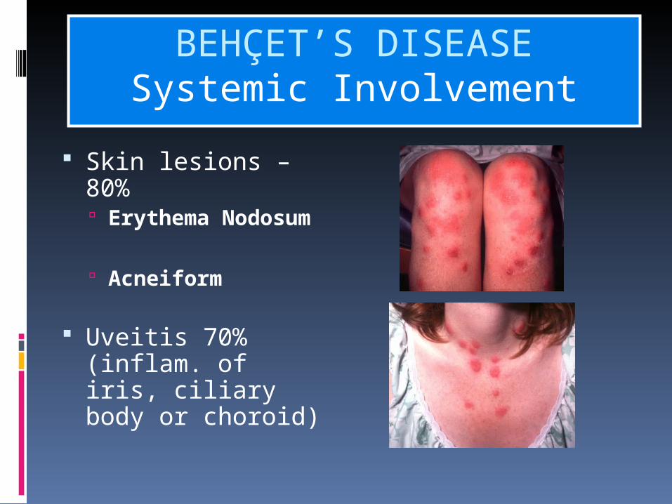
Skin lesions – 80% Erythema
Nodosum
Acneiform
Uveitis 70% (inflam. of iris, ciliary body or choroid)
BEHÇET’S DISEASESystemic Involvement

CNS involvement – strokes
Major vessels SVC obstruction
Increased skin response to trauma
BEHÇET’S DISEASESystemic Involvement
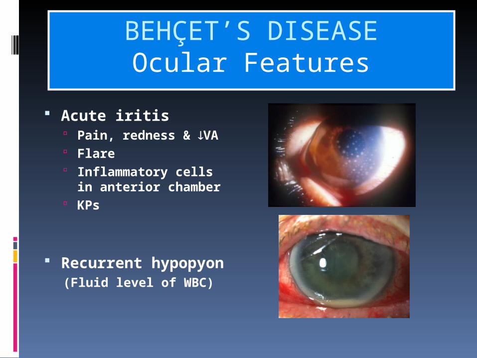
Acute iritis Pain, redness & VA Flare Inflammatory cells
in anterior chamber KPs
Recurrent hypopyon(Fluid level of WBC)
BEHÇET’S DISEASEOcular Features
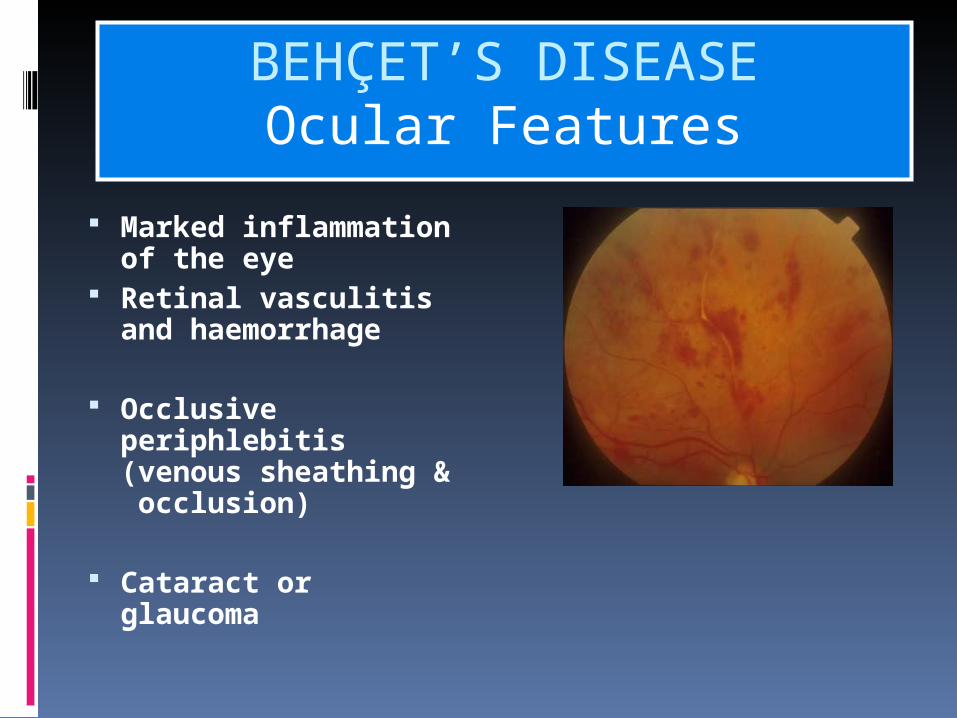
Marked inflammation of the eye
Retinal vasculitis and haemorrhage
Occlusive periphlebitis (venous sheathing & occlusion)
Cataract or glaucoma
BEHÇET’S DISEASEOcular Features

International criteria published in 1990 require
Oral ulcers 3 X /1 year + Any 2 of the following: 1. Recurrent genital ulcers2. eye lesions 3. Skin lesions4. Positive pathergy test 2mm plus papule developing over 24-48 hrs after
oblique insertion of a 20 gauge needle into skin.
BEHÇET’S DISEASEdiagnosis
Sarcoidosis
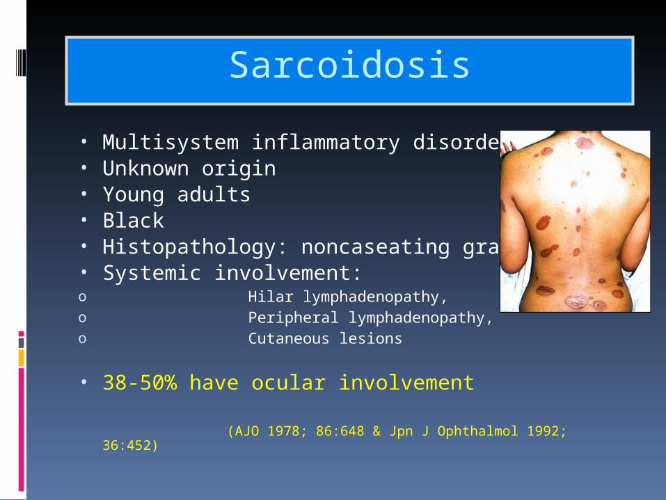
• Multisystem inflammatory disorder• Unknown origin• Young adults• Black• Histopathology: noncaseating granulomas• Systemic involvement: o Hilar lymphadenopathy,o Peripheral lymphadenopathy, o Cutaneous lesions
• 38-50% have ocular involvement (AJO 1978; 86:648 & Jpn
J Ophthalmol 1992; 36:452)

Sarcoidosis – Anterior Uveitis
• Most common ocular manifestation = chronic granulomatous uveitis
• Mutton fat KPs (15%) (AJO 1978; 86:648)
• Bilateral > unilateral
• Iris nodules (11.4-35%) (Ophthalmology 1986; 93: 511)
• AC angle nodules (49%) (Ophthalmology 1986; 93: 511)
• CB nodules (42%) (Ophthalmology 1986; 93: 511)
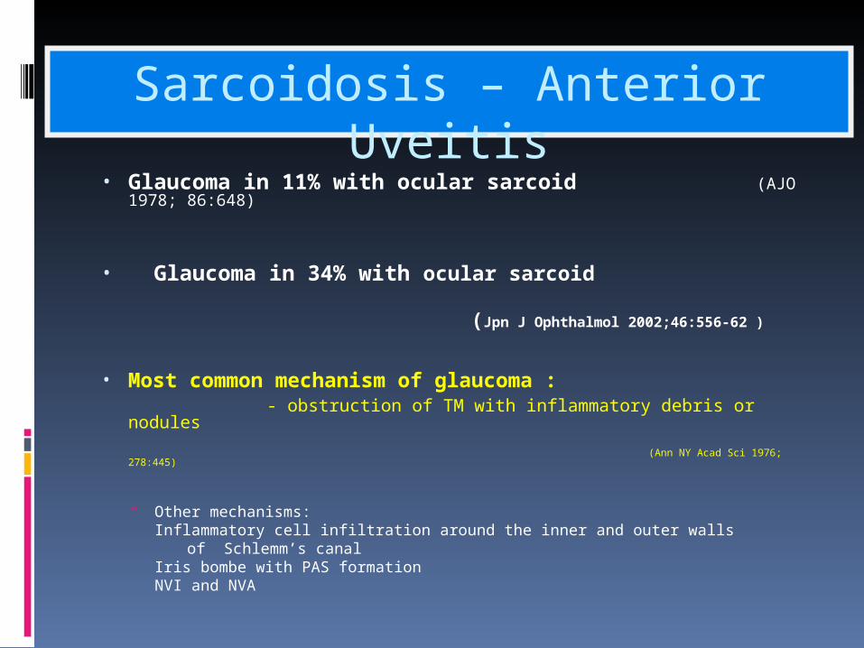
• Glaucoma in 11% with ocular sarcoid (AJO 1978; 86:648)
• Glaucoma in 34% with ocular sarcoid
(Jpn J Ophthalmol 2002;46:556-62 )
• Most common mechanism of glaucoma : - obstruction of TM with inflammatory debris or nodules
(Ann NY Acad Sci 1976; 278:445)
Other mechanisms: Inflammatory cell infiltration around the inner and outer walls
of Schlemm’s canalIris bombe with PAS formationNVI and NVA
Sarcoidosis – Anterior Uveitis
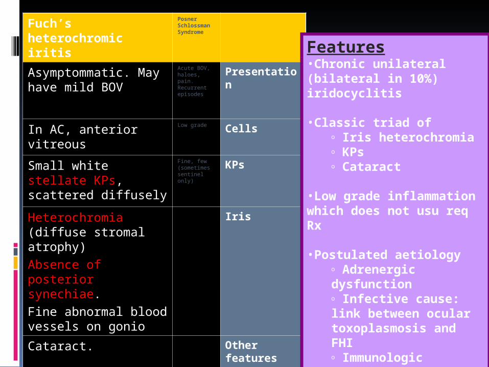
Posner Schlossman Syndrome
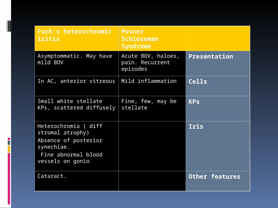
Fuch’s heterochromic iritis
PresentationAcute BOV, haloes, pain. Recurrent episodes
Asymptommatic. May have mild BOV
CellsMild inflammationIn AC, anterior vitreous
KPsFine, few, may be stellate
Small white stellate KPs, scattered diffusely
IrisHeterochromia ( diff stromal atrophy)Absence of posterior synechiae. Fine abnormal blood vessels on gonio
Other featuresCataract.
Posner Schlossman Syndrome
Fuch’s heterochromic iritis
Presentation
Acute BOV, haloes, pain. Recurrent episodes
Asymptommatic. May have mild BOV
CellsMild inflammation
In AC, anterior vitreous
KPsFine, few. May be stellate
Small white stellate KPs, scattered diffusely
IrisHeterochromia ( diff stromal atrophy)Absence of posterior synechiae. Fine abnormal blood vessels on gonio
Other features
Cataract.
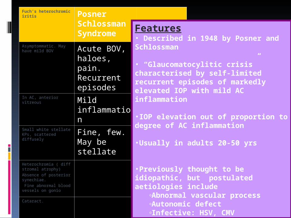
Features• Described in 1948 by Posner and Schlossman
• “Glaucomatocylitic crisis” characterised by self-limited recurrent episodes of markedly elevated IOP with mild AC inflammation
•IOP elevation out of proportion to degree of AC inflammation
•Usually in adults 20-50 yrs
•Previously thought to be idiopathic, but postulated aetiologies include
oAbnormal vascular processoAutonomic defectoInfective: HSV, CMV
Posner Schlossman Syndrome
Fuch’s heterochromic iritis
Presentation
Acute BOV, haloes, pain. Recurrent episodes
Asymptommatic. May have mild BOV
CellsLow gradeIn AC, anterior vitreous
KPsFine, few (sometimes sentinel only)
Small white stellate KPs, scattered diffusely
IrisHeterochromia (diffuse stromal atrophy)Absence of posterior synechiae.Fine abnormal blood vessels on gonio
Other features
Cataract.
Features•Chronic unilateral (bilateral in 10%) iridocyclitis
•Classic triad ofo Iris heterochromiao KPso Cataract
•Low grade inflammation which does not usu req Rx
•Postulated aetiologyo Adrenergic dysfunctiono Infective cause: link between ocular toxoplasmosis and FHIo Immunologic theories
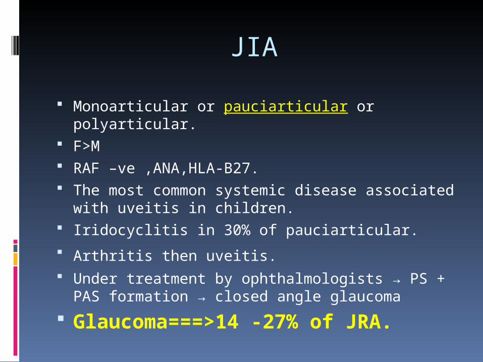
JIA
Monoarticular or pauciarticular or polyarticular. F>M RAF –ve ,ANA,HLA-B27. The most common systemic disease
associated with uveitis in children. Iridocyclitis in 30% of pauciarticular.
Arthritis then uveitis. Under treatment by ophthalmologists → PS +
PAS formation → closed angle glaucoma
Glaucoma===>14 -27% of JRA.
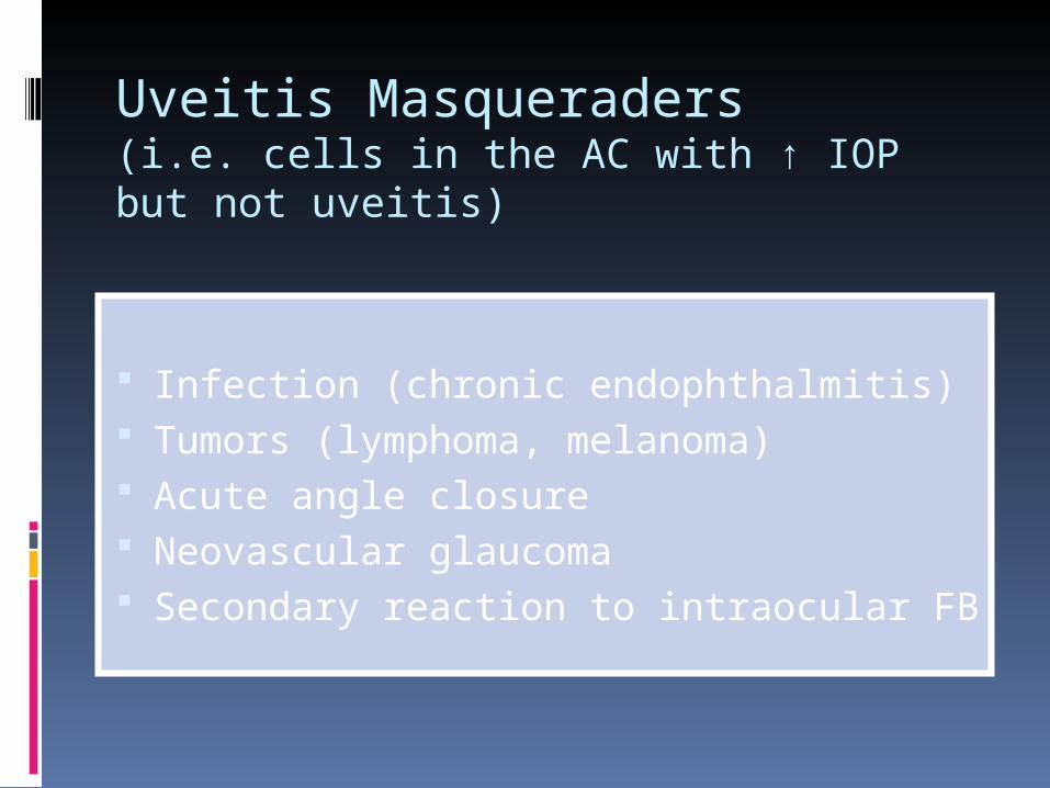
Infection (chronic endophthalmitis) Tumors (lymphoma, melanoma) Acute angle closure Neovascular glaucoma Secondary reaction to intraocular FB
Uveitis Masqueraders (i.e. cells in the AC with ↑ IOP but not uveitis)
First reported by:
Joseph Beer in -------- 1813Desmans in -------- 1821Mackenzie in -------- 1830

Glaucoma Associated with Uveitis
• Etiology of uveitis:• Topographic Types
Difference non-significant [P>0.05]
391391 eyeseyes F/U median 55 monthsF/U median 55 months
Neri P. J Glaucoma Neri P. J Glaucoma
2004;13:461-652004;13:461-65
ConclusionConclusion:: 1.1. Presence of glaucoma was associated with an increasing risk of visual loss. Presence of glaucoma was associated with an increasing risk of visual loss. 2.2. The incidence of glaucoma increased with time and similar among The incidence of glaucoma increased with time and similar among different types of uveitis. different types of uveitis.
IncidenceIncidence• 6.6% at 1 year• 11.2% at 4 years• 22.7% at 10 years
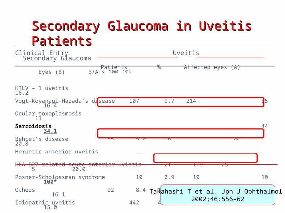
Secondary Glaucoma in Uveitis Secondary Glaucoma in Uveitis PatientsPatients
Clinical Entry Uveitis Secondary Glaucoma
Patients % Affected eyes (A) Eyes (B) B/A x 100 (%)
HTLV – 1 uveitis 194 17.7 260 4216.2
Vogt-Koyanagi-Harada’s disease 107 9.7 214 3516.4
Ocular toxoplasmosis 85 7.7 95 11 11.6
Sarcoidosis 71 6.5 129 4434.1
Behcet’s disease 55 5.0 96 2020.8
Herpetic anterior uveitis 22 2.0 23 7 30.4
HLA-B27-related acute anterior uvietis 21 1.9 25 520.0
Posner-Scholossman syndrome 10 0.9 10 10100*
Others 92 8.4 116 23 16.1
Idiopathic uveitis 442 40.2 636 9615.0
TOTAL 1099 100 1604 29318.3
*HTLV-I: human T-lymphotropic virus type1. HLA: human leukocyte antigen
Takahashi T et al. Jpn J Ophthalmol 2002;46:556-62
Glaucoma Associated with Uveitis
25% of uvetic patients: Ocular hypentensive
5-19% of uveitic patients: Develop S.G.

Risk factors for elevated IOP
in uveitis patients
1.1. ChronicityChronicity2.2. AgeAge3.3.CorticosterCorticoster
oidsoids4.4. ActivityActivity
Herbert H et al. J. Glaucoma 2004;13:96-99
JIA, and ANA JIA, and ANA positive positive uveitis uveitis without without evidence of evidence of arthritis.arthritis. seqandary glaucoma inseqandary glaucoma in children with uveitischildren with uveitis
Sijssens et al. Ophthalmology 2006;113:853-9
PATHOGENESIS
A. Biochemical and Cellular Changes in Aqueous Composition
B. Direct Involvement of the TMC. Corticosteroids Effects on the TMD. Morphologic Changes in the AC
Angle
1.1. ProteinsProteins
2.2. Inflammatory CellsInflammatory Cells
3.3. ProstaglandinsProstaglandins
4.4. Inflammatory Mediators (Cytokines) & Toxic Inflammatory Mediators (Cytokines) & Toxic AgentsAgents
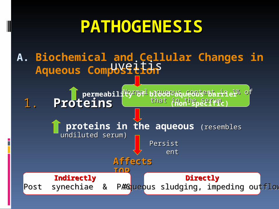
A. Biochemical and Cellular Changes in Aqueous Composition
PATHOGENESISPATHOGENESIS
Normal aqueous content is 1% ofNormal aqueous content is 1% of
that in the serumthat in the serum1.1. ProteinsProteins
uveitis
permeability of blood-aqueous barrier (non-specific)
proteins in the aqueous (resembles undiluted resembles undiluted serum)serum)
PersistePersistentnt
Affects IOPAffects IOP
IndirectlyIndirectlyPost synechiae & PASPost synechiae & PAS
DirectlyDirectlyAqueous sludging, impeding outflowAqueous sludging, impeding outflow
A.A. Biochemical and Cellular Changes in Biochemical and Cellular Changes in Aqueous CompositionAqueous Composition
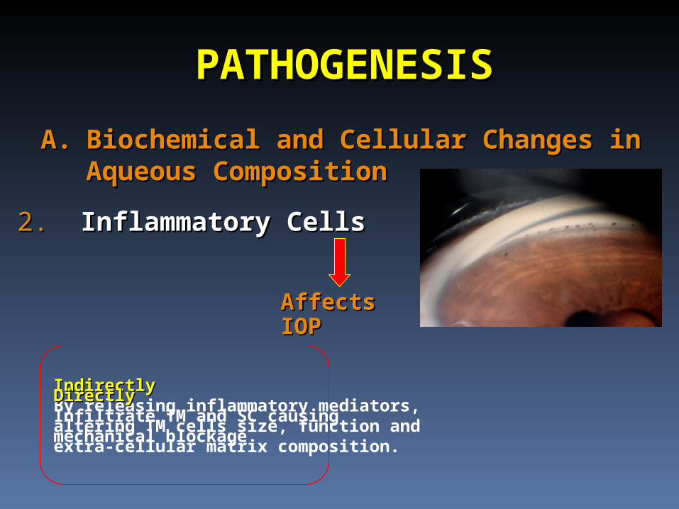
PATHOGENESISPATHOGENESIS
2.2. Inflammatory CellsInflammatory Cells
Affects IOPAffects IOP
IndirectlyIndirectlyBy releasing inflammatory mediators, altering TM cells size, function and extra-cellular matrix composition.
DirectlyDirectlyInfiltrate TM and SC causing mechanical blockage
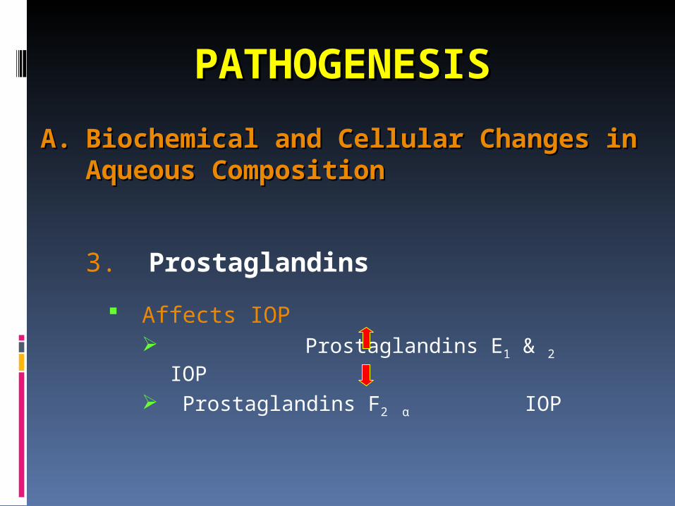
3. Prostaglandins
Affects IOP Prostaglandins E1 & 2 IOP
Prostaglandins F2 α IOP
A.A. Biochemical and Cellular Changes in Biochemical and Cellular Changes in Aqueous CompositionAqueous Composition
PATHOGENESISPATHOGENESIS
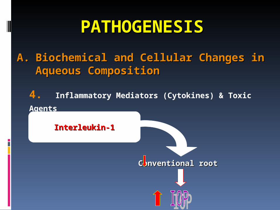
4. Inflammatory Mediators (Cytokines) & Toxic
Agents
A.A. Biochemical and Cellular Changes in Biochemical and Cellular Changes in Aqueous CompositionAqueous Composition
PATHOGENESISPATHOGENESIS
Conventional rootConventional root
Interleukin-1Interleukin-1
B. Direct Inflammation of the TM Posner-sehlossman Syndrome Fuchs’ Uveitis Herpetic Keratouveitis
PATHOGENESISPATHOGENESIS

C. Effect of Corticosteroids on the TM 35% of normal population, moderate
responders 4-6% high responders (>15mmHg) 50% or more of POAG population, high
responders
PATHOGENESISPATHOGENESIS
Weireb RN et al. Invest Ophthalmol Vis Sci 1985;26:170-5Weireb RN et al. Invest Ophthalmol Vis Sci 1985;26:170-5Levin DS, et al. Am J Ophtalmol 2002;133:196-202Levin DS, et al. Am J Ophtalmol 2002;133:196-202
Affects IOPAffects IOP
Reduction of aqueous Reduction of aqueous outflow BYoutflow BY
1.1. Alteration in cell sizeAlteration in cell size2.2. Cytoskeletal Cytoskeletal
organizationorganization3.3. Extra-cellular matrix Extra-cellular matrix
depositiondeposition Veda J et al. Invest Ophthalmol Vis Sci 2003;44:4772-9Veda J et al. Invest Ophthalmol Vis Sci 2003;44:4772-9
Velota et al. Curr Opn Ophthalmol 2004;15:136-140Velota et al. Curr Opn Ophthalmol 2004;15:136-140
Increase the aqueous
production

Incidence of steroid responsiveness
HighHigh(%)(%)ModerateModerate(%)(%)NonNon(%)(%)
General General PopulationPopulation5535356060Pts With Pts With POAGPOAG9090101000Siblings of Siblings of pts with pts with POAGPOAG
303050502020
Offspring of Offspring of pts with pts with POAGPOAG
2525707055
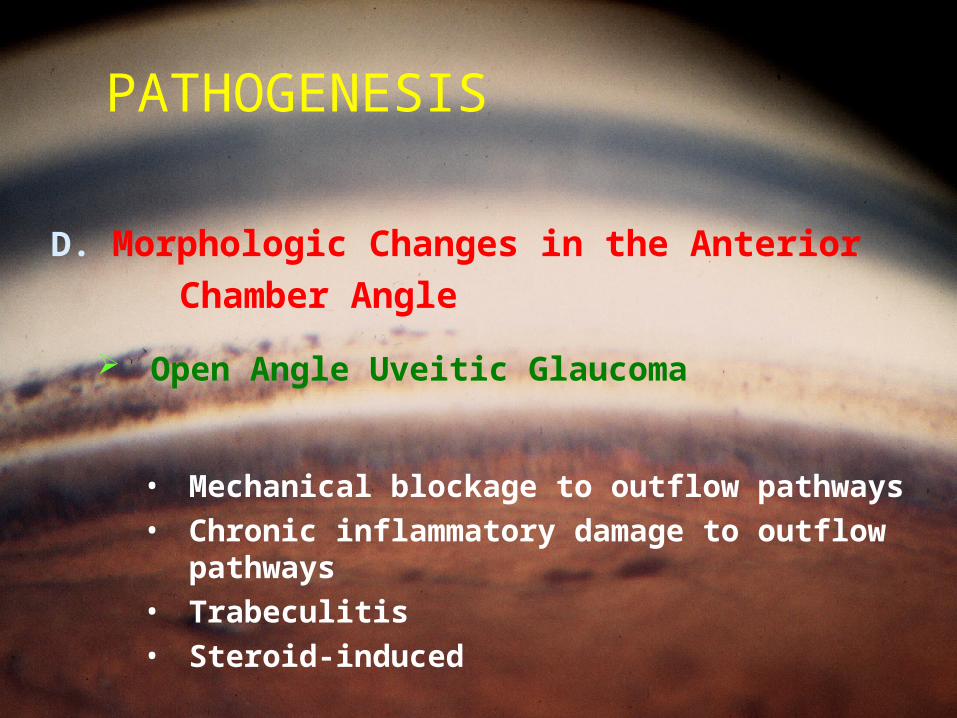
PATHOGENESIS
D. Morphologic Changes in the Anterior Chamber Angle
Open Angle Uveitic Glaucoma
• Mechanical blockage to outflow pathways
• Chronic inflammatory damage to outflow pathways
• Trabeculitis• Steroid-induced
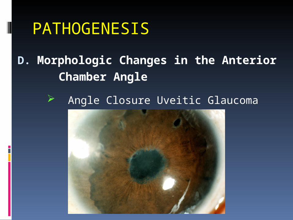
PATHOGENESIS
D. Morphologic Changes in the Anterior Chamber Angle
Angle Closure Uveitic Glaucoma
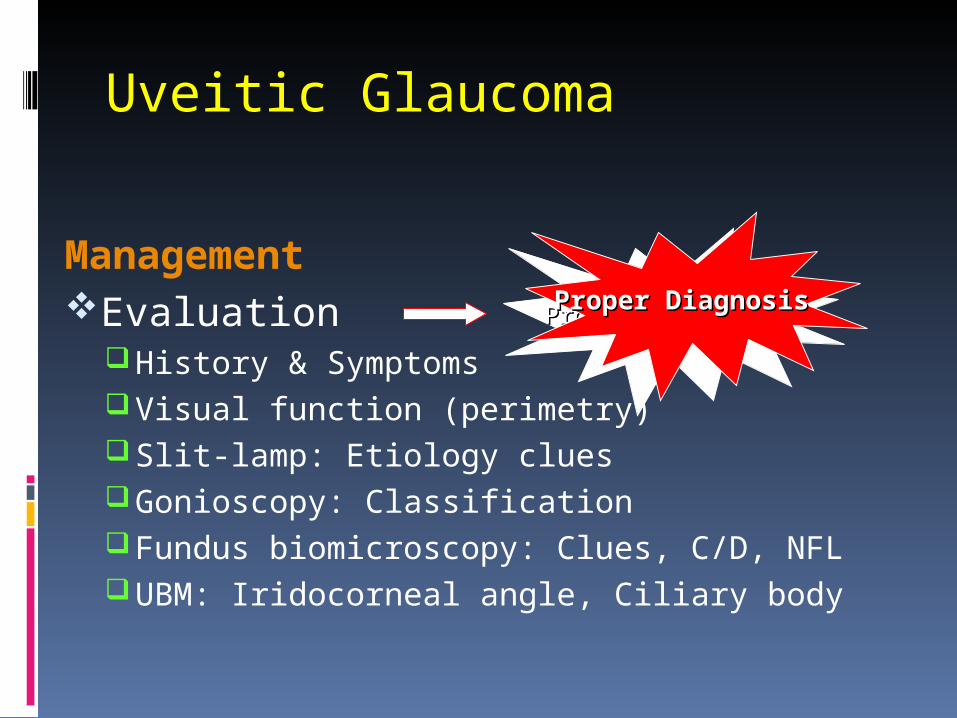
Uveitic Glaucoma
ManagementEvaluation
History & SymptomsVisual function (perimetry)Slit-lamp: Etiology cluesGonioscopy: ClassificationFundus biomicroscopy: Clues, C/D, NFLUBM: Iridocorneal angle, Ciliary body
Proper DiagnosisProper Diagnosis Proper DiagnosisProper Diagnosis

Uveitis Work Up
• CBC + differential• HLA• ESR + CRP• ACE, lysozyme• CXR +/- CT chest• FTA Abs + VDRL• TB skin test• Titers: toxocariasis, toxoplasmosis,
lyme

Management
Control Inflammation:
Undertreating
uveitis with corticosteroids to minimize IOP elevation at the expense of good control
of inflammation
isis
A A false false economyeconomy

Management
Medical therapy: Beta-blockers CA inhibitors Adrenergic agonists Prostaglandin analogues? Miotics - Avoid
Q3: What glaucoma drops have been associated with uveitis?
A3: Brimonidine (AJO 2000; 130:287)
Metipranolol (AJO 1997; 123:843)
prostaglandin analogues (Surv Ophthalmol 2002;
47 (suppl 1): S219)
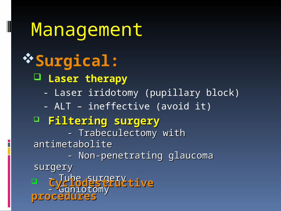
Management
Surgical: Laser therapy
- Laser iridotomy (pupillary block)- ALT – ineffective (avoid it)
Filtering surgeryFiltering surgery - Trabeculectomy with - Trabeculectomy with antimetaboliteantimetabolite - Non-penetrating glaucoma - Non-penetrating glaucoma surgerysurgery
- Tube surgery- Tube surgery- Goniotomy- Goniotomy
Cyclodestructive Cyclodestructive proceduresprocedures
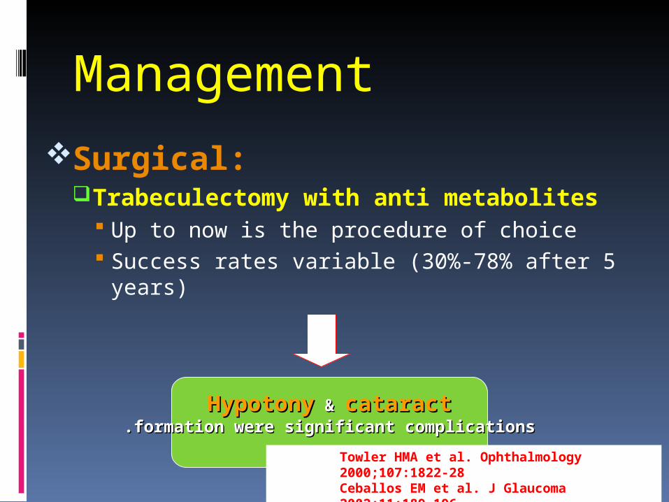
Management
Surgical:Trabeculectomy with anti metabolites
Up to now is the procedure of choice Success rates variable (30%-78% after 5
years)
HypotonyHypotony & & cataractcataract formation were significant complicationsformation were significant complications..
Towler HMA et al. Ophthalmology 2000;107:1822-28Ceballos EM et al. J Glaucoma 2002;11:189-196
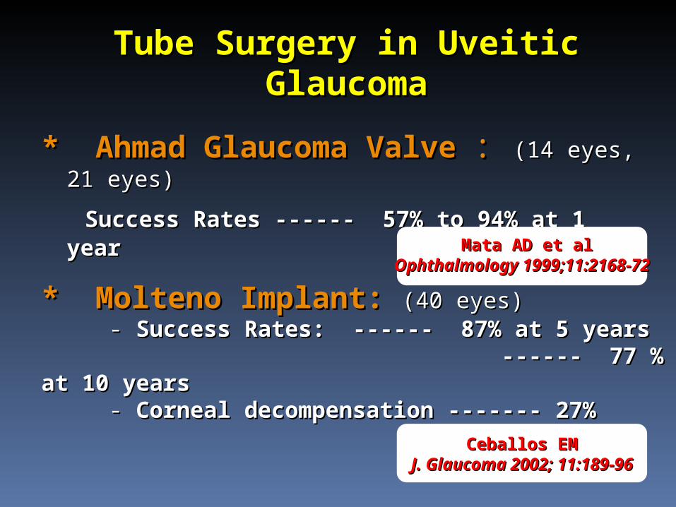
Tube Surgery in Uveitic Tube Surgery in Uveitic GlaucomaGlaucoma
* Ahmad Glaucoma Valve* Ahmad Glaucoma Valve :: (14 (14 eyes, 21 eyes)eyes, 21 eyes)
Success Rates ------ 57% to 94% at 1 yearSuccess Rates ------ 57% to 94% at 1 year
* Molteno Implant:* Molteno Implant: (40 eyes)(40 eyes) - - Success Rates: ------ 87% at 5 yearsSuccess Rates: ------ 87% at 5 years ------ 77 % at 10 years------ 77 % at 10 years - - Corneal decompensation ------- 27%Corneal decompensation ------- 27%
Mata AD et alMata AD et alOphthalmology 1999;11:2168-72Ophthalmology 1999;11:2168-72
Ceballos EMCeballos EMJ. Glaucoma 2002; 11:189-96J. Glaucoma 2002; 11:189-96
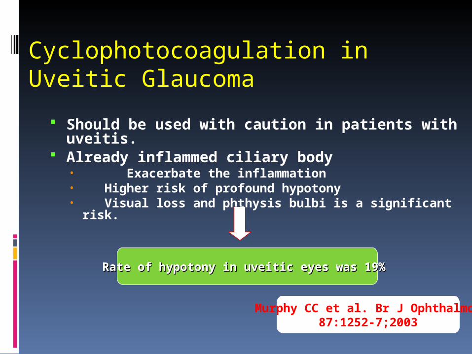
Cyclophotocoagulation in Uveitic Glaucoma
Should be used with caution in patients with uveitis.
Already inflammed ciliary body• Exacerbate the inflammation• Higher risk of profound hypotony• Visual loss and phthysis bulbi is a significant risk.
Murphy CC et al. Br J Ophthalmol 2003;87:1252-7
Rate of hypotony in uveitic eyes was 19%Rate of hypotony in uveitic eyes was 19%
Aside…DDx Krukenberg Spindle• PDS
– Young– Myopic– Male– 10% have glaucoma– ↑ risk RD
• Trauma• Iris infarction (e.g. zoster, ACG)• Tumor• Uveitis

سأل الممكن المستحيل : أين تقيم ؟ فأجابه في أحالم
العاجز