uvea and uveitis - in focus · feline lymphoplasmacytic uveitis (l/p uveitis) • 696 cases out of...
TRANSCRIPT

Uvea and Uveitis

Uveitis Organisms
• Blastomycosis• Cryptococcosis• Coccidioidomycosis• Histoplasmosis• Protothecosis• Aspergillosis

Blastomycosis167 cases in dogs, 10 cases in cats
• Blastomyces dermatitidis• Dogs more than cats• 32 cases in Goldens 30 in Labs • 98/167 less than 5 years old• Only 10 older than 10 years old• Mississippi River valley • Very local hotbeds• Soil fungus spread through inhalation from the soil• Encapsulated with broad-based budding


Blastomycosis

Uveitis Organisms
• Blastomycosis• Cryptococcosis• Coccidioidomycosis• Histoplasmosis• Protothecosis• Aspergillosis

Cryptococcosis11 cases in dogs, 27 cases in cats
• Cryptococcus neoformans• Spread from soil rich with pigeon droppings• Ohio River valley• Cats more than dogs• 7 to 10 micron poorly staining cell body
surrounded by mucinous capsule


Cryptococcosis
Alcian Blue PAS


Alcian Blue PAS


Uveitis Organisms
• Blastomycosis• Cryptococcosis• Coccidioidomycosis• Histoplasmosis• Protothecosis• Aspergillosis
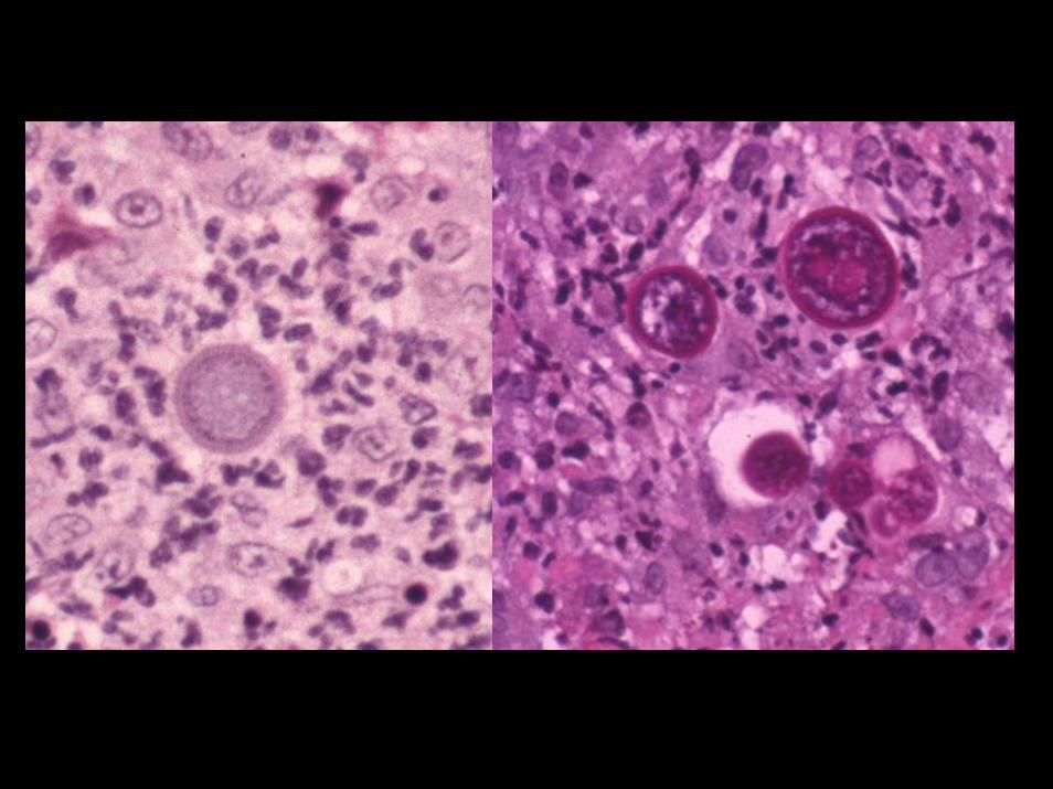
Coccidioidomycosis26 cases in dogs, 9 cases in cats
• Coccidioides immitis• Soil organism from Southwestern desert• Infects many species• Organism easier to find in cats than dogs• 20 to 40 micron spherule with enclosed
endospores• Does not have a budding form

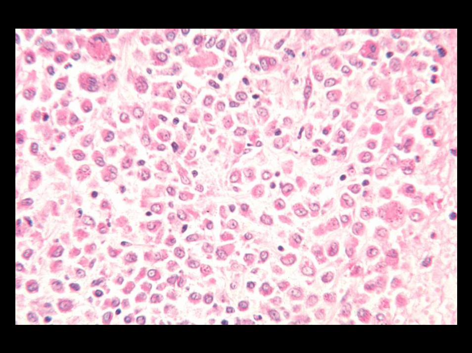
Coccidioidomycosis

Uveitis Organisms
• Blastomycosis• Cryptococcosis• Coccidioidomycosis• Histoplasmosis• Protothecosis• Aspergillosis

Histoplasmosis2 cases in dogs, 24 cases in cats
• Histoplasma capsulatum• Found in soil contaminated with bird
droppings• Affects many tissues• Ohio River valley• 4 micron intracellular organism

Histoplasmosis


Uveitis Organisms
• Blastomycosis• Cryptococcosis• Coccidioidomycosis• Histoplasmosis• Protothecosis• Aspergillosis

Protothecosis11 cases in dogs, 0 in cats
• Prototheca zopfii, Prototheca wickerhamii• Saprophytic achlorophyllus algae• Worldwide distribution infecting several
species• Associated with contaminated water and
susceptible host


Protothecosis

Uveitis Organisms
• Blastomycosis• Cryptococcosis• Coccidioidomycosis• Histoplasmosis• Protothecosis• Aspergillosis

Canine Systemic Aspergillosis24 Cases
• Worldwide distribution• German Shepherd breed at risk (13/24
cases)• Systemic disease with vasculitis• Poor prognosis

Systemic Aspergillosis


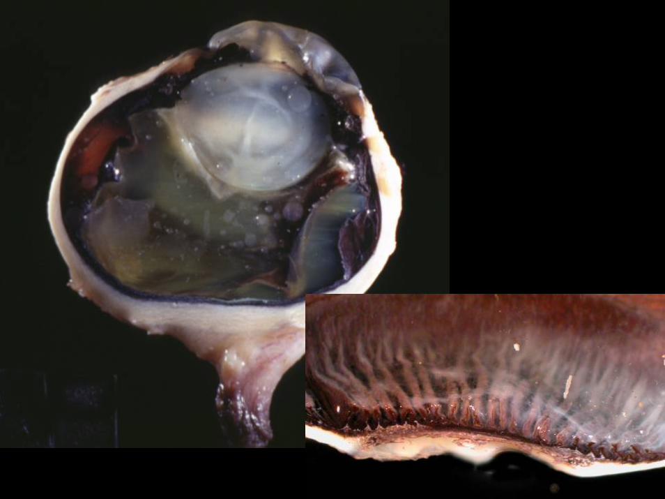
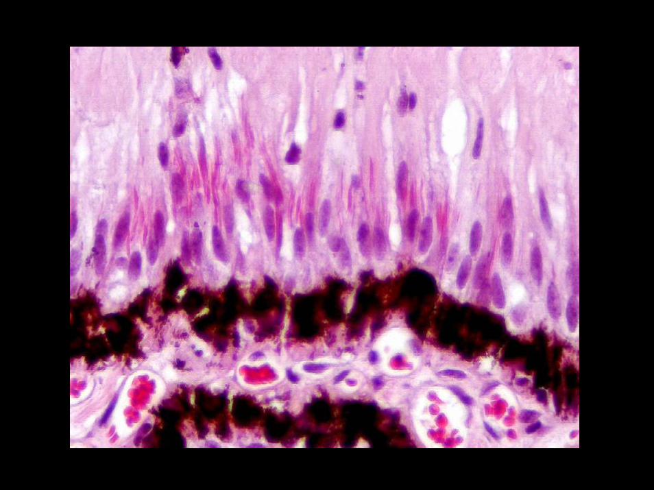
Equine Recurrent Uveitis (ERU)• Bilateral, but not always symmetrical • Cyclic Uveitis• Etiology
– Leptospirosis– Autoimmunity
• Classic morphologic features– Lymphoplasmacytic inflammation with lymphoid follicles– Lymphocytes within the ciliary epithelium– Linear hypereosinophilic cytoplasmic inclusions in the non-
pigmented ciliary epithelium– Amyloid deposition on the inner surface of the non-pigmented
ciliary epithelium


Pinealitis

West Nile Virus in Raptors
Canine Uveodermatologic Syndrome
Vogt-Koyanagi-Harada SyndromeVKH

Canine Uveodermatologic SyndromeVogt-Koyanagi-Harada Syndrome
VKH
• 125 cases in the COPLOW collection• Breeds
– Mixed breed…30– Akita...25– German Shepherd…8
• Bilateral symmetry is a distinctive feature– OS…42– OD…29– OU…44
• Glaucoma• Retinal detachment• Morphologically, relatively quiet eye


CanineVKH


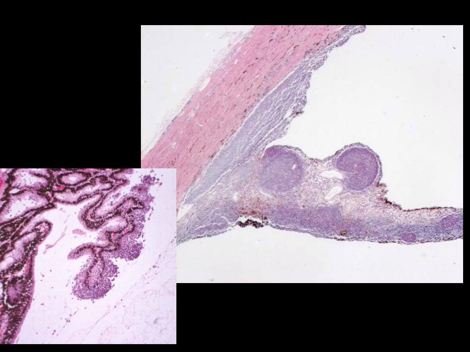
Canine Granulomatous Scleritis82 cases: 12 Cocker Spanials

Canine Granulomatous ScleritisNecrotizing scleritis
• The disease is defined by the scleralpredilection
• You may see granulomatous uveitis, like VKH
• You may see episcleritis, like NGE• 46 Females 31 Males
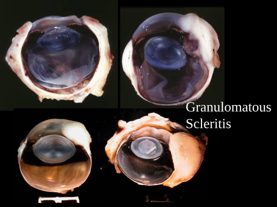
GranulomatousScleritis


Scleritis with Ectasia

Feline Lymphoplasmacytic Uveitis(L/P Uveitis)
• 696 cases out of 6,573 total feline cases (10%)• Second most common disease associated with glaucoma in the
COPLOW pathology collection (70% of L/P uveitis submissions have glaucoma also)
• There is vitreous degeneration that is underemphasized in this disease
• About 10% have lens protein in the anterior vitreous, secondary to rupture/leakage at the level of the posterior pole
• Many possible causes have been studied or suggested, but the cause is surely nonspecific and multifactorial


Alcian Blue PAS
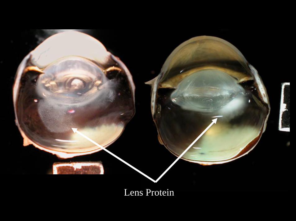
Lens Protein
