uva-dare (digital academic repository) structure … · download date: 28 jun 2018. ... chang c,...
TRANSCRIPT

UvA-DARE is a service provided by the library of the University of Amsterdam (http://dare.uva.nl)
UvA-DARE (Digital Academic Repository)
Structure-function analysis of barley NLR immune receptor MLA10 reveals its cellcompartment specific activity in cell death and disease resistance.Bai, S.; Liu, J.; Chang, C.; Zhang, L.; Maekawa, T.; Wang, Q.; Xiao, W.; Liu, Y.; Chai, J.;Takken, F.L.W.; Schulze-Lefert, P.; Shen, Q.H.Published in:PLoS Pathogens
DOI:10.1371/journal.ppat.1002752
Link to publication
Citation for published version (APA):Bai, S., Liu, J., Chang, C., Zhang, L., Maekawa, T., Wang, Q., ... Shen, Q. H. (2012). Structure-function analysisof barley NLR immune receptor MLA10 reveals its cell compartment specific activity in cell death and diseaseresistance. PLoS Pathogens, 8(6), [e10027]. DOI: 10.1371/journal.ppat.1002752
General rightsIt is not permitted to download or to forward/distribute the text or part of it without the consent of the author(s) and/or copyright holder(s),other than for strictly personal, individual use, unless the work is under an open content license (like Creative Commons).
Disclaimer/Complaints regulationsIf you believe that digital publication of certain material infringes any of your rights or (privacy) interests, please let the Library know, statingyour reasons. In case of a legitimate complaint, the Library will make the material inaccessible and/or remove it from the website. Please Askthe Library: http://uba.uva.nl/en/contact, or a letter to: Library of the University of Amsterdam, Secretariat, Singel 425, 1012 WP Amsterdam,The Netherlands. You will be contacted as soon as possible.
Download date: 28 Jul 2018

Structure-Function Analysis of Barley NLR ImmuneReceptor MLA10 Reveals Its Cell Compartment SpecificActivity in Cell Death and Disease ResistanceShiwei Bai1,2, Jie Liu1,2, Cheng Chang1,2, Ling Zhang1, Takaki Maekawa3, Qiuyun Wang1, Wenkai Xiao1,
Yule Liu4, Jijie Chai4, Frank L. W. Takken5,6, Paul Schulze-Lefert3, Qian-Hua Shen1*
1 State Key Laboratory of Plant Cell and Chromosome Engineering, Institute of Genetics and Developmental Biology, Chinese Academy of Sciences, Beijing, China,
2 Graduate University of Chinese Academy of Sciences, Beijing, China, 3 Department of Plant Microbe Interactions, Max-Planck Institut Pflanzenzuchtungsforschung,
Cologne, Germany, 4 School of Life Sciences, Tsinghua University, Beijing, China, 5 Molecular Plant Pathology, Swammerdam Institute for Life Sciences, University of
Amsterdam, Amsterdam, The Netherlands, 6 Centre for BioSystem Genomics, Wageningen, The Netherlands
Abstract
Plant intracellular immune receptors comprise a large number of multi-domain proteins resembling animal NOD-likereceptors (NLRs). Plant NLRs typically recognize isolate-specific pathogen-derived effectors, encoded by avirulence (AVR)genes, and trigger defense responses often associated with localized host cell death. The barley MLA gene is polymorphic innature and encodes NLRs of the coiled-coil (CC)-NB-LRR type that each detects a cognate isolate-specific effector of thebarley powdery mildew fungus. We report the systematic analyses of MLA10 activity in disease resistance and cell deathsignaling in barley and Nicotiana benthamiana. MLA10 CC domain-triggered cell death is regulated by highly conservedmotifs in the CC and the NB-ARC domains and by the C-terminal LRR of the receptor. Enforced MLA10 subcellularlocalization, by tagging with a nuclear localization sequence (NLS) or a nuclear export sequence (NES), shows that MLA10activity in cell death signaling is suppressed in the nucleus but enhanced in the cytoplasm. By contrast, nuclear localizedMLA10 is sufficient to mediate disease resistance against powdery mildew fungus. MLA10 retention in the cytoplasm wasachieved through attachment of a glucocorticoid receptor hormone-binding domain (GR), by which we reinforced the roleof cytoplasmic MLA10 in cell death signaling. Together with our data showing an essential and sufficient nuclear MLA10activity in disease resistance, this suggests a bifurcation of MLA10-triggered cell death and disease resistance signaling in acompartment-dependent manner.
Citation: Bai S, Liu J, Chang C, Zhang L, Maekawa T, et al. (2012) Structure-Function Analysis of Barley NLR Immune Receptor MLA10 Reveals Its CellCompartment Specific Activity in Cell Death and Disease Resistance. PLoS Pathog 8(6): e1002752. doi:10.1371/journal.ppat.1002752
Editor: Jeffery L. Dangl, The University of North Carolina at Chapel Hill, United States of America
Received November 8, 2011; Accepted April 30, 2012; Published June 7, 2012
Copyright: � 2012 Bai et al. This is an open-access article distributed under the terms of the Creative Commons Attribution License, which permits unrestricteduse, distribution, and reproduction in any medium, provided the original author and source are credited.
Funding: This work was supported by grants from National Basic Research Program of China (2011CB100700), the National Natural Science Foundation of China(31030007 and 30971876), the Chinese Academy of Sciences (KSCX2-YW-N-075), the Max Planck Society, a Deutsche Forschungsgemeinschaft grant (SFB670), andthe European Union BIOEXPLOIT grant (FOOD-CT-2005-513959). The funders had no role in study design, data collection and analysis, decision to publish, orpreparation os the manuscript.
Competing Interests: The authors have declared that no competing interests exist.
* E-mail: [email protected]
Introduction
Plants defend themselves against pathogens by mounting
effective, spatiotemporally fine-tuned immune responses. Two
major types of immune receptors are responsible for pathogen
recognition and subsequent defense induction [1]. One class
comprises membrane-localized pattern recognition receptors that
launch PAMP/MAMP-triggered immunity (PTI/MTI) upon
detection of pathogen/microbe-associated molecular pattern
(PAMP/MAMP). The second type are intracellular disease
resistance (R) proteins that trigger effector-triggered immunity
(ETI) after recognition of pathogen delivered effector proteins
[2,3]. Although PTI/MTI and ETI share some signaling pathways
and induce similar defense responses, ETI is more frequently
associated with the hypersensitive response (HR). The HR is
defined as a localized and rapid cell death response around
attempted pathogen infection sites [4–7].
Plant intracellular R proteins structurally resembling mamma-
lian NOD-like receptors (NLRs) are classified as STAND (signal
transduction ATPases with numerous domains) NTPases [8]. This
class of R proteins share a central conserved NB-ARC domain
that is highly conserved in the human apoptotic regulator APAF-1,
plant R proteins and CED-4 from C. elegans [9]. The NB-ARC
domain is believed to act as a molecular switch that regulates
STAND activity through binding and hydrolyzing nucleotides.
Plant NB-ARC domains consist of three subdomains, called the
NB, ARC1 and ARC2. All three subdomains contain many highly
conserved motifs whose functions have been intensively studied
[10,11]. The P-loop (Walker A) motif in the NB for example, was
shown to be required for nucleotide binding [12–14]. Mutations in
the P-loop motif result in loss-of-function of several NB-LRR
proteins [14–20]. The MHD motif in the ARC2 is predicted to act
as a phosphate sensor and to be involved in nucleotide-dependent
conformational changes [21]. Mutations in the MHD motif lead to
autoactivation of many NB-LRR proteins [18,21–25].
R proteins containing an NB-ARC domain are often referred to
as NB-LRR proteins because most of them carry a C-terminal
Leucine Rich Repeat domain (LRR). For many R proteins the
PLoS Pathogens | www.plospathogens.org 1 June 2012 | Volume 8 | Issue 6 | e1002752
LRR has been shown to determine pathogen recognition
specificity [26–31]. Plant NB-LRR proteins are broadly subdivid-
ed into two subclasses: the TNL and the CNL type. This
classification is based on their N-terminal domains, which either
resembles a TOLL/interleukin-1 receptor (TIR) or forms a coiled-
coil (CC) domain. The N-terminal TIR or CC domains are
thought to mediate downstream signaling, eventually leading to
induction of defense responses. Several lines of evidence are in
support for such a signaling function for the N-terminal domain of
R proteins [25,31–35]. The CC domain is defined as a loosely
conserved structure with at least three variants: CC, CCEDVID or
CCR. The CCEDVID variant is named after the highly conserved
‘‘EDVID’’ motif, which is absent in the other two variants. The
CCR class is named after their founding members the Arabidopsis
RPW8 proteins [36–38]. It is noteworthy that for Rx, a typical
CCEDVID-NB-LRR subtype R protein, its central NB and not its
N-terminal CCEDVID domain is sufficient to induce cell death [39].
For the Nicotiana benthamiana NRG1 and the Arabidopsis ADR1
proteins, both belonging to the CCR-NB-LRR subtype, their CCR
domains alone can trigger cell death [38]. For RPS2, RPS5 and
RPM1, all CC-NB-LRR proteins, it has been shown that their CC
domains are required for ectopic cell death, but it is unknown
whether their CCs alone are sufficient to induce defense signaling
[16,20,24]. For barley MLA10 its CCEDVID domain alone has
been shown to be required and sufficient to induce cell death [25].
Taken together these data do not allow generalities or predictions
on a signaling function for a particular CC domain or a CC
domain type.
The subcellular localization of plant R proteins is important for
their function. Several R proteins were shown to have a dynamic
nucleo-cytoplasmic distribution and to accumulate in the nucleus
in response to pathogen infection [40–42]. Although there are no
discernable nuclear localization signals (NLS) in the barley
MLA10 or the tobacco N proteins, their nuclear localization is
essential for effective resistance [43,44]. In addition, the activity of
the Arabidopsis RPS4, RRS1-R and snc1 have also been associated
with their nuclear localization [45–47]. Two recent studies on
RPS4 revealed that the RPS4-EDS1 signaling complex exists in
both nucleus and cytoplasm and each of these complexes can be
activated by AvrRps4 [48,49]. Strikingly, nuclear activation of
RPS4 by enforced AvrRps4 nuclear localization uncouples the
immune response from cell death signaling, however, full
immunity requires nucleo-cytoplasmic coordination of both
subcellular defense branches [49]. Studies on the potato Rx
protein revealed that its nucleocytoplasmic distribution is balanced
by its N-terminal and C-terminal domains and is facilitated by its
interacting partner RanGAP2 [50,51]. Intriguingly, hyperaccu-
mulation of Rx in the nucleus blocked its cell death signaling and
compromised resistance against PVX; whilst increasing the Rx
cytoplasmic pool by overexpressing RanGAP2 resulted in
potentiated defense signaling, leading to HR in the absence of
PVX-CP and enhanced resistance to PVX [51].
The barley MLA locus is highly polymorphic in nature and has
been subject to extensive functional diversification [52]. MLA
encodes mainly allelic CNL-type R proteins, designated MLA1,
MLA2 etc. Each MLA allele confers isolate-specific disease
resistance against the barley powdery mildew fungus (Blumeria
graminis f. sp. Hordei, Bgh). We have previously shown that MLA1
and MLA10 localize in the nucleus and cytoplasm before and after
Bgh inoculation during compatible and incompatible interactions.
Furthermore, in the nucleus MLA10 interacts with WRKY
transcription factors that act as repressors of MAMP-triggered
basal defenses; and importantly, the MLA10 nuclear pool is
required for disease resistance against the Bgh [44]. MLA proteins
harbor a largely invariant N-terminal CC domain of the CCEDVID
subtype. A recent study revealed that the CC of MLA forms a rod-
shaped homodimer in solution and that a MLA dimer defines the
minimal functional unit required for triggering cell death in barley
and N. benthamiana [25].
In this study, we first conducted structure and function analyses
of the MLA10 protein to better understand the regulation of the
activity of the invariant CC domain in triggering cell death by
using Agrobacterium-mediated transient expression in N. benthamiana
leaves. We then show distinct functions for the nuclear and
cytoplasmic MLA10 pools in disease resistance and cell death
induction. Remarkably, manipulation of cytoplasmic or nuclear
MLA10 levels reveals that the cytoplasmic pool of MLA10 alone is
sufficient to induce cell-death, whereas nuclear-localized MLA10
is unable to induce cell-death, but is capable of conferring disease
resistance.
Results
Only the CC Domain Has Cell Death Signaling Activitythat Is Modulated by Other MLA10 Domains
The CC domain of MLA10 dimerizes in solution and defines
the minimal functional unit required to trigger cell death [25]. To
better understand the involvement of MLA10 domains in cell
death induction a series of MLA10 fragments was generated.
These fragments include the CC, NB, NB-ARC and NB-ARC-
LRR alone, and the CC combined with one or multiple domains:
CC-NB, CC-NB-ARC and CC-NB-ARC-LRR, all fused with
36HA tag (Figure 1A). Upon expression of these fragments in N.
benthamiana leaves and subsequent trypan blue staining or
electrolyte leakage measurements, we observed a cell death
phenotype by expression of the CC domain alone or CC
domain-containing fragments (CC-NB, CC-NB-ARC and full-
length [FL] MLA10). We never observed a cell death phenotype
after expressing MLA10 fragments lacking the CC, i.e. NB, NB-
ARC or NB-ARC-LRR (Figure 1B, 1C). All fusion proteins
accumulated to detectable levels as revealed by immunoblotting
(Figure 1D). To make sure that the C-terminal tags used here or in
Author Summary
Plants utilize a multilayered immune system to protectthemselves against pathogens. One layer of innateimmunity is controlled by intracellular immune receptorscalled disease resistance (R) proteins. Plant R proteins arepowerful molecules capable of triggering host cell suicidethereby restricting pathogen growth. Therefore, it is crucialfor plants to control R protein activity in signaling celldeath to avoid harmful autoimmune responses. The BarleyMLA locus encodes a number of immune receptors thateach recognizes a specific powdery mildew fungal strain.Upon pathogen recognition MLAs trigger host defensesconcomitant with a rapid cell death response. We hereshow that MLA10 cell death-inducing activity is tightlyregulated by conserved motifs located in two of itsdomains and by specific cellular chaperone components.Furthermore, we show distinct functions for the nuclearand cytoplasmic MLA10 pools in disease resistance and celldeath signaling and provide evidence for a modeluncoupling MLA10 cell death signaling from its diseaseresistance activity. Our results suggest that plant immunereceptors integrate signals from multiple sub-cellularcompartments to coordinate effective immune responsesagainst pathogen attack.
Nucleocytoplasmic Dynamics in Cell Death Signaling
PLoS Pathogens | www.plospathogens.org 2 June 2012 | Volume 8 | Issue 6 | e1002752

experiments below do not interfere with functions, we tested cell
death activity of CC or CC-NB fused to different types of tag (Figure
S1). Cell death activity of CC was not affected by small tags such as
36HA or 36Myc, whilst the function of CC-NB retained when
fused to even larger tags, like YFP (Figure S1). These data indicate
that only the CC domain can initiate the cell death response. When
comparing the individual CC-containing fragments, we found that
the CC-NB reproducibly triggered the strongest cell death response.
The CC and CC-NB-ARC triggered comparatively weaker
responses, suggesting that the NB positively regulates cell death
whereas the ARC domain might exert a negatively regulatory effect
(Figure 1B, 1C). Notably, MLA10 FL protein triggered an even
stronger cell death response when compared to that of the CC-NB
(Figure 1B, 1C), indicating that the FL protein is partially auto-
active in the N. benthamiana heterologous expression system. In
summary, the CC domain of MLA10 is necessary and sufficient for
triggering cell death in N. benthamiana, and its activity is modulated
by other MLA10 domains.
Largely Invariant CC Sequence Is Critical for Its CellDeath-Inducing Activity
Previously, seventeen amino acid residues lining the interior
between the protomers of the MLA10 CC dimer were chosen for
Glutamate substitutions to assess their contributions to dimer
stability [25]. Substitutions of most of these 17 residues were
shown to reduce MLA10 self-interaction. In addition, all of these
substitutions, except L18E and F99E, compromised MLA10-
mediated disease resistance [25]. Here we tested whether the same
17 substitutions affect the cell death-inducing activity of the
MLA10 CC domain. We created CC variants each containing a
single substitution, i.e. L11E, L15E, L18E, L19E, F23E, L25E,
V29E, I33E, L36E, M43E, V69E, L72E, I76E, F83E, F99E,
M103E, and L110E. These CC variants, fused to a 36HA tag,
were transiently expressed in N. benthamiana. Significantly, except
for F99E, all substitutions diminished the cell death-inducing
activity of the CC domain (Figure 2A). Most variants accumulated
to similar levels (Figure 2B) although some showed reduced levels
Figure 1. MLA10 CC is the cell death signaling domain whose activity is regulated by other domains. (A) Schematic diagram of theMLA10 domain structure and the derived fragments expressed in N. benthamiana. Individual domains of MLA10 are represented by colored boxes,and the relative positions of relevant amino acids and motifs (in parentheses) are indicated on top (upper panel). Lower panel: MLA10 fragmentsconsisting only of the CC domain or the CC combined with other domains are drawn schematically in solid lines. All fragments were expressed as C-terminal -36HA fusions in N. benthamiana leaves using Agrobacterium-mediated transient transformation (Agro-infiltration). (B) Analysis of cell deathinducing activity of MLA10 fragments. MLA10 fragments or full-length(FL) protein fused with a C-terminal 36HA tag were expressed by Agro-infiltration in N. benthamiana (left), and cell-death triggered by each protein was visualized by trypan blue staining at 42 hrs post Agro-infiltration(hpi) (right). Red circles indicate cell death; white circles indicate no obvious cell death in the infiltrated area. (C) Quantification of cell-death inducingactivity of MLA10 fragments. Upon expression of the same fragment fusions as indicated in (B) by Agro-infiltration in N. benthamiana, electrolyteleakage was measured each hour from 28 to 34 hpi. Error bars were calculated from three replicates per time point and per construct. Experimentswere done at least twice with similar results. Letters (a–d) represent groups with significant differences [p,0.05, Tukey’s honest significant difference(HSD) test]. (D) Protein expression of MLA10 fragments. Total protein was extracted from N. benthamiana leaves at 40 hpi and MLA10-HA wasdetected by immunoblotting using an anti-HA antibody. Asterisk indicates non-specific signals throughout this article except specified. Ponceaustaining of Rubisco small subunit was used to show equal loading throughout this article except specified. EV: empty vector.doi:10.1371/journal.ppat.1002752.g001
Nucleocytoplasmic Dynamics in Cell Death Signaling
PLoS Pathogens | www.plospathogens.org 3 June 2012 | Volume 8 | Issue 6 | e1002752

(M43E, V69 and F99E). Three variants showed a reduced
mobility in the polyacrylamide gel (L72E, I76E and F83E) for
unknown reasons (Figure 2B, lower panel). The F99E substitution
is interesting because it is the only mutation that retained CC cell
death signaling activity. It is also noteworthy that the L18E
substitution abrogated the CC activity leading to cell death, whilst
full-length MLA10 containing the same mutation was shown to
retain wild-type-like disease resistance activity against Bgh [25]. To
exclude that the loss of cell death activity of CC(L18E) might be
due to altered subcellular protein localization, we constructed C-
terminal YFP fusions of CC(L18E) and FL(L18E), whose
expression were driven by 35S or Ubiquitin promoter for transient
expression in N. benthamiana or barley leaf cells, respectively (Figure
S2). As controls we generated CC(F83E)-YFP and FL(K207R)-
YFP fusions, the F83E and the P-loop K207R mutation rendering
MLA10 inactive (Figure 2A, [25], and below Figure 3). Upon
Figure 2. Glutamate substitution analyses in the MLA10 CC domain identify the F99E autoactive mutation. (A) Analysis of cell deathinducing activity of MLA10 CC mutant variants. MLA10 CC wild-type or mutant variants harboring indicated amino acid substitution were expressedin N. benthamiana leaves, and cell death induced by each protein was visualized by trypan blue staining at 42 hpi (upper panel). (B) Proteinexpression levels of each CC variant shown by Western blotting. Proteins were extracted from N. benthamiana leaves at 40 hpi and detection wasdone by immunoblotting with anti-HA antibody. (C) Comparison of cell death inducing activity of MLA10 FL and the FL(F99E) variant. FL and FL(F99E)were expressed on the same N. benthamiana leaf, and the amount of cell death induced by each protein was shown by Trypan blue staining at 24 hpi(upper panel); protein expression levels of FL and FL(F99E) were shown by protein immunoblotting analysis using anti-HA antibody (bottom panel),protein extracts were obtained at ,22 hpi. (D) Quantification of cell-death inducing activity of FL and FL(F99E). Upon expression of FL or FL(F99E) byAgro-infiltration in N. benthamiana, electrolyte leakage was measured each hour from 22 to 30 hpi, and then every two hours from 30 to 34 hpi;empty vector (EV) was included as a negative control. Error bars representing standard error (SE) were calculated from three replicates per time pointand per construct. Similar experiments were repeated at least twice with similar results. Letters (a–c) represent significant differences [p,0.05, Tukey’shonest significant difference (HSD) test].doi:10.1371/journal.ppat.1002752.g002
Nucleocytoplasmic Dynamics in Cell Death Signaling
PLoS Pathogens | www.plospathogens.org 4 June 2012 | Volume 8 | Issue 6 | e1002752
transient expression and confocal imaging we detected for
CC(L18E)-YFP or FL(L18E)-YFP in N. benthamiana or barley cells
similar nucleocytoplasmic distribution patterns compared to the
respective controls (Figure S2). Our data are consistent with
previously proposed sequence constraints acting on the CC
domain for MLA disease resistance [25,52] and assign a critical
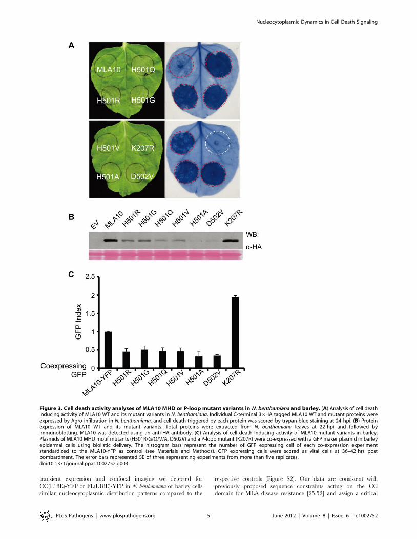
Figure 3. Cell death activity analyses of MLA10 MHD or P-loop mutant variants in N. benthamiana and barley. (A) Analysis of cell deathInducing activity of MLA10 WT and its mutant variants in N. benthamiana. Individual C-terminal 36HA tagged MLA10 WT and mutant proteins wereexpressed by Agro-infiltration in N. benthamiana, and cell-death triggered by each protein was scored by trypan blue staining at 24 hpi. (B) Proteinexpression of MLA10 WT and its mutant variants. Total proteins were extracted from N. benthamiana leaves at 22 hpi and followed byimmunoblotting. MLA10 was detected using an anti-HA antibody. (C) Analysis of cell death Inducing activity of MLA10 mutant variants in barley.Plasmids of MLA10 MHD motif mutants (H501R/G/Q/V/A, D502V) and a P-loop mutant (K207R) were co-expressed with a GFP maker plasmid in barleyepidermal cells using biolistic delivery. The histogram bars represent the number of GFP expressing cell of each co-expression experimentstandardized to the MLA10-YFP as control (see Materials and Methods). GFP expressing cells were scored as vital cells at 36–42 hrs postbombardment. The error bars represented SE of three representing experiments from more than five replicates.doi:10.1371/journal.ppat.1002752.g003
Nucleocytoplasmic Dynamics in Cell Death Signaling
PLoS Pathogens | www.plospathogens.org 5 June 2012 | Volume 8 | Issue 6 | e1002752
role of the invariant CC sequence to its cell death-inducing
activity. Because the tested substitutions are also needed for
efficient CC dimer formation [25], our new data substantiate a
previous suggestion that only the CC homodimer, which has a
characteristic surface charge segregation, is capable to initiate the
cell death response.
The F99E Substitution in the CC Domain AutoactivatesMLA10
To further characterize the F99E substitution, we introduced
this alteration in the MLA10 FL context and compared the cell
death-inducing activity of FL(F99E) to that of MLA10 FL in N.
benthamiana. At 24 hrs post Agrobacterium delivery MLA10 FL
triggered either no cell death or occasionally a weak cell death
response, whereas FL(F99E) induced extensive cell death
(Figure 2C), suggesting that the F99E mutation renders MLA10
highly autoactive. The lower protein accumulation level of
FL(F99E) in the leaf tissue might indirectly reflect its elevated
potency in triggering cell death or could be the consequence of a
higher turnover compared to MLA10 FL (Figure 2C). Ion leakage
quantification further confirmed the significantly elevated cell
death-inducing activity of FL(99E) as compared to FL MLA10
(Figure 2D). In summary, these results identify F99E as a novel
autoactive mutation located in the CC domain of MLA10.
The Role of the MLA10 P-loop and MHD Motif inTriggering Cell Death
The P-loop and the MHD motif (VHDM in MLA) are two
highly conserved motifs with important functions in regulating
NB-LRR protein activity. To better understand the MLA cell
death triggering activity, we generated MLA10 FL variants
carrying point mutations in these two motifs. These include a P-
loop variant FL(K207R) and six MHD variants, FL(H501R),
FL(H501G), FL(H501Q), FL(H501V), FL(H501A) and
FL(D502V). The MLA10 variants were each fused to a C-
terminal 36HA tag and their expression was driven by the 35S
promoter. After Agrobacterium infiltration in N. benthamiana leaves,
the cell-death phenotype for the variants was scored at 24 hrs post
infiltration (hpi) by trypan blue staining. At this time point MLA10
FL normally does not trigger cell death although occasionally
some cell death could be observed (Figure 3A, and below). By
contrast, all MHD variants induced massive cell death, whereas
the K207R P-loop mutant did not trigger any cell death at this or
at later time points. These results indicate that the MHD
mutations confer autoactivity of the receptor whereas the P-loop
mutation abolishes its cell death activity. Immunoblotting showed
that all fusion proteins accumulated to detectable levels in leaf
tissue (Fig. 3B). The lower protein accumulation level observed for
the MHD variants, compared to WT MLA10 and the K207R
variant, likely reflects their stronger cell death-inducing activity or
a higher turnover.
Since N. benthamiana is a heterologous expression system for
barley MLA10, we wondered whether the cell death phenotype
represents a genuine cell death signaling activity of the protein
variants. Therefore, we tested the activity of the same set of
MLA10 variants in barley using a transient gene expression assay
based on DNA-coated gold particle delivery to single plant cells
[28]. In this assay we quantified the number of barley leaf
epidermal cells that express a green fluorescent protein (GFP)
marker 36 to 40 hrs post co-delivery of a plasmid expressing the
individual MLA10 variants under the control of the ubiquitin
promoter. A higher or lower GFP index compared to the wild-type
MLA10-YFP fusion protein (index defined as 1.0) indicates a
weaker or stronger cell death-inducing activity for a given MLA
variant, respectively (Figure 3C). Expression of the FL(K207R) P-
loop variant resulted in a two-fold increase of GFP expressing cells
as compared to the wild-type MLA10-YFP fusion [44] (Figure 3C),
indicating that MLA10 cell death-inducing activity in barley
requires the P-loop motif. In contrast, expression of all MHD
variants led to a markedly reduction of GFP expressing cells by
more than one half as compared to the MLA10-YFP fusion
(Figure 3C), indicating that the MHD variants triggered autoac-
tivation also in barley. The FL(H501A) and FL(D502V) are likely
the two most active variants because their expression reproducibly
resulted in fewer GFP expressing cells than the other variants,
which is consistent with the lowest protein accumulation level for
these two variants in N. benthamiana (Figure 3B, 3C).
In summary, we demonstrated that an MLA10 P-loop mutant
lost its cell death-inducing activity whereas MLA10 MHD variants
triggered massive effector-independent cell death, i.e. autoactiva-
tion, in both barley and N. benthamiana. The functional conserva-
tion of MLA10 cell death-inducing activity in both plant species
suggests that the respective signaling components are likely
evolutionarily conserved across monocotyledonous and dicotyle-
donous plants.
MLA10 Cell Death-Inducing Activity Is Tightly RegulatedTo further examine the regulation of the MLA10-mediated cell
death activity by the P-loop motif, we introduced the K207R P-
loop mutation into the MLA10 N-terminal fragments (CC-NB,
CC-NB-ARC), and also combined this mutation with two
autoactive MHD mutations (H501A or D502V) in MLA10 FL.
The resulting K207R containing variants were then assessed for
their cell death-inducing activity in N. benthamiana (Figure 4A).
Unexpectedly, both CC-NB(K207R) and CC-NB-ARC(K207R)
triggered cell death, and similar to the CC-NB and CC-NB-ARC
fragments described above, the activity of the latter is weaker
(Figure 4A, and 1B). However, just as the FL(K207R), the two FL
variants, FL(K207R/H501A) and FL(K207R/D502V), were no
longer able to induce cell death (Figure 4A). This indicates that the
loss-of-function P-loop motif mutation in the receptor counteracts
the autoactivating effect of the MHD motif mutations.
The MLA R proteins harbor an invariant N-terminal CCEDVID
domain containing the conserved ‘‘EDVID’’ motif (EDVVD in
MLA10). Based on structure modeling it was proposed that the
EDVID motif in MLA10 might be important for receptor post-
activation signaling [25]. To gain more insight about its potential
function, we mutated the EDVVD sequence to AAVVD in three
MLA10 N-terminal fragments: CC, CC-NB and CC-NB-ARC,
and in MLA10 FL, and also combined this mutation with two
autoactive MHD mutations (H501A or D502V) in MLA10 FL,
respectively (Figure 4B). Interestingly, cell death-inducing activity
analyses showed that mutated N-terminal fragments,
CC(AAVVD), CC-NB(AAVVD) and CC-NB-ARC(AAVVD),
retained their cell death-inducing ability as their activity was
comparable to that of the CC, CC-NB and CC-NB-ARC,
respectively (Figure 4B, 1B). In contrast, the full-length variants,
FL(AAVVD), FL(AAVVD/H501A) and FL(AAVVD/D502V),
were unable to induce any cell death (Figure 4B). These results
indicate that MLA10 FL protein, irrespective of WT or MHD
variants, requires an intact EDVID motif for triggering cell death.
Taken together, these results show that MLA10 N-terminal
fragments (i.e. CC, CC-NB or CC-NB-ARC), lacking at least
the C-terminal LRR, are capable of triggering EDVID and P-loop
independent cell death. In contrast, MLA10 FL or FL MHD
mutants fully depend on an intact EDVID and P-loop motif to
trigger cell death.
Nucleocytoplasmic Dynamics in Cell Death Signaling
PLoS Pathogens | www.plospathogens.org 6 June 2012 | Volume 8 | Issue 6 | e1002752

To further test the requirement of EDVID and P-loop motif for
another autoactive mutant, we utilized the newly identified F99E
autoactive variant that carries a mutation in the N-terminal CC
(Figure 2). The F99E substitution was combined in FL MLA with
the K207R and AAVVD mutations, respectively, and the resulting
FL(F99E/AAVVD) and FL(F99E/K207R) variants were com-
pared for cell death activity to a series of controls, including
CC(F99E), CC-NB(F99E), CC-NB-ARC(F99E) and FL(F99E)
(Figure 4C, right panel). At 48 hpi, the F99E containing MLA10
N-terminal fragments, i.e. CC(F99E), CC-NB(F99E) triggered
obvious cell death while CC-NB-ARC(F99E) triggered a weaker
but still discernable cell death. These observations again indicate
the negative modulatory effect the ARC domain has on the CC-
mediated cell death activity. Notably, the F99E autoactivating
effect was strongest in the context of the full-length receptor
FL(F99E). Remarkably, FL(F99E/AAVVD) and FL(F99E/
K207R) did not trigger any visible cell death (Figure 4C, right
panel). These findings indicate that autoactivation in MLA10 FL
F99E-triggered cell death requires an intact EDVID and P-loop
motif.
It has been shown that allelic MLA proteins differentially
engage components of the intracellular chaperone machinery (e.g.
cytosolic HSP90, SGT1 and RAR1) to confer race-specific disease
resistance against the barley powdery mildew fungus [28,53–56].
For example, Barley Stripe Mosaic Virus (BSMV) mediated
silencing of RAR1, SGT1 and HSP90 led to breakdown of MLA13-
mediated resistance against Bgh [56]. The role of RAR1 in
controlling MLA steady state levels has been demonstrated for
both MLA1 and MLA6 [54]. To assess whether these chaperone
components are also required for MLA10-mediated cell death, we
used Tobacco Rattle Virus (TRV)-mediated VIGS to knock down
the expression of the homologous chaperone components in N.
benthamiana [57,58]. Subsequently, the MLA10 cell death inducing
activity was evaluated in these silenced lines using Agrobacterium-
mediated expression (Figure S3). We found that NbSGT1 silencing
eliminated the cell death-triggering activity of MLA10 N-terminal
fragments, including MLA10 CC, CC-NB and CC-NB-ARC, as
well as the FL protein and the two MHD mutant variants tested
(H501A and D502V), suggesting an essential role of NbSGT1 in
MLA10-triggered cell death in this heterologous expression system
(Figure S3, 2nd lane). In contrast to NbSGT1, NbRAR1 silencing
did not affect the cell death-inducing activity for any of these MLA
fragments or the FL proteins, indicating that RAR1 is not essential
for the MLA10-mediated cell death response (Figure S3, 3rd lane).
Interestingly, NbHSP90 is likely required for the MLA10-mediated
cell death, but this requirement could be partially overcome by the
expression of MHD mutant variants that confer a strong receptor
autoactivation (Figure S3, 4th lane).
Distinct Nuclear and Cytoplasmic MLA10 Activities inDisease Resistance and Cell Death Signaling
We have previously shown that MLA10 locates to both the
cytoplasm and the nucleus, and importantly, the nuclear pool is
required for its disease resistance [44]. In this study, we are
interested in how MLA10 subcellular partitioning relates to its cell
death-inducing activity and disease resistance function. First we
generated two fusion constructs, MLA10-YFP-NLS and MLA10-
YFP-nls, whose expression is driven by the Ubiquitin promoter
(NLS is a nuclear localization sequence from SV40 virus, while
‘nls’ is a mutated NLS and serves as a negative control; [59]).
Figure 4. Tightly regulated MLA10 cell death inducing activity. (A) Analysis of cell death triggering activity of MLA10 variants harboring theK207R P-loop mutation. The indicated N-terminal MLA10 fragments containing the K207R mutation, or FL variants harboring K207R alone or K207Rcombined with an MHD mutation (H501, D502V), were expressed in N. benthamiana leaves by Agro-infiltration. Cell death triggered by each fusionwas assessed by trypan blue staining at 42 hpi (upper panel). Protein expression levels of indicated MLA10 mutant fragments are shown (lowerpanel). Proteins were extracted at 40 hpi and detection was done by immunoblotting with anti-HA antibody. (B) Analysis of cell death activity ofMLA10 variants harboring mutations in the EDVID motif. The EDVID motif (EDVVD in MLA10) was mutated to AAVVD, and the indicated MLA10fragments or FL variants harboring indicated mutation(s) were expressed in N. benthamiana leaves by Agro-infiltration. Cell death triggered by eachvariant was assessed by trypan blue staining at 42 hpi (upper panel). Protein expression levels of indicated MLA10 mutant fragments are shown(lower panel). Proteins were extracted at 40 hpi and detection was done by immunoblotting with anti-HA antibody. (C) Analysis of cell death activityof F99E containing MLA10 fragments or FL variants. Indicated MLA10 fragments or FL variants harboring indicated mutation(s) were expressed in N.benthamiana leaves by Agro-infiltration. Cell death triggered by each protein was scored by trypan blue staining at 42 hpi (upper panel); Proteinwere extracts from N. benthamiana leaves and subjected to immunoblotting with anti-HA antibody (lower panel).doi:10.1371/journal.ppat.1002752.g004
Nucleocytoplasmic Dynamics in Cell Death Signaling
PLoS Pathogens | www.plospathogens.org 7 June 2012 | Volume 8 | Issue 6 | e1002752

Upon expression of MLA10-YFP-NLS in barley leaf epidermal
cells, YFP-derived fluorescence was exclusively detected in the
nucleus. The YFP signal overlapped with that of a nuclear marker
protein, CFP-WRKY2 [44], confirming its nuclear localization.
The fluorescence signal of the MLA10-YFP-nls variant clearly
partitioned to both nucleus and cytoplasm, similar to that of
MLA10-YFP (Figure 5A, and [44]). This experiment demonstrat-
ed that the NLS functions to localize MLA10-YFP into plant cell
nuclei. We wondered whether the nuclear localized MLA10-YFP-
NLS confers disease resistance against Bgh. To address this, we
utilized the single-cell transient expression assay [44], in which we
delivered gold particles coated with DNA plasmids coexpressing a
GUS reporter and either MLA10-YFP-NLS or MLA10-NLS into
barley epidermal cells. Subsequently, the frequency of fungal
structures formed inside the transformed cells (haustorium index;
[44]) was scored upon inoculation with a Bgh race carrying the
cognate AVRA10 effector. Included in these experiments are
control plasmids expressing MLA10-YFP and the MLA10(K207R)
P-loop mutant, respectively (Figure 5B). As expected the MLA10-
YFP fusion conferred resistance against Bgh infection, resulting in
an haustorium index around 29%, similar to our previous study
[44]. The K207R P-loop mutation reduced MLA10 resistance
Figure 5. Manipulation of MLA10 subcellular localization and the relevance to MLA10 cell death signaling and disease resistance.(A) Confocal images of barley leaf epidermal cells expressing MLA10 fusion proteins. Indicated fusion proteins were expressed in barley leafepidermal cells upon biolistic delivery, the confocal images were taken at 36 hrs post bombardment. Upper panel: A representative barley cellcoexpressing MLA10-YFP-NLS and a nucleus marker CFP-WRKY2 (2D z plane). Lower panel: a cell expressing MLA10-YFP-nls alone (2D z plane). NLS:nuclear localization signal; nls: mutated nuclear localization signal. Arrowheads mark the nucleus and scale bar is 50 mm. (B) Analyses of diseaseresistance activity of MLA10 fusions or mutant variants. Relative single cell resistance/susceptibility is shown by fungal haustorium index uponbiolistic delivery of plasmids expressing indicated protein and a GUS reporter into the barley leaf epidermal cells of a susceptible barley line (Goldenpromise). Bombarded leaves were inoculated with B. graminis fungal spores expressing AVRA10, and the fungal haustorium index was microscopicallyscored at 36 hrs post spore inoculation. Histogram bar represents average of three independent experiments and error bar represents SD. In the caseof MLA10(F99E) expression, the number of cells expressing GUS reporter were extremely low. (C) Analysis of cell death triggering activity of MLA10fusion proteins. Indicated MLA10 fusion proteins were expressed in N. benthamiana leaves by Agro-infiltration, and cell-death triggered by eachfusion protein was scored by trypan blue staining at 40 hpi. NES: nuclear exclusion signal; nes: mutated nuclear exclusion signal. (D) Proteinexpression of indicated MLA10 fusions. Proteins were extracted at 23 hpi and MLA was detected by immunoblotting using an anti-MLA27monoclonal antibody. Asterisk indicates non-specific signals. (E) Quantification of cell-death inducing activity of MLA10 fusion proteins. Uponexpression of indicated MLA10 fusion proteins by Agro-infiltration in N. benthamiana, ion leakage was measured each hour from 23 to 34 hpi. Errorbars (SE) were calculated from three replicates per time point and construct. Experiment was done at least twice with similar result. Letters (a–c)represent groups with significant differences [p,0.05, Tukey’s honest significant difference (HSD) test].doi:10.1371/journal.ppat.1002752.g005
Nucleocytoplasmic Dynamics in Cell Death Signaling
PLoS Pathogens | www.plospathogens.org 8 June 2012 | Volume 8 | Issue 6 | e1002752

activity as indicated by a haustorium index as high as 65%. In
contrast, MLA10-YFP-NLS and MLA10-NLS fusions mediated
similar levels of Bgh resistance as the MLA10-YFP control, with
haustorium indexes of 35% and 30% respectively (Figure 5B).
These data indicate that MLA10, forced to localize into the
nucleus, is capable of triggering resistance to Bgh. Previously, we
observed a haustorium index of 52% for the MLA10-YFP-NES
fusion, which was undetectable in the nucleus. Since a similar
haustorium index was obtained for the empty vector control, this
indicates its loss-of-function against Bgh [44]. Together, these data
suggest that the MLA10 nuclear pool is required for and capable
of mediating disease resistance against the Bgh fungus in barley.
In order to link MLA10-mediated cell death activity to a specific
subcellular receptor pool, we switched to the N. benthamiana
transient expression system. We made two pairs of MLA10
fusions, MLA10-NES vs. MLA10-nes (NES is a nuclear export
sequence, ‘nes’ is a mutated nonfunctional NES; [44]) and
MLA10-NLS vs. MLA10-nls. Expression of all MLA constructs
was driven by the 35S promoter (Figure 5C). Remarkably,
MLA10-NLS was unable to trigger cell death whereas MLA10-nls
retained cell death-inducing activity. Furthermore, expression of
MLA10-NES triggered a cell death response that was more severe
than the one initiated by MLA10-nes, MLA10-nls, and MLA10-
36HA at 36–40 hpi (Figure 5C). The stronger cell death activity
of MLA10-NES is not due to higher protein accumulation as our
anti-MLA27 antibody detected similar accumulation levels for
these fusion proteins (Figure 5D) (anti-MLA27 is a monoclonal
antibody raised against purified MLA27 that most likely cross-
reacts with a conserved epitope between MLA10 and MLA27).
Independent ion leakage measurements confirmed the results
obtained from the trypan blue staining assay and showed that the
stronger cell death-inducing activity of MLA10-NES was detect-
able at even earlier time points, compared to the WT receptor,
starting from around 24 hpi (Figure 5E). To corroborate these
data obtained from N. benthamiana, we decided to test in barley the
cell death inducing activity of MLA10-NES and MLA10-NLS
through the transient cell death assay used above (Figure 3C),
included in this assay as controls were the autoactive D502V
variant and the K207R non-functional P-loop variants (Figure S4).
Significantly, in barley epidermal cells we observed for MLA10-
NLS little cell-death inducing activity similar as the K207R, by
contrast, for MLA10-NES potentiated cell-death inducing activity
similar as the D502V (Figure S4).
It is intriguing that MLA10-NLS does not trigger cell-death,
while the same construct confers full Bgh resistance. One
explanation might be that the receptor needs to be activated for
cell-death induction. Since MLA10 mediate race-specific resis-
tance to Bgh isolates expressing the cognate effector AVRA10 [60]
we speculated that co-expression of AVRA10 in the nucleus might
activate MLA10. Since the NLS variant, in contrast to the non-
tagged MLA is not autoactive, this became testable. Therefore, we
made an AVRA10-YFP fusion and examined its subcellular
localization and ability to induce cell-death in N. benthamiana leaf
cells upon Agro-infiltration. Confocal imaging revealed accumu-
lation and distribution of the AVRA10-YFP fusion over both the
nucleus and the cytoplasm (Figure S5, left panel). Expression of
AVRA10-YFP alone did not induce cell-death, but also coexpres-
sion of AVRA10-YFP with MLA10-NLS did not induce cell-death
in N. benthamiana (Figure S5, right panel).
To further study the subcellular localization of MLA10 in N.
benthamiana, we constructed MLA10 CC-NB-YFP and MLA10-
YFP fusions with a C-terminal YFP sequence. The expression of
both constructs was driven by a 35S promoter. Upon Agro-
infiltration in N. benthamiana, we compared the fluorescence signal
as well as the cell death phenotype of CC-NB-YFP with MLA10-
YFP. Confocal imaging revealed that both fusion proteins
localized to the nucleus and the cytoplasm but the CC-NB variant
had a brighter YFP fluorescence signal and a clearer nuclear
localization than full-length MLA10, possibly due to differences in
protein folding and/or protein turnover (Figure S6). Since both
fusions triggered cell death, we decided to use CC-NB-YFP instead
of MLA10-YFP in the following experiments. We fused the C-
terminus of CC-NB-YFP to either an NES or NLS, and upon
expression confocal imaging showed that in most cells the CC-NB-
YFP-NES proteins localized to the cytoplasm, whilst the CC-NB-
YFP-NLS proteins exclusively localized in the nuclei (Figure 6A,
upper panel). Significantly, in cell death staining assays, CC-NB-
YFP-NES induced a much stronger cell death phenotype as
compared to the CC-NB-YFP fusion, whereas CC-NB-YFP-NLS
did not trigger any cell death (Figure 6A, lower panel). All these
fusion proteins accumulated to similar levels (Figure S7). These
results are consistent with those obtained with the MLA10 full-
length protein tagged with NES/NLS or nes/nls (compare to
Figure 5C).
In summary, the nuclear MLA10 pool alone cannot induce cell
death although it is sufficient to trigger disease resistance.
Accelerated MLA10 exclusion from the nucleus and/or enhanced
nucleocytoplasmic shuttling (i.e. in the NES tagged configuration)
enhanced MLA10-mediated cell death activity, but was shown
before to exhibit compromised MLA10-mediated disease resis-
tance activity (Figure 1 in [44]). An unanswered question is
whether the cytoplasmic MLA10 pool alone is sufficient to induce
cell death.
MLA10 FL Retained in Cytoplasm Does Trigger Cell DeathSignaling
Because the mammalian glucocorticoid receptor (GR) with a
hormone binding domain is folded into a larger HSP-containing
complex in the cytosol, it is possible to use this domain to retain
GR protein fusions in this cellular compartment and study their
cytoplasmic functions [61]. This strategy has also been successfully
used in plants [62–64]. Therefore, to particularly address whether
the MLA10 cytoplasmic portion is sufficient for cell death
signaling, we constructed CC-NB-YFP-GR fusions under the
control of the 35S promoter for expression in N. benthamiana.
Around 6 hrs after Agrobacterium infiltration we sprayed N.
benthamiana leaves with a buffer with or without dexamethasone
(Dex) and examined these leaves using confocal imaging after
,14 hrs. In the controls without Dex treatment, the YFP
fluorescence signals were largely restricted to the cytoplasm in
the majority of cells; expectedly, after Dex treatment the YFP
fluorescence signals were markedly intensified in most nuclei,
indicating Dex-induced nuclear relocation of a portion of CC-NB-
YFP-GR fusion (Figure 6B, left panel). In leaves expressing CC-
NB-YFP-GR, without applying a Dex treatment, no obvious cell
death could be observed, whereas upon Dex treatment cell death
was markedly induced in CC-NB-YFP-GR expressing leaves. The
extent of cell death for the latter was comparable to that of a CC-
NB-YFP control (Figure 6A left, 6B left panel). Compared to the
enhanced cell-death activity of CC-NB-YFP-NES, the lack of cell-
death-inducing activity of CC-NB-YFP-GR in the absence of Dex
induction could be explained by the need for a nuclear MLA
fraction to exert this activity. To examine this possibility, we co-
expressed the CC-NB-YFP-NLS and CC-NB-YFP-GR fusion
proteins in the same cells to accomplish localization of the CC-NB-
YFP in both the nucleus as well as in the cytoplasm (Figure 6B,
right panel). Upon coexpression of CC-NB-YFP-NLS and CC-
NB-YFP-GR, strong YFP signals could easily be detected in both
Nucleocytoplasmic Dynamics in Cell Death Signaling
PLoS Pathogens | www.plospathogens.org 9 June 2012 | Volume 8 | Issue 6 | e1002752

the nucleus and the cytoplasm in the same cell in the absence of
Dex, yet no obvious cell death was observed in the staining assays
(Figure 6B, right panel). Again, upon Dex treatment, cell death
was induced (Figure 6B, right panel). This finding indicates that,
even in the presence of nuclear-localized CC-NB-YFP-NLS,
cytoplasmically retained CC-NB-YFP-GR cannot induce cell
death. Since the GR steroid-binding domain is folded by the
large HSP-complex in the cytoplasm it is possible that the inability
of the CC-NB-YFP-GR fusion to induce cell-death was due to
steric hindrance by this chaperone complex. To investigate this
possibility we made GR fusions to MLA10 FL and MLA10-YFP to
have the endogenous 15 LRRs flanking the CC-NB to act as linker
and thereby relieve potential hindrance. The F99E and D502V
autoactive mutations were also introduced in MLA10-YFP-GR to
make FL(F99E)-YFP-GR and FL(D502V)-YFP-GR fusion con-
structs. Upon Agro-infiltration and expression of the MLA10 FL
fusions in N. benthamiana leaves MLA10-YFP-GR was retained in
the cytoplasm in the absence of Dex induction, similar to CC-NB-
YFP-GR (Figure S8, Figure 6B). Unexpectedly, in the absence of
Dex MLA10-GR and MLA10-YFP-GR induced cell-death at
similar levels, whereas the two autoactive fusions, FL(F99E)-YFP-
GR and FL(D502V)-YFP-GR induced a stronger cell-death
response as compared to the WT fusions (Figure 6C). These
results suggest that MLA10 FL proteins retained in the cytoplasm
Figure 6. MLA10 CC-NB or full-length trigger cell death signaling in the cytoplasm. (A) Analysis of cell death inducing activity of MLA10CC-NB fusion proteins. MLA10 CC-NB fusion proteins, CC-NB-YFP, CC-NB-YFP-NES and CC-NB-YFP-NLS, were expressed in N. benthamiana leaves byAgro-infiltration; confocal images were taken at ,22 hpi (upper panel) and cell-death triggered by each fusion protein was scored by trypan bluestaining at ,48 hpi (lower panel). Scale bar is 50 mm. (B) Analysis of cell death phenotype upon expression of MLA10 CC-NB-YFP-GR or co-expressionof MLA10 CC-NB-YFP-NLS and CC-NB-YFP-GR fusions before and after Dex treatment. The indicated fusion(s) were expressed in N. benthamianaleaves by Agro-infiltration. Buffer with/or without Dex was sprayed onto N. benthamiana leafs 20 hpi before the confocal images were taken (upperpanel). The cell-death phenotype of each fusion protein was scored by trypan blue staining at ,48 hrs post treatment with/or without Dex (lowerpanel). GR: Steroid binding domain of the mammalian glucocorticoid receptor. Scale bar is 50 mm. (C) Analysis of cell death activity of GR fusions ofMLA10 WT and autoactive variants. Indicated MLA10-GR fusion, and the C-terminal YFP-GR fusions of MLA10 WT and two autoactive mutant variantswere expressed in N. benthamiana leaves by Agro-infiltration. The cell-death phenotype was revealed by trypan blue staining at ,24 hrs postinfiltration without Dex induction.doi:10.1371/journal.ppat.1002752.g006
Nucleocytoplasmic Dynamics in Cell Death Signaling
PLoS Pathogens | www.plospathogens.org 10 June 2012 | Volume 8 | Issue 6 | e1002752

are capable of triggering cell-death, while truncated forms lacking
the LRR have to be released from the GR complex in order to
signal cell death.
Discussion
The Role of the CC Domain and the Regulation of MLA10Cell Death-Inducing Activity
Expression of MLA10 FL or CC containing fusions can induce
AVR-independent cell death by Agrobacterium-mediated transfor-
mation in N. benthamiana leaf cells (Figure 1, 2, and 3). We believe
that this partial auto-activation of MLA10 is unlikely due to an
overexpression effect since we did not observe a direct correlation
between protein stead-state levels and cell death activity (Figure 1
and 2C). Nevertheless, because for the FL receptor the cell death
response was shown to be entirely dependent on an intact P-loop
motif in the NB domain and the EDVID motif in the CC domain
(Fig. 3 and 4), our data imply that cell death signaling via the CC
domain is powered by receptor NTP hydrolysis and that pro-death
signaling components are evolutionarily conserved between
monocot and dicot plants.
The MLA proteins belong to the CCEDVID-NB-LRR subclass of
CNLs, whose members share the conserved EDVID motif [37,52].
Although the CCEDVID of several CNLs is required for ectopic cell
death, only the CCEDVID of MLA10 has been shown to date to be
sufficient for the induction of cell death signaling [16,20,24,25].
Our truncation and mutagenesis analyses confirmed that the
MLA10 CC domain is required and sufficient for triggering AVR-
independent, but SGT1-/HSP90-dependent cell death, pointing
to its role as a pro-cell death signaling domain (Figure 1, 2, 4, and
S3). This activity is likely dependent on a unique surface charge
segregation established upon CC homodimerization (Fig. 2 and
[25]). Such a signaling function is in contrast to some studies that
assign a specific role in pathogen recognition for the N-termini of
CNL proteins [20,39,65,66]. In the case of the Rx protein,
conferring resistance against potato PVX, its NB domain was
shown to mediate cell death signaling [39]. Thus, it appears that
immune receptors of the CCEDVID-NB-LRR subclass engage
distinct domains for triggering downstream cell death signaling.
What could be the role of the EDVID motif in MLA proteins?
Our data show that this motif plays an important role in MLA10-
triggered cell death signaling since it is needed for this activity of
MLA10 FL and autoactive variants (both for the MHD and the
F99E mutation; Figure 4B, 4C). While the EDVID motif in Rx
appears to be involved in mediating intramolecular interactions
between CC and the NB-ARC-LRR fragment [39], there is no
direct evidence to support a similar function in MLA; nevertheless,
molecular dynamics (MD) simulation of the MLA CC dimer
shows that its EDVID motif could be involved in both intra- and
intermolecular receptor interactions [25]. We could not resolve in
vivo the role of the EDVID motif in MLA10 intramolecular
interactions since we failed to detect an interaction between the
CC and the NBARC-LRR of MLA10 by Co-IP, although we
could reproduce the Rx intramolecular interactions in the N.
benthamiana system (data not shown). An explanation might be that
the interaction between MLA10 CC and NBARC-LRR domains
is too weak/or transient to be detected. However, considering that
the MLA10 CC does not only mediate MLA10 homodimerization
but also associates with different molecules (for example the
WRKY TFs, and other TFs according to our unpublished data)
for downstream signaling, it is also possible that MLA10 undergoes
different inter/intra-molecular interactions than those reported for
other NB-LRR proteins.
Mutagenesis and biochemical studies have indicated that the
NB-ARC domain acts as a molecular switch, whose status of
nucleotide binding and hydrolysis is critical for regulating plant
NB-LRR protein activity [12–14,19]. We mutagenized the
conserved P-loop and the MHD motifs in the MLA10 NB-ARC
domain to examine their function in MLA10 cell death-inducing
activity in both barley and N. benthamiana (Figure 3 and 4). Our
data support the current NB-LRR protein activation model
wherein the P-loop is involved in nucleotide binding and the
MHD relays nucleotide-dependent conformational changes
[10,21]. Subsequently, we combined the P-loop mutation
(K207R) with two MHD autoactivation mutations (H501A or
D502V, respectively) to test the combined effect of double
mutations on MLA10 cell death-inducing activity (Figure 4A).
These MLA10 double mutants completely lost their cell death-
inducing activity, consistent with the proposed model that MHD
mutation-induced autoactivation mimics the nucleotide-dependent
NB-LRR resistance protein activation that is normally triggered by
pathogen-derived AVR effectors [19,24]. It is possible that the
MLA10 receptor autoactivation by MHD motif mutations needs
bound NTP and that this NTP binding is compromised in
conjunction with the mutated P-loop motif.
The MLA10 N-terminal fragments (CC, CC-NB, CC-NB-
ARC) induce P-loop- or EDVID-independent cell death, whereas
the MLA10 WT or autoactive FL mutant variants (both the MHD
mutations or the F99E substitution) do require an intact P-loop
and EDVID motif to induce cell death (Figure 4). These data
suggest that the N-terminal fragments might mimic an MLA10
post-activation state that does no longer require the P-loop
function. It also indicates that the C-terminal LRR region of
MLA10 plays a negative regulatory role in MLA10 cell death-
inducing activity. It has been observed for several other NB-LRR
proteins that the LRR domain negatively regulates disease
resistance activity [16,20,29,33].
The Role of Nuclear and Cytoplasmic MLA10 Pools in CellDeath Signaling
To dissect the relation between the MLA10 subcellular
localization and cell death signaling activity, YFP fusions of
MLA10 FL and the CC-NB fragment were examined for their
subcellular localization. Not surprisingly, both fusions exhibited a
similar nucleocytoplasmic distribution in N. benthamiana leaf cells
(Figure 6, and S6). The utilization of NLS/NES and GR
sequences allowed us to examine the individual functions of the
MLA10 nuclear and cytoplasmic pools for cell death induction.
Significantly, enforced retention of full-length MLA10 or CC-NB
YFP fusions in the nucleus blocked its cell death-triggering activity
(Figure 5C, 6A). This finding suggests that either this MLA10
activity is inhibited in the nucleus or the mere nuclear localization
of MLA10 is insufficient for triggering cell death, or both.
Conversely, enhanced cytoplasmic localization of MLA10 FL or
the CC-NB fragment by fusing them to a NES signal clearly
increased their cell death-inducing activities (Figure 5C, 6A). This
enhanced receptor activity is likely attributable to enhanced
nuclear export- over import-rates (Figure 5C, 6A), implicating the
cytoplasm as compartment for MLA10 to initiate and/or amplify
post-activation cell death signaling. Furthermore, cytoplasmic
retention of YFP fusions of full-length MLA10 WT or two
autoactive mutant variants did not block their cell death-inducing
activity (Figure 6C), again suggesting that MLA10 triggers cell-
death signaling in the cytoplasm. In this context, the failure of
cytoplasmic CC-NB-YFP-GR to stimulate cell death in the
absence of Dex possibly reflects its enforced physical association
with the chaperone/folding machinery (i.e., direct binding
Nucleocytoplasmic Dynamics in Cell Death Signaling
PLoS Pathogens | www.plospathogens.org 11 June 2012 | Volume 8 | Issue 6 | e1002752

between the GR hormone binding domain and HSP90; [67]).
Along the same line, resumed cell death activation after Dex
treatment demonstrates that CC-NB-YFP-GR is per se signaling
competent, at least after its release from the cytoplasmic
chaperone/folding machinery.
Implications for MLA10 Activation and SignalingWe previously demonstrated that the MLA10-YFP-NES fusion
protein was compromised for MLA10 disease resistance activity
against the powdery mildew fungus due to depletion of the nuclear
receptor pool [44], suggesting that initiation of disease resistance
signaling demands either a threshold level of the receptor or
prolonged residence in this compartment. Together with the
finding that activated MLA10 interacts with HvWRKY1/2 in the
nucleus [44], we reasoned that enforced nuclear localization of the
receptors should be capable of triggering disease resistance
signaling. Indeed, upon transient expression in barley cells
MLA10-YFP-NLS fusion was found to localize exclusively in the
nucleus, and importantly both MLA10-YFP-NLS and MLA10-
NLS fusions confer disease resistance against Bgh in barley
(Figure 5A, 5B). In this context, it is worth noting that the activity
of cell death induction of MLA10 was enhanced for MLA10-NES
or CC-NB-YFP-NES fusions compared to the non-NES-tagged
versions. This enhanced receptor activity is likely attributable to
enhanced nuclear export over import rates (Figure 5C, 6A),
implicating the cytoplasm as compartment for MLA10 to initiate
and/or amplify post-activation cell death signaling. To integrate
our findings in a model, we propose a bifurcation of MLA10
disease resistance and cell-death signaling in a compartment-
dependent manner, thereby ‘uncoupling’ disease resistance
signaling from cell death induction. In this model, the nuclear
MLA10 pool triggers disease resistance signaling while the
cytoplasmic MLA10 pool induces cell-death signaling. Two
additional pieces of evidence support this model. First, the CC
domain containing the L18E substitution has diminished cell
death-inducing activity (Figure 2A) but FL MLA10 containing the
same mutation retains full disease resistance activity against the
cognate Bgh isolate [25]. This difference is not attributed to altered
subcellular distribution since CC(L18E)-YFP and FL(L18E)-YFP
localized to both nucleus and cytoplasm in N. benthamiana or barley
leaf cells respectively, similar to that of other mutants, for example,
the CC(F83E) or FL(K207R) P-loop mutants (Figure S2). Second,
RAR1 is required for MLA10 disease resistance function but not
for its cell death induction ([55]; Figure S3). Our model for MLA
functional bifurcation in disease resistance and cell-death signaling
appears distinct from Rx [50,51]. Rx hyperaccumulation in the
nucleus blocks not only cell-death but also resistance to PVX
whereas MLA10 enforced nuclear localization suppresses only the
cell-death signaling but not resistance to Bgh.
It has been shown that different types of plant disease resistance
proteins can be activated at diverse locations, for example, at the
membrane and in the cytoplasm or the nucleus or both
[24,35,41,48–50]. In this context, it is interesting that coexpression
of AVRA10 with MLA10-NLS did not induce cell-death in N.
benthamiana (Figure S5). It is possible that activated MLA10-NLS
might be suppressed in the nucleus for triggering cell-death. It may
also suggest that MLA10 activation by AVRA10 specifically occurs
in the cytoplasm and thus MLA10-NLS could not be activated in
the nucleus of N. benthamiana leaf cells. Unfortunately, we cannot
test this possibility in N. benthamiana by coexpressing AVRA10 with
MLA10-NES or MLA10-GR since these fusions themselves trigger
cell-death.
Our model also implies an important role of nucleocytoplasmic
distribution in both defense and cell-death signaling. One scenario
would be that activated MLA distributes across the nuclear
envelope so that a portion reaches its cytoplasmic destination to
trigger cell death signaling whereas another portion associates with
nuclear targets such as HvWRKY1/2 to induce disease resistance
responses. The notion that nucleocytoplasmic distribution is
important for defense signaling is in line with other studies
[42,68–70], for example, the nucleocytoplasmic partitioning of
potato Rx [48,49,68,69], and the nucleocytoplasmic localization of
several other plant immune receptors and immune regulators, e.g.
tobacco N, Arabidopsis RRS1-R, RPS4 and snc1 [43,45–47,71],
as well as Arabidopsis NPR1 and EDS1 [64,72]. The recent
studies linking EDS1-RPS4 interaction with immune receptor
activation consolidate the notion that proper nucleocytoplasmic
partitioning of immune regulators/receptors is important for
coordinated defense responses and support the existence of cell
compartment-specific functions for immune receptors [48,49].
Future experimentation will be needed to resolve where MLA is
activated inside host cells, directly or indirectly, and to identify
more targets of activated MLA in the cytoplasm and the nucleus.
Materials and Methods
Plant MaterialsBarley (Hordeum vulgare L.) cultivar Golden Promise plants were
grown in a growth chamber under a 16 hrs/8 hrs, 20uC/18uCday/night cycle with 70% relative humidity. Nicotiana benthamiana
plants were grown in greenhouse at 2461uC with a 16 hrs light
period.
Plasmid ConstructionAll oligonucleotide primers used in this study (marked SW66)
were purchased from Invitrogen Life Technologies (Beijing,
China), and are listed in supplemental Table S1. The following
gateway (GW) binary vectors derived from CTAPi [73] for
providing C-terminal tagged fusions were used in this study:
CTAPi-GW-36HA, CTAPi-GW-YFP, CTAPi-GW-mYFP and
CTAPi-GW-36Myc. To create these vectors, either 36HA, YFP,
mYFP (monomeric YFP), or 36Myc epitope tag followed by a stop
codon was inserted into CTAPi at the unique SpeI site.
Construction of plasmid pUbi-MLA10-YFP for expression of
MLA10-YFP fusion proteins under the control of the Ubiquitin
promoter was described previously [44]. MLA10 MHD motif
mutants (H501G/R/Q/V/A, D502V) and the P-loop mutant
(K207R) were generated by overlap extension PCR [74,75] using
two sets of primer pairs indicated in supplemental Table S2 on
template pUbi-MLA10-YFP. The consequent two PCR products
were joined in a second amplification step using primer pairs
SW09/SW10. A 1564-bp restriction fragment generated with AgeI
and BbsI containing the desired mutation was used to replace the
MLA10 wild-type sequence in pUbi-MLA10-YFP to obtain
MLA10 variants FL(H501G), FL(H501R), FL(H501Q),
FL(H501V), FL(H501A), FL(D502V) or FL(K207R) (FL for full-
length). The MLA10 FL wild-type and its mutant variants were
then transferred to the entry clone pDNOR201 by Gateway BP
reactions (Invitrogen) and subsequently to the binary vector
CTAPi-GW-36HA to obtain C-terminally 36HA-tagged MLA10
fusions.
MLA10 CC (wild-type/mutants), CC-NB, CC-NB-ARC, NB,
NB-ARC, and NB-LRR fragments were amplified from pUbi-
MLA10-YFP using primer pairs SW56/SW55, SW56/SW58,
SW56/SW33, SW32/SW58, SW32/SW33, SW32/SW20, re-
spectively. The single amino acid substitutions in MLA10 CC
domain have been described before [25]. The resulting PCR
products were cloned into pDNOR201 via a Gateway BP reaction
Nucleocytoplasmic Dynamics in Cell Death Signaling
PLoS Pathogens | www.plospathogens.org 12 June 2012 | Volume 8 | Issue 6 | e1002752

and subsequently to CTAPi-GW-36HA, generating C-terminally
36HA-tagged MLA10 fragments and MLA10 CC mutant variants.
MLA10 CC, CC variants or CC-NB fused with a C-terminal TAP,
YFP, mYFP or 36Myc epitope tag was constructed based on
CTAPi or its derivative vectors as described above. AVRA10 coding
sequences were amplified from the vector pPS1(AVRA10) [60] with
primers SW106/SW107. The amplification products were cloned
into pDNOR201 via a Gateway BP reaction and subsequently to
CTAPi-GW-YFP to create CTAPi- AVRA10-YFP.
GR fusions were generated based on the vector pBI-GR [62].
YFP fragments were amplified by PCR using primer pairs SW80/
SW75, and introduced into the XbaI/BamHI sites of pBI-GR to
create pBI-YFP-GR. YFP-GR and GR fragments were prepared
by amplification from pBI-YFP-GR with primers SW80/SW81
and SW82/SW81, and inserted into the unique SpeI site of
CTAPi, yielding a Gateway-compatible vector CTAPi-GW-YFP-
GR and CTAPi-GW-GR. MLA10 FL, FL(F99E), FL(D502V) and
MLA10 CC-NB fragments were then recombined into CTAPi-
GW-YFP-GR and CTAPi-GW-GR via a Gateway LR reaction to
obtain CTAPi-MLA10-YFP-GR, CTAPi-FL(F99E)-YFP-GR,
CTAPi-FL(D502V)-YFP-GR, CTAPi-CC-NB-YFP-GR and
CTAPi-MLA10-GR. For subcellular localization analysis, the
SV40 T-Ag NLS (QPKKKRKVGG)/PK1 NES (NELALK-
LAGLDINK) or their mutant nls (QPKKTRKVGG)/nes (NE-
LALKAAGADANK) were fused to the C-terminus of MLA10-
YFP or CC-NB-YFP by PCR using overhanging primers. The
cloning details are available upon request.
Single-Cell Transient Gene Expression AssaySingle-cell transient gene expression assays using biolistic delivery
of plasmid DNA into barley epidermal cells were described
previously [28]. A reporter plasmid (pUbi-GFP) and plasmids
expressing individual MLA10 or its mutant variants controlled by
the Ubiquitin promoter were mixed before coating of gold particles
(molar ratio of 1:1; 2.5 mg of total DNA). Single leaf epidermal cells
were transformed with a particle inflow gun of the model PDS-
1000/He (Bio-Rad). In the experiments analyzing cell-death activity
of MLA10 variants, all constructs were co-expressed with a GFP
marker. The numbers of GFP expressing cells were scored at 36–
42 hrs post bombardment using fluorescence microscope, which
represents viable cells. The number of GFP cells, obtained after
biolistic delivery of the control vector harboring functional MLA10-
YFP and a GFP marker, were used as cell-death activity reference
thus standardized to GFP Index equivalent to 1.000. GFP cell
numbers from other constructs in the same experiments were
standardized accordingly. To minimize the variations in the same
experiments, same amounts of gold particles and DNA for coating
were used and the barley leaves were also selected so that the leaf
area to receive the particles was roughly same.
Agrobacteria-Mediated Transient ExpressionAgrobacteria-mediated transient expression (Agro-infiltration)
using Agrobacterium tumefaciens strain GV3101 carrying the indicated
constructs was performed as described by van Ooijen et al. [21].
Agrobacteria suspensions were infiltrated at OD600 = 0.5 (for HR
inducing phenotype) or 1 (for protein isolation) into 4–5 weeks old
N. benthamiana leaves.
Trypan Blue StainingN. benthamiana leaves were boiled for 5 min in a 1:1 mixture of
ethanol and staining solution (10 ml lactic acid, 10 ml glycerol,
10 g phenol and 10 mg trypan blue, dissolved in 10 ml distilled
water) for staining. The leaves were then de-stained in 2.5 g ml21
chloral hydrate in distilled water.
Electrolyte Leakage AssaysElectrolyte leakage assays were performed as described previ-
ously [25] with following modifications. Briefly, four round leaf
discs (11 mm in diameter) per measurement were cut from the
infiltrated area at 22 hrs post Agro-infiltration, washed in
ultrapure water for 30 min, and subsequently transferred to a
60 mm-Petri dish containing 10 ml of ultrapure water supple-
mented with 0.001% Silwet L-77. Electrolyte leakage was
determined using a B-173 ion conductivity meter (Horiba) at the
indicated time points. Means and error bars were calculated from
three replicates per time point and construct. Analysis of variance
(ANOVA) was performed using SPSS (Version 13.0), and the
Tukey’s honestly significant difference (HSD) test (p,0.05) was
applied. Similar experiments were repeated at least twice.
Subcellular LocalizationCell suspensions of A. tumefaciens strain GV3101 containing the
indicated constructs were infiltrated into N. benthamiana leaves.
Confocal images were taken at the indicated time points using a
confocal laser-scanning microscope Zeiss LSM 710 (Carl-Zeiss).
Induced release of YFP-GR protein fusions were induced by
spraying the leaves with 30 mM DEX at 6 hrs post infiltration.
Protein AnalysesFor the protein accumulation analysis, leaf samples were
collected just prior to cell death occurring. Total soluble proteins
of three 11-mm-diameter leaf discs from the infiltrated area were
extracted in 200 ml of 26 Laemmli buffer [76], boiled and
centrifuged. Proteins from 20 ml of the obtained supernatant were
separated via SDS-PAGE and HA-tagged protein fraction was
detected by immunoblotting using rat anti-HA antibody (Roche,
11867423001) and anti-rat IgG conjugated with horseradish
peroxidase (HRP) (Sigma, A5795) for HA-tagged proteins, mouse
anti-c-Myc antibody (Sigma, M4439) and anti-mouse IgG
conjugated with HRP (Sigma, A9044) for myc-tagged proteins,
and mouse anti-GFP antibody (Roche, 11814460001) and anti-
mouse IgG conjugated with HRP for YFP (mYFP)-tagged
proteins, respectively. TAP-tagged proteins were detected using
peroxidase anti-peroxidase soluble complex (Sigma, P1291). NLS/
NES and their mutant nls/nes fused MLA10 proteins were
detected by antibody raised against MLA27. Pierce ECL western
blotting substrate was used for detection.
Virus-Induced Gene SilencingTobacco rattle virus-based virus induced gene silencing (VIGS)
was operated as described by Liu et al. [77] with some
modifications. pTRV1 and pTRV2-derived constructs were
transformed into A. tumefaciens strain GV3101. Agrobacteria were
grown overnight in LB to nearly saturated, spun down, and
resuspended in the same volume of infiltration buffer (10 mM
MgCl2, 100 mM acetosyringone and 10 mM MES). Cell cultures
containing pTRV1 were then mixed with those containing
pTRV2-derived constructs in 1:1 ratio before infiltration. Three
or four fully expanded leaves of three-weeks-old N. benthamiana
plants were infiltrated with the mixture. Three weeks after VIGS,
these plants were infiltrated with cell suspensions of Agrobacteria
containing the indicated constructs.
RNA Isolation and RT-PCR AnalysisTotal RNA of silenced and non-silenced N. benthamiana leaves
were extracted from three independent biological replicates using
TRizol solution (Invitrogen) and treated by RNase-free DNase I
(Takara) to remove the potential DNA contamination. First-strand
Nucleocytoplasmic Dynamics in Cell Death Signaling
PLoS Pathogens | www.plospathogens.org 13 June 2012 | Volume 8 | Issue 6 | e1002752
cDNA was synthesized using 2 mg of total RNA, Oligo(dT)18 and
M-MLV Reverse Transcriptase (Promega). The cDNAs were then
used as templates for Semi-quantitative RT-PCR using gene-
specific primers outside the region targeted for silencing. For
detection of NbRAR1, NbSGT1 and NbHSP90, primers SW87/
SW88, SW89/SW90 and SW91/SW92 were used, respectively.
Gene Accession NumbersSequence data for genes used in this article can be found under
GenBank accession numbers AY266445 (Mla10), AF494083
(NbSGT1), AY368904 (NbHSP90-1), AY368905 (NbHSP90-2),
AY438026 (NbRAR1), and DQ679913 (AVRA10).
Supporting Information
Figure S1 Analysis of cell death inducing activity ofepitope-tagged MLA10 fragments. (A) Analysis of cell death
inducing activity of MLA10 CC with various epitope tags. MLA10
CC alone or fused with different C-terminal tag (-36HA, -36Myc,
-mYFP or -TAP) were expressed by agro-infiltration in N.
benthamiana leaves, and cell-death triggered by each fusion was
scored by trypan blue staining 48 hpi (left panel). Proteins were
extracted from N. benthamiana leaves at 36 hpi and expression levels
of individual fusions were assessed by immunoblotting with
indicated antibodies (right). Asterisk indicates non-specific signals.
TAP: tandem affinity purification. (B) Analysis of cell death
inducing activity of MLA10 CC-NB with various epitope tags.
MLA10 CC-NB alone or fused with different C-terminal tag (-
36HA, -TAP, -YFP or -mYFP) were expressed by Agro-
infiltration in N. benthamiana leaves, and cell-death triggered by
each protein was scored by trypan blue staining 48 hrs post Agro-
infiltration (left). Proteins were extracted from N. benthamiana leaves
at 36 hpi and expression levels of individual fusion proteins were
assessed by immunoblotting with indicated antibodies (right).
(PDF)
Figure S2 Subcellular localization of YFP fusions ofMLA10 CC and FL mutant variants. Indicated YFP fusions
of MLA10 CC mutant variants were expressed respectively in N.
benthamiana leaves by Agro-infiltration, and confocal images were
taken at ,24 hpi post infiltration (upper panel). Indicated YFP
fusions of MLA10 FL variants were delivered into barley
epidermal cells through particle bombardment, and confocal
images were taken at ,36 hrs post bombardment. Images
represent z-stack 3D reconstruction (bottom panel). Scale bar
equals to 50 mm.
(PDF)
Figure S3 MLA10 cell death signaling activity is depen-dent on SGT1 and HSP90, but not RAR1. (A) MLA10 cell
death signaling in Sgt1-, Rar1- and Hsp90-silenced N. benthamiana
plants. Lower N. benthamiana leaf was Agro-infiltrated with TRV
vectors harboring fragments of NbSgt1, NbRar1 or NbHsp90 for
respective silencing of these genes. Four weeks after TRV
mediated silencing, indicated MLA10 fragments were expressed
in the upper leaves of the silenced plants, and three days after
agro-infiltration leaves were photographed (top row), and stained
with Trypan blue for cell death phenotype (bottom row). (B) TRV
vectors mediated silencing efficacy for Sgt1, Rar1 and Hsp90 in N.
benthamiana. Agrose gel electrophoresis pictures show semi-
quantitative RT-PCR products for Hsp90, Sgt1 and Rar1 using
cDNA templates from non-silenced or silenced plants (upper
panel), PCR cycle numbers were indicated above the gel picture,
and Actin was included as an internal control (lower panel). The
first strand cDNAs used as RT-PCR templates were synthesized
from total RNA isolated from non-silenced (TRV:00) or silenced
plants (TRV:Hsp90, TRV:Sgt1 and TRV:Rar1) using oligo (dT)
primer and reverse transcriptase.
(PDF)
Figure S4 Analysis of cell death Inducing activity ofMLA10 fusions and mutant variants in barley. The
Plasmids of WT MLA10, a MHD motif mutant (D502V), a P-
loop mutant (K207R) and two MLA10 fusions (MLA10-NLS and
MLA10-NES) were co-expressed respectively with a GFP maker
plasmid in barley epidermal cells using biolistic delivery. The
histogram bars represent the number of GFP expressing cell of
each co-expression experiment standardized to the MLA10-YFP
as control (see M&M). GFP expressing cells were scored as vital
cells at 36–42 hrs post bombardment. The error bars represented
SE of three representing experiments.
(PDF)
Figure S5 Analysis of cell death activity of AVRA10-YFPalone or coexpressing with MLA10-NLS. AVRA10 and
MLA10-NLS fusion were expressed alone or coexpressed in N.
benthamiana leaf by Agro-infiltration. Confocal image of N.
benthamiana cells expressing AVRA10-YFP fusion was taken at
,36 hrs post infiltration (left panel). Trypan blue staining for cell
death phenotype was done at 48 hpi (right panel). Scale bar equals
to 50 mm.
(PDF)
Figure S6 Comparisons for YFP signal intensity and celldeath inducing activity between CC-NB-YFP and MLA10-YFP. MLA10 CC-NB-YFP or MLA10-YFP were expressed
respectively in N. benthamiana leaves by Agro-infiltration, and
confocal images were taken at ,20 hpi (upper panel) and cell-
death triggered by each fusion protein was scored by trypan blue
staining at ,48 hpi (lower panel). Scale bar is 50 mm.
(PDF)
Figure S7 Protein immunoblotting analysis of CC-NBfusion variants with different tags before and after Dextreatment. Indicated MLA10 CC-NB fusions, CC-NB-YFP,
CC-NB-YFP-GR, CC-NB-YFP-NES and CC-NB-YFP-NLS,
were expressed respectively in N. benthamiana leaves by Agro-
infiltration. Buffer with/or without Dex were sprayed onto N.
benthamiana leaf surface at ,36 hpi before samples were collected
for crude protein extractions, and followed by immunoblotting
with an anti-GFP antibody (upper panel). GR: steroid binding
domain of the mammalian glucocorticoid receptor; Ponceau Red
staining of Rubisco indicates equal loading (lower panel).
(PDF)
Figure S8 Analyses of subcellular localization and celldeath activity of MLA10-YFP-GR without Dex treatment.MLA10-YFP-GR was expressed in N. benthamiana leaves by Agro-
infiltration. Confocal images were taken at ,24 hpi (left panel).
Trypan blue staining was done at ,48 hpi post Agro-infiltration
to reveal cell-death phenotype (right panel). GR: Steroid binding
domain of the mammalian glucocorticoid receptor. Scale bar
equals to 50 mm.
(PDF)
Table S1 Names and sequence of oligonucleotides usedin this study.
(DOC)
Table S2 Primer sets used for point mutation in thisstudy.
(DOC)
Nucleocytoplasmic Dynamics in Cell Death Signaling
PLoS Pathogens | www.plospathogens.org 14 June 2012 | Volume 8 | Issue 6 | e1002752

Table S3 Analysis of cell death inducing activity ofMLA10 mutant variants in barley cells through transientgene expression assay.(DOC)
Acknowledgments
We thank Yanjun Cheng for lab assistance and Dr. Bairu Zhang for
confocal imaging assistance, Dr. Peter Moffett for pBIN61 based vectors
containing Rx fragments and Co-IP protocol, and Dr. Christian Gu for
useful discussions.
Author Contributions
Conceived and designed the experiments: SB PSL QHS. Performed the
experiments: SB JL CC LZ QW WX. Analyzed the data: SB JL CC TM
QHS. Contributed reagents/materials/analysis tools: YL JC TM FLWT.
Wrote the paper: SB FLWT PSL QHS.
References
1. Jones JD, Dangl JL (2006) The plant immune system. Nature 444: 323–329.
2. Dodds PN, Rathjen JP (2010) Plant immunity: towards an integrated view of
plant-pathogen interactions. Nat Rev Genet 11: 539–548.
3. Maekawa T, Kufer TA, Schulze-Lefert P (2011) NLR functions in plant and
animal immune systems: so far and yet so close. Nat Immunol 12: 817–826.
4. Shirasu K, Schulze-Lefert P (2000) Regulators of cell death in disease resistance.
Plant Mol Biol 44: 371–385.
5. Tao Y, Xie Z, Chen W, Glazebrook J, Chang H-S, et al. (2003) Quantitative
nature of Arabidopsis responses during compatible and incompatible interac-
tions with the bacterial pathogen Pseudomonas syringae. Plant Cell 15: 317–330.
6. Tsuda K, Katagiri F (2010) Comparing signaling mechanisms engaged in
pattern-triggered and effector-triggered immunity. Curr Opin Plant Biol 13:
459–465.
7. Thomma BPHJ, Nurnberger T, Joosten MHAJ (2011) Of PAMPs and effectors:
the blurred PTI-ETI dichotomy. Plant Cell 23: 4–15.
8. Leipe DD, Koonin EV, Aravind L (2004) STAND, a class of P-loop NTPases
including animal and plant regulators of programmed cell death: multiple,
complex domain architectures, unusual phyletic patterns, and evolution by
horizontal gene transfer. J Mol Biol 343: 1–28.
9. van der Biezen EA, Jones JDG (1998) The NB-ARC domain: a novel signalling
motif shared by plant resistance gene products and regulators of cell death in
animals. Curr Biol 8: R226–R228.
10. Takken FLW, Albrecht M, Tameling WIL (2006) Resistance proteins: molecular
switches of plant defence. Curr Opin Plant Biol 9: 383–390.
11. Lukasik E, Takken FLW (2009) STANDing strong, resistance proteins
instigators of plant defence. Curr Opin Plant Biol 12: 1–10.
12. Tameling WIL, Elzinga SDJ, Darmin PS, Vossen JH, Takken FLW, et al. (2002)
The tomato R gene products I-2 and Mi-1 are functional ATP binding proteins
with ATPase activity. Plant Cell 14: 2929–2939.
13. Ueda H, Yamaguchi Y, Sano H (2006) Direct interaction between the tobacco
mosaic virus helicase domain and the ATP-bound resistance protein, N factor
during the hypersensitive response in tobacco plants. Plant Mol Biol 61: 31–45.
14. Williams SJ, Sornaraj P, deCourcy-Ireland E, Menz RI, Kobe B, et al. (2011) An
autoactive mutant of the M flax rust resistance protein has a preference for
binding ATP, whereas wild-type M protein binds ADP. Mol Plant Microbe
Interact 24: 897–906.
15. Dinesh-Kumar SP, Tham WH, Baker BJ (2000) Structure-function analysis of
the tobacco mosaic virus resistance gene N. Proc Natl Acad Sci U S A 97:
14789–14794.
16. Tao Y, Yuan F, Leister RT, Ausubel FM, Katagiri F (2000) Mutational analysis
of the arabidopsis nucleotide binding site-leucine-rich repeat resistance gene
RPS2. Plant Cell 12: 2541–2554.
17. Tornero P, Merritt P, Sadanandom A, Shirasu K, Innes RW, et al. (2002) RAR1
and NDR1 contribute quantitatively to disease resistance in Arabidopsis, and
their relative contributions are dependent on the R Gene assayed. Plant Cell 14:
1005–1015.
18. Howles P, Lawrence G, Finnegan J, McFadden H, Ayliffe M, et al. (2005)
Autoactive alleles of the flax L6 rust resistance gene induce non-race-specific rust
resistance associated with the hypersensitive response. Mol Plant Microbe
Interact 18: 570–582.
19. Tameling WIL, Vossen JH, Albrecht M, Lengauer T, Berden JA, et al. (2006)
Mutations in the NB-ARC domain of I-2 that impair ATP hydrolysis cause
autoactivation. Plant Physiol 140: 1233–1245.
20. Ade J, DeYoung BJ, Golstein C, Innes RW (2007) Indirect activation of a plant
nucleotide binding site-leucine-rich repeat protein by a bacterial protease. Proc
Natl Acad Sci U S A 104: 2531–2536.
21. van Ooijen G, Mayr G, Kasiem MMA, Albrecht M, Cornelissen BJC, et al.
(2008) Structure-function analysis of the NB-ARC domain of plant disease
resistance proteins. J Exp Bot 59: 1383–1397.
22. Bendahmane A, Farnham G, Moffett P, Baulcombe DC (2002) Constitutive
gain-of-function mutants in a nucleotide binding site-leucine rich repeat protein
encoded at the Rx locus of potato. Plant J 32: 195–204.
23. de la Fuente van Bentem S, Vossen JH, de Vries KJ, van Wees S,
Tameling WIL, et al. (2005) Heat shock protein 90 and its co-chaperone
protein phosphatase 5 interact with distinct regions of the tomato I-2 disease
resistance protein. Plant J 43: 284–298.
24. Gao Z, Chung E-H, Eitas TK, Dangl JL (2011) Plant intracellular innate
immune receptor Resistance to Pseudomonas syringae pv. maculicola 1 (RPM1) is
activated at, and functions on, the plasma membrane. Proc Natl Acad Sci U S A108: 7619–7624.
25. Maekawa T, Cheng W, Spiridon LN, Toller A, Lukasik E, et al. (2011) Coiled-
coil domain-dependent homodimerization of intracellular barley immunereceptors defines a minimal functional module for triggering cell death. Cell
Host Microbe 9: 187–199.
26. Ellis JG, Lawrence GJ, Luck JE, Dodds PN (1999) Identification of regions inalleles of the flax rust resistance gene L that determine differences in gene-for-
gene specificity. Plant Cell 11: 495–506.
27. Dodds PN, Lawrence GJ, Pryor T, Ellis JG (2000) Genetic analysis and
evolution of plant disease resistance genes. In: Dickinson M, Beynon J, eds.
Molecular Plant Pathology, Annual Plant Reviews. Sheffield, UK: AcademicPress. pp 88–107.
28. Shen QH, Zhou F, Bieri S, Haizel T, Shirasu K, et al. (2003) Recognitionspecificity and RAR1/SGT1 dependence in barley Mla disease resistance genes
to the powdery mildew fungus. Plant Cell 15: 732–744.
29. Rairdan GJ, Moffett P (2006) Distinct domains in the ARC region of the potatoresistance protein Rx mediate LRR binding and inhibition of activation. Plant
Cell 18: 2082–2093.
30. Rentel MC, Leonelli L, Dahlbeck D, Zhao B, Staskawicz BJ (2008) Recognition
of the Hyaloperonospora parasitica effector ATR13 triggers resistance against
oomycete, bacterial, and viral pathogens. Proc Natl Acad Sci USA 105:1091–1096.
31. Krasileva KV, Dahlbeck D, Staskawicz BJ (2010) Activation of an Arabidopsis
resistance protein is specified by the in planta association of its leucine-rich
repeat domain with the cognate oomycete effector. Plant Cell 22: 2444–2458.
32. Frost D, Way H, Howles P, Luck J, Manners J, et al. (2004) Tobacco transgenicfor the flax rust resistance gene L expresses allele-specific activation of defense
responses. Mol Plant Microbe Interact 17: 224–232.
33. Weaver ML, Swiderski M, Li Y, Jones J (2006) The Arabidopsis thaliana TIR-NB-LRR R-protein, RPP1A; protein localization and constitutive activation of
defence by truncated alleles in tobacco and Arabidopsis. Plant J 47: 829–840.
34. Swiderski MR, Birker D, Jones JDG (2009) The TIR domain of TIR-NB-LRR
resistance proteins is a signaling domain involved in cell death induction. Mol
Plant Microbe Interact 22: 157–165.
35. Bernoux M, Ve T, Williams S, Warren C, Hatters D, et al. (2011) Structural and
functional analysis of a plant resistance protein TIR domain reveals interfaces forself-association, signaling, and autoregulation. Cell Host Microbe 9: 200–211.
36. Xiao S, Ellwood S, Calis O, Patrick E, Li T, et al. (2001) Broad-spectrum
mildew resistance in Arabidopsis thaliana mediated by RPW8. Science 291:118–120.
37. Collier SM, Moffett P (2009) NB-LRRs work a ‘‘bait and switch’’ on pathogens.Trends Plant Sci 14: 521–529.
38. Collier SM, Hamel LP, Moffett P (2011) Cell death mediated by the N-terminal
domains of a unique and highly conserved class of NB-LRR protein. Mol PlantMicrobe Interact 24: 918–931.
39. Rairdan GJ, Collier SM, Sacco MA, Baldwin TT, Boettrich T, et al. (2008) The
coiled-coil and nucleotide binding domains of the potato Rx disease resistanceprotein function in pathogen recognition and signaling. Plant Cell 20: 739–751.
40. Shen QH, Schulze-Lefert P (2007) Rumble in the nuclear jungle: compartmen-talization, trafficking, and nuclear action of plant immune receptors. EMBO J
26: 4293–4301.
41. Bernoux M, Ellis JG, Dodds PN (2011) New insights in plant immunity signalingactivation. Curr Opin Plant Biol 14: 1–7.
42. Meier I, Somers DE (2011) Regulation of nucleocytoplasmic trafficking inplants. Curr Opin Plant Biol 14: 538–546.
43. Burch-Smith TM, Schiff M, Caplan JL, Tsao J, Czymmek K, et al. (2007) A
novel role for the TIR domain in association with pathogen-derived elicitors.PLoS Biol 5: e68.
44. Shen QH, Saijo Y, Mauch S, Biskup C, Bieri S, et al. (2007) Nuclear activity of
MLA immune receptors links isolate-specific and basal disease-resistanceresponses. Science 315: 1098–1103.
45. Deslandes L, Olivier J, Peeters N, Feng DX, Khounlotham M, et al. (2003)Physical interaction between RRS1-R, a protein conferring resistance to
bacterial wilt, and PopP2, a type III effector targeted to the plant nucleus. Proc
Natl Acad Sci U S A 100: 8024–8029.
46. Wirthmueller L, Zhang Y, Jones JDG, Parker JE (2007) Nuclear accumulation
of the Arabidopsis immune receptor RPS4 is necessary for triggering EDS1-dependent defense. Curr Biol 17: 2023–2029.
Nucleocytoplasmic Dynamics in Cell Death Signaling
PLoS Pathogens | www.plospathogens.org 15 June 2012 | Volume 8 | Issue 6 | e1002752

47. Cheng YT, Germain H, Wiermer M, Bi D, Xu F, et al. (2009) Nuclear pore
complex component MOS7/Nup88 is required for innate immunity and nuclearaccumulation of defense regulators in Arabidopsis. Plant Cell 21: 2503–2516.
48. Bhattacharjee S, Halane MK, Kim SH, Gassmann W (2011) Pathogen effectors
target Arabidopsis EDS1 and alter its interactions with immune regulators.Science 334: 1405–1408.
49. Heidrich K, Wirthmueller L, Tasset C, Pouzet C, Deslandes L, et al. (2011)Arabidopsis EDS1 connects pathogen effector recognition to cell compartment-
specific immune responses. Science 334: 1401–1404.
50. Slootweg E, Roosien J, Spiridon LN, Petrescu AJ, Tameling W, et al. (2010)Nucleocytoplasmic distribution is required for activation of resistance by the
potato NB-LRR receptor Rx1 and is balanced by its functional domains. PlantCell 22: 4195–4215.
51. Tameling WIL, Nooijen C, Ludwig N, Boter M, Slootweg E, et al. (2010)RanGAP2 mediates nucleocytoplasmic partitioning of the NB-LRR immune
receptor Rx in the Solanaceae, thereby dictating Rx function. Plant Cell 22:
4176–4194.52. Seeholzer S, Tsuchimatsu T, Jordan T, Bieri S, Pajonk S, et al. (2010) Diversity
at the Mla powdery mildew resistance locus from cultivated barley reveals sites ofpositive selection. Mol Plant Microbe Interact 23: 497–509.
53. Shirasu K, Schulze-Lefert P (2003) Complex formation, promiscuity and multi-
functionality: protein interactions in disease-resistance pathways. Trends PlantSci 8: 252–258.
54. Bieri S, Mauch S, Shen QH, Peart J, Devoto A, et al. (2004) RAR1 positivelycontrols steady state levels of barley MLA resistance proteins and enables
sufficient MLA6 accumulation for effective resistance. Plant Cell 16: 3480–3495.55. Halterman DA, Wise RP (2004) A single-amino acid substitution in the sixth
leucine-rich repeat of barley MLA6 and MLA13 alleviates dependence on
RAR1 for disease resistance signaling. Plant J 38: 215–226.56. Hein I, Barciszewska-Pacak M, Hrubikova K, Williamson S, Dinesen M, et al.
(2005) Virus-induced gene silencing-based functional characterization of genesassociated with powdery mildew resistance in barley. Plant physiol 138:
2155–2164.
57. Liu Y, Schiff M, Serino G, Deng X-W, Dinesh-Kumar SP (2002) Role of SCFubiquitin-ligase and the COP9 signalosome in the N gene-mediated resistance
response to Tobacco mosaic virus. Plant Cell 14: 1483–1496.58. Liu Y, Burch-Smith T, Schiff M, Feng S, Dinesh-Kumar SP (2004) Molecular
Chaperone Hsp90 Associates with Resistance Protein N and Its SignalingProteins SGT1 and Rar1 to Modulate an Innate Immune Response in Plants.
J Biol Chem 279: 2101–2108.
59. Lanford RE, Butel JS (1984) Construction and characterization of an SV40mutant defective in nuclear transport of T antigen. Cell 37: 801–813.
60. Ridout CJ, Skamnioti P, Porritt O, Sacristan S, Jones JDG, et al. (2006) Multipleavirulence paralogues in cereal powdery mildew fungi may contribute to parasite
fitness and defeat of plant resistance. Plant Cell 18: 2402–2414.
61. Dittmar KD, Demady DR, Stancato LF, Krishna P, Pratt WB (1997) Folding ofthe glucocorticoid receptor by the heat shock protein (hsp) 90-based chaperone
machinery. The role of p23 is to stabilize receptor?hsp90 heterocomplexes
formed by hsp90?p60?hsp70. J Biol Chem 272: 21213–21220.62. Simon R, Igeno MI, Coupland G (1996) Activation of floral meristem identity
genes in Arabidopsis. Nature 384: 59–62.
63. Gallois JL, Woodward C, Reddy GV, Sablowski R (2002) Combined SHOOTMERISTEMLESS and WUSCHEL trigger ectopic organogenesis in Arabidopsis.
Development 129: 3207–3217.64. Garcıa AV, Blanvillain-Baufume S, Huibers RP, Wiermer M, Li G, et al. (2010)
Balanced nuclear and cytoplasmic activities of EDS1 are required for a complete
plant innate immune response. PLoS Pathog 6: e1000970.65. Mackey D, Holt BF, Wiig A, Dangl JL (2002) RIN4 interacts with Pseudomonas
syringae type III effector molecules and is required for RPM1-mediated resistancein Arabidopsis. Cell 108: 743–754.
66. Mucyn TS, Clemente A, Andriotis VME, Balmuth AL, Oldroyd GED, et al.(2006) The tomato NBARC-LRR protein Prf interacts with Pto kinase in vivo to
regulate specific plant Immunity. Plant Cell 18: 2792–2806.
67. Xu M, Dittmar KD, Giannoukos G, Pratt WB, Simons SS (1998) Binding ofhsp90 to the glucocorticoid receptor requires a specific 7-amino acid sequence at
the amino terminus of the hormone-bindingdomain. J Biol Chem 273:13918–13924.
68. Garcıa AV, Parker JE (2009) Heaven’s Gate: nuclear accessibility and activities
of plant immune regulators. Trends Plant Sci 14: 479–487.69. Liu J, Coaker G (2008) Nuclear trafficking during plant innate immunity. Mol
Plant 1: 411–422.70. Wiermer M, Palma K, Zhang Y, Li X (2007) Should I stay or should I go?
Nucleocytoplasmic trafficking in plant innate immunity. Cell Microbiol 9:1880–1890.
71. Caplan JL, Mamillapalli P, Burch-Smith TM, Czymmek K, Dinesh-Kumar SP
(2008) Chloroplastic protein NRIP1 mediates innate immune receptorrecognition of a viral effector. Cell 132: 449–462.
72. Tada Y, Spoel SH, Pajerowska-Mukhtar K, Mou Z, Song J, et al. (2008) Plantimmunity requires conformational charges of NPR1 via S-nitrosylation and
thioredoxins. Science 321: 952–956.
73. Rohila JS, Chen M, Cerny R, Fromm ME (2004) Improved tandem affinitypurification tag and methods for isolation of protein heterocomplexes from
plants. Plant J 38: 172–181.74. Ho SN, Hunt HD, Horton RM, Pullen JK, Pease LR (1989) Site-directed
mutagenesis by overlap extension using the polymerase chain reaction. Gene 77:51–59.
75. Horton RM, Hunt HD, Ho SN, Pullen JK, Pease LR (1989) Engineering hybrid
genes without the use of restriction enzymes: gene splicing by overlap extension.Gene 77: 61–68.
76. Laemmli UK (1970) Cleavage of Structural Proteins during the Assembly of theHead of Bacteriophage T4. Nature 227: 680–685.
77. Liu Y, Schiff M, Marathe R, Dinesh-Kumar SP (2002) Tobacco Rar1, EDS1
and NPR1/NIM1 like genes are required for N-mediated resistance to tobaccomosaic virus. Plant J 30: 415–429.
Nucleocytoplasmic Dynamics in Cell Death Signaling
PLoS Pathogens | www.plospathogens.org 16 June 2012 | Volume 8 | Issue 6 | e1002752