uva-dare (digital academic repository) cell-derived ... fileplacenta 2007; 28: 928-935. 1dept. of...
TRANSCRIPT

UvA-DARE is a service provided by the library of the University of Amsterdam (http://dare.uva.nl)
UvA-DARE (Digital Academic Repository)
Cell-derived microparticles : composition and functionBiró, É.
Link to publication
Citation for published version (APA):Biró, É. (2008). Cell-derived microparticles : composition and function.
General rightsIt is not permitted to download or to forward/distribute the text or part of it without the consent of the author(s) and/or copyright holder(s),other than for strictly personal, individual use, unless the work is under an open content license (like Creative Commons).
Disclaimer/Complaints regulationsIf you believe that digital publication of certain material infringes any of your rights or (privacy) interests, please let the Library know, statingyour reasons. In case of a legitimate complaint, the Library will make the material inaccessible and/or remove it from the website. Please Askthe Library: http://uba.uva.nl/en/contact, or a letter to: Library of the University of Amsterdam, Secretariat, Singel 425, 1012 WP Amsterdam,The Netherlands. You will be contacted as soon as possible.
Download date: 04 Jun 2019

CHAPTER
1122
Cell-derived microparticles and complement activation in preeclampsia versus normal
pregnancy
Éva Biró1, Christianne A.R. Lok2, C. Erik Hack3, Joris A.M. van der
Post2, Marianne C.L. Schaap1, Augueste Sturk1, Rienk Nieuwland1
The first two authors contributed equally to this study
Placenta 2007; 28: 928-935.
1Dept. of Clinical Chemistry, Academic Medical Center, University of Amsterdam 2Dept. of Obstetrics and Gynecology, Academic Medical Center, University of Amsterdam
3Crucell, Leiden

CHAPTER 12
214
Abstract Background: Inflammation plays a major role in the vascular dysfunction seen in preeclampsia, and several studies suggest involvement of the complement system. Objectives: To investigate whether complement activation on the surface of microparticles is increased in plasma of preeclamptic patients versus healthy pregnant controls. Methods: Microparticles from plasma of preeclamptic (n = 10), healthy pregnant (n = 10) and healthy nonpregnant (n = 10) women were analyzed by flow cytometry for bound complement components (C1q, C4, C3) and complement activator molecules (C-reactive protein [CRP], serum amyloid P-component [SAP], immunoglobulin [Ig]M, IgG). Fluid phase complement activation products and activator molecules were also determined. Results: Levels of microparticles with bound complement components showed no increase in complement activation on the microparticle surface in preeclamptic women, in line with levels of fluid phase complement activation products. In healthy nonpregnant and pregnant women, bound CRP was associated with classical pathway activation on the microparticle surface, and in healthy pregnant women IgM and IgG molecules also contributed. In preeclamptic women, microparticles with bound SAP and those with IgG seemed to contribute to C1q binding without a clear association to further classical pathway activation. Furthermore, significantly increased levels of microparticles with bound CRP were present in preeclamptic compared with healthy pregnant women (median 178 × 106/L versus 47 × 106/L, P < 0.01), but without concomitant increases in complement activation. Conclusions: We found no evidence of increased complement activation on the microparticle surface in preeclamptic women. Microparticles with bound CRP were significantly increased, but in contrast to healthy pregnant and nonpregnant women, this was not associated with increased classical pathway activation on the surface of the microparticles.

MICROPARTICLES AND COMPLEMENT ACTIVATION IN PREECLAMPSIA
215
1122
ascular dysfunction plays a central role in preeclampsia. The early phase of the disease is characterized by suboptimal placentation [1,2] and hemodynamic maladaptation to pregnancy [3], while at later stages generalized vascular
dysfunction develops [4,5] leading to the clinical syndrome of preeclampsia. The pathogenesis of the disease is not yet fully understood, but inflammatory processes seem to play a major role [6,7]. An important inflammatory mechanism is complement activation. However, the role of the complement system in preeclampsia is not clear. Decreased levels of C4 and C3, decreased CH50 values [8], and increased plasma levels of the anaphylatoxins C3a and C5a [9-11] have been found in preeclamptic patients, as well as decreased C1-inhibitor antigen and activity and decreased C4b-binding protein in plasma [12,13]. Furthermore, in the placenta of preeclamptic patients, increased deposition of C1q, C3d and the terminal complement complex were found [14,15]. Activation products of the complement system induce chemotaxis and activation of leukocytes, and accordingly, Haeger et al. found a correlation between levels of C5a in preeclamptic patients and concentrations of elastase, a marker of neutrophil activation [11]. These results argue in favor of a role for the complement system in preeclampsia. In contrast, Massobrio et al. found no differences in serum C4, C3, or in plasma C3d concentrations when comparing preeclamptic, healthy pregnant, and healthy nonpregnant women [16], and Mellembakken et al. found only decreased plasma concentrations of C4 in preeclamptic patients versus healthy pregnant women, but no differences in plasma C3, C4b/c, C3b/c, C9, C4b-binding protein, or C1-inhibitor [17]. Thus, a role for the complement system in the development of preeclampsia is not yet definitely established.
Cell-derived microparticles are small vesicles released from cells upon activation or apoptosis. They may play a role in inflammatory processes by activating or altering the function of various cells types such as endothelial cells, monocytes, or neutrophil granulocytes, via transfer of bioactive molecules, or ligand-receptor interactions [18-21]. In addition, microparticles may also play a role in complement activation via the classical pathway, as demonstrated in vitro by Nauta et al. and Gasser et al. [22,23]. They showed the binding of C1q (the first component of the classical pathway of complement) to microparticles, and the deposition of C4 and C3, two components of the complement cascade downstream of C1q, that are known to bind covalently to complement activating surfaces [24,25].
Recently, in synovial fluid of patients suffering from rheumatoid arthritis (RA), we showed the presence of high levels of cell-derived microparticles with bound C1q, C4 and C3. Plasma of these patients and of healthy individuals contained much lower levels of such microparticles [26]. The results obtained in that study also suggested that in RA synovial fluid immunoglobulin G (IgG) and IgM molecules were involved in complement activation on the surface of the microparticles, while in plasma of both RA patients and healthy individuals, CRP [26]. All three of these molecules, as well as serum amyloid P component (SAP) can bind C1q and thereby activate the classical pathway [27-34]. Microparticles with
VV

CHAPTER 12
216
activated complement components on their surface are likely to play a role in the inflammatory processes in the joints of RA patients by chemotaxis and activation of leukocytes [26]. Similarly, if present in the circulation of preeclamptic patients, they might contribute to the generalized vascular dysfunction seen in this disorder.
In the present study we investigated whether complement activation on the surface of circulating microparticles is increased in patients suffering from preeclampsia compared with healthy pregnant controls. Levels of circulating microparticles with bound C1q and activated C4 and/or C3 were assessed, as well as levels of microparticles binding the complement activator molecules CRP, SAP, IgM and IgG.
Table 1. Demographic and clinical characteristics of the healthy nonpregnant, healthy pregnant and preeclamptic women.
Healthy
nonpregnant (n = 10)
Healthy pregnant (n = 10)
Preeclamptic (n = 10)
At time of sampling
Age (years) 31.0 (25.5 – 36.0) 31.5 (25.2 – 35.3) 30.6 (26.5 – 33.5)
Gestational age (weeks) – 31.0 (28.1 – 32.3) 29.4 (27.2 – 30.9)
Parity
Nulliparous 7 7 7
Multiparous 3 3 3
Blood pressure
Systolic (mmHg) 115.0 (107.5 – 126.5) 110.0 (107.5 – 115.0) 160.0 (142.5 – 175.0)
Diastolic (mmHg) 70.0 (65.0 – 80.0) 65.0 (60.0 – 75.0) 112.5 (98.8 – 120.0)
Body mass index (kg/m2) - 22.3 (20.6 – 30.9) 25.8 (20.3 – 40.8)
At delivery
Gestational age (weeks) - 39.5 (37.8 – 41.0) 33.9 (27.9 – 34.6)
Birth weight (g) - 3515 (2860 – 3690) 1160 (945 – 1818)
Data are presented as median (interquartile range), except for parity, where the numbers of nulliparous and multiparous women are provided.
Methods
Patients and healthy controls Preeclamptic (n = 10), healthy pregnant (n = 10) and healthy nonpregnant women (n = 10) were included in the study, both pregnant groups having singleton pregnancies. Preeclampsia was defined according to the International Society for the Study of

MICROPARTICLES AND COMPLEMENT ACTIVATION IN PREECLAMPSIA
217
1122
Hypertension in Pregnancy: diastolic blood pressure of at least 110 mmHg on any occasion, or at least 90 mmHg on two separate occasions at least four hours apart, and proteinuria of at least 300 mg protein/24 hours, both developing after 20 weeks gestational age and returning to normal values within 3 months after delivery [35]. The pregnant control group consisted of healthy normotensive women with uncomplicated pregnancies, not using any medication. The nonpregnant control group comprised healthy normotensive women, not using any medication including oral contraceptives. The women in the three groups were matched for age (± 5 years) and parity. The preeclamptic and healthy pregnant women were also matched for gestational age (± 2 weeks). The demographic and clinical characteristics of all study subjects are summarized in Table 1. Two of the women had received betamethasone 12 mg (the first of two identical doses, administered at an interval of 24h) the day before blood sampling, to promote fetal lung maturation. The other women either received no corticosteroids (n = 5) or received their first dose after blood sampling for this study (n = 3). The two women that had received corticosteroids did not form outliers when compared with the rest of the preeclamptic women (data not shown). This study was approved by the ethical committee of the Academic Medical Center of the University of Amsterdam and complies with the principles of the Declaration of Helsinki. All patients and healthy subjects had given their informed consent.
Sample collection Venous blood of patients and controls was collected without the use of a tourniquet into 0.1 volume of 105 mmol/L trisodium citrate. Blood cells were removed by centrifugation (1550 × g, 20 min, room temperature) immediately after sample collection, and the plasma was aliquotted, snap-frozen in liquid nitrogen and stored at –80°C.
Measurement of fluid phase complement activation products and complement activator molecules Plasma samples (250 μL aliquots) were thawed on melting ice and made microparticle-free by centrifugation at 19000 × g for 60 min at 4°C. The upper 200 μL of the microparticle-free supernatants were collected and analyzed for concentrations of the soluble complement activation products C4b/c (C4b, inactivated C4b and its further degradation product C4c) and C3b/c (C3b, inactivated C3b and its further degradation product C3c) as well as SAP, as described previously, by enzyme-linked immunosorbent assays [36,37]. CRP, IgM and IgG concentrations were analyzed on the automated clinical chemistry analyzer Modular Analytics P800 using Tina-quant reagents (Roche Diagnostics, Basel, Switzerland).
Flow cytometric analysis of microparticles and bound complement components or complement activator molecules Microparticles were isolated from plasma as we described previously [38]. Samples (250 μL aliquots) were thawed on melting ice, then centrifuged at 19000 × g for 30 min at room

CHAPTER 12
218
temperature to pellet the microparticles. Subsequently, 225 μL of the supernatants were removed, and 225 μL of phosphate-buffered saline (PBS; 154 mmol/L NaCl, 1.4 mmol/L phosphate, pH 7.4) containing 10.5 mmol/L trisodium citrate (PBS/citrate) were added. Microparticles were resuspended, then again pelleted by centrifugation, after which 225 μL of supernatant were again removed. To the remaining 25 μL microparticle pellet 75 μL of PBS/citrate buffer were added, and the microparticles were resuspended.
Flow cytometric analysis was performed using an indirect staining procedure [39]. Microparticles (5 μL aliquots) were incubated for 30 min at room temperature in a final volume of 50 μL of PBS containing 2.5 mmol/L CaCl2 (PBS/Ca, pH 7.4) and unlabeled mouse monoclonal antibodies against bound complement factors (C1q, C4, C3) or bound complement activator molecules (CRP, SAP, IgM, IgG), or the respective isotype-matched control antibodies [clones MOPC-31C (IgG1) and G155-178 (IgG2a) from Becton, Dickinson and Company (BD) Pharmingen, San José, CA, USA]. The monoclonal antibodies against C1q, C4, C3, CRP and SAP (clones C1q-2, C4-4, C3-15, 5G4, and SAP-14, respectively) were described previously [37,40-42]. Antibodies against the heavy chains of IgM and IgG molecules (clones MH15-1 and MH16-1, respectively) were obtained from Sanquin, Amsterdam, the Netherlands. After incubation with the antibodies, the microparticles were washed with 200 μL of PBS/Ca. Then, rabbit anti-mouse F(ab’)2-phycoerythrin [F(ab’)2-PE; Dako, Glostrup, Denmark; 5 μL] was added, and the mixtures were again incubated for 30 min at room temperature.
Subsequently, 400 μL of buffer were added and the microparticles analyzed on a FACSCalibur flow cytometer with CELLQuest 3.1 software [BD Immunocytometry Systems, San José, CA, USA]. Acquisition was performed for 1 minute per sample, during which the flow cytometer analyzed approximately 60 μL of the suspension. Forward scatter and side scatter were set at logarithmic gain. To identify marker positive events, thresholds were set based on microparticle samples incubated with similar concentrations of isotype-matched control antibodies. Calculation of the number of microparticles per liter plasma was based upon the particle count per unit time, the flow rate of the flow cytometer, and the net dilution during sample preparation of the analyzed microparticle suspension.
Statistical analysis Data were analyzed with GraphPad PRISM 3.02 (GraphPad Software, Inc., San Diego, CA, USA). Differences between groups were analyzed with the Friedman test, followed by Dunn’s post test. Correlations were determined using Spearman’s correlation test. Differences and correlations were considered significant at P < 0.05. Data are presented as median and interquartile range.

MICROPARTICLES AND COMPLEMENT ACTIVATION IN PREECLAMPSIA
219
1122
Table 2. Concentrations of fluid phase complement activation products and complement activator molecules in plasma of healthy nonpregnant, healthy pregnant and preeclamptic women.
Healthy
nonpregnant (n = 10)
P1
Healthy pregnant (n = 10)
P2
Preeclamptic
(n = 10)
P3
C4b/c (nmol/L) 4.1 (3.3 – 10.7) N.S. 9.4 (5.6 – 22.3) N.S. 10.3 (8.5 – 14.1) *
C3b/c (nmol/L) 14.8 (10.7 – 20.0) N.S. 19.5 (14.7 – 31.9) N.S. 20.6 (17.9 – 27.2) N.S.
CRP (mg/L) 1.3 (0.4 – 2.5) N.S. 4.8 (2.9 – 12.8) * 14.5 (6.6 – 61.2) ***
SAP (mg/L) 47.2 (38.8 – 67.4) N.S. 57.5 (57.1 – 70.4) N.S. 66.1 (52.0 – 81.4) *
IgM (g/L) 0.9 (0.6 – 1.6) N.S. 1.1 (0.6 – 1.7) N.S. 0.6 (0.5 – 1.8) N.S.
IgG (g/L) 9.8 (9.4 – 11.9) N.S. 7.8 (6.6 – 10.1) N.S. 6.3 (4.5 – 7.4) **
Data are presented as median (interquartile range). Differences were analyzed with the Friedman test, followed by Dunn’s post test. Two-tailed significance levels are provided (P), which were considered significant at P < 0.05. N.S., not significant; *P < 0.05; **P < 0.01; ***P < 0.001. 1Differences between healthy pregnant and healthy nonpregnant women. 2Differences between preeclamptic and healthy pregnant women. 3Differences between preeclamptic and nonpregnant women. CRP, C-reactive protein; IgG, immunoglobulin G; IgM, immunoglobulin M; SAP, serum amyloid P component.
Results
Healthy pregnant versus nonpregnant women Levels of the fluid phase complement activation products C4b/c and C3b/c in microparticle-free plasma of healthy pregnant women indicated a tendency towards increased complement activation when compared with healthy nonpregnant controls (Table 2) but the differences did not reach statistical significance (P > 0.05). Analyzing levels of the complement activator molecules in microparticle-free plasma (Table 2), we found no differences in concentrations of CRP, SAP, IgM or IgG between healthy pregnant and nonpregnant women (P > 0.05 for all).
The total concentration of microparticles did not differ between healthy pregnant and nonpregnant women [median and interquartile range 1207 (876 – 2057) × 106/L versus 1126 (910 – 2455) × 106/L, respectively, P > 0.05]. The concentrations of microparticles with the complement activator molecules CRP, SAP, IgM, and/or IgG bound to their surface are shown in Figure 1. No differences were found between healthy pregnant and nonpregnant women regarding these (P > 0.05 for all). Microparticles with C1q, C4 and/or C3 on their surface (Figure 2) were present at relatively low levels in plasma of both healthy pregnant and nonpregnant women, with no detectable differences regarding microparticles with bound C1q or C4 (P > 0.05 for both), and somewhat lower levels of

CHAPTER 12
220
microparticles with bound C3 in plasma of healthy pregnant versus healthy nonpregnant women (P < 0.05).
As shown in Table 3, in plasma of healthy pregnant women, the concentration of microparticles with CRP, IgM or IgG on their surface correlated with those binding C1q (CRP: r = 0.9394, P < 0.001; IgM: r = 0.8182, P < 0.01; IgG: r = 0.6565, P < 0.05), while in plasma of healthy nonpregnant women, only the concentrations of microparticles with CRP on their surface correlated with those binding C1q (r = 0.8328, P < 0.01). Furthermore, in healthy pregnant women, the concentrations of microparticles binding C1q correlated with those binding C4 (r = 0.7333, P < 0.05), and in healthy nonpregnant women, they correlated with those binding C4 (r = 0.7818, P < 0.05) as well as those binding C3 (r = 0.7576, P < 0.05). These results suggest that in healthy pregnant and nonpregnant women microparticles had induced classical pathway activation via bound CRP, and that in healthy pregnant women microparticle-bound IgM or IgG was also involved.
Healthynonpregnant
Healthypregnant
Preeclamptic0
200
400
600
800
1000
1200
1400
1600
Con
cent
ratio
n of
mic
ropa
rtic
les
with
bou
nd C
RP
(x10
6 /L)
N.S.
N.S.
**
Healthynonpregnant
Healthypregnant
Preeclamptic0
400
800
1200
1600
2000
2400
2800
3200
Con
cent
ratio
n of
mic
ropa
rtic
les
with
bou
nd S
AP (x
106 /
L) N.S.
N.S.
N.S.
Healthynonpregnant
Healthypregnant
Preeclamptic0
200
400
600
800
1000
1200
1400
1600
Con
cent
ratio
n of
mic
ropa
rtic
les
with
bou
nd Ig
M (x
106 /L
)
N.S.
N.S.
N.S.
Healthynonpregnant
Healthypregnant
Preeclamptic0
100
200
300
400
500
600
700
800
Con
cent
ratio
n of
mic
ropa
rtic
les
with
bou
nd Ig
G (x
106 /L
)
N.S.
N.S.
**
A B
C D
Healthynonpregnant
Healthypregnant
Preeclamptic0
200
400
600
800
1000
1200
1400
1600
Con
cent
ratio
n of
mic
ropa
rtic
les
with
bou
nd C
RP
(x10
6 /L)
N.S.
N.S.
**
Healthynonpregnant
Healthypregnant
Preeclamptic0
400
800
1200
1600
2000
2400
2800
3200
Con
cent
ratio
n of
mic
ropa
rtic
les
with
bou
nd S
AP (x
106 /
L) N.S.
N.S.
N.S.
Healthynonpregnant
Healthypregnant
Preeclamptic0
200
400
600
800
1000
1200
1400
1600
Con
cent
ratio
n of
mic
ropa
rtic
les
with
bou
nd Ig
M (x
106 /L
)
N.S.
N.S.
N.S.
Healthynonpregnant
Healthypregnant
Preeclamptic0
100
200
300
400
500
600
700
800
Con
cent
ratio
n of
mic
ropa
rtic
les
with
bou
nd Ig
G (x
106 /L
)
N.S.
N.S.
**
A B
C D
Figure 1. Concentrations of microparticles with bound complement activator molecules CRP (A), SAP (B), IgM (C) or IgG (D) in plasma of healthy nonpregnant, healthy pregnant, and preeclamptic women. Data are presented as median and quartiles. (The box extends from the 25th percentile to the 75th percentile, with a line at the median, and the whiskers show the minimum and maximum values.) Differences were analyzed with the Friedman test, followed by Dunn’s post test. Two-tailed significance levels are provided (P), which were considered significant at P < 0.05. N.S., not significant; **P < 0.01. CRP, C-reactive protein; IgG, immunoglobulin G; IgM, immunoglobulin M; SAP, serum amyloid P component.

MICROPARTICLES AND COMPLEMENT ACTIVATION IN PREECLAMPSIA
221
1122
Preeclamptic versus healthy pregnant women Levels of the fluid phase complement activation products C4b/c and C3b/c in microparticle-free plasma of preeclamptic patients did not differ from those in plasma of healthy pregnant controls (P > 0.05; Table 2). Likewise, there were no differences between preeclamptic and healthy pregnant women regarding plasma levels of SAP, IgM and IgG (P > 0.05 for all; Table 2). However, we found significantly increased concentrations of CRP in preeclamptic patients compared with healthy pregnant controls (P < 0.05).
The total concentration of microparticles in plasma did not differ between preeclamptic and healthy pregnant women [1232 (818 – 1653) × 106/L versus 1207 (876 – 2057) × 106/L, respectively, P > 0.05]. As shown in Figure 1, in preeclamptic patients, microparticles with bound CRP were present at significantly higher concentrations when compared with healthy pregnant women [178 (72 – 718) × 106/L versus 47 (15 – 75) × 106/L, P < 0.01), while the microparticles with SAP, IgM, and/or IgG on their surface were present at similar concentrations in the two groups (P > 0.05). In Figure 3 representative histogram plots of microparticles with bound CRP are provided.
Healthynonpregnant
Healthypregnant
Preeclamptic0
100
200
300
400
500
600
700
800
Con
cent
ratio
n of
mic
ropa
rtic
les
with
bou
nd C
1q (x
106 /L
) N.S.
N.S.
N.S.
Healthynonpregnant
Healthypregnant
Preeclamptic0
100
200
300
400
500
600
700
800
Con
cent
ratio
n of
mic
ropa
rtic
les
with
bou
nd C
4 (x
106 /L
) N.S.
N.S.N.S.
Healthynonpregnant
Healthypregnant
Preeclamptic0
100
200
300
400
500
600
700
800
Con
cent
ratio
n of
mic
ropa
rtic
les
with
bou
nd C
3 (x
106 /L
)
*
N.S.
N.S.
A B
C
Healthynonpregnant
Healthypregnant
Preeclamptic0
100
200
300
400
500
600
700
800
Con
cent
ratio
n of
mic
ropa
rtic
les
with
bou
nd C
1q (x
106 /L
) N.S.
N.S.
N.S.
Healthynonpregnant
Healthypregnant
Preeclamptic0
100
200
300
400
500
600
700
800
Con
cent
ratio
n of
mic
ropa
rtic
les
with
bou
nd C
4 (x
106 /L
) N.S.
N.S.N.S.
Healthynonpregnant
Healthypregnant
Preeclamptic0
100
200
300
400
500
600
700
800
Con
cent
ratio
n of
mic
ropa
rtic
les
with
bou
nd C
3 (x
106 /L
)
*
N.S.
N.S.
A B
C
Figure 2. Concentrations of microparticles with bound complement components C1q (A), C4 (B) or C3 (C) in plasma of healthy nonpregnant, healthy pregnant, and preeclamptic women. Data are presented as median and quartiles. (The box extends from the 25th percentile to the 75th percentile, with a line at the median, and the whiskers show the minimum and maximum values.) Differences were analyzed with the Friedman test, followed by Dunn’s post test. Two-tailed significance levels are provided (P), which were considered significant at P < 0.05. N.S., not significant; *P < 0.05.
CHAPTER 12
222
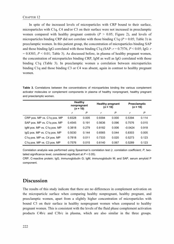
In spite of the increased levels of microparticles with CRP bound to their surface, microparticles with C1q, C4 and/or C3 on their surface were not increased in preeclamptic women compared with healthy pregnant controls (P > 0.05; Figure 2), and levels of microparticles binding CRP did not correlate with those binding C1q (P > 0.05; Table 3) in preeclamptic women. In this patient group, the concentration of microparticles binding SAP and those binding IgG correlated with those binding C1q (SAP: r = 0.7576, P < 0.05; IgG: r = 0.8303, P < 0.01; Table 3). As discussed before, in plasma of healthy pregnant women, the concentration of microparticles binding CRP, IgM as well as IgG correlated with those binding C1q (Table 3). In preeclamptic women a correlation between microparticles binding C1q and those binding C3 or C4 was absent, again in contrast to healthy pregnant women.
Table 3. Correlations between the concentrations of microparticles binding the various complement activator molecules or complement components in plasma of healthy nonpregnant, healthy pregnant and preeclamptic women.
Healthy
nonpregnant (n = 10)
Healthy pregnant (n = 10)
Preeclamptic (n = 10)
r P r P r P
CRP pos. MP vs. C1q pos. MP 0.8328 0.005 0.9394 0.000 0.5394 0.114
SAP pos. MP vs. C1q pos. MP 0.4545 0.191 0.5636 0.096 0.7576 0.015
IgM pos. MP vs. C1q pos. MP 0.3818 0.279 0.8182 0.006 -0.0424 0.918
IgG pos. MP vs. C1q pos. MP 0.5030 0.144 0.6565 0.044 0.8303 0.005
C1q pos. MP vs. C4 pos. MP 0.7818 0.011 0.7333 0.020 0.5273 0.123
C1q pos. MP vs. C3 pos. MP 0.7576 0.015 0.6140 0.067 0.5289 0.123
Correlation analysis was performed using Spearman’s correlation test (r, correlation coefficient; P, two-tailed significance level, considered significant at P < 0.05). CRP, C-reactive protein; IgG, immunoglobulin G; IgM, immunoglobulin M; and SAP, serum amyloid P component.
Discussion The results of this study indicate that there are no differences in complement activation on the microparticle surface when comparing healthy nonpregnant, healthy pregnant, and preeclamptic women, apart from a slightly higher concentration of microparticles with bound C3 on their surface in healthy nonpregnant women when compared to healthy pregnant women. This is consistent with the levels of the fluid phase complement activation products C4b/c and C3b/c in plasma, which are also similar in the three groups.

MICROPARTICLES AND COMPLEMENT ACTIVATION IN PREECLAMPSIA
223
1122
100 101 102 103 104FL2-Height
100 101 102 103 104FL2-Height
100 101 102 103 104FL2-Height
Cou
nts
0
5
10
15
CRP
Cou
nts
0
5
10
15
CRP
Cou
nts
0
5
10
15
CRP
A B C
100 101 102 103 104FL2-Height
100 101 102 103 104FL2-Height
100 101 102 103 104FL2-Height
Cou
nts
0
5
10
15
CRP
Cou
nts
0
5
10
15
CRP
Cou
nts
0
5
10
15
CRP100 101 102 103 104
FL2-Height100 101 102 103 104
FL2-Height100 101 102 103 104
FL2-Height
Cou
nts
0
5
10
15
CRP
Cou
nts
0
5
10
15
CRP
Cou
nts
0
5
10
15
CRP
Cou
nts
0
5
10
15
CRP
Cou
nts
0
5
10
15
CRP
Cou
nts
0
5
10
15
CRP
A B C
Figure 3. Representative histogram plots of microparticles with bound CRP in plasma of a healthy nonpregnant (A), a healthy pregnant (B), and a preeclamptic woman (C). Fluorescence intensity (x-axis) vs. microparticle count (y-axis) is shown. Binding of the isotype-matched control antibody is depicted with the filled histogram, and binding of the specific antibody with the open histogram. CRP, C-reactive protein.
Furthermore, our results suggest that in healthy nonpregnant and pregnant women, bound CRP molecules are involved in the classical pathway activation on the surface of microparticles, and that in healthy pregnant women IgM and especially IgG molecules also contribute. Interestingly, in plasma of preeclamptic women we found significantly higher levels of microparticles with bound CRP on their surface, compared with both healthy pregnant and nonpregnant women, but this was not associated with increased activation of the classical pathway of complement in preeclamptic women.
CRP binds to phosphorylcholine (the polar head group of phosphatidylcholine and sphingomyelin) in the outer leaflet of membranes in the presence of sufficient amounts of lysophosphatidylcholine [43], or to oxidized phosphatidylcholine [44]. Ligand-complexed CRP can bind C1q and activate the classical pathway of complement [27,31,42]. In a previous study [26], we found that in RA patients and healthy individuals levels of circulating microparticles with CRP on their surface correlated with levels of circulating microparticles with C1q. These in turn correlated with microparticles with bound C4 on their surface, suggesting classical pathway activation by CRP bound to the microparticles. The same phenomenon could be observed in the present study in plasma of healthy nonpregnant as well as healthy pregnant women. In plasma of preeclamptic women, however, such a relationship was absent. CRP is known to exert direct opsonic effects by binding to FcγRI and FcγRII on monocytes and neutrophils [45,46]. Possibly, in women with preeclampsia the balance between complement activation and direct opsonization by CRP is shifted towards the latter, or perhaps another mechanism is responsible for inhibition of complement activation in plasma of preeclamptic women. Interestingly, in a previous study we found that isolated microparticles from preeclamptic patients but not healthy pregnant women can cause endothelial dysfunction in isolated myometrial arteries from healthy pregnant women, but that this effect is abolished by the presence of plasma

CHAPTER 12
224
from the patients [47]. Explaining this phenomenon and our present results showing the absence of complement activation in the presence of relatively high concentrations of microparticles binding CRP will require further investigations.
It should be mentioned, that in contrast to healthy nonpregnant women, where only CRP seems to be involved in the low-level complement activation on the microparticle surface by binding and activating C1q, in healthy pregnant women IgM and IgG molecules also seem to participate. IgM molecules can bind to oxidized phospholipids and lysophospholipids [32,48] while the binding specificities of IgG molecules have not yet been elucidated. In preeclamptic women SAP (which binds to phosphatidylethanolamine [37,49,50]) and IgG seem to contribute, though in this study group microparticles binding C1q did not correlate with those binding C4 or C3, suggesting the classical pathway is either (partly) inhibited at the level of C1q, or that other pathways might also be involved. In spite of the questions that still remain open, these results do illustrate the altered inflammatory state of preeclamptic and even healthy pregnant women, when compared to healthy nonpregnant controls, as described previously [7].
There are data indicating that early-onset and late-onset preeclampsia might be qualitatively different diseases [51]. In line with this, in a study examining the extent of shedding of syncytiotrophoblast microparticles into the maternal circulation, Goswami et al. found a difference between early-onset (before 34 weeks of gestation) and late-onset (on or later than 34 weeks of gestation) preeclampsia, with increased levels in the former group and no difference in the latter, compared with matched healthy pregnant women [52]. In this regard it should be mentioned that our study group consisted entirely of early-onset preeclamptic women, and we observed no association between gestational age and the measured parameters within this group (data not shown). However, such a relationship cannot be excluded based on the present data. A study on preeclamptic women encompassing a wider range of gestational ages at disease onset, including women with later-onset preeclampsia than our study group, might reveal differences associated with gestational age at disease onset.
In conclusion, we did not find increased levels of microparticles with bound C1q, C4 or C3 in preeclamptic women, in accordance with similar levels of fluid phase complement activation products in these women. Plasma of preeclamptic women did contain significantly increased levels of microparticles binding CRP, but in contrast to healthy pregnant and nonpregnant women, this was not associated with classical pathway activation on the surface of the microparticles in these patients.
Acknowledgements The authors are grateful to L.M. Pronk for her technical assistance.

MICROPARTICLES AND COMPLEMENT ACTIVATION IN PREECLAMPSIA
225
1122
References [1] Pijnenborg R, Anthony J, Davey DA, Rees A, Tiltman A, Vercruysse L, van Assche A.
Placental bed spiral arteries in the hypertensive disorders of pregnancy. Br J Obstet Gynaecol 1991; 98: 648-655.
[2] Meekins JW, Pijnenborg R, Hanssens M, McFadyen IR, van Asshe A. A study of placental bed spiral arteries and trophoblast invasion in normal and severe pre-eclamptic pregnancies. Br J Obstet Gynaecol 1994; 101: 669-674.
[3] Easterling TR, Benedetti TJ, Schmucker BC, Millard SP. Maternal hemodynamics in normal and preeclamptic pregnancies: a longitudinal study. Obstet Gynecol 1990; 76: 1061-1069.
[4] Roberts JM. Endothelial dysfunction in preeclampsia. Semin Reprod Endocrinol 1998; 16: 5-15.
[5] Taylor RN, de Groot CJ, Cho YK, Lim KH. Circulating factors as markers and mediators of endothelial cell dysfunction in preeclampsia. Semin Reprod Endocrinol 1998; 16: 17-31.
[6] Taylor RN. Review: immunobiology of preeclampsia. Am J Reprod Immunol 1997; 37: 79-86. [7] Redman CW, Sacks GP, Sargent IL. Preeclampsia: an excessive maternal inflammatory
response to pregnancy. Am J Obstet Gynecol 1999; 180: 499-506. [8] Houwert-de Jong MH, Claas FH, Gmelig-Meyling FH, Kalsbeek GL, Valentijn RM, te Velde
ER, Schuurman HJ. Humoral immunity in normal and complicated pregnancy. Eur J Obstet Gynecol Reprod Biol 1985; 19: 205-214.
[9] Haeger M, Bengtson A, Karlsson K, Heideman M. Complement activation and anaphylatoxin (C3a and C5a) formation in preeclampsia and by amniotic fluid. Obstet Gynecol 1989; 73: 551-556.
[10] Haeger M, Unander M, Bengtsson A. Complement activation in relation to development of preeclampsia. Obstet Gynecol 1991; 78: 46-49.
[11] Haeger M, Unander M, Norder-Hansson B, Tylman M, Bengtsson A. Complement, neutrophil, and macrophage activation in women with severe preeclampsia and the syndrome of hemolysis, elevated liver enzymes, and low platelet count. Obstet Gynecol 1992; 79: 19-26.
[12] Halbmayer WM, Hopmeier P, Mannhalter C, Heuss F, Leodolter S, Rubi K, Fischer M. C1-esterase inhibitor in uncomplicated pregnancy and mild and moderate preeclampsia. Thromb Haemost 1991; 65: 134-138.
[13] Schjetlein R, Haugen G, Sandset PM, Wisloff F. Reduced C4b-binding protein in preeclampsia. Thromb Res 1997; 85: 153-158.
[14] Sinha D, Wells M, Faulk WP. Immunological studies of human placentae: complement components in pre-eclamptic chorionic villi. Clin Exp Immunol 1984; 56: 175-184.
[15] Tedesco F, Radillo O, Candussi G, Nazzaro A, Mollnes TE, Pecorari D. Immunohistochemical detection of terminal complement complex and S protein in normal and pre-eclamptic placentae. Clin Exp Immunol 1990; 80: 236-240.
[16] Massobrio M, Benedetto C, Bertini E, Tetta C, Camussi G. Immune complexes in preeclampsia and normal pregnancy. Am J Obstet Gynecol 1985; 152: 578-583.
[17] Mellembakken JR, Hogasen K, Mollnes TE, Hack CE, Abyholm T, Videm V. Increased systemic activation of neutrophils but not complement in preeclampsia. Obstet Gynecol 2001; 97: 371-374.

CHAPTER 12
226
[18] Jy W, Mao WW, Horstman L, Tao J, Ahn YS. Platelet microparticles bind, activate and aggregate neutrophils in vitro. Blood Cells Mol Dis 1995; 21: 217-231.
[19] Barry OP, Pratico D, Lawson JA, FitzGerald GA. Transcellular activation of platelets and endothelial cells by bioactive lipids in platelet microparticles. J Clin Invest 1997; 99: 2118-2127.
[20] Forlow SB, McEver RP, Nollert MU. Leukocyte-leukocyte interactions mediated by platelet microparticles under flow. Blood 2000; 95: 1317-1323.
[21] Mause SF, von Hundelshausen P, Zernecke A, Koenen RR, Weber C. Platelet microparticles: a transcellular delivery system for RANTES promoting monocyte recruitment on endothelium. Arterioscler Thromb Vasc Biol 2005; 25: 1512-1518.
[22] Nauta AJ, Trouw LA, Daha MR, Tijsma O, Nieuwland R, Schwaeble WJ, Gingras AR, Mantovani A, Hack EC, Roos A. Direct binding of C1q to apoptotic cells and cell blebs induces complement activation. Eur J Immunol 2002; 32: 1726-1736.
[23] Gasser O, Schifferli JA. Microparticles released by human neutrophils adhere to erythrocytes in the presence of complement. Exp Cell Res 2005; 307: 381-387.
[24] Campbell RD, Dodds AW, Porter RR. The binding of human complement component C4 to antibody-antigen aggregates. Biochem J 1980; 189: 67-80.
[25] Law SK, Lichtenberg NA, Levine RP. Evidence for an ester linkage between the labile binding site of C3b and receptive surfaces. J Immunol 1979; 123: 1388-1394.
[26] Biró É, Nieuwland R, Tak PP, Pronk LM, Schaap MC, Sturk A, Hack CE. Activated complement components and complement activator molecules on the surface of cell-derived microparticles in patients with rheumatoid arthritis and healthy individuals. Ann Rheum Dis 2007; 66: 1085-1092.
[27] Kaplan MH, Volanakis JE. Interaction of C-reactive protein complexes with the complement system. I. Consumption of human complement associated with the reaction of C-reactive protein with pneumococcal C-polysaccharide and with the choline phosphatides, lecithin and sphingomyelin. J Immunol 1974; 112: 2135-2147.
[28] Füst G, Medgyesi GA, Rajnavölgyi E, Csecsi-Nagy M, Czikora K, Gergely J. Possible mechanisms of the first step of the classical complement activation pathway: binding and activation of C1. Immunology 1978; 35: 873-884.
[29] Bruggemann M, Williams GT, Bindon CI, Clark MR, Walker MR, Jefferis R, Waldmann H, Neuberger MS. Comparison of the effector functions of human immunoglobulins using a matched set of chimeric antibodies. J Exp Med 1987; 166: 1351-1361.
[30] Ying SC, Gewurz AT, Jiang H, Gewurz H. Human serum amyloid P component oligomers bind and activate the classical complement pathway via residues 14-26 and 76-92 of the A chain collagen-like region of C1q. J Immunol 1993; 150: 169-176.
[31] Agrawal A, Shrive AK, Greenhough TJ, Volanakis JE. Topology and structure of the C1q-binding site on C-reactive protein. J Immunol 2001; 166: 3998-4004.
[32] Kim SJ, Gershov D, Ma X, Brot N, Elkon KB. I-PLA(2) activation during apoptosis promotes the exposure of membrane lysophosphatidylcholine leading to binding by natural immunoglobulin M antibodies and complement activation. J Exp Med 2002; 196: 655-665.
[33] Ciurana CL, Zwart B, van Mierlo G, Hack CE. Complement activation by necrotic cells in normal plasma environment compares to that by late apoptotic cells and involves predominantly IgM. Eur J Immunol 2004; 34: 2609-2619.

MICROPARTICLES AND COMPLEMENT ACTIVATION IN PREECLAMPSIA
227
1122
[34] Zwart B, Ciurana C, Rensink I, Manoe R, Hack CE, Aarden LA. Complement activation by apoptotic cells occurs predominantly via IgM and is limited to late apoptotic (secondary necrotic) cells. Autoimmunity 2004; 37: 95-102.
[35] Davey DA, MacGillivray I. The classification and definition of the hypertensive disorders of pregnancy. Am J Obstet Gynecol 1988; 158: 892-898.
[36] Wolbink GJ, Bollen J, Baars JW, ten Berge RJ, Swaak AJ, Paardekooper J, Hack CE. Application of a monoclonal antibody against a neoepitope on activated C4 in an ELISA for the quantification of complement activation via the classical pathway. J Immunol Methods 1993; 163: 67-76.
[37] Familian A, Zwart B, Huisman HG, Rensink I, Roem D, Hordijk PL, Aarden LA, Hack CE. Chromatin-independent binding of serum amyloid P component to apoptotic cells. J Immunol 2001; 167: 647-654.
[38] Berckmans RJ, Nieuwland R, Böing AN, Romijn FP, Hack CE, Sturk A. Cell-derived microparticles circulate in healthy humans and support low grade thrombin generation. Thromb Haemost 2001; 85: 639-646.
[39] Biró É, Nieuwland R, Sturk A. Measuring circulating cell-derived microparticles. J Thromb Haemost 2004; 2: 1843-1844.
[40] Hack CE, Paardekooper J, Smeenk RJ, Abbink J, Eerenberg AJ, Nuijens JH. Disruption of the internal thioester bond in the third component of complement (C3) results in the exposure of neodeterminants also present on activation products of C3. An analysis with monoclonal antibodies. J Immunol 1988; 141: 1602-1609.
[41] Hoekzema R, Martens M, Brouwer MC, Hack CE. The distortive mechanism for the activation of complement component C1 supported by studies with a monoclonal antibody against the "arms" of C1q. Mol Immunol 1988; 25: 485-494.
[42] Wolbink GJ, Brouwer MC, Buysmann S, Ten Berge IJ, Hack CE. CRP-mediated activation of complement in vivo: assessment by measuring circulating complement-C-reactive protein complexes. J Immunol 1996; 157: 473-479.
[43] Volanakis JE, Wirtz KW. Interaction of C-reactive protein with artificial phosphatidylcholine bilayers. Nature 1979; 281: 155-157.
[44] Chang MK, Binder CJ, Torzewski M, Witztum JL. C-reactive protein binds to both oxidized LDL and apoptotic cells through recognition of a common ligand: Phosphorylcholine of oxidized phospholipids. Proc Natl Acad Sci U S A 2002; 99: 13043-13048.
[45] Crowell RE, Du Clos TW, Montoya G, Heaphy E, Mold C. C-reactive protein receptors on the human monocytic cell line U-937. Evidence for additional binding to Fc gamma RI. J Immunol 1991; 147: 3445-3451.
[46] Bharadwaj D, Stein MP, Volzer M, Mold C, Du Clos TW. The major receptor for C-reactive protein on leukocytes is fcgamma receptor II. J Exp Med 1999; 190: 585-590.
[47] VanWijk MJ, Svedas E, Boer K, Nieuwland R, VanBavel E, Kublickiene KR. Isolated microparticles, but not whole plasma, from women with preeclampsia impair endothelium-dependent relaxation in isolated myometrial arteries from healthy pregnant women. Am J Obstet Gynecol 2002; 187: 1686-1693.
[48] Shaw PX, Horkko S, Chang MK, Curtiss LK, Palinski W, Silverman GJ, Witztum JL. Natural antibodies with the T15 idiotype may act in atherosclerosis, apoptotic clearance, and protective immunity. J Clin Invest 2000; 105: 1731-1740.

CHAPTER 12
228
[49] Schwalbe RA, Dahlback B, Coe JE, Nelsestuen GL. Pentraxin family of proteins interact specifically with phosphorylcholine and/or phosphorylethanolamine. Biochemistry 1992; 31: 4907-4915.
[50] Emsley J, White HE, O'Hara BP, Oliva G, Srinivasan N, Tickle IJ, Blundell TL, Pepys MB, Wood SP. Structure of pentameric human serum amyloid P component. Nature 1994; 367: 338-345.
[51] von Dadelszen P, Magee LA, Roberts JM. Subclassification of preeclampsia. Hypertens Pregnancy 2003; 22: 143-148.
[52] Goswami D, Tannetta DS, Magee LA, Fuchisawa A, Redman CW, Sargent IL, von Dadelszen P. Excess syncytiotrophoblast microparticle shedding is a feature of early-onset pre-eclampsia, but not normotensive intrauterine growth restriction. Placenta 2006; 27: 56-61.