uterocutaneous fistula following conservative management ...ircmj.portal.tools/73882.pdftors such as...
TRANSCRIPT

Iran Red Crescent Med J. 2017 January; 19(1):e41813.
Published online 2017 January 18.
doi: 10.5812/ircmj.41813.
Case Report
Uterocutaneous Fistula Following Conservative Management of
Placenta Increta: A Rare Case Report and Review of the Literature
Donya Khosravi,1 Maliheh Arab,2,3,* Behnaz Ghavami,4 Maryam Shokrpour,5 Samaneh Sheibani,1 and
Samaneh Saraeian11Assistant of Obstetrics and Gynecology, Imam Hossein Medical Center, Shahid Beheshti University of Medical Sciences, Tehran, IR Iran2Professor of Gynecology-oncology, Imam Hossein Medical Center, Shahid Beheshti University of Medical Sciences, Tehran, IR Iran3PhD of Medical Education, Shahid Beheshti University of Medical Sciences, Tehran, IR Iran4Shariati Medical Center, Tehran University of Medical Sciences, Tehran, IR Iran5Assistant Professor of Obstetrics and Gynecology, Arak University of Medical Sciences, Arak, IR Iran
*Corresponding author: Maliheh Arab, PhD, Professor of Gynecology-Oncology, Imam Hossein Medical Center, Shahid Beheshti University of Medical Sciences, Madani Street,Tehran, IR Iran. Fax: +98-21-77543634, E-mail: [email protected]
Received 2016 August 26; Revised 2016 December 06; Accepted 2017 January 14.
Abstract
Introduction: This was a report of uterocutaneous fistula as a rare complication of expectant management of placenta increta, anda review of the literature.Case Presentation: A 38- year- old pregnant woman in her third term of pregnancy was operated on in a secondary hospital inArak (a city located in the center of Iran) in April 2015. As a result of placenta incereta, the attached placenta was left in the uterus.On the 38th day, she was referred to our tertiary care hospital due to intermittent fever and bleeding. More examinations by MRIand hystrosalpingography revealed uterocutaneous fistula. She was operated on again, and surgical findings indicated coexistentuterine necrosis and uterocutaneous fistula.Conclusions: This case was the first reported morbidity of fistula in the uterocutaneous pathway.
Keywords: Placenta Accreta, Fistula, Disease Management, Iran
1. Introduction
Placenta accreta is abnormally attached placental villito the myometrium. The decidua basalis defect facilitatespassage of the villi into the myometrium. This pathophysi-ology is a barrier to normal hemostasis in the postpartumperiod. Sometimes, bleeding is severe and hysterectomyis necessary to save life. If the placenta is detached force-fully, severe bleeding (more than 3 lit) may occur, as it hasbeen reported in 40% of the placenta accreta cases (1). Se-vere bleeding after manual detachment of placenta is dueto opening of spiral sinuses, which is regarded as an im-portant morbidity (2). Transfusion is needed in about 90%of the patients, and more than 10 units of blood are trans-fused in 40% of the cases (3). The maternal mortality of pla-centa accreta in different studies is reported to be in therange of 0.3% to 7% (1, 3, 4).
There is a rise in the rate of placenta accreta parallelto the increase in the rate of cesarean delivery. There is aparticular increased risk in placenta previa. Placenta acc-reta is reported in a range of about 1 out of 533 (5, 6). The10- fold increase in placenta accreta risk during the past50 years may be due to the increase in cesarean deliveryand maternal age (6, 7). In addition to the strong risk fac-
tors such as previous cesarean scars and placenta previa,other known risk factors are multiparity, advanced mater-nal age, previous dilation and curettage, history of man-ual placenta removal, submucous myoma resulting in at-rophy of endometrial mucosa, recurrent abortion, gesta-tional product implanted in uterine diverticulum, and pre-vious radium insertion (7-9). Placenta accreta-percreta canbe diagnosed before delivery by sonography and MRI, es-pecially in patients with risk factors such as previous ce-sarean as placenta previa. Some abnormal placentationcases are diagnosed just after delivery (10, 11). In a study,about 25% of the placenta accreta cases were not diagnosedantenatally. Another study indicated pelvic MRI harbor-ing 19% false negative results (12). The American College ofObstetricians and Gynecologists confirmed hysterectomyas the optimal management of placenta accreta (13). Re-cent reports indicate similar morbidity of the conserva-tive approach in selected cases with primary hysterectomy.Nowadays, conservative management is considered in se-lected cases. Fertility preservation is the main aim of con-servative treatment (14, 15). Adjuvant treatment follow-ing conservative management including arterial ligation,uterine sutures, embolization of arteries, and methotrex-ate therapy are sometimes necessary (15, 16).
Copyright © 2017, Iranian Red Crescent Medical Journal. This is an open-access article distributed under the terms of the Creative Commons Attribution-NonCommercial 4.0International License (http://creativecommons.org/licenses/by-nc/4.0/) which permits copy and redistribute the material just in noncommercial usages, provided theoriginal work is properly cited.
Khosravi D et al.
Due to an increase in the tendency for conservativemanagement and fertility preservation, in addition to theincreased prevalence of placenta accreta, it seems reason-able to pay strong attention to complications and conse-quences of conservative management of placenta accreta.Previous studies found blood transfusion, prolonged hos-pital admission, infection, and secondary hysterectomy tobe the main complications of conservative management.In the present study, a rare complication of conservativemanagement of placenta accreta and the review of the lit-erature were presented.
2. Case Presentation
A 38-year- old female, who was in her third term of preg-nancy, was operated on in a secondary hospital due to totalplacenta previa and accreta in the background of her 2 pre-vious caesarian sections in Arak (a city located in the centerof Iran) in April 2015. She had been operated by Pfannen-stiel incision in the abdominal wall and kerr incision in theuterus.
Her surgical record indicated placental invasion intothe lower segment of the myometrium and cervical re-gion. About 8 cm3 of the placenta remained in the cav-ity. Ten units pack cell and 4 units fresh frozen plasmawere infused and a drain was left for 48 hours. A loweruterine segment had been packed in the operation fieldwith long gauze that was left for 24 hours and removedthrough the vagina. Because of continuous bleeding, in-trauterine balloon packing was alternated to the previ-ous packing for another 24 hours. Uterotonic drugs andmultiple antibiotic therapies had been prescribed. Dueto fever (38 - 39°C) in the postoperative period (days 1 - 3),triple antibiotherapy including Ampicillin, gentamycin,and clindamycin had been started. In the fourth day, dueto persistent fever, a new regimen (including meroperiem-vancomycin) was prescribed. On the fifth day, the resultsof the abdomino-pelvic sonography revealed a heteroge-neous collection similar to the retained placenta about 6cm in endometrium besides the fluid collection, and a fewair bubble about 8.5cm3 in the subcutaneous region abovethe cesarean scar. On the seventh day postoperation, shewas discharged without fever and in a good condition.
On the eighth day, the results of a postoperationabdomino-pelvic CT scan revealed a heterogeneous 10 ×6 cm mass in the lower segment of the abdominal cav-ity in favor of retained placenta. On the ninth day post-operation, 85 mg methotrexate was prescribed. On the24th day (during the follow-up), a 5 mm incisional de-fect with bloody discharge resulted in antibiotherapy andtransvaginal sonography. A mass of 8.5-cm, filling the
lower segment and cervical area, was reported in addi-tion to a 4-cm fluid collection in the abdominal wall, withdrainage pathway into abdominal wall. The result of theserum BHCG was negative. On the 25th postoperation,abdominal-pelvic MRI showed a heterogeneous mass of 6to 7 cm in the left side of the uterus, which was extendedto the near serosal surface. Extension outside the uteruswas not possible. On the 38th day postoperation, due to thecontinuous abdominal incision drainage and her medicalhistory, she was referred to our center, a tertiary hospitalin Tehran, Iran. Our hospital is a 570-bed, government gen-eral hospital, which is a referral center and offers service topatients nationwide.
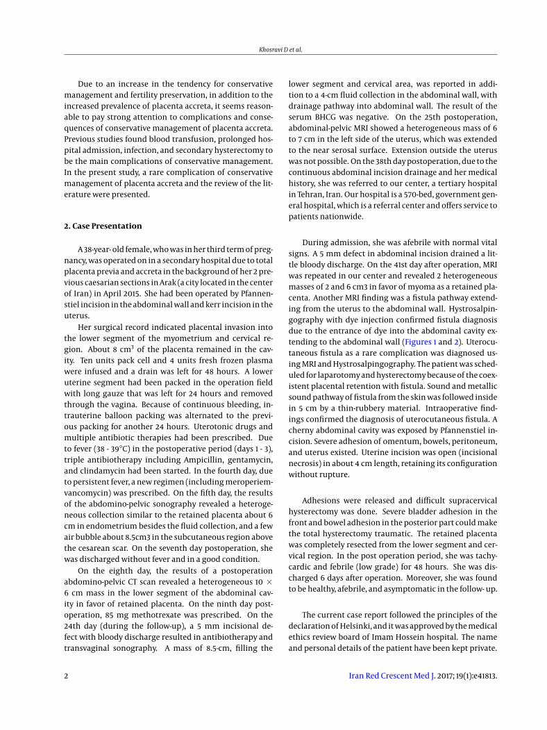
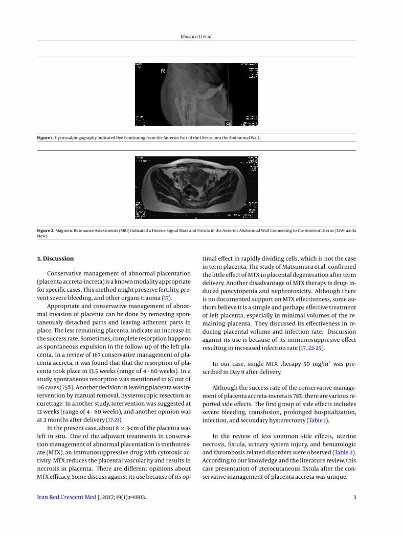
During admission, she was afebrile with normal vitalsigns. A 5 mm defect in abdominal incision drained a lit-tle bloody discharge. On the 41st day after operation, MRIwas repeated in our center and revealed 2 heterogeneousmasses of 2 and 6 cm3 in favor of myoma as a retained pla-centa. Another MRI finding was a fistula pathway extend-ing from the uterus to the abdominal wall. Hystrosalpin-gography with dye injection confirmed fistula diagnosisdue to the entrance of dye into the abdominal cavity ex-tending to the abdominal wall (Figures 1 and 2). Uterocu-taneous fistula as a rare complication was diagnosed us-ing MRI and Hystrosalpingography. The patient was sched-uled for laparotomy and hysterectomy because of the coex-istent placental retention with fistula. Sound and metallicsound pathway of fistula from the skin was followed insidein 5 cm by a thin-rubbery material. Intraoperative find-ings confirmed the diagnosis of uterocutaneous fistula. Acherny abdominal cavity was exposed by Pfannenstiel in-cision. Severe adhesion of omentum, bowels, peritoneum,and uterus existed. Uterine incision was open (incisionalnecrosis) in about 4 cm length, retaining its configurationwithout rupture.
Adhesions were released and difficult supracervicalhysterectomy was done. Severe bladder adhesion in thefront and bowel adhesion in the posterior part could makethe total hysterectomy traumatic. The retained placentawas completely resected from the lower segment and cer-vical region. In the post operation period, she was tachy-cardic and febrile (low grade) for 48 hours. She was dis-charged 6 days after operation. Moreover, she was foundto be healthy, afebrile, and asymptomatic in the follow- up.
The current case report followed the principles of thedeclaration of Helsinki, and it was approved by the medicalethics review board of Imam Hossein hospital. The nameand personal details of the patient have been kept private.
2 Iran Red Crescent Med J. 2017; 19(1):e41813.
Khosravi D et al.
Figure 1. Hystrosalpingography Indicated Dye Continuing from the Anterior Part of the Uterus Into the Abdominal Wall.
Figure 2. Magnetic Resonance Assessments (MRI) Indicated a Hetero- Signal Mass and Fistula in the Anterior Abdominal Wall Connecting to the Anterior Uterus (T2W- axillaview).
3. Discussion
Conservative management of abnormal placentation(placenta accreta-increta) is a known modality appropriatefor specific cases. This method might preserve fertility, pre-vent severe bleeding, and other organs trauma (17).
Appropriate and conservative management of abnor-mal invasion of placenta can be done by removing spon-taneously detached parts and leaving adherent parts inplace. The less remaining placenta, indicate an increase inthe success rate. Sometimes, complete resorption happensas spontaneous expulsion in the follow- up of the left pla-centa. In a review of 167 conservative management of pla-centa accreta, it was found that that the resorption of pla-centa took place in 13.5 weeks (range of 4 - 60 weeks). In astudy, spontaneous resorption was mentioned in 87 out of116 cases (75%). Another decision in leaving placenta was in-tervention by manual removal, hysteroscopic resection ascurettage. In another study, intervention was suggested at13 weeks (range of 4 - 60 weeks), and another opinion wasat 2 months after delivery (17-21).
In the present case, about 8× 3-cm of the placenta wasleft in situ. One of the adjuvant treatments in conserva-tion management of abnormal placentation is methotrex-ate (MTX), an immunosuppressive drug with cytotoxic ac-tivity. MTX reduces the placental vascularity and results innecrosis in placenta. There are different opinions aboutMTX efficacy. Some discuss against its use because of its op-
timal effect in rapidly dividing cells, which is not the casein term placenta. The study of Matsumura et al. confirmedthe little effect of MTX in placental degeneration after termdelivery. Another disadvantage of MTX therapy is drug- in-duced pancytopenia and nephrotoxicity. Although thereis no documented support on MTX effectiveness, some au-thors believe it is a simple and perhaps effective treatmentof left placenta, especially in minimal volumes of the re-maining placenta. They discussed its effectiveness in re-ducing placental volume and infection rate. Discussionagainst its use is because of its immunosuppresive effectresulting in increased infection rate (17, 22-25).
In our case, single MTX therapy 50 mg/m2 was pre-scribed in Day 9 after delivery.
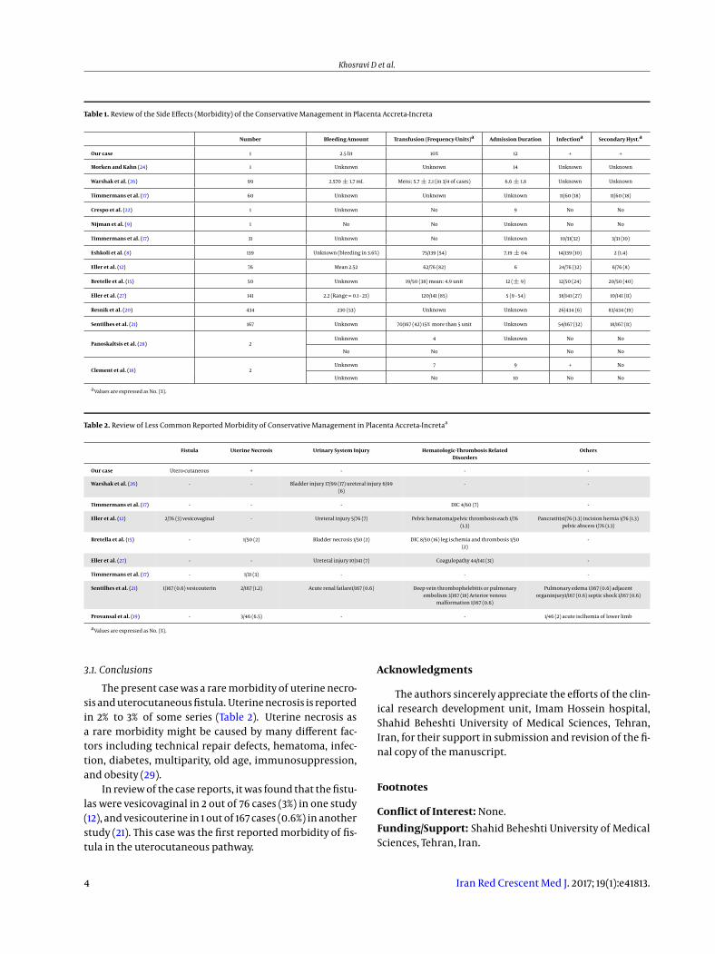
Although the success rate of the conservative manage-ment of placenta accreta-increta is 78%, there are various re-ported side effects. The first group of side effects includessevere bleeding, transfusion, prolonged hospitalization,infection, and secondary hysterectomy (Table 1).
In the review of less common side effects, uterinenecrosis, fistula, urinary system injury, and hematologicand thrombosis related disorders were observed (Table 2).According to our knowledge and the literature review, thiscase presentation of uterocutaneous fistula after the con-servative management of placenta accreta was unique.
Iran Red Crescent Med J. 2017; 19(1):e41813. 3
Khosravi D et al.
Table 1. Review of the Side Effects (Morbidity) of the Conservative Management in Placenta Accreta-Increta
Number Bleeding Amount Transfusion (Frequency-Units)a Admission Duration Infectiona Secondary Hyst.a
Our case 1 2.5 lit 10% 12 + +
Morken and Kahn (24) 1 Unknown Unknown 14 Unknown Unknown
Warshak et al. (26) 99 2.570 ± 1.7 mL Mens: 5.7 ± 2.1 (in 3/4 of cases) 6.6 ± 1.8 Unknown Unknown
Timmermans et al. (17) 60 Unknown Unknown Unknown 11/60 (18) 11/60 (18)
Crespo et al. (22) 1 Unknown No 9 No No
Nijman et al. (9) 1 No No Unknown No No
Timmermans et al. (17) 31 Unknown No Unknown 10/31(32) 3/31 (10)
Eshkoli et al. (8) 139 Unknown (bleeding in 3.6%) 75/139 (54) 7.19 ± 04 14/139 (10) 2 (1.4)
Eller et al. (12) 76 Mean 2.52 62/76 (82) 6 24/76 (32) 6/76 (8)
Bretelle et al. (15) 50 Unknown 19/50 (38) mean: 4.9 unit 12 (± 9) 12/50 (24) 20/50 (40)
Eller et al. (27) 141 2.2 (Range = 0.1 - 23) 120/141 (85) 5 (9 - 54) 38/141 (27) 10/141 (11)
Resnik et al. (20) 434 230 (53) Unknown Unknown 26/434 (6) 83/434 (19)
Sentilhes et al. (21) 167 Unknown 70/167 (42) 15% more than 5 unit Unknown 54/167 (32) 18/167 (11)
Panoskaltsis et al. (28) 2Unknown 4 Unknown No No
No No No No
Clement et al. (18) 2Unknown 7 9 + No
Unknown No 10 No No
a Values are expressed as No. (%).
Table 2. Review of Less Common Reported Morbidity of Conservative Management in Placenta Accreta-Incretaa
Fistula Uterine Necrosis Urinary System Injury Hematologic-Thrombosis RelatedDisorders
Others
Our case Utero-cutaneous + - - -
Warshak et al. (26) - - Bladder injury 17/99 (17) ureteral injury 6/99(6)
- -
Timmermans et al. (17) - - - DIC 4/60 (7) -
Eller et al. (12) 2/76 (3) vesicovaginal - Ureteral injury 5/76 (7) Pelvic hematoma/pelvic thrombosis each 1/76(1.3)
Pancratitis1/76 (1.3) incision hernia 1/76 (1.3)pelvic abscess 1/76 (1.3)
Bretella et al. (15) - 1/50 (2) Bladder necrosis 1/50 (2) DIC 8/50 (16) leg ischemia and thrombosis 1/50(2)
-
Eller et al. (27) - - Ureteral injury 10/141 (7) Coagulopathy 44/141 (31) -
Timmermans et al. (17) - 1/31 (3) - - -
Sentilhes et al. (21) 1/167 (0.6) vesicouterin 2/167 (1.2) Acute renal failare1/167 (0.6) Deep vein thrombophelebitis or pulmonaryembolism 3/167 (18) Arterior venous
malformation 1/167 (0.6)
Pulmonary edema 1/167 (0.6) adjacentorganinjury1/167 (0.6) septic shock 1/167 (0.6)
Provansal et al. (19) - 3/46 (6.5) - - 1/46 (2) acute isclhemia of lower limb
a Values are expressed as No. (%).
3.1. Conclusions
The present case was a rare morbidity of uterine necro-sis and uterocutaneous fistula. Uterine necrosis is reportedin 2% to 3% of some series (Table 2). Uterine necrosis asa rare morbidity might be caused by many different fac-tors including technical repair defects, hematoma, infec-tion, diabetes, multiparity, old age, immunosuppression,and obesity (29).
In review of the case reports, it was found that the fistu-las were vesicovaginal in 2 out of 76 cases (3%) in one study(12), and vesicouterine in 1 out of 167 cases (0.6%) in anotherstudy (21). This case was the first reported morbidity of fis-tula in the uterocutaneous pathway.
Acknowledgments
The authors sincerely appreciate the efforts of the clin-ical research development unit, Imam Hossein hospital,Shahid Beheshti University of Medical Sciences, Tehran,Iran, for their support in submission and revision of the fi-nal copy of the manuscript.
Footnotes
Conflict of Interest: None.
Funding/Support: Shahid Beheshti University of MedicalSciences, Tehran, Iran.
4 Iran Red Crescent Med J. 2017; 19(1):e41813.
Khosravi D et al.
References
1. Hudon L, Belfort MA, Broome DR. Diagnosis and management ofplacenta percreta: a review. Obstet Gynecol Surv. 1998;53(8):509–17.[PubMed: 9702791].
2. Lam H, Pun TC, Lam PW. Successful conservative management of pla-centa previa accreta during cesarean section. Int J Gynaecol Obstet.2004;86(1):31–2. doi: 10.1016/j.ijgo.2003.12.009. [PubMed: 15207668].
3. O’Brien JM, Barton JR, Donaldson ES. The management of placentapercreta: conservative and operative strategies. Am J Obstet Gynecol.1996;175(6):1632–8. [PubMed: 8987952].
4. Steins Bisschop CN, Schaap TP, Vogelvang TE, Scholten PC. Invasive pla-centation and uterus preserving treatment modalities: a systematicreview. Arch Gynecol Obstet. 2011;284(2):491–502. doi: 10.1007/s00404-011-1934-6. [PubMed: 21638046].
5. Silver RM, Landon MB, Rouse DJ, Leveno KJ, Spong CY, ThomEA, et al. Maternal morbidity associated with multiple repeatcesarean deliveries. Obstet Gynecol. 2006;107(6):1226–32. doi:10.1097/01.AOG.0000219750.79480.84. [PubMed: 16738145].
6. Wu S, Kocherginsky M, Hibbard JU. Abnormal placentation:twenty-year analysis. Am J Obstet Gynecol. 2005;192(5):1458–61.doi: 10.1016/j.ajog.2004.12.074. [PubMed: 15902137].
7. Miller DA, Chollet JA, Goodwin TM. Clinical risk factors for pla-centa previa-placenta accreta. Am J Obstet Gynecol. 1997;177(1):210–4.[PubMed: 9240608].
8. Eshkoli T, Weintraub AY, Sergienko R, Sheiner E. Placenta acc-reta: risk factors, perinatal outcomes, and consequences for sub-sequent births. Am J Obstet Gynecol. 2013;208(3):219 e1–7. doi:10.1016/j.ajog.2012.12.037. [PubMed: 23313722].
9. Nijman RG, Mantingh A, Aarnoudse JG. Persistent retained placentapercreta: methotrexate treatment and Doppler flow characteristics.BJOG. 2002;109(5):587–8. [PubMed: 12066955].
10. Levine D, Hulka CA, Ludmir J, Li W, Edelman RR. Placenta accreta:evaluation with color Doppler US, power Doppler US, and MR imag-ing.Radiology. 1997;205(3):773–6. doi: 10.1148/radiology.205.3.9393534.[PubMed: 9393534].
11. Megier P, Gorin V, Desroches A. [Ultrasonography of placenta previaat the third trimester of pregnancy: research for signs of placenta acc-reta/percreta and vasa previa. Prospective color and pulsed Dopplerultrasonography study of 45 cases]. J Gynecol Obstet Biol Reprod (Paris).1999;28(3):239–44. [PubMed: 10456306].
12. Eller AG, Porter TF, Soisson P, Silver RM. Optimal management strate-gies for placenta accreta. BJOG. 2009;116(5):648–54. doi: 10.1111/j.1471-0528.2008.02037.x. [PubMed: 19191778].
13. Bouvier-Colle MH, Varnoux N, Breart G. Maternal deaths and sub-standard care: the results of a confidential survey in France. Medi-cal Experts Committee. Eur J Obstet Gynecol Reprod Biol. 1995;58(1):3–7.[PubMed: 7758642].
14. Alanis M, Hurst BS, Marshburn PB, Matthews ML. Conservativemanagement of placenta increta with selective arterial emboliza-tion preserves future fertility and results in a favorable outcomein subsequent pregnancies. Fertil Steril. 2006;86(5):1514 e3–7. doi:10.1016/j.fertnstert.2006.02.128. [PubMed: 17007851].
15. Bretelle F, Courbiere B, Mazouni C, Agostini A, Cravello L, Bou-bli L, et al. Management of placenta accreta: morbidity andoutcome. Eur J Obstet Gynecol Reprod Biol. 2007;133(1):34–9. doi:10.1016/j.ejogrb.2006.07.050. [PubMed: 16965851].
16. Tsirulnikov MS. [Ligation of the uterine vessels during obstetricalhemorrhages. Immediate and long-term results (author’s transl)]. JGynecol Obstet Biol Reprod (Paris). 1979;8(8):751–3. [PubMed: 317962].
17. Timmermans S, van Hof AC, Duvekot JJ. Conservative manage-ment of abnormally invasive placentation. Obstet Gynecol Surv.2007;62(8):529–39. doi: 10.1097/01.ogx.0000271133.27011.05. [PubMed:17634154].
18. Clement D, Kayem G, Cabrol D. Conservative treatment of pla-centa percreta: a safe alternative. Eur J Obstet Gynecol ReprodBiol. 2004;114(1):108–9. doi: 10.1016/j.ejogrb.2003.06.008. [PubMed:15099881].
19. Provansal M, Courbiere B, Agostini A, D’Ercole C, Boubli L, BretelleF. Fertility and obstetric outcome after conservative managementof placenta accreta. Int J Gynaecol Obstet. 2010;109(2):147–50. doi:10.1016/j.ijgo.2009.12.011. [PubMed: 20152971].
20. Resnik R, Lockwood C, Levine D. Management of placenta accreta, inc-reta, and percreta. ; 2014.
21. Sentilhes L, Ambroselli C, Kayem G, Provansal M, Fernandez H,Perrotin F, et al. Maternal outcome after conservative treat-ment of placenta accreta. Obstet Gynecol. 2010;115(3):526–34. doi:10.1097/AOG.0b013e3181d066d4. [PubMed: 20177283].
22. Crespo R, Lapresta M, Madani B. Conservative treatment of placentaincreta with methotrexate. Int J Gynaecol Obstet. 2005;91(2):162–3. doi:10.1016/j.ijgo.2005.06.028. [PubMed: 16126207].
23. Matsumura N, Inoue T, Fukuoka M, Sagawa N, Fujii S. Changes in theserum levels of human chorionic gonadotropin and the pulsatility in-dex of uterine arteries during conservative management of retainedadherent placenta. J Obstet Gynaecol Res. 2000;26(2):81–7. [PubMed:10870298].
24. Morken NH, Kahn JA. Placenta accreta and methotrexate treatment.Acta Obstet Gynecol Scand. 2006;85(2):248–50. [PubMed: 16532926].
25. Widemann BC, Adamson PC. Understanding and managingmethotrexate nephrotoxicity. Oncologist. 2006;11(6):694–703. doi:10.1634/theoncologist.11-6-694. [PubMed: 16794248].
26. Warshak CR, Ramos GA, Eskander R, Benirschke K, Saenz CC,Kelly TF, et al. Effect of predelivery diagnosis in 99 consecutivecases of placenta accreta. Obstet Gynecol. 2010;115(1):65–9. doi:10.1097/AOG.0b013e3181c4f12a. [PubMed: 20027036].
27. Eller AG, Bennett MA, Sharshiner M, Masheter C, Soisson AP, DodsonM, et al. Maternal morbidity in cases of placenta accreta managed bya multidisciplinary care team compared with standard obstetric care.Obstet Gynecol. 2011;117(2 Pt 1):331–7. [PubMed: 21309195].
28. Panoskaltsis TA, Ascarelli A, de Souza N, Sims CD, Edmonds KD. Pla-centa increta: evaluation of radiological investigations and therapeu-tic options of conservative management. BJOG. 2000;107(6):802–6.[PubMed: 10847240].
29. Arab M, Khosravi D, Mehdighalb S, Kazemi SN, Nematihonar B, Jalaee-far AM. A rare case report of post cesarean uterine incisional necro-sis coexistant whit Ogilvie syndrome. Indian J Fund Appl Life Sci.2014;4(S3):515–8.
Iran Red Crescent Med J. 2017; 19(1):e41813. 5