uterine rupture
TRANSCRIPT
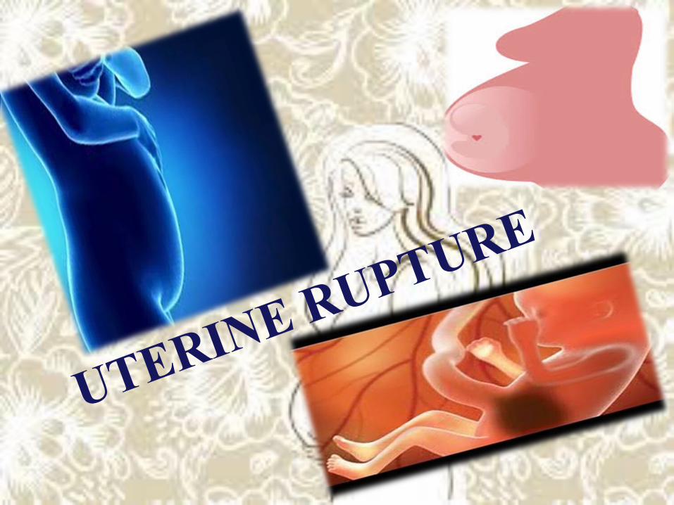
UTERINE RUPTURE

Definition
• A uterine rupture is a tear in the wall of the uterus, most often at the site of a previous c-section incision.
• Fortunately, these ruptures are relatively rare events – exceedingly rare for women who've never had a c-section, other uterine surgery, or a previous rupture. The vast majority of uterine ruptures occur during labor, but they can also happen before the onset of labor.

CAUSES• Cephalopelvic Disproportion- This is when the
mother’s pelvis is too small for the size of the baby, resulting in the baby being unable to pass through the birth canal.
• Grand multiparity. This is when the mother has given birth 5 or more times.
• Uncontrolled use of Pitocin ( Oxytocin). This is probably the leading cause of rupture of the unscarred uterus. Oxytocin can cause contractions to be too strong and too frequent, which puts a lot of strain on the uterus .

• Placental Abruption. This is when the placental lining separates from the uterus. This can cause the baby to be either partially or completely cut off from the mother’s circulation.
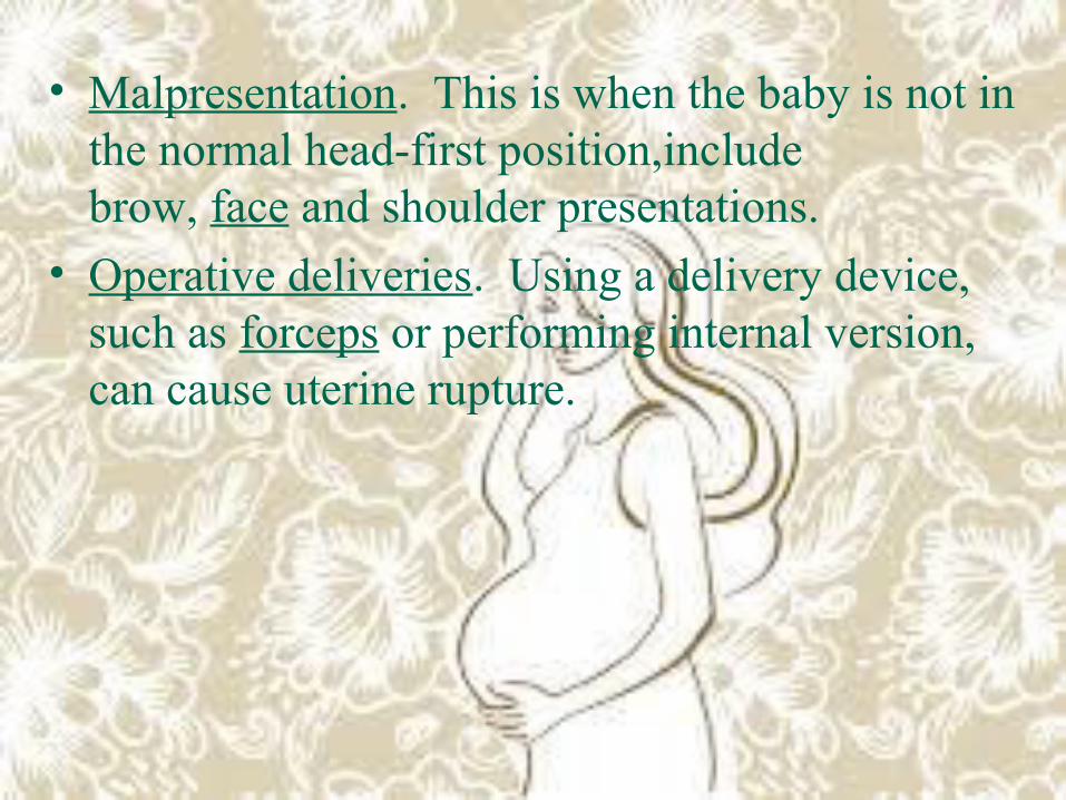
• Malpresentation. This is when the baby is not in the normal head-first position,include brow, face and shoulder presentations.
• Operative deliveries. Using a delivery device, such as forceps or performing internal version, can cause uterine rupture.

Risk Factor• Congenital uterine anomalies,• multiparity, • previous uterine myomectomy,• the number and type of previous cesarean
deliveries,• fetal macrosomia,
• labor induction, • uterine instrumentation, and • uterine trauma all increase the risk of uterine
rupture,

TYPES
• Incomplete rupture
• complete rupture

• In an Incomplete uterine rupture, the mother’s peritoneum remains intact. The peritoneum is the membrane that lines the abdominal cavity to support abdominal organs. It also acts as a channel for blood vessels and nerves. An incomplete uterine rupture is significantly less dangerous with fewer complications to the delivery process.


• During a Complete uterine rupture, the peritoneum tears and the contents of the mother’s uterus can spill into her peritoneal cavity. The peritoneal cavity is the fluid-filled gap that separates the abdomen walls and its organs. It is suggested that delivery via cesarean section (C-section) should occur within approximately 10 to 35 minutes after a complete uterine rupture occurs. The fetal morbidity rate increases dramatically after this period.


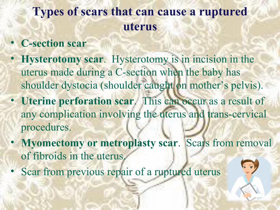
Types of scars that can cause a ruptured uterus
• C-section scar• Hysterotomy scar. Hysterotomy is in incision in the
uterus made during a C-section when the baby has shoulder dystocia (shoulder caught on mother’s pelvis).
• Uterine perforation scar. This can occur as a result of any complication involving the uterus and trans-cervical procedures.
• Myomectomy or metroplasty scar. Scars from removal of fibroids in the uterus.
• Scar from previous repair of a ruptured uterus

Signs and Symptoms• Vaginal bleeding• Sharp pain between contractions• Contractions that slow down or become less intense• Unusual abdominal pain or tenderness
• Recession of the fetal head (baby’s head moving back up into the birth canal)
• Bulging under the pubic bone (baby’s head has protruded outside of the uterine scar)
• Sharp onset of pain at the site of the previous scar• Uterine atony (loss of uterine muscle tone)• Maternal tachycardia (rapid heart rate) and hypotension

Nursing Management
1. . Monitor for the possibility of uterine rupture.
• In the presence of predisposing factors, monitor maternal labor pattern closely for hypertonicity or signs of weakening uterine muscle.
• Recognize signs of impending rupture, immediately notify the physician, and call for assistance.

2.Assist with rapid intervention. • If the client has signs of possible uterine rupture,
vaginal delivery is generally not attempted.• If symptoms are not severe, an emergency cesarean
delivery may be attempted and the uterine tear repaired.• If symptoms are severe, emergency laparotomy is
performed to attempt immediate delivery of the fetus and then establish homeostasis.
• Implement the following preparations for surgery.
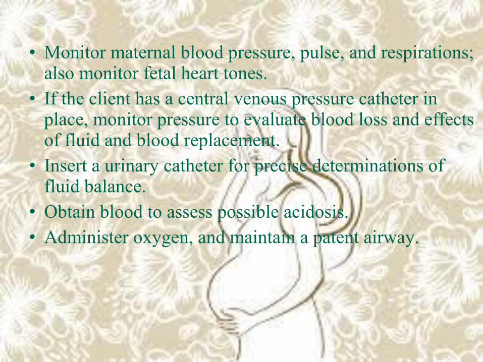
• Monitor maternal blood pressure, pulse, and respirations; also monitor fetal heart tones.
• If the client has a central venous pressure catheter in place, monitor pressure to evaluate blood loss and effects of fluid and blood replacement.
• Insert a urinary catheter for precise determinations of fluid balance.
• Obtain blood to assess possible acidosis.• Administer oxygen, and maintain a patent airway.

3. . Prevent and manage complications. Take these steps in order to prevent or limit hypovolemic shock:
• Oxygenate by providing 8 to 10 L/min using a closed mask.
• Restore circulating volume using one or more IV lines.
• Evaluate the cause, response to therapy, and fetal condition.
• Remedy the problem by preparing the client for surgery and administering antibiotics.

4. Provide physical and emotional support.• Provide support for the client’s partner and family
members once surgery has begun.• Inform the partner and family how they will receive
information about the mother and newborn and where to wait.

TREATMENT• Women’s general condition must be improved
giving blood transfusion, glucose solution)• immediate laparotomy
( is used to explore the mother’s abdominal wall and a C-section is performed.)
• Hysterectomy
(-is an operation to remove a woman's uterus. A woman may have a hysterectomy for different reasons, including:
THANKS FOR YOUR ATTENTION