uterine blood flow during pregnancy
TRANSCRIPT

Uterine Blood Flow During Pregnancy*
FRANK C. GREISS, JR., M.D.
Professor of Obstetrics and Gynecology, Bowman Gray School of Medicine, Wake Forest University, Winston-Salem, North Carolina
Of those parameters pertinent to fetal wellbeing, the delivery of an adequate amount of blood to the placenta seems to be the most crucial. At our present state of understanding, uterine blood flow (UBF) appears to be that parameter most often affected by physiologic and pathologic conditions, and it is certainly that factor most readily altered, either favorably or unfavorably, by the physician. In this presentation, normal changes in UBF during pregnancy will be reviewed briefly, those factors influencing uterine and placental vascular dynamics will be discussed, and the clinical relevance of these ol?servations will be illustrated on the background of varying levels of uteroplacental adequacy.
Homeostatic Changes in Uterine Blood Flow. While our most comprehensive knowledge of UBF changes during pregnancy has been determined in the ewe, correlative data in subhuman primates and women indicates that results are comparable, at least during the last half of the gestation period (Assali, Douglas, et al, 1953; Assali, Rauramo, et al, 1960; Lees, et al, 1971; Metcalfe, et al, 1955; Peterson and Behrman, 1969) . These changes are illustrated in Fig. 1. It can be seen that absolute UBF increases most markedly during the period of most rapid absolute fetal growth. Although fetal weight tends to increase more rapidly than UBF, the proportion of UBF perfusing the placenta progressively increases during this same time so that flow per unit weight of fetus, placenta, and uterus remains essentially constant. Similarly, the amount of oxygen extracted from each milliliter of blood also remains essentially constant (Huckabee, et al, 1961) indicating that the increase in placental blood flow is matching the increasing demands of the fetoplacental
* Presented at the 43rd Annual McGuire Lecture Series, December 3, 1971 , at the Medical College of Virginia, Richmond. (This investigation was supported by United States Public Health Service Grant No. HE-03941-12 from the National Heart Institute.)
52
!::111200 f 35 ~ 1100 lt ~ I~ ~1000 /l 30 100 ~ . 7
~,,1 90"'"' : 900 I x I
80~ ~ 800 I · ~ ' ~ 700 I
20 ~ 70"' ~ I 8" ~ 600 I 1'-, "I ~ 60 -.J"' Loo I ' /,// 15 ~
<tl_, I
50 ~~ I I ' x
i§ 400 I I ' '
} __, "'" I I ' ~ tl"-ii 300 I I \ 0/ 1.0~ "'"' ~ 200
'\ "/-l"L. ___ - --x ce~ \
\ I 0.5 1..-x
~ ;
~ 100 \.._ J j ~ __ , -o--o-l!'l "' 0 10 20 30 40 50 60 70 80 90 100 120 140
GESTATION OAY
Fig. I- Derived graph of relative and absolute uterine blood flows, fetal. weight and proportionate placental blood flow during ovine pregnancy (Barcroft, 1947; Greiss and Anderson, 1970; Huckabee, et al, 1961; Makowski, et al, 1968). During the last half of gestation, relative uterine blood flow is constant, and the proportion of uterine blood flow reaching the placenta progressively increases.
unit. After term gestation, however, available information indicates that placental blood flow decreases relative to fetoplacental requirements (Dixon, et al, 1963), blood oxygen extraction increases and fetal well-being is progressively compromised. Similarly, placental blood flow is reduced in patients with essential hypertension, chronic renal disease, preeclampsia or eclampsia, twin gestation and premature labors complicated by abnormal bleeding or infection (Browne and Veall, 1953; Dixon, et al, 1963; Johnston and Clayton, 1957; Landesman and Knapp, 1960; Morris, et al, 1955; Weis, et al, 1958). The severity of the reduction in flow with toxemia of pregnancy seems directly related to the severity of the disease process.
Dynamic Control of Placental Blood Flow. A schematic representation of those factors affecting placental and non-placental blood flow in the uterus is shown in Fig. 2. At term gestation in women, the proportion of total UBF perfusing the placenta probably exceeds 85 percent (Lees, et al, 1971; Makowski,
MCV QUARTERLY 8(1): 52-60, r972

GREISS: UTERINE BLOOD FLOW DURING PREGNANCY 53
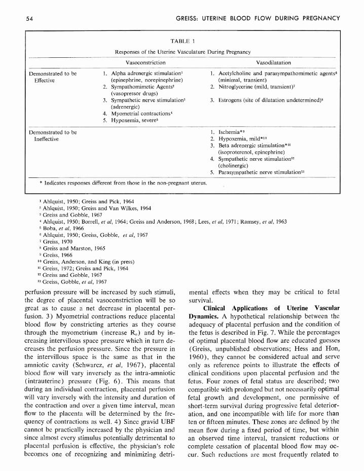
et al, 1968). Therefore, our primary attention should be directed to the second part of the equation accompanying the illustration. This shows that placental blood flow varies directly with the perfusion pressure (UABP-IUP-UVBP) and inversely with the resistance of the spiral arterioles supplying the intervillous space (Ripr) and the resistance imparted to the blood vessels as they course through the myometrium (Re). The effects of various stimuli on these resistances are summarized in Table I. All vasodilator stimuli exert inconsequential or no effect while all vasoconstrictor stimuli are capable of producing marked persistent reductions in flow. Four observations are of particular pertinence to the clinician: 1) Under normal circumstances, the placental vasculature approaches maximal vasodilatation (Greiss, 1966). This means that we have no effective way of further increasing placental blood flow. It also means that placental perfusion will vary directly with and in proportion to changes in systemic blood pressuret (Fig. 3), that is, a 30% decrease in systemic blood pressure will cause a 30% decrease in placental blood flow. 2) Endogenous or exogenous vasopressor hormones or drugs (Fig. 4)
Uterine Blood a Non-placental Blood + Placental Blood Flow Flow Flow
UABP-UVBP + UABP-IUP-UVBP
Ri N-PL + Re Ri PL. 1" Re
Amniotic Cavity
Intrauterine Pressure
Fig. 2- Schematic representation of those factors determining placental and non-placental uterine blood flow. Note that the reactivity of the non-placental vasculature (R •N- PL) is different from that of the placental vasculature (R,PL), the extrinsic vascular resistance (R0 ) imparted by contracting myometrium affects both vasculatures similarly, and that by increasing intrauterine and intervillous space pressures, myometrial contractions decrease placental perfusion pressure. UABP and UVBP indicate uterine arterial and venous pressures, and IUP equals intrauterine (intra-amniotic) pressure.
t For practical purposes in the absence of uterine contractions, perfusion pressure is determined primarily by arterial blood pressure.
and sympathetic nerve stimulation (Fig. 5) cause significant vasoconstriction (increase R;pL) usually proportionately greater than that induced in other peripheral vascular beds. This means that although
120
110
100
90
~ 80 ii " l'!
70
~ 60
i 50
· ~
~ 40
30
20
10
b• 1.03~ r • 0.992
• • Orl9lnal valuH
Experimenf 13-1
T .iZ.O S.D.
_L
10 20 30 40 ~O 60 70 80 90 IOO 110
A Uterine Blood Prnsure {orlerlol-venou1) (mm. H9.l
Fig. 3- Graph of the relationship between perfusion pressure and uterine blood flow at term gestation in the ewe indicating that the dominant placental vasculature is almost maximally dilated and that placental perfusion decreases linearly with decreases in the systemic blood pressure. (Reprinted with permission from Greiss. Am. J. Obstet. Gynec. 96: 41, 1966.)
UBF 335 •rot cc/min
'00 ~ ' BO ABP
mm Hg
' "t IUP 20 mm Hg
0
120 Pulse 180 t
beats/min. 60
Ewe 69-11
0
145d. Gestation
@
fNorepinephrine 40;r i.o.
Minutes
Fig. 4-0riginal tracing of the effect of norepinephrine on uterine blood flow in the gravid ewe. Circled numbers indicate uterine conductance values. Such profound vasoconstriction and decrease in uterine blood flow is typical of the action of endogenous adrenal medullary hormones and peripherally acting vasopressor agents. (Reprinted with permission from Greiss. Am. J. Obstet. Gynec. 112: 20, 1972.)
54 GREISS: UTERINE BLOOD FLOW DURING PREGNANCY
TABLE 1
Responses of the Uterine Vasculature During Pregnancy
Demonstrated to be Effective
Demonstrated to be Ineffective
Vasoconstriction
1. Alpha adrenergic stimulation1
(epinephrine, norepinephrine) 2. Sympathomimetic Agents2
( vasopressor drugs) 3. Sympathetic nerve stimulation3
( adrenergic) 4. Myometrial contractions4 5. Hypoxemia, severe•
V asodilatation
1. Acetylcholine and parasympathomimetic agents6 (minimal, transient)
2. Nitroglycerine (mild, transient)'
3. Estrogens (site of dilatation undetermined)B
1. Ischemia*' 2. Hypoxemia, mild* 10
3. Beta adrenergic stimulation* 11
(isoproterenol, epinephrine) 4. Sympathetic nerve stimulation12
(cholinergic) 5. Parasympathetic nerve stimulation1'
* Indicates responses different from those in the non-pregnant uterus.
1 Ahlquist, 1950; Greiss and Pick, 1964 2 Ahlquist, 1950; Greiss and Van Wilkes, 1964 a Greiss and Gobble, 1967 4 Ahlquist, 1950; Borrell, et al, 1964; Greiss and Anderson, 1968; Lees, et al, 1971; Ramsey, et al, 1963 ' Boba, et al, 1966 6 Ahlquist, 1950; Greiss, Gobble, et al, 1967 ' Greiss, 1970 s Greiss and Marston, 1965 • Greiss, 1966
10 Greiss, Anderson, and King (in press) 11 Greiss, 1972; Greiss and Pick, 1964 12 Greiss and Gobble, 1967 1a Greiss, Gobble, et al, 1967
perfusion pressure will be increased by such stimuli, the degree of placental vasoconstriction will be so great as to cause a net decrease in placental perfusion. 3) Myometrial contractions reduce placental blood flow by constricting arteries as they course through the myometrium (increase Re) and by increasing intervillous space pressure which in turn decreases the perfusion pressure. Since the pressure in the intervillous space is the same as that in the amniotic cavity (Schwarcz, et al, 1967), placental blood flow will vary inversely as the intra-amniotic (intrauterine) pressure (Fig. 6). This means that during an individual contraction, placental perfusion will vary inversely with the intensity and duration of the contraction and over a given time interval, mean flow to the placenta will be determined by the frequency of contractions as well. 4) Since gravid UBF cannot be practically increased by the physician and since almost every stimulus potentially detrimental to placental perfusion is effective, the physician's role becomes one of recognizing and minimizing detri-
mental effects when they may be critical to fetal survival.
Clinical Applications of Uterine Vascular Dynamics. A hypothetical relationship between the adequacy of placental perfusion and the condition of the fetus is described in Fig. 7. While the percentages of optimal placental blood flow are educated guesse~ (Greiss, unpublished observations; Hess and Hon, 1960), they cannot be considered actual and serve only as reference points to illustrate the effects of clinical conditions upon placental perfusion and the fetus. Four zones of fetal status are described; two compatible with prolonged but not necessarily optimal fetal growth and development, one permissive of short-term survival during progressive fetal deterioration, and one incompatible with life for more than ten or fifteen minutes. These zones are defined by the mean flow during a fixed period of time, but within an observed time interval, transient reductions or complete cessation of placental blood flow may occur. Such reductions are most frequently related to

GREISS: UTERINE BLOOD FLOW DURING PREGNANCY 55
~we 63-6 129 DAYS GESTATION
160 160
BP 80 80 (mm.Hg)
0 0
RUBF (cc./mi n.)
72 72
0 0
LUBF (cc./min.)
180 180
0 0 9.0 volts 12.0 volts
~ ~ Right SNS Left SNS
~5 mm./min. = paper speed
Fig. 5- 0riginal tracings of the effect of lumbar sympathetic nerve stimulation on uterine blood flow in the pregnant ewe. Although sympathetic tonus is low during resting condition~, profound uterine vasoconstriction is evoked by maternal hemorrhage. (Reprinted with permission from Greiss and Gobble. Am. J. Obstet. Gynec. 97 : 962, 1967.)
uterine contractions (Borr~ll, et al, 19~4; Ramsey, et al, 1963). Under optimal conditions, br.ief episodes of intervillous stasis evoke no evidence of fetal distress. However, when such episodes are superimposed upon a fetoplacental unit already co!llpromised with respect to placental perfusion, fetal hypoxia will be manifested · ini'tially by late d~celer'ation of the fetal heart rate with respect to uterine contractions, later by progressive fetal acidosis and finally by persistent bradycardia signaling incipient fetal demise (Hon and Quilligan, 1968) .
Labor. During strong myometrial contractions, placental perfusion temporarily ceases. For illustrative purposes, let us assume that intervillous flow stops for 30 seconds of a 45 second contraction and 45 seconds of a 60 second contraction. The effects of such contractions on mean placental blood flow are illustrated In ~ig. 8. As the quration and frequency of myometrial contractions increase, mean placental flow progressively decreases. If labor
begins when placental flow is optimal, even the most active contractions (solid line) cause no fetal jeopardy. However, when flow is suboptimal initially, active labor will evoke fetal distress (long dashed line) and when the pre-labor flow is borderline, even mild labor will cause distress or fetal death (short dasheq line). It is evident, therefore, that labor is inherently stressful to the fetus. This fact must be remembered whenever patients with high-risk pregnancies begin spontaneous or induced labor.
Hemorrhage. Maternal hemorrhage is a most potent stimulus of adrenal medullary hormone secretion and sympathetic nerve stimulation. Acute moderate hemorrhage (500c....750 ml. within 15 minutes) causes minimal changes in maternal blood pressure and pulse rate. However, these parameters are maintained by peripheral vasoconstriction with an inevitable decrease in placental blood flow (Fig. 9). Acute severe hemorrhage ( 1500 ml. in 15 minutes) evokes a further increase in peripheral and uterine

56 GREISS: ~TERINE BLOOD FLOW DURING PREGNANCY
UBF cc/min
20 15 10 · 5 0
127 MATERNAL 85
B.P. 42 mm Hg 0
RttESUS 66-2 TERM PREGNANCY
45- · IUP
mm Hg 30 15 o-.:?=~~~~~~"~~~~~--~~~~~
0 5 10 15 20 Minutes
25 30 35 40
Fig. 6---0riginal tracing of the effect of spontaneous uterine contractions on uterine blood flow in the term-pregnant Rhesus monkey The uterine blood flow trace is the inverse image of the intrauterine pressure pattern. Uterine blood flow achieves maximum levels during myometrial diastole only. Therefore, mean uterine blood flow will decrease as the frequency of contractions increases and the duration of myometrial diastole decreases. (Reprinted with permission from Greiss and Anderson. C/in. Obstet. Gynec. 11: 96, 1968.)
Hypothetical Relationship Between
Fetal Condition and Placental Blood Flow
Optimal Oxygenation
Normal Growth
to ·:·::::::::: ::::::: ;:;:;:: ;:;::;:: ::;::;: :::: :::::: ;::: / ( (
§~b~~~fi~~I 5;;~~~~fi~~) :,:,:: 7 z w IT IT ::: ?: :r: ;: ? ? > V>> :•:::• ( :} () ( ) > >> ( > : ? •: > •< ) •< ) ) ) ) ) )
I>::::::: :•::::: t > < () > < > ( > ? ) ) > > •> ?• ) { ) t { ? H \ ;::;:;: /) )) < :/ ( < : ? >> > { }) ) \. ) : ) ) > :. ;: ) ) ) > ) ? ;;::;::::;:: •:::::• )) / /: ( / > < > >
// •>• . . . . . . ::;:< :: ::,:,::: :::::::::::::;:: :::::;:::::;:::;: ;:;:::: ?/ )) }) ) ::: ) > '> : :; ) > ) ) ) ? i:' ::;:::: ::::::: :,::::: :,:,:::: :::::::: :':':: ·:·:·:·: ·:·.·.;. ·:·:·.:. >< · II<>••••••• ;:;:::: ;:;:;:; ::;::;: ;:::::: ;:;:::: :::,: ::,: :':':' ::::::: .,.,,. ::·:·
.._, 1naciiQuate" ox'Y9enatiOn:F>ro9ress1ve ' acidosis -Late Ft-!R_ deceleration-Limited survival time
---
Severe hypoxia - Persistent bradycardia = = Short survival time =
= = = = = --
Fig. 7- }Iypothetical relationship between placental blood flow and the status of the fetus. Of the many parameters determining fetal well-being, placental blood flow is the most variable and the parameter most subject to pathologic change·. The significance of detrimental influences on placental blood flow increases as the pre-stress level of flow decreases.
vasoconstriction with an associated decrease in systemic blOod pressure. Together, these two factors cause a marked decrease in placental perfusion ( Greiss, 1966). Figure 9 shows that acute severe hemorrhage may rapidly compromise the fetus even when the pre-hemorrhage status of placental per-
fusion is optimal. At slower rates of blood loss, the degree of peripheral vasoconstriction necessary to maintain maternal integrity will be less because of compensatory shifts of body fluids into the vascular compartment, and placental perfusion will not be reduced so drastically. Therapy for blood loss should

.... GREISS: UTERINE BLOOD FLOW DURING PREGNANCY 57
Hypothetical Relationship Between Fetal Condition and Placental Blood Flow
LABOR
Optimal
~=frequency (min) O 5.0 3.0 2.5 2.0 ~- . ti duration (sec.) __ 45 45 __ 60 __ 60 ~ . peak intensity (mmHg) ·. 60-80 60-80 . G0-80 · 60-80 a=tonus(mmHg) ~ 8-10 ~ 8-10 ~8-10 ~ 8-10
Fig. 8-Effect of uterine contractions on mean placental blood flow. As the duration and frequency of contractions increase, mean flow progressively decreases. If labor begins with optimal blood flow, placental perfusion is more than adequate even during very active contractions (solid line). However, if placental perfusion is borderline before labor such as occurs with toxemia, even mild contractions may cause fetal distress or demise (short dashed line).
be directed toward correcting the vascular volumecapacity discrepancy. Optimally, this should be accomplished with whole blood: However, in emergency situations when blood is not available or is being readied, rapid intravenous infusions of salt solutions or blood substitutes will still improve placental perfusion significantly (Boba, et al, 1966).
Non-hemorrhagic Hypotension. Except for hemorrhagic and septic shock, most circumstances that decrease maternal blood pressure do not change placental vascular resistance. Therefore, placental blood flow will decrease in proportion to the degree of hypotension. Such circumstances occur most frequently following spinal anesthesia for cesarean section. The effects of a typical clinical situation on placental blood flow are illustrated in Fig. 10. It should be evident that only those fetuses in the
suboptimal environment prior to anesthesia will be in jeopardy and only those in the lower suboptimal range may die. This is especially true since hypotension of this severity is usually corrected promptly. Generally, hypotension will respond to a combination of left-uterine displacement and a rapid intravenous infusion of 500-1000 ml. of salt or dextrose solution ( Greiss and Crandell, 1965). The former relieves pressure on the inferior vena cava, thus increasing venous return to the heart and cardiac output. The latter reduces the vascular volume-capacity disparity induced by sympathetic nerve paralysis and peripheral vasodilatation. However, some patients do not respond to these techniques, and vasopressor therapy becomes necessary. At this point, a knowledge of the vasomotor control of the placental blood vessels is requisite for the selection of that vasopressor agent

58 GREISS: UTERINE BLOOD FLOW DURING PREGNANCY
Hypothetical Relationship Between Fetal Condition and Placental Blood Flow
ACUTE HEMORRHAGE
Optimal
I~ 70
15
100 . ~5
30
7~ 40
Fig. 9- Effect of acute sudden hemorrhage on placental blood flow in the ewe. Prompt therapy with whole blood or blood volume expanders is necessary to protect the fetus even if pre-hemorrhage conditions were optimal.
most beneficial to placental perfusion. The effects of two agents, ephedrine and metaraminoP, on placental blood flow are depicted in Fig. 10 (Jam es, et al, 1970). By acting primarily on the heart with minimal uterine and peripheral vasoconstriction, ephedrine accomplishes the greatest improvement in placental blood flow. Metaraminol improves maternal blood pressure by acting in roughly equal amounts on the heart and peripheral blood vessels. Therefore, the beneficial effect of improved blood pressure is partially offset by placental vasoconstriction so that the fetal environment is improved less effectively. The difference between the two agents becomes significant when one considers flow responses in a fetus with initial borderline placental perfusion (short dashed line, Fig. 10) . Ephedrine therapy improved perfusion sufficiently to produce a live although probably depressed infant while metaraminol
t Aramine®, Merck Sharp & Dohme
therapy was inadequate to accomplish even this outcome. Not shown in Fig. 10 are the effects of primarily peripherally acting pressor agents such as phenylephi;ine. * Such agents cause so much placental vasoconstriction that despite restoration of normal blood pressure, placental perfusion is not improved and may even decrease further (Greiss and Crandell, 1965).
The above illustrations depict the effects of individual co.nditions upon placental blood flow . In clinical practice, it should be evident that such conditions may occur concomitantly, and actually there is a positive tendency for this to happen. Therefore, placental perfusion is often reduced by multiple factors. Our ability to minimize the effect of these factors on the. fetal environment may make the difference between a healthy child and a stillborn or retarded infant.
* Neo-Synephrine®, Winthrop Labs.

GREISS: UTERINE BLOOD FLOW DURING PREGNANCY 59
Hypothetical Relationship Between Fetal Condition and Placental Blood FI ow SPINAL HYPOTENSION 8 ITS THERAPY
-Ephedrine (heavy line) vs.
Prespinal B.P. 120/60
Spinal Hypotension Metarominol (light line)theropy B.P 70140 5' 10' 15'
B. P. brou t to 120 I 60
Fig. 10--Effect of hypotension induced by spinal anesthesia on placental perfusion. Only those fetuses with initial suboptimal placental blood flow will be affected by marked hypotension (dashed lines). If vasopressor therapy is required, appropriate selection of the drug according to its mode of action may be crucial to fetal survival.
REFERENCES AHLQUIST, R. P. The action of various drugs on the arterial blood flow of the pregnant canine uterus. /. Am. Pharm. Assoc. (Scient. Ed.) 39:370, 19'50.
AssALI, N. s., DOUGLAS, A., JR., BAIRD, w. w., NICHOLSON, D. B., AND SuYEMOTO, R. Measurement of uterine blood flow and uterine metabolism IV. Results in normal pregnancy. Am. I. Obstet. Gynec. 66:248, 1953.
AssALI, N. s., RAURAMO, L., AND PELTOMEN, T. Measurement of uterine blood flow and uterine metabolism VIII. Uterine and fetal blood flow and oxygen consumption in early human pregnancy. Am. l . Obstet. Gynec. 79 :86, 1960.
BARCROFT, J . Researches on Prenatal Life. Springfield : C. C. Thomas, 1947, p. 1.
BOBA, A., LINKIE, D . M., AND PLOTZ, E. J. Effects of vasopressor administration and fluid replacement on fetal bradycardia and hypoxia induced by maternal hemorrhage. Obstet. Gynecol. 27 :408, 1966. . .
BORRELL, U., INGMAN, F., OHLSON, L., AND WIQVJST, N. Effect of uterine contractions on the human uteroplacental blood circulation. Am. J. Obstet. Gynec. 89:881, 1964.
BROWNE, J. c. Mee., AND VEALL, N. Maternal placental blood flow./. Obstet. Gynaecol. Brit. Comm. 60:141, 1953.
DIXON, H. G ., BROWNE, J. C., AND DAVEY, D. A . Choriodecidual and myometrial blood flow. Lancet 2:369, 1963.
GREISS, F. C., JR. Pressure-flow relationship in the gravid uterine vascular bed. Am. l. Obstet. Gynec. 96: 41 , 1966.
GREISS, F. C., JR. Uterine vascular response to hemorrhage during pregnancy. Obstet. Gynecol. 27 : 549, 1966.
GREISS, F. C., JR. Differential reactivity of the myoendometrial and placental vasculatures: Vasodilatation. Am. J. Obstet. Gynec. 111 :61 1, 1971.
GREISS, F. C., JR. Differential reactivity of the myoendometrial and placental vasculatures: Adrenergic responses. Am. l . Obstet. Gynec. 112:20-30, 1972.

60 GREISS: UTERINE BLOOD FLOW DURING PREGNANCY
GREISS, F. C., JR. Unpublished observations.
GREiss, F. C., JR. AND ANDERSON, S. G. Uterine blood flow during labor. Clin. Obstet. Gynecol. 11 :96, 1968.
GREISS, F. c., JR. AND ANDERSON, s. G. Uterine blood flow during early ovine pregnancy. Am. J. Obstet. Gynec. 106: 30, 1970.
GREISS, F. c., JR., ANDERSON, s. G., AND KING, L. c. Uterine vascular bed: Effects of acute hypoxia. Am. J. Obstet. Gynec. In press.
GREISS, F. C., JR. AND CRANDELL, D. L. Therapy for hypotension induced by spinal anesthesia during pregnancy. JAMA 191:793, 1965.
GREISS, F. c., JR. AND GOBBLE, F. L. , JR. Effect of sympathetic nerve stimulation on the uterine vascular bed. Am. J. Obstet. Gynec. 97:962, 1967.
GREISS, F. c., JR., GOBBLE, F . L., JR., ANDERSON, s. G., AND McGuIRT, W. F. Effect of parasympathetic nerve stimulation on the uterine vascular bed. Am. J. Obstet. Gynec. 99: 1067, 19'67.
GREISS, F. C., JR., GOBBLE, F. L., JR., ANDERSON, S. G., AND McGUIRT, W. F. Effect of acetylcholine on the uterine vascular bed. Am. J. Obstet. Gynec. 99: 1073, 1967.
GREISS, F. c., JR. AND MARSTON, E. L. The uterine vascular bed: Effect of estrogens during ovine pregnancy. Am. J. Obstet. Gynec. 93:720, 1965.
GREISS, F. C., JR. AND PICK, J. R. JR. The uterine vascular bed: Adrenergic receptors. Obstet. Gynecol. 23 :209, 1964.
GREISS, F. C., JR. AND VAN WILKES, D. Effects of sympathomimetic drugs and angiotensin on the uterine vascular bed. Obstet. Gynecol. 23:925, 1964.
HEss, 0. W. AND HON, E. H. The electronic evaluation of fetal heart rate : III. The effect of an oxytocic agent for induction of labor. Am. J. Obstet. Gynec. 80: 558, 1960.
HoN, E. E. AND QUILLIGAN, E. J. Electronic evaluation of fetal heart rate. IX. Further observations on "pathologic" fetal bradycardia. Clin. Obstet. Gynecol. 11: 145, 1968.
HUCKABEE, W. E., METCALFE, J., PRYSTOWSKY, H., AND BARRON, D. H. Blood flow and oxygen consumption of the pregnant uterus. Am. J. Physiol. 200:274, 1961.
JAMES, F. M ., III, GREISS, F. C., JR., AND KEMP, R. A. An evaluation of vasopressor therapy for maternal hypotension during spinal anesthesia. Anesthesiology 33 :25, 1970.
JOHNSTON, T. AND CLAYTON, C. G. Diffusion of radioactive sodium in normotensive and preeclamptic pregnancies. Br. M ed. J. 5014:312, 1957.
LANDESMAN, R. AND KNAPP, R. c. Na24 uterine muscle clearance in late pregnancy. Am. J. Obstet. Gynec. 80:92, 1960.
LEES, M . H ., HILL, J. D., OCHSNER, A. J., Ill, THOMAS, C . L., AND NOVY, M. J. Maternal placental and myometrial blood .flow of the rhesus monkey during uterine contractions. Am. J. Obstet. Gynec. 110:68, 1971.
MAKOWSKI, E. L., MESCHIA, G., DROEGEMUELLER, W., AND BATTAGLIA, F. C. Distribution of uterine blood flow in the pregnant sheep. Am. J. Obstet. Gynec. 101 :409, 1968.
METCALFE, J ., ROMNEY, S. L., RAMSEY, L. H., REID, D. E ., AND BURWELL, C. S. Estimation of uterine blood flow in normal human pregnancy at term. J. Clin. Invest. 34: 1632, 1955.
MORRIS, N., OSBORN, s. B., AND WRIGHT, H. P. Effective circulation of the uterine wall in late pregnancy measured with 24NaCI. Lancet 268 : 323, 1955.
PETERSON, E . N . AND BEHRMAN, R. E. Changes in the cardiac output and uterine blood flow of the pregnant Macaca Mulatta. Am. J. Obstet. Gynec. 104 :988, 1969.
RAMSEY, E. M., CORNER, G. W., JR., AND DONNER, M. W. Serial and cineradioangiographic visualization of maternal circulation in the primate (hemochorial) placenta. Am. J. Obstet. Gynec. 86:213, 1963.
SCHWARCZ, R., JR. , ALTHABE, 0 ., JR., FISCH, L., AND PINTO, R. M. Relationships between intervillous space amniotic fluid and maternal arterial blood pressures. Am. J. Obstet . Gynec. 98:924, 1967.
WEIS, E. B., JR., BRUNS, P. D., AND TAYLOR, E. s. A comparative study of the disappearance of radioactive sodium from human uterine muscle in normal and abnormal pregnancy. Am. J. Obstet. Gynec. 76:340, 1958.