useful histological findings in incisional biopsies of ... · faculdade de odontologia avenida...
TRANSCRIPT
384
Srp Arh Celok Lek. 2016 Jul-Aug;144(7-8):384-390 DOI: 10.2298/SARH1608384I
ОРИГИНАЛНИ РАД / ORIGINAL ARTICLE UDC: 617.51/.53-006.6
Correspondence to:Jean Nunes DOS SANTOSFaculdade de OdontologiaAvenida Araújo Pinho, 62, CanelaSalvador-BA, Brazil [email protected]
SUMMARYIntroduction Oral squamous cell carcinoma (OSCC) is one of the most common head and neck cancers.Objective The aim of this study was to investigate the histopathological features of OSCC specimens ob-tained from incisional biopsies and to alert clinicians to the importance of more representative biopsies.Methods Forty-eight OSCC samples were obtained from incisional biopsies and classified by Bryne’s score. The following morphological features were analyzed: invasive front, invasiveness, apoptotic cells, atypical mitosis, giant cells, acantholysis, ulceration, necrosis, calcification, surface epithelium, granulation tissue, desmoplasia, tissue invasions, inflammatory infiltrate and tumor thickness. Results Ten (21%) cases were classified as high grade malignancies and 38 (79%) as low grade. Apoptotic cells (n = 26), atypical mitosis (1–2/20×; n = 38), giant cells (n = 8), acantholysis (n = 5), necrosis (n = 5), calcification (n = 1), granulation tissue (n = 32), desmoplasia (n = 4), perineural invasion (n = 2), muscular invasion (n = 8), invasion of salivary gland tissue (n = 3), vascular invasion (n = 10), and chronic inflamma-tion (n = 33) were observed. Vascular invasion (p = 0.04, Pearson’s χ2 test) and necrosis (p = 0.04, Pearson’s χ2 test) were significantly associated with cases of high-grade malignant tumors. Atypical mitosis was associated with a greatest tumor thickness (p = 0.04, Fischer’s exact test).Conclusion This study suggests that incisional biopsies may be useful and significant as they can show histopathological variables that are important to classify oral squamous cell carcinomas into low grade and high grade according to Bryne’s score, which was used in this study. Thus, more representative biopsies might be useful to achieve this and allow a more accurate planning.keywords: oral neoplasms; oral cancer, diagnosis; histopathological staging; carcinoma, squamous cell/pathology; carcinoma, squamos cell/surgery.
Useful histological findings in incisional biopsies of oral squamous cell carcinomaAdna Barros Ismerim1, Flávia Caló Aquino Xavier2, Maria Cristina Teixeira Cangussu3, Luciana Maria Pedreira Ramalho4, Ivan Marcelo Gonçalves Agra5, Jean Nunes dos Santos2
1Federal University of Bahia, School of Dentistry Salvador, Bahia, Brazil;2Federal University of Bahia, School of Dentistry, Laboratory of Surgical Pathology, Salvador, Bahia, Brazil;3Federal University of Bahia, School of Dentistry, Department of Dental Public Health, Salvador, Bahia;4Federal University of Bahia, School of Dentistry, Department of Stomatology, Salvador Bahia, Brazil;5Aristides Maltez Hospital, Department of Head and Neck Surgery, Salvador, Bahia, Brazil
INTRODUCTION
Squamous cell carcinoma (SCC) is one of the most common head and neck cancers. Tradi-tional risk factors such as smoking and alcohol consumption are frequently associated with this cancer [1]. Local invasion, regional lymph node involvement and distant metastasis are clinical parameters used to define the TNM stage (T: size or extent of the primary tumor; N: number of lymph nodes involved; M: presence or absence of metastases). This staging system continues to be the best predictor of prognosis and a fundamental tool for the definition of therapy [1, 2]. However, the TNM system does not consider individual histological features of tumors [3]. In this respect, tumors classified as being in an early stage and treated adequately may present an unfavorable evolution and may be fatal, whereas patients with more advanced tumors that have a poor prognosis can exhibit a favorable evolution, demonstrating a lack of accuracy of the TNM system in establishing a prognosis [3].
Careful evaluation of the histopathological features of SCC is of fundamental importance for its diagnosis. In addition, it provides an im-
portant parameter for the adoption of thera-peutic management and to predict the possible clinical course of the disease [4]. Several histo-logical grading systems designed to predict the biological behavior of oral squamous cell car-cinoma (OSCC) have been proposed [3, 5–8]. Furthermore, various histological parameters of prognostic value are being studied: inflam-matory infiltrate, tumor thickness, degree of keratinization, depth of tumor invasion, pat-tern of invasion, perineural invasion, vascular invasion, eosinophilia and foreign body reac-tion, among other morphological features [4, 6, 9, 10].
According to Foschini et al. [11], incisional biopsies from early oral SCCs could provide prognostic parameters to assess metastatic po-tential and avoid unnecessary dissection.
OBjECTIvE
The objective of the present study was to inves-tigate retrospectively the histopathological fea-tures of 48 cases of OSCC obtained from inci-sional biopsies, diagnosed at the Laboratory of Surgical Pathology in the School of Dentistry
385Srp Arh Celok Lek. 2016 Jul-Aug;144(7-8):384-390
www.srpskiarhiv.rs
of the Federal University of Bahia (Universidade Federal de Bahia – UFBA), in order to permit oral pathologists to issue a more detailed histopathological report and to alert clinicians to the importance of more representative biopsies.
METHODSSpecimens
In a retrospective study, OSCC specimens stored from 2002 to 2011 in the archives of the Surgical Pathology Service, School of Dentistry, UFBA, were analyzed. The sample consisted of 48 formalin-fixed paraffin-embedded (FFPE) oral SCC specimens obtained from incisional bi-opsies. The inclusion criterion was sufficient biological material in good condition in the paraffin blocks. Suf-ficient biological material was defined as stromal tissue infiltrated by an evident squamous carcinoma and useful for histopathological diagnosis. Clinical data such as gen-der, age, tumor location and gross pathology of the tumor were obtained from the anatomopathological records. This study was approved by the Ethics Committee of the School of Dentistry (Protocol # 235.138), Federal University of Bahia (FOUFBA).
Histopathological analysis
For histological analysis, 5 µm thick sections were cut from all FFPE specimens. The slides of each case were stained with hematoxylin-eosin and submitted to a light microscope histological assessment by two different ex-aminers. The cases were evaluated according to the system proposed by Bryne et al. [ 8], which takes into consid-eration four morphological features: degree of keratini-zation, nuclear pleomorphism, pattern of invasion, and inflammatory infiltrate. The mean score was obtained by the sum of scores attributed to each morphological fea-ture divided by the number of parameters used, in order to prevent possible bias. A score of 1.0 to 2.5 was classi-fied as low grade of malignancy and a score of 2.6 to 4.0 as high grade of malignancy. Furthermore, the following morphological features were analyzed as described by Woolgar and Triantafyllou [12]: invasive front (obvious/confusing), and invasiveness (microinvasive/invasive), giant cells, desmoplasia, perineural invasion, muscular invasion, glandular invasion, vascular invasion, presence of apoptotic cells, atypical mitosis (1–2/20×), acantholysis, ulceration, necrosis, calcification, surface epithelium and granulation tissue. The presence of stromal and paren-chymal eosinophils [9], lymphocytes and neutrophils [13] was also evaluated. All of histopathological features were compared with histological grade of malignancy. In addi-tion, the tumor thickness [14] of the incisional biopsies was compared with all histological findings. The tumor thickness was measured from de surface of the tumor to the deepest point of invasion.
Statistics
Differences between groups were evaluated using Pearson’s χ2 test or Fisher’s exact test. All data were analyzed with the Minitab® 15 (Minitab Inc., State College, PA, USA) statistical software. A p < 0.05 was set to be statistically significant.
RESULTS
Thirty-two (66.7%) of the 48 OSCC cases occurred in men, and 16 (33.3%) occurred in women. Patient age ranged from 27 to 92 years (58.12 ± 13.47 years). Twenty-nine pa-tients were 60 years old or younger (49.67 ± 6.20), and 19 were older than 60 years (72.45 ± 9.85). The most common site of OSCC was the floor of the mouth (n = 14), followed by the tongue (n = 13), palate (n = 9), gingiva (n = 4), lower lip (n = 1), buccal mucosa (n = 3) and vestibule mu-cosa (n = 1). Some SCCs did not have any site data (n = 3). The gross pathology ranged from 0.8 mm to 22 mm (mean: 9.1 mm).
A summary of histopathological findings is presented in Table 1, and Figures 1A–D highlight several histopatho-logical findings. Using the classification of Bryne et al. [ 8], 10 (21%) cases were classified as high grade of malignancy and 38 (79%) as low grade (Tables 1 and 2).
With respect to greatest tumor thickness and histologi-cal findings, only atypical mitosis was significantly associ-ated with greater tumor thickness, in which those OSCCs with a tumor thickness greater than 9 mm showed a high incidence of mitosis (p = 0.04, Fisher’s exact test) (Table 3).
This study found a chronic inflammatory infiltrate in 33 cases in addition to mixed (n = 12) and acute (n = 2)
Table 1. Morphological features according to the system proposed by Bryne et al. [8]
Histopathological features n = 48 (%)Keratinization (grade)
1 – intense 13 27.12 – moderate 5 10.43 – minimal 20 41.74 – absent 10 20.8
Nuclear pleomorphism (grade)1 (>75% mature cells): minimal 20 41.72 (50–70% mature cells): moderate 15 31.23 (25–50% mature cells): abundant 7 14.64 (0–25% mature cells): extreme 6 12.5
Pattern of invasion (grade)I 14 29.2II 24 50III 9 18.7IV 1 2.1
Lymphoplasmacytic infiltrate (grade)1 – intense 16 33.42 – moderate 10 20.83 – mild 21 43.74 – absent 1 2.1
n – number of cases
386
doi: 10.2298/SARH1608384I
Ismerim A. B. et al. Useful histological findings in incisional biopsies of oral squamous cell carcinoma
inflammatory infiltrates and a confusing front in 34 cas-es. We did not observe microinvasive cancer. In addition, there was no association between clinical data and histo-logical features (p > 0.05, Fisher’s exact test).
Association between the morphological parameters and malignancy grade
Inflammatory infiltrate and malignancy grade
Lymphocytes invading the tumor parenchyma were ob-served in 17 (35.4%) cases, most of them low-grade ma-lignant tumors (n = 13, 76.5%), but the result was not sta-tistically significant (p = 0.73, Pearson’s χ2 test). Similarly, neutrophils invading the tumor parenchyma were observed in four (8.3%) cases, three of them low-grade malignant
tumors (p = 0.79, Pearson’s χ2 test). In addition, eosinophils were detected in seven (14.6%) cases, most of them (n = 5) low-grade malignant tumors (p = 0.58, Pearson’s χ2 test).
Table 3. Histopathological features of the OSCCs and tumor thickness.
Histopathological Features Tumor Thickness p*>3, ≤9 m (n) >9 mm (n)
Inflammatory infiltratePresent 18 26 0.28Absent 4
Keratinization
Present 14 24 0.14Absent 1 9
NecrosisPresent 1Absent 13 34 1.00
CalcificationPresent 1 0.29Absent 13 34
AcantholysisPresent 3 2 0.14Absent 11 32
UlcerationPresent 6 11 0.52Absent 8 23
Surface epitheliumPresent 13 30 1.00Absent 1 4
Granulation tissuePresent 11 25 1.00Absent 3 9
Giant cellsPresent 4 6 0.45Absent 10 28
vascular invasionPresent 5 6 0.26Absent 9 28
Perineural invasionPresent 2 1.00Absent 14 32
Glandular invasionPresent 3 0.54Absent 14 31
Muscular invasionPresent 3 4 0.40Absent 11 30
DesmoplasiaPresent 1 3 1.00Absent 13 31
Atypical mitosisPresent 8 30 0.04Absent 6 4
Apoptotic cellsPresent 2 24 0.00Absent 12 10
*Fisher’s exact test n – number of cases
Table 2. Histopathological features of the OSCCs and malignancy grade
Histopathological features
High grade(n = 10) (%) Low grade
(n = 38) (%) p*
Necrosis 2 4.2 3 6.3 0.04Calcification 1 2.11Acantholysis 5 10.4 0.19Ulceration 3 6.2 14 29.2 0.68Surface epithelium 8 16.7 35 72.9 0.13Granulation tissue 8 16.7 28 58.3 0.68Giant cells 1 2.1 9 18.7 0.34Vascular invasion 4 8.4 6 12.5 0.04Perineural invasion 1 2.1 1 2.1 0.56Glandular invasion 3 6.2 0.16Muscle invasion 1 2.1 7 14.6 0.52Desmoplasia 1 2.1 3 6.2 0.62Atypical mitoses 7 14.6 26 54.2 0.1Apoptotic cells 2 4.2 24 50 0.72Lymphocytes 4 8.4 13 27.1 0.73Neutrophils 1 2.1 3 6.2 0.79Eosinophils 2 2.1 5 10.4 0.58
* Pearson’s χ2 testn – number of cases
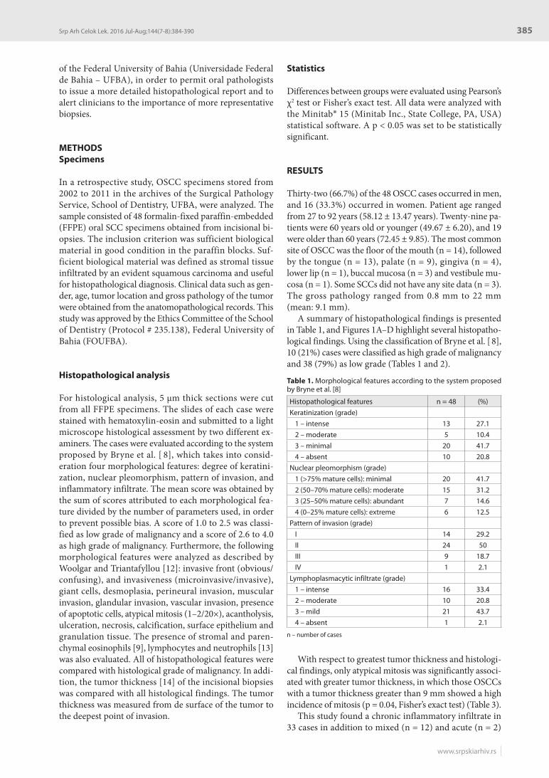
Figure 1. A – low grade OSCC with obvious invasive front (hematoxylin and eosin staining); B – low grade OSCC exhibiting perineural invasion (hematoxylin and eosin staining); C – high grade OSCC exhibiting mus-cular invasion (hematoxylin and eosin staining); D – high grade OSCC invading salivary gland (hematoxylin and eosin staining)
387Srp Arh Celok Lek. 2016 Jul-Aug;144(7-8):384-390
www.srpskiarhiv.rs
Vascular, glandular, muscle, and perineural invasion and malignancy grade
No significant association was observed between grade of malignancy and glandular (p = 0.16), muscle (p = 0.52) or perineural invasion (p = 0.56, Pearson’s χ2 test for all as-sociations). However, vascular invasion was significantly associated with cases of high-grade malignant tumors (p = 0.04, Pearson χ2 test).
Apoptotic cells and malignancy grade
Apoptotic cells were detected in 26 (54.1%) cases; only two of them were high-grade malignant SCCs, and 24 were low-grade tumors. There was no statistically significant association (p = 0.72, Pearson’s χ2 test).
Giant cells and malignancy grade
Giant cells were detected in 10 (20.8%) cases, including only one high-grade malignant tumor. No association was observed between the presence of giant cells and grade of malignancy (p = 0.34, Pearson’s χ2 test).
Necrosis and malignancy grade
Necrosis was observed in five (10.4%) cases and was as-sociated with high grade of malignancy (p = 0.04, Pearson’s χ2 test).
DISCUSSION
Although oral pathologists encounter difficulties in the histopathological analysis of incisional biopsies of OSCCs due to their size and depth, it is important that the histo-pathological reports include detailed morphological in-formation with more precise definitions and descriptions rather than only the proposed diagnostic. Careful descrip-tion of the observed histopathological features can be of high prognostic value and may contribute to the choice of treatment in some cases. Sometimes the incisional biopsy may not be representative of the entire tumor or may not comprise the more advanced part of the tumor, and a new biopsy may therefore be required. In view of these con-siderations, the present study evaluated a series of cases of OSCC, focusing on these individual histopathological features in incisional biopsies, in an attempt to associate these features with the histological grade of malignant tu-mors and other aspects such as the greatest diameter of the incisional biopsies. These histopathological features are discussed below.
The invasive front of the tumor is not always obvious [12], as observed in the present study, in which most cases exhibited an invasive front with a confusing pattern. Ac-cording to Woolgar and Triantafyllou [12], it is important that an incisional biopsy be of sufficient size and depth to include part of the advancing invasive front of the tumor. Ideally, the deep invasive front should be included, but if
not, as in the case of large tumors, the peripheral (lateral) front is often sufficiently representative to permit provi-sional evaluation. In the present study, the advancing front was not always included, with many biopsies not having the ideal size or depth to permit visualization of the in-vasive front.
Different regions of the same tumor frequently exhibit different patterns of invasion, but the most aggressive pat-tern should be recorded. For this purpose, Bryne et al. [8]
proposed a traditional classification evaluating the predic-tive value of the pattern of invasion at the tumor interface on a scale from I to IV: pattern I, well-demarcated, broad tumor invasion; pattern II, finger-like tumor invasion or through separate tumor islands, with a stellate appearance, forming strands, cords or solid bands; pattern III, invasive islands of the tumor with more than 15 cells per island; pattern IV, invasive tumor islands with fewer than 15 cells per island, including a single invasive cell. This scale was expanded by Brandwein-Gensler et al. [6], who included pattern V, defined by the presence of dispersed satellites at a distance of 1 mm or more from the tumor. In the present study, the invasion patterns I and II were predominated.
Some authors suggested the classification of a tumor as microinvasive if it is confined to the papillary lamina propria, or as superficially invasive if it remains confined to the reticular (deep) lamina propria without involvement of submucosal tissues. However, the term “superficially in-vasive” cannot be applied to tumors located in the alveolar process of the mandible or in the gingival mucosa due to the absence of submucosa [12]. In the present study, there were only four cases of OSCC in the gingival mucosa and all of them were low-grade tumors.
Accumulation of giant cells in SCCs is a rare finding but has been reported in cutaneous SCCs [4, 15]. In the present study, giant cells were detected in eight cases of OSCC. Despite the non-specificity of giant cells observed in this study, it was not possible to associate the presence of these cells with the reactive nature of the tumor, although macrophages were frequently observed in the center of some tumor islands, and a foreign body-type giant cell reaction may occur near areas of keratinization.
Desmoplasia is a histological pattern characterized by the presence of hyaline stroma and a minimal cell infiltrate consisting of spindle-shaped tumor cells or, occasionally, by a fascicular pattern [12, 16]. In non-cutaneous tumors, the presence of desmoplasia has been associated with a malignant phenotype [16]. In the present study, desmo-plasia was observed in four cases of OSCC. Despite the importance of this finding, little attention has been given to desmoplasia in histological grading systems of OSCCs. However, its presence seems to be important for tumori-genesis and metastasis [12].
Perineural invasion is an important predictor of surviv-al in patients with SCC of the oral cavity and oropharynx [17]. According to Chatzistamou et al. [14], the shape of the tumor (ill- or well-defined) is also associated with the presence of perineural invasion, as its incidence is almost twice as high for ill-defined tumors as for well-defined ones. Other authors report that tumors presenting peri-
388
doi: 10.2298/SARH1608384I
neural invasion are more likely to develop metastases [18]. The depth of the biopsy may have influenced this finding, as only two cases exhibited perineural invasion. However, in 202 tongue SCCs, Rodrigues et al. [2] observed peri-neural invasion in almost 15% of samples.
There is extensive discussion about whether sialad-enotropism should be regarded as invasion, considering its association with increased local recurrence [19]. It is therefore important to include the depth and extent of SCCs in the histopathological report [12]. In the present study, glandular invasion was observed in only three cases of SCC. These were low-grade malignant tumors that oc-curred in the floor of the mouth, tongue border, and ret-romolar region, sites where well-differentiated glandular tissue is usually found.
Vascular invasion can be identified when tumor cell ag-gregates are found within clear spaces that are completely lined by endothelial cells. Invasion of thin-walled vessels is more common, whereas invasion of muscular vessels is rare [12]. According to Rodrigues et al. [2], vascular inva-sion is more frequent than perineural invasion in tongue SCC, and this was also seen in this study in incisional bi-opsies. In the present study, four (36.3%) of the 11 cases with vascular invasion were high-grade malignant tumors and this difference was significant. According to Chandler et al. [20], muscular invasion influences the type and ex-tent of treatment of OSSC. In the present study, muscular invasion was observed in only eight cases of OSCC, most commonly in the tongue.
In this study, apoptotic cells were detected in 26 (54.1%) cases of OSCC. Apoptosis plays an important role in the removal of aberrant cells that can lead to the development of tumors. The relationship between cell growth and cell death (apoptosis) in cancer will predict the rate of tumor growth. Apoptosis is increased in pre-malignant and malig-nant oral lesions and is the main pathway of cell regulation [21]. On the other hand, according to Aoyama et al. [22], the presence or absence of mitosis is a significant indicator of neoplastic atypical squamous lesion of the uterine cervix. In the present study, atypical mitosis was found in 38 cases of OSCC and their presence was found significant in those cases with tumor thickness larger than 9 mm.
In the present study, 10.4% (n = 5) of OSCCs exhibited tumor islands with acantholysis. Honings et al. [23] found no association between the presence of acantholysis and tumor prognosis in SCC of the trachea.
The degree of keratinization of oral SCCs is a parameter of the histological grading system proposed by Bryne et al. [8]. According to Matos et al. [10], keratinization tends to be low in metastatic SCCs. This seems to be true since in the present study no keratinization was observed in 70% (n = 7) of high-grade malignant SCCs.
A surface epithelium was seen in most of the OSCC cases studied (n = 43). This finding was significant when
associated with well-differentiated OSCC. Occasionally, the origin of the overlying surface epithelium is less obvi-ous in some tumors present in the lamina propria or sub-mucosa. This is often the case when the area biopsied is an intact area adjacent to the tumor [12]. On the other hand, in a 2013 study of 36 OSCCs, 50% showed ulceration [24], but this feature was present in 37 cases of the present study. Other minor findings included calcification and necrosis. Necrosis was observed in five (10.4%) of the cases studied and was associated with high grade of malignancy. With respect to necrosis, Honings et al. [23] found no associa-tion between the presence of this histological feature and tumor prognosis in SCC of the trachea.
The chronic inflammatory response influences the prognosis of SCC, with a high-density [6, 9, 14] or low-density [10] infiltrate being associated with a better prog-nosis or regional metastases, respectively. According to Chatzistamou et al. [14], the presence of a stromal chronic inflammatory infiltrate is associated with the shape of the tumor (ill- or well-defined). In this respect, well-defined tumors exhibit an intense chronic inflammatory infiltrate and are associated with a favorable prognosis. On the other hand, tumors not associated with stromal inflammation exhibit a more aggressive growth pattern accompanied by a more expansive shape (ill-defined). However, there is clear evidence that the immune and inflammatory cells can invoke both tumor-promoting and tumor-antagoniz-ing effects, depending on the context and cell types [2].
The role of eosinophils in carcinogenesis is still a mat-ter of discussion. In the present study, eosinophils were more frequently detected in low-grade malignant tumors (74.1%), but the difference was not significant. Despite this, Goldsmith et al. [25] showed eosinophilia to be the variable that most influenced a less aggressive clinical course of SCCs. Lundqvist et al. [9] found no prognostic value in the presence of eosinophils in most of the SCC cases studied, and there was no correlation with treatment response or recurrence.
Tumor-infiltrating neutrophils have been associated with a poor clinical course of different types of cancer [18]. In the present study, neutrophils invading the tumor parenchyma were observed in four cases of SCC, three of them low-grade malignant tumors. These findings differ from those found by Trellakis et al. [13], who found that a high neutrophil infiltration was independently associated with a poor prognosis of head and neck SCCs. Further studies are necessary to clarify this matter.
In conclusion, this study suggests that incisional biop-sies may have considerable significance as they can show histopathological variables that are important to classify OSCCs into low grade and high grade in accordance with Bryne’s score used in this study. Thus, more representative biopsies might be useful to achieve this and allow a more accurate planning.
Ismerim A. B. et al. Useful histological findings in incisional biopsies of oral squamous cell carcinoma
389Srp Arh Celok Lek. 2016 Jul-Aug;144(7-8):384-390
www.srpskiarhiv.rs
REFERENCES
1. Ribeiro ACP, Silva ARS, Simonato LE, Salzedas LMP, Sundefeld MLMM, Soubhia AMP. Clinical and histopathological analysis of oral squamous cell carcinoma in young people. A descriptive study in Brazilians. Br J Oral Maxillofac Surgery. 2009; 47:95–8. [DOI: 10.1016/j.bjoms.2008.05.004] [PMID: 18586366]
2. Rodrigues PC, Miguel MC, Bagordakis E, Fonseca FP, de Aquino SN, Santos-Silva AR, et al. Clinicopathological prognostic factors of oral tongue squamous cell carcinoma: a retrospective study of 202 cases. Int. J Oral Maxillofac Surg. 2014; 43:795–801. [DOI: 10.1016/j.ijom.2014.01.014] [PMID: 24583139]
3. Anneroth G, Batsakis J, Luna M. Review of the literature and a recommended system of malignancy grading in oral squamous cell carcinomas. Scand J Dent Res. 1987; 95:229–49. [PMID: 3299675]
4. Wooff J, Werner D, Murphy J, Walsh N. Osteoclast-like giant cell reaction associated with cutaneous squamous cell carcinoma: a report of 2 cases and review of the literature. Am J Dermatopathol. 2009; 31:282–7. [DOI: 10.1097/DAD.0b013e31819cf6f4] [PMID: 19384071]
5. Barnes L, Eveson JW, Reichart P, Sidransky D. World Health Organization Classification of Tumours. Pathology and Genetics of Head and Neck Tumours. Lyon, IARC, 2005.
6. Brandwein-Gensler M, Teixeira MS, Lewis CM, Lee B, Rolnitzky L, Hille JJ, et al. Oral squamous cell carcinoma: Histologic risk assessment, but not margin status, is strongly predictive of local disease-free and overall survival. Am J Surg Pathol. 2005; 29:167–78. [PMID: 15644773]
7. Broders AC. The microscopic grading of cancer. Surg Clin North Am. 1941; 21:947–62.
8. Bryne M. Is the invasive front of an oral carcinoma the most important area for prognostication? Oral Dis. 1998; 4(2):70–7. [PMID: 9680893]
9. Lundqvist L, Stenlund H, Laurell G, Nylander K. The importance of stromal inflammation in squamous cell carcinoma of the tongue. J Oral Pathol Med. 2012; 41:379–83. [DOI: 10.1111/j.1600-0714.2011.01107.x.] [PMID: 22084865]
10. Matos FR, Lima ENA, Queiroz LMG, Silveira EJD. Perineural Invasion, and Risk Score Can Indicate Concurrent Metastasis in Squamous Cell Carcinoma of the Tongue. J Oral MaxillofacSurg. 2012; 70:1703–10. [DOI: 10.1016/j.joms.2011.08.023] [PMID: 22154400]
11. Foschini MP, Leonardi E, Eusebi LH, Farnedi A, Poli T, Tarsitano A, et al. Podoplanin and E-cadherin expression in preoperative incisional biopsies of oral squamous cell carcinoma is related to lymph node metastases. Int J SurgPathol. 2013; 21:133–4. [DOI: 10.1177/1066896912471851] [PMID: 23349470]
12. Woolgar JA, Triantafyllou A. Pitfalss and procedures in the histopathological diagnosis of oral and oropharyngeal squamous cell carcinoma and a review of the role of pathology in prognosis. Oral Oncology. 2009; 45:361–85. [DOI: 10.1016/j.oraloncology.2008.07.016] [PMID: 18849188]
13. Trellakis S, Bruderek K, Dumitru CA, Gholaman H, Gu X, Bankfalvi A, et al. Polymorphonuclear granulocytes in human head and neck cancer: enhanced inflammatory activity, modulation by cancer cells
and expansion in advanced disease. Int J Cancer. 2011; 129:2183–93. [DOI: 10.1002/ijc.25892] [PMID: 21190185]
14. Chatzistamou I, Rodriguez J, Jouffroy T, Girod A, Point D, Sklavounou A, et al. Prognostic significance of tumor shape and stromal chronic inflammatory infiltration in squamous cell carcinomas of the oral tongue. J Oral Pathol Med. 2010; 39:667. [DOI: 10.1111/j.1600-0714.2010.00911.x.] [PMID: 20618607]
15. Yozu M, Glengarry J, Ahmed SS. Cutaneous squamous cell carcinoma associated with proliferation of osteoclast-like giant cells. J Pak Med Assoc. 2011; 61:922–5. [PMID: 22360041]
16. Abbas O, Mahalingam M. Desmoplasia: not always a bad thing. Histopathology. 2011; 58:643–59. [DOI: 10.1111/j.1365-2559.2010.03617.x] [PMID: 20718872]
17. Rahima B, Shingaki S, Nagata M, Saito C. Prognostic significance of perineural invasion in oral and oropharyngeal carcinoma. Oral Surg Oral Med Oral Pathol Oral Radiol Endod. 2004; 97:423–31. [PMID: 15088027]
18. Gu FM, Gao Q, Shi GM, Zhang X, Wang J, Jiang JH, et al. Intratumoral IL17+ cells and neutrophils show strong prognostic significance inintrahepatic cholangiocarcinoma. Ann SurgOncol. 2012; 19:2506–14.
19. Daley TD, Lovas JG, Peters E, Wysocki GP, McGaw TW. Salivary gland duct involvement in oral epithelial dysplasia and squamous cell carcinoma. Oral Surg Oral Med Oral Pathol Oral Radiol Endod. 1996; 81:186–92. [PMID: 8665313]
20. Chandler K, Vance C, Budnick S, Muller S. Muscle invasion in oral tongue squamous cell carcinoma as a predictor of nodal status and local recurrence: just as effective as depth of invasion? Head NeckPathol. 2011; 5:359–63. [DOI: 10.1007/s12105-011-0296-5] [PMID: 21892763]
21. Loro LL, Vintermyr OK, Johannessen AC. Cell death regulation in oral squamous cell carcinoma: methodological considerations and clinical significance. J Oral Pathol Med. 2003; 32:125–38. [PMID: 12581382]
22. Aoyama C, Liu P, Ostrzega N, Holschneider CH. Histologic and immunohistochemical characteristics of neoplastic and nonneoplastic subgroups of atypical squamous lesions of the uterine cervix. Am J ClinPathol. 2005; 123:699–706. [DOI: 10.1309/FB900YXTF7D2HE99] [PMID: 15981809]
23. Honings J, Gaissert HA, Ruangchira-Urai R, Wain JC, Wright CD, Mathisen DJ, et al. Pathologic characteristics of resected squamous cell carcinoma of the trachea: prognostic factors based on an analysis of 59 cases. Virchows Arch. 2009; 455:423–9. [DOI: 10.1007/s00428-009-0843-6] [PMID: 19838727]
24. Allon I, Alon DM, Anavi Y, Kaplan I. The significance of surface ulceration as a sign of malignancy in exophytic oral mucosal lesions: myth or fact? Head Neck Pathol. 2013; 7:149–54. [DOI: 10.1007/s12105-012-0413-0] [PMID: 23242858]
25. Goldsmith MM, Belchis DA, Cresson DH, Merritt WD, Askin FB. The importance of the eosinophil in head and neck cancer. Otolaryngol Head Neck Surg. 1992; 106:106–27. [PMID: 1734363]
390
doi: 10.2298/SARH1608384I
КРАТАК САДРжАЈУвод Орални сквамоцелуларни карцином (ОСЦК) један је од најчешћих канцера главе и врата.Циљ рада Циљ ове студије био је да се истраже хистопа-толошке особине узорака ОСЦК добијених инцизијалним биопсијама, те да се скрене пажња клиничарима на важност репрезентативнијих биопсија.Методе рада Обухваћена су 48 узорка ОСЦК добијена ин-цизијалним биопсијама и класификованих према Бринеовој скали. Анализиранe су следеће карактеристике: инвазивни фронт, инвазивност, апоптотичне ћелије, атипична митоза, џиновске ћелије, акантолиза, улцерација, некроза, калци-фикација, површински епителијум, гранулационо ткиво, десмоплазија, инвазије ткива, инфламаторни инфилтрат и дебљина тумора.Резултати Десет (21%) случајева класификовано је као ма-лигнитет високог степена, а 38 (79%) као малигнитет ниског степена. Уочени су апоптотичне ћелије (n = 26), атипична митоза (1–2/20×; n = 38), џиновске ћелије (n = 8), акантолиза
(n = 5), некроза (n = 5), калцификација (n = 1), гранулационо ткиво (n = 32), десмоплазија (n = 4), перинеурална инвазија (n = 2), мускуларна инвазија (n = 8), инвазија ткива пљува-чне жлезде (n = 3), васкуларна инвазија (n = 10) и хронично запаљење (n = 33). Васкуларна инвазија (p = 0,04, Пирсонов χ2-тест) и некроза (p = 0,04, Пирсонов χ2-тест) значајно су по-везани са случајевима тумора високог степена малигнитета. Атипична митоза је повезана са великом дебљином тумора (p = 0,04, Фишеров егзактни тест).Закључак Ова студија сугерише да инцизијалне биопсије могу имати велику важност, јер могу указати на хистопа-толошке чиниоце који су значајни за поделу ОСЦК на оне високог и ниског степена према Бринеовој скали, коришће-ној у овом раду. Стога би репрезентативније биопсије могле бити од користи како би се постигао наведени циљ и како би било омогућено боље планирање.Кључне речи: оралне неоплазме; орални карцином, дија-гноза; хистопатолошко градирање; сквамоцелуларни кар-цином, патологија; сквамоцелуларни карцином, хирургија
Значај инцизионе биопсије оралног сквамоцелуларног карцинома за валидност хистопатолошког налазаАдна Барос Исмерим1, Флавија Кало Акино Шавијер2, Марија Кристина Теишеира Кангусу3, Лусијана Марија Педреира Рамаљо4, Иван Марсело Гонсалвес Агра5, Жеан Нунес дос Сантос2
1Државни универзитет Баије, Стоматолошки факултет, Салвадор, Баија, Бразил;2Државни универзитет Баије, Стоматолошки факултет, Лабораторија за хируршку патологију, Салвадор, Баија, Бразил;3Државни универзитет Баије, Стоматолошки факултет, Катедра за превентивну стоматологију, Салвадор, Баија, Бразил;4Државни универзитет Баије, Стоматолошки факултет, Катедра за стоматологију, Салвадор, Баија, Бразил;5Болница „Аристидес Малтез“, Одељење за хирургију главе и врата, Салвадор, Баија, Бразил
Примљен • Received: 17/07/2015 Ревизија • Revision: 29/10/2015 Прихваћен • Accepted: 09/12/2015
Ismerim B. A. et al. Useful histological findings in incisional biopsies of oral squamous cell carcinoma