use of cone beam computed tomography in the diagnosis...
TRANSCRIPT

www.pucrs.br/repositorio
Use of cone beam computed tomography in the diagnosis, planning and
follow up of a type III dens invaginatus case
Vier-Pelisser FV, Pelisser A, Recuero LC, Só MVR, Borba MG, Figueiredo JAP.
Use of cone beam computed tomography in the diagnosis, planning and follow up
of a type III dens invaginatus case. Int Endod J 2012; 45(2): 198-208.
PMID: 21978185 [PubMed - indexed for MEDLINE]
http://dx.doi.org/10.1111/j.1365-2591.2011.01956.x
International Endodontic Journal (ISSN: 0143-2885)
The PDF publisher version of this paper is not available in the PUCRS Institutional
Repository because this is a RoMEO yellow journal (Archiving Policy: Can archive
pre-print) but author cannot archive publisher's version/PDF.
A versão PDF da editora não está disponível no Repositório Institucional da
PUCRS porque este é um periódico classificado como RoMEO amarelo (Política
de Arquivamento: Pode arquivar a versão preprint ), mas o autor não pode
arquivar a versão/PDF do editor.

Use of cone beam computed tomography in the diagnosis, planning and
follow up of a type III dens invaginatus case
F. V. Vier-Pelisser1,
A. Pelisser2,
L. C. Recuero3,
M. V. R. Só4,
M. G. Borba1,
J. A. P. Figueiredo1
1Post-Graduate Program in Dentistry, Pontifical Catholic University of Rio Grande
do Sul - PUCRS, Porto Alegre, RS, Brazil
2Private Pratice, São Sebastião do Caí, RS, Brazil;
3School of Dentistry, Universidade Cruzeiro do Sul (UNICSUL), Caxias do Sul, RS,
Brazil
4Department of Endodontics, Federal University of Rio Grande do Sul - UFRGS,
Porto Alegre, RS, Brazil
Running Title: Treatment of type III dens invaginatus
Corresponding Author:
Profa. Dra. Fabiana Vieira Vier-Pelisser
E-mail: [email protected]

Abstract
Aim To present the case of a maxillary left lateral incisor with Oehlers’ type III dens
invaginatus in which cone beam computed tomography (CBCT) was used as an
adjunctive resource in the diagnosis as well as in the planning and two-year follow-
up of the nonsurgical/surgical treatment.
Summary The tooth had two root canals: a primary (main) canal with vital pulp that
appeared to be closed apically and an invaginated canal that was necrotic, wide-
open at the portal of exit and associated with a large chronic periapical lesion
extending to the apex of the maxillary left central incisor. Radiographic tracking of a
sinus tract in the labial gingiva of the affected tooth with a gutta-percha point
revealed its origin to be the invagination. The CBCT scans revealed that the
periapical radiolucency was significantly larger than seen radiographically and an
increased thickness of the buccal cortical plate. Conventional root canal treatment
of the primary canal was under taken. As nonsurgical access to the invaginated
canal was not possible, endodontic surgery was performed for curettage of the
lesion, root-end cavity preparation using ultrasonic tips and root canal filling with
white mineral trioxide aggregate. CBCT scanning after 17 months and clinical and
radiographic follow-up after 24 months revealed complete periapical repair and
absence of symptoms. Key learning points: The combination of non-surgical and
surgical treatments produced periapical repair in a tooth with type III dens
invaginatus with two root canals. CBCT may aid the diagnosis as well as the
management plan and follow-up of teeth with this developmental anomaly.
Key words: anomalies, cone beam computed tomography, dens invaginatus,
endodontic treatment, endodontic surgery.

Introduction
Dens invaginatus is a developmental anomaly that results in an enamel-
lined cavity intruding into the crown or root before the mineralisation phase (Shafer
et al. 1983, Reddy et al. 2008). Over the years several theories have been
proposed to explain the aetiology of these invaginations: constriction of the dental
arch on the enamel organ, a retardation or acceleration of growth of the internal
enamel epithelium, a distortion of the enamel organ during tooth development or
inadequate nutrition of a portion of a single tooth germ (Souza & Bramante 1998).
The classification system proposed by Oehlers (Oehlers 1957) is probably
the most clinically relevant and is by far the most commonly used in clinical
studies, case reports and case series. The cases are grouped in three major
categories, according to the depth of the invagination and the existence of
communication with the pulp tissue or periodontal ligament, regardless of the
affected tooth (anterior, posterior, supernumerary). A single tooth can present
multiple invaginations, but each one may fall into a different classification: type I is
an enamel-lined minor invagination occurring within the confines of the tooth crown
not extending beyond the cementoenamel junction; in type II, the invagination
extends apically beyond the cementoenamel junction, but remains as a blind sac
and it may or may not communicate with the dental pulp; in type III, the
invagination penetrates through the root, perforating the apical area and having a
second foramen in the apical or periodontal area, but there is no immediate
communication with the pulp.
The most frequently affected tooth is the maxillary lateral incisor (Shafer et
al 1983, Rotstein et al 1987,Yeh et al. 1999, Alani & Bishop 2008), with a reported
incidence of approximately 1% and a bilateral involvement of under 1%. In
decreasing order of frequency, other teeth that develop this anomaly are the
maxillary central incisors, premolars, canines, and molars (Yeh et al. 1999).
The clinical appearance of dens invaginatus varies considerably. The crown
of affected teeth can have normal morphology or it can also be associated with
unusual forms such as greater buccolingual dimension, peg-shaped, barrel-
shaped, conical shapes and talon cusps (Souza & Bramante 1998, Reddy et al

2008). A deep foramen caecum might be the first clinical sign indicating the
presence of an invaginated tooth. As this area is difficult to access and clean,
caries develop with subsequent pulp necrosis and evolution to apical pathosis (Yeh
et al 1999, Jung 2004).
Dens invaginatus may require treatment that can range from less invasive
procedures to the combination of different therapies (Hülsmann 1997) associated
with advanced technical resources for diagnosis and treatment planning (Reddy et
al 2008, Alani & Bishop 2008). In case of pulp involvement, root canal treatment
has been recommended with high success rates (Rotstein et al 1987, Szajkis &
Kaufman 1993). Endodontic surgery is the treatment modality indicated when
endodontic therapy fails, or when endodontic treatment or retreatment is
impossible or would not achieve better results. It is also indicated for cases of
severe forms of dens invaginatus. In other cases, combined treatment may be
necessary, that is, root canal treatment followed by endodontic surgery due to the
complexity of the root morphology or in case of large apical lesions (Rotstein et al.
1987, Souza & Bramante 1998, Schmitz et al. 2010).
The bulky and irregular volume of the invaginated canal makes cleaning and
shaping difficult. The use of ultrasonic tips has been described as being of great
efficacy in these cases (Skoner & Wallace 2008). Lateral condensation of
thermoplasticized gutta-percha has been recommended for canal filling when
conventional access to the invaginated canal is possible (Rotstein et al. 1987,
Lichota et al. 2008). Mineral trioxide aggregate (MTA) has also been largely used
in the treatment of invaginated teeth because of its biocompatibility, reparative
capacity by stimulation of mineralised tissue deposition, superior sealing capacity
in the presence of moisture, and reduction in the treatment time (Jaramillo et al.
2006, Sathorn & Parashos 2007, Reddy et al. 2008, Demartis et al. 2009).
Conventional radiography has an important role in the evaluation of the
complex morphology of the root canal system, but they only provide two-
dimensional representation of a three-dimensional structure (Jung 2004, Reddy et
al. 2008). The use of more advanced imaging methods, such as cone beam
computed tomography (CBCT), has become ever more common in a number of

dental specialties. Endodontic applications of CBCT include the diagnosis of
periapical lesions due to pulpal inflammation, more detailed visualisation of canals,
elucidation of internal and external resorption, and detection of root fractures
(Tyndall & Rathore 2008). CBCT has been specifically designed to produce
undistorted three-dimensional reconstruction of the maxillofacial skeleton as well
as three-dimensional images of the teeth and their surrounding tissues (Al-Rawy et
al. 2010). This is usually achieved with a substantially lower effective radiation
dose compared to conventional CT, though higher than that of conventional dental
radiography techniques (Roberts et al. 2009). Periapical disease may be detected
sooner using CBCT compared to periapical images, revealing the actual size,
extent, nature and position of periapical and resorptive lesions. In addition, CBCT
scans provide valuable information about dental anatomy (Patel 2009), and can be
used prior to periapical surgery to assess the thickness of the cortical and
cancellous bone and the inclination of roots in relation to the surrounding jaw (Patel
et al. 2007), and thus help plan the treatment (Reddy et al. 2008, Demartis et al.
2009).
This paper describes the use of CBCT as an auxiliary resource in the
diagnosis as well as in the planning and 2-year follow-up of the non-
surgical/surgical treatment of a maxillary left lateral incisor with Oehlers’ type III
dens invaginatus in which the invaginated canal was associated with chronic apical
periodontitis and the primary (main) canal contained vital tissue.
Case Report
A 12-year-old boy was referred by a general dentist for root canal treatment
of the maxillary left lateral incisor tooth. The patient was under fixed orthodontic
treatment. Root canal treatment of the maxillary left central incisor had been
iniciated by the general dentist after confirmation of pulp necrosis, though without
remission of a sinus tract in the labial gingiva of the lateral incisor. Radiographic
tracking of the sinus tract with a gutta-percha point revealed its origin to be in wide-
open apical end of an invaginated canal of this tooth.

In spite of the sinus tract indicating development of necrosis, the lateral
incisor responded to cold and hot stimuli. The tooth presented greater mesiodistal
width than usual (Fig. 1a), normal color, normal gingival tissues and probing depth
(<3mm), and a composite resin restoration on the palatal aspect (Fig. 1b). Review
of medical history was uneventful and no previous dental trauma was reported.
Radiographic examination revealed that the tooth had Oehlers’ type III dens
invaginatus and two root canals: a separate primary (main) canal that appeared to
be closed apically and an invaginated canal that was wide-open at the portal of exit
and associated with a large periapical lesion extending to the apex of the maxillary
left central incisor (Fig. 1c). Approximately two thirds of the invagination seemed to
be lined by enamel. The lack of communication between the vital pulp tissue in the
main canal and the necrotic tissue in the invaginated canal explained the positive
response of the tooth to the pulp tests. Radiographic examination of the
contralateral tooth did not reveal a malformation.
A CBCT scan of the involved teeth was performed as a complementary
examination (i-CAT CBCT scanner, Imaging Sciences International, Inc, Hatfield,
Pennsylvania, USA), at 120KVp, in sections of 1.0mm thickness. The CBCT scans
revealed the periapical radiolucency was larger than seen radiographically,
measuring 2.0 x 1.5 cm axially (Fig. 2a). The two canals were visualised: the main
canal located distally, which tapered progressively towards the apex; and an
invaginated canal mesial to and separate from the main root canal, which was
obliterated in the cervical third, appeared wide in the middle third, and then
became discontinuous in the apical third (Fig. 2a). An increased thickness of the
buccal cortical plate was observed (Fig. 2b) and the lesion was in continuity with
the incisive foramen (Fig. 2).
As non-surgical access to the invaginated canal was not possible,
endodontic surgery was planned. Conventional root canal treatment of the main
canal was achieved prior to surgery due to the high risk of rupture of its
neurovascular bundle during curettage of the periapical lesion. After local
anaesthesia with 2% lidocaine and epinephrine 1:100,000 (DFL, Rio de Janeiro,
RJ, Brazil), access to the pulp chamber was gained under rubber dam isolation

and the working length was established 1 mm short of the apex using an electronic
apex locator (Bingo 1020; Forum, Rio Comprido, RJ, Brazil) followed by
radiographic confirmation (Fig. 3a). The canal was instrumented according to a
crown-down technique to a size 50 Flexofile instrument (Dentsply Maillefer,
Ballaigues, Switzerland) followed by a step-back preparation from the working
length at 1 mm increments to a size 70 Flexofile instrument. Throughout the
preparation, the canal was copiously irrigated with 2.5% sodium hypochlorite
(Farmácia Botânica, São Sebastião do Caí, RS, Brazil), followed by a final flush
with 17% EDTA (Biodinâmica Europa S.L., Ibiporã, PR, Brazil) for 3 min and
neutralisation with 2.5% sodium hypochlorite. The canal was the dried with sterile
absorbent paper points filled with lateral condensation of gutta-percha cones
(Tanari, Manaus, AM, Brazil) and AHPlus sealer (Dentsply DeTrey, Kontanz,
Germany). The root canal treatment of the maxillary left central incisor was also
completed following the same protocol (Fig. 3b). The access cavities of both teeth
were restored with Filtek-Z250 composite resin (3M ESPE, St. Paul, MN, USA).
The orthodontist was asked to discontinue temporarily the application of
forces to the maxillary teeth. Endodontic surgery was performed in a subsequent
session. After local anaesthesia with 2% mepivacaine (DFL, Rio de Janeiro, RJ,
Brazil), incision and elevation of a full-thickness flap (Fig. 3c), the large lesion
involving the roots of the central and lateral incisors was curetted and root-end
resection was performed in both teeth (Fig. 3d). Root-end cavity preparation of the
invaginated canal was carried out with ultrasonic tips followed by root canal filling
with white MTA (Angelus, Londrina, PR, Brazil) (Fig 3e).
The flap was repositioned, sutured with nylon 4.0 sutures (Somerville,
Jaboatão dos Guararapes, PE, Brazil) and an immediate postoperative periapical
radiograph was taken (Figure 4A). Sutures were removed one week later. The
histopathological diagnosis of the lesion was chronic periapical abscess.
Radiographic examination 3 months (Fig. 4b), 6 months and 12 months (Fig. 4c)
postoperatively showed ongoing periapical repair.
The CBCT scan was repeated 17 months after treatment because the
buccal cortical plate on the left side (adjacent to the dens in dent) exhibited an

increased volume in comparison to the same region on the right side, although
much smaller than observed in the previous examination. This clinical finding,
however, was not supported by any radiographic evidence, as the 3-, 6- and 12-
month postoperative radiographic images showed good repair of the area. Sagittal
CBCT sections of the left central and lateral incisors revealed a discrete residual
apical rarefaction associated with the maxillary left lateral incisor and discrete
periodontal ligament widening of the maxillary left central incisor, probably due to
the orthodontic treatment (Fig. 5a,b). The axial CBCT sections of the maxillary left
lateral incisor showed complete filling of the main and invaginated canals as well
as apical repair. In addition, the axial CBCT sections allowed the assumption taht
the anatomical feature of type III dens invaginatus may cause a thick buccal plate
(Fig. 5c-e). At 24 months, the radiographic examination revealed an excellent
periapical repair with the patient having remained symptom-free.
Discussion
This paper presents an interesting case of Oehlers’ type III dens invaginatus
in a maxillary left lateral incisor with two root canals, a primary canal with vital
tissue and a invaginated canal with necrotic tissue, which was associated with a
periapical lesion extending to apex of the maxillary left central incisor, as confirmed
by apical axial CBCT sections (Fig. 2a). Periapical radiographs are limited in
revealing the type, extension and complex morphology of dens invaginatus as well
as the actual bone loss compared to tomographic techniques. More advanced
imaging techniques, such as CBCT, may aid the diagnosis as well as the
management plan and follow-up of teeth with this dental developmental anomaly.
Although the maxillary left lateral incisor was responsive to the pulp
sensibility tests, a sinus tract had developed on the labial gingiva close to the
apical region of this tooth. Radiographic tracking of the sinus tract revealed it
originated from the invaginated canal. An explanation for the contamination of this
canal could be the presence of a cavitated carious lesion in the palatal surface of
this tooth, which had greater mesiodistal width than usual (Pai et al. 2004) and had
been previously removed and restored with composite resin. The presence of

caries in this region could have led to bacterial penetration into the invaginated
canal, causing apical periodontitis. Hülsmann (1997) has reported that the enamel
is fragile and hypomineralized in invagination. It is possible that the dentist who
placed the original composite filling did not notice the presence of a small
perforation that served as a pathway of communication between the floor of the
cavity and the invaginated canal.
De Smit et al. (1984) performed a histological investigation of six
invaginated human maxillary incisors, and found that the outlines of the
invaginations resembled bottles with narrow necks directed at the incisal edge of
the teeth. Structurally normal enamel forms a uniform layer between dentine and
inner connective tissue of the invagination. At the entrance of the invaginated
portion, both enamel layers are so close that only one band of connective tissue
can be observed. Apically, the centrally positioned connective tissue was richly
supplied with blood vessels and lined with layers of epithelial tissue, demonstrating
various stages of an involutive enamel organ. De Smit et al. (1984) believed that
after eruption, these teeth would lose this blood supply in the central portion of the
invagination, resulting in necrosis of this tissue. This could be another explanation
for the development of chronic apical periodontitis associated with the invaginated
canal.
Because of their unusual root canal anatomy and wide apical foramen, teeth
with Oehlers’ type III dens invaginatus can be treated with a combination of non-
surgical and surgical procedures or extraction, as the last resort in more severe
cases (Pai et al. 2004). The complex anatomy of these teeth makes conventional
root canal treatment challenging, especially in those cases with a wide open
foramen. An immature apex causes additional problems due to the possibility of
overfilling and the difficulty in achieving a satisfactory apical seal (Sathorn &
Parashos, 2007, Reddy et al. 2008). Therefore, although conservative root canal
treatment is one of the possibilities for Oehlers’ type III dens invaginatus (Chen et
al. 1998, Holtzman 1998, Gonçalves et al. 2002, Jung, 2004, Pai et al. 2004,
Jaramillo et al. 2006, Lichota et al 2008, Demartis et al. 2009, Kusgoz et al. 2009),
non-surgical and surgical endodontic interventions were combined in the present

case (Ortiz et al. 2004, Reddy et al. 2008, Schmitz et al. 2010). Surgical apical
access was necessary because it was not possible to locate the canal a coronal
access due to an accentuated cervical narrowing, as shown on the axial CBCT
scans (Fig. 2a) and also because the invaginated canal had an unusual anatomy
and was wide-open at its portal of exit. Cleaning and shaping of the dens track was
achieved by root canal preparation with ultrasonic tips (Skoner & Wallace 1994,
Girsch & McClammy 2002). Skoner & Wallace (1994) affirmed that the use of
ultrasonic tips improves significantly the surgical phase of the treatment. The use
of an operative microscope could have aided locating the canal in the cervical third
(Jung 2004). However, as an operative microscope was not available, conventional
access to the canal was not attempted because of the risk of accidents such as
lateral perforation with burs.
Most authors (Yeh et al. 1999, Jung 2004, Pai et al. 2004, Jaramillo et al
2006, Schmitz et al. 2010) recommend successive changes of calcium hydroxide
dressing in immature teeth to stimulate the formation of a mineralised tissue barrier
prior to root filling. However, this treatment protocol requires longer chairtime and
several clinical sessions. In the present case, as the patient lived in a city far from
the dental office and was undergoing orthodontic treatment, surgical treatment of
the invaginated canal seemed to be indicated. It should be mentioned that the
application of orthodontic forces to the tooth was discontinued until apical repair
was observed.
Kusgoz et al. (2009) presented a case of type III dens invaginatus that was
resolved successfully with non-surgical root canal treatment and exhibited healing
after 18 months without any need for further surgical intervention despite the
complex anatomy of dens invaginatus. However, the treatment of the invaginated
canal was performed exclusively in its enamel-lined portion. It is known that the
invagination can be completely lined by enamel, but cementum is frequently seen
in its apical portion (Oehlers 1957). In the present case, the apical lesion was
extensive, also reaching the maxillary left central incisor (Figs. 1a, 2, 3d,e).
Therefore, the treatment of choice, that is, disinfection and retrofilling of the apical
portion of the canal not lined by enamel with MTA would increase the chances of

success since bacteria could have remained in this part of the canal. In addition,
MTA sets in the presence of moisture, is biocompatible and stimulates apical repair
(Torabinejad et al. 1998, Economides et al. 2003), being the material of choice in a
number of cases of dens invaginatus (Reddy et al. 2008, Jaramillo et al. 2006,
Demartis et al. 2009).
Teeth with Oehlers’ type III dens invaginatus present two canals with (Chen
et al. 1998, Yeh et al. 1999, Jaramillo et al. 2006, Lichota et al. 2008, Demartis et
al. 2009) or without (Szajkis & Kaufman, 1993, Holtzman 1998, Gonçalves et al.
2002, Kusgoz et al. 2009) communication between them. Endodontic treatment of
the primary canal is indicated when there is communication with a necrotic
invaginated canal (Chen et al. 1998, Lichota et al. 2008, Demartis et al. 2009).
However, several authors have stated that root canal treatment of the primary
canal is not necessary if it is not communicating with the invaginated canal or when
pulp necrosis is not diagnosed (Szajkis & Kaufman 1993, Holtzman 1998,
Gonçalves et al. 2002). In the present case, although the canals were separated,
endodontic treatment of the primary canal was done prior to surgery because
rupture of its neurovascular bundle could occur during surgical removal of the
apical lesion, causing necrosis and worsening the prognosis of the case.
This case illustrates that the combination of treatments produced periapical
repair in a tooth with type III dens invaginatus with two root canals. While the
primary canal was treated by conventional non-surgical therapy, endodontic
surgery was necessary to treat the invaginated canal with a wide apical opening
associated with the large periapical lesion. In addition, CBCT was an important
auxiliary resource in the diagnosis, treatment planning and follow-up. CBCT has
shown great benefit in the localisation and identification of root canals (Baratto
Filho et al. 2009) provides the unique possibility of evaluating periapical lesions
three-dimensionally with respect to their extension, size and nature (Patel 2009). In
the present case, it was possible to determine that the lesion extended from the
maxillary left central incisor to the maxillary left lateral incisor as well as its intimate
relationship with the nasopalatine (incisive) foramen, the presence of increased
thickness of the buccal cortical plate. The CBCT scans revealed that the

invagination was narrowed in the cervical third and lined by enamel, which made it
difficult reaching the invaginated portion via coronal access.
CBCT could have been used as a diagnostic aid solely in the initial phase of
the treatment, respecting the “ALARA” Radiation Principle (As Low As Reasonably
Achievable). Nevertheless, the CBCT scan was repeated 17 months after
treatment because the buccal cortical plate on the left side (adjacent to the dens in
dent) exhibited an increased thickness in comparison to the same region on the
right side, although much smaller than observed in the previous examination. This
clinical finding, however, was not supported by any radiographic evidence as the 3-
, 6- and 12-month postoperative radiographic images showed good repair of the
area. It was unclear if the buccal plate thickness was due to the maintenance of the
lesion, or if it was due to the anatomical feature of the dens invaginatus, as the
patient was not seen before the lesion was present. It was not possible to evaluate
this feature precisely using through bi-dimensional periapical radiographs, and the
decision to request another CBCT was because if the lesion persisted, this could
lead to a new surgery, or even to the progression of a lesion. CBCT provided an
accurate view of the repair process and assuring that healing was ocorring.
Following of the new CBCT scan, it became clear that it was probably the
anatomical feature of type III dens invaginatus that caused the remaining thickness
on the buccal plate, and no further intervention was necessary. Therefore, in this
case, repeating the CBCT scan was useful to avoid misinterpretations. The
decision to repeat the CBCT was also supported by the fact that the extent of the
lesion was much greater in the initial CBCT scan than it appeared in the initial
radiographic view. In the same way, the follow-up radiographic views showed
complete healing, whilst discrete residual apical rarefaction associated with the
maxillary left lateral incisor and discrete periodontal ligament widening of the
maxillary left central incisor could still be seen on the new CBCT scan. In addition,
it confirmed the periapical repair as well as the quality of the root filling of the
invaginated canal with MTA (Al-Rawy et al. 2010).

Conclusion
The use of CBCT contributed to determine the actual extension of the
chronic apical periodontitis associated with a maxillary left lateral incisor with type
III dens invaginatus and provided more details of the internal anatomy of this
developmental dental anomaly. .
References
Alani A, Bishop K (2008) Dens invaginatus. Part 1: classification, prevalence and
aetiology. International Endodontic Journal 41, 1123-36.
Al-Rawy B, Hassan B, Vandenberge B, Jacobs R (2010) Accuracy assessment of
three-dimensional surface reconstructions of teeth from cone-beam computed
tomography scans. Journal of Oral Rehabilitation 37, 352-8.
Baratto Filho F, Zaitter S, Haragushiku GA, De Campos EA, Abuabara EA, Correr
GM (2009) Analysis of the internal anatomy of maxillary first molars by using
different methods. Journal of Endodontics 35, 337-42.
Chen YH, Tseng CC, Harn WM (1998) Dens invaginatus: Review of formation and
morphology with 2 cases reports. Oral Surgery, Oral Medicine, Oral Pathology,
Oral Radiology, and Endodontology 86, 347-52.
De Smit A, Jansen HW, Dermaut L (1984) An histological investigation of
invaginated human incisors. Journal de Biologie Buccale 12, 201-9.
Demartis P, Dessì C, Cotti M, Cotti E (2009) Endodontic treatment and hypotheses
on an unusual case of dens invaginatus. Journal of Endodontics 35, 417-21.
Economides N, Pantelidou O, Kokkas A, Tziafas D (2003) Short term periradicular
tissue response to MTA as root end filling material. International Endodontic
Journal 36, 44-8.
Girsch WJ, McClammy TV (2002) Microscopic removal of dens invaginatus.
Journal of Endodontics 28, 336-9.
Gonçalves A, Gonçalves M, Oliveira DP, Gonçalves N (2002) Dens invaginatus
type III: report of a case and 10-year radiographic follow-up. International
Endodontic Journal 35, 873-9.

Holtzman L (1998) Conservative treatment os supernumerary maxillary incisor with
dens invaginatus. Journal of Endodontics 24, 378-0.
Hülsmann M (1997) Dens invaginatus: aetiology, classification, prevalence,
diagnosis, and treatment considerations. International Endodontic Journal 30, 79-
90.
Jaramillo A, Fernández R, Villa P (2006) Endodontic treatment of dens invaginatus:
A 5-year follow-up. Oral Surgery, Oral Medicine, Oral Pathology, Oral Radiology,
and Endodontology 101, 15-21.
Jung M (2004) Endodontic treatment of dens invaginatus type III with three root
canals and open apical foramen. International Endodontic Journal 37, 205-13.
Kusgoz AK, Yildirim T, Kayipmaz S, Saricaoglu S (2009) Nonsurgical endodontic
treatment of type III dens invaginatus in maxillary canine: an 18-month follow-up.
Oral Surgery, Oral Medicine, Oral Pathology, Oral Radiology, and Endodontology
107, 103-6.
Lichota D, Lipski M, Wozniak K, Buczkowska-Radlinska J (2008) Endodontic
treatment of a maxillary canine with type 3 dens invaginatus and large periradicular
lesion: a case report. Journal of Endodontics 34, 756-8.
Oehlers FA (1957) Dens invaginatus: variations of the infolding process and
associated anterior crown forms. Oral Surgery, Oral Medicine, Oral Pathology, Oral
Radiology, and Endodontology 10, 1204-18.
Ortiz P, Weisleder R, Villareal De Justus Y (2004) Combined therapy in the
treatment of dens invaginatus: case report. Journal of Endodontics 30, 672-4.
Pai SF, Yang SF, Lin LM (2004) Nonsurgical endodontic treatment os dens
invaginatus with large periradicular lesion: a case report. Journal of Endodontics
30, 597-600.
Patel S (2009) New dimensions in endodontic imaging: Part 2. Cone Beam
computed tomography. International Endodontic Journal 42, 463-75.
Patel S, Dawood A, Ford TP, Whaites E (2007) The potential applications of cone
beam computed tomography in the management of endodontic problems.
International Endodontic Journal 40, 818-30.

Reddy YP, Karpagavinayagam K, Subbarao CV (2008) Management of dens
invaginatus diagnosed by spiral computed tomography: a case report. Journal of
Endodontics 34, 1138-42.
Roberts JA, Drage NA, Davies J, Thomas DW (2009) Effective dose from cone
beam CT examinations in dentistry. British Journal of Radiology 82, 35-40.
Rotstein I, Stabholz A, Heling I, Friedman S (1987) Clinical consideration in
treatment of dens invaginatus. Endodontics & Dental Traumatology 3, 249-54.
Sathorn C, Parashos P (2007) Contemporary treatment of class II dens
invaginatus. International Endodontic Journal 40, 308-16.
Schmitz MS, Montagner F, Flores CB, Morari VH, Quesada GA, Gomes BP (2010).
Management of dens invaginatus type I and open apex: report of three cases.
Journal of Endodontics 36, 1079-85.
Shafer WG, Hine MK, Levy B (1983). A textbook of pathology. 4th ed. Philadelphia:
WB Saunders Co.
Skoner JR, Wallace JA (1994) Dens invaginatus: another use for the ultrasonics.
Journal of Endodontics 20, 138-40.
Souza SMG, Bramante CM (1998) Dens invaginatus: treatment choices.
Endodontics & Dental Traumatology 14, 152-8.
Szajkis S, Kaufman AY (1993) Root infolding treatment: A conservative approach
in endodontics. Journal of Endodontics 19, 576-8.
Torabinejad M, Ford TR, Abedi HR, Kariyawasam SP, Tang HM (1998) Tissue
reaction to implanted root end filling materials in the tibia and mandible of guinea
pigs. Journal of Endodontics 24, 468-71.
Tyndall DA, Rathore S (2008) Cone-beam CT diagnostic applications: caries,
periodontal bone assessment, and endodontic applications. Dental Clinics of North
America 52, 825-41.
Yeh SC, Lin YT, Lu SY (1999) Dens invaginatus in the maxillary lateral incisor:
treatment of 3 cases. Oral Surgery, Oral Medicine, Oral Pathology, Oral Radiology,
and Endodontology 87, 628-31.

Figure Legends
Figure 1 Panel of preoperative images. (a) Buccal clinical photograph showing
tooth 22 had greater mesiodistal width than usual and normal color; (b) Palatal
clinical photograph showing a composite resin restoration (white arrowhead); (c)
Periapical radiograph showing the tooth 22 had an Oehlers’ type III dens
invaginatus and two root canals as well as a periapical radiolucency extending
from the wide-open apical extent of the invaginated canal to the apex of tooth 21.
Note that approximately two thirds of the invagination is lined by enamel (white
arrowhead).
Figure 2 CBCT images. (a) Axial CBCT sections from the cervical, middle and
apical thirds of the dens invaginatus. Note that the apical osteolytic zone is
significantly larger than seen radiographically, measuring 2.0 x 1.5 cm axially. The
two canals can be seen: the main canal (white arrowhead) located distally, which
tapered progressively towards the apex; and an invaginated canal mesial (black
arrowhead) to and separate from the main root canal, which was obliterated in the
cervical third, appeared wide in the middle third, and then became discontinuous in
the apical third. (b) The sagittal CBCT sections of tooth 22 (top) show an increased
thickness of the buccal cortical plate, the involvement of the tooth with the
periapical lesion and the lesion in continuity with the incisive foramen. The sagittal
CBCT sections of tooth 22 with the dens invaginatus (bottom) show different
aspects of the invaginated and main canals.
Figure 3 Periapical radiographs showing root canal length determination (a) and
filling (b) of tooth 22 with Oehlers’ type III dens invaginatus. Note completion of root
filling of tooth 21. Endodontic surgery performed after nonsurgical treatment of the
main canal. (c) Exposure of the lesion after full-thickness flap reflection. (d) Bone
window enlarged for removal of the lesion involved the apices of teeth 22 and 21

followed by root-end resection of both teeth. (e) Root canal filling of the invaginated
canal with white MTA.
Figure 4 Periapical radiographs taken immediately (a), 3 months (b), 12 months (c)
and 24 months (d) after endodontic surgery showing periapical repair.
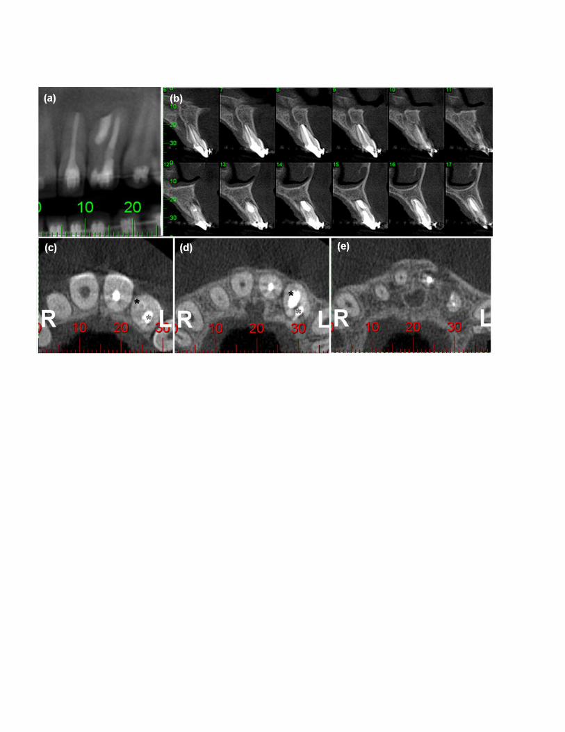
Figure 5 Seventeen-month postoperative CBCT images. (a) Coronal CBCT
section showing apical healing. (b) Sagittal CBCT sections showing discrete
residual apical rarefaction associated with tooth 21 and discrete periodontal
ligament widening of the tooth 22 (dens invaginatus). Panel of axial CBCT
sections. (c) Image of the cervical third showing filling of the main canal (grey
arrow) and invagination lined by enamel (black arrow), (d) Image of the middle third
showing complete filling of the main canal (grey arrow) and the invagination (black
arrow), (e) Image of the apical third showing healing.
Figure 1

Figure 2
Figure 3

Figure 4
Figure 5