urinary system - class videos for anatomy and
TRANSCRIPT
1
Human Anatomy and
Physiology IILaboratory
Anatomy of the Urinary System
This lab involves the exercise in the lab manual entitled “Anatomy of the Urinary System”. In this lab you will look at urinary system histology, and anatomy.Complete the review sheet from the exercise and take the online urinary system quiz. As an alternate your instructor may have you submit a drawing of kidney tissue from the Virtual Microsocpe or other histology site.There are also videos showing cadaver dissection of the urinary tract and sheep kidney.Click on the sound icon for the audio file (mp3 format) for each slide. There is also a link to a dowloadable mp4 video which can be played on an iPod.

2
Organs of The Urinary SystemAdrenal gland
Kidney
Ureter
Urinary bladder
Urethra
Renal artery and vein
Stores urine
Carries urine to the bladder
Expels urine from the bladder
Maintains homeostasis of the blood.

3
Structure of the Kidney
Pyramid
Cortex Ureter
Renal pelvis
Calyx
Renal column
Medulla
}
Papilla
The kidney is composed of several layers and is covered with a fibrous capsule, the renal capsule. The outer layer of the kidney is the cortex. It contains the major (upper) portion of the nephrons. The middle layer of the kidney is the medulla. It is composed of the triangular shaped pyramidsand the renal columns. The pyramids contain the collecting tubules and loops of Henle, the lower portion of the nephrons. These tubules run nearly parallel to one another and give the pyramids a grain which leads to their points or papillae. The renal columns are regions between the pyramids in which blood vessels run to and from the cortex. The papilla of each pyramid projects into a funnel-shaped area known as the calyx. The calyces (plural of calyx) collect the urine released from the papillae and allow itto drain into a large area known as the renal pelvis and then into the ureter.

4
Kidney Section
Renal capsule
Cortex
Pyramids
Calyx
Renal pelvis
Ureter
Renal column
Renal papilla
This view of the kidney shows not only the regions mentioned previously but also the manner in which blood vessels supply these regions.

5
Kidney Vascularization
Renal vein
Renal arteryLobar artery
and vein
Interlobar artery and vein
Arcuate artery and vein
Interlobular artery and vein
12
3
4
5
Segmental artery
The blood supply of the kidney is paramount in its function. The two kidneys receive between 15 and 20% of the body's systemic blood flow at rest. The renal artery branches into lobar and then interlobar arteries. These pass through the renal columns toward the cortex. Arcuate arteries branch into the cortex and lead to interlobular arteries which distribute the blood evenly throughout the cortex to the afferent arterioles which serve the nephrons. Blood flow leaving the nephrons returns by veins of the corresponding names.

6
Nephron Vasculature
Cortical Nephron Juxtamedullary nephron
Cortex
Medulla
Peritubularcapillaries
Vasa recta
Convoluted tubules
Convoluted tubules
Collecting tube
Loop of Henle
Loop of Henle
Interlobular artery
Cortical nephrons have short loops of Henle which barely enter the medulla. Longer loops which dip much further into the medulla belong to juxtamedullary nephrons. These nephrons are important for concentrating the urine by absorbing extra water.

Orientation of the Nephron Structures
Cortex
Capsule GlomerulusDistal convoluted tubule (DCT)
Juxtamedullarynephron
Outer medulla
Medulla
Inner medulla
Proximal convoluted tubule (PCT)
Cortical nephronMedullary ray -contains only straight tubules
Collecting tubule
Ascending limbDescending limb }Loop of
Henle
Collecting duct
Papilla
Here you see the relationship of the nephron types to the parts of the kidney.

8
Normal Human Kidney
Renal cyst
Fetal lobules
Here is a normal adult kidney. The capsule has been removed and a pattern of “fetal lobules” still persists, as it sometimes does. The hilus at the mid left contains some adipose tissue. At the lower right is a smooth-surfaced, small, clear fluid-filled simple renal cyst. Such cysts occur either singly or scattered around the renal parenchyma and are not uncommon in adults. Only when cysts are large and extensive do they have the potential to interfere with kidney structure and function.

9
Kidney Section
cortex pyramid Renal columncalyx
In a sectioned human kidney can easily be seen the regions shown in previous slides. Much of the hilus (notch) of the kidney is filled with the fat, the yellowish tissue.

Glomerulus
Bowman's Capsule
Convoluted Tubule
A medullary ray is a group of straight tubules of the loop of Henleand collecting tubules, along with blood vessels which project from the cortex into the medulla.
Structure of the Cortex
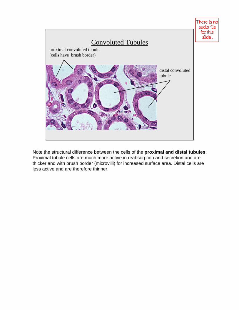
Convoluted Tubulesproximal convoluted tubule (cells have brush border)
distal convoluted tubule
Note the structural difference between the cells of the proximal and distal tubules. Proximal tubule cells are much more active in reabsorption and secretion and are thicker and with brush border (microvilli) for increased surface area. Distal cells are less active and are therefore thinner.

The collecting ducts have simple cuboidal epithelia as do some of the other tubule segments. A group of blood vessels called thevasa recta parallel these tubules running into the medulla. There are no arteries or arterioles supplying blood to the medullary structures, only the vasa recta.
Renal Medulla
In the medulla, the collecting tubes (ducts) and loops of Henle run parallel with one another as they travel through the pyramids. Therefore these tubules appear elongated when compared with those in the cortex. Also seen in the medulla are the blood vessels of the vasa recta which surround the long loops of Henle from the juxtamedullary nephrons.

Two layers of smooth muscle (three near the bladder) move urine by peristalsis.
Mucosa of transitional epithelium allows expansion and damping of pressure.
Low power
High power
The Ureters
Transitional epithelial lining allows both the bladder and ureter to stretch

Three layers of smooth muscle in the urinary bladder, called thedetrusor muscle, cause a compression during micturition. The bladder wall has extensive rugae, flattened in this distended bladder. The transitional epithelial lining (on the right side) permits expansion as the bladder fills with urine.
Urinary Bladder

15
The Urinary Bladder
ureters
rugaeUreter
openings
3-layered detrusormuscle
Trigone
Urethra
Prostate gland
Bulbourethral gland
Urethra
Urine travels to the urinary bladder through the ureter by peristalsis. The ureter has two layers of smooth muscle which work like smooth muscle in the intestine, except they are in reversed position (longitudinal toward the inside, circular toward the outside). The ureter is lined with transitional epithelium to allow for stretch and reduce back pressure on the kidney. The bladder is also lined with transitional epithelium and has many rugae for expansion. The bladder's detrusor muscle consists of three layers like the stomach's and also serves for compression. At the lower end of the bladder the ureteral openings form a triangle with the urethra which is called the trigone. The trigone has longitudinal folds which funnel the urine toward the urethra. These folds help squeeze the ureteral openings closed when micturition occurs. The urethra varies from a short tubule in females to a longer tubule in males with several sections (see diagram). Near the bladder the urethra is lined with transitional epithelium and near the external os it is stratified squamous, while in the middle it is pseudostratified columnar epithelium.

16
Lab Protocol1) Complete the Review Sheet for this exercise .
2) Take the quiz on the urinary system.
3) Use ADAM to identify structures of the digestive system. (See next slide)
4) View the cadaver video on the urinary tract.

17
ADAM Interactive Anatomy
Dissectible Anatomy, Male, Anterior, Window centered on abdomen, Begin with Layer Indicator at 234, scroll to Layer Indicator 238 and 242 for the external and internal anatomy of the kidney, and the ureter.
Atlas Anatomy, Male, Anterior, System, Urinary, Renal Arteries
Atlas Anatomy, Male, Anterior, System, Urinary, Diagram of Nephron
Atlas Anatomy, Male, Anterior, System, Urinary, Diagram of RenalGlomerulus