urease inhibition potential and molecular docking of ... · university of swabi urease inhibition...
TRANSCRIPT

Urease inhibition potential and molecular docking of dihydroquercetin anddihydromyricetin isolated from Picea smithiana (wall) Boiss.
Kashif Bashir1, Bashir Ahmad1, Abdur Rauf2*, Sami Bawazeer3, Khaliq Ur Rahman2, TayyebaRehman4, Muhammad Saleem5, Rao Saeed Ahmed5, Huang Linfang6, Rabia Ikram7
1Center of Biotechnology and Microbiology, University of Peshawar, Peshawar-KPK-25120, KPK, Pakistan2Department of Chemistry, University of Swabi, Ambar-23561, Khyber Pakhtunkhwa, Pakistan3Department of EMS. Paramedic, College of Public Health and Health Informatics, Umm Al-Qura University, Makkah,Saudi Arabia4Department of Pharmacy and Alternative Medicine, Islmia University of Bahalwalpur Pakistan5Department of Chemistry, University of Education, Lahore, Campus Dera Ghazi Khan Pakistan6Institute of Medicinal Plants Development (IMPLAD), Peking Union Medical College, Chinese Academy of MedicalSciences (CAMS) No.151, Malianwa North Road, HaiDian District, Beijing, PR China7Department of Chemistry, Center for Natural Products and Drug Research (CENAR), University of Malaysia, Malaysia
Abstract
Three flavanonol namely quercetin, dihydroquercetin and dihydromyricetin were isolated from ethylacetate fraction of Picea smithiana (wall) Boiss by using standard isolation schemes. The isolatedflavanonol were evaluated for their enzyme inhibition potential against urease, carbonic anhydrase andPhosphodiesterases-I. Flavanonol showed significant activity against urease with IC50 value of 29.73 ±1.22 µM, While quercetin and dihydroquercetin were found to be weak inhibitors of urease with IC50values of 208.87 ± 2.11 and 202.87 ± 2.01 µM respectively. Thiourea was used as a standard for theinhibition of urease enzyme (IC50=21 ± 0.12). On the other hand flavanonol 2 showed potent activityagainst phosphodieseterase-1 when compared with the standard EDTA (IC50=273 ± 1.69 µM). Newlyidentified inhibitors of enzyme may lead for the discovery of new drug to treat urolithiasis, andcardiovascular associated disorder. In silico drug designing plays an important role in the discovery ofnew inhibitors against the target. Compounds 1-3 were screen for docking study. The dockedconformations of compounds 1-3 and reference thiourea showed potency of compounds 1-3. From thedocking statistics, it is observed that the binding affinity of compound 3 is -8.5 kcal/mol (Autodock vinadocking energies), which is best than the standard thiourea (-3.4 kcal/mol).
Keywords: Picea smithiana, Dihydroquercetin, Dihydromyricetin, Urease, Carbonic anhydrase, EDTA, Urolithiasis,Cardiovascular associated disorder.
Accepted on October 23, 2017
IntroductionPhyto-compounds serve as a lead compound for the productionof modern medicines [1]. Synthetic drugs are costly and haveadverse side effects such as rendering the patientsimmunocompromised, apart from leading evolution of drugresistant strains of bacterial pathogens [2]. Ethno-botanicalstudies play their role in revealing important plant species interm of crude drug discovery [3]. This traditional knowledge ofmedicinal plants is important for discovery and synthesis ofmainstream drugs [4]. In this regard, phytochemical, biologicaland pharmacological studies backed the drug industry
remarkably [5]. A plethora of plants have been reported toexert health benefits yet there are many poorly evaluatedspecies. This study deals with one of such promising specie,Picea smithiana (aka West Himalayan spruce) which belongsto the family Pinaceae. This tree grows at high altitude, hillyareas of different areas of Pakistan such as Dir, Kaghan, Swat,Chitral, Kashmir, Gilgit and Kurram while it can also be foundin Afghanistan, India and Nepal [6]. The genus Picea is arepertoire of phytochemicals like glycoside, lignans, flavonoidsetc. [7]. Some of the traditional uses of the plant include therelief of skin diseases, renal disorders, eye diseases, diabetesand inflammations [8].
ISSN 0970-938Xwww.biomedres.info
Biomedical Research 2017; 28 (22): 10026-10032
Biomed Res 2017 Volume 28 Issue 22 10026

Phosphodiesterase (PDE, EC 3.1.4.1) family is a ubiquitousenzyme group that catalyzes the hydrolysis of phosphodiesterbonds. PDEs comprising at least 11 distinct enzyme familiesare responsible for the hydrolysis of cyclic AdenosineMonophosphate (cAMP) and cyclic GuanosineMonophosphate (cGMP). Both cAMP and, to a lesser extent,cGMP possess an important role in the regulation of inotropicmechanisms in the human myocardium. However, cAMP hasnumerous effects in other tissues and differentphosphodiesterase iso-enzymes are found in many other tissues[9]. Drugs that inhibit the action of PDE (thus reducing thebreakdown of cAMP and cGMP) exert a therapeutic action onthe heart, lung, and vasculature as well as on platelet functionand inflammatory mechanisms. Many of these drugs affectmore than one iso-enzyme widely distributed in tissues. As aresult, PDE Inhibitors (PDEI) can have a multiplicity ofeffects. For example; theophylline displays activity on the lungas well as cardiac and vascular effects, while amrinone affectscardiac, vascular, and platelet functions. In fact, sildenafil(Viagra®) was originally studied as a possible anti-anginalagent [9]. Up to date, many phenolic compounds such aschromones; e.g. 5-(hydroxymethyl)-7-methoxy-2-methylchromone [10], flavonoids; e.g. amentoflavone,bilobetin, sequoiaflavone, and ginkgetin [10], stilbenes; e.g.trans-4', 5-dihydroxy-3-methoxystilbene-5-O-(R-L-rhamnopyranosyl-(1,6))-β-D-glucopyranoside [11], have beenreported to inhibitory potential against PDE enzyme familyand, therefore, dihydromyricetin (3) appear to be a successfulsource for showing to ascertain new PDE1 inhibitor.
Experimental
ReagentsSnake venom phosphodiesterase I (P4631), caffeine (C0750),and bis-(p-nitrophenyl) phosphate (N3002) were purchasedfrom Sigma-Aldrich Co. (Milan, Italy). Organic solvents suchas n-hexane, chloroform (CHCl3), ethyl acetate (EtOAc), andn-butanol (BuOH) were obtained from Suzhou AusunChemical Co., Lit. (Suzhou,China).Unless otherwise stated, allthe other reagents were from Searle Pakistan Ltd. (Karachi,Pakistan).
Plant materialPicea Smithiana (wall) Boiss was collected from Hilly areas ofSwat, Khyber Pakhtunkhwa, Pakistan in the month of July,2015 and it was successively identified by Dr. Lal Badshah,Department of Botany, University of Peshawar, Pakistan.
Extraction and isolationCoarsely powdered aerial parts of Picea Smithiana (wall) Boiss(16 kg) were subjected to maceration at room temperature for15 d with commercial grade methanol with periodic shakingand mixing. After 15 d it was filtered with the help of muslincloth which separated the methanol soluble material in theform of filtrate while the remaining insoluble plant remained inthe form of residue. The filtrate of plant material obtained as a
result of soaking contains the solvent (methanol) and solventsoluble part of plant. So to remove the solvent it was passedthrough the rotary evaporator under vacuum at 40°C. As aresult about 1200 grams dark green colored crude methanolicextract (crude MeOH extract) was obtained. This extract wassuspended in about 600 ml distilled water to form a solution.This suspension was then separated into various fractions withthe help of a separating funnel by adding solvents of differentpolarity starting from less polar to more polar i.e. n-hexane (3× 400 ml), CHCl3 (3 × 400 ml), EtOAc (3 × 400 ml) andBuOH (3 × 400 ml) which yielded 160 g of n-hexane, 90 g ofCHCl3, 120 g of EtOAc, 145 g of BuOH and 130 g of aqueousfractions
The EtOAc fraction was subjected to repeated columnchromatography and TLC in order to obtain the isolatedcompounds 1-3 (Figure 1). Their structures were established bycomparing their spectral data and physical constants with dataalready present in literature [12,13].
7
65 10
9
8
4 3
2O1HO
1'
6'
5'
4'
3'
2'OH
OH
OOH
OH
7
6
5
10
9
8
4
3
2
O1 1'
6'
5'
4'
3'
2'
HO
OH
OH
OH O
OH
7
6
5
10
9
8
4
3
2
O1
1'
6'
5'
4'
3'
2'
HO
OH
OH
OH
OH O
OH
1 2
3
Figure 1. Structures of isolated compounds (1-3) from EtOAc fractionof P. smithiana.
Enzyme inhibitory assayAnti-urease assay was performed according to the mentionedmethod [14] with slight modifications. First 5 µl of testcompound, 25 µl (0.25 mg ̸ml) of enzyme were incubated at37°C for 15 min. Then 55 µl of substrate (urea) was added andre-incubated at the same condition. After incubation,absorbance was measured at 630 nm, and data was recorded aspre read. Then 45 µL of phenol and 70 µL of alkali reagentwas added to the mixture and incubated for 50 min. Afterincubation absorbance was measured at 630 nm and taken asafter read. Thiourea was taken as positive control and methanolwas taken as control.
Phosphodiesterase-I inhibition assayIn this assay, the PDE-I activity against snake venom (SigmaP-4631) (EC 3.1.4.1) was assayed using the reported methodwith some modifications [15]. Tris-HCl buffer 33 mM (pH8.8), 30 mM Mg-Acetate was added as a cofactor with0.000742 U of the enzyme as a final concentration using 96-well flat bottom plate as well as 0.33 mM bis (p-nitrophenyl)
Bashir/Ahmad/Rauf/Bawazeer/Rahman/Rehman/Saleem/Ahmed/Linfang/Ikram
Biomed Res 2017 Volume 28 Issue 2210027

phosphate (Sigma N-3002) as a substrate. EDTA (EthyleneDiamine Tetra Acetic acid) (E. Merck, Germany) was used asreference. After 30 min of incubation, the enzyme activity wasmonitored at 37°C on a microtiter plate readerspectrophotometer (Molecular Devices, USA) by following therelease of p-nitrophenol from p-nitrophenyl phosphate at 410nm. All the reactions were performed in triplicate, and theinitial rates were measured as the rates of changes in theOD/min (optical density/minute) and used in subsequentcalculations.
Carbonic anhydrase and α-chemotrypsin assay were performedfollowing previously reported methods [9].
Molecular dockingThe three dimensional (3D) X-ray crystallographic structure ofurease enzyme (PDB ID: 4GY7) from jack bean was retrievedfrom Protein Data Bank (PDB) [16]. Geometry optimizationand crystal structure refinement was carried out by utilizingSwiss pdb viewer v4.1.0 program [16]. The compounds (1-3)and standard thiourea structures were prepared for docking byusing ChemSketch [17] and Avogadro’s software [18].Docking studies were carried out through AutoDock Vina [19]and initially method optimizations of the docking softwarewere carried out.
PyRx tools was connected with AutoDock Vina [20]. Removalof solvent molecules, hydrogen addition and gasteiger chargescalculation was carried out [21]. Furthermore, all the defaultparameters were maintained for docking through AutoDockVina [22,23]. Interaction analysis of docked complexes wasperformed by LIGPLOT+ version v.1.4.5 [24] and PyMOLversion 1.7.2 [25].
Results and Discussion
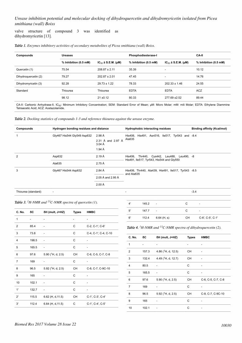
Enzyme inhibition assayA number of diseases are caused due to the over expression ofenzyme urease and phosphodiesterase-I. Helicobactor pyloriproduces urease which causes gastric ulcer and urolithiasiswhile cardiovascular diseases and erectile dysfunction areassociated with over expression of phosphodiesterase-I. Newand novel discoveries are important to treat various ailmentassociated with hyperactivities of these enzymes [13,26]. In thepresent study three secondary metabolites; quercetin (1)dihydroquercetin (2) dihydromyricetin (3) were isolated fromPicea Smithiana (wall) Boiss. The biological activities of thesecompounds are presented in Table 1.
Among these compounds, quercetin (1) and dihydroquercetin(2) showed selective inhibition against urease and were foundinactive against CA-II and phosphodiesterase-I. On the otherhand, dihydromyricetin (3) showed potent activity againsturease and phosphodiesterase-I with IC50=29.73 ± 1.22, 202.33± 1.46 µM, respectively, when compared with the standardinhibitors (Thiourea IC50=21 ± 0.12, EDTA IC50=277.69 ±2.52 µM). Keeping in view the above results and discussion
dihydromyricetin (3) can lead as potential chromosphere for hitto lead optimization.
Molecular dockingIn silico drug designing plays an important role in thediscovery of new inhibitors against the target. This processrequires less time and is cost effective. Computational dockingstudies of the compounds 1-3 were carried out to analyse theirbinding pattern in the active site of the target (urease) enzyme.The docking studies were carried out by the AutoDock Vina.The docking procedure was optimized. Generally dockingstudies shows that, if a compound contribute lesser interactionenergies then that compound has best activities against thetargeted enzyme. It can be predicted from the compound thatthere are certain necessary features in the compounds whichare responsible for their potency. The docking detail ofcompound 1-3 and reference thiourea are presented in theTable 2.
Figure 2. The best docked conformation of compounds (1-3) (shownby red color sticks) along with superimposed standard thiourea (balland sticks blue color) against the Urease enzyme (cyan color). Thesebinding conformations were produced in the binding site, wheremetals (green dots nickel atoms) are already present in the active siteof crystal structure.
The best docked conformation of the compound 3 wasanalysed it was based upon the hydrogen and hydrophobicinteractions. The docking energies of compound 3 are best thanthe reference thiourea and all other compounds. It means thatthere are certain features in the structure of this compoundwhich are responsible for mediating the biological activities.The docked conformations of compounds 1-3 and referencethiourea are shown in the Figure 2. From the docking statistics,it is observed that the binding affinity of compound 3 is -8.5kcal/mol (Autodock vina docking energies), which is best thanthe standard thiourea (-3.4 kcal/mol) (Table 2).
The interaction analysis revealed that, there are four hydrogenbonds formed by compound 3 in the binding site of ureaseenzyme, that hydrogen bond (2.84 Å) observed from Gly467with the -OH group of compound 3 (Figure 3). Here, we alsoinvestigated other two hydrogen bonds from the His544,
Urease inhibition potential and molecular docking of dihydroquercetin and dihydromyricetin isolated from Piceasmithiana (wall) Boiss
Biomed Res 2017 Volume 28 Issue 22 10028

distance of 2.05 Å and 2.95 Å with compound 3. The Asp632in the binding pocket of urease enzyme also showed hydrogenbonding contact with a distance of 2.00 Å. The hydrophobicinteractions which were also observed from the surroundingresidues in the binding site of urease enzyme, these residuesincluding His406, Thr440, Ala439, His491, Ile517, Tyr543 andAla635 forms seven hydrophobic contacts with compound 3.The interaction profile of compounds 1 and 2 is shown in theFigures 4 and 5.
Figure 3. The 2D (left) and 3D (right) interaction profile ofcompound 3 with the active site of urease enzyme. In the above 2Dimage half-moon shows the hydrophobic interactions while hydrogenbond is represented by dotted green lines with distance in Angstrom.
Figure 4. The 2D (left) and 3D (right) interaction profile ofcompound 2 with the active site of urease enzyme.
Figure 5. The 2D (left) and 3D (right) interaction profile ofcompound 1 with the active site of urease enzyme.
Quercetin (1)Compound 1 was purified as colorless crystalline solid fromEtOAc fraction of P. smithiana. The molecular formula of thiscompound was identified as C15H12O7 (304.23) by EI-MS, ESIspectrum.
IR (KBr, vmax in cm-1) 3583, 1665, 2926, 1585 and 1465. UVλmax (nm): 340, 265. 1H-NMR (600 MHz, MeOD) δh: 5.90(1H, d, H-6, j=2.5), 5.92 (1H, d, H-8, j=2.5), 6.62 (H, d, H-2’ ,j=11.5) and 6.64 (H, d, H-3’), 6.64 (H, s) respectively; 13C-NMR (150 MHz, MeOD), δc: 85.4 (CH, C-2), 73.8 (CH, C-3),198.5 (C, C-4), 165.5 (C, C-5), 97.6 (CH, C-6), 169.0 (C, C-7),96.5 (CH, C-8), 165.0 (C, C-9), 102.1 (C, C-9), 129.3 (C,C-1’), 115.5 (CH, C-2’), 112.4 (CH, C-3’), 145.2 (C, C-4’),147.7 (C, C-5’) and 112.4 (CH, C-6’) (Table 3). The HMBCcorrelations confirmed the position of all five hydroxyl group(OH) at 3, 5, 7, 4’ and 5’. Based on spectroscopic data andcomparing data with literature reported valve structure ofcompound 1 was identified as quercetin [12].
Dihydroquercetin (2)Compound 2 was purified as colorless crystalline solid fromEtOAc fraction of P. smithiana. The molecular formula of thiscompound was identified as C15H12O7 (302.18) by EI-MS, ESIspectrum.
IR (KBr, vmax in cm-1) 3580, 1664, 2925, 1583 and 1460. UVλmax (nm): 335, 260. 1H-NMR (600 MHz, MeOD) δh: 4.86(1H, d, H-2, j=12.5), 4.49 (1H, d, H-3, j=12.7), 5.90 (1H, d,H-6, j=2.5), 5.92 (1H, d, H-8, j=2.5), 6.62 (H, d, H-2’, j=11.5)and 6.64 (H, d, H-3’), 6.64 (H, s) respectively; 13C-NMR (150MHz, MeOD), δc: 157.3 (C, C-2), 132.4 (C, C-3), 80.5 (C,C-4), 165.5 (C, C-5), 97.6 (CH, C-6), 169.0 (C, C-7), 96.5(CH, C-8), 165.0 (C, C-9), 102.1 (C, C-9), 129.3 (C, C-1’),115.5 (CH, C-2’), 112.4 (CH, C-3’), 145.2 (C, C-4’), 147.7 (C,C-5’) and 112.4 (CH, C-6’) (Table 4). The HMBC correlationsconfirmed the position of all five hydroxyl group (OH) at 3, 5,7, 4’ and 5’. Based on spectroscopic data and comparing datawith literature reported valve structure of compound 2 wasidentified as dihydroquercetin [12].
Dihydromyricetin (3)Compound 3 was purified as light yellow powder from EtOAcfraction of P. smithiana. The molecular formula of thiscompound was identified as C15H12O8 (320.24) by EI-MS, ESIspectrum.
IR (KBr, vmax in cm-1) 3590, 1660, 2924, 15, 88 and 1460.UV λmax (nm): 345, 270. 1H-NMR (600 MHz, MeOD) δh: 4.86(1H, d, H-2, j=12.5), 4.49 (1H, d, H-3, j=12.7), 5.90 (1H, d,H-6, j=2.5), 5.92 (1H, d, H-8, j=2.5), 6.64 (2H, H-2’) and 6.64(2H, s, H-6’) respectively; 13C-NMR (150 MHz, MeOD), δc:85.4 (CH, C-2), 73.8 (CH, C-3), 198.5 (C, C-4), 165.5 (C,C-5), 97.6 (CH, C-6), 169.0 (C, C-7), 96.5 (CH, C-8), 165.0(C, C-9), 102.1 (C, C-9), 129.3 (C, C-1’), 109.1 (CH, C-2’),148.7 (C, C-3’), 135.1 (C, C-4’), 148.7 (C, C-5’) and 109.1(CH, C-6’) (Table 5).
The HMBC correlations confirmed the position of all fivehydroxyl group (OH) at 3, 5, 7, 3’, 4’ and 5’. Based onspectroscopic data and comparing data with literature reported
Bashir/Ahmad/Rauf/Bawazeer/Rahman/Rehman/Saleem/Ahmed/Linfang/Ikram
Biomed Res 2017 Volume 28 Issue 2210029
valve structure of compound 3 was identified asdihydromyricetin [13].
Table 1. Enzymes inhibitory activities of secondary metabolites of Picea smithiana (wall) Boiss.
Compounds
Ureases Phosphodiesterase-I CA-II
% Inhibition (0.5 mM) IC50 ± S.E.M. (µM) % Inhibition (0.5 mM) IC50 ± S.E.M. (µM) % Inhibition (0.5 mM)
Quercetin (1) 75.54 208.87 ± 2.11 35.39 - 10.12
Dihydroquercetin (2) 79.27 202.87 ± 2.01 47.45 - 14.76
Dihydromyricetin (3) 92.28 29.73 ± 1.22 78.33 202.33 ± 1.46 24.55
Standard Thiourea Thiourea EDTA EDTA ACZ
98.12 21 ±0.12 80.33 277.69 ±2.52 89.44
CA-II: Carbonic Anhydrase-II, IC50: Minimum Inhibitory Concentration; SEM: Standard Error of Mean; µM: Micro Molar; mM: mili Molar; EDTA: Ethylene DiammineTetraacetic Acid; ACZ: Acetazolamide.
Table 2. Docking statistics of compounds 1-3 and reference thiourea against the urease enzyme.
Compounds Hydrogen bonding residues and distance Hydrophobic interacting residues Binding affinity (Kcal/mol)
1 Gly467 His544 Gly549 Asp632 2.98 Å
2.31 Å and 2.97 Å3.04 Å
1.94 Å
His406, His491, Asn516, Ile517, Tyr543 andAla635
-8.4
2
Asp632 2.19 Å His406, Thr440, Cys442, Leu488, Leu490,His491, Ile517, Tyr543, His544 and Gly550
-8
Ala635 2.75 Å
3 Gly467 His544 Asp632
2.84 Å His406, Thr440, Ala439, His491, Ile517, Tyr543and Ala635
-8.5
2.05 Å and 2.95 Å
2.00 Å
Thiourea (standard) - - -3.4
Table 3. 1H-NMR and 13C-NMR spectra of quercetin (1).
C. No. δC δH (mult, J=HZ) Types HMBC
1 - - - -
2 85.4 - C C-2, C-1′, C-6′
3 73.8 - C C-4, C-1′, C-4, C-10
4 198.5 - C -
5 165.5 - C -
6 97.6 5.90 (1H, d, 2.5) CH C-6, C-5, C-7, C-8
7 169 - C -
8 96.5 5.92 (1H, d, 2.5) CH C-8, C-7, C-9C-10
9 165 - C -
10 102.1 - C -
1′ 132.7 - C -
2′ 115.5 6.62 (H, d,11.5) CH C-1′, C-3′, C-4′
3′ 112.4 6.64 (H, d,11.5) C C-1′, C-4′, C-5′
4′ 145.2 - C -
5′ 147.7 - C -
6′ 112.4 6.64 (H, s) CH C-6′, C-5′, C-1′
Table 4. 1H-NMR and 13C-NMR spectra of dihydroquercetin (2).
C. No. δC δH (mult, J=HZ) Types HMBC
1 - - - -
2 157.3 4.86 (1H, d, 12.5) CH -
3 132.4 4.49 (1H, d, 12.7) CH -
4 80.5 - C -
5 165.5 - C -
6 97.6 5.90 (1H, d, 2.5) CH C-6, C-5, C-7, C-8
7 169 - C -
8 96.5 5.92 (1H, d, 2.5) CH C-8, C-7, C-9C-10
9 165 - C -
10 102.1 - C -
Urease inhibition potential and molecular docking of dihydroquercetin and dihydromyricetin isolated from Piceasmithiana (wall) Boiss
Biomed Res 2017 Volume 28 Issue 22 10030

1′ 132.7 - C -
2′ 115.5 6.62 (H, d,11.5) CH C-1′, C-3′, C-4′
3′ 112.4 6.64 (H, d,11.5) C C-1′, C-4′, C-5′
4′ 145.2 - C -
5′ 147.7 - C -
6′ 112.4 6.64 (H, s) CH C-6′, C-5′, C-1′
Table 5. 1H-NMR and 13C-NMR spectra of dihydromyricetin (3).
C. No. δC δH (mult, J=HZ) Types HMBC
1 - - - -
2 85.4 4.86 (1H, d, 12.5) CH C-2, C-1′, C-6′
3 73.8 4.49 (1H, d, 12.7) CH C-4, C-1′, C-4, C-10
4 198.5 - C -
5 165.5 - C -
6 97.6 5.90 (1H, d, 2.5) CH C-6, C-5, C-7, C-8
7 169 - C -
8 96.5 5.92 (1H, d, 2.5) CH C-8, C-7, C-9C-10
9 165 - C -
10 102.1 - C -
1′ 129.3 - C -
2′ 109.1 6.64 (H, s) CH C-2′, C-3′, C-4′
3′ 148.7 - C -
4′ 135.1 - C -
5′ 148.7 - C -
6′ 109.1 6.64 (H, s) CH C-6′, C-5′, C-1′
References1. Patrick GL. An introduction to medicinal chemistry. Oxford
Univ Press 2013.2. Johann S, Pizzolatti MG, Donnici CL, Resende MAD.
Antifungal properties of plants used in Brazilian traditionalmedicine against clinically relevant fungal pathogens. BrazJ Microbiol 2007; 38: 632-637.
3. Farombi EO. African indigenous plants withchemotherapeutic potentials and biotechnological approachto the production of bioactive prophylactic agents. Afr JBiotechnol 2003; 2: 662-671.
4. Teklehaymanot T, Giday M. Ethnobotanical study ofmedicinal plants used by people in Zegie Peninsula,Northwestern Ethiopia. J Ethnobiol Ethnomed 2007; 3:12-22.
5. Srinivasan K, Natarajan D, Mohanasundari C,Venkatakrishnan C, Nagamurugan N. Antibacterial,preliminary phytochemical and pharmacognosticalscreening on the leaves of Vicoa indica (L.) DC. JPharmacol Exp Ther 2007; 6: 109-113.
6. Jan G, Khan MA, Jan F. Traditional medicinal andeconomic uses of gymnosperms of Dir Kohistan Valleys,NWFP, Pakistan. Ethnobot Leaflets 2009; 13: 1509-1521.
7. Kuo YH1, Yeh MH, Lin HC. New abietane-type diterpenesfrom the heartwood of Picea morrisonicola. Chem PharmBull (Tokyo) 2004; 52: 861-863.
8. Ummara U, Bokhari TZ, Altaf A, Younis U, Dasti AA.Pharmacological study of Shogran valley flora, Pakistan.Int J Sci Eng Res 2013; 4: 1-9.
9. Soderling SH, Beavo JA. Regulation of cAMP and cGMPsignaling: New phosphodiesterases and new functions. CurrOpin Cell Biol 2000; 12: 174-179.
10. Corbin JD1, Francis SH. Cyclic GMP phosphodiesterase-5:target of sildenafil. J Biol Chem 1999; 274: 13729-13732.
11. Mehats C, Andersen CB, Filopanti M, Jin SLC, Conti M.Cyclic nucleotide phosphodiesterases and their role inendocrine cell signaling. Trends Endocrinol Metab 2002;13: 29-35.
12. Vercruysse SAR, Delcour JA, Dondeyne P. Isolation ofquercetin, myricetin, and their respective dihydro-compounds by Sephadex LH-20 chromatography. JChromatography A 1985; 324: 495-497.
13. Chaturvedula VSP, Huang R. Isolation and NMR spectralstudies of dihydromyricetin. J Pharmacog Phytochem 2013;2: 113-115.
14. Kakiuchi S, Yamazaki R, Teshima Y, Uenishi K, MiyamotoE. Multiple cyclic nucleotide phosphodiesterase activitiesfrom rat tissues and occurrence of a calcium-plus-magnesium-ion-dependent phosphodiesterase and itsprotein activator. Biochem J 1975; 146: 109-120.
15. Perry MJ, Higgs GA. Chemotherapeutic potential ofphosphodiesterase inhibitors. Curr Opin Chem Biol 1998;2: 472-481.
16. Guex N, Peitsch MC. Swiss-model and the swiss-pdbviewer: An environment for comparative protein modeling.Electrophoresis 1997; 18: 2714-2723.
17. Li Z, Wan H, Shi Y, Ouyang P. Personal experience withfour kinds of chemical structure drawing software: Reviewon chemdraw, chemwindow, isis/draw, and chemsketch. JChem Inf Comput Sci 2004; 44: 1886-1890.
18. Hanwell MD, Curtis DE, Lonie DC, Vandermeersch T,Zurek E, Hutchison GR. Avogadro: An advanced semanticchemical editor, visualization, and analysis platform. JCheminform 2014; 4: 17.
19. Trott O, Olson AJ. Autodock vina: Improving the speedand accuracy of docking with a new scoring function,efficient optimization, and multithreading. J Comput Chem2010; 31: 455-461.
20. Yellamma K, Nagaraju S, Peera K, Praveen K. To designnovel lead molecules for the enzyme, ache associated withalzheimers disease. Int J Pharm Sci Res 2013; 22: 296-302.
21. Chang MW, Ayeni C, Breuer S, Torbett BE. Virtualscreening for hiv protease inhibitors: A comparison ofautodock 4 and vina. PLoS One 2010; 5: 11955.
22. Jacob RB, Andersen T, McDougal OM. Accessible high-throughput virtual screening molecular docking software
Bashir/Ahmad/Rauf/Bawazeer/Rahman/Rehman/Saleem/Ahmed/Linfang/Ikram
Biomed Res 2017 Volume 28 Issue 2210031

for students and educators. PLoS Comput Biol 2012; 8:1002499.
23. Kumar A, Kumar S, Jain S, Kumar P, Goyal R. Study ofbinding of pyridoacridine alkaloids on topoisomerase iiusing in silico tools. Med Chem Res 2013; 22: 5431-5441.
24. Laskowski RA, Swindells MB. Ligplot+: Multiple ligand–protein interaction diagrams for drug discovery. ACS Publ2011
25. DeLano WL. The pymol molecular graphics system 2002.http://www.pymol.org
26. Rauf A, Uddin G, Raza M, Patel S, Bawazeer S, Ben-Hadda T, Jehan N, Mabkhot YN, Khan A, Mubarak MS.Urease inhibition potential of Di-naphthodiospyrol fromDiospyros lotus roots. Nat Prod Res 2017; 31: 1214-1218.
*Correspondence toAbdur Rauf
Head Department of Chemistry
University of Swabi
Pakistan
E-mail:[email protected]
Phone number: +923469488944
Urease inhibition potential and molecular docking of dihydroquercetin and dihydromyricetin isolated from Piceasmithiana (wall) Boiss
Biomed Res 2017 Volume 28 Issue 22 10032