uptake and survival of vibrio vulnificus in...
TRANSCRIPT
1
UPTAKE AND SURVIVAL OF Vibrio vulnificus IN OYSTERS
By
MILAN SRIVASTAVA
A THESIS PRESENTED TO THE GRADUATE SCHOOL OF THE UNIVERSITY OF FLORIDA IN PARTIAL FULFILLMENT
OF THE REQUIREMENTS FOR THE DEGREE OF MASTER OF SCIENCE
UNIVERSITY OF FLORIDA
2007

2
© 2007 Milan Srivastava

3
To my family and friends

4
ACKNOWLEDGMENTS
The substantial amount of work done in this study could not have been completed without
the help of many people. First, I would like to extend my most profound gratitude to my advisor
Dr. Anita C. Wright. I want to thank her for believing in me and giving me an opportunity to
continue my graduate school. I am thankful for her encouragement throughout my research work
and polish my skills that helped me become a researcher. I would also like to extend my thanks
my committee members, Dr. Rodrick E. Gary for his guidance and valuable suggestions. Special
thanks to Dr. Max Teplitski for his insightful suggestions on improving on my poster
presentation in Marine biotech conference, and in preparing this document.
My research would not have been possible without the help of Ms. Jennette Villeda and
Ms. Melissa Evans, my lab mates and above all my friends. These two people stood by me in
tough times with the shoulders to lean on. Ms. Villeda joined our lab during my initial phases of
research and provided the helping hand whenever I needed it. Her reliable hands helped me a lot
in developing the methodology of this project. Special thanks to Melissa Evans for being my
support pillar. She was always present with the positive attitude and warming hug on every other
tough day in the graduate school. I would like to thank for her love and support, for
accompanying me to the lab after hours to finish up experiments, reading my thesis again and
again, and for listening to me ranting and raving about life as graduate student. Special note of
thanks goes to Dr. Maria Chatzidaki-Livanis for sharing her technical knowledge for the benefit
of my research work and motivation to do well all the time. I would also like to thank Dr.
Melissa Jones, for all her easy access and suggestions in the molecular work involved in this
research project. All my Lab mates, Mr. Mike Hubbard, Mr. Koo-Whang Chang, Ms. Lina
Jacques deserve a special note of thanks for always being handy and helpful throughout my
research project.
5
My friends were my pillars for the moral support and encouragement; in particular Mr.
Chambal and Ms. Sumita Pandey deserves a special note of thanks. They were filling elements in
the dip during my graduate school. I can’t even thank enough my family, my parents, my in-laws
for their unbending support and loving words, which always helped me through thick and thin.
Last but not least, my deepest gratitude and love goes to my husband, Saurabh Srivastava, for his
love and support when the pressures of life overwhelmed me. He gave me the lifetime of
encouragement and instilled in me the confidence to know that I can accomplish anything I set
my mind to. His valuable positive suggestions and role as closet critique made a remarkable
difference in the formulation of this document

6
TABLE OF CONTENTS page
ACKNOWLEDGMENTS ...............................................................................................................4
LIST OF TABLES...........................................................................................................................8
LIST OF FIGURES .........................................................................................................................9
LIST OF ABBREVIATIONS........................................................................................................10
ABSTRACT...................................................................................................................................12
CHAPTER
1 INTRODUCTION ..................................................................................................................14
V. vulnificus Distribution and Occurrence..............................................................................15 V. vulnificus Pathogenesis ......................................................................................................16 Potential V. vulnificus Secreted Virulence Factors.................................................................17 Lipopolysaccharide and Capsular Polysaccharide..................................................................18 The Genetics of CPS and Phase Variation .............................................................................19 V. vulnificus Flagella ..............................................................................................................21 V. vulnificus Type IV Pilus.....................................................................................................23 V. vulnificus Surface Structures and Environmental Survival................................................24 Goals and Objectives ..............................................................................................................25
2 MATERIAL AND METHODS..............................................................................................29
Bacterial Strains and Culture Conditions ...............................................................................29 Generation of a Double Mutant for pilA and wzb...................................................................32 Oyster Model for V. vulnificus Infection ................................................................................33 Bacterial Inoculation and Determination of Bacterial Content in Oysters .............................35 Dissection of Oyster Tissues ..................................................................................................36 Evaluation of Phase Variation in Oysters...............................................................................37 Competition Studies................................................................................................................39 Statistical Analysis..................................................................................................................40
3 DEVELOPMENT OF OYSTER MODEL OF INFECTION.................................................44
Optimization of Tetracycline Treatment ................................................................................44 Recovery of V. vulnificus in Post Tetracycline Treated Oysters ............................................45 Effects of Extended Incubation Post Tetracycline Treated Oysters .......................................45 Bacterial Recovery in Tetracycline and Non-tetracycline Treated Oysters ...........................45

7
4 ROLE OF CAPSULAR POLYSACCHARIDE IN SURVIVAL OF V. vulnificus IN OYSTERS...............................................................................................................................51
Distribution of CPS Mutant and Phase Variants in Oyster Tissues........................................52 Recovery of V. vulnificus CPS Strains in Oysters after Extended Inoculation ......................52 Phase variation of V. vulnificus in Oysters .............................................................................53 Confirmation of Phase Variation of Translucent V. vulnificus with Growth Plasmid............54
5 ROLE OF TYPE IV PILUS IN SURVIVAL OF V. vulnificus IN OYSTERS......................63
Distribution of V. vulnificus pilA Mutants in Oyster Tissues .................................................63 Recovery of V. vulnificus ∆pilA Mutants in Oysters after Extended Inoculation ..................64 Survival of V. vulnificus in Oysters as a Result of Bacterial Competition .............................64
6 ROLE OF FLAGELLA IN SURVIVAL OF V. vulnificus IN OYSTERS.............................70
Motility Test of V. vulnificus Strains......................................................................................70 Distribution of V. vulnificus Flagella Mutants in Oyster Tissues...........................................71
7 DISCUSSION AND CONCLUSION ....................................................................................76
LIST OF REFERENCES...............................................................................................................91
BIOGRAPHICAL SKETCH .........................................................................................................97
8
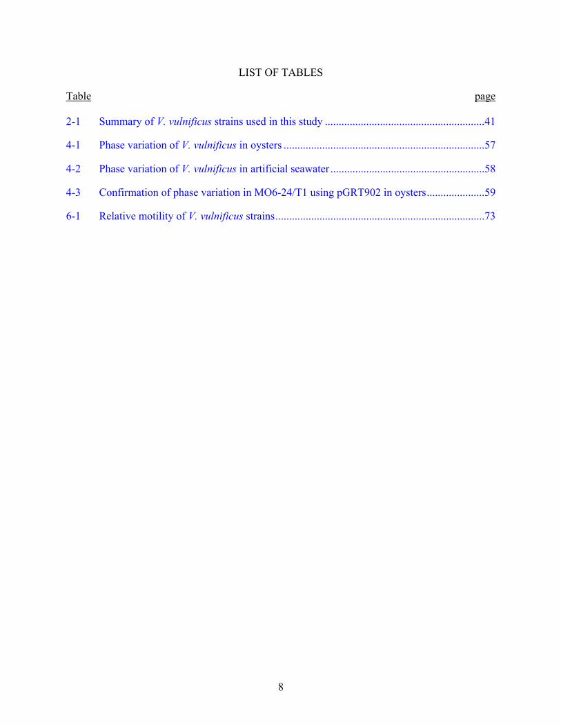
LIST OF TABLES
Table page 2-1 Summary of V. vulnificus strains used in this study ..........................................................41
4-1 Phase variation of V. vulnificus in oysters .........................................................................57
4-2 Phase variation of V. vulnificus in artificial seawater ........................................................58
4-3 Confirmation of phase variation in MO6-24/T1 using pGRT902 in oysters.....................59
6-1 Relative motility of V. vulnificus strains............................................................................73
9
LIST OF FIGURES
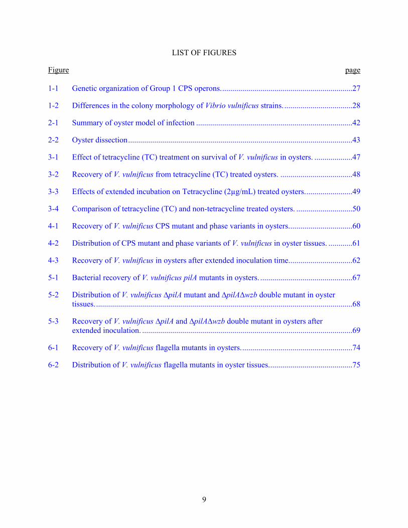
Figure page 1-1 Genetic organization of Group 1 CPS operons..................................................................27
1-2 Differences in the colony morphology of Vibrio vulnificus strains...................................28
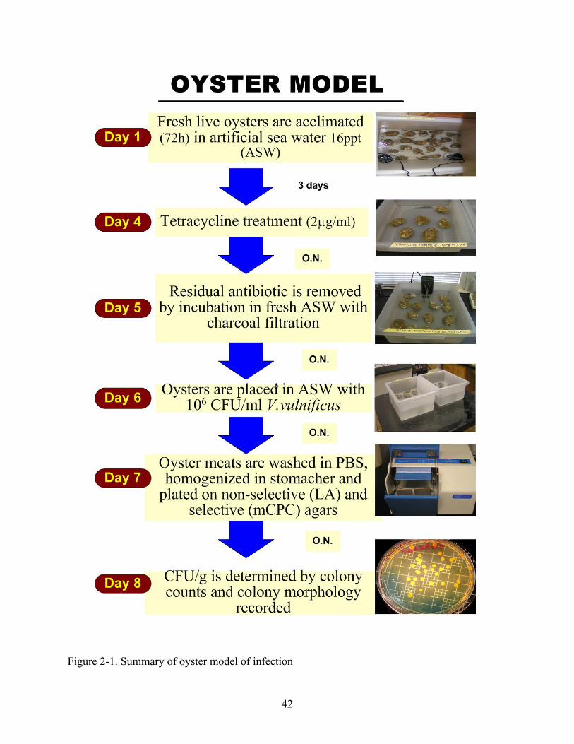
2-1 Summary of oyster model of infection ..............................................................................42
2-2 Oyster dissection................................................................................................................43
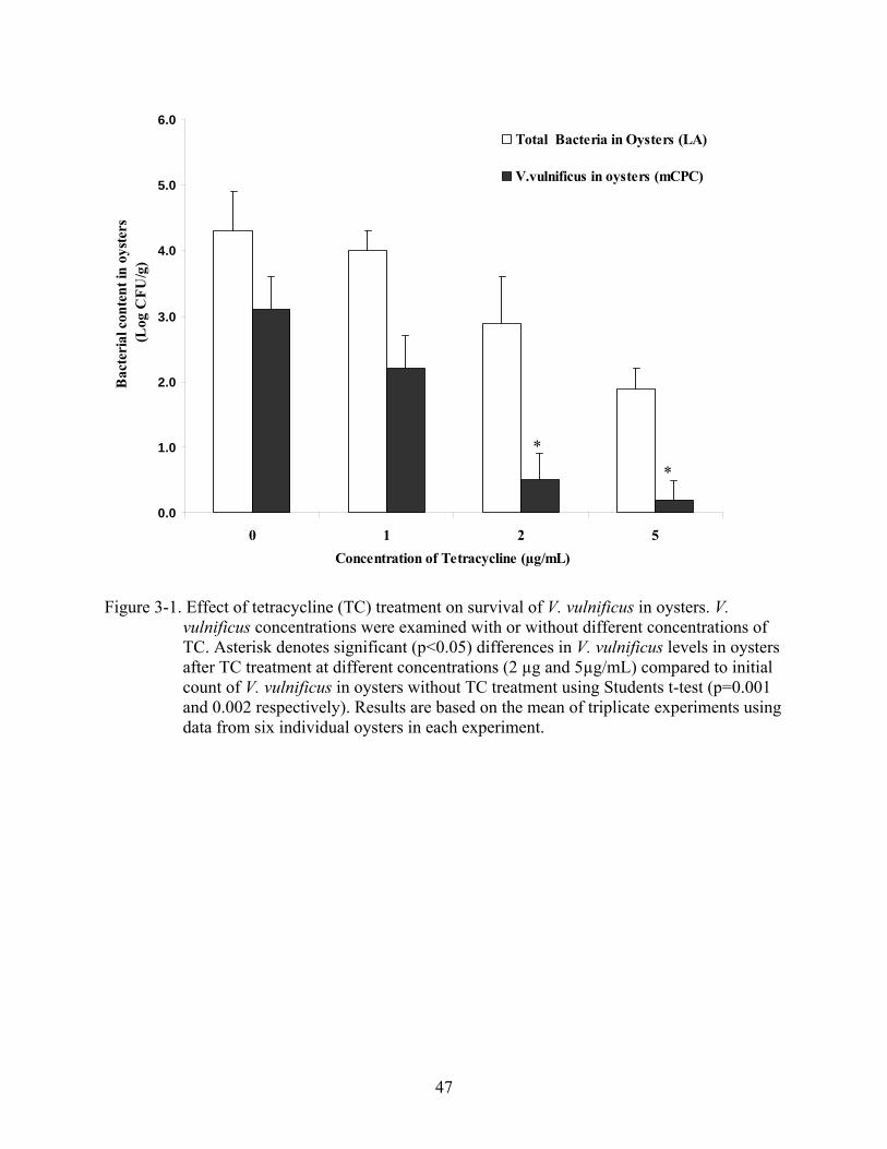
3-1 Effect of tetracycline (TC) treatment on survival of V. vulnificus in oysters. ...................47
3-2 Recovery of V. vulnificus from tetracycline (TC) treated oysters. ....................................48
3-3 Effects of extended incubation on Tetracycline (2µg/mL) treated oysters........................49
3-4 Comparison of tetracycline (TC) and non-tetracycline treated oysters. ............................50
4-1 Recovery of V. vulnificus CPS mutant and phase variants in oysters................................60
4-2 Distribution of CPS mutant and phase variants of V. vulnificus in oyster tissues. ............61
4-3 Recovery of V. vulnificus in oysters after extended inoculation time................................62
5-1 Bacterial recovery of V. vulnificus pilA mutants in oysters. ..............................................67
5-2 Distribution of V. vulnificus ∆pilA mutant and ∆pilA∆wzb double mutant in oyster tissues.................................................................................................................................68
5-3 Recovery of V. vulnificus ∆pilA and ∆pilA∆wzb double mutant in oysters after extended inoculation. .........................................................................................................69
6-1 Recovery of V. vulnificus flagella mutants in oysters........................................................74
6-2 Distribution of V. vulnificus flagella mutants in oyster tissues..........................................75
10
LIST OF ABBREVIATIONS
APW Alkaline peptone water
ASW Artificial sea water
AI Autoinducer
CDC Centers for Disease Control and Prevention
CPS Capsular polysaccharide
CFU Colony forming units
oC Degree centigrade
EPS Extra-polymeric substance
FDA Food and Drug Administration
Kan Kanamycin
LA Luria-Bretani agar
LB Luria-Bretani broth
LD50 Lethal dose 50%
LPS Lipopolysaccharide
mCPC Modified Cellobiose-Polymyxin B-Colistin
MSHA Mannose-sensitive hemagglutinin
NPW3 Neutral peptone water 3
OMP Outer membrane protein
PBS Phosphate buffered saline
PCR Polymerase chain reaction
PHT Post harvest treatment
Pol Polymixin B
ppt Parts per thousand
rpm Rotations per minute

11
RTX Repeats in the structural toxin
TC Tetracyline
TCP Toxin-coregulated pilus
TR1 Translucent Genotype 1 (intact CPS operon)
TR2 Translucent Genotype 2 (deletion of wzb)
VBNC Viable but non-culturable
12
Abstract of Thesis Presented to the Graduate School of the University of Florida in Partial Fulfillment of the
Requirements for the Degree of Master of Science
UPTAKE AND SURVIVAL OF Vibrio vulnificus IN OYSTERS
By
Milan Srivastava
December 2007
Chair: Anita C. Wright Major: Food Science and Human Nutrition
Vibrio vulnificus is a halophilic, gram-negative, opportunistic pathogen that is associated
with plankton and shellfish (oysters, clams, and mussels). This bacterium exhibits distinct
seasonality and is frequently isolated at temperatures greater than 20oC and is associated with
consumption of contaminated raw oysters. Adaptations in the surface structures of V. vulnificus
may influence the environmental reservoirs of disease. Additionally, survival of V. vulnificus
may also be dependent on phase variation of these cell surface structures. This research study
examined the contributions of known virulence factors, such as capsular polysaccharide (CPS),
pili, and flagella to the survival of V. vulnificus in oysters, using mutational analysis in an oyster
model of infection. Oysters (Crassostrea virginica) were acclimated in artificial seawater
(16ppt), and background V. vulnificus was reduced to <10 CFU/gram of oyster meat with
tetracycline (2µg/mL) treatment, followed by transferring to fresh artificial seawater (ASW) with
charcoal filtration to remove the residual antibiotic. Survival in inoculated oysters (106 CFU/mL)
was determined by plate count on non-selective (total bacteria count) and selective (V. vulnificus
count) agars. Strains included virulent, encapsulated wild type strain with opaque colonies;
translucent reversible phase variant (T1) with reduced CPS and virulence, rugose (wrinkled
colonies) phase variant with enhanced biofilm; or mutants with deletion in CPS (∆wzb), and in

13
the operon for Type IV Pilus (∆pilA) and double deletion mutant (∆pilA∆wzb), or deletions in
either one (∆flaCDE) or both (∆flaCDE∆flaFBA) flagellar genetic loci and flagellar motor
(∆motAB) components. Wild type opaque V. vulnificus was recovered from oysters at
significantly higher levels as compared to the rugose variant (p=0.005), or to ∆wzb (p=0.025),
∆pilA (p=0.01), ∆pilA∆wzb (p=0.002), or ∆CDE/∆FBA (p=0.03) deletion mutants. On the other
hand rugose, ∆pilA and ∆pilA∆wzb strains showed greater recovery in seawater compared to
oysters, indicating that in vitro biofilm function may be independent of survival of V. vulnificus
in oysters. Translucent phase variants (T1) did not differ from the wild type, and both T1 and
rugose phase variants reverted to opaque morphotype at high frequency (72 and 100%,
respectively) in oysters, while maintaining their stable morphology in the seawater. Competition
studies confirmed that encapsulation contributes to the survival of V. vulnificus in oysters.
Distribution of strains differed somewhat in oyster gills and intestinal tract, but significant
reductions in recovery from the hemolymph were observed for rugose variant, ∆wzb, ∆pilA,
∆pilA∆wzb mutants and for all the flagella mutants as compared to the wild type. Thus, surface
structures such as CPS, pili and flagella, and motility of V. vulnificus contribute not only to
survival in whole oysters but also to dissemination of the bacterium, especially to the
hemolymph of the oyster. Furthermore, observations of phase variation within the oyster host
indicate that variable expression of CPS is a survival strategy of V. vulnificus in oysters.
The research study described herein may ultimately lead to an understanding of the
contribution of surface structures of V. vulnificus in their molluscan shellfish host and thereby
aid in designing post harvest treatment methods to bring about more efficient reduction of this
potential pathogen to safe levels in seafood for human consumption.

14
CHAPTER 1 INTRODUCTION
Vibrio vulnificus is the most common cause of seafood associated deaths in Florida (Hlady
et al., 1993; Hlady and Klontz, 1996). V. vulnificus is a Gram-negative, flagellated, curved
bacterium that was first identified and described by the Centers for Disease Control and
Prevention (CDC) in 1976 (Hollis., 1987). V. vulnificus belongs to the family of Vibrionaceae
and is a mesophilic and obligate halophilic bacterium. Formerly referred as the “lactose-positive”
Vibrio (Farmer, 1979), the ability of V. vulnificus to ferment lactose distinguishes this bacterium
from other member of Vibrio genus. V. vulnificus exists naturally in sediments, coastal waters
and resides in high numbers in filter-feeding shellfish (oysters, clams and mussels) (Tamplin and
Capers, 1992; DePaola et al., 1994; Wright et al., 1996; Motes et al., 1998). Oysters harvested
during warm months, when the water temperature is greater than 22 oC from the Gulf of Mexico,
have high concentrations of V. vulnificus that may reach or exceed 105 bacteria per gram of
oyster meats (Murphy and Oliver, 1992; Kaspar and Tamplin, 1993; Levine, 1993; Wright et al.,
1996; Kelley et al., 1997). Approximately 80% of all reported V. vulnificus infections occur
when the level of this bacterium in the environmental reservoir and marine environment are high,
typically between the months of May and October (Hlady et al., 1993; Kelley et al., 1997; Motes
et al., 1998). V. vulnificus infections are usually associated with the consumption of
contaminated molluscan shellfish.
V. vulnificus infections in humans include primary septicemia and wound infections.
Ingestion of V. vulnificus contaminated oysters is the most common mode of exposure that can
result in primary septicemia in immuno deficient individuals (FDA, 1992; as reviewed by Gulig
et al., 2005; Ross et al., 1994; CDC, 1996; Hlady and Klontz, 1996). Wound infection can occur
as a result of exposure of open and/or breached skin surface to the sea water or to the handling
15
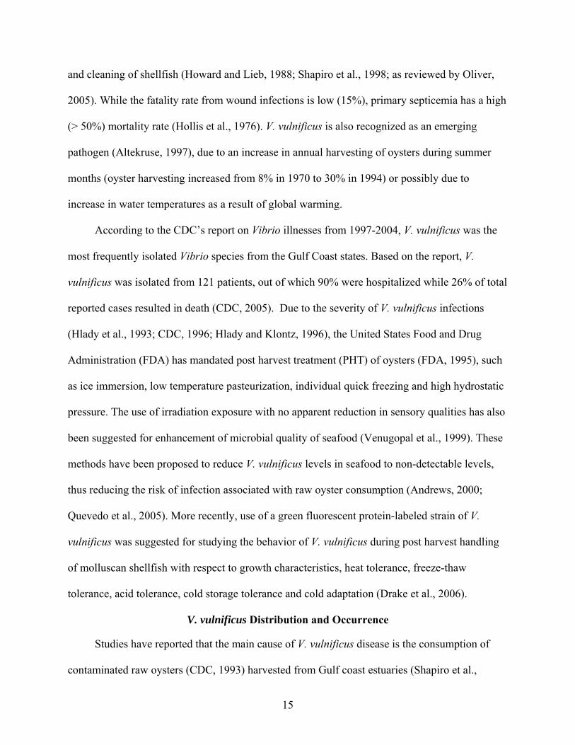
and cleaning of shellfish (Howard and Lieb, 1988; Shapiro et al., 1998; as reviewed by Oliver,
2005). While the fatality rate from wound infections is low (15%), primary septicemia has a high
(> 50%) mortality rate (Hollis et al., 1976). V. vulnificus is also recognized as an emerging
pathogen (Altekruse, 1997), due to an increase in annual harvesting of oysters during summer
months (oyster harvesting increased from 8% in 1970 to 30% in 1994) or possibly due to
increase in water temperatures as a result of global warming.
According to the CDC’s report on Vibrio illnesses from 1997-2004, V. vulnificus was the
most frequently isolated Vibrio species from the Gulf Coast states. Based on the report, V.
vulnificus was isolated from 121 patients, out of which 90% were hospitalized while 26% of total
reported cases resulted in death (CDC, 2005). Due to the severity of V. vulnificus infections
(Hlady et al., 1993; CDC, 1996; Hlady and Klontz, 1996), the United States Food and Drug
Administration (FDA) has mandated post harvest treatment (PHT) of oysters (FDA, 1995), such
as ice immersion, low temperature pasteurization, individual quick freezing and high hydrostatic
pressure. The use of irradiation exposure with no apparent reduction in sensory qualities has also
been suggested for enhancement of microbial quality of seafood (Venugopal et al., 1999). These
methods have been proposed to reduce V. vulnificus levels in seafood to non-detectable levels,
thus reducing the risk of infection associated with raw oyster consumption (Andrews, 2000;
Quevedo et al., 2005). More recently, use of a green fluorescent protein-labeled strain of V.
vulnificus was suggested for studying the behavior of V. vulnificus during post harvest handling
of molluscan shellfish with respect to growth characteristics, heat tolerance, freeze-thaw
tolerance, acid tolerance, cold storage tolerance and cold adaptation (Drake et al., 2006).
V. vulnificus Distribution and Occurrence
Studies have reported that the main cause of V. vulnificus disease is the consumption of
contaminated raw oysters (CDC, 1993) harvested from Gulf coast estuaries (Shapiro et al.,
16
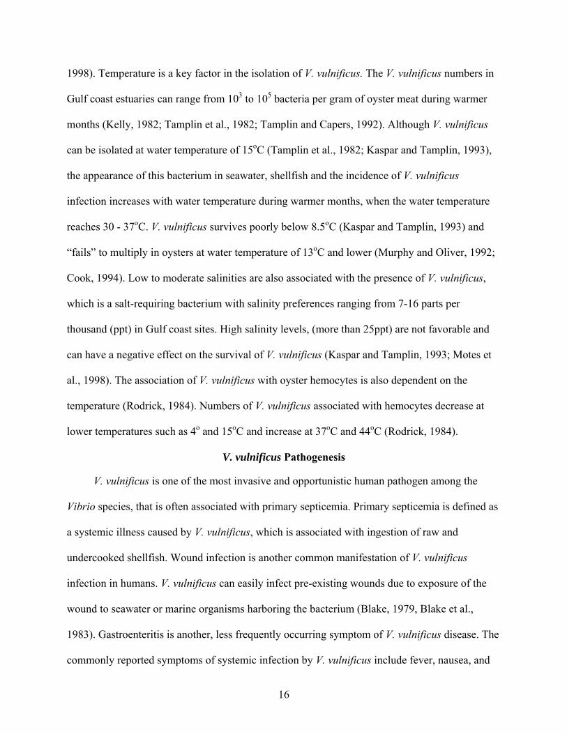
1998). Temperature is a key factor in the isolation of V. vulnificus. The V. vulnificus numbers in
Gulf coast estuaries can range from 103 to 105 bacteria per gram of oyster meat during warmer
months (Kelly, 1982; Tamplin et al., 1982; Tamplin and Capers, 1992). Although V. vulnificus
can be isolated at water temperature of 15oC (Tamplin et al., 1982; Kaspar and Tamplin, 1993),
the appearance of this bacterium in seawater, shellfish and the incidence of V. vulnificus
infection increases with water temperature during warmer months, when the water temperature
reaches 30 - 37oC. V. vulnificus survives poorly below 8.5oC (Kaspar and Tamplin, 1993) and
“fails” to multiply in oysters at water temperature of 13oC and lower (Murphy and Oliver, 1992;
Cook, 1994). Low to moderate salinities are also associated with the presence of V. vulnificus,
which is a salt-requiring bacterium with salinity preferences ranging from 7-16 parts per
thousand (ppt) in Gulf coast sites. High salinity levels, (more than 25ppt) are not favorable and
can have a negative effect on the survival of V. vulnificus (Kaspar and Tamplin, 1993; Motes et
al., 1998). The association of V. vulnificus with oyster hemocytes is also dependent on the
temperature (Rodrick, 1984). Numbers of V. vulnificus associated with hemocytes decrease at
lower temperatures such as 4o and 15oC and increase at 37oC and 44oC (Rodrick, 1984).
V. vulnificus Pathogenesis
V. vulnificus is one of the most invasive and opportunistic human pathogen among the
Vibrio species, that is often associated with primary septicemia. Primary septicemia is defined as
a systemic illness caused by V. vulnificus, which is associated with ingestion of raw and
undercooked shellfish. Wound infection is another common manifestation of V. vulnificus
infection in humans. V. vulnificus can easily infect pre-existing wounds due to exposure of the
wound to seawater or marine organisms harboring the bacterium (Blake, 1979, Blake et al.,
1983). Gastroenteritis is another, less frequently occurring symptom of V. vulnificus disease. The
commonly reported symptoms of systemic infection by V. vulnificus include fever, nausea, and

17
hypotension (Blake et al., 1979; Klontz et al., 1988). Development of secondary bullous lesions
on legs and feet is another feature of primary septicemia and they are characterized by fluid-
filled blisters, typically resulting in tissue and muscle destruction (Tacket et al., 1984; Klontz et
al., 1988).
The severity of V. vulnificus infections and the infectious dose required for appearance of
this disease are dependent upon a number of host factors. People who are most susceptible to V.
vulnificus infection usually have underlying health conditions such as alcoholism, liver disease
(hepatitis, cirrhosis), diabetes mellitus, cancer, hemochromatosis (iron-overload) and immune
system dysfunction (Hlady et al., 1993; CDC, 2005; as reviewed by Gulig et al., 2005). The
infectious dose of V. vulnificus that causes disease in humans is not known. However, people
with a recent history of gastro-intestinal illness and infection of skin and open wounds have a
higher risk of getting V. vulnificus infections (Klontz et al., 1988; Hlady and Klontz, 1996).
Thus, host immune status is important for the pathogenesis of V. vulnificus (as reviewed by
Gulig et al., 2005).
Potential V. vulnificus Secreted Virulence Factors
V. vulnificus exhibits multiple virulence factors that may be involved in or required for the
manifestation of this disease in humans. Iron is important for bacterial growth, and bacteria have
mechanisms to scavenge iron from the host through the production of siderophores. V. vulnificus
produces hydroxymate and phenolate (catechol) siderophores for the acquisition of iron from
mammalian host to cause fulminating septicemia and invasive wound infection in animal models
(Wright et al., 1981; Simpson and Oliver, 1983; Litwin et al., 1996). Both clinical and
environmental strains of V. vulnificus expresses secreted factors such as cytolysin/hemolysin
(vvhA gene) and metalloprotease (vvpE), which were initially thought to contribute to
pathogenicity in mammalian models. However, mutations in either of the two genes indicated no
18
apparent role of these proteins in the virulence of this bacterium (Wright and Morris, 1991).
Quorum sensing has also been related to the regulation of gene expression and virulence of V.
vulnificus (Kim et al., 2003). It has been reported that autoinducer-2 (AI-2) communication
molecules play an important role in the stress response in starvation and stationary growth phase
of V. vulnificus (McDougald et al., 2006) and may be important for the virulence of V. vulnificus.
Exotoxin (s) belonging to the family of pore-forming proteins, named as RTX toxins (repeats in
the structural toxin), may also play an important role in virulence in many gram-negative
bacterial pathogens. The RTX toxin operon consists of four genes namely rtxA, rtxB, rtxC, and
rtxD. RTX toxin is encoded by rtxA. The transportation and delivery of RTX toxin outside the
bacterial cell is facilitated by rtxB and rtxD (Welch, 1992). It has been shown that RtxA toxin
cause pore formation in red blood cells, and necrotic cell death in Hep-2 cells (Lee et al. 2007).
A rtxA mutant in V. vulnificus exhibited a 100-fold increase in lethal dose 50% (LD50) in mouse
model suggesting that RTX toxin plays a critical role in virulence of V. vulnificus (Lee et al.,
2007).
Lipopolysaccharide and Capsular Polysaccharide
The expression of lipopolysaccharide (LPS) on the cell surface was also thought to
contribute to the virulence and toxic shock of V. vulnificus (Martin and Siebeling, 1991).
However, LPS from V. vulnificus was less pyrogenic than the LPS from other Gram- negative
pathogens (McPherson et al., 1991; Powell et al., 1997).On the other hand, studies have
suggested a positive relationship between the degree of capsular polysaccharide (CPS) expressed
and virulence in animal models (Yoshida et al., 1985; Simpson et al., 1987; Wright et al., 1990;
Wright et al., 1999; Wright et al., 2001; Chatzidaki-Livanis et al., 2006).
V. vulnificus expresses an extracellular acidic capsular polysaccharide on its cell surface.
There is a relation between the capsular expression, the colony opacity and the virulence of V.
19
vulnificus (Amako, 1984). Colonies that exhibit capsule have an opaque phenotype, but these
cells can also undergo a reversible switch to a translucent phenotype, characterized by reduced or
patchy expression of capsule (Simpson et al., 1987; Wright et al., 1990). Presence of capsule is
correlated with virulence in animal models, antiphagocytic activity, tissue invasiveness and
resistance to the bactericidal activity of normal human serum. On the other hand, loss of capsule
is accompanied by decrease in virulence, hydrophilicity and serum susceptibility (Wright et al.,
1990). Additionally, unencapsulated strains have significantly higher LD50 than the wild type
encapsulated strain (Wright et al., 1990). Heterogeneous capsular types have been found among
the various clinical and environmental isolates of V. vulnificus (Hayat et al., 1993). Thus,
different V. vulnificus strains have differences in their CPS composition, and are likely to use
different metabolic pathways for biosynthesis of CPS (Reddy et al., 1992; Hayat et al., 1993).
However, most strains isolated from human infections or oysters appear to be encapsulated
(Simpson et al., 1987; Stelma et al., 1992; Wright et al., 1996). Expression of CPS can also vary
depending on the growth phase and other environmental conditions, especially temperature
(Wright et al., 1999; Wright et al., 2001). It has been reported that surface expression of CPS
increases during logarithmic growth phase and decreases during stationary phase in the wild type
strain. Additionally, greater CPS is expressed during growth at 30oC as compared to 37oC
(Wright et al., 1999).
The Genetics of CPS and Phase Variation
Both CPS expression and virulence are associated with opaque colony morphology.
However, opaque colonies can spontaneously revert to the translucent phenotype, reduced or
patchy expression of surface polysaccharide (Figure 1-2), by a process called phase variation.
The V. vulnificus opaque strain exhibits a reversible-phase variation to a translucent morphotype
that occurs within a population at a rate of 10-3 to 10-4 (Wright et al., 1990; Wright et al., 1999;
20
Wright et al., 2001). The avirulent, unencapsulated translucent, spontaneous phase variant of V.
vulnificus can also revert back to the original opaque, encapsulated phenotype (Wright et al.,
1999).
Epimerase genes encoding the CPS biosynthetic gene (Zuppardo and Siebeling, 1998) and
wza, encoding a CPS outer membrane transporter (Wright et al., 2001) have been reported. More
recently, the latter gene was found to reside within the group 1 CPS operon of V. vulnificus
strains, and the entire operon was sequenced for opaque and translucent strains (Figure 1-1). V.
vulnificus CPS genes show homology in the organization and sequence of previously described
group 1 CPS operons in E. coli (Wright et al., 1999; Chatzidaki-Livanis et al., 2006). E. coli
group 1 capsule is defined by the presence of wza-wzb-wzc genes in the CPS operon, and a
similar gene cluster was found in V. vulnificus (Chatzidaki-Livanis et al., 2006) (Figure 1-1).
Wza is an outer membrane lipoprotein that is involved in surface assembly of group 1 capsules
and transportation of polysaccharide to the outer surface (Drummelsmith and Whitfield, 1999).
Wzb is a cytoplasmic acid phosphatase that functions to catalyze the removal of phosphates from
Wzc. Wzc is tyrosine kinase, located in the plasma membrane, is involved in the surface
assembly of the capsular layer (Drummelsmith and Whitfield, 1999). Multiple genotypes (T1, T2
and T3) from the translucent isolates of V. vulnificus were identified (Chatzidaki-Livanis et al.,
2006). T1 (MO6-24/T1) strain with reduced CPS expression showed a CPS operon that was
identical to that of the opaque strain. MO6-24/T2 (∆wzb) cells showed a deletion mutation in the
wzb, resulting in acapsular colonies locked in the translucent phase, which were unable to revert
to opaque colony morphology (Chatzidaki-Livanis et al., 2006). T3 (MO6-24/T3) strains had
more extensive genetic deletions that also included the wzb. Complementation of the CPS
deletion mutant with wzb, restored the opaque phenotype, and electron microscopy confirmed

21
that the strain recovered the surface expression of CPS (Chatzidaki-Livanis et al., 2006). Thus,
different mechanisms were proposed to be responsible for reversible phase variation in CPS
expression versus irreversible genetic deletions in V. vulnificus (Chatzidaki-Livanis et al., 2006).
However, the precise role of phase variation in Vibrio species is less clear, and the genetic
mechanism (s) responsible for phase variation is still unknown.
V. vulnificus also produces a rugose or wrinkled colony type from both opaque and
translucent strains at high frequencies, that can switch back to opaque or translucent colony
morphology (Figure 1-2C) (Grau et al., 2005). Rugose colonies show enhanced biofilm
formation and survival under adverse environmental conditions (Grau et al., 2005). In V.
cholerae, these rugose variants express alternate CPS composition with neutral (glucose and
galactose) sugars (Yildiz and Schoolnik, 1999) as opposed to the acidic sugar (uronic acid)
expressed by Group 1 CPS. However, the composition of rugose CPS in V. vulnificus is
unknown. Upon further characterization of rugose strains, it was found that the V. vulnificus
rugose strain is relatively less motile and more resistant to serum killing than the parental opaque
or translucent version. Despite their decreased motility, the rugose strain was reported to possess
a polar flagellum.
V. vulnificus Flagella
Flagella help in the initial absorption of bacteria to surfaces, biofilm substrates, and
invasion of host (McCarter, 2001; Harshey, 2003). Flagellum based motility is required for the
localization of V. vulnificus to sites of infection or for invasion in the host cell (Lee et al., 2004).
McCarter studied the genetic and molecular characterization of the polar flagellum of Vibrio
parahaemolyticus (McCarter, 1995). It was reported that multiple (six) flagellin genes encode the
filament subunits of the flagellum, namely flaA, flaB, flaC, flaD, flaE and flaF, organized in two
genetic loci, flaFBA and flaCDE (McCarter, 1995). Further analysis revealed that none of the six

22
flagellin genes were essential for filament formation, and loss of a single flagellin gene has no
significant effect on the motility or the flagella structure (McCarter, 1995). However, deletion of
the ΔflaCDE genetic locus showed reduced motility, but deletion of both loci
(ΔflaFBAΔflaCDE) completely abolished the motility and the flagella expression (McCarter,
1995; Tucker, 2006). More recently, (Tucker, 2006) examined the roles of flagella, motility, and
chemotaxis in the virulence of V. vulnificus using a mouse model of disease. It was found that a
mutant with a deletion in the ΔflaFBA locus was equally motile and virulent for either localized
skin or systemic liver infection as compared to that of wild-type. On the other hand, deletions in
the ΔflaCDE locus resulted in strain with reduced motility and virulence (both localized and
systemic) as compared to the wild-type. Furthermore, deletion of flagella motor genes (ΔmotAB)
resulted in a non-motile strain that showed attenuated skin infection in the mouse model.
Complementation of this mutant with cloned motAB fully restored the motility to levels of the
wild-type. The ΔflaFBAΔflaCDE double deletion mutant was also non-motile and showed
attenuated virulence for systemic infection in a mouse model. Other studies have focused on the
role of flagellar basal body rod proteins (flgC) as a potential virulence determinant of V.
vulnificus (Ran Kim, 2003). A transposon insertion mutation in the ΔflgC gene showed
decreased motility, biofilm formation, cytotoxicity to the He-Le cells and virulence in mice
models. Furthermore, flagella related motility mutant (∆flgE) was also less virulent and deficient
in the biofilm formation to INT-407 cells (Lee et al., 2006). Recently, expression of methyl-
accepting chemotaxis protein was found to be during V. vulnificus infection, and it was theorized
that MCP might play an important role in invasion of V. vulnificus during gastrointestinal
infection (Kim et al., 2003). Furthermore, it was reported that defects in chemotaxis can alter the
ability of V. vulnificus to cause disease in animal models (Tucker, 2006).

23
V. vulnificus Type IV Pilus
Expression of pili on V. vulnificus cells was identified by electron microscopy, and more
pilus fibers were seen on clinical isolates from blood or wounds than environmental isolates
(Gander and LaRocco, 1989). Presence of pilus-like structures on V. vulnificus can facilitate
adherence, attachment and colonization to the HEp-2 cells of host surface receptor (Paranjpye et
al., 1998). Type IV pili are common to many gram-negative bacteria that allows for flagellum-
independent movement, termed as twitching motility. Genes encoding proteins required for the
biogenesis of type IV pili in V. vulnificus have been reported (Paranjpye and Strom, 2005). It has
been shown that mutations in a gene encoding IV prepilin peptidase/N-methyltransferase, vvpD
or pilD, results in a loss of all pili expression on the cell surface of V. vulnificus, which
significantly decreases cell cytotoxicity in Chinese Hamster Ovary (CHO) cells, adherence to
HEp-2 cells and reduces virulence in mouse model (Paranjpye and Strom, 2005).
The amino acid sequence of V. vulnificus type IV pilin (PilA) shares extensive homology
to group A type IV pilin expressed by many pathogens, including V. cholerae (PilA) and P.
aeruginosa (PilA). The V. vulnificus pilA is part of an operon that also includes three other pilus
biogenesis genes (pilBCD), that encodes for pilin precursor protein in the type IV pilus
biogenesis gene cluster. A deletion in the V. vulnificus ∆pilA, resulted in reduced biofilm
formation, decreased adherence to HEp-2 cells, and attenuated virulence in iron dextran-treated
mouse models (Paranjpye and Strom, 2005). However, pili were still present on the surface of
the ΔpilA mutant strain as shown by transmission electron microscope, suggesting that V.
vulnificus produces other type (s) of pili. The genome of V. vulnificus also encodes a second type
IV pilin, mannose-sensitive hemagglutinin (MSHA) that is homologous to V. cholerae MSHA,
but carries only a single prepilin peptidase gene (Yamaichi et al., 1999). Therefore, the loss of all
surface pili on the ΔpilD mutant suggests that pilD processes both type IV pilins of V. vulnificus
24
(Paranjpye et al., 1998). Recently, it has been reported the pilA and pilD of V. vulnificus in the
colonization of bacterium in oysters by comparing the uptake and persistence of the wild type V.
vulnificus to that of the pilA and pilD mutant strains (Paranjpye et al., 2007). The authors
reported that expression of pilA and pilD are important for V. vulnificus to persist in American
oysters, Crassostrea virginica.
V. vulnificus Surface Structures and Environmental Survival
Biofilm formation is an essential mode of bacterial survival in the natural environment, as
reviewed by (Watnick and Kolter, 1999). Biofilms are complex interactions of surface structures
of bacteria, constituting a protected community that allows bacteria to attach to surfaces,
providing an adaptive advantage for enhanced survival under adverse conditions (Watnick and
Kolter, 2000). Surface structures of bacteria such as flagella, fimbriae, pili, and extra polymeric
substances that are major determinants of virulence, helps in biofilm formation. For example, V.
cholerae motility genes, motA and motB, are required for flagellar rotation and initiating cell-to-
surface contact in biofilm formation. The mannose-sensitive hemagglutinin (MSHA) pilus helps
the bacterium pull onto the abiotic surface, leading to the attachment of V. cholerae EI Tor
(Watnick and Kolter, 1999). Alternatively, V. cholerae EI Tor does not use virulence associated
toxin coregulated pilus (TCP) to form biofilms. Extra polymeric substances are necessary to
stabilize cell-to-cell interactions and formation of 3-dimension biofilms (Watnick and Kolter,
1999). Polysaccharides are not always critical to initial attachment, but are considered major
constitutes of the complex architecture of the later stages of biofilm formation. Expression of
capsular polysaccharide is also important for virulence in animal models (Yoshida et al., 1985;
Wright et al., 1999; Wright et al., 2001), but it inhibits biofilm formation in V. vulnificus (Joseph
and Wright, 2004). On the other hand, motility of V. vulnificus is reported to be both a potential
virulence factor and an important determinant for initial cell-to-surface contact and colonization

25
in the host. In this regard, surface expression of pili (type IV pilus) (Paranjpye et al., 1998;
Paranjpye and Strom, 2005; Paranjpye et al., 2007) and flagellar motility (Lee et al., 2004; Lee et
al., 2006; Tucker, 2006) are also reported to contribute to both biofilm formation and to the
virulence of V. vulnificus in animal models.
Surface structures of V. vulnificus such as CPS, flagella, flagellar motility and type IV pili,
that are associated with biofilms and virulence, may also provide adaptations for increased
survival of V. vulnificus in their oyster host. Vibrio species attach to algae and plankton (Hood,
1997; Chiavelli, 2001). Oysters being filter-feeders trap suspended food particles including
bacteria and concentrate Vibrios in their tissues (Tamplin and Capers, 1992; Harris-Young et al.,
1993; Kennedy, 1999). Expression of CPS facilitates the survival of V. vulnificus by providing
resistance to phagocytosis by oyster hemocytes (Harris-Young et al., 1995). The degree of
encapsulation may also provide resistance to lysis by oyster lysozyme, as V. vulnificus opaque
strain is more resistant to the intracellular bactericidal effects of oyster hemocytes than the
translucent strain (Harris-Young et al., 1995). Moreover, it has been proposed that reversion of
phase variation from translucent to the opaque phenotype may also allow the bacterium to regain
the CPS expression and enhance survival (Chatzidaki-Livanis et al., 2006). Expression of pilA
and pilD are important for the persistence of V. vulnificus in American oysters (Paranjpye et al.,
2007); however, the role of flagella and motility in the survival of V. vulnificus in oysters in not
clear.
Goals and Objectives
The overall goal of this research study was to examine the hypothesis that different surface
structures of V. vulnificus, such as CPS, pili and flagella, contribute to the survival of V.
vulnificus in an oyster model. These surface structures of V. vulnificus, which are virulence
factors in mammalian models, may also provide adaptations for survival in oysters. Furthermore,

26
phase variation of cell surface structures such as CPS can potentially influence the behavior of
this bacterium in oysters. For validation of the hypothesis, mutational analysis was used to
examine these variables in an oyster model of infection. Survival of mutant and phase variants of
V. vulnificus was compared to the wild type encapsulated strain. The specific objectives of this
research include the following items:
1 To develop an oyster model of infection in order to assess the contribution of surface structures such as CPS, pili and flagella expression in the survival and colonization of V. vulnificus in an Eastern oyster, Crassostrea virginica.
2 To examine the uptake and distribution of V. vulnificus mutant and phase variants in hemolymph, gills and digestive tract of oyster tissues.
3 To examine the relative importance of different surface structures of V. vulnificus at different stages (extended inoculation up to 72 hours) of colonization.
4 To examine the rate of survival of V. vulnificus in oysters as a result of bacterial competition between wild type and wzb deletion mutant and ∆wzb∆pilA double mutant strain.
5 To examine the phase variation in oysters using the growth plasmid pGTR902 into V. vulnificus as a marker for the appearance of opaque colonies resulting from phase variation of translucent to the opaque phenotype. These experiments should distinguish phase variation from die off within a population

27
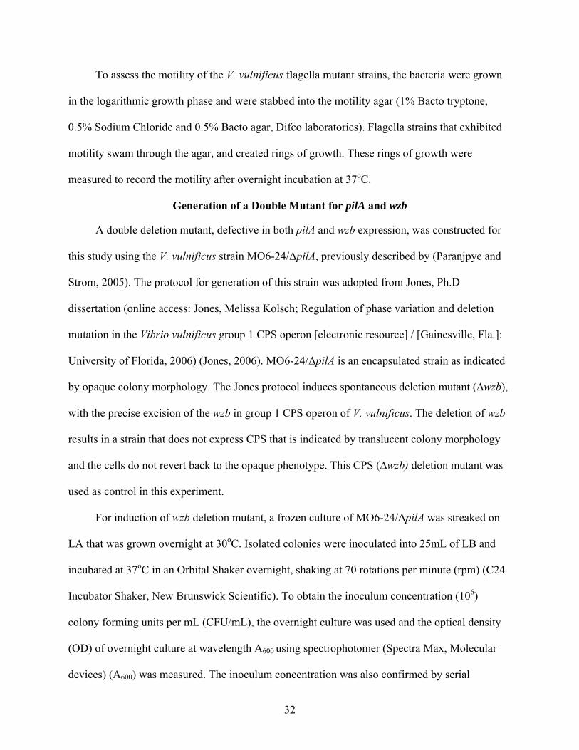
Figure 1-1. Genetic organization of Group 1 CPS operons. Source: (Chatzidaki-Livanis et al.,
2006)

28
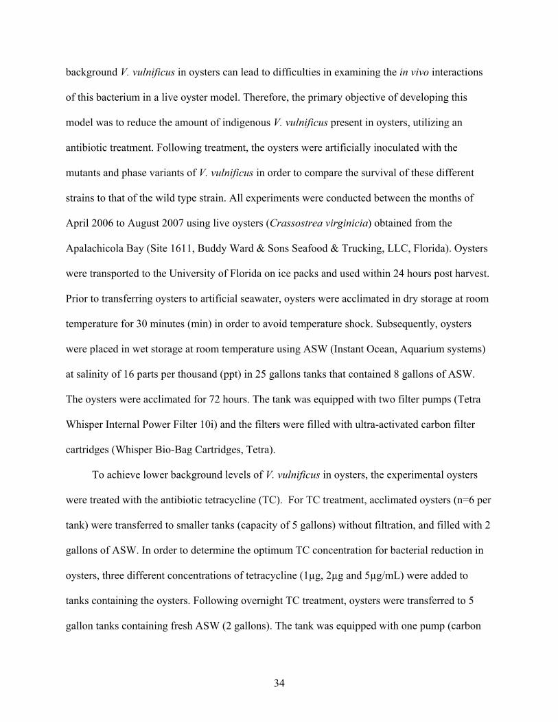
A B
C
Figure 1-2. Differences in the colony morphology of Vibrio vulnificus strains. A) Opaque colonies, B) Translucent colonies, and C) Rugose (wrinkled) colonies
.

29
CHAPTER 2 MATERIAL AND METHODS
Bacterial Strains and Culture Conditions
The capsular polysaccharide (CPS) phase variants, CPS (wzb) deletion mutant of V.
vulnificus (Chatzidaki-Livanis et al., 2006), pili mutants (Paranjpye and Strom, 2005) and
flagella mutants of CMCP6 (Tucker, 2006) used in this research study are summarized in Table
2-1. All strains were stored in Luria-Bertani broth (LB; 1.0% tryptone, 0.5% yeast extract, and
1.0% NaCl) with 50% glycerol at -80oC. The strains were recovered from frozen stock by
streaking for isolation on Luria-Bertani agar, LA (LB with 1.5% Bacto Agar) and incubated at
37°C. For V. vulnificus plate counts, a species-specific medium , modified cellobiose-polymyxin
B-colistin (mCPC) agar, prepared with 1.0% peptone, 0.5% beef extract, 2.0% NaCl, 0.1% of the
1000X dye stock solution (4.0% bromothymol blue, 4.0% cresol red in 95% Methanol), and 10%
of filtered antibiotic solution (1.0% cellobiose, 3.0% colistin, 1.3 % polymyxin B dissolved in
100mL distilled water), as described in Bacteriological Analytical Manual (BAM), 2001 was
used. When required, kanamycin (50-300 μg/mL) and polymyxin B (50 μg/mL) were added to
LA and LB, to facilitate the growth of antibiotic resistant strains. The Escherichia coli (E. coli)
strain S17-λ pir with pGTR902 (provided by Dr. Paul Gulig, University of Florida) was used for
introducing antibiotic resistance marker in V. vulnificus. The E. coli strain was grown in LB with
kanamycin (50μg/mL) and arabinose (1%).
V. vulnificus CPS phase variants and the deletion mutant of MO6-24/O were from a
previous study by (Chatzidaki-Livanis et al., 2006). MO6-24/Opaque is a wild type encapsulated
clinical isolate that expresses CPS on the cell surface, marked by opaque colonies on solid
medium (LA) and is virulent in animal models. MO6-24/T1 is a reversible phase variant derived
from MO6-24/O, with undefined mutation that shows reduced or patchy CPS expression on the

30
cell surface. The strain is marked by translucent colonies and is less virulent in animal models.
MO6-24/∆wzb is an irreversible deletion mutant derived from MO6-24/O with precise deletion
of wzb of the CPS operon, eliminating CPS surface expression and locking the strain into
translucent phenotype (Chatzidaki-Livanis et al., 2006). Complementation of wzb in trans
restores the CPS expression, and translucent colonies return back to the opaque phenotype
(Chatzidaki-Livanis et al., 2006). Another reversible phase mutant of opaque is MO6-24/
Rugose, known for enhanced biofilm formation, is marked by wrinkled and dry morphotype.
This strain is relatively less motile than the parental opaque strain, but yet possesses a polar
flagellum (Grau et al., 2005).
V. vulnificus pili mutant was kindly provided by Dr. Rohinee Paranjype, and consisted of
strain MO6-24/PilA with deletion mutation in pilA (Paranjpye and Strom, 2005). This pilA is a
part of an operon and is clustered with three other pilus biogenesis genes, pilBCD. PilA is the
precursor of a substrate of PilD, and mutations in pilD, the gene encoding the type IV leader
peptidaseN-methyltransferase (type IV prepilin peptidase), result in the absence of pili on the
surface of V. vulnificus (Paranjpye et al., 1998). MO6-24/∆pilA strain has a specific deletion
mutation in pilA in MO6-24/O parent strain, but does produce other type(s) of pili such as
mannose-sensitive haemagglutinin (MSHA), homologous to the V. cholerae MSHA. This strain
shows intact CPS operon and is marked by opaque colonies on solid medium (LA). However,
this strain is defective in biofilm formation, adherence to epithelial cells and virulence in mouse
model (Paranjpye and Strom, 2005). Complementation of pilA mutation restores PilA expression,
adherence to Hep-2 cells, biofilm formation on borosilicate glass surface, and virulence in iron
dextran-treated mouse model (Paranjpye and Strom, 2005). Another strain used in this study was
MO6-24∆pilA∆wzb, double deletion mutant derived from MO6-24∆pilA that has a precise

31
deletion of wzb of the CPS operon. Deletion of wzb eliminated the CPS surface expression and as
a result the cells were locked in the translucent phenotype (This study).
Flagellar mutants were kindly provided by Dr. Paul Gulig and were described previously
in the M.S. thesis of Matt Tucker (online access: Tucker, Matthew S; Analysis of flagella,
chemotaxis, and motility in the virulence of Vibrio vulnificus [electronic resource] / [Gainesville,
Fla.] : University of Florida, 2006) (Tucker, 2006). V. vulnificus flagella has six flagellin genes,
namely flaA, flaB, flaF, flaC, flaD and flaE that are organized into two genetic loci, flaFBA and
flaCDE. Briefly, these mutants consisted of mutations in one (∆flaCDE) or both
(∆flaFBA∆flaCDE) genetic loci encoding the genes for the production of flagella and motility.
Using the CMCP6 strain, deletion was made in the gene locus of FLA 677(∆flaCDE) strain
(Tucker, 2006). This resulted in a strain with reduced motility as compared to the wild type, but
had flagella and caused skin infections similar to that of the wild type but was absent in the liver
(Tucker, 2006). Strain FLA 711 (∆flaFBA∆flaCDE) is a double mutant, with deletion in all
flagella genes flaC, flaD, and flaE, flaF, flaB and flaA. Deletion of both genetic loci of V.
vulnificus flagella, resulted in a non-motile, non- flagellated strain that showed attenuated
virulence in both skin and liver in a mouse model. Mutations in flagellar propulsion (motility)
due to the deletion of motAB resulted in a non-motile and flagellated strain FLA 674 (∆motAB),
that was capable of causing skin infection but showed no systemic infection in mouse model
(Tucker, 2006). Complementation of FLA (∆motAB) strain with cloned motAB in trans fully
restored the motility and the virulence to the levels of the wild type (Tucker, 2006). However,
complementation to the strains FLA 677 (∆CDE) and FLA 711 (∆flaFBA∆flaCDE) restored the
motility but the mutants were not virulent to the levels of wild type strain in the mouse model
(Personal communication with Dr. Paul Gulig).
32
To assess the motility of the V. vulnificus flagella mutant strains, the bacteria were grown
in the logarithmic growth phase and were stabbed into the motility agar (1% Bacto tryptone,
0.5% Sodium Chloride and 0.5% Bacto agar, Difco laboratories). Flagella strains that exhibited
motility swam through the agar, and created rings of growth. These rings of growth were
measured to record the motility after overnight incubation at 37oC.
Generation of a Double Mutant for pilA and wzb
A double deletion mutant, defective in both pilA and wzb expression, was constructed for
this study using the V. vulnificus strain MO6-24/ΔpilA, previously described by (Paranjpye and
Strom, 2005). The protocol for generation of this strain was adopted from Jones, Ph.D
dissertation (online access: Jones, Melissa Kolsch; Regulation of phase variation and deletion
mutation in the Vibrio vulnificus group 1 CPS operon [electronic resource] / [Gainesville, Fla.]:
University of Florida, 2006) (Jones, 2006). MO6-24/ΔpilA is an encapsulated strain as indicated
by opaque colony morphology. The Jones protocol induces spontaneous deletion mutant (∆wzb),
with the precise excision of the wzb in group 1 CPS operon of V. vulnificus. The deletion of wzb
results in a strain that does not express CPS that is indicated by translucent colony morphology
and the cells do not revert back to the opaque phenotype. This CPS (∆wzb) deletion mutant was
used as control in this experiment.
For induction of wzb deletion mutant, a frozen culture of MO6-24/ΔpilA was streaked on
LA that was grown overnight at 30oC. Isolated colonies were inoculated into 25mL of LB and
incubated at 37oC in an Orbital Shaker overnight, shaking at 70 rotations per minute (rpm) (C24
Incubator Shaker, New Brunswick Scientific). To obtain the inoculum concentration (106)
colony forming units per mL (CFU/mL), the overnight culture was used and the optical density
(OD) of overnight culture at wavelength A600 using spectrophotomer (Spectra Max, Molecular
devices) (A600) was measured. The inoculum concentration was also confirmed by serial

33
dilutions and plate counts. An inoculum of 1mL at 106 CFU/mL with concentration (106
CFU/mL) was then centrifuged at 13,000 rpm (Eppendorf 5810R), suspended in PBS twice, and
transferred into Neutral Peptone water 3 (NPW3; 10 g of protease peptone 3, 10g of NaCl in 1
litre (L) of water at pH 7.0) broth. This culture was incubated, statically at 37oC. On days 1, 2, 3,
and 7 post incubations, samples were serially diluted in phosphate buffer saline (PBS) and spread
plated on LA, to determine changes in the colony morphology. Mutations were indicated by
appearance of translucent colonies and confirmed by polymerase chain reaction (PCR) as
previously described by (Chatzidaki-Livanis et al., 2006).
DNA for PCR was extracted using the boiling extraction method (Chatzidaki-Livanis et
al., 2006) and amplified by PCR under the following conditions: incubation at 94°C for 5 min,
25 cycles of 94°C for 1 min, 56°C for 1 min, and 72°C for 1 min with a final 7 min extension at
72°C on a thermocycler (Eppendorf Master Cycler). Primers for wzaF1 (5´-
gacgattccagcaggctctta-3´) and wzcR2 (5´tccatcatcgcaaaatgcaagctg-3´) were used for the
amplification (Chatzidaki-Livanis et al., 2006). The PCR products were visualized on 1%
agarose gels with ethidium bromide and compared to MO6-24/opaque, MO6-24/T1, and MO6-
24/ Δwzb standards. Amplicon size was determined by comparison to the Hi-Lo DNA ladder. A
negative control without template was also included the assay. The MO6-24/ΔpilAΔwzb strain
was confirmed by a decreased size of PCR amplicon that was equivalent to the CPS (Δwzb)
control. When available, the strain was stored in LB with glycerol (50%) in -80oC.
Oyster Model for V. vulnificus Infection
An oyster model of infection was designed to study the role of bacterial surface structures,
such as CPS, pili and flagella, in the survival of V. vulnificus in live oysters. High background
levels of V. vulnificus are present in the oysters during the summer month (April to November)
(Kelly, 1982; Murphy and Oliver, 1992; Wright et al., 1996; Motes et al., 1998). Presence of
34
background V. vulnificus in oysters can lead to difficulties in examining the in vivo interactions
of this bacterium in a live oyster model. Therefore, the primary objective of developing this
model was to reduce the amount of indigenous V. vulnificus present in oysters, utilizing an
antibiotic treatment. Following treatment, the oysters were artificially inoculated with the
mutants and phase variants of V. vulnificus in order to compare the survival of these different
strains to that of the wild type strain. All experiments were conducted between the months of
April 2006 to August 2007 using live oysters (Crassostrea virginicia) obtained from the
Apalachicola Bay (Site 1611, Buddy Ward & Sons Seafood & Trucking, LLC, Florida). Oysters
were transported to the University of Florida on ice packs and used within 24 hours post harvest.
Prior to transferring oysters to artificial seawater, oysters were acclimated in dry storage at room
temperature for 30 minutes (min) in order to avoid temperature shock. Subsequently, oysters
were placed in wet storage at room temperature using ASW (Instant Ocean, Aquarium systems)
at salinity of 16 parts per thousand (ppt) in 25 gallons tanks that contained 8 gallons of ASW.
The oysters were acclimated for 72 hours. The tank was equipped with two filter pumps (Tetra
Whisper Internal Power Filter 10i) and the filters were filled with ultra-activated carbon filter
cartridges (Whisper Bio-Bag Cartridges, Tetra).
To achieve lower background levels of V. vulnificus in oysters, the experimental oysters
were treated with the antibiotic tetracycline (TC). For TC treatment, acclimated oysters (n=6 per
tank) were transferred to smaller tanks (capacity of 5 gallons) without filtration, and filled with 2
gallons of ASW. In order to determine the optimum TC concentration for bacterial reduction in
oysters, three different concentrations of tetracycline (1µg, 2µg and 5µg/mL) were added to
tanks containing the oysters. Following overnight TC treatment, oysters were transferred to 5
gallon tanks containing fresh ASW (2 gallons). The tank was equipped with one pump (carbon
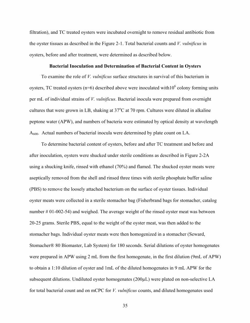
35
filtration), and TC treated oysters were incubated overnight to remove residual antibiotic from
the oyster tissues as described in the Figure 2-1. Total bacterial counts and V. vulnificus in
oysters, before and after treatment, were determined as described below.
Bacterial Inoculation and Determination of Bacterial Content in Oysters
To examine the role of V. vulnificus surface structures in survival of this bacterium in
oysters, TC treated oysters (n=6) described above were inoculated with106 colony forming units
per mL of individual strains of V. vulnificus. Bacterial inocula were prepared from overnight
cultures that were grown in LB, shaking at 37oC at 70 rpm. Cultures were diluted in alkaline
peptone water (APW), and numbers of bacteria were estimated by optical density at wavelength
A600. Actual numbers of bacterial inocula were determined by plate count on LA.
To determine bacterial content of oysters, before and after TC treatment and before and
after inoculation, oysters were shucked under sterile conditions as described in Figure 2-2A
using a shucking knife, rinsed with ethanol (70%) and flamed. The shucked oyster meats were
aseptically removed from the shell and rinsed three times with sterile phosphate buffer saline
(PBS) to remove the loosely attached bacterium on the surface of oyster tissues. Individual
oyster meats were collected in a sterile stomacher bag (Fisherbrand bags for stomacher, catalog
number # 01-002-54) and weighed. The average weight of the rinsed oyster meat was between
20-25 grams. Sterile PBS, equal to the weight of the oyster meat, was then added to the
stomacher bags. Individual oyster meats were then homogenized in a stomacher (Seward,
Stomacher® 80 Biomaster, Lab System) for 180 seconds. Serial dilutions of oyster homogenates
were prepared in APW using 2 mL from the first homogenate, in the first dilution (9mL of APW)
to obtain a 1:10 dilution of oyster and 1mL of the diluted homogenates in 9 mL APW for the
subsequent dilutions. Undiluted oyster homogenates (200µL) were plated on non-selective LA
for total bacterial count and on mCPC for V. vulnificus counts, and diluted homogenates used
36
100µL for plate counts. The LA and mCPC plates were incubated for 24 hours at 37oC and 40oC,
respectively.
After incubation, Log CFU/mL of inocula and bacterial recovery of V. vulnificus strains
(Log CFU/gram of oyster) from oysters were calculated. Isolates of V. vulnificus strains,
recovered after oyster passage were also examined for changes in colony morphology on LA
plates. Colony morphology was recorded, and results were summarized as changes in colony
morphology following oyster infections, recorded as a percentage (%) of total colonies on LA.
Each experiment used at least six oysters for each strain inoculated and included an un-
inoculated control (n=6). Bacterial content was determined for individual oyster meats. TC-
treated oysters (n=6) were also inoculated with 100µL of APW without V. vulnificus inocula as a
negative control for the study. Oysters without any TC treatment (n=6) were also examined in
order to determine the initial levels of bacterium present in oysters. Some experiments were
conducted in winter oysters that have natural reductions in bacterial load, in order to compare the
bacterial recovery in TC treated oysters versus non-TC treated oysters. All experiments were
repeated in triplicate.
Dissection of Oyster Tissues
Hemolymph, digestive tract, and gills of oysters were examined in order to determine the
distribution of V. vulnificus in oyster tissues using the oyster model of infection. Oyster
hemolymph was collected by drilling a notch in the oyster shell with a power drill, avoiding
contact with oyster tissue. With a 21-gauge needle, oyster hemolymph was withdrawn into a
sterile 5-mL syringe. For dissections, oysters were shucked using sterile shucking knife (Figure
2-2A). Using sterile scissors and forceps, the gills (located directly underneath the mantle) and
the digestive tract of oysters were dissected. The dissected oyster tissues were rinsed with sterile
PBS three times and weighed in stomacher bag. Equal amounts of PBS were added and the

37
oyster meats were homogenized. As described above, serial dilutions were made in APW;
homogenates were plated on LA and mCPC, incubated at 37 and 40oC for 24 hours, to determine
the survival of total and V. vulnificus bacteria (Log CFU/gram of oyster tissue), respectively.
Evaluation of Phase Variation in Oysters
A growth plasmid (pGTR902) was used to determine if appearance of opaque colonies
recovered from oyster infection are a result of phase variation of the translucent to the opaque
phenotype or just a reflection of die-off within the translucent population. E. coli S17-λ pir
containing the growth plasmid pGTR902 was provided by Dr. Paul Gulig at the University of
Florida. This plasmid was conjugated into V. vulnificus MO6-24/T1. This plasmid has a
kanamycin resistant gene marker (Starks et al., 2000). Additionally, the plasmid only replicates
in the presence of arabinose. Therefore, absence of arabinose will result in loss of the plasmid in
daughter cells (growing population) and those cells will not grow when subsequently plated on
antibiotic medium (LA with kanamycin and arabinose). On the other hand, in the presence of
arabinose, the growing cells will inherit the plasmid in the daughter cells. Thus, the appearance
of kanamycin opaque colonies on antibiotic medium will be indicative of phase variation within
the originally translucent culture.
The growth plasmid was conjugated to V. vulnificus MO6-24/T1, followed by plating the
culture on LA with arabinose (1%), kanamycin and polymyxin B. Briefly, an isolated colony of
MO6-24/T1 was grown in LB at 37oC overnight. E. coli with pGTR902 was also grown
overnight in LB with kanamycin (50µg/mL) with 1% arabinose. Each culture (1 mL) was
centrifuged for 10 min at 13,000 rpm and resuspended in 1mL of LB, to a total of three times.
Final pellets were resuspended in 5mL LB, and the cultures were incubated at 37oC with shaking
for 2.5 hours. E. coli with pGTR902 was then incubated statically at 37oC for 30 min to re-grow
sex pili. For conjugation, 200µL of E. coli S17 was added to 200µL of MO6-24/T1 and

38
transferred to a 0.45 µm filter (Millipore® filters Bedford, Mass., USA) placed on a 3 mm
Whatman filter paper and dried for one hour. Dried filters were the placed on the LA, filter cell
side up, and incubated overnight at 37oC. Following incubation, the filters were transferred to the
LB with kanamycin (300 µg/mL), polymyxin B (50 µg/mL) and arabinose (1%) and incubated
for one hour with shaking at 37oC. Cultures (150µL) were spread onto antibiotic LA (kanamycin
and polymyxin and arabinose) and incubated overnight at 37oC. The original MO6-24/T1
inoculum was also plated as a negative control on LA /kanamycin (50 µg/mL), polymyxin B (50
µg/mL), arabinose 1%. Colonies that grew on LA (300 µg/mL), polymyxin B (50 µg/mL),
arabinose 1%, were stored. The presence of plasmid in MO6-24/T1 was confirmed by plasmid
extraction kit (Promega, DNA purification, SV Minipreps, Catalog # A1460), for potential MO6-
24/T1/pGTR902 strains. The extraction was simultaneously performed on E. coli S17/pGTR902
as a positive control. The extracted plasmid was visualized on 1% agarose gel, and the band
sizes were compared. The isolates having same band size as the E. coli S17/pGTR902 confirmed
the presence of plasmid in MO6-24/T1, and positive colonies were streaked onto LA with
kanamycin (150µg/mL) for storage at -80oC.
The transconjugan MO6-24/T1 pGTR902 only replicates in the presence of arabinose, and
growth under non-permissive conditions results in loss of plasmid in the newly generated cells.
Consequently, the growing populations under conditions without arabinose were negative for the
plasmid and for antibiotic resistance (LA with kanamycin and arabinose). This property of the
plasmid was used for distinguishing the original inoculum from the growing population, as
kanamycin positive cells indicated the original inoculum. Additionally, the appearance of
kanamycin opaque colonies will indicate phase variation within the originally translucent culture.
Therefore, MO6-24/T1 pGTR902 was inoculated in the oyster model of infection, and the

39
inoculum concentration was determined by plate count on LA. Recovery of bacteria after 24
hours of inoculation was determined by plating the oyster homogenate on LA and on LA with
arabinose (1%) and kanamycin (300µg/mL). The plates were incubated at 30oC. The colonies
were examined for changes in colony morphology and phase variation. Opaque cells that
retained antibiotic resistance were presumed to be derived from phase variation of surviving
translucent cells. Calculation of death within the original population was determined by the
killing proportion, which was calculated using the following equation:
inoculum initial in the bacteria containing pGTR902 ofion Concentratoysters from recovered bacteria containing pGTR902 ofion Concentratproportion Killing =
Competition Studies
For competition studies, CPS deletion mutant (wzb) and double deletion mutant (pilA/wzb)
of V. vulnificus were used. The purpose of this study was to examine the recovery of V.
vulnificus strains in a mixed culture as a result of enhanced survival. Mixed overnight cultures of
either opaque and CPS deletion mutant (wzb) or opaque and ΔpilAΔwzb double mutant, were
inoculated (106 CFU/mL) in oysters using the oyster model of infection. The relative survival of
each V. vulnificus strain recovered from oysters was determined by examining the colony
morphology. V. vulnificus CPS (Δwzb) mutant is locked in the translucent phase and does not
show phase variation to opaque colony morphology (Chatzidaki-Livanis et al., 2006). Therefore,
difference in the colony morphology was used as a marker to distinguish the recovery of
different bacterial strains (opaque versus translucent) in the mixed culture. Experiments with the
pure culture of MO6-24/O (opaque) were also conducted simultaneously as a control to
determine any changes in the recovered colony morphology of opaque cells. The colony
morphology recovered after oyster passage on LA (polymyxin 50) incubated at 30oC for 24 hours,
was recorded as a percentage of opaque and translucent colonies of total colonies recovered.

40
Recovery of wild type encapsulated opaque strain was compared with to recovered translucent
colonies from oysters incubated with the mixed culture of opaque and Δwzb mutant. Similarly,
for the oysters incubated with the mixed culture of opaque-ΔpilAΔwzb mutant, the survival of
opaque strain was compared to that of recovered translucent ΔpilAΔwzb double mutant.
Statistical Analysis
Student’s t-test was used to evaluate the survival of V. vulnificus in oysters by comparing
the mutants and phase variants of V. vulnificus on selective media (mCPC). Bacterial
concentrations were log transformed and average and standard deviation of oysters within one
experiment and within multiple experiments were calculated. All the strains were compared to
the wild type encapsulated opaque strain to calculate the significant differences in the survival of
V. vulnificus strains in the oysters using a Student’s t-test two samples assuming unequal
variance, α = 0.05 in Microsoft Excel, 2003.

41
Table 2-1. Summary of V. vulnificus strains used in this study Strain Description MO6-24/O Wild-type, encapsulated, virulent, clinical isolate (Chatzidaki-Livanis
et al., 2006) CMCP6 Wild-type, encapsulated, virulent, clinical isolate (Lee et al., 2004) MO6-24/T1 Reversible phase variant, patchy CPS expression, reduced virulence
(Chatzidaki-Livanis et al., 2006) MO6-24/T2 (Δwzb)
Deletion mutant with precise deletion of wzb, no CPS surface expression (Chatzidaki-Livanis et al., 2006)
MO6-24/R (Rugose)
Reversible phase variant with enhanced biofilm formation and wrinkled (rugose) colonies (Grau et al., 2005)
MO6-24/ΔpilA (ΔpilA)
Specific deletion mutation in pilA, but with intact CPS operon, marked by opaque colonies (Paranjpye and Strom, 2005)
MO6-24/ΔpilA/T2 (ΔpilAΔwzb)
Deletion mutant derived from MO6-24/ΔpilA with precise deletion of wzb gene of CPS operon, eliminating CPS surface expression, marked by translucent colonies (This study)
FLA 677 (∆flaCDE)
Virulent strain with deletion in one gene locus (∆CDE), reduced motility, showed flagella, wild-type level of skin infection but was absent in liver (Tucker, 2006)
FLA 711 (∆flaCDE∆flaFBA)
Double deletion mutant (∆flaCDE∆flaFBA) replacing all the flagella genes, showed attenuated virulence in both skin and liver of mouse model (Tucker, 2006)
FLA 674 (∆motAB)
Deletion of (∆motAB) resulting in no motility but showed flagella, produced local infection but no systemic infection (Tucker, 2006)
42
Figure 2-1. Summary of oyster model of infection

43
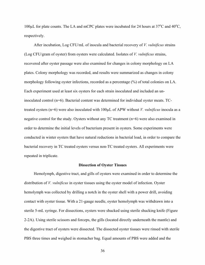
A
C Figure 2-2. Oyster dissection. Oysters are shucked using A) sterile oyster knife, and B) gills and
C) oyster digestive tract tissue removed. (Source: http://www.mdsg.umd.edu/issues/ chesapeake/oysters/education/anatlab/lab_i.htm , Last accessed 08/22/2007)
Gill Tissue
B

44
CHAPTER 3 DEVELOPMENT OF OYSTER MODEL OF INFECTION
Presence of indigenous V. vulnificus in oysters can complicate the experimental
investigation into the interaction of these bacteria with their molluscan shellfish host. The
problem is particularly severe during summer months when levels of V. vulnificus may approach
or exceed 105 bacteria per gram of oysters (Tamplin and Capers, 1992; Kaspar and Tamplin,
1993; Wright et al., 1996; Motes et al., 1998). In order to facilitate the development of an in vivo
oyster model of infection, antibiotic treatment was used to reduce the background V. vulnificus
levels in oysters. V. vulnificus is particularly sensitive to tetracycline (Bowdre et al., 1983);
therefore, tetracycline (TC) was selected as a pre-treatment prior to the artificial inoculation of
oysters with V. vulnificus
Optimization of Tetracycline Treatment
As described in Materials and Methods (Chapter 2), oysters were acclimated in artificial
sea water (ASW) with filtration for several days prior to treatment. Following acclimation, the
oysters were immersed in ASW containing the antibiotic, followed by transfer of oysters to the
fresh ASW with charcoal filtration overnight to remove the residual antibiotic. Three different
concentrations (1µg/mL, 2µg/mL, and 5µg/mL) of tetracycline were tested for the removal of
background V. vulnificus levels in the oysters. As shown in Figure 3-1, pre-treatment with
tetracycline at 2µg/mL and 5µg/mL resulted in significant reduction (less than 10 CFU per gram
of oyster meat) of V. vulnificus level oysters, compared to oysters without any TC treatment
(p=0.001 and 0.002 respectively). On the other hand, oysters treated with TC concentration of
1µg/mL did not show significant difference in reduction in V. vulnificus levels when compared to
non-treated oysters.

45
Recovery of V. vulnificus in Post Tetracycline Treated Oysters
Tetracycline-treated oysters were inoculated with V. vulnificus (106 CFU/mL) to examine
the bacterial recovery after the tetracycline treatment. Figure 3-2 illustrates that a significantly
higher recovery of V. vulnificus was seen in oysters treated with 2µg/mL of TC as compared to
the bacterial recovery seen in oysters that received TC concentration of 5µg/mL (p=0.01). It is
possible that high concentration of 5µg/mL of TC may have inhibited the survival of V.
vulnificus in oysters due to the accumulation of excess TC in oyster tissues. Therefore, pre-
treatment with 2µg/mL of tetracycline was chosen as the optimum concentration to remove the
background V. vulnificus contents in oysters.
Effects of Extended Incubation Post Tetracycline Treated Oysters
Tetracycline (2µg/mL) treated oysters were examined for the effectiveness of antibiotic
treatment to maintain low levels of V. vulnificus for time period of 24, 48 and 72 hours. Figure 3-
3 shows that the levels of V. vulnificus in TC treated oysters without V. vulnificus inocula were
significantly lower after 24, 48 and 72 hours (p=0.001, p=0.01 and p=0.03 respectively), as
compared to the initial V. vulnificus contents in oysters. These data suggest that tetracycline was
effective in reducing the V. vulnificus levels in oysters, which subsequently facilitated the in vivo
experimental assays.
Bacterial Recovery in Tetracycline and Non-tetracycline Treated Oysters
Antibiotic treatment of oysters reduced the V. vulnificus levels in the oysters; however
there is a possibility that concomitant reduction in non-V. vulnificus bacterial counts may alter
the results independently from other variables within the experimental oysters. The natural levels
of V. vulnificus in oysters are reduced during winter months as compared to the levels in warmer
months (Tamplin and Capers, 1992; Kaspar and Tamplin, 1993; Wright et al., 1996; Motes et al.,
1998). Therefore, in order to examine the suitability of tetracycline treatment in the oyster model
46
of infection, the survival of artificially inoculated V. vulnificus was compared in TC treated and
non-TC treated winter oysters. As shown in Figure 3-4, no significant differences were seen in
the survival of V. vulnificus strains in TC-treated oysters compared to non-TC -treated oysters.
These results show that treatment with tetracycline reduces the background V. vulnificus levels in
oysters without altering the survival of artificially inoculated bacteria in this in vivo assay.
47
0.0
1.0
2.0
3.0
4.0
5.0
6.0
0 1 2 5Concentration of Tetracycline (µg/mL)
Bac
teri
al c
onte
nt in
oys
ters
(L
og C
FU/g
) Total Bacteria in Oysters (LA)
V.vulnificus in oysters (mCPC)
Figure 3-1. Effect of tetracycline (TC) treatment on survival of V. vulnificus in oysters. V. vulnificus concentrations were examined with or without different concentrations of TC. Asterisk denotes significant (p<0.05) differences in V. vulnificus levels in oysters after TC treatment at different concentrations (2 µg and 5µg/mL) compared to initial count of V. vulnificus in oysters without TC treatment using Students t-test (p=0.001 and 0.002 respectively). Results are based on the mean of triplicate experiments using data from six individual oysters in each experiment.
* *

48
0.0
1.0
2.0
3.0
4.0
5.0
6.0
Pre-inoculation(2µg/mL)
Post-inoculation(2µg/mL)
Pre-inoculation(5µg/mL)
Post-inoculation(5µg/mL)
Tetracycline concentration
Bac
teri
al c
onte
nt in
oys
ters
(L
og C
FU/g
)Total Bacterial counts (LA)
V.vulnificus counts (mCPC)
Figure 3-2. Recovery of V. vulnificus from tetracycline (TC) treated oysters. V. vulnificus (Log
CFU/g) contents were examined in oysters receiving TC (either 2µg/mL or 5µg/mL) before and after artificial inoculation of bacteria. Asterisk denotes significantly higher bacterial recovery (p<0.05) in TC oysters at 2µg/mL (p= 0.01) as compared with the TC oysters at 5µg/mL using Student’s t-test. Data are based on the means of triplicate experiments using data from six individual oysters in each experiment.
*

49
0.0
1.0
2.0
3.0
4.0
5.0
6.0
Prior TC (2µg/mL) 24 h 48 h 72 h
Hours of incubation (h)
Bac
teri
al c
onte
nt in
oys
ters
(L
og C
FU/g
) Total bacterial counts (LA)
V.vulnificus counts (mCPC)
Figure 3-3. Effects of extended incubation on Tetracycline (2µg/mL) treated oysters. Asterisk
denotes significant (p<0.05) difference in V. vulnificus counts in TC (2µg/mL) oysters after 24h, 48 h and 72 hours of extended incubation as compared to initial V. vulnificus in oysters (p= 0.001, 0.01 and 0.03 respectively) using the Student’s t-test. Data are based on the means of triplicate experiments using data from six individual oysters in each experiment.
* *
*
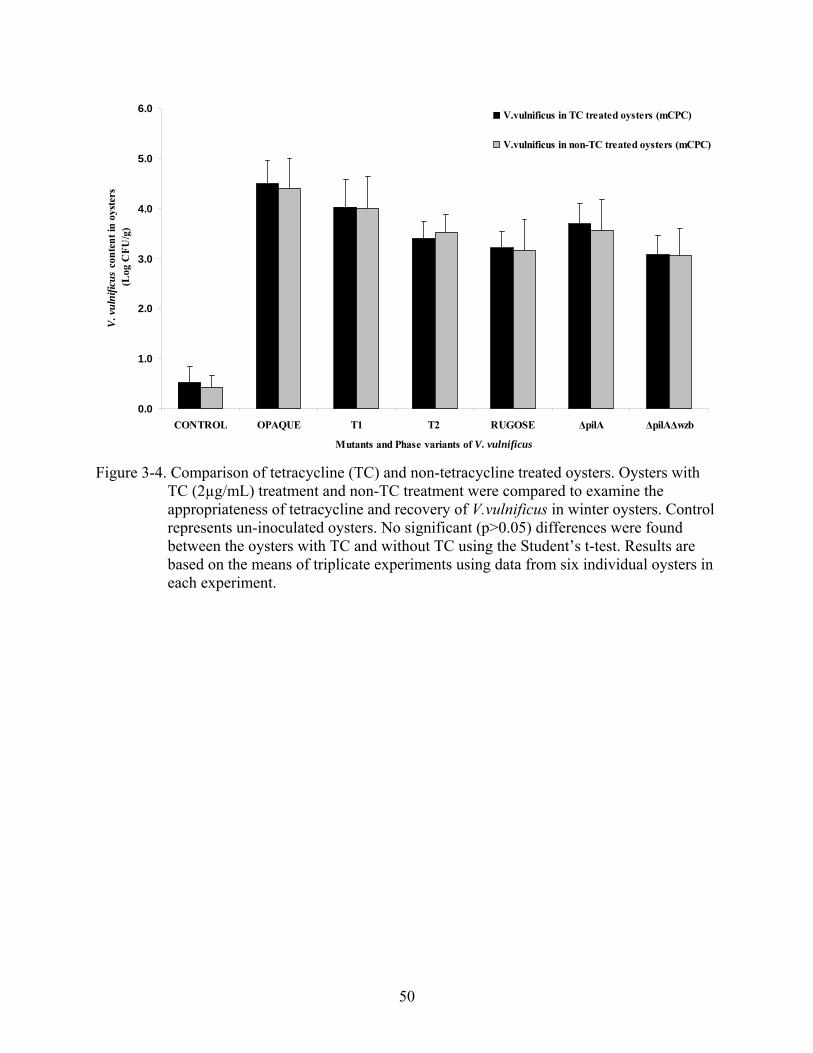
50
0.0
1.0
2.0
3.0
4.0
5.0
6.0
CONTROL OPAQUE T1 T2 RUGOSE ΔpilA ΔpilAΔwzb
Mutants and Phase variants of V. vulnificus
V. v
ulni
ficus
cont
ent i
n oy
ster
s(L
og C
FU/g
)V.vulnificus in TC treated oysters (mCPC)
V.vulnificus in non-TC treated oysters (mCPC)
Figure 3-4. Comparison of tetracycline (TC) and non-tetracycline treated oysters. Oysters with
TC (2µg/mL) treatment and non-TC treatment were compared to examine the appropriateness of tetracycline and recovery of V.vulnificus in winter oysters. Control represents un-inoculated oysters. No significant (p>0.05) differences were found between the oysters with TC and without TC using the Student’s t-test. Results are based on the means of triplicate experiments using data from six individual oysters in each experiment.

51
CHAPTER 4 ROLE OF CAPSULAR POLYSACCHARIDE IN SURVIVAL OF V. vulnificus IN OYSTERS
Encapsulation has been shown to contribute to resistance of bacteria to ingestion and
degradation by oyster hemocytes (Harris-Young et al., 1995). Therefore, the role of capsular
polysaccharide (CPS) in survival of V. vulnificus in oysters was examined using V. vulnificus
CPS mutant and phase variants (Table 2-1). Tetracycline (TC) treatment of 2µg/mL was used to
reduce the background V. vulnificus in oysters using the oyster model of infection followed by
artificial inoculation (106 CFU/mL) of oysters with CPS mutants and phase variants (Chapter 3).
Figure 4-1 shows that wild type encapsulated V. vulnificus survived in oysters at
significantly higher levels as compared to the CPS (wzb) deletion mutant with no capsule
expression (p= 0.025), after 24 hours of inoculation in oysters (Figure 4-1). However, the
translucent phase variant, which shows reduced capsule expression and can revert back to the
wild type, did not differ significantly from the opaque strain. These results suggest that
expression of CPS may confer an advantage for enhanced survival of V. vulnificus in oysters. A
translucent phase variant of V. vulnificus has been previously shown to have enhanced biofilm
formation (Joseph and Wright, 2004). Therefore, these data suggest that in vitro biofilm
formation may be independent of survival of V. vulnificus in oysters. Unlike group 1 CPS, other
capsular types have been implicated in promoting biofilm in V. cholerae and V. vulnificus (Yildiz
and Schoolnik, 1999; Grau et al., 2005). These strains are identified by the formation of rugose
or wrinkled colonies. Survival of V. vulnificus rugose variant was found in significantly lower
amounts in oysters (p=0.005) when compared to the wild type opaque strain. Survival
comparison of rugose and opaque strains in artificial seawater, showed that rugose had a higher
rate of recovery (p=0.03) than the wild type opaque strain. Based on these results, it can be

52
suggested that rugose strains are better adapted to survive in artificial seawater as compared to
survival in oysters.
Distribution of CPS Mutant and Phase Variants in Oyster Tissues
Vibrio species attach to plankton found in estuarine and marine environment. Molluscan
shellfish such as oysters filter-feed on the plankton by drawing the water using their gills and the
beating of cilia. Suspended plankton and food particles including bacteria get trapped in the
mucus of gills and are transported to the mouth. From the mouth, plankton including attached
bacteria is transported to the digestive tract and subsequently bacteria are disseminated into the
hemolymph and concentrated in oyster tissues (Kennedy, 1999). Therefore, CPS mutant and
phase variants were examined for the distribution of V. vulnificus in gills, digestive tract and the
hemolymph of oysters. As shown in Figure 4-2, significant differences were not found in the
survival of V. vulnificus CPS mutant and phase variants in the digestive tract of oysters.
However, survival of the wild-type encapsulated opaque strain was significantly higher in the
hemolymph of the oyster as compared to that of CPS (wzb) deletion mutant (p=0.03) and the
rugose phase variant (p=0.02). Moreover, distribution of the opaque strain was significantly
higher in gills of the oyster as compared to that of CPS (wzb) deletion mutant and rugose phase
variant (p=0.02, 0.01, respectively). Conversely, the survival of the translucent phase variant
(T1) did not differ from the wild type opaque strain. These results indicate that expression of
CPS did not appear to alter the initial attachment of bacteria to the intestine but is important for
the persistence of V. vulnificus in gills and dissemination to the hemolymph of oysters.
Recovery of V. vulnificus CPS Strains in Oysters after Extended Inoculation
V. vulnificus CPS mutant and phase variants were also examined for survival in
colonization in an oyster model after extended inoculation periods from 24 to 72 hours. As
shown in Figure 4-3, the survival of the encapsulated opaque strain was significantly higher as

53
compared to that of V. vulnificus CPS (wzb) deletion mutant after 24, 48 and 72 hours of
inoculation (p=0.03, p=0.04 and p=0.01, respectively. ). Survival of the rugose strain was also
found to be significantly lower than the survival of the wild type strain at 24 hours (p=0.009) and
48 hours (p=0.02) of inoculation. However, after 72 hours of inoculation, the survival of rugose
and the opaque was not found to be significantly different. On the other hand, the survival of
translucent strain with reduced capsular expression did not differ from the wild type
encapsulated strain at all during the extended periods of inoculation (24, 48 and 72 hours). This
study suggests that expression of capsule is important for the long term survival of V. vulnificus
in oysters.
Phase variation of V. vulnificus in Oysters
V. vulnificus exhibits phase variation in the expression of CPS, which is manifested by
changes in the colony morphology of opaque encapsulated strains by switching to translucent
colony morphology that is indicative of reduced CPS expression. Some translucent strains may
also revert to the opaque phenotype. It is hypothesized that survival of V. vulnificus in oysters
may be dependent on its ability to phase shift the expression of cell surface structures in response
to adverse environmental conditions (Chatzidaki-Livanis et al., 2006). In order to validate the
hypothesis, isolates of V. vulnificus strains were recovered from oysters and ASW following
oyster infection. The colony forming units from the recovered isolates were examined for
changes in the colony morphology that would indicate phase variation from the original
phenotype, on solid medium (LA), incubated for 24 hours at 30oC.
Results showed that opaque strains maintained stable morphology after oyster passage, and
all the colonies recovered from oysters were opaque on LA plates. However, both translucent
phase variant (T1) and rugose strains, showed changes in colony morphology by switching to the
opaque morphology at high frequency (Table 4-2). Nearly 72% of the original translucent

54
colonies inoculated in oysters were opaque on LA plates, whereas, the remaining 28% of
colonies maintained the translucent phenotype after oyster passage. Interestingly, 100% of the
initially rugose strain appeared opaque on LA plates, 24 hours post inoculation. Furthermore,
after extended incubation of rugose cells on LA plates for another 24 hours, originally rugose
strains produced atypical opaque colonies; those were either larger (68.9%) or smaller (9.9%)
than the typical wild type opaque phenotype, while 22% of colonies reverted back to the rugose
morphotype. These data suggest that phase variation of CPS expression in V. vulnificus could be
a survival strategy for enhanced survival. Opaque strains are reported to resist the phagocytosis
action of oyster hemocytes; therefore, it is possible that phase variation of translucent and rugose
cells to the opaque phenotype could provide adaptations to these V. vulnificus strains for better
endurance.
V. vulnificus isolates were also recovered from the artificial seawater to examine changes
in the colony morphology on LA, 24 hours post inoculation. Data showed that all the V.
vulnificus isolates recovered from water samples exhibited their original morphotype, and no
phase variation to opaque colonies was observed in any of the V. vulnificus strains (Table 4-3). In
contrast to translucent and rugose colonies, that showed phase variation to opaque colonies in
oysters, these strains maintained their stable colony morphology in artificial sea water on LA, 24
hours post inoculation in oysters. Rugose cells were also monitored for extended incubation of
LA plates for another 24 hours (a total of 48 hours on LA), and were found to maintain their
wrinkled colony morphology (Table 4-2). These results suggest that V. vulnificus does not
demonstrate phase variation in artificial seawater but exhibits phase variation in oysters.
Confirmation of Phase Variation of Translucent V. vulnificus with Growth Plasmid
In order to determine whether or not the appearance of opaque colonies from originally
translucent strains was a result of phase variation or just the result of die-off within the
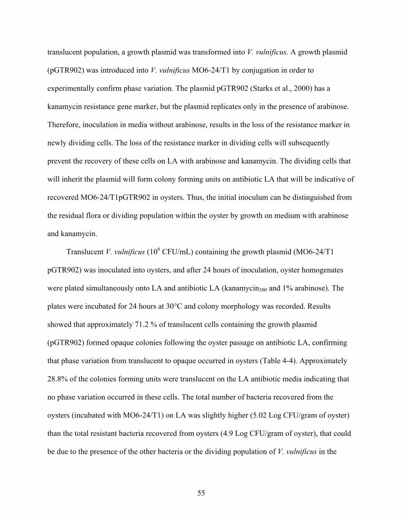
55
translucent population, a growth plasmid was transformed into V. vulnificus. A growth plasmid
(pGTR902) was introduced into V. vulnificus MO6-24/T1 by conjugation in order to
experimentally confirm phase variation. The plasmid pGTR902 (Starks et al., 2000) has a
kanamycin resistance gene marker, but the plasmid replicates only in the presence of arabinose.
Therefore, inoculation in media without arabinose, results in the loss of the resistance marker in
newly dividing cells. The loss of the resistance marker in dividing cells will subsequently
prevent the recovery of these cells on LA with arabinose and kanamycin. The dividing cells that
will inherit the plasmid will form colony forming units on antibiotic LA that will be indicative of
recovered MO6-24/T1pGTR902 in oysters. Thus, the initial inoculum can be distinguished from
the residual flora or dividing population within the oyster by growth on medium with arabinose
and kanamycin.
Translucent V. vulnificus (106 CFU/mL) containing the growth plasmid (MO6-24/T1
pGTR902) was inoculated into oysters, and after 24 hours of inoculation, oyster homogenates
were plated simultaneously onto LA and antibiotic LA (kanamycin300 and 1% arabinose). The
plates were incubated for 24 hours at 30°C and colony morphology was recorded. Results
showed that approximately 71.2 % of translucent cells containing the growth plasmid
(pGTR902) formed opaque colonies following the oyster passage on antibiotic LA, confirming
that phase variation from translucent to opaque occurred in oysters (Table 4-4). Approximately
28.8% of the colonies forming units were translucent on the LA antibiotic media indicating that
no phase variation occurred in these cells. The total number of bacteria recovered from the
oysters (incubated with MO6-24/T1) on LA was slightly higher (5.02 Log CFU/gram of oyster)
than the total resistant bacteria recovered from oysters (4.9 Log CFU/gram of oyster), that could
be due to the presence of the other bacteria or the dividing population of V. vulnificus in the

56
oyster tissue. However, the difference in the total bacterial counts (LA) and the total resistant
bacteria on antibiotic LA was not significant (Table 4-4). Approximately one Log CFU/mL of
the cells were killed as indicated by the killing proportion, which measures the concentration of
bacteria retaining pGTR902 after oyster passage divided by concentration of bacteria with
pGTR902 initially inoculated into culture. Experiments with a pure culture of MO6-24/T1
without plasmid were also conducted, serving as a control. The control experiments also showed
similar results of phase variation from translucent to opaque colonies. As shown in Table 4-4,
approximately, 70.4 % of original translucent strain inoculated in the oysters appeared opaque on
LA, 24 hours post inoculation in oysters and remaining 29.6% of the colonies appeared
translucent. These data confirm the phase variation of translucent colonies to the opaque
phenotype in the oysters.
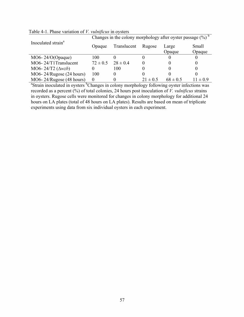
57
Table 4-1. Phase variation of V. vulnificus in oysters Changes in the colony morphology after oyster passage (%) b
Inoculated straina Opaque Translucent Rugose Large
Opaque Small Opaque
MO6- 24/O(Opaque) 100 0 0 0 0 MO6- 24/T1Translucent 72 ± 0.5 28 ± 0.4 0 0 0 MO6- 24/T2 (∆wzb) 0 100 0 0 0 MO6- 24/Rugose (24 hours) 100 0 0 0 0 MO6- 24/Rugose (48 hours) 0 0 21 ± 0.5 68 ± 0.5 11 ± 0.9 aStrain inoculated in oysters bChanges in colony morphology following oyster infections was recorded as a percent (%) of total colonies, 24 hours post inoculation of V. vulnificus strains in oysters. Rugose cells were monitored for changes in colony morphology for additional 24 hours on LA plates (total of 48 hours on LA plates). Results are based on mean of triplicate experiments using data from six individual oysters in each experiment.

58
Table 4-2. Phase variation of V. vulnificus in artificial seawater Changes in the colony morphology in water sample (%) b
Inoculated straina Opaque Translucent Rugose Large Opaque
Small Opaque
MO6- 24/O (Opaque) 100 0 0 0 0 MO6- 24/T1 (Translucent) 0 100 0 0 0 MO6- 24/T2 (∆wzb) 0 100 0 0 0 MO6- 24/Rugose (24 hours) 0 0 100 0 0 `MO6- 24/Rugose (48 hours) 0 0 100 0 0 aV. vulnificus strains inoculated in oysters bChanges in colony morphology following oyster infections in artificial seawater was recorded, 24 hours post incubation of V. vulnificus strains in oysters. Rugose cells were monitored for changes in colony morphology for additional 24 hours (a total of 48 hours) on LA plates. Data are based on mean of triplicate experiments using data from six individual oysters in each experiment.

59
Table 4-3. Confirmation of phase variation in MO6-24/T1 using pGRT902 in oysters Bacterial recoveryb Phase Variationc (%) Strain
inoculateda Total bacteria
Resistant bacteria
Translucent Opaque Killing Proportiond
MO6-24/T1 pGRT902
5.06± 0.4 4.89 ± 0.2 28.8 ± 0.4 71.2 ± 0.3 1.2 ±0.2
MO6-24/T1 5.03 ± 0.3 N.G 29.6 ± 0.7 70.4 ± 0.4 N.A. a Strain inoculated in the oysters b Bacteria (Log CFU/g) were recovered on LA (total) or antibiotic LA (total resistant) from oysters, inoculated with the cultures of V. vulnificus, 24 hours post inoculation. NG= no growth.c Phase variation of V. vulnificus in oysters; bacteria recovered from oysters was calculated as a fraction of total colony forming units on LA for (MO6-24/T1) and LA with kanamycin and arabinose for (MO6-24/T1pGRT902). d Killing proportion (Log CFU/mL) was calculated by concentration of bacteria with pGTR902 initially inoculated into the oysters dividing the concentration bacteria of retaining pGTR902 in the oyster. NA= not applicable
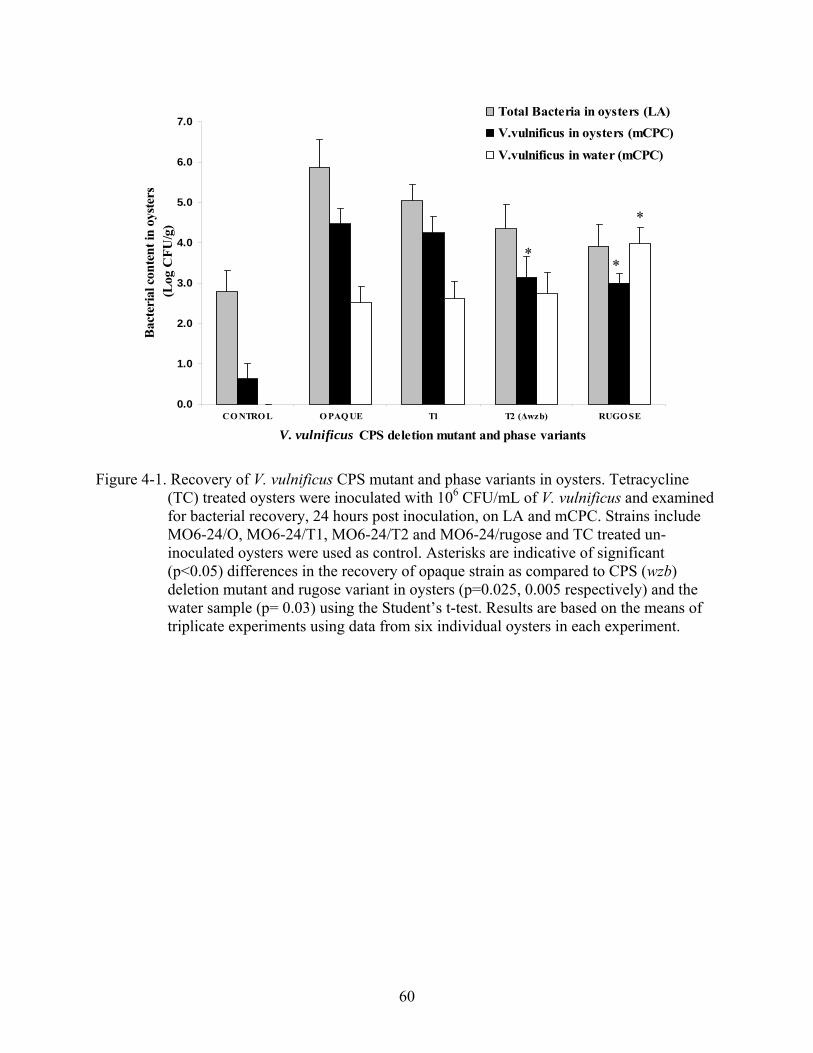
60
0.0
1.0
2.0
3.0
4.0
5.0
6.0
7.0
CO NTRO L O PAQ UE T1 T2 (Δwzb) RUGO SE
V. vulnificus CPS deletion mutant and phase variants
Bac
teri
al c
onte
nt in
oys
ters
(L
og C
FU/g
)
Total Bacteria in oysters (LA) V.vulnificus in oysters (mCPC)
V.vulnificus in water (mCPC)
Figure 4-1. Recovery of V. vulnificus CPS mutant and phase variants in oysters. Tetracycline (TC) treated oysters were inoculated with 106 CFU/mL of V. vulnificus and examined for bacterial recovery, 24 hours post inoculation, on LA and mCPC. Strains include MO6-24/O, MO6-24/T1, MO6-24/T2 and MO6-24/rugose and TC treated un-inoculated oysters were used as control. Asterisks are indicative of significant (p<0.05) differences in the recovery of opaque strain as compared to CPS (wzb) deletion mutant and rugose variant in oysters (p=0.025, 0.005 respectively) and the water sample (p= 0.03) using the Student’s t-test. Results are based on the means of triplicate experiments using data from six individual oysters in each experiment.
* *
*

61
0.0
0.5
1.0
1.5
2.0
2.5
3.0
CONTROL OPAQUE T1 T2 (∆wzb) RUGOSE
V. vulnificus CPS mutant and phase variants
V. v
ulni
ficus
con
tent
in o
yste
rs
(Log
CFU
/g)
V.vulnificus in Hemolymph(mCPC)V.vulnificus in Gills (mCPC)
V.vulnificus in Digestive tract (mCPC)
Figure 4-2. Distribution of CPS mutant and phase variants of V. vulnificus in oyster tissues. Oysters were inoculated with 106 CFU/mL of mutants and phase variants of V. vulnificus. Gills, intestine and hemolymph of oysters were dissected and examined for the distribution of V. vulnificus after 24 hours post inoculation; by examining growth on mCPC. Asterisk denotes significant (p<0.05) difference in recovery of wild type opaque strain with the CPS ∆wzb mutant and rugose in the hemolymph of the oysters (p=0.03, p=0.02 respectively) using the Student’s t-test. Significant differences were also seen in the distribution of opaque strain as compared to the CPS ∆wzb mutant and rugose in oyster gills (p=0.02 and p=0.01). Data are based on the means of triplicate experiments using data from six individual oysters in each experiment.
**
* *

62
0.0
1.0
2.0
3.0
4.0
5.0
6.0
CONTROL OPAQUE T1 T2 (∆wzb) RUGOSE
V. vulnificus CPS mutant and Phase Variants
V. v
ulni
ficus
cont
ent i
n oy
ster
s(L
og C
FU/g
)V.vulnificus in oysters (24h)
V.vulnificus in oysters (48h)
V.vulnificus in oysters (72h)
Figure 4-3. Recovery of V. vulnificus in oysters after extended inoculation time. Survival of V.
vulnificus was examined after 24, 48 and 72 hours of inoculation of V. vulnificus in oysters, and by examining the growth of strains on mCPC. Asterisk denotes significant (p<0.05) difference in the recovery of wild type strain with CPS mutants and phase variants in oysters at extended inoculation times. Recovery of opaque strain was significantly higher than T2 at 24, 48 and 72 hours of inoculation (p=0.03, p=0.04, p=0.01 respectively). Recovery of rugose strain was significantly lower than the opaque strain at 24 and 48 hour of inoculation (p=0.009 and p=0.02) using the Student’s t-test. Results are based on of the means of triplicate experiments using data from six individual oysters in each experiment.
**
**
*

63
CHAPTER 5 ROLE OF TYPE IV PILUS IN SURVIVAL OF V. vulnificus IN OYSTERS
V. vulnificus type IV pilus (∆pilA) has been associated with biofilm formation and
virulence in mammalian models (Paranjpye and Strom, 2005). To analyze the role of pilA and
additive effects of wzb and pilA in the survival of V. vulnificus in oysters, V. vulnificus pilA
deletion mutant and ∆pilA∆wzb double mutant (described in Chapter 2), were examined using
the oyster model of infection. Wild type encapsulated opaque strain survived in significantly
higher levels in oysters as compared to the V.vulnificus ∆pilA (p=0.01) mutant and ∆pilA∆wzb
double mutant (p=0.002), as shown in Figure 5-1. These data indicate that expression of both
pilA and wzb is important for the survival of V. vulnificus in oysters. The lower level of the
double deletion mutant (∆pilA∆wzb) as compared to the V. vulnificus strains with a single
mutation, indicated the additive contribution of PilA and encapsulation in the survival of V.
vulnificus in oysters. Both pilA deletion mutant and the ∆pil∆wzb double deletion mutant showed
higher recovery in artificial seawater (ASW) as compared to the wild-type encapsulated strain
(p= 0.04 and p=0.01, respectively).
Distribution of V. vulnificus pilA Mutants in Oyster Tissues
Hemolymph, gills and digestive tract of oysters were dissected to examine the distribution
of V. vulnificus ∆pilA and V. vulnificus∆ pilA∆wzb mutants in oyster tissues. Figure 5-2 shows
that there were significant differences in the distribution of the wild type encapsulated opaque
strain in the hemolymph of the oyster as compared to that of ∆pilA and ∆pilA∆wzb mutant strains
(p=0.02 and 0.01, respectively). On the other hand, there was no significant difference in the
distribution of V. vulnificus ∆pilA and ∆pilA∆wzb mutants in the digestive tract of oyster as
compared to the wild type opaque strain. The levels of wild type were significantly higher in the
gills of the oysters (p=0.04) as compared to the ∆pilA∆wzb strain, however, the ∆pilA mutant did

64
not differ from the wild type encapsulated strain. These data suggest that both type IV pilus and
encapsulation help in the dissemination of V. vulnificus to the hemolymph of oysters. Expression
of pili and CPS did not appear to alter the initial attachment of bacteria to the intestine. However,
the lower levels of V. vulnificus ∆pilA∆wzb in gills suggest that encapsulation, but not type IV
pilus is important for the persistence of V. vulnificus in oyster gills, as results were similar to
those observed for CPS (∆wzb) mutant alone (Figure4-2).
Recovery of V. vulnificus ∆pilA Mutants in Oysters after Extended Inoculation
Survival of V. vulnificus ∆pilA and ∆pilA∆wzb double mutant was also examined after
extended inoculation of oysters for up to 72 hours. Figure 5-3 shows that the survival of
encapsulated opaque strain was significantly higher than the V. vulnificus ∆pilA mutant after 24,
48 and 72 hours of inoculation (p=0.02, 0.03 and 0.04 respectively). Significant differences were
also seen in the recovery of V. vulnificus ∆pilA∆wzb double mutant (p=0.005, 0.03 and 0.01,
respectively) after 24, 48 and 72 hours of extended inoculation as compared to the wild type
strain (Figure 5-3). These results suggest that expression of both wzb and pilA are important for
the long term survival of V. vulnificus in oysters.
Survival of V. vulnificus in Oysters as a Result of Bacterial Competition
In the natural environment, bacteria in a mixed community compete for available nutrients,
and more successful species may outgrow the other members of the community for enhanced
survival. The purpose of this study was to examine the relative survival of V. vulnificus strains as
a result of bacterial competition in oysters. Mixed overnight cultures of opaque and CPS deletion
mutant (wzb) or opaque and ΔpilAΔwzb double mutant were inoculated (106 CFU/mL) in oysters
using the oyster model of infection. V. vulnificus (wzb) deletion mutant is locked in the
translucent phase and does not show phase variation to opaque morphotype (Chatzidaki-Livanis
et al., 2006). Therefore, difference in colony morphology was used to monitor the recovery of

65
opaque, ∆wzb and ∆pilA ∆wzb strains from oysters. Oysters were also incubated with the pure
culture of MO6-24/O (opaque) simultaneously as a control, to determine recovered colony
morphology of opaque strains after oyster passage. The majority of colonies recovered from
oysters inoculated with mixed culture of either opaque and wzb deletion mutant or opaque and
∆pilA∆wzb double mutant, were always wild type encapsulated opaque cells (Table 5-1). Results
showed that the survival of opaque was significantly higher than that of CPS (∆wzb) mutant
(p=0.002) and ΔpilAΔwzb double mutant (p=0.001). Additionally, significant differences were
also seen in the recovery of translucent strains from oysters incubated with opaque and ∆wzb
mutant as compared to that of opaque and ∆pilA∆wzb (p= 0.03).
These data support the previous results with individual strains (Figures 4-1, 2 and 3) and
confirm that expression of CPS is important for the survival of V. vulnificus in oysters. These
results also suggest the additive contribution of pilA and wzb in the increased survival of V.
vulnificus strains in oysters. Results also suggest that encapsulation may provide protective
adaptations for increased survival of opaque strains within the mixed bacterial community in
aquatic habitats and within molluscan shellfish host, particularly oysters.
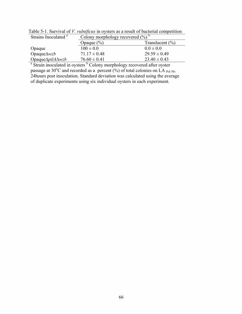
66
Table 5-1. Survival of V. vulnificus in oysters as a result of bacterial competition Colony morphology recovered (%) b Strains Inoculated a Opaque (%) Translucent (%)
Opaque 100 ± 0.0 0.0 ± 0.0 Opaque∆wzb 71.17 ± 0.48 29.59 ± 0.49 Opaque∆pilA∆wzb 76.60 ± 0.41 23.40 ± 0.43 a Strain inoculated in oysters b Colony morphology recovered after oyster passage at 30oC and recorded as a percent (%) of total colonies on LA Pol 50, 24hours post inoculation. Standard deviation was calculated using the average of duplicate experiments using six individual oysters in each experiment.
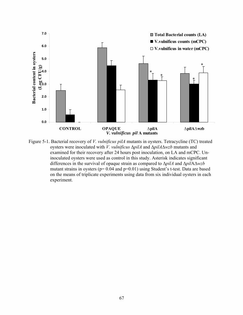
67
0.0
1.0
2.0
3.0
4.0
5.0
6.0
7.0
CONTROL OPAQUE ∆pilA ∆pilA∆wzbV. vulnificus pil A mutants
Bac
teri
al c
onte
nt in
oys
ters
(L
og C
FU/g
)Total Bacterial counts (LA)V.vulnificus counts (mCPC)V.vulnificus in water (mCPC)
Figure 5-1. Bacterial recovery of V. vulnificus pilA mutants in oysters. Tetracycline (TC) treated
oysters were inoculated with V. vulnificus ∆pilA and ∆pilA∆wzb mutants and examined for their recovery after 24 hours post inoculation, on LA and mCPC. Un-inoculated oysters were used as control in this study. Asterisk indicates significant differences in the survival of opaque strain as compared to ∆pilA and ∆pilA∆wzb mutant strains in oysters (p= 0.04 and p=0.01) using Student’s t-test. Data are based on the means of triplicate experiments using data from six individual oysters in each experiment.
**
*
*
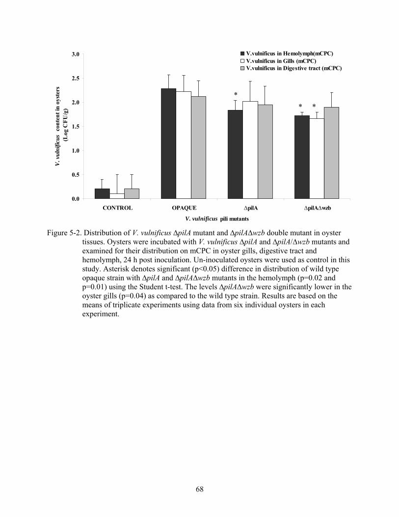
68
0.0
0.5
1.0
1.5
2.0
2.5
3.0
CONTROL OPAQUE ∆pilA ∆pilA∆wzb
V. vulnificus pili mutants
V. v
ulni
ficus
con
tent
in o
yste
rs (L
og C
FU/g
)
V.vulnificus in Hemolymph(mCPC)V.vulnificus in Gills (mCPC)V.vulnificus in Digestive tract (mCPC)
Figure 5-2. Distribution of V. vulnificus ∆pilA mutant and ∆pilA∆wzb double mutant in oyster
tissues. Oysters were incubated with V. vulnificus ∆pilA and ∆pilA/∆wzb mutants and examined for their distribution on mCPC in oyster gills, digestive tract and hemolymph, 24 h post inoculation. Un-inoculated oysters were used as control in this study. Asterisk denotes significant (p<0.05) difference in distribution of wild type opaque strain with ∆pilA and ∆pilA∆wzb mutants in the hemolymph (p=0.02 and p=0.01) using the Student t-test. The levels ∆pilA∆wzb were significantly lower in the oyster gills (p=0.04) as compared to the wild type strain. Results are based on the means of triplicate experiments using data from six individual oysters in each experiment.
** *

69
0.0
1.0
2.0
3.0
4.0
5.0
CONTROL OPAQUE ∆ pilA ∆pilA/∆wzbV. vulnificus pilA mutants
V. v
ulni
ficus
cont
ent i
n oy
ster
s(L
og C
FU/g
)V.vulnificus in oysters (24h) V.vulnificus in oysters (48h) V.vulnificus in oysters (72h)
Figure 5-3: Recovery of V. vulnificus ∆pilA and ∆pilA∆wzb double mutant in oysters after
extended inoculation. Survival of V. vulnificus pilA mutant strains was examined after 24, 48 and 72 hours of inoculation in oysters, as determined by growth on mCPC at 40oC. Asterisk denotes significant (p<0.05) difference in the survival of wild type opaque as compared to the ∆pilA mutant (p= 0.02, 0.03 and 0.04) and ∆pilA∆wzb double mutant (p=0.005, 0.03 and 0.01) after 24, 48 and 72 hours of inoculation using Student’s t-test. Results are based on the means of triplicate experiments using data from six individual oysters in each experiment.
**
*
*
*
*

70
CHAPTER 6 ROLE OF FLAGELLA IN SURVIVAL OF V. vulnificus IN OYSTERS
V. vulnificus possesses a single flagellum, and motility is important for the
attachment/initial steps of adhesion to surfaces, biofilm formation and virulence (Harshey, 2003;
Lee et al., 2004). However, the role of flagella and flagella-related motility in the survival of V.
vulnificus in oysters is less clear. Therefore, mutational analysis was used to examine the
survival of V. vulnificus strains that were either defective or lacking flagella or the flagellar
motor in oysters. The V. vulnificus flagella mutants (detailed description in Chapter 3) were
constructed by (Tucker, 2006) and provided by Dr. Paul Gulig, University of Florida.
Motility Test of V. vulnificus Strains
V. vulnificus flagella mutant strains used in this study were either defective in motility or
were non-motile (Tucker, 2006). The V. vulnificus rugose strain is also reported to possess a
polar flagellum, but has relatively reduced motility as compared to the opaque and translucent
strains (Grau et al., 2005). Therefore, to compare the relative motility of V. vulnificus strains, the
motility agar test was performed on the strains as shown in Table 6-1. Results from motility test
were in agreement with (Grau et al., 2005), and showed the reduced motility of rugose strain, as
compared to the encapsulated opaque strain. The motility of rugose was also found to be similar
to that of the flagella mutant strain ∆ flaCDE. The flagella mutant ∆flaCDE∆flaFBA strain,
which lacks flagella showed the least motility as expected due to the deletion of all six flagellar
genes. The flagellated strain, FLA(∆motAB), showed comparatively less motility than the wild
type strain, due to the deletions in genes that encode flagellar propulsion, as described by (Grau
et al., 2005)
Motility-deficient strains were assayed for survival in oysters using the oyster model of
infection. As shown in Figure 6-1, the survival of non-flagellated V. vulnificus double mutant

71
strain fla∆CDE∆ flaFBA, was significantly lower in artificially inoculated oysters as compared to
the wild type CMCP6 strain (p=0.03). However, the survival of V. vulnificus deletion mutant ∆
flaCDE and flagellar motor mutant (∆motAB) strain was not different from the wild type CMCP6
strain. Additionally, no significant differences were found in the levels of above mentioned
flagella mutants in artificial seawater (ASW) as compared to the levels of the wild type strain.
V. vulnificus rugose strain showed similar motility to ∆flaCDE mutant strain and was also
found in significantly lower levels in oysters (p=0.005), but the levels of rugose were higher in
the ASW as compared to the wild type strain (p=0.03). These results differed from flagellated
mutants with defective motility i.e. no significant difference was found in ∆ flaCDE and
FLA(∆motAB) mutant strain, as compared to the wild type strain, suggesting that motility is not
the only reason for low levels of rugose V. vulnificus in oysters. Furthermore, unlike rugose,
these strains did not show increase in numbers in ASW. On the other hand, the loss of both
flagella and motility (∆flaCDE∆ flaFBA) resulted in significant decrease in survival of vulnificus
in oysters, Figure 6-1.
Distribution of V. vulnificus Flagella Mutants in Oyster Tissues
It is possible that flagella and flagella-related motility are important for the dissemination
of V. vulnificus to hemolymph and internal organs of oysters. To experimentally examine the
distribution of V. vulnificus in the hemolymph, gills and digestive tract were dissected and
examined for distribution of V. vulnificus flagella and motility mutant strains. Results showed
that the wild type encapsulated strain (CMCP6) survived better in the hemolymph of oysters as
compared to all the three V. vulnificus flagella mutants. As shown in Figure 6-2, significant
differences were found in the survival of wild type V. vulnificus in the hemolymph of oysters as
compared to that of fla∆CDE, FLA (∆motAB) and ∆ flaCDE∆flaFBA mutant strains (p=0.04,
p=0.04 and p=0.02 respectively).

72
Significant differences found in the survival of ∆flaCDE, FLA(∆motAB) and ∆flaCDE∆
flaFBA in the hemolymph of the oyster as compared to the wild type strain suggests that
expression of flagella and flagellar motility are important for the dissemination of V. vulnificus to
the hemolymph of oysters. However, the survival of all three flagella mutants, such as ∆flaCDE,
FLA (∆motAB) and ∆flaCDE∆flaFBA did not differ from the wild type CMCP6 strain in the gills
and digestive tract of the oyster. These results indicate that expression of flagella and flagella
related motility may not contribute to the initial attachment of bacteria to the digestive tract and
the gills of the oyster but may be important in dissemination of bacteria to hemolymph.
On the other hand, V. vulnificus rugose, which showed similar motility to ∆flaCDE mutant
strain, was found to be in significantly lower levels in both the hemolymph and the gills of the
oyster, but the levels of this bacterium did not differ in intestines (Chapter 4). This contrast in the
survival of the rugose strain with the flagella mutant strain (∆flaCDE) indicates that reduced
motility is not the sole contributing factor towards the lower levels of rugose in the whole oyster
and oyster tissues. These data support the hypothesis that perhaps the altered expression of the
rugose phenotype and the CPS may be related to the lower level of this bacterium in oysters.
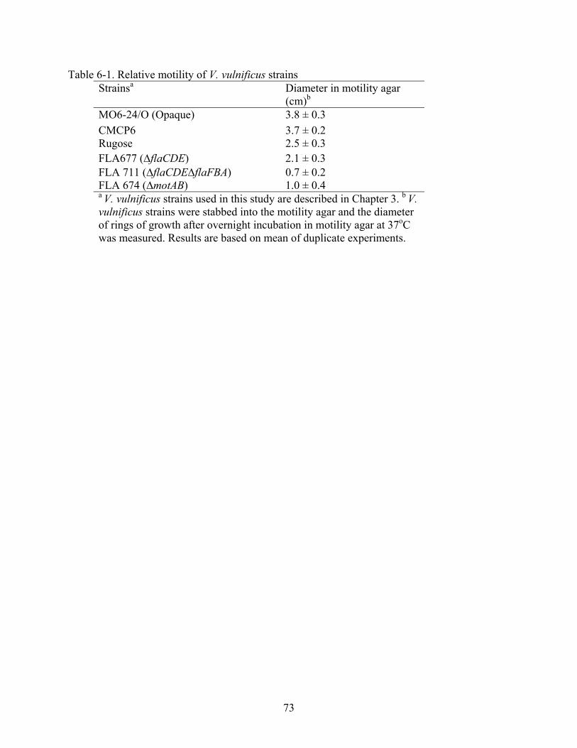
73
Table 6-1. Relative motility of V. vulnificus strains Strainsa Diameter in motility agar
(cm)b MO6-24/O (Opaque) 3.8 ± 0.3 CMCP6 3.7 ± 0.2 Rugose 2.5 ± 0.3 FLA677 (∆flaCDE) 2.1 ± 0.3 FLA 711 (∆flaCDE∆flaFBA) 0.7 ± 0.2 FLA 674 (∆motAB) 1.0 ± 0.4 a V. vulnificus strains used in this study are described in Chapter 3. b V. vulnificus strains were stabbed into the motility agar and the diameter of rings of growth after overnight incubation in motility agar at 37oC was measured. Results are based on mean of duplicate experiments.

74
0.0
1.0
2.0
3.0
4.0
5.0
6.0
7.0
Control CMCP6 ∆flaCDE ∆flaCDE∆flaFBA ∆motAB
V. vulnificus Flagella mutants
Bac
teri
al c
onte
nt in
oys
ters
(L
og C
FU/g
)
Total Bacteria in Oysters (LA)
V.vulnificus in oysters (mCPC)
V.vulnificus in water (mCPC)
Figure 6-1. Recovery of V. vulnificus flagella mutants in oysters. Survival of V. vulnificus flagella mutants in oysters were examined, 24 hours post inoculation, on LA and mCPC. Un-inoculated oysters were used as control for this study. Asterisk denotes significant (p<0.05) difference in the recovery of wild type CMCP6 strain in oysters as compared to the V. vulnificus (∆flaCDE∆flaFBA) strain using Student’s t-test (p=0.03). Results are based on the means of triplicate experiments using data from six individual oysters in each experiment.
*

75
0.0
0.5
1.0
1.5
2.0
2.5
3.0
Control CMCP6 ∆flaCDE ∆flaCDE∆flaFBA ∆motAB
V. vulnificus flagella mutants
V. v
ulni
ficus
con
tent
in o
yste
rs(L
og C
FU/g
)
V.vulnificus in hemolymph (mCPC) V.vulnificus in gills (mCPC)V.vulnificus in digestive tract(mCPC)
Figure 6-2. Distribution of V. vulnificus flagella mutants in oyster tissues. Hemolymph, gills and the intestine of oysters were examined for the distribution of V. vulnificus in oyster tissues, 24 hours post inoculation. Asterisk denotes significant (p<0.05) difference in survival of CMCP6 strain in the hemolymph of the oysters as compared to the V. vulnificus ∆flaCDE, ∆motAB and ∆flaCDE∆flaFBA mutants using Student’s t-test, (p= 0.04, 0.04 and 0.02 respectively). Results are based on the means of triplicate experiments using data from six individual oysters in each experiment.
*
**
76
CHAPTER 7 DISCUSSION AND CONCLUSION
Vibrio vulnificus naturally inhabits in warm estuarine environments, possibly existing in
mutual association with molluscan shellfish such as oysters (Tamplin and Capers, 1992; Harris-
Young et al., 1993; Harris-Young et al., 1995). Environmental factors favoring the growth of this
bacterium are low salinity levels (7-21 ppt) and warmer water temperatures (Kelly, 1982; Kaspar
and Tamplin, 1993). Occurrence of V. vulnificus has been positively correlated with warm water
temperature (Kelly, 1982; Kelly and Dinuzzo, 1985; Murphy and Oliver, 1992; Wright et al.,
1996; Motes et al., 1998), which is also linked with an increase in V. vulnificus related illnesses
(CDC, 1993; Hlady et al., 1993; CDC, 1996; Hlady and Klontz, 1996; FDA, 2003; CDC, 2005b,
a). Vibrios attach to plankton found in the estuarine environment. Filter-feeding shellfish such as
oysters (Crassostrea virginica) feed on plankton including bacteria, and concentrate the bacteria
in their tissues (Blake, 1983; Tamplin and Capers, 1992; Harris-Young et al., 1993; Harris-
Young et al., 1995). Raw oysters contaminated with this bacterium act as a vector of V.
vulnificus infections in humans, particularly as a result of consumption of raw oysters harvested
from warm waters in the Gulf of Mexico (Kelly and Dinuzzo, 1985; Hlady et al., 1993; CDC,
1996; FDA, 2003; CDC, 2005b).
This research study examined the possibility that surface structures of V. vulnificus may
provide an adaptation for increased survival of this bacterium in their environmental oyster
reservoir. Bacterial surface structure such as capsular polysaccharide (CPS), pili and flagella can
help anchor the bacterium to nutrient-rich surfaces of the host and may also provide protection
against the host defense mechanisms by avoiding the phagocytosis (Harris-Young et al., 1995).
These functions can lead to an increased endurance of V. vulnificus in the oyster host and,

77
subsequently may facilitate human infection by increasing bacterial contamination of food
intended for human consumption.
Surface structures of V. vulnificus also contribute to biofilm formation and increased
survival under hostile environments. Capsular polysaccharide in V. vulnificus inhibits in vitro
biofilm formation (Joseph and Wright, 2004), but it appears to be important for the survival of V.
vulnificus in oyster hemocytes (Harris-Young et al., 1993; Harris-Young et al., 1995).
Expression of pilD and pilA are important for the persistence of V. vulnificus in American oysters
(Paranjpye et al., 2007). Other studies have focused on the role of V. vulnificus flagella and
flagellar motility in biofilm formation and virulence in mouse model (Kim 2003; Lee et al.,
2004). However, the role of these structures in the survival of V. vulnificus in oysters has not
been established. Therefore, this research study was conducted to gain better understanding on
the contribution of these surface structures to the survival of V. vulnificus in oysters.
Presence of indigenous V. vulnificus in oysters can complicate the in vivo investigation,
particularly during summer months when V. vulnificus levels may approach or exceed to 105
bacteria per gram of oyster meat (Kelly, 1982; Oliver et al., 1983; Oliver, 1989; Wright et al.,
1996; Motes et al., 1998). Therefore, to facilitate the in vivo examination of V. vulnificus in their
environmental reservoir of disease, an oyster model of infection that utilizes antibiotic treatment
was developed. V. vulnificus is particularly sensitive to tetracycline (TC) (Bowdre et al., 1983);
therefore, oysters were bathed overnight in different concentrations of TC. Subsequently, the TC
treated oysters were transferred to fresh artificial seawater with overnight charcoal filtration to
remove residual antibiotic. Data showed that the reduction of V. vulnificus levels in oysters was
accomplished with an increase in concentration of TC. The greatest reduction in V. vulnificus
levels was seen with TC concentration of 5µg/mL. However, the bacterial recovery post TC

78
treatment at 5µg/mL was significantly reduced, presumably due to the excess accumulation of
TC in the oyster tissues, leading to inhibition of V. vulnificus recovery in oysters. Therefore, TC
concentration of 2µg/mL was used as optimum concentration for V. vulnificus reduction and was
shown to maintain low V. vulnificus levels in oysters after 24, 48 and 72 hours of post TC
treatment. These data showed that TC was effective in reducing the V. vulnificus levels in oysters
and facilitating in vivo experimental assays.
Environmental conditions, such as water temperature, play an important role in the
proliferation of V. vulnificus in molluscan shellfish and their natural surroundings. The levels of
V. vulnificus are lower during winter months as compared to summer months (Oliver et al., 1983;
Kaspar and Tamplin, 1993; Cook, 1994; Wright et al., 1996; Motes et al., 1998). Treatment of
oysters with TC reduced both the total bacterial count and the V. vulnificus counts in oysters, but
it is possible that it may have additional consequences that could alter the outcome of
experiments. Therefore, different V. vulnificus strains were evaluated for their survival in oysters
with or without TC treatment, using winter oysters that had a natural reduction of bacteria.
Statistical analysis on the survival of both the total bacteria and V. vulnificus recovered from
oysters using winter oysters revealed no significant differences as a consequence of TC treatment
for any of the V. vulnificus strains that were tested. Additionally, bacterial contents in TC-treated
summer oysters did not differ from bacterial levels found in winter oysters. Therefore, these data
imply that antibiotic treatment of oysters “artificially simulates” the bacterial content similar to
that of winter oysters, providing a model for in vivo mutational analysis of V. vulnificus in
oysters.
The role of capsular polysaccharide in survival of V. vulnificus in oysters was verified
using the oyster model of infection. The survival of the encapsulated opaque strain was

79
consistently higher than the CPS deletion mutant (∆wzb) and translucent phase variant (T1) in
oysters at 24 hours post inoculation. Significant differences were noted in the survival of CPS
deletion mutant (T2) as compared to the wild type encapsulated opaque strain. However, the
partially encapsulated CPS translucent phase variant did not differ significantly from the wild
type strain. Thus, the data suggest that degree of encapsulation contributes to the survival of V.
vulnificus in oysters. Alternatively, the instability of translucent phase variants may have caused
the switching of cells to the more resistant opaque phenotype.
Survival of the rugose phenotype was found to be significantly lower in oysters as
compared to the wild type strain. On the other hand, high levels of rugose were recovered from
artificial seawater. These data propose the possibility that the type of CPS may be related to the
lower survival of rugose strain in oysters as well as to the higher survival of rugose strains in
artificial seawater. Differences in the composition of CPS have been reported for V. vulnificus
and rugose V. cholerae that may explain the observed discrepancies in the survival of rugose and
opaque strains in oysters. A higher net negative charge on the cell surface due to polysaccharide
capsule results in a greater degree of resistance to phagocytosis, as reviewed by (Roberts, 1996).
Biochemical analysis of Group 1 CPS revealed that V. vulnificus MO6-24 produces highly
charged and acidic CPS (uronic acid), and is composed of four sugar residues three residues of 2-
acetomido-2, 6- dideoxyhexopyranose and one residue of 2-acetomido hexouronate (Reddy et
al., 1992; Hayat et al., 1993). On the other hand, the polysaccharide structure of rugose V.
cholerae EPS is composed of neutral sugars (glucose and galactose) that are more hydrophobic
(Yildiz and Schoolnik, 1999; Ali et al., 2002) in contrast to the hydrophilic V. vulnificus CPS
(Reddy et al., 1992; Hayat et al., 1993; Wright et al., 1999). The EPS of the rugose phase variant
of V. cholerae O1 TSI-4 contains N-acetyl-D-glucosamine, D-mannose, 6-deoxy-D-galactose,

80
and D-galactose and contains equal amounts of 4-linked galactose and 4-linked glucose (Yildiz
and Schoolnik, 1999). It is likely that hydrophobic EPS produced by wrinkled rugose colonies
promote cell aggregation, leading to the attachment of bacteria to one another in the ASW
medium; however, this aggregation may inhibit uptake by oyster or enhanced degradation by
hemocytes. Therefore, the differences in the polysaccharide composition of V. cholerae rugose
may provide clues to increased survival of opaque V. vulnificus in oysters, as well as the
increased numbers of rugose strains in seawater. Unfortunately, the composition of rugose in V.
vulnificus has not been determined, and further investigation will be required to understand the
role of polysaccharide in survival of rugose in oysters.
The survival of CPS mutant and phase variants was also examined for persistence at
extended inoculation up to 24, 48 and 72 hours in whole oysters. In agreement with the results at
24 hours post inoculation, the survival of encapsulated opaque strain was always highest as
compared to other CPS phase variants and mutants of V. vulnificus, and the translucent phase
variant did not differ significantly from the wild type opaque strain at any of the extended
inoculation time. Survival of the opaque strain was always significantly higher than the CPS
(∆wzb) deletion mutant at 24, 48 and 72 hours of extended inoculation periods. The rugose phase
variant also differed significantly at 24 and 48 hours of extended inoculation but did not differ
from the opaque, after 72 hours post inoculation. A possible explanation for this lack of
significant difference at this time point could be that the initial concentration of rugose cells in
oysters was so low as compared to the opaque strain that further reduction by oyster defense
mechanism was not possible or the surviving rugose cells were better adapted to survive and
resist further destruction. Alternatively, survival of rugose strain in oysters could be enhanced

81
after extended inoculation by continued seeding from their relatively high numbers surviving in
the seawater.
Molluscan shellfish such as oysters are filter feeders accumulate indigenous bacteria
attached to plankton or algae and concentrate the bacteria in their tissues (Kumazawa, 1991;
Hood, 1997; Maugeri et al., 2006). To determine the distribution of V. vulnificus in oyster
tissues, hemolymph, intestines and gills of oysters were examined. Data consistently showed
increased survival of encapsulated opaque in hemolymph of oysters as compared to V. vulnificus
CPS deletion (∆wzb) mutant and rugose phase variant. However, there were no differences in the
survival of CPS V. vulnificus strains in the intestinal tract of the oyster. Only rugose and CPS
deletion mutant were found to be deficient in attachment to the oyster gills. These data suggest
that CPS contributes to the attachment of V. vulnificus in the gills but not the gut and is required
for the dissemination of the bacterium to the hemolymph of the oyster.
Phase variation has been observed in many bacteria by different surface structures and
involves capsule, pili, fimbriae, and outer membrane proteins that can be recognized by their
effect on colony morphology, as reviewed by (van der Woude and Baumler, 2004). Phase
variation in V. vulnificus is exhibited by changes in colony morphology that reflect alteration of
CPS surface expression. V. vulnificus phase variation is marked by reversible changes in colony
morphology such as opaque (encapsulated, virulent) versus translucent (decreased CPS,
attenuated virulence) and smooth versus rugose (wrinkled) phenotype (Wright et al., 1999;
Wright et al., 2001; Grau et al., 2005; Chatzidaki-Livanis et al., 2006; Hilton et al., 2006). In a
recent study, an irreversible deletion mutation in V. vulnificus CPS operon was reported. MO6-
24/∆wzb is a result of a deletion mutation of wzb in the group 1 CPS operon, thus locking the cell
in a translucent phase that is unable to revert to opaque morphotype (Chatzidaki-Livanis et al.,

82
2006). To examine the phase variation in oysters, V. vulnificus strains recovered after oyster
passage were monitored for changes in the colony morphology on LA plates. Results
demonstrated that the opaque strain maintained a stable phenotype, while the translucent and
rugose phase variant switched to the opaque morphotype at a high frequency (72 and 100%,
respectively) following oyster infection. Moreover, rugose strains were unstable on LA plates
and after extended incubation of rugose cells on LA for additional 24 hours (total of 48 hours),
atypical opaque colonies were observed or the cells reverted back to the rugose (wrinkled)
morphotype. V. vulnificus isolates from the water samples contrasted greatly from those
recovered for oysters. No changes in the colony morphology of V. vulnificus strains were
observed in any of isolate recovered from the ASW. Thus, phase variation was specific to oyster
passage and did not occur in cells that remained in seawater.
In order to experimentally confirm this phase variation, a growth plasmid (pGTR902) was
introduced into V. vulnificus translucent strain to track the growing population during oyster
infection. Results showed that approximately 72% cells that were initially translucent contained
the growth plasmid now formed opaque colonies on solid medium when recovered from oysters,
which conformed that phase variation from translucent to opaque phenotype had occurred in
vivo. The growth plasmid was also used to monitor the killing proportion of the translucent cells.
Results showed that only about 1 Log CFU/mL of the translucent inoculum died during oyster
passage, indicating that phase variation rather than die off of translucent cells in the oysters was
primarily responsible for appearance of opaque cells. Overall, these data confirmed the phase
variation of V. vulnificus translucent to opaque phenotype in oysters.
Formation of biofilm and attachment of microbial communities to the nutrient-rich
surfaces serve as a survival mechanism for bacteria in adverse environments, as reviewed by

83
(Davey and O'Toole G, 2000). Although, expression of group 1 CPS in V. vulnificus inhibits the
in vitro biofilm formation, results from this study showed that it promotes survival of V.
vulnificus in oysters. Thus, it is possible that some facets of in vitro biofilm formation may be
independent of in vivo survival of this bacterium. Unlike group 1 CPS, other capsular types are
associated with the formation of biofilm, which are marked by rugose or wrinkled colonies in V.
cholerae (Watnick et al., 1999; Yildiz and Schoolnik, 1999) and V. vulnificus (Grau et al., 2005).
Comparing the survival of V. vulnificus CPS mutants and phase variants in oysters, it was found
that among all the strains examined, rugose strain showed least survival in the oysters. On the
other hand, rugose was found in significantly higher concentrations in ASW samples. These data
imply that the rugose strains are probably better adapted for increased survival in sea water as
compared to the oysters and that in vivo survival of V. vulnificus in oysters is not dependent in
biofilm formation.
Pili are important for survival of bacteria in biofilm formation and oysters (Paranjpye and
Strom, 2005; Paranjpye et al., 2007) and it is possible that they are likely to contribute towards
dissemination of V. vulnificus to the internal tissues of oysters. Therefore, the role of type IV
pilus in the survival of V. vulnificus in oysters was investigated. V. vulnificus ∆pilA, deficient in
adherence to epithelial cells, biofilm formation and virulence in animal models (Paranjpye and
Strom, 2005), was used in this study. A double deletion mutant of MO6-24/∆pilA with deletions
in pilA and wzb was constructed in this study to evaluate the additive roles of pilA and
encapsulation (wzb) in survival of V. vulnificus in oysters. Results showed that the survival of
wild-type encapsulated strain was significantly higher than that of V. vulnificus pili mutant
strains. These findings were in agreement with (Paranjpye et al., 2007) that deletion of pilD and
pilA genes significantly reduced the ability of V. vulnificus to colonize and persist in oysters. The

84
additive effects of pili and encapsulation in the survival of V. vulnificus in oysters were
evidenced by the lower survival of V. vulnificus ∆pilA∆wzb double mutant in oysters as
compared to the single deletion mutants of V. vulnificus. Significantly higher amounts of ∆pilA
and ∆pilA∆wzb mutants were found in ASW samples as compared to the wild type, similar to
observations regarding rugose strain, which indicated that in vitro biofilm formation was
independent of survival of these bacteria in oysters.
Distribution of V. vulnificus∆ pilA mutants was also examined in the hemolymph, gills and
intestines of the oyster. Results showed significantly higher levels of the encapsulated wild-type
strain in the hemolymph of oyster as compared to levels of ∆pilA and ∆pilA∆wzb double mutant.
Compared to the double ∆pilA∆wzb mutant, opaque strain was recovered in significantly higher
levels from the gills; however, the ∆pilA mutant did not differ from the wild type strain.
Additionally, no significant differences were seen in the distribution of V. vulnificus opaque,
∆pilA and ∆pilA∆wzb double deletion mutants in the intestinal tract of oyster. Based on these
results, it can be concluded that although the expression of pilA and capsule may not be
important for the initial attachment in the gut, they contribute towards the dissemination of V.
vulnificus to the hemolymph of oyster. Furthermore, this study again confirmed that the
expression of CPS is also important for the distribution of V. vulnificus in the gills of the oyster.
In a natural environment, bacteria compete with each other for attachment to nutrient rich
surfaces for enhanced survival. In this regard, in vivo competition studies can indicate the fitness
for survival of different bacteria in host. Experiments were conducted to confirm the enhanced
survival of wild type V. vulnificus as compared to the CPS deletion mutant and the pili double
deletion mutant in oysters. Therefore, oysters were incubated with the mixed cultures of V.
vulnificus opaque and CPS mutant (∆wzb) or opaque and ∆pilA∆wzb double deletion mutant.

85
Both of these wzb deletion mutants are locked in the translucent phase, therefore, examination of
colony morphology was used as a marker to distinguish these mutants from wild type
(Chatzidaki-Livanis et al., 2006). Results revealed that majority of strains recovered from the
oysters were always encapsulated opaque strain, and only 25-30% of colonies recovered were
translucent. Results also showed that the recovery of the double deletion mutant was further
reduced as compared to the single deletion mutant, which also confirmed that the additive effects
of pilA and wzb contributes to the enhanced survival of V. vulnificus in oysters.
V. vulnificus rugose strain is relatively less motile than either opaque or translucent phase
variant, but yet possesses a polar flagellum (Grau et al., 2005). Therefore, it is possible that
reduced motility of the rugose strain resulted in the lower levels of this strain in oysters, as
motility also contributes to biofilm formation and invasion of host tissues (McCarter, 2001;
Harshey, 2003). V. vulnificus possesses six flagellin structural genes flaA, flaB, flaF, flaC, flaD
and flaE organized in two distinct genetic loci, namely flaFBA and flaCDE (Tucker, 2006). To
examine the role of motility in the uptake and survival of V. vulnificus in oysters, flagella mutant
strains were tested using the oyster model of infection. Results from survival of V. vulnificus
flagella mutants in oysters showed that the survival of the double deletion mutant
(∆flaCDE∆flaFBA), lacking all flagella genes, was significantly lower than the wild type
CMCP6 strain in the whole oyster preparation. However, the survival of flagella mutant strain
with defective flagella and reduced motility (∆flaCDE), or the strain with no flagellar motor
components (∆motAB), did not differ from the wild type in whole oyster preparations. A similar
conclusion was also reported by Lee et al., 2004, as flagellum-deficient and non-motile V.
vulnificus mutant (∆flgE gene, encoding the flagellar hook protein), showed decreased virulence
in iron dextran-treated mice, defect in adherence to the cells, and less biofilm formation on a

86
polystyrene surface. Flagella mutant strains were also analyzed for distribution of V. vulnificus in
the hemolymph, gills and the digestive tract of the oyster. Data showed significantly higher
survival of V. vulnificus wild type strain as compared to all flagella mutant strains in the
hemolymph of the oyster. However, the distribution of flagella mutants did not differ from the
wild type strain in gills and the digestive tract of oysters. Overall, these results suggest that
motility or flagella may not be a contributing factor to the initial attachment of V. vulnificus in
oyster tissues, but expressions of both is probably important for the survival and dissemination of
V. vulnificus to the hemolymph of oysters.
Comparative analysis on the motility of all the V. vulnificus strains examined, revealed that
the motility of rugose was similar to the single gene locus deletion flagella mutant (∆flaCDE),
but data for survival of ∆flaCDE mutant strain in whole oysters differed greatly from the survival
of rugose V. vulnificus in oysters. The rugose phase variant showed lower survival in oysters but
was found to be in greatest number in seawater. On the other hand, the ∆flaCDE mutant did not
differ from wild type in either seawater or oysters. Moreover, rugose was also found in
significantly lower levels than the wild type in oyster gills, but the survival of the flagellar
(∆flaCDE) deletion mutant was not significantly different in the gills as compared to the wild
type. Therefore, it can be concluded that reduced motility of rugose was not the only sole factor
contributing to the outcome of this strain in oysters. Further, it is likely that an alternate
composition of rugose CPS, reported for V. cholerae (Yildiz and Schoolnik, 1999), could be
related to the lower survival of rugose in oysters.
However, it is difficult to sort out the contribution of flagella to attachment versus motility
in the mutational analysis employed in this study, as loss of ligand also results in loss of motility.
V. vulnificus express H antigen in the core proteins of the polar flagella, and an anti-flagellar
87
(anti-H) antibody is produced in rabbits immunized with flagellar core protein prepared from V.
vulnificus (Simonson and Siebeling, 1986). Therefore, additional experiments utilizing antibody
to block attachment to oyster would be useful in drawing conclusions about the role of flagella in
attachment versus motility and subsequently interactions within the oyster tissues. Furthermore,
oysters are filter-feeders and use their gills to filter particles out of the water. Through the
beating of cilia on the gills, water currents are generated and the gills transport water and
planktons towards the mouth. Therefore, it is also possible that the action of oyster filtration was
contributing greatly towards the uptake of V. vulnificus flagella mutants in oysters, and the
contribution of bacterial motility may be minor compared to the filtering activity of the oyster.
In conclusion, this research study demonstrated the contribution of surface structures in the
survival of V. vulnificus in their molluscan shellfish host, Crassostrea virginica. V. vulnificus
attaches to plankton in seawater, and oysters are bivalve mollusks that feed by filtering water and
plankton, ingesting bacteria and concentrating in the tissues (Maugeri et al., 2006). Bacteriolytic,
heat stable lysozyme is present in the hemolymph of oysters (McDade, 1967a), which provides
active defense against bacteria (Rodrick, 1974). However, V. vulnificus disseminates in the
hemolymph and concentrates in oyster tissues (Kennedy, 1999), possibly by avoiding the lysis
by lysozyme. Little is known about the biology of V. vulnificus in oyster survival, but studies
have reported that expression of V. vulnificus CPS provides resistance to phagocytosis and
degradation by oyster hemocytes (Harris-Young et al., 1995). It has been shown that a protease
from the oyster parasite Perkinsus marinus may inhibit the phagocytic function of oyster
hemocytes (Tall et al., 1999). More recently, it has been reported that expression of pilD and pilA
is important for persistence of V. vulnificus in oysters (Paranjpye et al., 2007). The present study

88
provides evidence that surface structures of V. vulnificus may also have important roles in
survival of this bacterium in the oyster host.
Survival of bacteria in hostile environments has been studied in extensively detail in vitro.
Formation of biofilms requires a cooperative effort by surface structures of bacteria such as
flagella, pili, outer membrane proteins, and EPS. Results from this research study suggest that
surface structures of V. vulnificus that are involved in biofilm formation and virulence contribute
differentially towards the survival of V. vulnificus in oysters. The survival strategy of V.
vulnificus in oysters appears to be multi-factorial. The expression of capsular polysaccharide,
pili, and flagella together with motility, are all important for survival of V. vulnificus in the
environmental reservoirs of the disease. As evidenced by lower survival of V. vulnificus
∆pil∆wzb double mutant in oysters, it can be concluded that surface structures of V. vulnificus
put together an additive effort to fight the defense mechanism of oysters, leading to increased
endurance in their oyster host. Although CPS expression of V. vulnificus inhibits the biofilm
formation (Joseph and Wright, 2004), results from this study showed that it is important for the
survival of this bacterium in oysters. Therefore, it can be concluded that some facets of in vitro
biofilm formation may be different from in vivo survival of V. vulnificus in oysters. On the other
hand, expression of pili, flagella and flagellar motility, that are important surface structures
contributing to the biofilm formation (Lee et al., 2004; Paranjpye and Strom, 2005; Lee et al.,
2006), also appears to be important for survival of V. vulnificus in oysters.
Studies have reported occurrence of vast genetic variation among the stains of V.
vulnificus. It has also been shown that individual oyster can harbor numerous genetically
divergent V. vulnificus strains (Buchrieser et al., 1995). However, most of the strains isolated
from environmental reservoir appear to be encapsulated and are virulent as clinical strains in
89
animal models (Tison and Kelly, 1986; Simpson et al., 1987; Tilton and Ryan, 1987; Wright et
al., 1996; DePaola et al., 2003). Evaluation of environmental strains recovered from the
Chesapeake Bay also showed only encapsulated opaque phenotype (Wright et al., 1996). Results
from this study showed that encapsulation contributes to the increased survival of this bacterium
in the mixed V. vulnificus culture, supporting the hypothesis that CPS expression determines the
enhanced survival of V. vulnificus in its environmental reservoir.
This is the first study to demonstrate that phase variation of V. vulnificus CPS occurs
within the oyster host. Phase-variable surface structure of V. vulnificus such as capsular
polysaccharide can play a role in the adaptation of bacteria to adverse conditions in the host
(Henderson, 1999; van der Woude and Baumler, 2004; Chatzidaki-Livanis et al., 2006). As
compared to the translucent phase variant, the encapsulated opaque V. vulnificus appears to be
better adapted for survival in oysters, due to its ability to avoid host immune responses (Harris-
Young et al., 1995). On the other hand, translucent cells may be better adapted for survival in
aquatic medium through enhanced biofilm formation (Joseph and Wright, 2004). It is possible
that V. vulnificus translucent cells attached to plankton, contribute the uptake and concentration
of V. vulnificus in oysters during filter-feeding. Once inside the oyster, translucent cells are likely
to switch back to the opaque phenotype. This phase variation of translucent cells to opaque cells
provides a survival strategy for V. vulnificus and a possible explanation as to why the majority of
the strains isolated from raw oysters are always encapsulated and virulent. Therefore, susceptible
individuals who consume raw oysters are more likely to encounter the pathogenic opaque form
of V. vulnificus that can lead to V. vulnificus disease.
This research study describes the role of V. vulnificus surface structures for increased
survival in their molluscan shellfish host. Understanding the parameters that influence the

90
survival of V. vulnificus in their environmental reservoir is vital. Better understanding of these
parameters can help in developing improved post-harvest processes, which will effectively
reduce the level of V. vulnificus in edible oyster, and therefore, reduce the risks of seafood borne
disease in humans.

91
LIST OF REFERENCES
Ali, A., Rashid, M.H., and Karaolis, D.K.R. (2002) High-Frequency Rugose Exopolysaccharide Production by Vibrio cholerae. Appl Environ Microbiol 68: 5773-5778.
Altekruse, S.F., Cohen, M.L., and Swerdlow, D.L. (1997) Emerging Foodborne Diseases.
Emerg Infect Dis. 3. Amako, K., Okada, K., and Miake, S. (1984) Evidence for the presence of a capsule in Vibrio
vulnificus. J Gen Microbiol. 130: 2741-2743. Blake, P.A. (1983) Vibrios on the half shell: what the walrus and the carpenter didn't know. Ann
Intern Med. 99: 558-559. Blake, P.A., Merson, M. H., Weaver, R. E., Hollis, D. G., and Heublein, P. C. (1979) Disease
caused by a marine Vibrio. Clinical characteristics and epidemiology. N Engl J Med 300. Bowdre, J.H., Hull, J.H., and Cocchetto, D.M. (1983) Antibiotic efficacy against Vibrio
vulnificus in the mouse: superiority of tetracycline. J Pharmacol Exp Ther 225: 595-598. Buchrieser, C., Gangar, V.V., Murphree, R.L., Tamplin, M.L., and Kaspar, C.W. (1995) Multiple
Vibrio vulnificus strains in oysters as demonstrated by clamped homogeneous electric field gel electrophoresis. Appl Environ Microbiol 61: 1163-1168.
Center for Disease Control, CDC (1993) Vibrio vulnificus infections associated with raw oyster
consumption--Florida, 1981-1992. MMWR Morb Mortal Wkly Rep 42: 405-407. Center for Disease Control, CDC (2005) Annual Summaries of Surveillance of Outbreaks of
Vibrio infection, 1997 - 2004. CDC. (2005) Vibrio vulnificus. In Technical information. Coordinating Center for Infectious
Diseases / Division of Bacterial and Mycotic Diseases: Centers for Disease Control and Prevention.
Chatzidaki-Livanis, M., Jones, M.K., and Wright, A.C. (2006) Genetic variation in the Vibrio
vulnificus group 1 capsular polysaccharide operon. J Bacteriol. 188: 1987-1998. Chiavelli, D.A., Jane, W. M., and Ronald, K. T. (2001) The Mannose-Sensitive Hemagglutinin
of Vibrio cholerae Promotes Adherence to Zooplankton. Appl Environ Microbiol. 67: 3220 –3225. .
Davey, M.E., and O'Toole, G. A. (2000) Microbial biofilms: from ecology to molecular genetics.
Microbiol Mol Biol Rev 64: 847-867. DePaola, A., Nordstrom, J.L., Dalsgaard, A., Forslund, A., Oliver, J., Bates, T. et al. (2003)
Analysis of Vibrio vulnificus from market oysters and septicemia cases for virulence markers. Appl Environ Microbiol 69: 4006-4011.

92
Drake, S.L., Elhanafi, D., Bang, W., Drake, M.A., Green, D.P., and Jaykus, L.A. (2006) Validation of a green fluorescent protein-labeled strain of Vibrio vulnificus for use in the evaluation of postharvest strategies for handling of raw oysters. Appl Environ Microbiol 72: 7205-7211.
Drummelsmith, J., and Whitfield, C. (1999) Gene products required for surface expression of the
capsular form of the group 1 K antigen in Escherichia coli (O9a:K30). Mol Microbiol 31: 1321-1332.
Farmer, J.J., (1979) Vibrio ("Beneckea") vulnificus, the bacterium associated with sepsis,
septicaemia, and the sea. Lancet 2: 903. Food and Drug Administration FDA (1995) Procedures for the safe and sanitary processing and
importing of fish and fishery products. Federal Register 60: 65096- 65186. Gander, R.M., and LaRocco, M.T. (1989) Detection of piluslike structures on clinical and
environmental isolates of Vibrio vulnificus. J Clin Microbiol. 27: 1015-1021. Genthner, F.J., Volety, A.K., Oliver, L.M., and Fisher, W.S. (1999) Factors influencing in vitro
killing of bacteria by hemocytes of the Eastern oyster (Crassostrea virginica). Appl Environ Microbiol 65: 3015-3020.
Grau, B.L., Henk, M.C., and Pettis, G.S. (2005) High-frequency phase variation of Vibrio
vulnificus 1003: isolation and characterization of a rugose phenotypic variant. J Bacteriol 187: 2519-2525.
Gulig, P.A., Bourdage, K.L., and Starks, A.M. (2005) Molecular Pathogenesis of Vibrio
vulnificus. J Microbiol 43 Spec No: 118-131. Harris-Young, L., Tamplin, M.L., Mason, J.W., Aldrich, H.C., and Jackson, J.K. (1995) Viability
of Vibrio vulnificus in association with hemocytes of the American oyster (Crassostrea virginica). Appl Environ Microbiol 61: 52-57.
Hayat, U., Reddy, G.P., Bush, C.A., Johnson, J.A., Wright, A.C., and Morris, J.G., Jr. (1993)
Capsular types of Vibrio vulnificus: an analysis of strains from clinical and environmental sources. J Infect Dis 168: 758-762.
Henderson, I.R., Owen, P., and Natraro, J.P. (1999) Molecular switches - the ON and OFF of
bacterial phase variation. Mol Microbiol 33: 919-932. Hlady, W.G., Mullen, R.C., and Hopkin, R.S. (1993) Vibrio vulnificus from raw oysters. Leading
cause of reported deaths from foodborne illness in Florida. J Fla Med Assoc. 80: 536-538. Hollis, D.G., Weaver, R.E., Baker, C.N., and Thornsberry, C. (1976) Halophilic Vibrio species
isolated from blood cultures. J Clin Microbiol 3: 425-431.

93
Hollis., R. (1987) Vibrio vulnificus. Infect Control 8: 430-433. Hood, M.A., and Winter, P. A. (1997) Attachment of Vibrio cholerae under various
environmental conditions and to selected substrates. FEMS Microbiol Eco. 22: 215-223. Jones, M.K. (2006) Regulatin of phase variation and deletion mutation in the Vibrio vulnificus
group 1 CPS operon. In Food Science and Human Nutrition. Gainesville: University of Florida.
Joseph, L.A., and Wright, A.C. (2004) Expression of Vibrio vulnificus capsular polysaccharide
inhibits biofilm formation. J Bacteriol. 186: 889-893. Kaspar, C.W., and Tamplin, M.L. (1993) Effects of temperature and salinity on the survival of
Vibrio vulnificus in seawater and shellfish. Appl Environ Microbiol 59: 2425-2429. Kennedy, V.S., Newell, R.I.E., and Eble, A. F. (1999) The Eastern Oysters. Crassostrea
virginica: Maryland Sea Grant College, University of Maryland system, College Park. Kim YR, L.S., Kim, C.M., Kim, S.Y., Shin, E.K., Shin, D.H., Chung, S.S., Choy, H.E.,
Progulske-Fox A., Hillman, J.D., Handfield, M., and Rhee, J.H. (2003) Characterization and Pathogenic significance of Vibrio vulnificus antigens preferentially expressed in septicemic patients. Infect Immun 71: 5461-5471.
Lee, J.H., Rho, J.B., Park, K.J., Kim, C.B., Han, Y.S., Choi, S.H., Lee, K.H., Park, S.J. (2004)
Role of flagellum and motility in pathogenesis of Vibrio vulnificus. Infect Immun 72: 4905-4910.
Lee, J.H., Kim, M.W., Kim, B.S., Kim, S.M., Lee, B.C., Kim, T.S., and Choi, S.H. (2007 )
Identification and characterization of the Vibrio vulnificus rtxA essential for cytotoxicity in vitro and virulence in mice. J Microbiol 45: 146-152.
Lee, S.E., Kim, S.Y., Jeong, B.C., Kim, Y.R., Bae, S.J., Ahn, O.S. Lee, J.J., Song, H.C., Kim,
J.M., Choy, H.E., Chung, S.S., Kweon, M.N, Rhee, J.H. (2006) A bacterial flagellin, Vibrio vulnificus FlaB, has a strong mucosal adjuvant activity to induce protective immunity. Infect Immun 74: 694-702.
Martin, S.J., and Siebeling, R.J. (1991) Identification of Vibrio vulnificus O serovars with
antilipopolysaccharide monoclonal antibody. J Clin Microbiol 29: 1684-1688. Maugeri, T.L., Carbone, M., Fera, M.T., and Gugliandolo, C. (2006) Detection and
differentiation of Vibrio vulnificus in seawater and plankton of a coastal zone of the Mediterranean Sea. Res Microbiol 157: 194-200.
McCarter, L.L. (1995) Genetic and molecular characterization of the polar flagellum of Vibrio
parahaemolyticus. J. Bacteriol 177: 1595-1609.
94
McDade, J.E., and Tripp, M.R. (1967a) Lysozyme in the hemoplymph of the oyster, Crassostrea virginica. J Invertebr Pathol 9: 531-535.
McDougald, D., Lin, W.H., Rice, S.A., and Kjelleberg, S. (2006) The role of quorum sensing
and the effect of environmental conditions on biofilm formation by strains of Vibrio vulnificus. Biofouling 22: 133-144.
Oliver, J.D. (2005) Wound infections caused by Vibrio vulnificus and other marine bacteria.
Epidemiol Infect 133: 383-391. Paranjpye, R.N., and Strom, M.S. (2005) A Vibrio vulnificus type IV pilin contributes to biofilm
formation, adherence to epithelial cells, and virulence. Infect Immun 73: 1411-1422. Paranjpye, R.N., Johnson, A.B., Baxter, A.E., and Strom, M.S. (2007) Role of type IV pilins of
Vibrio vulnificus in persistence in oysters, Crassostrea virginica. Appl Environ Microbiol. Ran Kim Y., and Rhee, J.H. (2003) Flagellar basal body flg operon as a virulence determinant of
Vibrio vulnificus. Biochem Biophys Res Commun. 304: 405-410. Roberts, I.S. (1996) The biochemistry and genetics of capsular polysaccharide production in
bacteria. Annu Rev Microbiol. 50: 285-315. Rodrick, G.E., and Cheng, T.C. (1974) Kinetic properties of Lysozyme from the hemolymph of
Crassostrea virginica. J Invertebr Pathol 24: 41-48. Rodrick, G.E., and Ulrich, S. A. (1984) Micropscopical studies on the hemocytes of bivalves and
their phagocytic interaction with selected bacteria. Helgolander Meeresuntersuchungen 37: 167- 176.
Ross, E.E., Guyer, L., Varnes, J., and Rodrick, G. (1994) Vibrio vulnificus and molluscan
shellfish: The necessity of education for high-risk individuals. J Am Diet Assoc 94: 312-314.
Simonson, J., and Siebeling, R.J. (1986) Rapid serological identification of Vibrio vulnificus by
anti-H coagglutination. Appl Environ Microbiol 52: 1299-1304. Simpson, L.M., White, V.K., Zane, S.F., and Oliver, J.D. (1987) Correlation between virulence
and colony morphology in Vibrio vulnificus. Infect Immun 55: 269-272. Starks, A.M., Schoeb, T.R., Tamplin, M.L., Parveen, S., Doyle, T.J., Bomeisl, P.E. et al. (2000)
Pathogenesis of infection by clinical and environmental strains of Vibrio vulnificus in iron-dextran-treated mice. Infect Immun 68: 5785-5793.
Tall, B.D., La Peyre, J.F., Bier, J.W., Miliotis, M.D., Hanes, D.E., Kothary, M.H. et al. (1999)
Perkinsus marinus extracellular protease modulates survival of Vibrio vulnificus in eastern oyster (Crassostrea virginica) hemocytes. Appl Environ Microbiol 65: 4261-4263.
95
Tilton, R.C., and Ryan, R.W. (1987) Clinical and ecological characteristics of Vibrio vulnificus
in the northeastern United States. Diagn Microbiol Infect Dis 6: 109-117. Tison, D.L., and Kelly, M.T. (1986) Virulence of Vibrio vulnificus strains from marine
environments. Appl Environ Microbiol 51: 1004-1006. Tucker, M.S. (2006) Analysis of flagella, motility and chemotaxis in the pathoegenesis of Vibrio
vulnificus. In Department of molecular biology. Gainesville: University of Florida. van der Woude, M.W., and Baumler, A.J. (2004) Phase and antigenic variation in bacteria. Clin
Microbiol Rev 17: 581-611. Venugopal, V., Doke, S.N., and Thomas, P. (1999) Radiation processing to improve the quality
of fishery products. Crit Rev Food Sci Nutr 39: 391-440. Watnick, P., and Kolter, R. (2000) Biofilm, city of microbes. J Bacteriol 182: 2675-2679. Watnick, P.I., and Kolter, R. (1999) Steps in the development of a Vibrio cholerae El Tor
biofilm. Mol Microbiol 34: 586-595. Welch, R.A., Forestier, C., Lobo, A., Pellett, S., Thomas, W., and Rowe, G. (1992) The synthesis
and function of the Escherichia coli hemolysins and related RTX exotoxins. FEMS Microbiol Immunol 105: 29-36.
Wright, A.C., and Morris, J.G., Jr. (1991) The extracellular cytolysin of Vibrio vulnificus:
inactivation and relationship to virulence in mice. Infect Immun 59: 192-197. Wright, A.C., Simpson, L.M., Oliver, J.D., and Morris, J.G., Jr. (1990) Phenotypic evaluation of
acapsular transposon mutants of Vibrio vulnificus. Infect Immun 58: 1769-1773. Wright, A.C., Powell, J.L., Kaper, J.B., and Morris, J.G., Jr. (2001) Identification of a group 1-
like capsular polysaccharide operon for Vibrio vulnificus. Infect Immun 69: 6893-6901. Wright, A.C., Hill, R.T., Johnson, J.A., Roghman, M.C., Colwell, R.R., and Morris, J.G., Jr.
(1996) Distribution of Vibrio vulnificus in the Chesapeake Bay. Appl Environ Microbiol 62: 717-724.
Wright, A.C., Powell, J.L., Tanner, M.K., Ensor, L.A., Karpas, A.B., Morris, J.G., Jr., and
Sztein, M.B. (1999) Differential expression of Vibrio vulnificus capsular polysaccharide. Infect Immun 67: 2250-2257.
Yamaichi, Y., Iida, T., Park, K.S., Yamamoto, K., and Honda, T. (1999) Physical and genetic
map of the genome of Vibrio parahaemolyticus: presence of two chromosomes in Vibrio species. Mol Microbiol 31: 1513-1521.

96
Yildiz, F.H., and Schoolnik, G.K. (1999) Vibrio cholerae O1 El Tor: Identification of a gene cluster required for the rugose colony type, exopolysaccharide production, chlorine resistance, and biofilm formation. PNAS 96: 4028-4033.
Zuppardo, A.B., and Siebeling, R.J. (1998) An epimerase gene essential for capsule synthesis in
Vibrio vulnificus. Infect Immun 66: 2601-2606.

97
BIOGRAPHICAL SKETCH
Milan Srivastava was born in the State of Bihar in India. She completed her undergraduate
degree from Allahabad University in 1999, where she was awarded a gold medal for being first
in her graduating class. She continued her studies and obtained a master’s degree in food science
and applied nutrition from Allahabad Agricultural Institute (Deemed University) (AAIDU) in
2001. At AAIDU she was selected the vice president of academic affairs and she also
participated in regional level seminar entitled “Diabetes and Diet” as a project leader. She
secured first position in her graduating class of masters degree and was awarded a gold medal.
She then started working as a sales & technical division Officer for Molecular Diagnostics Pvt.
Ltd (MDPL) in Pune, India. At MDPL she supervised a 40-member sales and technical team of
the Pune sub-division. She was responsible for overall training of the executives, developing and
implementing strategies for marketing and technical support.
She started her masters education at University of Florida in food microbiology under the
guidance of Dr. Anita C. Wright in 2006. During her graduate studies at University of Florida
she presented her research work at Florida Marine Biotechnology Summit V, 2006, where she
was awarded the Best Student Research award. She was also awarded the William Angnes
Brown graduate scholarship in 2007 for excellence in academics in food science and human
nutrition department. After completing her graduate studies she plans to work in the field of food
microbiology.