upper cervical protocol & results for 300 meniere’s patients sixth international symposium on...
TRANSCRIPT
Upper Cervical Protocol & Results for 300 Meniere’s Patients
Upper Cervical Protocol & Results for 300 Meniere’s Patients
Sixth International Symposium on Meniere’s Disease, Kyoto, Japan
Dr. Michael T. Burcon, B.Ph., D.C.Grand Rapids, MI USABurconChiropractic.comMenieresResearch.com
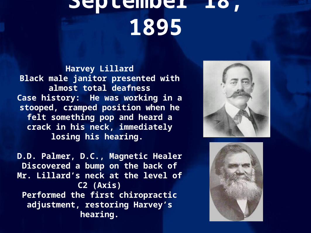
September 18, 1895
Harvey LillardBlack male janitor presented with almost
total deafnessCase history: He was working in a
stooped, cramped position when he felt something pop and heard a crack in his neck, immediately losing his hearing.
D.D. Palmer, D.C., Magnetic Healer
Discovered a bump on the back of Mr. Lillard’s neck at the level of C2 (Axis)
Performed the first chiropractic adjustment, restoring Harvey’s hearing.

BJ Palmer, DC
Son of DD Palmer
Took over Palmer Chiropractic College from his father
Started researching upper cervical specific chiropractic in
1931

Endolymphatic Hydrops
“The accumulation of the fluid of the membranous labyrinth of the ear, thought to be caused by the over production or under absorption of that fluid,” MerckManual.
Question: What is the Cause of the problem?

Meniere’s Disease Meniere’s Disease is a Syndrome is a Syndrome
caused by caused by WhiplashWhiplash

It takes an average of 15 years from the time of the trauma before
the onset of symptoms.
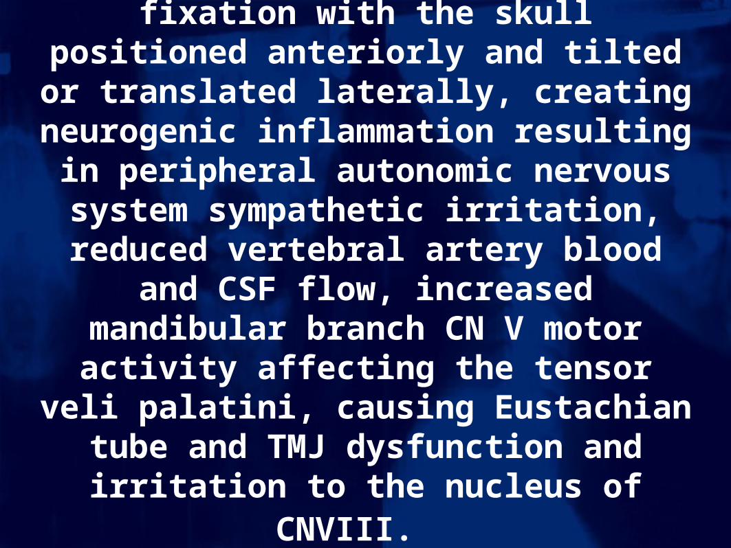
WHIPLASH:
Cervical subluxation complex comprised of vertebral facet fixation with the skull
positioned anteriorly and tilted or translated laterally, creating neurogenic inflammation resulting in peripheral autonomic nervous
system sympathetic irritation, reduced vertebral artery blood and CSF flow,
increased mandibular branch CN V motor activity affecting the tensor veli palatini,
causing Eustachian tube and TMJ dysfunction and irritation to the nucleus of CNVIII.

All of the following conditions exhibit hyper-activation of the Trigeminal ganglion when
symptomatic on PET scan:
Meniere’s diseaseMigraine headache
Trigeminal neuralgiaBell’s palsy
Additionally, patients with one of these conditions are twice as likely to experience another one of these
conditions in their lifetime.
More than 9 out of 10 benefit from cervical specific chiropractic care.

Normal
Anterior Occiput
Posterior Atlas
Rear ended auto accident

Posterior C5 Subluxation

Head tilt will make you dizzy

Right Head Translation
“T-Bone” Vehicular Accident

Chiropractic TreatmentDetailed case history including letter from ENT
and copies of tests used to DX MD
Titronics TyTron C-3000 cervical thermographs
Modified Prill leg check analysis
Modified Blair Cervical X-rays
Adjustments as determined by pattern work
15 minute rest after adjustment with re-check

Thermography

Pre and Post Adjustment Graphs of Patient with Right Unilateral Meniere’s
C5 Adjusted PIL with Pierce technique
Atlas adjusted PIL with Blair technique
Followed by 15 minute rest before re-scan

Cervical Syndromes“Most significant indication of upper cervical subluxation,” Dr Burcon.
Derifield/Thompson Cervical Syndrome Test- Hold patient’s shoes with thumbs under the heel, while applying very mild cephalic pressure. Lift the legs one inch off from the table,
keeping the shoes one inch apart. Compare the welts to estimate the leg length differential. Notate differential of short leg to closest
1/8 inch. Instruct patient to slowly turn their head to the right, then to the left. If the legs change length only while turning to the
right, notate the amount of change as a right cervical syndrome (RCS). If the legs change length only while turning to the left,
notate the amount of change as a left cervical syndrome (LCS). If the leg length changes while turning the head in both directions,
notate the total amount of change as a bilateral cervical syndrome (BLCS). If there is no change in leg length when the head is
turned, there is no cervical syndrome. Perform following tests to determine which upper cervical vertebrae is subluxated.
First Published by Ruth Jackson, MD in 1956

Modified Blair X-RaysAll 300 consecutive Meniere’s patients tested positive
for upper cervical subluxations.
3 Cervical X-rays taken and analyzed:Lateral, A-P Open Mouth & Nasium.
All 300 film studies showed evidence of upper cervical subluxation and whiplash, although cervical
trauma was denied by over 50% of these patients.

4 Blair Atlas Subluxation Listings
Anterior and Superior on the Right (ASR)Anterior and Superior on the Left (ASL)Posterior and Inferior on the Right (PIR)Posterior and Inferior on the Left (PIL)

Atlas listings for 300 Patients
0- Anterior and Superior on opposite side of involved ear
18- Anterior and Superior on the side of the involved ear
12- Posterior and Inferior on the side of the involved ear
270- Posterior and Inferior on the opposite side of the involved ear

Side Posture with Drop Upper Cervical Adjustment
BJ Palmer, DC

Pre-Adjustment (C1 PIL) 6 Weeks Post (Juxta)
Patient with Right Patient is off Medication
Unilateral Meniere’s and Symptom Free
Lesion

Upper Cervical Protocol forTen Meniere’s Patients
Same paper published in Upper Cervical Subluxation Complex,
A Review of the Chiropractic and Medical Literature, by Kirk Ericksen. Lippincott, Williams & Wilkens, 2004

MENIERE’S QUESTIONAIRE
Thank you for participating in our Meniere’s study. Please answer the following questions and return this form to Burcon Chiropractic. Answer questions with a number between 0 and 10, with 0 representing that you do not have that problem, 10 representing that the problem is the worst you can imagine. In column A put the number for how you felt before your first adjustment at Burcon Chiropractic. In column B put the number for how you have been feeling since your last adjustment. Patient Name _______________________________ Today’s date __________________ Major Symptoms A B 1. Vertigo, dizziness or lack of balance: __________ __________ 2. Loss of hearing: __________ __________ 3. Tinnitus (ringing in the ears): __________ __________ Secondary Symptoms 4. Nausea/vomiting: __________ __________ 5. Ear Pressure: __________ __________ 6. Migraines: __________ __________ 7. Headaches: __________ __________ 8. Brain fog: __________ __________ 9. Neck stiffness/pain: __________ __________ 10. Sinus pressure/pain: __________ __________ Comments:

10
7.5
3
2
1.4
0
1
2
3
4
5
6
7
8
9
10
1 2 3 4 5
Scale, Pre Adjust, 6 Weeks, 1 Year, 2 Years
VERTIGO