upconversion nanoparticles: from hydrophobic to ......upconversion nanoparticles: from hydrophobic...
TRANSCRIPT
Upconversion Nanoparticles: From Hydrophobic to HydrophilicSurfacesVerena Muhr, Stefan Wilhelm, Thomas Hirsch, and Otto S. Wolfbeis*
Institute of Analytical Chemistry, Chemo- and Biosensors, University of Regensburg, 93040 Regensburg, Germany
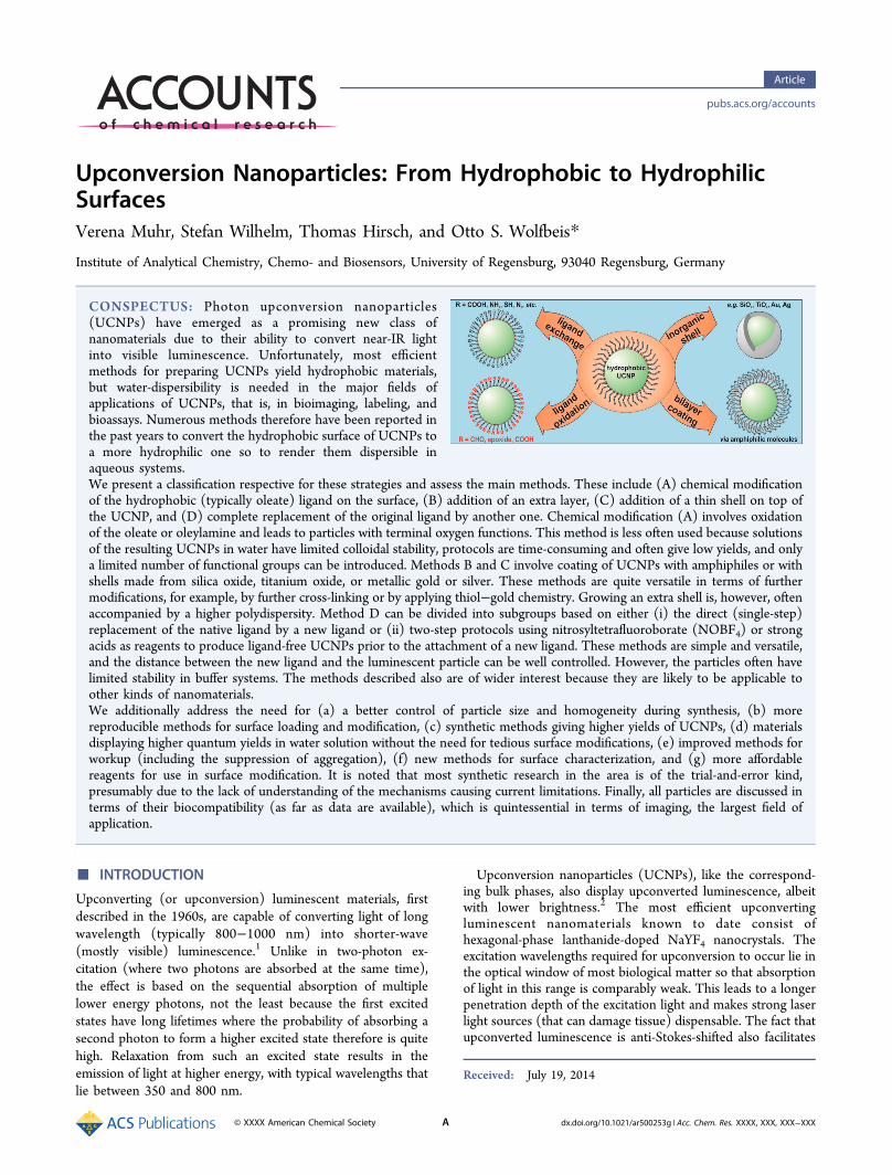
CONSPECTUS: Photon upconversion nanoparticles(UCNPs) have emerged as a promising new class ofnanomaterials due to their ability to convert near-IR lightinto visible luminescence. Unfortunately, most efficientmethods for preparing UCNPs yield hydrophobic materials,but water-dispersibility is needed in the major fields ofapplications of UCNPs, that is, in bioimaging, labeling, andbioassays. Numerous methods therefore have been reported inthe past years to convert the hydrophobic surface of UCNPs toa more hydrophilic one so to render them dispersible inaqueous systems.We present a classification respective for these strategies and assess the main methods. These include (A) chemical modificationof the hydrophobic (typically oleate) ligand on the surface, (B) addition of an extra layer, (C) addition of a thin shell on top ofthe UCNP, and (D) complete replacement of the original ligand by another one. Chemical modification (A) involves oxidationof the oleate or oleylamine and leads to particles with terminal oxygen functions. This method is less often used because solutionsof the resulting UCNPs in water have limited colloidal stability, protocols are time-consuming and often give low yields, and onlya limited number of functional groups can be introduced. Methods B and C involve coating of UCNPs with amphiphiles or withshells made from silica oxide, titanium oxide, or metallic gold or silver. These methods are quite versatile in terms of furthermodifications, for example, by further cross-linking or by applying thiol−gold chemistry. Growing an extra shell is, however, oftenaccompanied by a higher polydispersity. Method D can be divided into subgroups based on either (i) the direct (single-step)replacement of the native ligand by a new ligand or (ii) two-step protocols using nitrosyltetrafluoroborate (NOBF4) or strongacids as reagents to produce ligand-free UCNPs prior to the attachment of a new ligand. These methods are simple and versatile,and the distance between the new ligand and the luminescent particle can be well controlled. However, the particles often havelimited stability in buffer systems. The methods described also are of wider interest because they are likely to be applicable toother kinds of nanomaterials.We additionally address the need for (a) a better control of particle size and homogeneity during synthesis, (b) morereproducible methods for surface loading and modification, (c) synthetic methods giving higher yields of UCNPs, (d) materialsdisplaying higher quantum yields in water solution without the need for tedious surface modifications, (e) improved methods forworkup (including the suppression of aggregation), (f) new methods for surface characterization, and (g) more affordablereagents for use in surface modification. It is noted that most synthetic research in the area is of the trial-and-error kind,presumably due to the lack of understanding of the mechanisms causing current limitations. Finally, all particles are discussed interms of their biocompatibility (as far as data are available), which is quintessential in terms of imaging, the largest field ofapplication.
■ INTRODUCTION
Upconverting (or upconversion) luminescent materials, firstdescribed in the 1960s, are capable of converting light of longwavelength (typically 800−1000 nm) into shorter-wave(mostly visible) luminescence.1 Unlike in two-photon ex-citation (where two photons are absorbed at the same time),the effect is based on the sequential absorption of multiplelower energy photons, not the least because the first excitedstates have long lifetimes where the probability of absorbing asecond photon to form a higher excited state therefore is quitehigh. Relaxation from such an excited state results in theemission of light at higher energy, with typical wavelengths thatlie between 350 and 800 nm.
Upconversion nanoparticles (UCNPs), like the correspond-ing bulk phases, also display upconverted luminescence, albeitwith lower brightness.2 The most efficient upconvertingluminescent nanomaterials known to date consist ofhexagonal-phase lanthanide-doped NaYF4 nanocrystals. Theexcitation wavelengths required for upconversion to occur lie inthe optical window of most biological matter so that absorptionof light in this range is comparably weak. This leads to a longerpenetration depth of the excitation light and makes strong laserlight sources (that can damage tissue) dispensable. The fact thatupconverted luminescence is anti-Stokes-shifted also facilitates
Received: July 19, 2014
Article
pubs.acs.org/accounts
© XXXX American Chemical Society A dx.doi.org/10.1021/ar500253g | Acc. Chem. Res. XXXX, XXX, XXX−XXX
the separation of luminescence from Raman bands and otherscattered light. If luminescence of biomatter is induced at all, itwill occur far in the NIR. In other words, the intensity ofbackground visible fluorescence (i.e., in the region whereupconverted luminescence occurs) is virtually zero.In contrast to quantum dots, the color of the emission of
UCNPs does not depend on the size of the particles. UCNPsare not known to be cytotoxic, are chemically stable, andneither blink nor bleach. On the other hand, their quantumyields depend on their size, on the kind of surface coating, andon the power density of the laser used for photoexcitation.3
These features have been discussed in numerous reviews thatcan be easily found. Their outstanding features make UCNPshighly interesting materials for purposes including photo-dynamic therapy,4 photoinduced drug delivery,5 (targeted) cellimaging,6 sensing of fundamental parameters such as pHvalues,7 oxygen,8 ammonia,9 heavy metal ions,10 or CO2,
11
screening,12 and immunoassays.13
Initially, UCNPs were produced by top-down strategies.Such methods yield stable colloidal solutions, but the particleswere rather polydisperse and fairly large, leading to slow or nocellular uptake. A comparative study on upconversionluminescence and cell bioimaging based on single-stepsynthesized hydrophilic UCNPs capped with various functionalgroups has been presented by Tsang et al.14 To overcome thelimitations of high polydispersity and poor flexibility in terms ofsurface modification (by either small molecules or thinadditional layers), bottom-up strategies have been developedwith the aim to synthesize small and monodisperse nano-particles possessing bright upconversion luminescence. Themost common strategies are coprecipitation, thermal decom-position, and solvothermal syntheses.15 Depending on the kindof host lattice and the protocol used for synthesis, UCNPs canbe prepared in shapes such as symmetrical spheres, rods, andeven plates. The reviews by Chen et al.15 and DaCosta et al.16
provide an overview of synthetic strategies and materialproperties. DeCosta et al. also have introduced a system forthe classification of methods of making apolar surfaces ofUCNPs more polar (based on ligand exchange, ligandoxidation, ligand absorption, layer-by-layer assembly, ligand-free modifications, and silanizations). Other reviews areavailable on lanthanide-doped luminescent nanoprobes (witha small section on UCNPs)17 and on UCNPs for use in small-animal imaging (with a short section on surface modifica-tion).18 The groups of Selvin19 and Li20 have briefly reviewedmethods for surface engineering of UCNPs, while other reviewsare ignoring the need for converting hydrophobic to hydro-philic surfaces.21
Hexagonal phase NaYF4 nanoparticles doped with trivalentlanthanides (Ln3+) are by far most often used. In 2008, Li andZhang22 presented an efficient protocol for preparation ofhydrophobic (oleate-capped) UCNPs. It involves heating of rareearth chlorides in a mixture of octadecene and oleic acid, first togenerate the respective oleate salts, which act as in situprecursors. The addition of ammonium fluoride and sodiumhydroxide and an increase in the reaction temperature to 300°C leads to the formation of highly monodisperse, oleate-capped, hexagonal UCNPs that can be dispersed in nonpolarsolvents. Oleylamine (OlAm) may be added to, or even used inplace of oleic acid.23 This bottom-up method in high-boilingsolvents is said to be superior to others with respect to themonodispersity, shape uniformity, and phase purity of theresulting UCNPs. On the other hand, it suffers from the
disadvantage of giving UCNPs that are dispersible inhydrophobic solvents only and not in aqueous solutionsincluding buffers. If intended for use in biosciences, waterdispersibility and colloidal stability in buffers is, however,mandatory. In addition, these particles are highly inert in beingdevoid of any useful functional group on their surface. In orderto exploit the large potential of UCNPs, appropriate functionalgroups have to be introduced.This Account throws a critical look at the methods for
surface modification and functionalization that lead to UCNPsfor use in aqueous media. In terms of bioimaging (which is themost widespread application of such particles at present),features such as small size, brightness, and tunable emission andexcitation spectra also are paramount.24 We summarize generalprinciples, discuss advantages and disadvantages of thestrategies, and give selected examples for respective applica-tions.
■ SURFACE MODIFICATION OF HYDROPHOBICUPCONVERSION NANOPARTICLES
A large variety of methods for surface modification have beendeveloped to convert hydrophobic UCNPs into more hydro-philic particles. They often are not limited to UCNPs but alsomay be applied to other types of nanoparticles.25 Therespective strategies can be categorized into four groups: (1)chemical modification of the hydrophobic (usually oleate oroleylamine) ligand on the surface; (2) bilayer coating withamphiphilic molecules or polymers, (3) addition of an extralayer or shell on top of the UCNP; and (4) completereplacement of the original ligand by another one. These will bediscussed in the following.Modification of the Original Ligand
The direct modification of the hydrophobic ligand on aparticle’s surface to generate hydrophilic UCNPs is simple butnot common. It is based on the oxidation of the carbon−carbondouble bond of the oleate or oleylamine. Depending on thereagents employed, it can lead to the formation of carboxygroups or epoxy groups. Oxidizing agents include theLemieux−von Rudloff reagent,26 ozone,27 and 3-chloroper-oxy-benzoic acid.28 In addition to the formation of reactivegroups, the dispersibility of the resulting particles in water isstrongly enhanced. Such particles then may been covalentlycoupled to other species, for example, to the cancer drugdoxorubicin in order to enable controlled drug delivery29 or topoly(ethylene glycol) in order to impart biocompatibility.28
Notwithstanding this, oxidative surface modification is rarelyused because dispersions in water have poor colloidal stabilityand only a limited number of ligands (aldehydes, epoxides, orcarboxylic acids) can be introduced.Amphiphilic Coatings
This technique involves coating of the UCNPs with moleculescontaining long alkyl chains to form a bilayer that is stabilizedvia van-der-Waals interactions between the hydrophobic oleateand the new coating. Amphiphilic molecules are preferred inthis context because they (a) undergo strong van-der-Waalsinteraction, (b) enable the surface charge to be easily altered,and (c) may even be deposited as a so-called layer-by-layercoating, that is, in the form of multiple layers of alternatingcharge. If oleate-capped UCNPs are treated with long-chainamphiphiles, their hydrophobic tails intercalate between theoleate chains, while their hydrophilic head groups are directedoutward. This results in the formation of a bilayer around the
Accounts of Chemical Research Article
dx.doi.org/10.1021/ar500253g | Acc. Chem. Res. XXXX, XXX, XXX−XXXB
UCNP as shown in Figure 1. The hydrophilic head groupsrender the particles well dispersible in water.
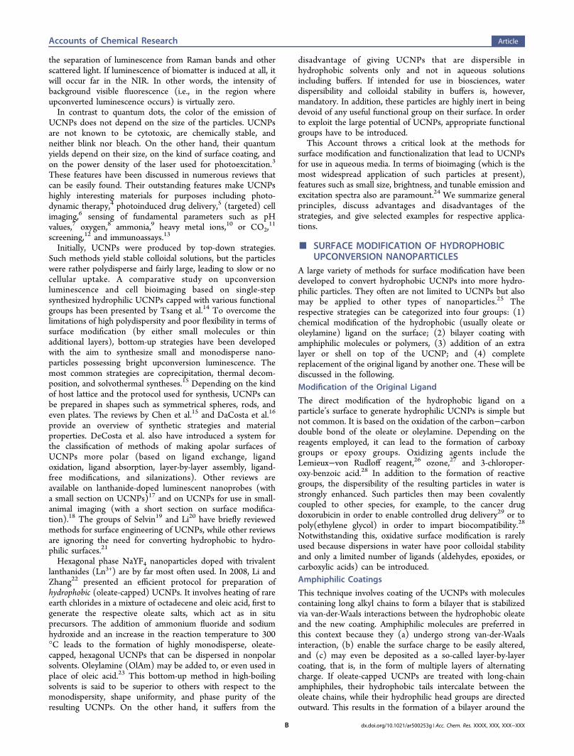
Phospholipids (PLs) have often been used to modifysurfaces. Resulting particles (not only UCNPs) are readilyinternalized by cells, are nonimmunogenic, and possess a longfunctional lifetime even in vivo. PLs have been widely used indrug delivery. Quite a variety of PLs are commercially availablewith various kinds of head groups such as maleimide (forbinding the particle to protein thiol groups), biotin (with itshigh affinity for streptavidin), and several others. Phospholipidsalso are known with highly different chain lengths, and variationin length is often accomplished by incorporating poly(ethyleneglycol) (PEG) units, which has the beneficial effect of impartingbiocompatibility.30,31 In a typical example, maleimide and folatehead groups were used to conjugate UCNPs to goldnanoparticles and image HeLa cells (see Figure 2).19
While phospholipids with polar head groups and PEGspacers are easy to use, they are difficult to make and purify andexpensive if commercially available. The following calculationmay reflect the costs to be expected in a typical experiment:The surface area of one single nanocrystal with a diameter of 20nm is 1250 nm2. If 1 μmol of such particles is to be coveredwith, say, a PEGylated distearoyl phospholipid (with a size of∼80 Å2), the total surface to be coated is as large as 760 m2.This requires, roughly, 4.5 g of the phospholipid, which willactually cost more than US $30,000. This number may be evenhigher if phospholipids are applied in excess to warrantcomplete coverage of the surface. Obviously, less expensivemethods are desirable to create bilayers. Zhao et al.32 havecoated UCNPs with the detergent Tween 80 to obtain particlesfor use as a carrier for doxorubicin that was trapped in thehydrophobic bilayer. Other long-chain alkylammonium derivedsurfactants were tested by the Yang group,33 but the colloidalstability of the particles in water was poor. Subsequent surfacemodification with silica was required. This will be discussed inthe next section.In another approach, amphiphilic polymers were used in
place of the relatively small surfactants as shown by the Parakgroup34 in order to modify the surface of gold nanoparticles,quantum dots, or iron oxide particles. Poly(maleic anhydride-alt-1-octadecene) (PMAO) is a widely used polymericamphiphile35,36 because it contains multiple alkyl chains permolecule and has a weak chelating effect, which stabilizes thesurface coating against ligand detachment. Particles coated withPMAO display good temporal stability in aqueous media, whichcan be further increased by reacting the anhydride groups withbis(hexamethylene)triamine (BHMT).37 This method enabled
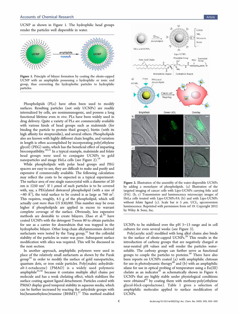
UCNPs to be stabilized over the pH 3−13 range and in cellcultures for even several weeks (see Figure 3).Poly(acrylic acid) modified with long alkyl chains also binds
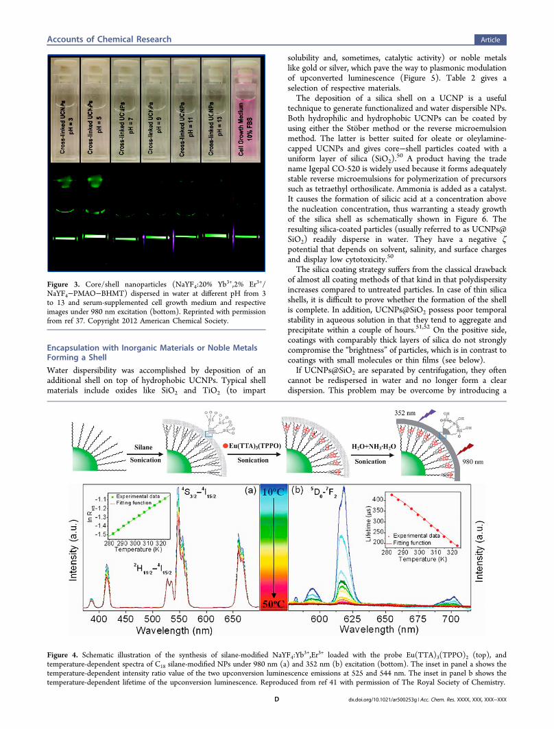
to the surface of oleate-capped UCNPs.38 This results in theintroduction of carboxy groups that are negatively charged atnear-neutral pH values and will render the particles water-soluble. The carboxy groups can further serve as functionalgroups to couple the particles to proteins.39 There have alsobeen reports on UCNPs coated (a) with amphiphilic chitosanfor use in photodynamic therapy40 and (b) with an amphiphilicsilane for use in optical probing of temperature using a Eu(III)chelate as an indicator41 as schematically shown in Figure 4.UCNPs that are highly stable under physiological conditionswere obtained42 by coating them with methoxy-poly(ethyleneglycol-block-caprolactone). Table 1 gives a selection ofamphiphilic molecules applied to surface modification ofUCNPs.
Figure 1. Principle of bilayer formation by coating the oleate-cappedUCNP with an amphiphile possessing a hydrophilic or ionic endgroup, thus converting the hydrophobic particles to hydrophilicparticles.
Figure 2. Illustration of the assembly of the water-dispersible UCNPsby adding a monolayer of phospholipids. (a) Illustration of thetargeted imaging of cancer cells with Lipo-UCNPs carrying folic acid(FA). (b, c) Transmission and luminescence microscopy images ofHeLa cells treated with Lipo-UCNPs-FA (b) and with Lipo-UCNPswithout folate ligand (c). Scale bar is 5 μm. UCL, upconversionluminescence. Reprinted with permission from ref 19. Copyright 2012by Wiley & Sons, Inc.
Accounts of Chemical Research Article
dx.doi.org/10.1021/ar500253g | Acc. Chem. Res. XXXX, XXX, XXX−XXXC
Encapsulation with Inorganic Materials or Noble MetalsForming a Shell
Water dispersibility was accomplished by deposition of anadditional shell on top of hydrophobic UCNPs. Typical shellmaterials include oxides like SiO2 and TiO2 (to impart
solubility and, sometimes, catalytic activity) or noble metalslike gold or silver, which pave the way to plasmonic modulationof upconverted luminescence (Figure 5). Table 2 gives aselection of respective materials.The deposition of a silica shell on a UCNP is a useful
technique to generate functionalized and water dispersible NPs.Both hydrophilic and hydrophobic UCNPs can be coated byusing either the Stober method or the reverse microemulsionmethod. The latter is better suited for oleate or oleylamine-capped UCNPs and gives core−shell particles coated with auniform layer of silica (SiO2).
50 A product having the tradename Igepal CO-520 is widely used because it forms adequatelystable reverse microemulsions for polymerization of precursorssuch as tetraethyl orthosilicate. Ammonia is added as a catalyst.It causes the formation of silicic acid at a concentration abovethe nucleation concentration, thus warranting a steady growthof the silica shell as schematically shown in Figure 6. Theresulting silica-coated particles (usually referred to as UCNPs@SiO2) readily disperse in water. They have a negative ζpotential that depends on solvent, salinity, and surface chargesand display low cytotoxicity.50
The silica coating strategy suffers from the classical drawbackof almost all coating methods of that kind in that polydispersityincreases compared to untreated particles. In case of thin silicashells, it is difficult to prove whether the formation of the shellis complete. In addition, UCNPs@SiO2 possess poor temporalstability in aqueous solution in that they tend to aggregate andprecipitate within a couple of hours.51,52 On the positive side,coatings with comparably thick layers of silica do not stronglycompromise the “brightness” of particles, which is in contrast tocoatings with small molecules or thin films (see below).If UCNPs@SiO2 are separated by centrifugation, they often
cannot be redispersed in water and no longer form a cleardispersion. This problem may be overcome by introducing a
Figure 3. Core/shell nanoparticles (NaYF4:20% Yb3+,2% Er3+/NaYF4−PMAO−BHMT) dispersed in water at different pH from 3to 13 and serum-supplemented cell growth medium and respectiveimages under 980 nm excitation (bottom). Reprinted with permissionfrom ref 37. Copyright 2012 American Chemical Society.
Figure 4. Schematic illustration of the synthesis of silane-modified NaYF4:Yb3+,Er3+ loaded with the probe Eu(TTA)3(TPPO)2 (top), and
temperature-dependent spectra of C18 silane-modified NPs under 980 nm (a) and 352 nm (b) excitation (bottom). The inset in panel a shows thetemperature-dependent intensity ratio value of the two upconversion luminescence emissions at 525 and 544 nm. The inset in panel b shows thetemperature-dependent lifetime of the upconversion luminescence. Reproduced from ref 41 with permission of The Royal Society of Chemistry.
Accounts of Chemical Research Article
dx.doi.org/10.1021/ar500253g | Acc. Chem. Res. XXXX, XXX, XXX−XXXD
high density of surface charges, which will reduce the tendencytoward aggregation.53 It was shown that agarose gel electro-phoresis (AGE) is well suited for the purification of silica-coated UCNPs.54 The silica shell of a fraction of the particles
was doped with a fluorescent dye for direct detection. The shellwas prepared by reverse microemulsion and resulted inindividual nanoparticles but also in aggregates that wereseparated and isolated. The preparation of an ultrathincarboxylated silica shell, in contrast, yielded nonaggregatedUCNPs that can be directly used for protein conjugation.Functional groups can be created on the surface of the
UCNPs in two ways. In one, the preformed UCNPs@SiO2particles are modified with organically modified silanizingagents. In the other, functional organosilanes are added duringthe polymerization process, which leads to the formation of theshell so that postsynthetic modification is not needed.Organosilanes that have been used in either method aresummarized in Table 3.Amino-functionalized UCNPs@SiO2 may be prepared by
adding aminopropyltriethoxysilane to the microemulsion.44
The water-dispersible UCNPs can then be conjugated to folicacid to enable targeting of tumor cells. Similarly, silica-coatedNaYF4:Yb,Er UCNPs were further endowed with folic acid andanti-Her2 antibody to label the folate receptors and Her2receptors of certain cells. Our group has reported thepreparation of protein-reactive hydrophilic particles (Figure7) by modifying the surface of UCNPs@SiO2 with a silane-modified poly(ethylene glycol) with a terminal N-hydrox-ysuccinimide group.58 The nanoparticles were then conjugatedto proteins as verified by surface plasmon resonance spectros-copy.UCNPs coated with mesoporous silica were modified with
azo groups via silanization and then loaded with the cancerdrug doxorubicin.5 The azo groups acted as motors to triggerthe controlled release of the drug under photoexcitation at 980nm. Mesoporous silica shells are characterized by a largespecific surface area and a pore size that can be fine-tuned (seeFigure 8). Other UCNPs were coated with mesoporous silicaand loaded with photosensitizers such as zinc(II) phthalocya-nine, which causes the formation of singlet oxygen upon NIRexcitation.4 Li et al.60 have incorporated doxorubicin intoparticles coated with mesoporous silica, which then werestudied with respect to cellular uptake and cytotoxicity. Theirpotential for imaging of nasopharyngeal epidermal carcinomacells was demonstrated.61 More recently, core−shell−shellparticles of the type β-NaYF4:Yb,Er@SiO2@mSiO2 have beenreported,45 again for use in imaging and drug storage anddelivery. So-called yolk−shell UCNPs were obtained byforming a hollow mesoporous silica shell around NaLu-F4:Yb,Er,Tm nanoparticles.62 Their large cavities were loadedwith a chromophore to construct nanoprobes for cysteine,homocysteine, and cyanide.Replacement of the Native Ligand
Ligand exchange is a versatile strategy to modify the surface ofUCNPs. Two major methods are known. One is based on
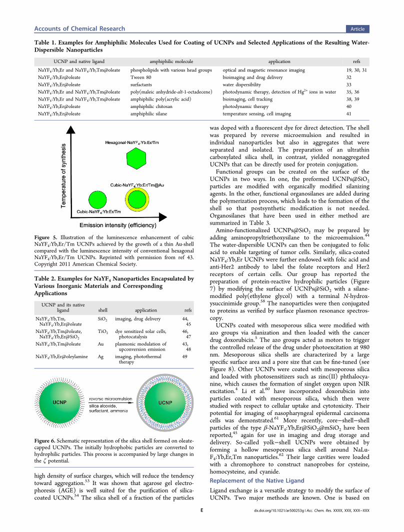
Table 1. Examples for Amphiphilic Molecules Used for Coating of UCNPs and Selected Applications of the Resulting Water-Dispersible Nanoparticles
UCNP and native ligand amphiphilic molecule application refs
NaYF4:Yb,Er and NaYF4:Yb,Tm@oleate phospholipids with various head groups optical and magnetic resonance imaging 19, 30, 31NaYF4:Yb,Er@oleate Tween 80 bioimaging and drug delivery 32NaYF4:Yb,Er@oleate surfactants water dispersibility 33NaYF4:Yb,Er and NaYF4:Yb,Tm@oleate poly(maleic anhydride-alt-1-octadecene) photodynamic therapy, detection of Hg2+ ions in water 35, 36NaYF4:Yb,Er and NaYF4:Yb,Tm@oleate amphiphilic poly(acrylic acid) bioimaging, cell tracking 38, 39NaYF4:Yb,Er@oleate amphiphilic chitosan photodynamic therapy 40NaYF4:Yb,Er@oleate amphiphilic silane temperature sensing, cell imaging 41
Figure 5. Illustration of the luminescence enhancement of cubicNaYF4:Yb,Er/Tm UCNPs achieved by the growth of a thin Au-shellcompared with the luminescence intensity of conventional hexagonalNaYF4:Yb,Er/Tm UCNPs. Reprinted with permission from ref 43.Copyright 2011 American Chemical Society.
Table 2. Examples for NaYF4 Nanoparticles Encapsulated byVarious Inorganic Materials and CorrespondingApplications
UCNP and its nativeligand shell application refs
NaYF4:Yb,Tm,NaYF4:Yb,Er@oleate
SiO2 imaging, drug delivery 44,45
NaYF4:Yb,Tm@oleate,NaYF4:Yb,Er@SiO2
TiO2 dye sensitized solar cells,photocatalysis
46,47
NaYF4:Yb,Tm@oleate Au plasmonic modulation ofupconversion emission
43,48
NaYF4:Yb,Er@oleylamine Ag imaging, photothermaltherapy
49
Figure 6. Schematic representation of the silica shell formed on oleate-capped UCNPs. The initially hydrophobic particles are converted tohydrophilic particles. This process is accompanied by large changes inthe ζ potential.
Accounts of Chemical Research Article
dx.doi.org/10.1021/ar500253g | Acc. Chem. Res. XXXX, XXX, XXX−XXXE
direct exchange of the first ligand by the new one; the other isbased on two-step strategies using NOBF4 or acid treatmentwith HCl to strip off the oleate or oleylamine and subsequentattachment of a new coating. Unfortunately, practically allcoatings with small molecules (for example via oleatereplacement or the NOBF4 technique) for phase transfer toaqueous solvents drastically reduce the “brightness” of UCNPs.Coating the particles with NaYF4, in contrast, does not causesuch an effect. However, a systematic study on the effect ofsmall-molecule coatings on quantum yields and luminescencedecay times has not been presented so far.Direct (Single Step) Replacement of the Native Ligand by aNew Ligand
In this case, the native ligand on the UCNP is (almost)completely displaced by another ligand that is supposed to bemore polar to confer water solubility. Ideally, it contains afunctional group that coordinates to the surface of the UCNPso that it can easily replace the native ligand. The strength ofinteraction is likely to increase in the order −SH, −NH2,−COOH, −PO3H, but no comparative study covering thedifferent binding strengths is available. Respective methods arefairly simple, at least in principle, but work up is tedious and
more challenging than the chemical reaction itself. In a typicalprocedure, oleate-capped UCNPs and the new ligand arestirred for 4 h to several days, usually at elevated temper-ature.63,49 The protocols have to be optimized for each singleligand because each ligand requires specific reaction conditionsin terms of concentrations, stirring time, temperature, and needfor an inert atmosphere. Furthermore, the particles tend toaggregate during ligand exchange.64 The group of Perez-Prietoused heterobifunctional PEG with a thiol group at one end andan amine or carboxylic group at the other.65 In this protocol,the PEG ligands are used as both the capping ligand and thewater-stabilizing agent. PEG moieties can function aspolydentate ligands and bind to lanthanide ions. Representativereagents and cappings and the properties and application of theresulting hydrophilic UCNPs are summarized in Table 4.Ligands usually have to be added in excess in order to displace
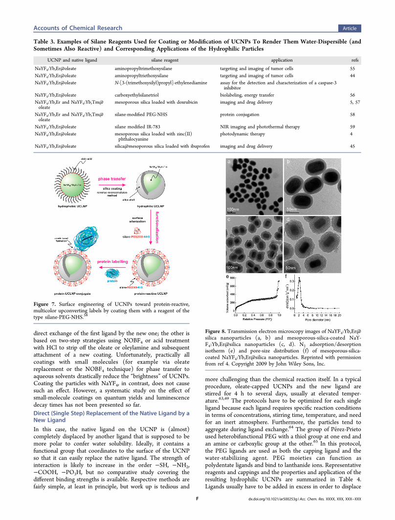
Table 3. Examples of Silane Reagents Used for Coating or Modification of UCNPs To Render Them Water-Dispersible (andSometimes Also Reactive) and Corresponding Applications of the Hydrophilic Particles
UCNP and native ligand silane reagent application refs
NaYF4:Yb,Er@oleate aminopropyltrimethoxysilane targeting and imaging of tumor cells 55NaYF4:Yb,Er@oleate aminopropyltriethoxysilane targeting and imaging of tumor cells 44NaYF4:Yb,Er@oleate N-[3-(trimethoxysilyl)propyl]-ethylenediamine assay for the detection and characterization of a caspase-3
inhibitorNaYF4:Yb,Er@oleate carboxyethylsilanetriol biolabeling, energy transfer 56NaYF4:Yb,Er and NaYF4:Yb,Tm@oleate
mesoporous silica loaded with doxrubicin imaging and drug delivery 5, 57
NaYF4:Yb,Er and NaYF4:Yb,Tm@oleate
silane-modified PEG-NHS protein conjugation 58
NaYF4:Yb,Er@oleate silane modified IR-783 NIR imaging and photothermal therapy 59NaYF4:Yb,Er@oleate mesoporous silica loaded with zinc(II)
phthalocyaninephotodynamic therapy 4
NaYF4:Yb,Er@oleate silica@mesoporous silica loaded with ibuprofen imaging and drug delivery 45
Figure 7. Surface engineering of UCNPs toward protein-reactive,multicolor upconverting labels by coating them with a reagent of thetype silane-PEG-NHS.58
Figure 8. Transmission electron microscopy images of NaYF4:Yb,Er@silica nanoparticles (a, b) and mesoporous-silica-coated NaY-F4:Yb,Er@silica nanoparticles (c, d). N2 adsorption/desorptionisotherm (e) and pore-size distribution (f) of mesoporous-silica-coated NaYF4:Yb,Er@silica nanoparticles. Reprinted with permissionfrom ref 4. Copyright 2009 by John Wiley Sons, Inc.
Accounts of Chemical Research Article
dx.doi.org/10.1021/ar500253g | Acc. Chem. Res. XXXX, XXX, XXX−XXXF
the former ligand. Even organic polymers may be used in thisreplacement strategy as can be seen in Table 4.The introduction of PEG chains not only imparts hydro-
philicity but also results in improved biocompatibility whenused in imaging or cell targeting. Ultrasmall core−shell UCNPsof the type NaYF4:Yb,Tm@SiO2 were further modified withPEG and found to be bound by MCF-7 tumors,77 while otherswere coated with similarly hydrophilic multihydroxy dendriticmolecules to provide water dispersibility and hydrophilicity.78
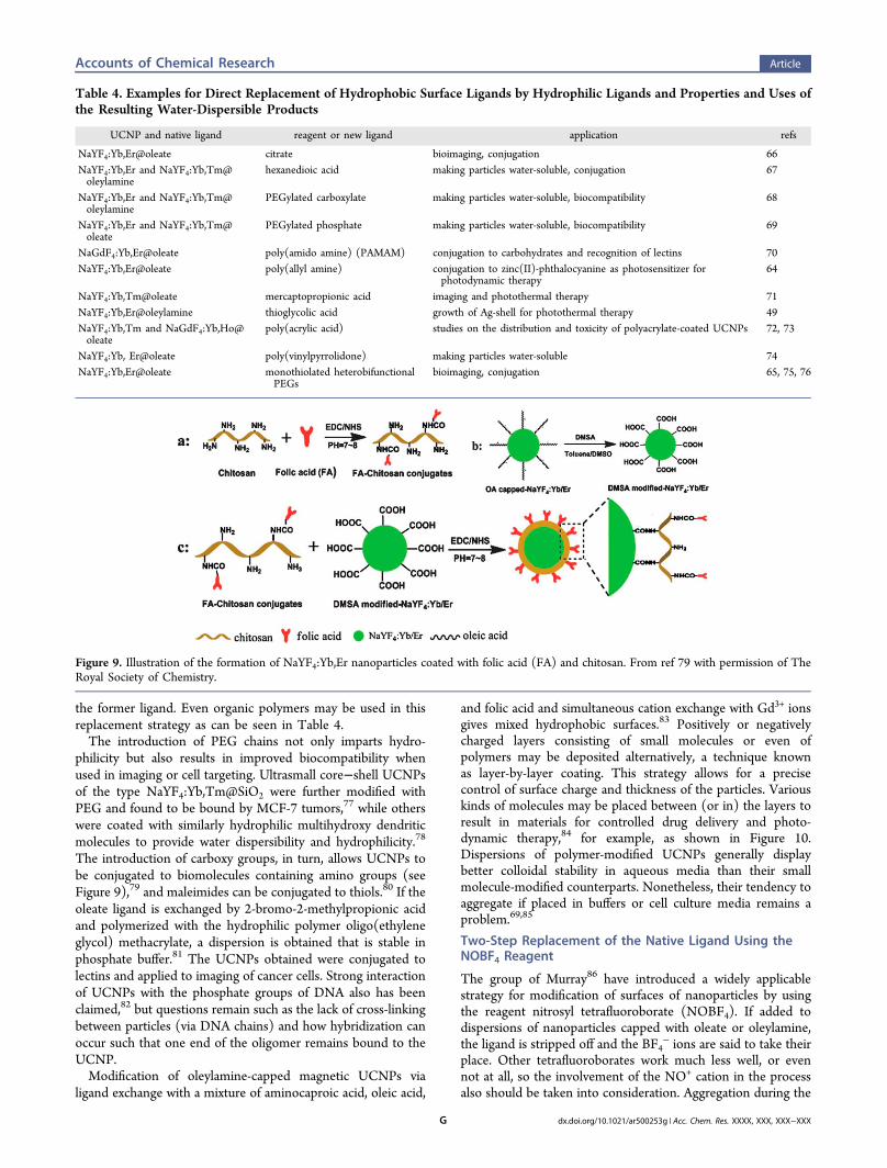
The introduction of carboxy groups, in turn, allows UCNPs tobe conjugated to biomolecules containing amino groups (seeFigure 9),79 and maleimides can be conjugated to thiols.80 If theoleate ligand is exchanged by 2-bromo-2-methylpropionic acidand polymerized with the hydrophilic polymer oligo(ethyleneglycol) methacrylate, a dispersion is obtained that is stable inphosphate buffer.81 The UCNPs obtained were conjugated tolectins and applied to imaging of cancer cells. Strong interactionof UCNPs with the phosphate groups of DNA also has beenclaimed,82 but questions remain such as the lack of cross-linkingbetween particles (via DNA chains) and how hybridization canoccur such that one end of the oligomer remains bound to theUCNP.Modification of oleylamine-capped magnetic UCNPs via
ligand exchange with a mixture of aminocaproic acid, oleic acid,
and folic acid and simultaneous cation exchange with Gd3+ ionsgives mixed hydrophobic surfaces.83 Positively or negativelycharged layers consisting of small molecules or even ofpolymers may be deposited alternatively, a technique knownas layer-by-layer coating. This strategy allows for a precisecontrol of surface charge and thickness of the particles. Variouskinds of molecules may be placed between (or in) the layers toresult in materials for controlled drug delivery and photo-dynamic therapy,84 for example, as shown in Figure 10.Dispersions of polymer-modified UCNPs generally displaybetter colloidal stability in aqueous media than their smallmolecule-modified counterparts. Nonetheless, their tendency toaggregate if placed in buffers or cell culture media remains aproblem.69,85
Two-Step Replacement of the Native Ligand Using theNOBF4 Reagent
The group of Murray86 have introduced a widely applicablestrategy for modification of surfaces of nanoparticles by usingthe reagent nitrosyl tetrafluoroborate (NOBF4). If added todispersions of nanoparticles capped with oleate or oleylamine,the ligand is stripped off and the BF4
− ions are said to take theirplace. Other tetrafluoroborates work much less well, or evennot at all, so the involvement of the NO+ cation in the processalso should be taken into consideration. Aggregation during the
Table 4. Examples for Direct Replacement of Hydrophobic Surface Ligands by Hydrophilic Ligands and Properties and Uses ofthe Resulting Water-Dispersible Products
UCNP and native ligand reagent or new ligand application refs
NaYF4:Yb,Er@oleate citrate bioimaging, conjugation 66NaYF4:Yb,Er and NaYF4:Yb,Tm@oleylamine
hexanedioic acid making particles water-soluble, conjugation 67
NaYF4:Yb,Er and NaYF4:Yb,Tm@oleylamine
PEGylated carboxylate making particles water-soluble, biocompatibility 68
NaYF4:Yb,Er and NaYF4:Yb,Tm@oleate
PEGylated phosphate making particles water-soluble, biocompatibility 69
NaGdF4:Yb,Er@oleate poly(amido amine) (PAMAM) conjugation to carbohydrates and recognition of lectins 70NaYF4:Yb,Er@oleate poly(allyl amine) conjugation to zinc(II)-phthalocyanine as photosensitizer for
photodynamic therapy64
NaYF4:Yb,Tm@oleate mercaptopropionic acid imaging and photothermal therapy 71NaYF4:Yb,Er@oleylamine thioglycolic acid growth of Ag-shell for photothermal therapy 49NaYF4:Yb,Tm and NaGdF4:Yb,Ho@oleate
poly(acrylic acid) studies on the distribution and toxicity of polyacrylate-coated UCNPs 72, 73
NaYF4:Yb, Er@oleate poly(vinylpyrrolidone) making particles water-soluble 74NaYF4:Yb,Er@oleate monothiolated heterobifunctional
PEGsbioimaging, conjugation 65, 75, 76
Figure 9. Illustration of the formation of NaYF4:Yb,Er nanoparticles coated with folic acid (FA) and chitosan. From ref 79 with permission of TheRoyal Society of Chemistry.
Accounts of Chemical Research Article
dx.doi.org/10.1021/ar500253g | Acc. Chem. Res. XXXX, XXX, XXX−XXXG
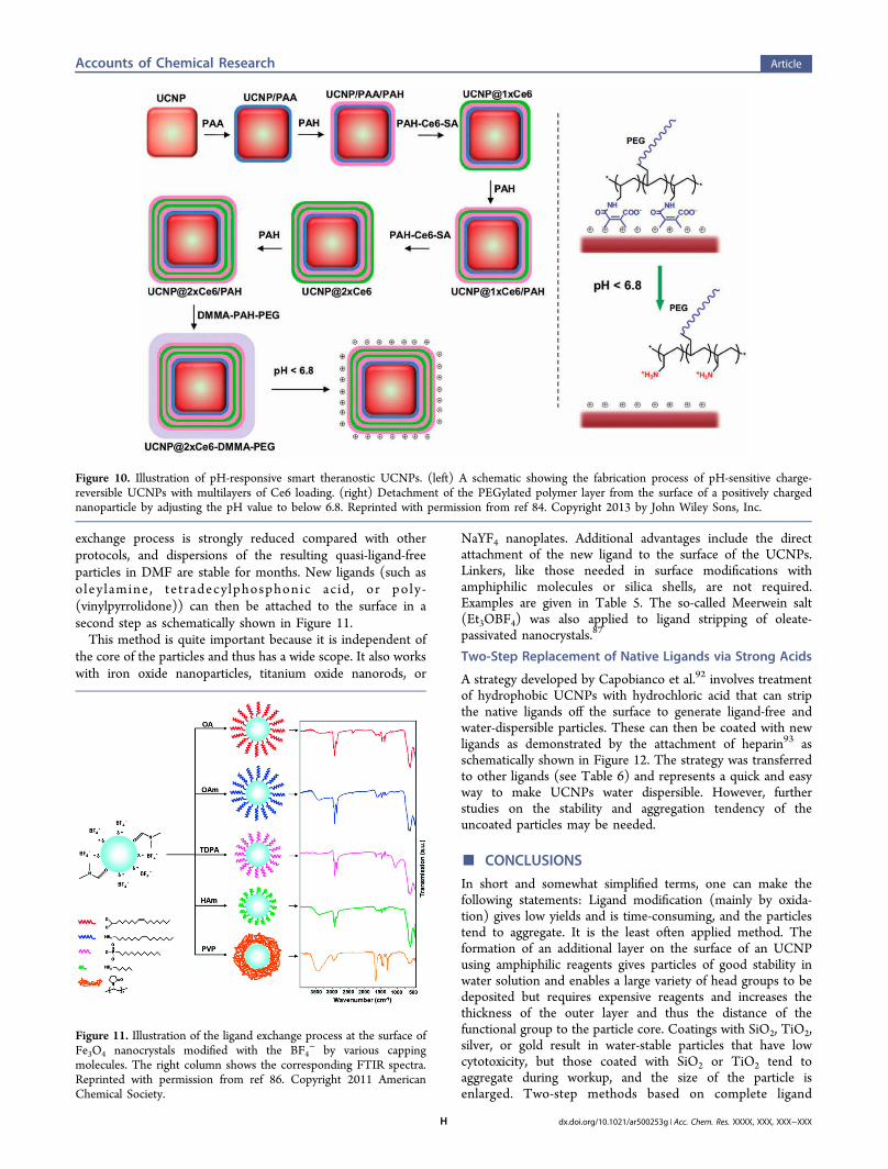
exchange process is strongly reduced compared with otherprotocols, and dispersions of the resulting quasi-ligand-freeparticles in DMF are stable for months. New ligands (such asoley lamine , te tradecylphosphonic ac id , or poly-(vinylpyrrolidone)) can then be attached to the surface in asecond step as schematically shown in Figure 11.This method is quite important because it is independent of
the core of the particles and thus has a wide scope. It also workswith iron oxide nanoparticles, titanium oxide nanorods, or
NaYF4 nanoplates. Additional advantages include the directattachment of the new ligand to the surface of the UCNPs.Linkers, like those needed in surface modifications withamphiphilic molecules or silica shells, are not required.Examples are given in Table 5. The so-called Meerwein salt(Et3OBF4) was also applied to ligand stripping of oleate-passivated nanocrystals.87
Two-Step Replacement of Native Ligands via Strong Acids
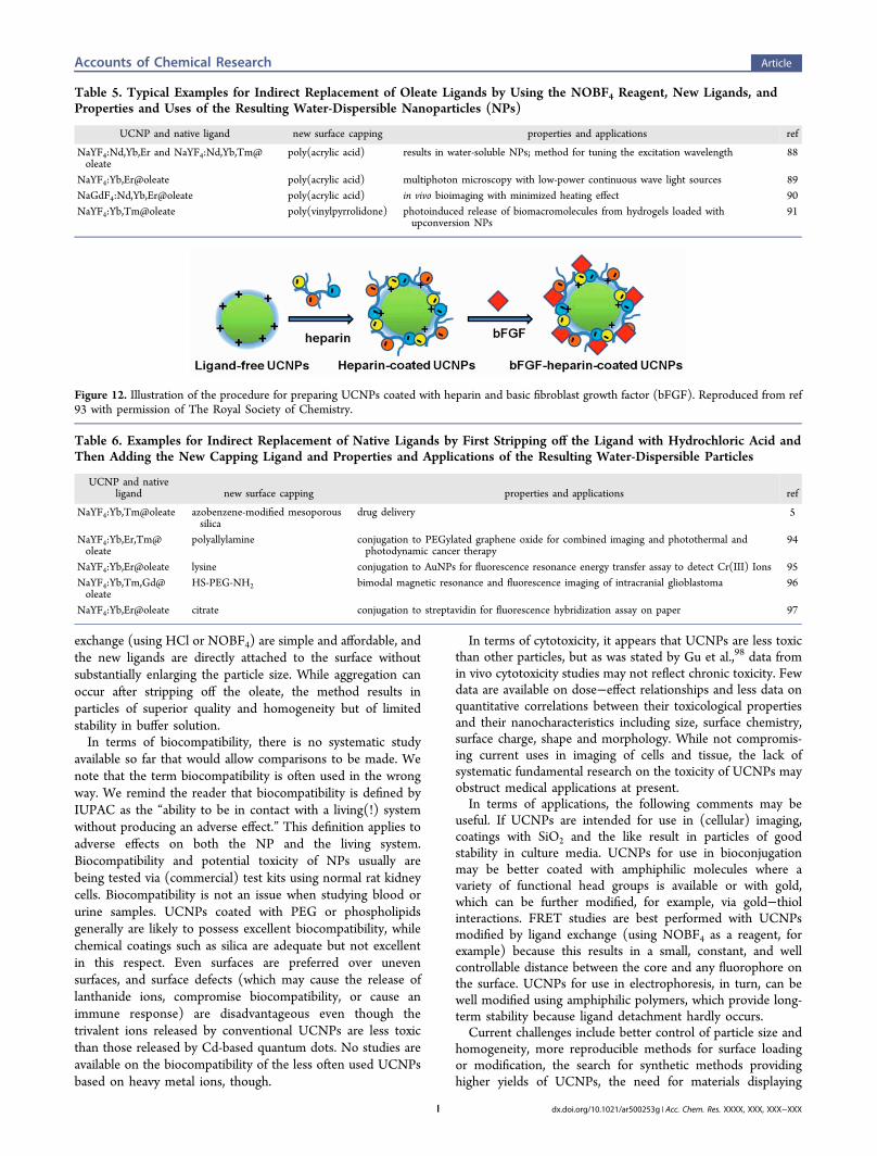
A strategy developed by Capobianco et al.92 involves treatmentof hydrophobic UCNPs with hydrochloric acid that can stripthe native ligands off the surface to generate ligand-free andwater-dispersible particles. These can then be coated with newligands as demonstrated by the attachment of heparin93 asschematically shown in Figure 12. The strategy was transferredto other ligands (see Table 6) and represents a quick and easyway to make UCNPs water dispersible. However, furtherstudies on the stability and aggregation tendency of theuncoated particles may be needed.
■ CONCLUSIONS
In short and somewhat simplified terms, one can make thefollowing statements: Ligand modification (mainly by oxida-tion) gives low yields and is time-consuming, and the particlestend to aggregate. It is the least often applied method. Theformation of an additional layer on the surface of an UCNPusing amphiphilic reagents gives particles of good stability inwater solution and enables a large variety of head groups to bedeposited but requires expensive reagents and increases thethickness of the outer layer and thus the distance of thefunctional group to the particle core. Coatings with SiO2, TiO2,silver, or gold result in water-stable particles that have lowcytotoxicity, but those coated with SiO2 or TiO2 tend toaggregate during workup, and the size of the particle isenlarged. Two-step methods based on complete ligand
Figure 10. Illustration of pH-responsive smart theranostic UCNPs. (left) A schematic showing the fabrication process of pH-sensitive charge-reversible UCNPs with multilayers of Ce6 loading. (right) Detachment of the PEGylated polymer layer from the surface of a positively chargednanoparticle by adjusting the pH value to below 6.8. Reprinted with permission from ref 84. Copyright 2013 by John Wiley Sons, Inc.
Figure 11. Illustration of the ligand exchange process at the surface ofFe3O4 nanocrystals modified with the BF4
− by various cappingmolecules. The right column shows the corresponding FTIR spectra.Reprinted with permission from ref 86. Copyright 2011 AmericanChemical Society.
Accounts of Chemical Research Article
dx.doi.org/10.1021/ar500253g | Acc. Chem. Res. XXXX, XXX, XXX−XXXH
exchange (using HCl or NOBF4) are simple and affordable, andthe new ligands are directly attached to the surface withoutsubstantially enlarging the particle size. While aggregation canoccur after stripping off the oleate, the method results inparticles of superior quality and homogeneity but of limitedstability in buffer solution.In terms of biocompatibility, there is no systematic study
available so far that would allow comparisons to be made. Wenote that the term biocompatibility is often used in the wrongway. We remind the reader that biocompatibility is defined byIUPAC as the “ability to be in contact with a living(!) systemwithout producing an adverse effect.” This definition applies toadverse effects on both the NP and the living system.Biocompatibility and potential toxicity of NPs usually arebeing tested via (commercial) test kits using normal rat kidneycells. Biocompatibility is not an issue when studying blood orurine samples. UCNPs coated with PEG or phospholipidsgenerally are likely to possess excellent biocompatibility, whilechemical coatings such as silica are adequate but not excellentin this respect. Even surfaces are preferred over unevensurfaces, and surface defects (which may cause the release oflanthanide ions, compromise biocompatibility, or cause animmune response) are disadvantageous even though thetrivalent ions released by conventional UCNPs are less toxicthan those released by Cd-based quantum dots. No studies areavailable on the biocompatibility of the less often used UCNPsbased on heavy metal ions, though.
In terms of cytotoxicity, it appears that UCNPs are less toxicthan other particles, but as was stated by Gu et al.,98 data fromin vivo cytotoxicity studies may not reflect chronic toxicity. Fewdata are available on dose−effect relationships and less data onquantitative correlations between their toxicological propertiesand their nanocharacteristics including size, surface chemistry,surface charge, shape and morphology. While not compromis-ing current uses in imaging of cells and tissue, the lack ofsystematic fundamental research on the toxicity of UCNPs mayobstruct medical applications at present.In terms of applications, the following comments may be
useful. If UCNPs are intended for use in (cellular) imaging,coatings with SiO2 and the like result in particles of goodstability in culture media. UCNPs for use in bioconjugationmay be better coated with amphiphilic molecules where avariety of functional head groups is available or with gold,which can be further modified, for example, via gold−thiolinteractions. FRET studies are best performed with UCNPsmodified by ligand exchange (using NOBF4 as a reagent, forexample) because this results in a small, constant, and wellcontrollable distance between the core and any fluorophore onthe surface. UCNPs for use in electrophoresis, in turn, can bewell modified using amphiphilic polymers, which provide long-term stability because ligand detachment hardly occurs.Current challenges include better control of particle size and
homogeneity, more reproducible methods for surface loadingor modification, the search for synthetic methods providinghigher yields of UCNPs, the need for materials displaying
Table 5. Typical Examples for Indirect Replacement of Oleate Ligands by Using the NOBF4 Reagent, New Ligands, andProperties and Uses of the Resulting Water-Dispersible Nanoparticles (NPs)
UCNP and native ligand new surface capping properties and applications ref
NaYF4:Nd,Yb,Er and NaYF4:Nd,Yb,Tm@oleate
poly(acrylic acid) results in water-soluble NPs; method for tuning the excitation wavelength 88
NaYF4:Yb,Er@oleate poly(acrylic acid) multiphoton microscopy with low-power continuous wave light sources 89NaGdF4:Nd,Yb,Er@oleate poly(acrylic acid) in vivo bioimaging with minimized heating effect 90NaYF4:Yb,Tm@oleate poly(vinylpyrrolidone) photoinduced release of biomacromolecules from hydrogels loaded with
upconversion NPs91
Figure 12. Illustration of the procedure for preparing UCNPs coated with heparin and basic fibroblast growth factor (bFGF). Reproduced from ref93 with permission of The Royal Society of Chemistry.
Table 6. Examples for Indirect Replacement of Native Ligands by First Stripping off the Ligand with Hydrochloric Acid andThen Adding the New Capping Ligand and Properties and Applications of the Resulting Water-Dispersible Particles
UCNP and nativeligand new surface capping properties and applications ref
NaYF4:Yb,Tm@oleate azobenzene-modified mesoporoussilica
drug delivery 5
NaYF4:Yb,Er,Tm@oleate
polyallylamine conjugation to PEGylated graphene oxide for combined imaging and photothermal andphotodynamic cancer therapy
94
NaYF4:Yb,Er@oleate lysine conjugation to AuNPs for fluorescence resonance energy transfer assay to detect Cr(III) Ions 95NaYF4:Yb,Tm,Gd@oleate
HS-PEG-NH2 bimodal magnetic resonance and fluorescence imaging of intracranial glioblastoma 96
NaYF4:Yb,Er@oleate citrate conjugation to streptavidin for fluorescence hybridization assay on paper 97
Accounts of Chemical Research Article
dx.doi.org/10.1021/ar500253g | Acc. Chem. Res. XXXX, XXX, XXX−XXXI
higher quantum yields in water solution (ideally withouttedious surface modification), improved methods for workup(including the suppression of aggregation), new methods forsurface characterization, and the design of more affordablereagents for surface modifications. Unfortunately, muchsynthetic research in the area is of the trial-and-error kinddue to the lack of understanding of the mechanisms causing theabove limitations. Better control of the reproducibility ofparticle size and composition requires experimental skill,chemicals of high purity, nonleaching labware (glass!), andthe careful exclusion of oxygen. Surface loading can be testedbest via thermogravimetric analysis (TGA), which presently isthe method of choice but requires 10−15 mg of particles.Interestingly, inductively coupled plasma mass spectrometry,which is a powerful technique, is not often applied, possiblybecause of costs. The fight against aggregation is never-ending.No single good method can be recommended becauseaggregation tendency strongly depends on the kind of surfaceand its charge. A simple rule of thumb tells that particles withnegatively charged surfaces tend to aggregate in the presence ofdivalent ions, while positively charged do (less) so in thepresence of bivalent anions. One also notes the lack of a fastmethod for the determination of the degree of aggregation andsedimentation. Despite these challenges, UCNPs are consid-ered to represent very promising new materials as evidenced bythe almost exponential increase in the number of articlescovering the subject.
■ AUTHOR INFORMATIONCorresponding Author
*E-mail: [email protected]. Website: www.wolfbeis.de.Notes
The authors declare no competing financial interest.
Biographies
Verena Muhr is currently pursuing her Ph.D. in the group of ProfessorAntje J. Baeumner at the University of Regensburg, where she receivedher M.S. in 2013. Her research focuses on methods for the surfacemodification of nanomaterials, especially of upconverting nanoparticlesfor use in sensors.
Stefan Wilhelm received his Ph.D. from the University of Regensburgin 2014. He is currently a Postdoctoral Fellow in the group of Prof.Warren Chan at the Institute of Biomaterials and BiomedicalEngineering at the University of Toronto, Canada. His researchinterests are in nanoparticle engineering, tumor targeting, therapeuticdelivery, and optical bioimaging.
Thomas Hirsch obtained a Ph.D. in chemistry in 2008 at the Instituteof Analytical Chemistry, Chemo- and Biosensors, University ofRegensburg, under the supervision of Prof. Wolfbeis. He is now asenior research associate in the group of Prof. Antje Baeumner. Hisscientific interests are mainly in new nanomaterials for use inelectrochemical and optical detection schemes and (bio)sensors.
Otto S. Wolfbeis was a Full Professor of Analytical and InterfaceChemistry at the University of Regensburg from 1995 to 2012. He hasauthored numerous papers on optical (fiber) chemical sensors (mainlyfor oxygen), fluorescent probes, labels (mainly for proteins), andchemical and enzymatic assays, on nanomaterials (such asupconversion nanoparticles) for use in sensing schemes, and onmethods of fluorescence (including fluorescence lifetime imaging). Hehas acted as the (co)organizer of several conferences related tofluorescence spectroscopy (MAF) and to chemical sensors and
biosensors (Europtrode). Several of his optical sensors have beencommercialized. His current h-index is 80. He served on the board ofAngewandte Chemie (Wiley), is the editor in chief of Microchimica Acta(Springer), and is one of the three editors of Methods and Applicationsin Fluorescence (IOPP). Also see www.wolfbeis.de.
■ REFERENCES(1) Auzel, F. Upconversion and Anti-Stokes Processes with f and dIons in Solids. Chem. Rev. 2004, 104, 139−173.(2) Boyer, J.-C.; van Veggel, F. C. J. M. Absolute Quantum YieldMeasurements of Colloidal NaYF4:Er
3+,Yb3+ Upconverting Nano-particles. Nanoscale 2010, 2, 1417−1419.(3) van Veggel, F. C. J. M.; Dong, C.; Johnson, N. J. J.; Pichaandi, J.Ln3+-Doped Nanoparticles for Upconversion and Magnetic ResonanceImaging: Some Critical Notes on Recent Progress and Some Aspectsto Be Considered. Nanoscale 2012, 4, 7309−7321.(4) Qian, H. S.; Guo, H. C.; Ho, P. C.-L.; Mahendran, R.; Zhang, Y.Mesoporous-Silica-Coated Up-Conversion Fluorescent Nanoparticlesfor Photodynamic Therapy. Small 2009, 5, 2285−2290.(5) Liu, J.; Bu, W.; Pan, L.; Shi, J. NIR-Triggered Anticancer DrugDelivery by Upconverting Nanoparticles with Integrated Azobenzene-Modified Mesoporous Silica. Angew. Chem., Int. Ed. 2013, 52, 4375−4379.(6) Mader, H. S.; Kele, P.; Saleh, S. M.; Wolfbeis, O. S. UpconvertingLuminescent Nanoparticles for Use in Bioconjugation and Bioimaging.Curr. Opin. Chem. Biol. 2010, 14, 582−596.(7) Arppe, R.; Nareoja, T.; Nylund, S.; Mattsson, L.; Koho, S.;Rosenholm, J. M.; Soukka, T.; Schaferling, M. Photon UpconversionSensitized Nanoprobes for Sensing and Imaging of pH. Nanoscale2014, 6, 6837−6843.(8) Achatz, D. E.; Meier, R. J.; Fischer, L. H.; Wolfbeis, O. S.Luminescent Sensing of Oxygen Using a Quenchable Probe andUpconverting Nanoparticles. Angew. Chem., Int. Ed. 2011, 50, 260−263.(9) Mader, H. S.; Wolfbeis, O. S. Optical Ammonia Sensor Based onUpconverting Luminescent Nanoparticles. Anal. Chem. 2010, 82,5002−5004.(10) Saleh, S. M.; Ali, R.; Wolfbeis, O. S. Quenching of theLuminescence of Upconverting Luminescent Nanoparticles by HeavyMetal Ions. Chem.Eur. J. 2011, 17, 14611−14617.(11) Ali, R.; Saleh, S. M.; Meier, R. J.; Azab, H. A.; Abdelgawad, I. I.;Wolfbeis, O. S. Upconverting Nanoparticle Based Optical Sensor forCarbon Dioxide. Sens. Actuators, B 2010, 150, 126−131.(12) Achatz, D. E.; Ali, R.; Wolfbeis, O. S. Fluorescent Sensing,Biosensing, and Screening Using Upconverting Nanoparticles. Top.Curr. Chem. 2011, 300, 29−50.(13) Wang, M.; Hou, W.; Mi, C.-C.; Wang, W.-X.; Xu, Z.-R.; Teng,H.-H.; Mao, C.-B.; Xu, S.-K. Immunoassay of Goat AntihumanImmunoglobulin G Antibody Based on Luminescence ResonanceEnergy Transfer between Near-Infrared Responsive NaYF4:Yb, ErUpconversion Fluorescent Nanoparticles and Gold Nanoparticles.Anal. Chem. 2009, 81, 8783−8789.(14) Tsang, M.-K.; Chan, C.-F.; Wong, K.-L.; Hao, J. ComparativeStudies of Upconversion Luminescence Characteristics and CellBioimaging Based on One-step Synthesized Upconversion Nano-particles Capped with Different Functional Groups. J. Lumin. 2015,157, 172−178.(15) Chen, G.; Qiu, H.; Prasad, P. N.; Chen, X. UpconversionNanoparticles: Design, Nanochemistry, and Applications in Thera-nostics. Chem. Rev. 2014, 114, 5161−5214.(16) DaCosta, M. V.; Doughan, S.; Han, Y.; Krull, U. J. LanthanideUpconversion Nanoparticles and Applications in Bioassays andBioimaging: A Review. Anal. Chim. Acta 2014, 832, 1−33.(17) Liu, Y.; Tu, D.; Zhu, H.; Chen, X. Lanthanide-DopedLuminescent Nanoprobes: Controlled Synthesis, Optical Spectrosco-py, and Bioapplications. Chem. Soc. Rev. 2013, 42, 6924−6958.(18) Zhou, J.; Liu, Z.; Li, F. Upconversion Nanophosphors for Small-Animal Imaging. Chem. Soc. Rev. 2012, 41, 1323−1349.
Accounts of Chemical Research Article
dx.doi.org/10.1021/ar500253g | Acc. Chem. Res. XXXX, XXX, XXX−XXXJ
(19) Li, L.-L.; Zhang, R.; Yin, L.; Zheng, K.; Qin, W.; Selvin, P. R.;Lu, Y. Biomimetic Surface Engineering of Lanthanide-DopedUpconversion Nanoparticles as Versatile Bioprobes. Angew. Chem.2012, 124, 6225−6229.(20) Feng, W.; Zhu, X.; Li, F. Recent Advances in the Optimizationand Functionalization of Upconversion Nanomaterials for in vivoBioapplications. NPG Asia Mater. 2013, 5, No. e75.(21) Rao, L.; Lu, W.; Ren, G.; Wang, H.; Yi, Z.; Liu, H.; Zeng, S.Monodispersed LaF3 Nanocrystals: Shape-Controllable Synthesis,Excitation-Power-Dependent Multi-color Tuning and Intense Near-Infrared Upconversion Emission. Nanotechnology 2014, 25,No. 065703.(22) Li, Z.; Zhang, Y.; Jiang, S. Multicolor Core/Shell-StructuredUpconversion Fluorescent Nanoparticles. Adv. Mater. 2008, 20, 4765−4769.(23) Chan, E. M.; Han, G.; Goldberg, J. D.; Gargas, D. J.; Ostrowski,A. D.; Schuck, P. J.; Cohen, B. C.; Milliron, D. J. CombinatorialDiscovery of Lanthanide-Doped Nanocrystals with Spectrally PureUpconverted Emission. Nano Lett. 2012, 12, 3839−3845.(24) Sun, L.-D.; Wang, Y.-F.; Yan, C.-H. Paradigms and Challengesfor Bioapplication of Rare Earth Upconversion Luminescent Nano-particles: Small Size and Tunable Emission/Excitation Spectra. Acc.Chem. Res. 2014, 47, 1001−1009.(25) Erathodiyil, N.; Ying, J. Y. Functionalization of InorganicNanoparticles for Bioimaging Applications. Acc. Chem. Res. 2011, 44,925−935.(26) Chen, Z.; Chen, H.; Hu, H.; Yu, M.; Li, F.; Zhang, Q.; Zhou, Z.;Yi, T.; Huang, C. Versatile Synthesis Strategy for Carboxylic Acid-Functionalized Upconverting Nanophosphors as Biological Labels. J.Am. Chem. Soc. 2008, 130, 3023−3029.(27) Zhou, H.-P.; Xu, C.-H.; Sun, W.; Yan, C.-H. Clean and FlexibleModification Strategy for Carboxyl/Aldehyde-Functionalized Upcon-version Nanoparticles and their Optical Applications. Adv. Funct.Mater. 2009, 19, 3892−3900.(28) Hu, H.; Yu, M.; Li, F.; Chen, Z.; Gao, X.; Xiong, L.; Huang, C.Facile Epoxidation Strategy for Producing Amphiphilic Up-ConvertingRare-Earth Nanophosphors as Biological Labels. Chem. Mater. 2008,20, 7003−7009.(29) Dai, Y.; Yang, D.; Ma, P.; Kang, X.; Zhang, X.; Li, C.; Hou, Z.;Cheng, Z.; Lin, J. Doxorubicin Conjugated NaYF4:Yb
3+/Tm3+
Nanoparticles for Therapy and Sensing of Drug Delivery byLuminescence Resonance Energy Transfer. Biomaterials 2012, 33,8704−8713.(30) Park, Y. I.; Kim, J. H.; Lee, K. T.; Jeon, K.-S.; Na, H. B.; Yu, J.H.; Kim, H. M.; Lee, N.; Choi, S. H.; Baik, S.-I.; Kim, H.; Park, S. P.;Park, B.-J.; Kim, Y. W.; Lee, S. H.; Yoon, S.-Y.; Song, I. C.; Moon, W.K.; Suh, Y. D.; Hyeon, T. Nonblinking and NonbleachingUpconverting Nanoparticles as an Optical Imaging Nanoprobe andT1 Magnetic Resonance Imaging Contrast Agent. Adv. Mater. 2009,21, 4467−4471.(31) Nam, S. H.; Bae, Y. M.; Park, Y. I.; Kim, J. H.; Kim, H. M.; Choi,J. S.; Lee, K. T.; Hyeon, T.; Suh, Y. D. Long-Term Real-Time Trackingof Lanthanide Ion Doped Upconverting Nanoparticles in Living Cells.Angew. Chem. 2011, 123, 6217−6221.(32) Ren, W.; Tian, G.; Jian, S.; Gu, Z.; Zhou, L.; Yan, L.; Jin, S.; Yin,W.; Zhao, Y. Tween-Coated NaYF4:Yb,Er/NaYF4 Core/ShellUpconversion Nanoparticles for Bioimaging and Drug Delivery. RSCAdv. 2012, 2, 7037−7041.(33) Liang, S.; Zhang, X.; Wu, Z.; Liu, Y.; Zhang, H.; Sun, H.; Sun,H.; Yang, B. Decoration of Up-Converting NaYF4:Yb,Er(Tm)Nanoparticles with Surfactant Bilayer. A Versatile Strategy to PerformOil-to-Water Phase Transfer and Subsequently Surface Silication.CrystEngComm 2012, 14, 3484−3489.(34) Pellegrino, T.; Manna, L.; Kudera, S.; Liedl, T.; Koktysh, D.;Rogach, A. L.; Keller, S.; Radler, J.; Natile, G.; Parak, W. J.Hydrophobic Nanocrystals Coated with an Amphiphilic PolymerShell: A General Route to Water Soluble Nanocrystals. Nano Lett.2004, 4, 703−707.
(35) Wang, C.; Tao, H.; Cheng, L.; Liu, Z. Near-Infrared LightInduced in vivo Photodynamic Therapy of Cancer Based onUpconversion Nanoparticles. Biomaterials 2011, 32, 6145−6154.(36) Li, X.; Wu, Y.; Liu, Y.; Zou, X.; Yao, L.; Li, F.; Feng, W.Cyclometallated Ruthenium Complex-Modified Upconversion Nano-phosphors for Selective Detection of Hg2+ Ions in Water. Nanoscale2014, 6, 1020−1028.(37) Jiang, G.; Pichaandi, J.; Johnson, N. J. J.; Burke, R. D.; vanVeggel, F. C. J. M. An Effective Polymer Cross-Linking Strategy ToObtain Stable Dispersions of Upconverting NaYF4 Nanoparticles inBuffers and Biological Growth Media for Biolabeling Applications.Langmuir 2012, 28, 3239−3247.(38) Yi, G.-S.; Chow, G.-M. Water-Soluble NaYF4:Yb,Er(Tm)/NaYF4/Polymer Core/Shell/Shell Nanoparticles with SignificantEnhancement of Upconversion Fluorescence. Chem. Mater. 2007,19, 341−343.(39) Cheng, L.; Yang, K.; Zhang, S.; Shao, M.; Lee, S.; Liu, Z. Highly-Sensitive Multiplexed in vivo Imaging Using PEG-ylated UpconversionNanoparticles. Nano Res. 2010, 3, 722−732.(40) Cui, S.; Chen, H.; Zhu, H.; Tian, J.; Chi, X.; Qian, Z.; Achilefu,S.; Gu, Y. Amphiphilic Chitosan Modified Upconversion Nano-particles for in vivo Photodynamic Therapy Induced by Near-InfraredLight. J. Mater. Chem. 2012, 22, 4861−4873.(41) Chen, B.; Dong, B.; Wang, J.; Zhang, S.; Xu, L.; Yu, W.; Song,H. Amphiphilic Silane Modified NaYF4:Yb,Er Loaded with Eu-(TTA)3(TPPO)2 Nanoparticles and their Multi-Functions: DualMode Temperature Sensing and Cell Imaging. Nanoscale 2013, 5,8541−8549.(42) Budijono, S. J.; Shan, J.; Yao, N.; Miura, Y.; Hoye, T.; Austin, R.H.; Ju, Y.; Prud’homme, R. K. Synthesis of Stable Block-Copolymer-Protected NaYF4:Yb
3+, Er3+ Up-Converting Phosphor Nanoparticles.Chem. Mater. 2010, 22, 311−318.(43) Sudheendra, L.; Ortalan, V.; Dey, S.; Browning, N. D.; Kennedy,I. M. Plasmonic Enhanced Emissions from Cubic NaYF4:Yb:Er/TmNanophosphors. Chem. Mater. 2011, 23, 2987−2993.(44) Hu, H.; Xiong, L.; Zhou, J.; Li, F.; Cao, T.; Huang, C.Multimodal-Luminescence Core−Shell Nanocomposites for TargetedImaging of Tumor Cells. Chem.Eur. J. 2009, 15, 3577−3584.(45) Liu, B.; Li, C.; Yang, D.; Hou, Z.; Ma, P.; Cheng, Z.; Lian, H.;Huang, S.; Lin, J. Upconversion-Luminescent Core/Mesoporous-Silica-Shell-Structured β-NaYF4:Yb
3+,Er3+@SiO2@mSiO2 CompositeNanospheres: Fabrication and Drug-Storage/Release Properties. Eur. J.Inorg. Chem. 2014, 2014, 1906−1913.(46) Liang, L.; Liu, Y.; Zhao, X.-Z. Double-Shell β-NaYF4:Yb
3+, Er3+/SiO2/TiO2 Submicroplates as a Scattering and Upconverting Layer forEfficient Dye-Sensitized Solar Cells. Chem. Commun. 2013, 49, 3958−3960.(47) Zhang, Y.; Hong, Z. Synthesis of Lanthanide-Doped NaYF4@TiO2 Core-Shell Composites with Highly Crystalline and TunableTiO2 Shells Under Mild Conditions and Their Upconversion-BasedPhotocatalysis. Nanoscale 2013, 5, 8930−8933.(48) Zhang, H.; Li, Y.; Ivanov, I. A.; Qu, Y.; Huang, Y.; Duan, X.Plasmonic Modulation of the Upconversion Fluorescence inNaYF4:Yb/Tm Hexaplate Nanocrystals Using Gold Nanoparticles orNanoshells. Angew. Chem., Int. Ed. 2010, 49, 2865−2868.(49) Dong, B.; Xu, S.; Sun, J.; Bi, S.; Li, D.; Bai, X.; Wang, Y.; Wang,L.; Song, H. Multifunctional NaYF4:Yb
3+,Er3+@Ag Core/Shell Nano-composites: Integration of Upconversion Imaging and PhotothermalTherapy. J. Mater. Chem. 2011, 21, 6193−6200.(50) Jalil, R. A.; Zhang, Y. Biocompatibility of Silica Coated NaYF4Upconversion Fluorescent Nanocrystals. Biomaterials 2008, 29, 4122−4128.(51) Wang, M.; Mi, C.; Zhang, Y.; Liu, J.; Li, F.; Mao, C.; Xu, S. NIR-Responsive Silica-Coated NaYbF4:Er/Tm/Ho Upconversion Fluores-cent Nanoparticles with Tunable Emission Colors and TheirApplications in Immunolabeling and Fluorescent Imaging of CancerCells. J. Phys. Chem. C 2009, 113, 19021−19027.(52) Idris, N. M.; Gnanasammandhan, M. K.; Zhang, J.; Ho, P. C.;Mahendran, R.; Zhang, Y. In Vivo Photodynamic Therapy Using
Accounts of Chemical Research Article
dx.doi.org/10.1021/ar500253g | Acc. Chem. Res. XXXX, XXX, XXX−XXXK
Upconversion Nanoparticles as Remote-Controlled Nanotransducers.Nat. Med. 2012, 18, 1580−1585.(53) Bagwe, R. P.; Hilliard, L. R.; Tan, W. Surface Modification ofSilica Nanoparticles to Reduce Aggregation and Nonspecific Binding.Langmuir 2006, 22, 4357−4362.(54) Hlavacek, A.; Sedlmeier, A.; Skladal, P.; Gorris, H. H.Electrophoretic Characterization and Purification of Silica-CoatedPhoton-Upconverting Nanoparticles and Their Bioconjugates. ACSAppl. Mater. Interfaces 2014, 6, 6930−6935.(55) Wang, M.; Mi, C.-C.; Wang, W.-X.; Liu, C.-H.; Wu, Y.-F.; Xu,Z.-R.; Mao, C.-B.; Xu, S.-K. Immunolabeling and NIR-ExcitedFluorescent Imaging of HeLa Cells by Using NaYF4:Yb,ErUpconversion Nanoparticles. ACS Nano 2009, 3, 1580−1586.(56) Liu, F.; Zhao, Q.; You, H.; Wang, Z. Synthesis of StableCarboxy-Terminated NaYF4: Yb3+, Er3+@SiO2 Nanoparticles withUltrathin Shell for Biolabeling Applications. Nanoscale 2013, 5, 1047−1053.(57) Li, C.; Hou, Z.; Dai, Y.; Yang, D.; Cheng, Z.; Ma, P.; Lin, J. A.Facile Fabrication of Upconversion Luminescent and MesoporousCore-Shell Structured β-NaYF4:Yb
3+, Er3+@mSiO2 NanocompositeSpheres for Anti-Cancer Drug Delivery and Cell Imaging. Biomater.Sci. 2013, 1, 213−223.(58) Wilhelm, S.; Hirsch, T.; Patterson, W. M.; Scheucher, E.; Mayr,T.; Wolfbeis, O. S. Multicolor Upconversion Nanoparticles for ProteinConjugation. Theranostics 2013, 3, 239−248.(59) Shan, G.; Weissleder, R.; Hilderbrand, S. A. UpconvertingOrganic Dye Doped Core-Shell Nano-Composites for Dual-ModalityNIR Imaging and Photo-Thermal Therapy. Theranostics 2013, 3, 267−274.(60) Li, C.; Hou, Z.; Dai, Y.; Yang, D.; Cheng, Z.; Ma, P.; Lin, J. AFacile Fabrication of Upconversion Luminescent and MesoporousCore-Shell Structured b-NaYF4:Yb
3+, Er3+@mSiO2 NanocompositeSpheres for Anti-Cancer Drug Delivery and Cell Imaging. Biomater.Sci. 2013, 1, 213−223.(61) Sun, L.; Liu, T.; Qiu, Y.; Liu, J.; Shi, L.; Wolfbeis, O. DirectFormation of Mesoporous Upconverting Core-Shell Nanoparticles forBioimaging of Living Cells. Microchim. Acta 2013, 180, 1−7.(62) Zhao, L.; Peng, J.; Chen, M.; Liu, Y.; Yao, L.; Feng, W.; Li, F.Yolk-Shell Upconversion Nanocomposites for LRET Sensing ofCysteine/Homocysteine. ACS Appl. Mater. Interfaces 2014, 6,11190−11197.(63) Schafer, H.; Ptacek, P.; Kompe, K.; Haase, M. Lanthanide-Doped NaYF4 Nanocrystals in Aqueous Solution Displaying StrongUp-Conversion Emission. Chem. Mater. 2007, 19, 1396−1400.(64) Xia, L.; Kong, X.; Liu, X.; Tu, L.; Zhang, Y.; Chang, Y.; Liu, K.;Shen, D.; Zhao, H.; Zhang, H. An Upconversion Nanoparticle - ZincPhthalocyanine Based Nanophotosensitizer for Photodynamic Ther-apy. Biomaterials 2014, 35, 4146−4156.(65) Voliani, V.; Gonzalez-Bejar, M.; Herranz-Perez, V.; Duran-Moreno, M.; Signore, G.; Garcia-Verdugo, J. M.; Perez-Prieto, J.Orthogonal Functionalization of Upconverting NaYF4 Nanocrystals.Chem.Eur. J. 2013, 19, 13538−13546.(66) Cao, T.; Yang, T.; Gao, Y.; Yang, Y.; Hu, H.; Li, F. Water-Soluble NaYF4:Yb/Er Upconversion Nanophosphors: Synthesis,Characteristics and Application in Bioimaging. Inorg. Chem. Commun.2010, 13, 392−394.(67) Zhang, Q.; Song, K.; Zhao, J.; Kong, X.; Sun, Y.; Liu, X.; Zhang,Y.; Zeng, Q.; Zhang, H. Hexanedioic Acid Mediated Surface-Ligand-Exchange Process for Transferring NaYF4:Yb/Er (or Yb/Tm) Up-Converting Nanoparticles from Hydrophobic to Hydrophilic. J. ColloidInterface Sci. 2009, 336, 171−175.(68) Yi, G. S.; Chow, G. M. Synthesis of Hexagonal-PhaseNaYF4:Yb,Er and NaYF4:Yb,Tm Nanocrystals with Efficient Up-Conversion Fluorescence. Adv. Funct. Mater. 2006, 16, 2324−2329.(69) Boyer, J.-C.; Manseau, M.-P.; Murray, J. I.; van Veggel, F. C. J.M. Surface Modification of Upconverting NaYF4 Nanoparticles withPEG-Phosphate Ligands for NIR (800 nm) Biolabeling within theBiological Window. Langmuir 2010, 26, 1157−1164.
(70) Bogdan, N.; Vetrone, F.; Roy, R.; Capobianco, J. A.Carbohydrate-Coated Lanthanide-Doped Upconverting Nanoparticlesfor Lectin Recognition. J. Mater. Chem. 2010, 20, 7543−7550.(71) Nyk, M.; Kumar, R.; Ohulchanskyy, T. Y.; Bergey, E. J.; Prasad,P. N. High Contrast in Vitro and in Vivo PhotoluminescenceBioimaging Using Near Infrared to Near Infrared Up-Conversion inTm3+ and Yb3+ Doped Fluoride Nanophosphors. Nano Lett. 2008, 8,3834−3838.(72) Xiong, L.; Yang, T.; Yang, Y.; Xu, C.; Li, F. Long-Term in VivoBiodistribution Imaging and Toxicity of Polyacrylic Acid-CoatedUpconversion Nanophosphors. Biomaterials 2010, 31, 7078−7085.(73) Naccache, R.; Vetrone, F.; Mahalingam, V.; Cuccia, L. A.;Capobianco, J. A. Controlled Synthesis and Water Dispersibility ofHexagonal Phase NaGdF4:Ho
3+/Yb3+ Nanoparticles. Chem. Mater.2009, 21, 717−723.(74) Johnson, N. J. J.; Sangeetha, N. M.; Boyer, J.-C.; van Veggel, F.C J. M. Facile Ligand-Exchange with Polyvinylpyrrolidone andSubsequent Silica Coating of Hydrophobic Upconverting β-NaYF4:Yb
3+/Er3+ Nanoparticles. Nanoscale 2010, 2, 771−777.(75) Gonzalez-Bejar, M.; Liras, M.; Frances-Soriano, L.; Voliani, V.;Herranz-Perez, V.; Duran-Morena, M.; Garcia-Verdugo, J. M.; Alarcon,E. I.; Scaiano, J. C.; Perez-Prieto, J. NIR Excitation of UpconversionNanohybrids Containing a Surface Grafted Bodipy Induces Oxygen-Mediated Cancer Cell Death. J. Mater. Chem. B 2014, 2, 4554−4563.(76) Liras, M.; Gonzales-Bejar, M.; Peinado, El; Frances-Soriano, L.;Perez-Prieto, J.; Quijada-Garrido, I.; García, O. Thin AmphiphilicPolymer-Capped Upconversion Nanoparticles: Enhanced Emissionand Thermoresponsive Properties. Chem. Mater. 2014, 26, 4014−4022.(77) Zhu, X.; Da Silva, B.; Zou, X.; Shen, B.; Sun, Y.; Feng, W.; Li, F.Intra-Arterial Infusion of PEGylated Upconversion Nanophosphors toImprove the Initial Uptake by Tumors in Vivo. RSC Adv. 2014, 4,23580−23584.(78) Zhou, L.; He, B.; Huang, J.; Cheng, Z.; Xu, X.; Wei, C.Multihydroxy Dendritic Upconversion Nanoparticles with EnhancedWater Dispersibility and Surface Functionality for Bioimaging. ACSAppl. Mater. Interfaces 2014, 6, 7719−7727.(79) Chen, Q.; Wang, X.; Chen, F.; Zhang, Q.; Dong, B.; Yang, H.;Liu, G.; Zhu, Y. Functionalization of Upconverted LuminescentNaYF4:Yb/Er Nanocrystals by Folic Acid-Chitosan Conjugates forTargeted Lung Cancer Cell Imaging. J. Mater. Chem. 2011, 21, 7661−7667.(80) Liebherr, R. B.; Soukka, T.; Wolfbeis, O. S.; Gorris, H. H.Maleimide Activation of Photon Upconverting Nanoparticles forBioconjugation. Nanotechnology 2012, 23, No. 485103.(81) Zhang, W.; Peng, B.; Tian, F.; Qin, W.; Qian, X. FacilePreparation of Well-Defined Hydrophilic Core-Shell UpconversionNanoparticles for Selective Cell Membrane Glycan Labeling andCancer Cell Imaging. Anal. Chem. 2014, 86, 482−489.(82) Li, L.-L.; Wu, P.; Hwang, K.; Lu, Y. An Exceptionally SimpleStrategy for DNA-Functionalized Up-Conversion Nanoparticles asBiocompatible Agents for Nanoassembly, DNA Delivery, and Imaging.J. Am. Chem. Soc. 2013, 135, 2411−2414.(83) Liu, Q.; Sun, Y.; Li, C.; Zhou, J.; Li, C.; Yang, T.; Zhang, X.; Yi,T.; Wu, D.; Li, F. 18F-Labeled Magnetic-Upconversion Nano-phosphors via Rare-Earth Cation-Assisted Ligand Assembly. ACSNano 2011, 5, 3146−3157.(84) Wang, C.; Cheng, L.; Liu, Y.; Wang, X.; Ma, X.; Deng, Z.; Li, Y.;Liu, Z. Imaging-Guided pH-Sensitive Photodynamic Therapy UsingCharge Reversible Upconversion Nanoparticles under Near-InfraredLight. Adv. Funct. Mater. 2013, 23, 3077−3086.(85) Budijono, S. J.; Shan, J.; Yao, N.; Miura, Y.; Hoye, T.; Austin, R.H.; Ju, Y.; Prud’homme, R. K. Synthesis of Stable Block-Copolymer-Protected NaYF4:Yb
3+, Er3+ Up-Converting Phosphor Nanoparticles.Chem. Mater. 2010, 22, 311−318.(86) Dong, A.; Ye, X.; Chen, J.; Kang, Y.; Gordon, T.; Kikkawa, J. M.;Murray, C. B. A Generalized Ligand-Exchange Strategy EnablingSequential Surface Functionalization of Colloidal Nanocrystals. J. Am.Chem. Soc. 2011, 133, 998−1006.
Accounts of Chemical Research Article
dx.doi.org/10.1021/ar500253g | Acc. Chem. Res. XXXX, XXX, XXX−XXXL
(87) Rosen, E. L.; Buonsanti, R.; Llordes, A.; Sawvel, A. M.; Milliron,D. J.; Helm, B. A. Exceptionally Mild Reactive Stripping of NativeLigands from Nanocrystal Surfaces by Using Meerwein’s Salt. Angew.Chem. 2012, 51, 684−689.(88) Shen, J.; Chen, G.; Vu, A.-M.; Fan, W.; Bilsel, O. S.; Chang, C.-C.; Han, G. Engineering the Upconversion Nanoparticle ExcitationWavelength: Cascade Sensitization of Tri-Doped UpconversionColloidal Nanoparticles at 800 nm. Adv. Opt. Mater. 2013, 1, 644−650.(89) Esipova, T. V.; Ye, X.; Collins, J. E.; Sakadzic, S.; Mandeville, E.T.; Murray, C. B.; Vinogradov, S. A. Dendritic UpconvertingNanoparticles Enable in vivo Multiphoton Microscopy with Low-Power Continuous Wave Sources. Proc. Natl. Acad. Sci. U.S.A. 2012,109, 20826−20831.(90) Wang, Y.-F.; Liu, G.-Y.; Sun, L.-D.; Xiao, J.-W.; Zhou, J.-C.; Yan,C.-H. Nd3+-Sensitized Upconversion Nanophosphors: Efficient in VivoBioimaging Probes with Minimized Heating Effect. ACS Nano 2013, 7,7200−7206.(91) Yan, B.; Boyer, J.-C.; Habault, D.; Branda, N. R.; Zhao, Y. NearInfrared Light Triggered Release of Biomacromolecules from Hydro-gels Loaded with Upconversion Nanoparticles. J. Am. Chem. Soc. 2012,134, 16558−16561.(92) Bogdan, N.; Vetrone, F.; Ozin, G. A.; Capobianco, J. A.Synthesis of Ligand-Free Colloidally Stable Water Dispersible BrightlyLuminescent Lanthanide-Doped Upconverting Nanoparticles. NanoLett. 2011, 11, 835−840.(93) Bogdan, N.; Rodríguez, E. M.; Sanz-Rodríguez, F.; de la Cruz,M. C. I.; Juarranz, A.; Jaque, D.; Sole, J. G.; Capobianco, J. A. Bio-Functionalization of Ligand-Free Upconverting Lanthanide DopedNanoparticles for Bio-Imaging and Cell Targeting. Nanoscale 2012, 4,3647−3650.(94) Wang, Y.; Wang, H.; Liu, D.; Song, S.; Wang, X.; Zhang, H.Graphene Oxide Covalently Grafted Upconversion Nanoparticles forCombined NIR Mediated Imaging and Photothermal/PhotodynamicCancer Therapy. Biomaterials 2013, 34, 7715−7724.(95) Liu, B.; Tan, H.; Chen, Y. Upconversion Nanoparticle-BasedFluorescence Resonance Energy Transfer Assay for Cr(III) Ions inUrine. Anal. Chim. Acta 2013, 761, 178−185.(96) Ni, D.; Zhang, J.; Bu, W.; Xing, H.; Han, F.; Xiao, Q.; Yao, Z.;Chen, F.; He, Q.; Liu, J.; Zhang, S.; Fan, W.; Zhou, L.; Peng, W.; Shi, J.Dual-Targeting Upconversion Nanoprobes across the Blood−BrainBarrier for Magnetic Resonance/Fluorescence Imaging of IntracranialGlioblastoma. ACS Nano 2014, 8, 1231−1242.(97) Zhou, F.; Noor, M. O.; Krull, U. J. Luminescence ResonanceEnergy Transfer-Based Nucleic Acid Hybridization Assay on CellulosePaper with Upconverting Phosphor as Donors. Anal. Chem. 2014, 86,2719−2726.(98) Gu, Z.; Yan, L.; Tian, G.; Li, S.; Chai, Z.; Zhao, Y. RecentAdvances in Design and Fabrication of Upconversion Nanoparticlesand Their Safe Theranostic Applications. Adv. Mater. 2013, 25, 3758−3779.
Accounts of Chemical Research Article
dx.doi.org/10.1021/ar500253g | Acc. Chem. Res. XXXX, XXX, XXX−XXXM