unraveling the host innate immune response to a respiratory … · mr. surendran ramakrishnan...
TRANSCRIPT
Unraveling the host innate immune response to a respiratory model
of Brucella abortus
Naveen Surendran
Dissertation submitted to the faculty of the Virginia Polytechnic Institute and State University in partial fulfillment of the requirement of the degree of
Doctor of Philosophy in
Biomedical & Veterinary Sciences
Sharon G. Witonsky Nammalwar Sriranganathan
Stephen M. Boyle Kurt L. Zimmerman
Elizabeth (Hiltbold) Schwartz
May 24, 2010
Blacksburg, Virginia Keywords: Brucella abortus, innate immunity, dendritic cells, vaccine strains RB51 and RB51SOD, toll-like receptors, intranasal vaccination

Unraveling the host innate immune response to a respiratory model of Brucella abortus
Naveen Surendran
Abstract Brucella are Gram-negative intracellular bacteria that cause abortion and infertility in
livestock and chronic disease in humans. The Centers for Disease Control and Prevention (CDC)
categorizes them as class B pathogens due to their zoonotic potential. Currently, there are no
efficacious Brucella vaccines for humans available. Very few studies have focused on
identifying protective vaccines against respiratory exposure. Protection by B. abortus rough
vaccine strains RB51 and RB51SOD is through strong CD4+ Th1 and CD8+ Tc1 adaptive
immunity. However, limited information is available on how they stimulate innate immunity.
This knowledge is critical for improving these vaccines for their potential use in humans.
Dendritic cells (DCs) play a crucial role bridging innate and adaptive immunity.
Therefore, enhancing the ability of rough vaccine strains to induce DC maturation and function
could be critical for upregulating protective T-cell responses. Herein, we demonstrated that live
vaccine strain RB51 induced significantly better (p≤0.05) DC maturation and function in vitro
and upon intranasal inoculation in vivo compared to strain RB51SOD or strain 2308. Due to
safety concerns of live vaccines, irradiated and heat killed vaccines were also tested; only live
strain RB51 infected DCs induced significant (p≤0.05) DC function based on TNF -α and IL-12
secretion.
DC activation occurs through Toll-like receptors (TLRs) 2, 4 and 9. Our study reported
that strain RB51 induced significant (p≤0.05) DC activation compared to strain 2308 , which was
not dependent on a specific TLR. However, strain RB51 induced TNF – α production was TLR2

iii
and TLR9 dependent and IL-12 production was TLR2 and TLR4 dependent. TLR4 KO mice had
significantly (p≤0.05) higher number of strain RB51 colonies present at day 14 post infection.
By unraveling the innate immune responses to Brucella, the ultimate goal of these studies
is to develop a protective vaccine for animals and people against respiratory challenge. As such,
we tested several vaccination strategies. Despite enhanced DC activation and function achieved
by vaccine strains, they failed to protect mice against intranasal challenge with strain 2308.
Future experiments will address host-pathogen interaction at the lung microenvironment and
elucidate immune mechanisms that will enhance protection against aerosol exposure.

iv
Dedication
To
My Dear Father
Mr. Surendran Ramakrishnan
(1948-2010)

v
Acknowledgements
My sincere gratitude and heart-felt thank you goes first of all to my advisor Dr. Sharon G.
Witonsky for being a wonderful mentor for my dissertation research. Her exceptional mentoring
skills, knowledge, patience, perceptiveness, tireless perfectionism, ethics, kindness, care and
support inspired and guided me through the PhD program and shaped my research career.
Among my committee members, I am indebted to Dr. Nammalwar Sriranganathan for his
scientific and personal support throughout the program as well as for encouraging me to develop
a research philosophy. I would like to thank Dr. Stephen M. Boyle for his valuable questions
which always helped me to address research problems from different perspectives. Dr. Kurt L.
Zimmerman, with his knowledge and expertise in pathology, was of immense help to me in
trying to understand the histopathological lesions while going through innumerable slides. I
would like to thank Dr. Elizabeth (Hiltbold) Schwartz for giving me the new direction in
research by introducing us to the world of dendritic cells. I would also like to thank the deceased
member of my committee, Dr. Robert B. Duncan Jr. who supported me in the initial stages of my
research. I thank Dr. Ramesh Vemulapalli for sharing his valuable time to be my external
reviewer.
Center for Molecular Medicine and Infectious Diseases (CMMID) was an excellent place
to work with cheerful and kind neighbors; together they fostered a good research environment. I
extend my thanks to Heather Lawler who taught me the basic bench top skills in the laboratory at
the beginning of my program. I thank Dr. Rochelle Lewis who was my colleague in the lab for
her sense of humor and company. I would like to thank Ms. Betty Mitchell for the invaluable
help with organ preparation and CFU calculations. I was fortunate to get help from Bettina Heid,
a highly resourceful person with tremendous knowledge in lab techniques. She taught me how to

vi
trouble shoot efficiently, where to find information and how important it is to be detail-oriented.
I extend my sincere thanks to her for the help. My Bio-Safety level -3 (BSL-3) lab experiences
would not have been complete without Ms. Kay Carlson. As the BSL-3 lab supervisor, she
inducted me into the laboratory and safe guarded BSL-3 principles while taking pain to explain
and teach us all of those safety practices. I would also like to thank Ms. Nancy Tenpenny, glass
ware staff Ms. Doris Tickle and Ms. Debbie Saville and the animal care staff for their helping
hand at times of need.
I cannot forget and am thankful to the informative sessions I had with Dr. Stephen Werre
who helped with the statistical analysis of most of my data. I would like to thank Ms. Melissa
Makris for her great effort to help me with flow cytometry and analysis of results. I also
acknowledge the help from Joan Kalnitsky, the previous flow laboratory technician.
There is no better word to acknowledge the exceptional support provided by the Research
and Graduate Studies for the administrative, financial and emotional needs of my graduate
education. I sincerely extend my thanks to Dr. Roger Avery and to Ms. Becky Jones, Ms. Cindy
Booth and Ms. Tara Vipperman Craig. I belong to the Department of Large Animal Clinical
Sciences and I acknowledge the invaluable help from Ms. Ruth Meade in arranging and
organizing all my paper work and helping me to meet the conference deadlines. As the
department head, Dr. David Hodgson was of great help in my professional development.
I cherish my friendship with all the different people I interacted with or received help
from during my five year PhD program in Virginia Tech. I would like to thank Dr. Ranga
Appuhamy, Dr. Vahida and family, Dr. Ashish Ranjan, Drs. Pradeep, Bisi and family, Dr. Sunish
Mohanan, Dr. Joby and family, Karthik and family, Dr. Binu Velayudhan and family, Dr.
Gopakumar and family, Dr. Subbiah and family, Dr. Ramanathan Kasimanickyam, Dr. Parthiban

vii
Rajasekaran, Dr. Mohammed Naguieb Seleem, Dr. Sheela Ramamoorthy, Clifton Cassidy, Dr.
Andrew Herbert, Sumanth Kumar, Abdul Gafoor, Neeta Jain, Deena Khan, Cheryl Ryder and
Alba Hall.
I thank my father for giving me the motivation, courage and confidence and my mother
for her unconditional support, love and sacrifice. I thank my wife Saritha for her love,
encouragement, care and patience; my sister and family for their great support; and to Saritha’s
parents and brother for being supportive and caring.

viii
Table of Contents
Titles Page
Abstract……………………………………………………………………………
Dedication…………………………………………………………………………
Acknowledgements………………………………………………………………..
Table of contents…………………………………………………………………..
List of figures ……………………………………………………………………..
List of tables ……………………………………………………………………...
List of abbreviations……………………………………………………………….
Chapter 1: Literature review
Historical overview………………………………………………………………..
General characteristics of Brucella………………………………………………..
Transmission, pathogenesis and diagnosis in domestic animals………………….
Prevention, control and eradication of animal brucellosis………………………...
Use of mouse model in Brucella research to study host immune response............
Intracellular adaptation of Brucella……………………………………………….
Brucella virulence factors…………………………………………………………
Lipopolysaccharide (LPS)…………………………………………………………
The two-component BvrR/BvrS system…………………………………………..
Cyclic β-1,2 glucan………………………………………………………………..
Type IV secretion system………………………………………………………….
ii
iv
v
viii
xii
xv
xvi
1
1
2
3
5
5
5
7
7
8
8
8
ix
Protective antigens of Brucella - O-side chain……………………………………
Outer membrane proteins (OMPs)………………………………………………...
Superoxide dismutase (SOD)……………………………………………………...
L7/L12 ribosomal proteins………………………………………………………...
Antibodies to Brucella……………………………………………………………
Cell Mediated Immunity to Brucella……………………………………………..
Brucella vaccines – Live vaccines ………………………………………………..
Live vaccines – B. abortus strain 19………………………………………………
B. melitensis Rev 1………………………………………………………………..
B. suis strain 2…………………………………………………………………….
B. abortus strain 45/20…………………………………………………………….
B. abortus strain RB51…………………………………………………………….
Killed vaccines…………………………………………………………………….
Recombinant vector vaccines……………………………………………………...
Subunit vaccines…………………………………………………………………..
DNA vaccines……………………………………………………………………..
Zoonosis…………………………………………………………………………..
Human brucellosis vaccines..................................................................................
Bioterrorism...........................................................................................................
Introduction and rationale………………………………………………………
References...............................................................................................................
Chapter 2: Live Brucella abortus rough vaccine strain RB51 stimulates
9
9
10
11
11
12
14
14
14
15
15
15
17
18
18
19
19
21
21
22
28
46

x
enhanced innate immune response in vitro compared to rough vaccine strain
RB51SOD and virulent smooth strain 2308 in murine bone-marrow derived
dendritic cells.
Abstract……………………………………………………………………………
Introduction………………………………………………………………………..
Materials and Methods…………………………………………………………….
Results……………………………………………………………………………..
Discussion…………………………………………………………………………
References…………………………………………………………………………
Tables and Figures………………………………………………………………...
Chapter 3: Enhanced dendritic cell activation by heat killed or gamma-
irradiated Brucella abortus rough vaccine strain RB51 compared to virulent
smooth strain 2308.
Abstract……………………………………………………………………………
Introduction………………………………………………………………………..
Materials and Methods…………………………………………………………….
Results……………………………………………………………………………..
Discussion…………………………………………………………………………
References…………………………………………………………………………
Tables and Figures………………………………………………………………...
Chapter 4: The ability of Brucella abortus rough vaccine and smooth
46
47
50
53
55
64
72
78
78
79
81
83
87
92
99
103
xi
pathogenic strains to elicit innate immunity in a murine respiratory model.
Abstract……………………………………………………………………………
Introduction………………………………………………………………………..
Materials and Methods…………………………………………………………….
Results……………………………………………………………………………..
Discussion…………………………………………………………………………
References…………………………………………………………………………
Tables and Figures………………………………………………………………...
Chapter 5: Role of TLRs in Brucella abortus mediated murine dendritic cell
activation in vitro and clearance of pulmonary infection in vivo.
Abstract……………………………………………………………………………
Introduction………………………………………………………………………..
Materials and Methods…………………………………………………………….
Results……………………………………………………………………………..
Discussion…………………………………………………………………………
References…………………………………………………………………………
Tables and Figures………………………………………………………………...
Chapter 6: Efficacy of vaccination strategies against intranasal challenge
with Brucella abortus in BALB/c model.
Abstract……………………………………………………………………………
Introduction………………………………………………………………………..
103
104
107
112
116
123
130
137
137
138
140
143
148
154
161
168
168
169
xii
Materials and Methods…………………………………………………………….
Results……………………………………………………………………………..
Discussion…………………………………………………………………………
References…………………………………………………………………………
Tables and Figures………………………………………………………………...
Overall Summary and Discussion………………………………………………
171
174
176
180
185
190
List of Figures
Figure Description Page
1
2
3
4
Chapter 2
Figure 1: Bone marrow cells after 6 days of culture are
predominantly CD11c+ immature dendritic cells.
Figure 2: B. abortus Rough vaccine strain RB51
significantly upregulates MHC class II expression on
immature BMDCs.
Figure 3: B. abortus rough vaccine strain RB51
significantly upregulates costimulatory marker
expression on immature BMDCs.
Figure 4: B. abortus rough vaccine strain RB51 induces
higher TNF-α and IL-12 secretion.
72
73
75
77

xiii
1A
1B-1E
2
1
2
3
4
5
Chapter 3
Fig. 1A: Day 6 harvested BMDCs show an immature
phenotype.
Fig. 1B-1E: Heat killed or irradiated B. abortus rough
vaccine strain RB51 induced greater DC maturation than
corresponding smooth strain 2308.
Fig. 2: Heat killed or irradiated B. abortus rough vaccine
strain RB51 do not induce significant TNF – α and IL-12
secretion.
Chapter 4
Figure 1. Bacterial clearance from BALB/c mice
following IN infection with B. abortus rough and smooth
strains.
Figure 2. In vivo dendritic cell (DC) maturation in
response to IN infection with B. abortus rough and
smooth strains.
Figure 3. IFN-γ secretion in BAL following IN infection
with B. abortus rough and smooth strains.
Figure 4. Variation in pneumonia severity score
following IN infection with B. abortus rough and smooth
strains.
Figure 5. Histopathology of lungs from BALB/c mice
following intranasal infection with B. abortus strains
99
100
102
130
131
132
133
135
xiv
1
2
3
4
5
1
2
3
4
compared to saline control.
Chapter 5
Figure 1: E. coli LPS downregulated CD11c expression
on bone marrow derived cells.
Figure 2: Rough strain RB51 up-regulated MHC class II
and co-stimulatory marker expression in control and TLR
KO BMDCs.
Figure 3: Rough strain RB51 activated BMDCs
irrespective of TLR KO status.
Figure 4: Rough strain RB51 induced DC – TNF-α and
IL-12 secretion was TLR dependent.
Figure 5: Pulmonary clearance of rough strain RB51 is
TLR4 dependent.
Chapter 6
Figure 1. IN vaccination – IN challenge study.
Figure 2. IN vaccination – IN boost – IN challenge study.
Figure 3. Optimal dose and route of vaccination.
Figure 4. Prime-boost vaccination strategy.
161
163
165
166
167
185
186
187
188

xv
List of Tables
Table Description Page
1
2
3
I
1
Chapter 4
Table 1. Primers used for amplification
Table 2. Dosage and route of administration of Brucella
strains for innate experiment.
Table 3. Histopathological changes in lung tissue after
intranasal inoculation with saline or rough and smooth
strains of B. abortus.
Chapter 5
Table I. Comparison of significant changes among
BALB/c control and TLR KO mice for BMDC cell
surface marker expression.
Chapter 6
Table 1. Experimental design of vaccination and
challenge studies
109
110
134
162
171

xvi
List of Abbreviations
Acronym Expansion
BMDC
IN
MLN
IP
SC
ID
IM
PI
TLR
MyD88
TRIF
IFN – γ
TNF – α
IL – 12, 4, 10
HK
IR
MOI
SOD
DC
Bone marrow derived dendritic cells
Intranasal
Mediastinal lymph node
Intraperitoneal
Subcutaneous
Intradermal
Intramuscular
Post infection
Toll-like receptors
Myeloid differentiation factor 88
TIR-domain-containing adapter-inducing interferon-β
Interferon – gamma
Tumor necrosis factor – alpha
Interleukin – 12, 4, 10
Heat killed
Irradiated
Multiplicities of infection
Superoxide dismutase
Dendritic cell

1
CHAPTER 1
Literature Review
Historical overview: Brucellosis is a world-wide zoonotic disease affecting both domesticated
and wild animals including marine mammals, which is caused by bacterial organisms of the
genus Brucella (1). The first description of clinical conditions characteristic of brucellosis was
written as early as 450 B.C. by Hippocrates (99). Recently, Capasso et al. found vertebral lesions
suggestive of brucellosis in skeletal remains of Roman residents buried alive at Herculaneum by
the tremendous volcanic eruption of Mount Vesuvius in August 79 A.D (20). In 1751, Cleghorn,
a British army surgeon stationed in Mediterranean island of Minorca, described chronic cases of
relapsing febrile illness and related it to Hippocrates’s description of a similar disease (99).
However, the cause of the disease was unknown until 1887, when Sir David Bruce first isolated
Micrococcus melitensis from the spleen of a British soldier who died from maltese fever in Malta
(81, 99, 111). Ten years later, M. L. Hughes published a monograph detailing the clinical and
pathological conditions in 844 human patients and coined the term “undulant fever” to describe
the relapsing nature of the fever (99). In that same year, a Danish investigator Bang identified
“Bacillus of abortion” (i.e., B. abortus) from placentas and fetuses of cattle affected with
contagious abortion (81). The first recognized human case of brucellosis in the United States of
America (USA) was reported in 1898 in an army officer based in Puerto Rico (81). It was only
by 1905 that Brucella was recognized as a zoonotic agent by Zammit after isolating B. melitensis
from goat’s milk (81, 111). In 1918, Alice Evans showed that Bang’s organism was identical to
that described by Bruce in 1887 and renamed the genus to Brucella in honor of Sir David Bruce
(81). B. suis was isolated in 1914 by Traum from an aborted pig fetus in United States (US) (81).
During 1953-66, three more species of Brucella were identified from sheep (B. ovis), desert

2
wood rat (B. neotomae) and dogs (B. canis) (81). The concept of land based distribution of
brucellosis was changed by 1994 when a bacterial isolate from the aborted fetus of a bottle nose
dolphin was characterized as nontypical Brucella spp. (45) Since then new Brucella species have
been isolated from different marine mammals (B. cetaceae and B. pinnipediae)(45). By April
2003, zoonotic nature of marine Brucellae was documented by showing its ability to cause
abortions in cattle and neurologic disease in humans (45). As of 2009, eight different Brucella
species have been recognized (81).
General characteristics of Brucella: Brucella spp. are small (0.5 to 0.7 μm by 0.5 to 1.5 μm),
nonmotile, nonsporulating, nonfermenting, microaerophilic Gram-negative coccobacilli (99,
110). Although classically considered as facultative intracellular organisms, they can survive in
open environment and bacteriological media to some extent. Brucellae grow best on trypticase
soybased or other enriched media with a typical doubling time of 2 hours (99). Growth occurs
aerobically and is enhanced by 5-10% CO2. Brucellae produce urease, oxidize nitrite to nitrate,
and are oxidase and catalase positive (99). The genus Brucella belongs to the order Rhizobiales
within the class alpha-proteobacteria along with Ochrobactrum, Rhizobium, Rhodobacter,
Agrobacterium, Bartonella and Rickettsia (81, 110). Although DNA hybridization studies carried
out within the genus revealed high degree of homology (>90%) between the different species and
it was proposed that Brucella should be grouped as biovars of a single species, the current
classification based on host specificity and pathogenicity is preferred (81). At present, eight
Brucella species are recognized; six of them affect terrestrial animals: B. abortus, B. melitensis,
B. suis, B. ovis, B. canis and B. neotomae and two affect marine mammals: B. cetaceae and B.
pinnipediae (81, 110). Different Brucella species are further subdivided into biovars (B. abortus,
B. melitensis and B. suis into seven, three and five biovars respectively) based on serotyping,
3
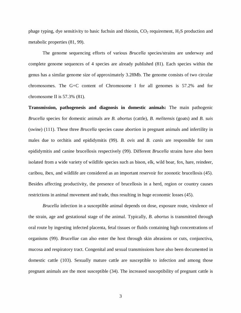
phage typing, dye sensitivity to basic fuchsin and thionin, CO2 requirement, H2S production and
metabolic properties (81, 99).
The genome sequencing efforts of various Brucella species/strains are underway and
complete genome sequences of 4 species are already published (81). Each species within the
genus has a similar genome size of approximately 3.28Mb. The genome consists of two circular
chromosomes. The G+C content of Chromosome I for all genomes is 57.2% and for
chromosome II is 57.3% (81).
Transmission, pathogenesis and diagnosis in domestic animals: The main pathogenic
Brucella species for domestic animals are B. abortus (cattle), B. melitensis (goats) and B. suis
(swine) (111). These three Brucella species cause abortion in pregnant animals and infertility in
males due to orchitis and epididymitis (99). B. ovis and B. canis are responsible for ram
epididymitis and canine brucellosis respectively (99). Different Brucella strains have also been
isolated from a wide variety of wildlife species such as bison, elk, wild boar, fox, hare, reindeer,
caribou, ibex, and wildlife are considered as an important reservoir for zoonotic brucellosis (45).
Besides affecting productivity, the presence of brucellosis in a herd, region or country causes
restrictions in animal movement and trade, thus resulting in huge economic losses (45).
Brucella infection in a susceptible animal depends on dose, exposure route, virulence of
the strain, age and gestational stage of the animal. Typically, B. abortus is transmitted through
oral route by ingesting infected placenta, fetal tissues or fluids containing high concentrations of
organisms (99). Brucellae can also enter the host through skin abrasions or cuts, conjunctiva,
mucosa and respiratory tract. Congenital and sexual transmissions have also been documented in
domestic cattle (103). Sexually mature cattle are susceptible to infection and among those
pregnant animals are the most susceptible (34). The increased susceptibility of pregnant cattle is

4
thought to be related to the concentration of sugar erythritol in the gravid bovine uterus (34,
105).
Upon ingestion, the organisms reach the gastro intestinal tract, and they are phagocytosed
by lymphoepithelial cells of gut-associated lymphoid tissue and gain access to submucosa (99).
In the submucosa, organisms are rapidly ingested by neutrophils and phagocytosed by
macrophages (99). Inside macrophages, Brucella escape death by inhibiting phago-lysosomal
fusion, survive and reach the reticuloendothelial system of local lymph nodes, this leads to local
lymphadenopathy eventually resulting in bacteremia (25, 99). Brucella then spread through the
circulation to the spleen, liver, mammary gland, joints, kidneys, bone marrow and reproductive
tract establishing a systemic infection (99).
In ruminants, Brucella target embryonic and trophoblastic tissue with high concentrations
of erythritol in such tissues as pregnant uterus, fetal tissues and male genital tract (105). B.
abortus infection in cattle may cause late term abortions, still births, retained placentas, sterility,
lymphoplasmacytic mastitis and tissue granulomas (34). The infected animal will shed virulent
Brucella through milk, aborted secretions and afterbirth (34).
Brucellae can be cultured from bones, joints, eyes and brain in adult cattle and from the
stomach, lung and spleen of the bovine fetus (33). Culture of Brucella from aborted material,
milk or tissues collected at autopsy provides a definitive diagnosis (34). Serology is usually the
most practicable of diagnosis methods. In cattle, World Health Organisation (WHO)
recommends the Rose Bengal plate Test (RBT) for screening and ELISA or complement fixation
for confirmation of infected individual animals. Screening of milk samples by milk ring test or
ELISA is useful for surveillance (34). No single serological test is reliable for confirmation of
infection in individual animals in sheep, goats and pigs (34). Serological tests should be used on
5
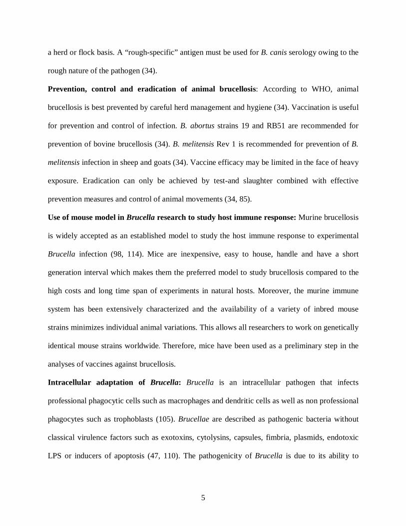
a herd or flock basis. A “rough-specific” antigen must be used for B. canis serology owing to the
rough nature of the pathogen (34).
Prevention, control and eradication of animal brucellosis: According to WHO, animal
brucellosis is best prevented by careful herd management and hygiene (34). Vaccination is useful
for prevention and control of infection. B. abortus strains 19 and RB51 are recommended for
prevention of bovine brucellosis (34). B. melitensis Rev 1 is recommended for prevention of B.
melitensis infection in sheep and goats (34). Vaccine efficacy may be limited in the face of heavy
exposure. Eradication can only be achieved by test-and slaughter combined with effective
prevention measures and control of animal movements (34, 85).
Use of mouse model in Brucella research to study host immune response: Murine brucellosis
is widely accepted as an established model to study the host immune response to experimental
Brucella infection (98, 114). Mice are inexpensive, easy to house, handle and have a short
generation interval which makes them the preferred model to study brucellosis compared to the
high costs and long time span of experiments in natural hosts. Moreover, the murine immune
system has been extensively characterized and the availability of a variety of inbred mouse
strains minimizes individual animal variations. This allows all researchers to work on genetically
identical mouse strains worldwide. Therefore, mice have been used as a preliminary step in the
analyses of vaccines against brucellosis.
Intracellular adaptation of Brucella: Brucella is an intracellular pathogen that infects
professional phagocytic cells such as macrophages and dendritic cells as well as non professional
phagocytes such as trophoblasts (105). Brucellae are described as pathogenic bacteria without
classical virulence factors such as exotoxins, cytolysins, capsules, fimbria, plasmids, endotoxic
LPS or inducers of apoptosis (47, 110). The pathogenicity of Brucella is due to its ability to

6
adapt to the environmental conditions encountered in its intracellular replicative niche including
low levels of nutrients and oxygen, acidic pH and reactive oxygen intermediates (63). This
ability is believed to be responsible for establishment of chronic infection. Brucella had a long
standing co-evolution with its replication niche which makes the pathogen well adapted to the
intracellular environment. For instance, Brucella has the ability to control its own intracellular
trafficking to avoid lysosomal degradation, replicate extensively within the host cell and not
induce apoptosis (25, 48, 50). Brucella expresses a non-canonical LPS with very low
endotoxicity which plays an essential role in the entry of the organism into the phagocytic cell
through interactions with particular receptors within the lipid rafts located on the host cell plasma
membrane (21, 65). Although immediately after entry Brucella containing vacuoles (BCV)
within the host cell interact with the early compartments of endocytic pathway, Brucella with an
intact O-side chain on its LPS avoids fusion of the BCV with lysosome (25). An additional
mechanism by which Brucella avoids lysosomal fusion is by secreting cyclic β-1,2-glucan which
extracts cholesterol from lipid rafts of vacuole membrane preventing phagosomal maturation (6).
After surviving the early destruction within the macrophages, BCVs continues to interact with
endocytic pathway until vacuolar acidification occurs which is required for intracellular
expression of type IV secretion system (25). At this stage BCVs segregate themselves from the
endocytic pathway and start to physically interact with the endoplasmic reticulum (ER) to
become mature replication proficient vacuoles (25, 105). Brucellae virB mutants fail to acquire
ER markers and become ER – derived organelles that ultimately fuse with lysosomes (25, 105).
Therefore, Brucella possesses VirB type IV secretion machinery as well to reach its replication
permissive niche for intracellular survival (25). Moreover, recently it has been shown that

7
Brucella uses its LPS and lipidated outer membrane proteins (L-OMP19) to inhibit MHC class II
antigen expression of host cells to prevent antigen presentation to T-lymphocytes (9, 65).
Brucella virulence factors: Brucella uses a number of virulence factors/mechanisms for
avoiding or suppressing bactericidal responses and for invading and surviving within the host
cell.
Lipopolysaccharides (LPS): LPS is vital to the structural and functional integrity of the Gram-
negative bacterial outer membrane (21). The LPS is composed of Lipid A, a core
oligosaccharide, and an O-side chain polysaccharide (65). LPS of rough Brucella strains do not
have O-side chain (108). In contrast to enterobacteria, such as Escherichia coli (E. coli), Brucella
spp. possesses a nonclassical LPS (21). B. abortus lipid A has a diaminoglucose backbone
(rather than glucosamine) and acyl groups are longer (C18–C19 or C28 rather than C12 and C14)
and are linked to the core by simple amide bonds (rather than ester and amide bonds) (21, 65).
Highly purified B. abortus LPS is several hundred times less active and toxic than the classical
E. coli LPS and is a poor inducer of respiratory burst, bactericidal nitrogen intermediates and
lysozyme secretion (102). Brucella O-side chain blocks deposition of complement factor C1Q to
the outer membrane protein targets and impairs anti-microbial host responses (65). Brucella spp.
are resistant to a large variety of anti-bacterial proteins, including defensin NP-2, lactoferrin,
cecropines, lysozyme, bactenecin-derived peptides and the defensin-like antibiotic polymyxin B,
as well as to crude lysosomal extracts from polymorphonuclear leukocytes (75, 104). Brucella
LPS forms LPS macrodomains enriched with MHC II molecules which inhibit efficient antigen
presentation and downregulate T-cell activation (65). Moreover, pathogenic Brucella smooth
strains enter cells using their LPS to interact with cell surface lipid rafts to avoid fusion with
8
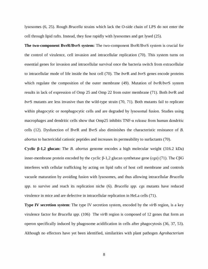
lysosomes (6, 25). Rough Brucella strains which lack the O-side chain of LPS do not enter the
cell through lipid rafts. Instead, they fuse rapidly with lysosomes and get lysed (25).
The two-component BvrR/BvrS system: The two-component BvrR/BvrS system is crucial for
the control of virulence, cell invasion and intracellular replication (70). This system turns on
essential genes for invasion and intracellular survival once the bacteria switch from extracellular
to intracellular mode of life inside the host cell (70). The bvrR and bvrS genes encode proteins
which regulate the composition of the outer membrane (49). Mutation of bvrR/bvrS system
results in lack of expression of Omp 25 and Omp 22 from outer membrane (71). Both bvrR and
bvrS mutants are less invasive than the wild-type strain (70, 71). Both mutants fail to replicate
within phagocytic or nonphagocytic cells and are degraded by lysosomal fusion. Studies using
macrophages and dendritic cells show that Omp25 inhibits TNF-α release from human dendritic
cells (12). Dysfunction of BvrR and BvrS also diminishes the characteristic resistance of B.
abortus to bactericidal cationic peptides and increases its permeability to surfactants (70).
Cyclic β-1,2 glucan: The B. abortus genome encodes a high molecular weight (316.2 kDa)
inner-membrane protein encoded by the cyclic β-1,2 glucan synthetase gene (cgs) (71). The CβG
interferes with cellular trafficking by acting on lipid rafts of host cell membrane and controls
vacuole maturation by avoiding fusion with lysosomes, and thus allowing intracellular Brucella
spp. to survive and reach its replication niche (6). Brucella spp. cgs mutants have reduced
virulence in mice and are defective in intracellular replication in HeLa cells (71).
Type IV secretion system: The type IV secretion system, encoded by the virB region, is a key
virulence factor for Brucella spp. (106) The virB region is composed of 12 genes that form an
operon specifically induced by phagosome acidification in cells after phagocytosis (36, 37, 53).
Although no effectors have yet been identified, similarities with plant pathogen Agrobacterium
9
tumefaciens suggest that Brucella spp. use their type IV secretion system to secrete effector
molecules into the host cytosol (22, 29, 30, 106). Upon entering the macrophage through lipid
rafts, the Brucella containing vacuole (BCV) avoid fusion with lysosomes and start interacting
with endoplasmic reticulum (ER) to reach their replicative niche in the ER (26, 27). The
acquisition of ER membranes requires a functional virB apparatus for sustained interactions and
fusion events between the BCV and ER elements (26). Brucella virB mutants have shown to
loose their ability to multiply in HeLa cells (27, 86). However, Billard et al. documented that
type IV secretion system is not involved in the inhibition of DC maturation (14).
Protective antigens of Brucella - O-side chain: The oligosaccharide chain (O-side chain) is the
most exposed, major antigenic determinant of Brucella spp.(46) This N-formylperosamine O-
polysaccharide of LPS stimulates the major proportion of antibody response in animals and
humans infected with pathogenic Brucella species (83). Although protection against brucellosis
is mainly cell mediated, Arraya et al. demonstrated that passive transfer of immune serum with
O-sidechain antibodies conferred protection in mice against virulent Brucella challenge (5, 78).
Moreover, studies in mice by Vemulapalli et al. using B. abortus vaccine strain RB51 expressing
wboA gene, which expresses low quantities of O-side chain, demonstrated enhanced protection
against challenge with virulent strain 2308 compared to mice vaccinated with strain RB51 not
expressing O-side chain (122, 125). However, it seems antibody mediated protection is
dependent on the host animal as O-side chain antibodies in bovines do not additionally enhance
protection (83).
Outer membrane proteins (OMPs): OMPs were identified as early as 1980s by using
monoclonal antibodies (MAb) and immunogold techniques (31, 127). Two major B. abortus
OMPs were identified and designated as group 2 and 3 proteins based on molecular mass

10
representing 36-38 and 25-27 kDa OMPs respectively (127). Additionally, several low molecular
weight proteins including Omp10, Omp16 and Omp19 have been identified as minor OMPs and
as lipoproteins (31). Group 2 and 3 proteins have shown to be strongly associated with
peptidoglycan (31). Group 2 proteins were also identified as porin proteins (31). Omp31 from B.
suis, B. melitensis and B. ovis was shown to be a hemin binding protein (HBP), which is
expressed under reduced iron conditions and helps obtain iron from the host (35). However,
group 2 and 3 OMPs from rough B. abortus and B. melitensis did not protect against smooth B.
abortus and B. melitensis challenge in mouse models (31, 78). Additionally, major OMPs only
induced low antibody levels and served as poor immunogens in B. abortus infected cattle.
Gonzalez et al. demonstrated that the outer membrane proteins are more exposed on rough
strains than on smooth Brucella due to the absence of O-polysaccharide of the LPS (46).
Therefore, the lack of steric hindrance caused by O-side chain to MAbs against major OMPs
might explain the protection afforded by major OMPs in mice against rough B. ovis challenge
infection (17, 24). In contrast, Zwerdling et al. and Pasquevich et al. both have shown that minor
outer membrane proteins such as Omp16 and Omp19 were immunostimulatory (97, 130).
Pasquevich et al. demonstrated that both Omp16 and Omp19 in its unlipidated version stimulated
antigen specific CD4+ and CD8+ T-cells to provide systemic and oral protection to B. abortus
infection in mice (97). In summary, major OMPs appear to be less relevant as protective antigens
against smooth Brucella infection. Although recently, major OMP25 have shown to play a
potential role as virulence factors by limiting the host response while inhibiting TNF-alpha
secretion from DCs upon infection with smooth B. suis (12).
Superoxide dismutase (SOD): Brucella Cu/Zn SOD is a protective periplasmic antigen (123).
Reactive oxygen intermediates (ROI) are harmful to Brucella and production of ROIs is one

11
mechanism adopted by the host to limit intracellular replication of Brucella (43, 59). SODs are a
family of metallo-enzymes that catalyze the dismutation of superoxide into hydrogen peroxide
and molecular oxygen, thus preventing damage to Brucella by ROIs (76). Brucella Cu/Zn SOD
with copper and zinc at their active sites is encoded by sodC gene and is highly conserved among
Brucella biovars (112). However, the inability to produce Cu/Zn SOD by B. abortus does not
significantly impair its virulence in mice and mutants were able to establish chronic infection in
mice (66, 115). Therefore, SOD cannot be considered a virulence factor of Brucella (110).
Antigenic properties of Brucella Cu-Zn SOD have been demonstrated under several
experimental conditions. Recombinant E. coli expressing Brucella Cu/Zn SOD and strain RB51
overexpressing SOD have been shown to protect mice against challenge with pathogenic
Brucella (93, 123). SOD specific IFN-gamma levels have been detected in vaccinated mice
(123).
L7/L12 ribosomal proteins: CD4+ T cells play an important role in protecting against Brucella
infection. Ribosomal preparations have been used as vaccines against several pathogens,
including B. abortus, conferring some degree of protection. Oliveira et al. demonstrated that in
mice recombinant B. abortus L7/L12 protein stimulated CD4 Th1 - cell response with IFN-γ
secretion (90, 91). Antibody and delayed type hypersensitivity (DTH) responses to this protein
have also been demonstrated in cattle and mice (64, 91). However, it is not clear whether such
subunit vaccinations will provide long term protection in the host.
Antibodies to Brucella: The significance of humoral immunity in murine brucellosis has been
demonstrated by many passive-transfer experiments (5, 78). Brucella LPS and O-antigen of the
Brucella LPS are the two immunodominant structures against which antibodies are shown to be
produced (5, 78, 94). Passive transfer of sera containing LPS antibodies to mice protected against
12
challenge with virulent B. abortus (5, 32, 78). Antibodies to Brucella O-antigen reduced
bacterial infection in mice or conferred partial protection against virulent Brucella infection in
murine models (32). IgG2a and IgG3 are the dominant antibody isotypes detected in mice
suggesting a Th1 immune response against brucellosis (39, 113).
B. abortus infection induces production of IgM, IgG1, IgG2a and IgA antibody isotypes
in both milk and sera of cattle (84). Although humoral immunity plays a role in resistance to
brucellosis, the data suggest that cell mediated immunity is most critical. B. abortus vaccine
strain RB51 lacking the O-side chain of LPS, which therefore does not induce any O-side chain
antibodies, still provides good protection (52, 108, 109). Therefore, while passive transfer studies
in mice support a role for humoral immunity, based on these other studies, CMI provides
adequate immunity.
Cell Mediated Immunity to Brucella: Similar to most intracellular bacterial infections, T-cell
mediated immunity plays a significant role in protecting against virulent Brucella infection (52).
This is best demonstrated by results from B. abortus vaccine strain RB51 studies. Protection
conferred by strain RB51 can only be transferred by immune T cells and not by antibodies (52,
109). Protective functions of adaptive immune response in brucellosis can be classified in to 2
mechanisms (62). The first mechanism is IFN-γ production by CD4+, CD8+ and γδ T-cells which
stimulates macrophage antimicrobial activity and hampers intracellular survival of Brucella. The
second mechanism of T cell mediated immunity is the lysis of infected cells by specific CD8+
and γδ T cells.
Some of the studies which demonstrated the critical role of CMI were adoptive transfer
experiments. In these studies, CD4+, CD8+ and whole T-cell populations from immunized
BALB/c mice which were transferred into infected mice enhanced protection indicating that both
13
CD4+ and CD8+ T-cell subsets are involved in protection (4). Additionally, the critical role of
IFN-γ in resistance to Brucella infection has been demonstrated by in vivo antibody
neutralization experiments (79, 89). Although IFN-gamma can be produced by CD4, CD8, NK
and gamma delta cells, CD4 cells are the major T-cell population based on number and they
secrete most of the IFN- γ; all of this suggests a critical role for CD4 T-cells and associated
IFN—γ production (62). However, experiments with αβ-/- and β2-m-/- mice infected with B.
abortus strain 19 suggest that CD8 T-cell deficient mice have decreased clearance compared to
CD4 T-cell deficient mice and wild type mice implying a more critical role of CD8 vs. CD4 T-
cells (89). Mouse models of brucellosis have revealed that Brucella resistant C57BL/6 mice
require IFN-γ throughout the course of infection and mice died in its absence (79). In
comparison, Brucella susceptible BALB/c mice which failed to produce IFN-γ after first weeks
of infection relied on CD8 T-cells and TNF-α to control infection (79). In cattle younger than 1
year, the major T-cell population is γδ T cells suggesting a significant role of γδ T cells in
Brucella infected calves although their role has not been characterized in vivo (116).
B. abortus induces a CD4 Th1 and CD8 TC1 immune response and inhibits Th2 type
immune responses. The mechanisms associated with regulation of CD4 Th1 and CD8 Tc1
responses are less clear. DC mediated cytokines such as IFN-γ, IL-12 and TNF- α often direct
the T-cell response towards a CD4 Th1 CD8 Tc1 response (62). In vivo depletion of endogenous
IL-12, which is produced by DCs and macrophages, exacerbated Brucella infection and reduced
IFN-γ production (128). Additionally, decreased TNF-α, via TNF-α-receptor knockout mice
(TNF-r-/-), were also severely deficient in IL-12 production; these mice had aggravated Brucella
infection (129).
14
Brucella vaccines – live vaccines: Prevention is better than cure. Historically the most
successful vaccines against brucellosis were live attenuated vaccines compared to killed vaccines
(42). Live attenuated vaccines provide long lasting cell mediated immunity and Brucella can
replicate within the host leading to a longer half-life and better immune response, and thus
making it more efficacious and less expensive (42). Compared to subunit or DNA vaccines, live
attenuated vaccines contain intact bacteria with all the immunogenic components that can be
involved in protection making it more efficient (62). However, some live attenuated vaccines
may cause abortion in pregnant animals and safety concerns limit their use in humans.
Live vaccines - B. abortus strain 19: This vaccine has been used extensively in brucellosis
eradication program in the United States, prior to the introduction of strain RB51 in 1996. Strain
19 is a live attenuated smooth strain (85). The molecular basis of attenuation is not known. This
was first described in 1930 (18). Anecdotal references indicated that strain 19 was originally
isolated from the milk of a Jersey cow as a virulent strain in 1923. But after being kept in the
laboratory for over a year at room temperature it developed a deletion in the erythritol gene (41);
this attenuated the strain (3). While strain 19 conferred protection against virulent B. abortus in
cattle, abortions can develop in pregnant animals (10). Additionally, it has the disadvantage of
inducing O-side chain antibodies that can interfere with diagnostic tests to differentiate infected
and vaccinated animals (109).
B. melitensis Rev. 1: Rev. 1 vaccine is a live attenuated spontaneous mutant derived from
virulent B. melitensis (109). The strain is resistant to streptomycin (38). It stimulates protection
against B. melitensis infection in sheep and goats and also protects rams against B. ovis infection
(109). The use of Rev. 1 in cattle indicates that it provides better protection than strain 19 (109).

15
Depending upon the dose administered, abortion occurs with variable frequency (109). Rev. 1 is
a smooth strain and it interferes with diagnostic tests.
B. suis strain 2: This is a live attenuated smooth strain derived from biovar 1 of B. suis. It is
used as an oral vaccine in China to protect cattle, goats, sheep and pigs (15, 80). Although it
induces O-side chain antibodies, they disappear by one year post vaccination (80).
B. abortus strain 45/20: B. abortus smooth strain 45/0 was isolated from a cow in 1922 (109).
After 20 passages in guinea pigs, rough strain 45/20 bearing at least one unknown mutation was
obtained. This strain protects guinea pigs and cattle from Brucella infection (109). However,
when used as a live vaccine, strain 45/20 was not stable and reverted back to smooth virulent
form (108). The reversion to smooth strain resulted in vaccine induced antibodies which
interfered with diagnostic tests. This defeated the purpose of using rough strains.
B. abortus strain RB51: Vaccine strain RB51 is a stable, rifampin-resistant, rough mutant of B.
abortus strain 2308 (108). It was derived by serial passage of parental strain 2308 on Trypticase
soy agar supplemented with varying concentrations of rifampin and pencillin (108). “R” stands
for rough and “B” stands for Brucella; 51 refer to an internal laboratory nomenclature used at the
time it was derived and not the passage number (108). Strain RB51 is devoid of O-side chain and
is stable after multiple passages in vitro and in vivo through various species of animals (108).
Colonies of strain RB51 are rough in morphology as indicated by their ability to absorb crystal
violet as well as auto-agglutinate while in suspension (108). Biochemically, strain RB51 has the
ability to use erythritol unlike strain 19 (108). In February 1996, the USDA Animal Plant Health
Inspection service (APHIS) approved the use of B. abortus strain RB51 as the official calf hood
vaccine for protection against brucellosis. The recommended dose for calves between the ages of
16
4-12 months vaccinated subcutaneously (SC) is 1-3.4 X 10¹⁰ organisms (117). It induces
protection in cattle against virulent B. abortus at a level similar to that conferred by strain 19.
There are a number of advantages for strain RB51 over other vaccines. It does not
produce clinical signs post vaccination; there are no local reaction at the site of injection (28). It
is rapidly cleared from blood stream as early as 2 weeks post inoculation and it is not shed in the
nasal secretions, saliva or urine. Thus it is unable to spread from vaccinated to non-vaccinated
animals through these routes (28, 108). Pregnant cattle can be safely vaccinated SC with 109
organisms of strain RB51 without inducing abortion or placentitis. Mouse studies revealed that
protective immunity induced by strain RB51 is solely mediated by T-cells with a polarized type
1 cytokine profile which is the desired type of protection against intracellular pathogens (52). In
a murine model, strain RB51 protected against challenge with B. abortus, B. melitensis, B. suis,
and B. ovis (108). Moreover, the lack of O-side chain with strain RB51 prevents O-antigen
specific antibody formation and interference with diagnostic tests (108).
Although strain RB51 is extremely stable, the exact nature for its avirulence is not known
(108). It is thought that the strain possesses at least two mutations in its LPS biosynthetic
pathway. One being the presence of an IS711 element in the wboA gene responsible for synthesis
of O-side chain (124). Complementation experiments using wboA gene showed that the strain
produces O-side chain while maintaining a rough phenotype, but the O-side chain remains in the
cytoplasm indicating the possibility for at least one more mutation (121). This second mutation is
thought to be in the wzt gene that codes for an ABC type transporter which is involved in the
translocation of the O-side chain across the inner membrane of Brucella (44).
Recombinant strain RB51 vaccine overexpressing homologous protective antigens such
as B. abortus Cu/Zn SOD (superoxide dismutase; approximately 10 times the normal level),

17
designated RB51SOD, induced significantly increased protection against challenge with virulent
strain 2308 in BALB/c mice (123). Complementation of strain RB51 with a functional copy of
wboA gene, RB51wboA, produced intracytoplasmic O-side chain and completely protected mice
against virulent strain 2308 infection (122). Additionally, recombinant strain RB51SODWboA,
which overexpressed SOD with simultaneous expression of O-side chain in the cytoplasm,
induced better protection than strain RB51 or RB51SOD against strain 2308 challenge (119).
The ability of strain RB51 to induce CD4 Th1 and CD8 TC1 polarized response with high levels
of IFN-γ made it an attractive candidate for heterologous expression of protective antigens
belonging to other intracellular pathogens. Development of strain RB51 as a vector for
expression of heterologous antigens has met with success when E. coli, Mycobacterium and
Neospora caninum antigens were successfully expressed in strain RB51 (96, 100, 101, 121, 124).
Vemulapalli et al. had demonstrated that strain RB51 exposed to an appropriate minimum
dose of gamma radiation were unable to replicate but retained their ability to stimulate Th1
immune responses and protected mice against virulent challenge with strain 2308 (107).
Additionally, Magnani et al. showed that irradiated B. melitensis protected against virulent B.
melitensis challenge (74). By contrast, Lee et al., found that irradiated (higher dose of irradiation)
strain RB51 with or without IL-12 as an adjuvant, did not protect against strain 2308 challenge
(68, 69).
Killed vaccines: Killed vaccines can be safer alternatives to live attenuated vaccine strains and a
variety of killed vaccines have been developed for protection against brucellosis. However,
killed vaccines without adjuvants had only limited success and protection compared to live
attenuated strains. B. melitensis H38 was smooth formalin killed vaccine in mineral oil adjuvant
used for vaccination in goats and sheep (109). It protected against abortions but induced positive

18
serology to vaccine and caused unacceptable local reactions at the inoculation site (109). B.
abortus strain 45/20 when used as a bacterin incorporated in adjuvants gave varying results
regarding protective efficacy and positive serology (109). Most investigators considered that two
vaccinations were necessary for protection. Strain 45/20 did not induce abortions when used as
bacterin (109). However, batch to batch variations in properties of the vaccine, variability of
reported protection, severe local reactions and unpredictable serology prompted the
discontinuation of this killed vaccine (109).
Recombinant vector vaccines: Brucella protective antigens such as Cu/Zn SOD were expressed
using vaccinia virus and insect baculovirus vectors although these vaccines were not successful
in eliciting effective protection against virulent Brucella challenge (7, 120). Similarly, Gram-
negative soil bacterium Ochrobactrum anthropi, closest genetic relative of Brucella, had been
used to express Cu/Zn SOD antigen (51). Vaccination of mice with recombinant O. anthropi
induced mixed Th1-Th2 immunity with high IFN-γ and IL-4 levels. It was non-protective unless
co-administered with CpG adjuvant which polarized the cytokine response to Th1 profile (51).
Subunit vaccines: The concept of subunit vaccines in brucellosis is based on generation of
memory Th1 cells by immunization with T-cell antigen (62). The strategy is to identify those
Brucella antigens that are responsible for T-cell mediated response. Until now periplasmic
binding protein P39, bacterioferritin and L7/L12 proteins, Omp 31 have been purified and tested
as subunit vaccines with adjuvants (2, 90). Mice vaccinated with these proteins showed only a
partial protection when challenged. The enzyme lumazine synthase from Brucella spp. (BLS) is
highly immunogenic and stable. Goldbaum et al. (2007), showed that a recombinant chimera of
10 copies of protective antigen OMP31 on a scaffold of BLS (rBLSOmp31) provided good
protection level against Brucella ovis (23). The rBLSOmp31 vaccine induced greater protection
19
than vaccination with co-delivery of both recombinant proteins (rBLS + rOmp31) (23).
Additionally, the former protected similarly compared to control vaccine Brucella melitensis
strain Rev.1 (23). The chimera induced humoral as well as BLS and peptide specific T-cell
responses (23, 40, 118). Recently, Pasquevich et al. (2009) demonstrated that immunization with
recombinant Brucella species outer membrane protein Omp16 or Omp19 in adjuvant induces
specific CD4+ and CD8+ T-cells as well as systemic and oral protection against Brucella abortus
challenge (97).
DNA vaccines: DNA vaccines involve the injection of plasmid DNA encoding protective
antigens in to the host. No other cellular or subcellular components are included in the vaccine
(109). The type of immune response elicited depends upon the antigen’s characteristics, route of
administration and the presence of immunostimulatory DNA sequences. So far, DNA vaccines
expressing various Brucella antigens such as Cu/Zn SOD, BLS, L7/L12, P39, heat shock protein
GroEL and OMPs have been tested by different research groups in mice with variable levels of
protection upon challenge infection (2, 64, 67, 118). However the question remains as to whether
DNA vaccines encoding Brucella antigens would induce an effective long term protection.
Zoonosis: Zoonosis is any infectious disease that can be transmitted (in some instances, by a
vector) from non-human animals, both wild and domestic, to humans or vice versa (1).
Brucellosis is an established zoonosis (34). All Brucella species with the exception of B. ovis and
B. neotomae can infect humans (34). B. melitensis is the most important zoonotic agent among
Brucella species although most human cases of brucellosis are caused by B. abortus. B.
melitensis, B. suis and B. abortus are the most infectious of the genus in their order of
pathogenicity (34). The incidence of human disease is closely related to the prevalence of
infection in livestock and to the practices that allow potential exposure of humans to infected
20
animals or their products (45). The risk group is Abattoir workers, meat inspectors, animal
handlers, veterinarians, and lab workers (61).
Consumption of unpasteurized cow, small ruminant or camelid milk and milk products is
considered to be the main route of infection (45). Clinically symptoms are, in the acute form (<8
weeks from illness onset), nonspecific and "flu-like" symptoms including fever, sweats, malaise,
anorexia, headache, myalgia, and back pain (34, 45). In the undulant form (<1 year from illness
onset), symptoms include undulant fevers, arthritis, and epididymo-orchitis in males (34).
Neurologic symptoms may occur acutely in up to 5% of cases. In the chronic form (>1 year from
onset), symptoms may include chronic fatigue syndrome, depression, and arthritis. Human-to-
human transmissions by tissue transplantation or sexual contact have occasionally been reported
but are very rare (34). Therefore, control and eradication of the disease from the natural animal
reservoirs have important public health implications (1, 34, 45). A definitive diagnosis in acute
human brucellosis includes, isolation of Brucella from blood or other tissues is definitive (34).
However, culture is often negative, especially in long-standing disease. Serology is the most
generally useful diagnostic procedure. The Rose Bengal test (RBT), tube agglutination and
ELISA procedures are recommended (34). Methods which differentiate IgM and IgG can
distinguish active vs. past infection. The critical element in the treatment of all forms of human
brucellosis is the administration of effective antibiotics for an adequate length of time (34).
Treatment of uncomplicated cases in adults and children eight years of age and older are by
using a combination of antibiotics; doxycycline 100 mg twice a day for six weeks + streptomycin
1 g daily for two to three weeks or doxycycline 100 mg twice a day for six weeks + rifampicin
600–900 mg daily for six weeks (34). However, for these acute cases the relapse rate is 10-20 %
and in chronic phase, eradication is difficult since Brucella spp. are localized intracellularly and
21
most antibiotics do not actively pass through cell membranes (34). There are no safe and
effective commercially available vaccines to protect against human brucellosis.
Human brucellosis vaccines: Safe and protective vaccines against human brucellosis are not
commercially available (34). However, numerous vaccines have been tested in people in the past
with limited success. B. abortus strain 19-BA was used in the former USSR (109). This strain 19
derived vaccine (1 x 10⁹ CFUs) given by skin scarification (epicutaneous route) induced
protection for a shorter duration (5-6 months, maximum up to 1 year) but with a high frequency
of hypersensitivity reactions occurring in 76% of those vaccinated (34, 109). Attenuated strains
of B. abortus 84-C and 104-M were also given epicutaneously or as aerosols in USSR and China
respectively (109). Although considered effective, these vaccines induced serious adverse
reactions and are no longer in use. Emphasis for safer non living vaccines led to the development
of subunit vaccines for use in humans. The French developed a vaccine utilizing phenol-
insoluble peptidoglycan fraction of B. melitensis M15 which was administered subcutaneously
and supposedly offered protection for 2 years (34, 109). However, conclusive evidence of
protective efficacy is not available and the vaccine is not at present in production. Another sub-
cellular fraction namely “Brucella chemical vaccine” (BCV) was developed from an acetic acid
extracted polysaccharide-protein fraction in Russia (34). This vaccine given intramuscularly does
not elicit severe hypersensitivity reactions but evidence of protective efficacy from controlled
clinical trials is not available.
Renewed interest in Brucella as a potential bio-terror weapon illustrates the need for
developing an effective vaccine against human brucellosis.
Bioterrorism: Brucella has been traditionally considered as a biological weapon (95). B.
melitensis and B. suis have been developed experimentally as biological weapons by many state
22
sponsored programs during World War II (95). Brucellosis remains the most common
anthropozoonosis worldwide and its significance as a potential bioterrorism agent makes it in to
the category B biodefense research list of Center for Disease Control and Prevention (CDC) (95).
Although Brucella can enter the human host through skin abrasions or cuts, the conjunctiva or by
consuming unpasteurized dairy products, the most important means of transmission in a
bioterrorism event is airborne transmission (61). They are relatively stable in aerosol form and a
small inoculum (10-100 bacteria) will induce human disease (61). The organism is easily
obtained worldwide in contrast to other agents and easy to develop antibiotic resistant strains
(95). The disease is severely debilitating, infectious to both humans and livestock, has vague
clinical characteristics delaying rapid diagnosis and requires combined antibiotic regimen for a
prolonged period to treat the disease (34, 45). Additionally, there are no human vaccines
available. According to Godfroid et al., in a theoretical model of a bioterrorist attack and in the
absence of an intervention program for 100,000 persons exposed, a B. melitensis cloud would
result in 82,500 cases of brucellosis requiring extended therapy, with 413 deaths. The economic
impact of such a brucellosis bioterrorist attack would cost $ 477.7 million per 100,000 persons
exposed (45). Therefore, the development of a vaccine for brucellosis suitable for humans would
be an ideal solution to prepare for a bioterror threat. One of the theoretical vaccine targets for the
future that could be considered for humans, whose efficacy has been proven in animals, is B.
abortus strain RB51.
Introduction and rationale
In spite of the documented evidence that an infectious aerosol dose of 10-100 Brucella can cause
human disease (16), its potential use as a bioterror agent and the absence of an efficacious

23
vaccine for use in humans, very few studies have focused on vaccine efficacy associated with
respiratory challenge. Most Brucella studies have predominantly focused on non-respiratory
routes of vaccination and challenge, such as vaccinating animals intraperitoneal (IP),
subcutaneous (SC), or intravenous (IV) followed by IP or (IV) challenge infection (60, 108). By
contrast, Mense et al. demonstrated that intranasal (IN) inoculation of virulent B. melitensis 16M
can cause chronic infection in BALB/c mice (77). In a different study, Ficht et al. also
demonstrated that aerosol infection with B. abortus caused chronic infection at lower intranasal
doses compared to IN B. melitensis infection (4 x 10² vs. 1 x 10⁴ CFUs/mouse) respectively (61).
These studies demonstrated that Brucella species can cause chronic infection either via or
subsequent to respiratory infection. However, contrary to the fact that IP vaccination protected
against brucellosis in mouse models, IP vaccination did not protect against aerosol challenge.
Both Ficht et al. and Olsen et al. failed to show clearance of B. melitensis and/or B. abortus from
lung upon aerosol challenge following IP vaccination with protective vaccine strains including B.
abortus strain RB51 (61, 92).
B. abortus strain RB51 is a USDA approved live attenuated rough vaccine used in the
United States and many other countries against cattle brucellosis. Another live attenuated B.
abortus strain RB51SOD which overexpresses Cu-Zn superoxide dismutase had been shown to
elicit better protection than strain RB51 when vaccinated IP against IP challenge with virulent B.
abortus in mice (123). Protection against brucellosis induced by both these vaccine strains is
mediated through a strong CD4+ Th1 and CD8+ Tc1 adaptive immune response (52).
Nevertheless, vaccine strain RB51 failed to protect mice against IN challenge with virulent B.
abortus when vaccinated IP (92).

24
Part of the reason for this lack of protection may be due to the route of vaccination. Based
on continuously developing knowledge, it is expected that mucosal vaccination would enhance
mucosal protective immune response against aerosol challenge (82). Therefore, an IN
vaccination with either vaccine strains RB51 or RB51SOD was expected to protect mice against
IN challenge with virulent B. abortus. However, preliminary data from our laboratory
demonstrated that intranasal vaccination alone with strains RB51 or RB51SOD would not elicit
protection against intranasal challenge with virulent B. abortus strain 2308 in BALB/c mice.
This finding warranted further exploration into the events which led to lack of protective
immunity elicited by these vaccine strains. Although it has been proven that both vaccine strains
induce protection through T-cell mediated immunity, limited information is available on how
they stimulate innate immune response which results in protective CMI. This knowledge is
critical to improving these protective animal vaccines for their ultimate use in humans against
aerosol brucellosis infection.
A robust innate immune response is necessary to initiate a strong adaptive immune
response. Dendritic cells (DCs) and macrophages are the two antigen presenting cells of the
innate immune system (8). DCs are the better antigen presenting cells (APC) and are more
susceptible to Brucella infection. DCs recognize and capture antigen, and subsequently migrate
to secondary lymphoid organs (8). There the DCs present the antigens to naïve T-lymphocytes,
thus resulting in the initiation of specific adaptive immune responses (8). Based on DC activation
status and the cytokines they produce, DCs prime T cell phenotype and function (Th1/Th2 or
regulatory T cells or Th17 cells) (8). Inadequate DC activation characterized by high expression
of MHC class II and costimulatory markers and limited cytokine production might lead to T-cell
tolerance (72). Thus, DCs play a crucial role in bridging the innate and adaptive immune

25
response by acting as the key mediator. Therefore, the enhanced ability of rough vaccine strains
for inducing DC maturation and function could be critical for a protective T-cell response.
There are no published data on how rough vaccine strains RB51 or RB51SOD affect DC
maturation, activation and function. However, there are contradictory data on the effects of strain
2308 on DC maturation (13, 73, 130). Previous studies have established the use of murine bone
marrow derived dendritic cells (BMDCs) as a model system for studying the effects of bacterial
infection on DC phenotype in vitro (73). In chapter 2 of this dissertation, we discuss the effect
of vaccine strains RB51, RB51SOD and pathogenic B. abortus strain 2308 on DC phenotype and
cytokine production using murine BMDCs.
Although it is important to improve the protective ability of live rough vaccine strain
RB51 by delineating its innate immune activation ability, safety concerns limit their ultimate use
in humans. Therefore, ideally, heat killed (HK) or irradiated (IR) strain RB51 vaccine which still
induces efficacious protective immune responses has the potential as a safer human vaccine.
Previous studies have shown that both HK and IR B. abortus strains induce Th1 immunity (54,
56, 107). However, the differential ability of live, HK and IR rough and smooth strains of B.
abortus to stimulate BMDC activation and function at the same doses has not been reported in
literature. Chapter 3 of my dissertation presents the data from our in vitro study designed to
determine whether HK and IR strain RB51 stimulated comparable innate responses to live
vaccine strain RB51 for exploring their use as vaccine in people and animals.
The above mentioned in vitro studies helped us delineate rough vs. smooth B. abortus
strain mediated DC activation and function. However, in an accidental or deliberate aerosol
Brucella exposure, the organism is directly delivered to the pulmonary airways and airway
epithelium. Although pulmonary DCs comprise only a small fraction of innate immune cells in

26
lung compared to alveolar macrophages, they have the unique ability to migrate to the draining
lymph node with the captured antigen to activate naïve T-cells (57). However, it is expected that
pulmonary DCs behave differently in some significant respects to BMDCs. Given the inability of
vaccine strains RB51 and RB51SOD to protect against IN challenge with pathogenic strain 2308
upon IN vaccination, it is crucial to understand the differential ability of these vaccine strains
given IN to stimulate innate immunity in vivo. Additionally these studies will also provide
information as to whether B. abortus pathogenic strain 2308 limits the proinflammatory response
in the lungs. The ability to minimize the innate immune response may allow both Brucella
species to subvert the immune response and allow for systemic spread. To our knowledge, no
studies have been published which characterize the in vivo innate immune response including the
associated histopathological changes to IN inoculation of either B. abortus pathogenic strain
2308 or rough vaccine strains RB51 or RB51SOD. In chapter 4, we evaluated the differential
ability of B. abortus rough vaccine strains RB51, RB51SOD and smooth pathogenic strain 2308
to elicit pulmonary DC activation and function in vivo. We also assessed the vaccine and virulent
strain induced histopathological changes in lung at day 3, 5, 7 and 14 post infection (PI).
While considering DCs as the major mediator of host innate response, in order for the
activation to occur, DCs must first recognize rough and/or smooth strains of B. abortus. DCs
recognize microbes via host cell membrane receptors called Toll-Like Receptors (TLRs) (58).
Upon recognition of microbial products, TLRs transduce signals via common adaptor molecules
to activate their host cells (58, 87). Published literature suggest that B. abortus signals through
multiple TLRs such as TLR2 (outer membrane proteins), TLR4 (lipopolysaccharide) and TLR9
(CpG DNA) (11, 19, 55, 88, 126, 130). However, there are contradictory data on the most crucial
TLRs in recognition of B. abortus by DCs. Weiss et al. demonstrate that Brucella signals through

27
TLR2, TLR4, and MyD88; the latter is most critical for clearance (126). Although TLR2 and
TLR4 both signal through MyD88, their studies suggest an additional role for TLR9 molecule.
Subsequently, Oliveira et al. also suggested a prominent role for TLR9 in DC, IL-12 production
and Brucella clearance (88). Zwerdling et al. provided data suggests that Brucella signals
through TLR2 and TLR4 (130). Despite how individual TLRs activate a cell, there is a dearth in
information whether a difference in TLR preference exists between rough and smooth strains of
Brucella in mediating DC activation. Identifying a differential TLR activation, if it exists,
between B. abortus rough vaccine strain RB51 and smooth strain 2308 will help us to improve
innate immune stimulating ability of strain RB51 by using TLR agonist adjuvants. In order to
address this question, we infected TLR2, TLR4, TLR9 KO BALB/c BMDCs and wild type
control BMDCs with rough strain RB51 and smooth strain 2308 to analyze the difference in DC
activation and function. Additionally, no published studies have addressed the role of TLRs in
the clearance of rough or smooth B. abortus strains from intranasally infected mice. It is critical
to know if the pulmonary clearance of smooth B. abortus is directly related to recognition of
bacteria through a particular TLR to identify the best strategy to induce enhanced clearance. In
our study, we infected BALB/c control mice as well as TLR2, TLR4, TLR9 KO mice with either
vaccination dose of strain RB51 or challenge dose of strain 2308 to assess TLR dependent
clearance of Brucella strains. Chapter 5 of my dissertation discuss the role of TLR2, TLR4 and
TLR9 in the differential activation of DCs upon infection with B. abortus rough and smooth
strains in vitro and in the clearance of a Brucella challenge in vivo.
Concurrent with unraveling the innate immune response to Brucella vaccine and
pathogenic strains, the ultimate goal of the experiments which constitute this dissertation is to
develop a protective vaccine for animals and people against respiratory challenge with Brucella.
28
Although our initial efforts with IN vaccination of strain RB51 or RB51SOD failed to protect
against IN challenge with strain 2308, we performed subsequent experiments to identify alternate
vaccination strategies which might yield protection. Those strategies included testing different
vaccination routes, doses, booster vaccination, various prime-boost strategies (involving
systemic and IN vaccination routes) using B. abortus vaccine strains RB51 and RB51SOD to
protect against intranasal exposure to pathogenic B. abortus 2308. Chapter 6 describes the
results and conclusions from those experiments.
In summary, experiments from this dissertation research determined the extent to which
B. abortus vaccine and pathogenic strain mediated DC activation and function in vitro and in
vivo as well as the roles played by various TLRs in inducing DC response in vitro and pulmonary
clearance in vivo. Experiments were also designed to test different vaccination strategies to
protect against an aerosol challenge with virulent strain B. abortus.
References
1. Acha, P., Szyfres, B. (ed.). 2001. Zoonoses and communicable diseases common to man
and animal. , vol. I, Bacterioses and Mycoses. Pan American Health Organization, WHO.
2. Al-Mariri, A., A. Tibor, P. Mertens, X. De Bolle, P. Michel, J. Godefroid, K.
Walravens, and J. J. Letesson. 2001. Protection of BALB/c mice against Brucella
abortus 544 challenge by vaccination with bacterioferritin or P39 recombinant proteins
with CpG oligodeoxynucleotides as adjuvant. Infect Immun 69:4816-4822.
3. Alton, G. G. 1978. Recent developments in vaccination against bovine brucellosis. Aust
Vet J 54:551-557.
29
4. Araya, L. N., P. H. Elzer, G. E. Rowe, F. M. Enright, and A. J. Winter. 1989.
Temporal development of protective cell-mediated and humoral immunity in BALB/c
mice infected with Brucella abortus. J Immunol 143:3330-3337.
5. Araya, L. N., and A. J. Winter. 1990. Comparative protection of mice against virulent
and attenuated strains of Brucella abortus by passive transfer of immune T cells or
serum. Infect Immun 58:254-256.
6. Arellano-Reynoso, B., N. Lapaque, S. Salcedo, G. Briones, A. E. Ciocchini, R.
Ugalde, E. Moreno, I. Moriyon, and J. P. Gorvel. 2005. Cyclic beta-1,2-glucan is a
Brucella virulence factor required for intracellular survival. Nat Immunol 6:618-625.
7. Bae, J. E., G. G. Schurig, and T. E. Toth. 2002. Mice immune responses to Brucella
abortus heat shock proteins. Use of baculovirus recombinant-expressing whole insect
cells, purified Brucella abortus recombinant proteins, and a vaccinia virus recombinant
as immunogens. Vet Microbiol 88:189-202.
8. Banchereau, J., and R. M. Steinman. 1998. Dendritic cells and the control of
immunity. Nature 392:245-252.
9. Barrionuevo, P., J. Cassataro, M. V. Delpino, A. Zwerdling, K. A. Pasquevich, C.
Garcia Samartino, J. C. Wallach, C. A. Fossati, and G. H. Giambartolomei. 2008.
Brucella abortus inhibits major histocompatibility complex class II expression and
antigen processing through interleukin-6 secretion via Toll-like receptor 2. Infect Immun
76:250-262.
10. Beckett, F. W., and S. C. MacDiarmid. 1985. The effect of reduced-dose Brucella
abortus strain 19 vaccination in accredited dairy herds. Br Vet J 141:507-514.

30
11. Berguer, P. M., J. Mundinano, I. Piazzon, and F. A. Goldbaum. 2006. A polymeric
bacterial protein activates dendritic cells via TLR4. J Immunol 176:2366-2372.
12. Billard, E., J. Dornand, and A. Gross. 2007. Brucella suis prevents human dendritic
cell maturation and antigen presentation through regulation of tumor necrosis factor alpha
secretion. Infect Immun 75:4980-4989.
13. Billard, E., J. Dornand, and A. Gross. 2007. Interaction of Brucella suis and Brucella
abortus rough strains with human dendritic cells. Infect Immun 75:5916-5923.
14. Billard, E., J. Dornand, and A. Gross. 2008. VirB type IV secretory system does not
contribute to Brucella suis' avoidance of human dendritic cell maturation. FEMS
Immunol Med Microbiol 53:404-412.
15. Blasco, J. M., C. Marin, M. P. Jimenez de Bagues, and M. Barberan. 1993. Efficacy
of Brucella suis strain 2 vaccine against Brucella ovis in rams. Vaccine 11:1291-1294.
16. Bossi, P., A. Tegnell, A. Baka, F. Van Loock, J. Hendriks, A. Werner, H. Maidhof,
and G. Gouvras. 2004. Bichat guidelines for the clinical management of brucellosis and
bioterrorism-related brucellosis. Euro Surveill 9:E15-16.
17. Bowden, R. A., A. Cloeckaert, M. S. Zygmunt, and G. Dubray. 1995. Outer-
membrane protein- and rough lipopolysaccharide-specific monoclonal antibodies protect
mice against Brucella ovis. J Med Microbiol 43:344-347.
18. Buck, J. M. 1930. Studies of vaccination during calfhood to prevent bovine infectious
abortion. J. Agric. Res. 41:667.
19. Campos, M. A., G. M. Rosinha, I. C. Almeida, X. S. Salgueiro, B. W. Jarvis, G. A.
Splitter, N. Qureshi, O. Bruna-Romero, R. T. Gazzinelli, and S. C. Oliveira. 2004.
31
Role of Toll-like receptor 4 in induction of cell-mediated immunity and resistance to
Brucella abortus infection in mice. Infect Immun 72:176-186.
20. Capasso, L. 1999. Brucellosis at Herculaneum (79 AD). Int. J. Osteoarchaeol. 9:277-
288.
21. Cardoso, P. G., G. C. Macedo, V. Azevedo, and S. C. Oliveira. 2006. Brucella spp
noncanonical LPS: structure, biosynthesis, and interaction with host immune system.
Microb Cell Fact 5:13.
22. Cascales, E., and P. J. Christie. 2003. The versatile bacterial type IV secretion systems.
Nat Rev Microbiol 1:137-149.
23. Cassataro, J., K. A. Pasquevich, S. M. Estein, D. A. Laplagne, C. A. Velikovsky, S.
de la Barrera, R. Bowden, C. A. Fossati, G. H. Giambartolomei, and F. A.
Goldbaum. 2007. A recombinant subunit vaccine based on the insertion of 27 amino
acids from Omp31 to the N-terminus of BLS induced a similar degree of protection
against B. ovis than Rev.1 vaccination. Vaccine 25:4437-4446.
24. Cassataro, J., C. A. Velikovsky, S. de la Barrera, S. M. Estein, L. Bruno, R. Bowden,
K. A. Pasquevich, C. A. Fossati, and G. H. Giambartolomei. 2005. A DNA vaccine
coding for the Brucella outer membrane protein 31 confers protection against B.
melitensis and B. ovis infection by eliciting a specific cytotoxic response. Infect Immun
73:6537-6546.
25. Celli, J. 2006. Surviving inside a macrophage: the many ways of Brucella. Res Microbiol
157:93-98.
32
26. Celli, J., C. de Chastellier, D. M. Franchini, J. Pizarro-Cerda, E. Moreno, and J. P.
Gorvel. 2003. Brucella evades macrophage killing via VirB-dependent sustained
interactions with the endoplasmic reticulum. J Exp Med 198:545-556.
27. Celli, J., and J. P. Gorvel. 2004. Organelle robbery: Brucella interactions with the
endoplasmic reticulum. Curr Opin Microbiol 7:93-97.
28. Cheville, N. F., M. G. Stevens, A. E. Jensen, F. M. Tatum, and S. M. Halling. 1993.
Immune responses and protection against infection and abortion in cattle experimentally
vaccinated with mutant strains of Brucella abortus. Am J Vet Res 54:1591-1597.
29. Christie, P. J. 2004. Type IV secretion: the Agrobacterium VirB/D4 and related
conjugation systems. Biochim Biophys Acta 1694:219-234.
30. Christie, P. J., K. Atmakuri, V. Krishnamoorthy, S. Jakubowski, and E. Cascales.
2005. Biogenesis, architecture, and function of bacterial type IV secretion systems. Annu
Rev Microbiol 59:451-485.
31. Cloeckaert, A., P. de Wergifosse, G. Dubray, and J. N. Limet. 1990. Identification of
seven surface-exposed Brucella outer membrane proteins by use of monoclonal
antibodies: immunogold labeling for electron microscopy and enzyme-linked
immunosorbent assay. Infect Immun 58:3980-3987.
32. Cloeckaert, A., I. Jacques, N. Bosseray, J. N. Limet, R. Bowden, G. Dubray, and M.
Plommet. 1991. Protection conferred on mice by monoclonal antibodies directed against
outer-membrane-protein antigens of Brucella. J Med Microbiol 34:175-180.
33. Colby, L. A. 1997. The humoral immune response of Elk (Cervus elaphus nelsoni) and
mice to vaccination with Brucella abortus strain RB51. Virginia Polytechnic Institute and
State University.
33
34. Corbel, M. J. (ed.). 2006. Brucellosis in humans and animals. World Health
Organization.
35. Delpino, M. V., J. Cassataro, C. A. Fossati, F. A. Goldbaum, and P. C. Baldi. 2006.
Brucella outer membrane protein Omp31 is a haemin-binding protein. Microbes Infect
8:1203-1208.
36. Delrue, R. M., C. Deschamps, S. Leonard, C. Nijskens, I. Danese, J. M. Schaus, S.
Bonnot, J. Ferooz, A. Tibor, X. De Bolle, and J. J. Letesson. 2005. A quorum-sensing
regulator controls expression of both the type IV secretion system and the flagellar
apparatus of Brucella melitensis. Cell Microbiol 7:1151-1161.
37. Delrue, R. M., M. Martinez-Lorenzo, P. Lestrate, I. Danese, V. Bielarz, P. Mertens,
X. De Bolle, A. Tibor, J. P. Gorvel, and J. J. Letesson. 2001. Identification of Brucella
spp. genes involved in intracellular trafficking. Cell Microbiol 3:487-497.
38. Elberg, S. S., and K. Faunce, Jr. 1957. Immunization against Brucella infection. VI.
Immunity conferred on goats by a nondependent mutant from a streptomycin-dependent
mutant strain of Brucella melitensis. J Bacteriol 73:211-217.
39. Elzer, P. H., R. H. Jacobson, K. H. Nielsen, J. T. Douglas, and A. J. Winter. 1994.
BALB/c mice infected with Brucella abortus express protracted polyclonal responses of
both IgG2a and IgG3 isotypes. Immunol Lett 42:145-150.
40. Estein, S. M., M. A. Fiorentino, F. A. Paolicchi, M. Clausse, J. Manazza, J.
Cassataro, G. H. Giambartolomei, L. M. Coria, V. Zylberman, C. A. Fossati, R.
Kjeken, and F. A. Goldbaum. 2009. The polymeric antigen BLSOmp31 confers
protection against Brucella ovis infection in rams. Vaccine 27:6704-6711.
34
41. Ewalt, D. R., P. F. Ross, and J. B. Payeur. 1990. Identification of Brucella abortus
strain 19 by decreased ability to utilize erythritol as determined by gas liquid
chromatography. J Vet Diagn Invest 2:120-122.
42. Ficht, T. A., M. M. Kahl-McDonagh, A. M. Arenas-Gamboa, and A. C. Rice-Ficht.
2009. Brucellosis: the case for live, attenuated vaccines. Vaccine 27 Suppl 4:D40-43.
43. Gee, J. M., M. W. Valderas, M. E. Kovach, V. K. Grippe, G. T. Robertson, W. L.
Ng, J. M. Richardson, M. E. Winkler, and R. M. Roop, 2nd. 2005. The Brucella
abortus Cu,Zn superoxide dismutase is required for optimal resistance to oxidative killing
by murine macrophages and wild-type virulence in experimentally infected mice. Infect
Immun 73:2873-2880.
44. Godfroid, F., A. Cloeckaert, B. Taminiau, I. Danese, A. Tibor, X. de Bolle, P.
Mertens, and J. J. Letesson. 2000. Genetic organisation of the lipopolysaccharide O-
antigen biosynthesis region of Brucella melitensis 16M (wbk). Res Microbiol 151:655-
668.
45. Godfroid, J., A. Cloeckaert, J. P. Liautard, S. Kohler, D. Fretin, K. Walravens, B.
Garin-Bastuji, and J. J. Letesson. 2005. From the discovery of the Malta fever's agent
to the discovery of a marine mammal reservoir, brucellosis has continuously been a re-
emerging zoonosis. Vet Res 36:313-326.
46. Gonzalez, D., M. J. Grillo, M. J. De Miguel, T. Ali, V. Arce-Gorvel, R. M. Delrue, R.
Conde-Alvarez, P. Munoz, I. Lopez-Goni, M. Iriarte, C. M. Marin, A. Weintraub,
G. Widmalm, M. Zygmunt, J. J. Letesson, J. P. Gorvel, J. M. Blasco, and I.
Moriyon. 2008. Brucellosis vaccines: assessment of Brucella melitensis

35
lipopolysaccharide rough mutants defective in core and O-polysaccharide synthesis and
export. PLoS ONE 3:e2760.
47. Gorvel, J. P., and E. Moreno. 2002. Brucella intracellular life: from invasion to
intracellular replication. Vet Microbiol 90:281-297.
48. Gross, A., A. Terraza, S. Ouahrani-Bettache, J. P. Liautard, and J. Dornand. 2000.
In vitro Brucella suis infection prevents the programmed cell death of human monocytic
cells. Infect Immun 68:342-351.
49. Guzman-Verri, C., L. Manterola, A. Sola-Landa, A. Parra, A. Cloeckaert, J. Garin,
J. P. Gorvel, I. Moriyon, E. Moreno, and I. Lopez-Goni. 2002. The two-component
system BvrR/BvrS essential for Brucella abortus virulence regulates the expression of
outer membrane proteins with counterparts in members of the Rhizobiaceae. Proc Natl
Acad Sci U S A 99:12375-12380.
50. He, Y., S. Reichow, S. Ramamoorthy, X. Ding, R. Lathigra, J. C. Craig, B. W.
Sobral, G. G. Schurig, N. Sriranganathan, and S. M. Boyle. 2006. Brucella melitensis
triggers time-dependent modulation of apoptosis and down-regulation of mitochondrion-
associated gene expression in mouse macrophages. Infect Immun 74:5035-5046.
51. He, Y., R. Vemulapalli, and G. G. Schurig. 2002. Recombinant Ochrobactrum anthropi
expressing Brucella abortus Cu,Zn superoxide dismutase protects mice against B.
abortus infection only after switching of immune responses to Th1 type. Infect Immun
70:2535-2543.
52. He, Y., R. Vemulapalli, A. Zeytun, and G. G. Schurig. 2001. Induction of specific
cytotoxic lymphocytes in mice vaccinated with Brucella abortus RB51. Infect Immun
69:5502-5508.

36
53. Hong, P. C., R. M. Tsolis, and T. A. Ficht. 2000. Identification of genes required for
chronic persistence of Brucella abortus in mice. Infect Immun 68:4102-4107.
54. Huang, L., A. M. Krieg, N. Eller, and D. E. Scott. 1999. Induction and regulation of
Th1-inducing cytokines by bacterial DNA, lipopolysaccharide, and heat-inactivated
bacteria. Infect Immun 67:6257-6263.
55. Huang, L. Y., J. Aliberti, C. A. Leifer, D. M. Segal, A. Sher, D. T. Golenbock, and B.
Golding. 2003. Heat-killed Brucella abortus induces TNF and IL-12p40 by distinct
MyD88-dependent pathways: TNF, unlike IL-12p40 secretion, is Toll-like receptor 2
dependent. J Immunol 171:1441-1446.
56. Huang, L. Y., K. J. Ishii, S. Akira, J. Aliberti, and B. Golding. 2005. Th1-like
cytokine induction by heat-killed Brucella abortus is dependent on triggering of TLR9. J
Immunol 175:3964-3970.
57. Jakubzick, C., F. Tacke, J. Llodra, N. van Rooijen, and G. J. Randolph. 2006.
Modulation of dendritic cell trafficking to and from the airways. J Immunol 176:3578-
3584.
58. Janeway, C. A., Jr., and R. Medzhitov. 2002. Innate immune recognition. Annu Rev
Immunol 20:197-216.
59. Jiang, X., B. Leonard, R. Benson, and C. L. Baldwin. 1993. Macrophage control of
Brucella abortus: role of reactive oxygen intermediates and nitric oxide. Cell Immunol
151:309-319.
60. Jimenez de Bagues, M. P., P. H. Elzer, S. M. Jones, J. M. Blasco, F. M. Enright, G.
G. Schurig, and A. J. Winter. 1994. Vaccination with Brucella abortus rough mutant
37
RB51 protects BALB/c mice against virulent strains of Brucella abortus, Brucella
melitensis, and Brucella ovis. Infect Immun 62:4990-4996.
61. Kahl-McDonagh, M. M., A. M. Arenas-Gamboa, and T. A. Ficht. 2007. Aerosol
infection of BALB/c mice with Brucella melitensis and Brucella abortus and protective
efficacy against aerosol challenge. Infect Immun 75:4923-4932.
62. Ko, J., and G. A. Splitter. 2003. Molecular host-pathogen interaction in brucellosis:
current understanding and future approaches to vaccine development for mice and
humans. Clin Microbiol Rev 16:65-78.
63. Kohler, S., V. Foulongne, S. Ouahrani-Bettache, G. Bourg, J. Teyssier, M. Ramuz,
and J. P. Liautard. 2002. The analysis of the intramacrophagic virulome of Brucella
suis deciphers the environment encountered by the pathogen inside the macrophage host
cell. Proc Natl Acad Sci U S A 99:15711-15716.
64. Kurar, E., and G. A. Splitter. 1997. Nucleic acid vaccination of Brucella abortus
ribosomal L7/L12 gene elicits immune response. Vaccine 15:1851-1857.
65. Lapaque, N., I. Moriyon, E. Moreno, and J. P. Gorvel. 2005. Brucella
lipopolysaccharide acts as a virulence factor. Curr Opin Microbiol 8:60-66.
66. Latimer, E., J. Simmers, N. Sriranganathan, R. M. Roop, 2nd, G. G. Schurig, and S.
M. Boyle. 1992. Brucella abortus deficient in copper/zinc superoxide dismutase is
virulent in BALB/c mice. Microb Pathog 12:105-113.
67. Leclerq, S., J. S. Harms, G. M. Rosinha, V. Azevedo, and S. C. Oliveira. 2002.
Induction of a th1-type of immune response but not protective immunity by intramuscular
DNA immunisation with Brucella abortus GroEL heat-shock gene. J Med Microbiol
51:20-26.

38
68. Lee, I. K., S. C. Olsen, and C. A. Bolin. 2001. Effects of exogenous recombinant
interleukin-12 on immune responses and protection against Brucella abortus in a murine
model. Can J Vet Res 65:223-228.
69. Lee, I. K., S. C. Olsen, M. Kehrli, and C. A. Bolin. 1999. The adjuvant effect of a
single dose of interleukin-12 on murine immune responses to live or killed Brucella
abortus strain RB51. Can J Vet Res 63:284-287.
70. Lopez-Goni, I., C. Guzman-Verri, L. Manterola, A. Sola-Landa, I. Moriyon, and E.
Moreno. 2002. Regulation of Brucella virulence by the two-component system
BvrR/BvrS. Vet Microbiol 90:329-339.
71. Lopez-Goni, I., Moriyon, I. (ed.). 2004. Brucella: Molecular and Cellular Biology, 1 ed.
Horizon Bioscience, England.
72. Lutz, M. B., and G. Schuler. 2002. Immature, semi-mature and fully mature dendritic
cells: which signals induce tolerance or immunity? Trends Immunol 23:445-449.
73. Macedo, G. C., D. M. Magnani, N. B. Carvalho, O. Bruna-Romero, R. T. Gazzinelli,
and S. C. Oliveira. 2008. Central role of MyD88-dependent dendritic cell maturation
and proinflammatory cytokine production to control Brucella abortus infection. J
Immunol 180:1080-1087.
74. Magnani, D. M., J. S. Harms, M. A. Durward, and G. A. Splitter. 2009. Nondividing
but metabolically active gamma-irradiated Brucella melitensis is protective against
virulent B. melitensis challenge in mice. Infect Immun 77:5181-5189.
75. Martinez de Tejada, G., J. Pizarro-Cerda, E. Moreno, and I. Moriyon. 1995. The
outer membranes of Brucella spp. are resistant to bactericidal cationic peptides. Infect
Immun 63:3054-3061.

39
76. McCord, J. M., B. B. Keele, Jr., and I. Fridovich. 1971. An enzyme-based theory of
obligate anaerobiosis: the physiological function of superoxide dismutase. Proc Natl
Acad Sci U S A 68:1024-1027.
77. Mense, M. G., L. L. Van De Verg, A. K. Bhattacharjee, J. L. Garrett, J. A. Hart, L.
E. Lindler, T. L. Hadfield, and D. L. Hoover. 2001. Bacteriologic and histologic
features in mice after intranasal inoculation of Brucella melitensis. Am J Vet Res 62:398-
405.
78. Montaraz, J. A., A. J. Winter, D. M. Hunter, B. A. Sowa, A. M. Wu, and L. G.
Adams. 1986. Protection against Brucella abortus in mice with O-polysaccharide-
specific monoclonal antibodies. Infect Immun 51:961-963.
79. Murphy, E. A., J. Sathiyaseelan, M. A. Parent, B. Zou, and C. L. Baldwin. 2001.
Interferon-gamma is crucial for surviving a Brucella abortus infection in both resistant
C57BL/6 and susceptible BALB/c mice. Immunology 103:511-518.
80. Mustafa, A. A., and M. Abusowa. 1993. Field-oriented trial of the Chinese Brucella
suis strain 2 vaccine on sheep and goats in Libya. Vet Res 24:422-429.
81. Nene, V., Kole, Chittaranjan. (ed.). 2009. Genome Mapping and Genomics in Animal-
Associated Microbes, 1 ed. Springer-Verlag, Berlin Heidelberg.
82. Neutra, M. R., and P. A. Kozlowski. 2006. Mucosal vaccines: the promise and the
challenge. Nat Rev Immunol 6:148-158.
83. Nielsen, K. 2002. Diagnosis of brucellosis by serology. Vet Microbiol 90:447-459.
84. Nielsen, K., and J. R. Duncan. 1988. Antibody isotype response in adult cattle
vaccinated with Brucella abortus S19. Vet Immunol Immunopathol 19:205-214.
85. Nielsen, K., Duncan, J. R. (ed.). 1990. Animal Brucellosis. CRC press, Inc., Florida.

40
86. O'Callaghan, D., C. Cazevieille, A. Allardet-Servent, M. L. Boschiroli, G. Bourg, V.
Foulongne, P. Frutos, Y. Kulakov, and M. Ramuz. 1999. A homologue of the
Agrobacterium tumefaciens VirB and Bordetella pertussis Ptl type IV secretion systems
is essential for intracellular survival of Brucella suis. Mol Microbiol 33:1210-1220.
87. O'Neill, L. A. 2006. How Toll-like receptors signal: what we know and what we don't
know. Curr Opin Immunol 18:3-9.
88. Oliveira, S. C., F. S. de Oliveira, G. C. Macedo, L. A. de Almeida, and N. B.
Carvalho. 2008. The role of innate immune receptors in the control of Brucella abortus
infection: toll-like receptors and beyond. Microbes Infect 10:1005-1009.
89. Oliveira, S. C., and G. A. Splitter. 1995. CD8+ type 1 CD44hi CD45 RBlo T
lymphocytes control intracellular Brucella abortus infection as demonstrated in major
histocompatibility complex class I- and class II-deficient mice. Eur J Immunol 25:2551-
2557.
90. Oliveira, S. C., and G. A. Splitter. 1996. Immunization of mice with recombinant
L7/L12 ribosomal protein confers protection against Brucella abortus infection. Vaccine
14:959-962.
91. Oliveira, S. C., Y. Zhu, and G. A. Splitter. 1994. Recombinant L7/L12 ribosomal
protein and gamma-irradiated Brucella abortus induce a T-helper 1 subset response from
murine CD4+ T cells. Immunology 83:659-664.
92. Olsen, S. C., W. R. Waters, and W. S. Stoffregen. 2007. An aerosolized Brucella spp.
challenge model for laboratory animals. Zoonoses Public Health 54:281-285.
93. Onate, A. A., R. Vemulapalli, E. Andrews, G. G. Schurig, S. Boyle, and H. Folch.
1999. Vaccination with live Escherichia coli expressing Brucella abortus Cu/Zn

41
superoxide dismutase protects mice against virulent B. abortus. Infect Immun 67:986-
988.
94. Palmer, D. A., and J. T. Douglas. 1989. Analysis of Brucella lipopolysaccharide with
specific and cross-reacting monoclonal antibodies. J Clin Microbiol 27:2331-2337.
95. Pappas, G., P. Panagopoulou, L. Christou, and N. Akritidis. 2006. Brucella as a
biological weapon. Cell Mol Life Sci 63:2229-2236.
96. Pasnik, D. J., R. Vemulapalli, S. A. Smith, and G. G. Schurig. 2003. A recombinant
vaccine expressing a mammalian Mycobacterium sp. antigen is immunostimulatory but
not protective in striped bass. Vet Immunol Immunopathol 95:43-52.
97. Pasquevich, K. A., S. M. Estein, C. G. Samartino, A. Zwerdling, L. M. Coria, P.
Barrionuevo, C. A. Fossati, G. H. Giambartolomei, and J. Cassataro. 2009.
Immunization with recombinant Brucella species outer membrane protein Omp16 or
Omp19 in adjuvant induces specific CD4+ and CD8+ T cells as well as systemic and oral
protection against Brucella abortus infection. Infect Immun 77:436-445.
98. Plommet, M. 1987. Brucellosis and immunity: humoral and cellular components in mice.
Ann Inst Pasteur Microbiol 138:105-110.
99. Purcell, B. K., Hoover, D. L., Friedlander, A.M. (ed.). 2007. Medical aspects of
biological warfare. Department of Defense, Office of The Surgeon General, US Army,
Borden Institute., Washington D.C.
100. Ramamoorthy, S., N. Sanakkayala, R. Vemulapalli, R. B. Duncan, D. S. Lindsay, G.
S. Schurig, S. M. Boyle, R. Kasimanickam, and N. Sriranganathan. 2007. Prevention
of lethal experimental infection of C57BL/6 mice by vaccination with Brucella abortus
strain RB51 expressing Neospora caninum antigens. Int J Parasitol 37:1521-1529.
42
101. Ramamoorthy, S., N. Sanakkayala, R. Vemulapalli, N. Jain, D. S. Lindsay, G. S.
Schurig, S. M. Boyle, and N. Sriranganathan. 2007. Prevention of vertical
transmission of Neospora caninum in C57BL/6 mice vaccinated with Brucella abortus
strain RB51 expressing N. caninum protective antigens. Int J Parasitol 37:1531-1538.
102. Rasool, O., E. Freer, E. Moreno, and C. Jarstrand. 1992. Effect of Brucella abortus
lipopolysaccharide on oxidative metabolism and lysozyme release by human neutrophils.
Infect Immun 60:1699-1702.
103. Ray, W. C., R. R. Brown, D. A. Stringfellow, P. R. Schnurrenberger, C. M. Scanlan,
and A. I. Swann. 1988. Bovine brucellosis: an investigation of latency in progeny of
culture-positive cows. J Am Vet Med Assoc 192:182-186.
104. Riley, L. K., and D. C. Robertson. 1984. Ingestion and intracellular survival of Brucella
abortus in human and bovine polymorphonuclear leukocytes. Infect Immun 46:224-230.
105. Roop, R. M., 2nd, B. H. Bellaire, M. W. Valderas, and J. A. Cardelli. 2004.
Adaptation of the Brucellae to their intracellular niche. Mol Microbiol 52:621-630.
106. Roux, C. M., H. G. Rolan, R. L. Santos, P. D. Beremand, T. L. Thomas, L. G.
Adams, and R. M. Tsolis. 2007. Brucella requires a functional Type IV secretion system
to elicit innate immune responses in mice. Cell Microbiol 9:1851-1869.
107. Sanakkayala, N., A. Sokolovska, J. Gulani, H. Hogenesch, N. Sriranganathan, S. M.
Boyle, G. G. Schurig, and R. Vemulapalli. 2005. Induction of antigen-specific Th1-
type immune responses by gamma-irradiated recombinant Brucella abortus RB51. Clin
Diagn Lab Immunol 12:1429-1436.
43
108. Schurig, G. G., R. M. Roop, 2nd, T. Bagchi, S. Boyle, D. Buhrman, and N.
Sriranganathan. 1991. Biological properties of RB51; a stable rough strain of Brucella
abortus. Vet Microbiol 28:171-188.
109. Schurig, G. G., N. Sriranganathan, and M. J. Corbel. 2002. Brucellosis vaccines: past,
present and future. Vet Microbiol 90:479-496.
110. Seleem, M. N., S. M. Boyle, and N. Sriranganathan. 2008. Brucella: a pathogen
without classic virulence genes. Vet Microbiol 129:1-14.
111. Seleem, M. N., S. M. Boyle, and N. Sriranganathan. Brucellosis: a re-emerging
zoonosis. Vet Microbiol 140:392-398.
112. Stabel, T. J., Z. Sha, and J. E. Mayfield. 1994. Periplasmic location of Brucella abortus
Cu/Zn superoxide dismutase. Vet Microbiol 38:307-314.
113. Stevens, T. L., A. Bossie, V. M. Sanders, R. Fernandez-Botran, R. L. Coffman, T. R.
Mosmann, and E. S. Vitetta. 1988. Regulation of antibody isotype secretion by subsets
of antigen-specific helper T cells. Nature 334:255-258.
114. Sulitzeanu, D. 1965. Mechanism of immunity against brucella. Nature 205:1086-1088.
115. Tatum, F. M., P. G. Detilleux, J. M. Sacks, and S. M. Halling. 1992. Construction of
Cu-Zn superoxide dismutase deletion mutants of Brucella abortus: analysis of survival in
vitro in epithelial and phagocytic cells and in vivo in mice. Infect Immun 60:2863-2869.
116. Tizard, I. R. (ed.). 2008. Veterinary Immunology: An Introduction, 8 ed. Elsevier.
117. USDA-APHIS. 2003. Brucellosis eradication: Uniform methods and Rules, p. 121. In
USDA-APHIS (ed.). USDA-APHIS.
118. Velikovsky, C. A., F. A. Goldbaum, J. Cassataro, S. Estein, R. A. Bowden, L. Bruno,
C. A. Fossati, and G. H. Giambartolomei. 2003. Brucella lumazine synthase elicits a

44
mixed Th1-Th2 immune response and reduces infection in mice challenged with Brucella
abortus 544 independently of the adjuvant formulation used. Infect Immun 71:5750-
5755.
119. Vemulapalli, R., A. Contreras, N. Sanakkayala, N. Sriranganathan, S. M. Boyle, and
G. G. Schurig. 2004. Enhanced efficacy of recombinant Brucella abortus RB51 vaccines
against B. melitensis infection in mice. Vet Microbiol 102:237-245.
120. Vemulapalli, R., S. Cravero, C. L. Calvert, T. E. Toth, N. Sriranganathan, S. M.
Boyle, O. L. Rossetti, and G. G. Schurig. 2000. Characterization of specific immune
responses of mice inoculated with recombinant vaccinia virus expressing an 18-
kilodalton outer membrane protein of Brucella abortus. Clin Diagn Lab Immunol 7:114-
118.
121. Vemulapalli, R., Y. He, S. M. Boyle, N. Sriranganathan, and G. G. Schurig. 2000.
Brucella abortus strain RB51 as a vector for heterologous protein expression and
induction of specific Th1 type immune responses. Infect Immun 68:3290-3296.
122. Vemulapalli, R., Y. He, L. S. Buccolo, S. M. Boyle, N. Sriranganathan, and G. G.
Schurig. 2000. Complementation of Brucella abortus RB51 with a functional wboA
gene results in O-antigen synthesis and enhanced vaccine efficacy but no change in rough
phenotype and attenuation. Infect Immun 68:3927-3932.
123. Vemulapalli, R., Y. He, S. Cravero, N. Sriranganathan, S. M. Boyle, and G. G.
Schurig. 2000. Overexpression of protective antigen as a novel approach to enhance
vaccine efficacy of Brucella abortus strain RB51. Infect Immun 68:3286-3289.

45
124. Vemulapalli, R., Y. He, N. Sriranganathan, S. M. Boyle, and G. G. Schurig. 2002.
Brucella abortus RB51: enhancing vaccine efficacy and developing multivalent vaccines.
Vet Microbiol 90:521-532.
125. Vemulapalli, R., J. R. McQuiston, G. G. Schurig, N. Sriranganathan, S. M. Halling,
and S. M. Boyle. 1999. Identification of an IS711 element interrupting the wboA gene of
Brucella abortus vaccine strain RB51 and a PCR assay to distinguish strain RB51 from
other Brucella species and strains. Clin Diagn Lab Immunol 6:760-764.
126. Weiss, D. S., K. Takeda, S. Akira, A. Zychlinsky, and E. Moreno. 2005. MyD88, but
not toll-like receptors 4 and 2, is required for efficient clearance of Brucella abortus.
Infect Immun 73:5137-5143.
127. Winter, A. J. 1987. Outer membrane proteins of Brucella. Ann Inst Pasteur Microbiol
138:87-89.
128. Zhan, Y., and C. Cheers. 1995. Endogenous interleukin-12 is involved in resistance to
Brucella abortus infection. Infect Immun 63:1387-1390.
129. Zhan, Y., Z. Liu, and C. Cheers. 1996. Tumor necrosis factor alpha and interleukin-12
contribute to resistance to the intracellular bacterium Brucella abortus by different
mechanisms. Infect Immun 64:2782-2786.
130. Zwerdling, A., M. V. Delpino, P. Barrionuevo, J. Cassataro, K. A. Pasquevich, C.
Garcia Samartino, C. A. Fossati, and G. H. Giambartolomei. 2008. Brucella
lipoproteins mimic dendritic cell maturation induced by Brucella abortus. Microbes
Infect 10:1346-1354.

46
Chapter 2
Live Brucella abortus rough vaccine strain RB51 stimulates enhanced innate immune
response in vitro compared to rough vaccine strain RB51SOD and virulent smooth strain
2308 in murine bone-marrow derived dendritic cells.
Naveen Surendrana, Elizabeth M. Hiltboldc, Bettina Heida, Nammalwar Sriranganathanb,
Stephen M. Boyleb, Kurt L. Zimmermanb, Melissa R. Makrisb, Sharon. G. Witonskya*.
(Revised version accepted to Veterinary Microbiology)
Abstract
Brucella spp. are Gram-negative, coccobacillary, facultative intracellular pathogens. B.
abortus strain 2308 is a pathogenic strain affecting cattle and humans. Rough B. abortus strain
RB51, which lacks the O-side chain of lipopolysaccharide (LPS), is the live attenuated USDA
approved vaccine for cattle in the United States. Strain RB51SOD, which over-expresses Cu-Zn
superoxide dismutase (SOD), has been shown to confer better protection than strain RB51 in a
murine model. Protection against brucellosis is mediated by a strong CD4+ Th1 and CD8+ Tc1
adaptive immune response. In order to stimulate a robust adaptive response, a solid innate
immune response, including that mediated by dendritic cells, is essential. As dendritic cells
(DCs) are highly susceptible to Brucella infection, it is possible that pathogenic strains could
limit the innate and thereby adaptive immune response. By contrast, vaccine strains could limit
or bolster the innate and subsequent adaptive immune response. Identifying how Brucella
vaccines stimulate innate and adaptive immunity is critical to enhance vaccine efficacy. The
ability of rough vaccine strains RB51 and RB51SOD to stimulate DC function has not been

47
characterized. We report that live rough vaccine strain RB51 induced significantly better
(p≤0.05) DC maturation and function compared to either strain RB51SOD or smooth virulent
strain 2308, based on costimulatory marker expression and cytokine production.
Keywords: BRUCELLA ABORTUS; DENDRITIC CELLS; INNATE IMMUNITY
Introduction
Brucella spp. are small coccobacillary, Gram-negative, facultative intracellular α2-
proteobacteria (Acha, 2001). Brucellosis is a world-wide zoonotic disease affecting a broad
range of hosts including humans, cattle, goats, sheep, pigs, dogs and marine mammals (Corbel,
2006). In addition to its potential use as a bioterror agent, it causes infertility, abortion and
reduced productivity in livestock, all resulting in massive economic losses to industry.
Brucellosis is one of the most common anthropozoonoses in the world with more than half a
million new human cases reported annually (Pappas et al., 2006). In humans, it can cause acute
infection characterized by undulant fever and general malaise; this can evolve into chronic and
debilitating disease if untreated quickly. Three of the most infectious species in humans are B.
melitensis, B. abortus and B. suis. Of these, smooth B. abortus strain 2308 is one of the species
affecting cattle.
In contrast to these smooth strains, mutant strains have been studied for their potential
use as vaccines. B. abortus strain RB51 is a spontaneous naturally occurring mutant of
B. abortus 2308 and lacks the N-formylperosamine O-polysaccharide of LPS (Schurig et al.,
1991). B. abortus strain RB51SOD was generated by overexpressing the gene encoding Cu-Zn
SOD on a broad host range plasmid (Vemulapalli et al., 2000). Also derived from B. abortus is a

48
rough strain 45/20, whose mutation is unknown and reversible (Schurig et al., 1991), therefore, it
is not a good candidate for human vaccine (Schurig et al., 1991).
By contrast, both strains RB51 and RB51SOD have stable mutations and thus are
potential vaccine candidates for people. Both strain RB51 and RB51SOD carry an IS711-
disrupted wboA gene (putative glycosyl transferase gene, resulting in the lack of the O-side
chain), as well as resistance to rifampicin (Vemulapalli et al., 1999).
Challenge studies in mice demonstrated that strain RB51SOD protects better than strain
RB51 (Vemulapalli et al., 2000). Enhanced protection by strain RB51SOD is thought to be at
least partially mediated by increased CD8 cytotoxic T- lymphocyte activity (He, unpublished, Va
Tech). However, there are no published studies assessing the mechanism of enhanced protection
between strain RB51 and strain RB51SOD. It has not been demonstrated whether strain
RB51SOD upregulates DC mediated immunity versus strain RB51. It is possible that even if
strain RB51SOD does not upregulate and/or limits DC mediated function, the adaptive immune
response still enhances CD8 function such that strain RB51SOD provides greater protection than
strain RB51 in murine models. It is also possible that overexpressed SOD is functional, acting to
decrease inflammation that could result in decreased innate, including DC function. Unpublished
data (manuscript in preparation) demonstrates that strain RB51SOD does not stimulate as
significant inflammatory pulmonary infiltrate when administered intranasally compared to strain
RB51. These data support, despite the fact that strain RB51SOD provides better immunity in
mouse IP vaccination and IP challenge studies, the increased protection is not associated with
enhanced DC function compared to strain RB51. Additional information regarding strain
RB51SOD’s ability to protect in vivo arises from a report by Olsen et al., (22) in which strain
RB51SOD did not show better protection than RB51 in bison against challenge with B. abortus.

49
Thus, although murine IP challenge studies demonstrated that strain RB51SOD has enhanced
protection vs. strain RB51, there are other data that question the mechanism and extent of
protection provided by strain RB51SOD.
In the studies described here we used both rough strains RB51 and RB51SOD. Smooth
pathogenic and rough vaccine strains can infect DCs and macrophages. Upon infection, Brucella
infect mononuclear phagocytes and prevent phago-lysosome fusion. Thus, Brucella find a
replicative niche within phagocytes which provide a means for infected phagocytes to
disseminate bacteria throughout the body (Celli, 2006). In comparing the roles of macrophages
and DCs, although murine macrophages serve as classic in vitro models for Brucella infection
studies, their role is limited as to the innate immune response. By contrast, as dendritic cells
(DCs) bridge the innate and adaptive immune response and DCs are highly susceptible to
Brucella infection, DCs are a better model for assessing Brucella-mediated innate immune
responses (Billard et al., 2005). DCs are critical in recognizing, capturing and presenting antigen
to naïve and memory T-cells to stimulate the adaptive immune response. Upon pathogen
recognition, DCs mature, upregulating both co-stimulatory molecules and cytokine production.
The nature of the DC response (i.e., cytokines) dictates the direction of the T-cell response (i.e.,
T-helper-1 (Th1); Th2, Th17 or regulatory T-cells (T-regs). During this process, DCs often
migrate from the site of antigen exposure to secondary lymphoid organs.
As DCs have been demonstrated to be an important cellular target for Brucella infection
(Billard et al., 2005), DC infection studies are warranted to define the mechanisms of activation
and/or inhibition involving both smooth and rough Brucella strains.
For the majority of intracellular bacteria such as Salmonella, Listeria, and Francisella
(Bosio and Dow, 2005; Brzoza et al., 2004; Svensson et al., 2000), infection will induce DC

50
maturation. By contrast, pathogenic smooth B. abortus strain 2308, possessing the smooth O-
chain of LPS, only weakly stimulates DC maturation compared to rough B. abortus strain 45/20
(Billard et al., 2007b).
In order to have a reliable in vitro model, we used the already established murine BMDC
model, including controls, to characterize the effects of B. abortus infection on BMDC
phenotype and cytokine production. Our goal for this study was to determine the differential
ability of live B. abortus rough and smooth strains to induce BMDC activation and function. We
hypothesized that live rough vaccine strains, as opposed to smooth virulent strain 2308, would
stimulate increased BMDC activation and function based on costimulatory molecule cell surface
expression and cytokine production.
Materials and Methods
Mice: Female 6-8 weeks old BALB/c mice were obtained from Charles River Laboratories Inc.,
Wilmington, MA. Mice were used under animal care protocols approved by the Institutional
Animal Care and Use Committee at Virginia Tech.
Dendritic cell preparation: Bone marrow-derived DCs (BMDCs) were generated, as previously
described (Inaba et al., 1992). Briefly, tibias and fibulas of 7-8 weeks old BALB/c mice were
incised and bone marrow (BM) cells removed. Following red blood cell lysis and filtration, the
cells were resuspended and plated in RPMI 1640 complete media with 10% non heat-inactivated
fetal bovine serum and 20ng/ml rGM-CSF (Invitrogen, Carlsbad, CA). The cells were incubated
at 37°C in 5% C02. Fresh media containing rGM-CSF was added at days 2, 4 and 5 and
harvested on day 6. The cells harvested on day 6 were typically 70% CD11c+ and displayed low
51
levels of MHC class II, CD40 and CD86, consistent with immature DCs. Flow cytometry was
performed to confirm immature DC status (Inaba et al., 1992).
Brucella strains: Live attenuated rough B. abortus strains RB51, RB51SOD and virulent smooth
strain 2308 were used from our stock culture collection (Schurig et al., 1991; Vemulapalli et al.,
2000). All experiments with Brucella were performed in our CDC approved Biosafety Level
(BSL)-3 facility.
Infection experiments: On day 6, DCs were harvested and plated at 5 X 10⁵ cells/well in 24
well plates and infected with strain RB51, RB51SOD or strain 2308 at each of the three
multiplicities of infection (MOI) 1:1 (DC:Brucella), 1:10 and 1:100. Infection was enhanced by a
short spin at 1300 rpm (400 x g) for 5 minutes at room temperature. The infected cells were
incubated for 4 hours at 37°C in 5% C02. The infection was terminated by washing the cells with
gentamicin (Sigma-Aldrich, St. Louis, MO) at 30μg/ml. The cells were then incubated for an
additional 20 hours in complete media with 10ng/ml rGM-CSF and 30μg/ml gentamicin. Control
samples were maintained by incubating cells with media (negative control) or Escherichia coli
LPS 0111:B4 (Sigma) (positive control) (100ng/ml) following the exact same procedure for
infection. In replicate experiments, at each time point for each treatment, an aliquot of cells was
collected to determine bacteria cell numbers. Cells were washed to remove non-intracellular
bacteria, and total BMDCs were counted. BMDCs were then lysed by treating with 1 ml/well of
0.1% Triton x-100 in sterile distilled water for 10 minutes, mixed well and 10-fold serial
dilutions were plated onto TSA plates. Intracellular bacteria were counted and number of
bacteria per cell was determined.

52
Viability and infection controls: To quantitate and assess viability, at each time point and with
each treatment, Trypan blue was used to differentiate viable and dead cells. Total live and dead
BMDC numbers were determined.
Staining and flow cytometry: The cells were harvested 24 hours following infection, and they
were stained with the following monoclonal antibodies at 0.1 - 0.2 μg per million cells for FACS
analysis: PE-Texas red conjugated anti-CD11c, Biotin-conjugated anti-CD40, Streptavidin Tri-
color conjugate, PE-conjugated anti-CD86 were all acquired from Caltag (Invitrogen), and PE-
conjugated anti I-A/I-E, acquired from BD Pharmingen, San Jose, CA. Cells were washed and
analyzed by BD FACSAria™ flow cytometer.
Cytokine analysis: For cytokine measurement, culture supernatants from Brucella infected
BMDCs were collected after 24 hours of incubation and stored at -80°C. TNF-α, IL-12p70
(bioactive form of IL-12) and IL-4 cytokine levels were subsequently measured using indirect
sandwich ELISAs (BD Pharmingen).
Statistical analysis: A normal probability plot was generated to assess if each of the outcomes
followed an approximate Gaussian distribution. As the data had a Gaussian distribution, the
effect of treatments on expression of various DC maturation and activation markers were tested
using a mixed model ANOVA with treatment as a fixed effect and day as a blocking factor
(Tukey procedure for multiple comparisons). After a logarithmic (to base e) transformation,
TNF-α data was also analyzed using the above mentioned procedure. For IL-12p70, the treatments
were compared using the exact Kruskal-Wallis test. The main p-value for this test which applies
to the overall dataset for the effect of variable treatments (including samples from all different
MOIs per treatment) was > 0.05 (0.0889). By this method, all different MOIs were analyzed
together; therefore, there was no consideration if only certain MOIs have a significant effect. As
53
the pattern of IL-12p70 secretion between different treatments was similar to TNF-α and we used
Dunn’s procedure for two-way comparisons as a post hoc test on IL-12p70 data. Significance was
set at p ≤ 0.05. All analyses were performed using the SAS system (Cary, NC, USA).
Results
Harvested bone marrow cells at day 6 were found to be predominantly (70%) CD11c+
immature dendritic cells: CD11c+ expression on the harvested cells was determined to
calculate the yield and percentage of BMDCs following 6 days of culture and after each
treatment. Fig. 1A depicts the level of CD11c+ expression on bone marrow cells prior to and
following each treatment as a percentage of total cells collected (harvested). Bone marrow cells
were gated based on size and granularity and almost 70% of the total gated cells expressed
CD11c+ on day 6. Following overnight infection with all treatments except LPS, the percentage
of CD11c+ cells increased to 84-90% (Fig. 1A) of total gated cells compared to day 6 media only
(p<0.05). By contrast, with E. coli LPS treatment, only 71.65% of cells were CD11c+. In
addition, >99% of all CD11c+ cells from all treatments were positive for expression of CD11b
(data not shown). On day 6, non-stimulated (day 6) CD11c+ BMDCs expressed an immature
phenotype based on surface expression of characteristic maturation markers MHC class II, CD40
and CD86 (Fig.1B). LPS significantly upregulated DC maturation markers MHC class II, CD40
and CD86 compared to unstimulated BMDCs (24 hr media and day 6) (p<0.05). Media only (24
hr media) samples had similar expression patterns of surface markers as that of day 6
unstimulated BMDCs.
Rough vaccine strain RB51 significantly up-regulated MHC class II expression on BMDCs
compared to strain 2308: LPS stimulated DC maturation characterized by significant
upregulation of MHC class II high expression compared to media only (Fig. 1B) (p<0.05). At
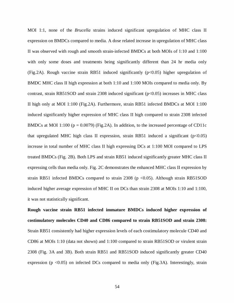
54
MOI 1:1, none of the Brucella strains induced significant upregulation of MHC class II
expression on BMDCs compared to media. A dose related increase in upregulation of MHC class
II was observed with rough and smooth strain-infected BMDCs at both MOIs of 1:10 and 1:100
with only some doses and treatments being significantly different than 24 hr media only
(Fig.2A). Rough vaccine strain RB51 induced significantly (p<0.05) higher upregulation of
BMDC MHC class II high expression at both 1:10 and 1:100 MOIs compared to media only. By
contrast, strain RB51SOD and strain 2308 induced significant (p<0.05) increases in MHC class
II high only at MOI 1:100 (Fig.2A). Furthermore, strain RB51 infected BMDCs at MOI 1:100
induced significantly higher expression of MHC class II high compared to strain 2308 infected
BMDCs at MOI 1:100 (p = 0.0079) (Fig.2A). In addition, to the increased percentage of CD11c
that upregulated MHC high class II expression, strain RB51 induced a significant (p<0.05)
increase in total number of MHC class II high expressing DCs at 1:100 MOI compared to LPS
treated BMDCs (Fig. 2B). Both LPS and strain RB51 induced significantly greater MHC class II
expressing cells than media only. Fig. 2C demonstrates the enhanced MHC class II expression by
strain RB51 infected BMDCs compared to strain 2308 (p <0.05). Although strain RB51SOD
induced higher average expression of MHC II on DCs than strain 2308 at MOIs 1:10 and 1:100,
it was not statistically significant.
Rough vaccine strain RB51 infected immature BMDCs induced higher expression of
costimulatory molecules CD40 and CD86 compared to strain RB51SOD and strain 2308:
Strain RB51 consistently had higher expression levels of each costimulatory molecule CD40 and
CD86 at MOIs 1:10 (data not shown) and 1:100 compared to strain RB51SOD or virulent strain
2308 (Fig. 3A and 3B). Both strain RB51 and RB51SOD induced significantly greater CD40
expression (p <0.05) on infected DCs compared to media only (Fig.3A). Interestingly, strain
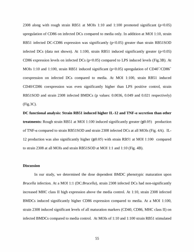
55
2308 along with rough strain RB51 at MOIs 1:10 and 1:100 promoted significant (p<0.05)
upregulation of CD86 on infected DCs compared to media only. In addition at MOI 1:10, strain
RB51 infected DC-CD86 expression was significantly (p<0.05) greater than strain RB51SOD
infected DCs (data not shown). At 1:100, strain RB51 induced significantly greater (p<0.05)
CD86 expression levels on infected DCs (p<0.05) compared to LPS induced levels (Fig.3B). At
MOIs 1:10 and 1:100, strain RB51 induced significant (p<0.05) upregulation of CD40+/CD86+
coexpression on infected DCs compared to media. At MOI 1:100, strain RB51 induced
CD40/CD86 coexpression was even significantly higher than LPS positive control, strain
RB51SOD and strain 2308 infected BMDCs (p values: 0.0036, 0.049 and 0.021 respectively)
(Fig.3C).
DC functional analysis: Strain RB51 induced higher IL-12 and TNF-α secretion than other
treatments: Rough strain RB51 at MOI 1:100 induced significantly greater (p≤0.05) production
of TNF-α compared to strain RB51SOD and strain 2308 infected DCs at all MOIs (Fig. 4A). IL-
12 production was also significantly higher (p≤0.05) with strain RB51 at MOI 1:100 compared
to strain 2308 at all MOIs and strain RB51SOD at MOI 1:1 and 1:10 (Fig. 4B).
Discussion
In our study, we determined the dose dependent BMDC phenotypic maturation upon
Brucella infection. At a MOI 1:1 (DC:Brucella), strain 2308 infected DCs had non-significantly
increased MHC class II high expression above the media control. At 1:10, strain 2308 infected
BMDCs induced significantly higher CD86 expression compared to media. At a MOI 1:100,
strain 2308 induced significant levels of all maturation markers (CD40, CD86, MHC class II) on
infected BMDCs compared to media control. At MOIs of 1:10 and 1:100 strain RB51 stimulated

56
significantly higher expression of MHC class II than strain 2308. At 1:100, strain RB51 infected
BMDCs had greater CD40/CD86 expression than strain 2308 (Figures 2A-C, 3A-3C).
Additionally, strain RB51 stimulated enhanced BMDC function more than strain 2308 as well
based on TNF-alpha and IL-12p70 cytokine production. These data illustrated that strain RB51
enhanced BMDC maturation and function greater than strain 2308.
In comparing our results with others, recent publications show conflicting reports on the
effects of strain 2308 on DC maturation. These differences can at least partially be explained
based on differing cell and Brucella concentration. Billard et al. (Billard et al., 2007a, b) and
Salcedo et al. (Salcedo et al., 2008) reported that smooth strain 2308 inhibited DC maturation
whereas Zwerdling et al. (Zwerdling et al., 2008) and Macedo et al. (Macedo et al., 2008), who
both used higher concentrations of DCs, reported that smooth strain 2308 induced DC
maturation. In our studies, we also found that there was a dose dependent response. Having
shown the differential ability of Brucella rough and smooth strains in DC maturation, the
questions remain as to what are the minimal requirements of DC activation/function needed to
promote CD4 Th1 immunity. Recently it has been shown that DCs can bias the T-cell response
towards a CD4 Th1, Th2 T-reg or Th17 phenotype/population. The requirements by DCs and T-
cells for each specific DC mediated T-cell responses are not fully understood.
With regard to tolerance/anergy, certain types of immature and mature DCs can induce
tolerance (Steinman et al., 2003). Some immature DCs in peripheral tissues expressing low
levels of MHC class II and co-stimulatory molecules can induce T-cell anergy or regulatory T-
cells (Jonuleit et al., 2000). In addition, other murine BMDC studies show that stimulated DCs
can induce tolerance (Akbari et al., 2001). Despite the high expression of MHC class II and
costimulatory markers, these tolerogenic DCs do not produce proinflammatory cytokines,
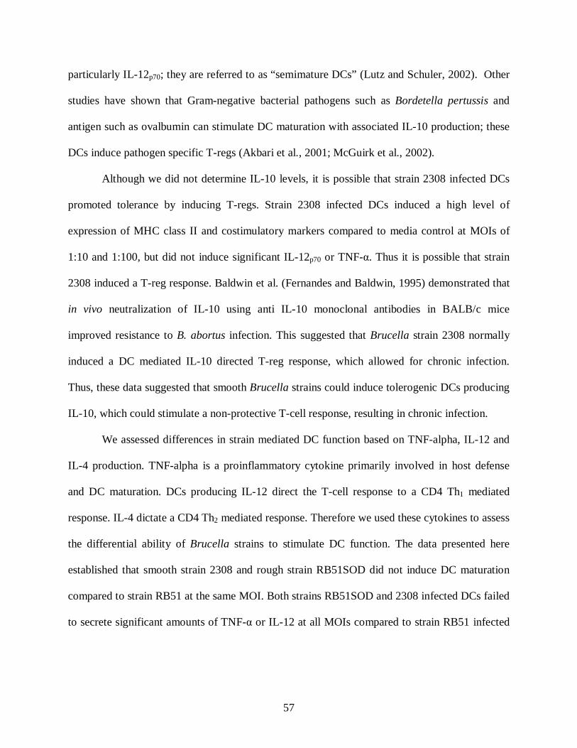
57
particularly IL-12p70; they are referred to as “semimature DCs” (Lutz and Schuler, 2002). Other
studies have shown that Gram-negative bacterial pathogens such as Bordetella pertussis and
antigen such as ovalbumin can stimulate DC maturation with associated IL-10 production; these
DCs induce pathogen specific T-regs (Akbari et al., 2001; McGuirk et al., 2002).
Although we did not determine IL-10 levels, it is possible that strain 2308 infected DCs
promoted tolerance by inducing T-regs. Strain 2308 infected DCs induced a high level of
expression of MHC class II and costimulatory markers compared to media control at MOIs of
1:10 and 1:100, but did not induce significant IL-12p70 or TNF-α. Thus it is possible that strain
2308 induced a T-reg response. Baldwin et al. (Fernandes and Baldwin, 1995) demonstrated that
in vivo neutralization of IL-10 using anti IL-10 monoclonal antibodies in BALB/c mice
improved resistance to B. abortus infection. This suggested that Brucella strain 2308 normally
induced a DC mediated IL-10 directed T-reg response, which allowed for chronic infection.
Thus, these data suggested that smooth Brucella strains could induce tolerogenic DCs producing
IL-10, which could stimulate a non-protective T-cell response, resulting in chronic infection.
We assessed differences in strain mediated DC function based on TNF-alpha, IL-12 and
IL-4 production. TNF-alpha is a proinflammatory cytokine primarily involved in host defense
and DC maturation. DCs producing IL-12 direct the T-cell response to a CD4 Th1 mediated
response. IL-4 dictate a CD4 Th2 mediated response. Therefore we used these cytokines to assess
the differential ability of Brucella strains to stimulate DC function. The data presented here
established that smooth strain 2308 and rough strain RB51SOD did not induce DC maturation
compared to strain RB51 at the same MOI. Both strains RB51SOD and 2308 infected DCs failed
to secrete significant amounts of TNF-α or IL-12 at all MOIs compared to strain RB51 infected
58
BMDCs at MOI 1:100 (Figure 4A and 4B). The lack of TNF-α and IL-12 secretion by strains
RB51SOD and 2308 infected DCs support weaker DC function.
By comparison, studies with strain 2308 infected DCs by Billard et al. (Billard et al.,
2007a) and Salcedo et al. (Salcedo et al., 2008) also corroborated the impaired DC - TNF-α and
IL-12 secretion. Billard et al. (Billard et al., 2007a) additionally determined that Brucella outer
membrane protein Omp25 blocked TNF-α secretion by smooth strain infected DCs. Omp-25 is
expressed on both rough and smooth strains of B. abortus, yet RB51 infected DCs at MOI 1:100
stimulated higher TNF-α secretion. This suggested that there were multiple mechanisms
regulating TNF-alpha production.
Analyzing the IL-12 (IL-12p70) secretion, only strain RB51 at MOI 1:100 induced DCs to
produce substantial levels of IL-12p70 (Figure 4B). For a protective CD4 Th1 and CD8 Tc1 T-cell
response, DC derived IL-12 p70 is required. Thus, these data suggested that if cell-mediated
immunity (CMI) was dictated only by the DC response that only strain RB51 would trigger a
protective CD4 Th1 response compared to strain RB51SOD or strain 2308. This further explains
the better innate immune stimulation by vaccine strain RB51 compared to strain RB51SOD.
As strain RB51SOD and strain 2308 infected BMDCs did not induce significant IL-12 or
TNF-alpha to direct a CD4 Th1 response, IL-4 production was assessed. However, none of the
strains induced IL-4 secretion. These data suggested that smooth vs. rough strains do not induce
an increased DC mediated IL-4 response which would bias towards a CD4 Th2 polarization.
Clearly, these data do support that strain RB51 vs. strains RB51SOD and 2308 have an
increased vs. decreased bias towards a DC mediated CD4 Th1 CD8 Tc1 response. As these
differences for strains RB51SOD and 2308 are not mediated by IL-4 and a CD4 Th2 response,

59
the differences could still be mediated by either IL-10 biasing towards a T-reg or IL-17 biasing
towards a Th17 response.
With this study, for the first time, we evaluated the in vitro innate immune response of
immature murine BMDCs to B. abortus rough vaccine strains RB51 and RB51SOD infection
compared to smooth strain 2308. Understanding the rough vaccine vs. smooth pathogenic strain
induced changes in DC phenotype will help identify the mechanism(s) by which rough Brucella
strains modulate the immune response towards a CD4 Th1 profile. It will also allow us to begin
to define differences in DC function between rough strains (RB51 vs. RB51SOD). This study
demonstrated that rough vaccine strain RB51 induced strong DC maturation and function
compared to strain RB51SOD or pathogenic strain 2308 at corresponding MOIs. Additionally,
there is a dose dependent positive correlation between infection dose and DC phenotypic
maturation following rough or smooth Brucella infection.
There are several possible explanations for these differences between smooth and rough
strains. Some likely explanations include differences in viability/infectivity. Additional
differences in Brucella specific components/factors (i.e. LPS, outermembrane proteins, virulence
factors) could alter DC mediated activation and function.
In order to determine that the differences in DC mediated function were not due to
differences in viability of Brucella and/or BMDCs, DC viability and Brucella numbers were
analyzed. DC viability was analyzed in all experiments at 24 hrs. These data showed that at 4hrs
there were relatively similar levels of Brucella: BMDCs. Data are from 1 of 3 replicates and the
counts denote number of intracellular Brucella per 100 cells. For just the 1:100 MOI: at 1 hr,
Brucella: BMDCs for RB51 were 35,254; RB51SOD 16,000; 2308 4,535. For 4hrs, RB51 6,330;
RB51SOD 8,760; 2308 19,420; At 24 hrs, strain RB51 124; RB51SOD 378; strain 2308 2,125.
60
Therefore, while initial 1 hr time point and other data (Pei and Ficht, 2004) supported that
rough strains were internalized more rapidly than smooth strains, by 4 hrs, which was when
BMDCs were washed with gentamicin, there were much fewer rough vs. smooth strains of
bacteria. At 24 hrs, rough strains were still less than smooth strains. This suggested that although
rough strains may be internalized and could possibly have more bacteria within activated DCs at
1 hour, by 4 hrs and 24 hrs smooth strains remained higher than rough strains in sufficient
numbers to stimulate BMDCs. These data would suggest that even though the number of rough
bacteria was greater earlier in the response, as the smooth bacteria had higher numbers at 24 hrs,
should have been sufficient to stimulate the BMDCs, if the stimulation differences between
strains were regulated only by numbers of bacteria. Thus, the differences are attributable to other
characteristics/components of the rough vs. smooth bacteria. It is possible that rough strains
could stimulate the BMDCs with large numbers of cells early and that smooth strains are
inhibitory and at greater numbers of bacteria at the later time point exert an even more significant
inhibitory response. However, there are still characteristically different properties between
smooth and rough strains which are associated with the differences in BMDC function.
Some of the additional potential differences to be explored include LPS, VirB,
outermembrane protein expression, and Brucella Toll Interleukin 1 receptor family containing
protein (Btp1). Barquero-Calvo et al. (Barquero-Calvo et al., 2009) recently demonstrated that
LPS from smooth Brucella does not bind to TLR signaling molecule MD2 as well as Salmonella
LPS. This supports that LPS has a role in lack of stimulation by smooth strains. Both rough B.
abortus vaccine strains lack the O-side chain of the LPS making them rough. However, by
treating human immature DCs either with rough LPS (100 ng) from B. abortus rough strain
45/20 or with smooth LPS (200 ng) from strain 2308, Billard et al. (Billard et al., 2007b)
61
established that Brucella LPS does not stimulate DC maturation and there are no differences in
DC maturation between rough or smooth Brucella LPS, thus ruling out the possibility for
implicating these strains of rough LPS in enhanced DC activation.
Besides LPS, Type IV secretion system encoded by VirB is a virulence factor involved in
the control of host defense. VirB controls maturation of the Brucella-containing vacuole into a
replication permissive organelle. Unfortunately, recent studies have shown that VirB mutants
similar to wild type Brucella do not control DC maturation (Billard et al., 2008). Thus, this
supports the notion that VirB does not have a significant effect in DC mediated function.
Another component that has been demonstrated to down-modulate DC maturation through TLR2
is Btp1 (Salcedo et al., 2008). Btp1 decreased CD40 and CD80 activation of DCs as well as
decreasing DC mediated IL-6, IFN-beta, TNF alpha, IL-12 production (13). However, it is
unlikely that rough vaccine vs. smooth pathogenic strains differ with respect to Btp1 expression.
Thus Btp1 is unlikely to have a significant contributing role in the differences between rough and
smooth strains.
There is potential that variations in composition or expression of outer membrane
proteins on rough vs. smooth strains which could explain differences in DC function. Zwerdling
et al. (Zwerdling et al., 2008) and Pasquevich et al. (Pasquevich et al., 2009) both have shown
that outer membrane proteins Omp16 and Omp19 are immunostimulatory. Pasquevich et al.
(Pasquevich et al., 2009) demonstrated that both Omp16 and Omp19 in its unlipidated version
stimulates specific CD4+ and CD8+ T-cells to provide systemic and oral protection to B. abortus
infection. Additionally, Gonzalez et al. (Gonzalez et al., 2008) demonstrated that the outer
membrane proteins are more exposed on rough strains than on smooth Brucella due to the
absence of O-polysaccharide of the LPS. These recent findings suggested that the relatively more
62
exposed Omps on the surface of rough vaccine strains RB51 and RB51SOD could stimulate
stronger DC maturation and function compared to smooth strain 2308. Additionally, Zwerdling
et al. (Zwerdling et al., 2008) described lipidated Omp19 upregulating DC maturation.
The above mentioned discussion provides a few plausible reasons to explain the
differences between rough and smooth strains. With regard to differences between strains RB51
and RB51SOD, at this point, we can only speculate as to why strain RB51SOD does not
stimulate as well as strain RB51. We do not expect differences in LPS or omps which could be
present between rough and smooth strains to be present between strains RB51 and RB51SOD.
Therefore, this suggests that there are other possible explanations for differences between strain
RB51 and RB51SOD. Strain RB51SOD overexpresses SOD and this was demonstrated by
western blot analysis. In future studies, the next step would be to determine whether SOD is
functional. As SOD downregulates oxidative damage, it is expected that superoxide (SO) would
be decreased. Theoretically, if SOD decreases SO and oxidative inflammation, the inflammatory
response associated with infection would be decreased. Decreased DC activation and function
could be a consequence. Consistent with this explanation is our preliminary data (not shown,
manuscript in preparation) which demonstrates that mice infected IN with strain RB51SOD vs.
RB51 show markedly decreased inflammatory infiltrate in the lungs. The next step, in future
studies, would be to determine in these experiments whether mice infected with strain RB51SOD
vs. RB51 have decreased SO and cytokine mediators in the lungs. This would also support our
theory. These findings substantiate our data which demonstrates that strain RB51SOD induces
substantially less DC activation and function. Our proposed theory, which will be investigated in
future studies, is that strain RB51SOD overproduces functional SOD which downregulates the
inflammatory response, and subsequently also including DC mediated activation and function.

63
While these data are not consistent with the studies demonstrating that strain RB51SOD
protects better than strain RB51 in mice vaccinated IP and challenged IP with B. abortus strain
2308 (Vemulapalli et al., 2000), Olsen demonstrated that strain RB51SOD when administered
subcutaneously did not protect bison against B. abortus strain 2308 (Olsen et al., 2009). Thus,
although strain RB51SOD provided better protection in mice, it did not provide better protection
in bison. This provides support that strain RB51SOD is not a superior vaccine in all
species/trials.
Additionally, it is still possible that while strain RB51SOD does not upregulate DC
activation and function in a DC mediated CD4 Th1 response, it is still possible that strain
RB51SOD could provide enhanced protection. There are a few possible explanations for these
results. It is possible that SOD is an immunodominant antigen, and despite the limited DC
response, because SOD is immunodominant, there is still a marked CD4 and CD8 Brucella
specific response, which results in enhanced protection vs. strain RB51. Another possibility is
that strain RB51SOD may direct the DC mediated T-cell response towards a Th17 response,
which could still result in enhanced bacterial clearance. These possible explanations for strain
RB51SOD enhanced protection will be investigated in future studies. In this paper, we have
focused on demonstrating that these differences between strain RB51 and RB51SOD do exist.
These data provide information on why in some challenge models, strain RB51SOD may not be
a more protective vaccine. In addition, if one’s goal were to identify a more efficacious vaccine,
then generating a recombinant RB51SOD vaccine that upregulated DC function would be ideal
based on previous mouse model data (Vemulapalli et al., 2000). However, based on the data
presented here, choosing another vaccine or vector other than strain RB51SOD may allow for
64
enhanced protection. Thus these data provide valuable information on the introductory
mechanisms of strain RB51SOD mediated protection.
In conclusion, our study demonstrated that live B. abortus rough vaccine strain RB51
stimulated enhanced murine BMDC maturation responses in vitro compared to rough strain
RB51SOD and virulent smooth strain 2308. The BMDC maturation response upon infection with
rough or smooth B. abortus strains was dose dependent with maximum DC maturation response
to rough strain RB51 at MOIs 1:10 and 1:100. Further experiments are needed to understand the
mechanism of enhanced DC function associated with strain RB51; the causes for reduced DC
maturation by strain RB51SOD, and the limited ability of strain 2308 to induce DC maturation
and function.
Acknowledgements: The authors acknowledge Dr. Stephen Werre for help with the statistical
analysis of the data.
References
Acha, P., Szyfres, B. 2001. Zoonoses and communicable diseases common to man and animal.
(Pan American Health Organization, WHO).
Akbari, O., DeKruyff, R.H., Umetsu, D.T., 2001, Pulmonary dendritic cells producing IL-10
mediate tolerance induced by respiratory exposure to antigen. Nat Immunol 2, 725-731.
Barquero-Calvo, E., Conde-Alvarez, R., Chacon-Diaz, C., Quesada-Lobo, L., Martirosyan, A.,
Guzman-Verri, C., Iriarte, M., Mancek-Keber, M., Jerala, R., Gorvel, J.P., Moriyon, I.,
Moreno, E., Chaves-Olarte, E., 2009, The differential interaction of Brucella and
65
ochrobactrum with innate immunity reveals traits related to the evolution of stealthy
pathogens. PLoS ONE 4, e5893.
Billard, E., Cazevieille, C., Dornand, J., Gross, A., 2005, High susceptibility of human dendritic
cells to invasion by the intracellular pathogens Brucella suis, B. abortus, and B.
melitensis. Infect Immun 73, 8418-8424.
Billard, E., Dornand, J., Gross, A., 2007a, Brucella suis prevents human dendritic cell maturation
and antigen presentation through regulation of tumor necrosis factor alpha secretion.
Infect Immun 75, 4980-4989.
Billard, E., Dornand, J., Gross, A., 2007b, Interaction of Brucella suis and Brucella abortus
rough strains with human dendritic cells. Infect Immun 75, 5916-5923.
Billard, E., Dornand, J., Gross, A., 2008, VirB type IV secretory system does not contribute to
Brucella suis' avoidance of human dendritic cell maturation. FEMS Immunol Med
Microbiol 53, 404-412.
Bosio, C.M., Dow, S.W., 2005, Francisella tularensis induces aberrant activation of pulmonary
dendritic cells. J Immunol 175, 6792-6801.
Brzoza, K.L., Rockel, A.B., Hiltbold, E.M., 2004, Cytoplasmic entry of Listeria monocytogenes
enhances dendritic cell maturation and T cell differentiation and function. J Immunol
173, 2641-2651.
Celli, J., 2006, Surviving inside a macrophage: the many ways of Brucella. Res Microbiol 157,
93-98.
Corbel, M.J. 2006. Brucellosis in humans and animals (World Health Organization).
Fernandes, D.M., Baldwin, C.L., 1995, Interleukin-10 downregulates protective immunity to
Brucella abortus. Infect Immun 63, 1130-1133.

66
Gonzalez, D., Grillo, M.J., De Miguel, M.J., Ali, T., Arce-Gorvel, V., Delrue, R.M., Conde-
Alvarez, R., Munoz, P., Lopez-Goni, I., Iriarte, M., Marin, C.M., Weintraub, A.,
Widmalm, G., Zygmunt, M., Letesson, J.J., Gorvel, J.P., Blasco, J.M., Moriyon, I., 2008,
Brucellosis vaccines: assessment of Brucella melitensis lipopolysaccharide rough mutants
defective in core and O-polysaccharide synthesis and export. PLoS ONE 3, e2760.
Inaba, K., Inaba, M., Romani, N., Aya, H., Deguchi, M., Ikehara, S., Muramatsu, S., Steinman,
R.M., 1992, Generation of large numbers of dendritic cells from mouse bone marrow
cultures supplemented with granulocyte/macrophage colony-stimulating factor. J Exp
Med 176, 1693-1702.
Jonuleit, H., Schmitt, E., Schuler, G., Knop, J., Enk, A.H., 2000, Induction of interleukin 10-
producing, nonproliferating CD4(+) T cells with regulatory properties by repetitive
stimulation with allogeneic immature human dendritic cells. J Exp Med 192, 1213-1222.
Lutz, M.B., Schuler, G., 2002, Immature, semi-mature and fully mature dendritic cells: which
signals induce tolerance or immunity? Trends Immunol 23, 445-449.
Macedo, G.C., Magnani, D.M., Carvalho, N.B., Bruna-Romero, O., Gazzinelli, R.T., Oliveira,
S.C., 2008, Central role of MyD88-dependent dendritic cell maturation and
proinflammatory cytokine production to control Brucella abortus infection. J Immunol
180, 1080-1087.
McGuirk, P., McCann, C., Mills, K.H., 2002, Pathogen-specific T regulatory 1 cells induced in
the respiratory tract by a bacterial molecule that stimulates interleukin 10 production by
dendritic cells: a novel strategy for evasion of protective T helper type 1 responses by
Bordetella pertussis. J Exp Med 195, 221-231.

67
Olsen, S.C., Boyle, S.M., Schurig, G.G., Sriranganathan, N.N., 2009, Immune responses and
protection against experimental challenge after vaccination of bison with Brucella
abortus strain RB51 or RB51 overexpressing superoxide dismutase and
glycosyltransferase genes. Clin Vaccine Immunol 16, 535-540.
Pappas, G., Panagopoulou, P., Christou, L., Akritidis, N., 2006, Brucella as a biological weapon.
Cell Mol Life Sci 63, 2229-2236.
Pasquevich, K.A., Estein, S.M., Samartino, C.G., Zwerdling, A., Coria, L.M., Barrionuevo, P.,
Fossati, C.A., Giambartolomei, G.H., Cassataro, J., 2009, Immunization with
recombinant Brucella species outer membrane protein Omp16 or Omp19 in adjuvant
induces specific CD4+ and CD8+ T cells as well as systemic and oral protection against
Brucella abortus infection. Infect Immun 77, 436-445.
Pei, J., Ficht, T.A., 2004, Brucella abortus rough mutants are cytopathic for macrophages in
culture. Infect Immun 72, 440-450.
Salcedo, S.P., Marchesini, M.I., Lelouard, H., Fugier, E., Jolly, G., Balor, S., Muller, A.,
Lapaque, N., Demaria, O., Alexopoulou, L., Comerci, D.J., Ugalde, R.A., Pierre, P.,
Gorvel, J.P., 2008, Brucella control of dendritic cell maturation is dependent on the TIR-
containing protein Btp1. PLoS Pathog 4, e21.
Schurig, G.G., Roop, R.M., 2nd, Bagchi, T., Boyle, S., Buhrman, D., Sriranganathan, N., 1991,
Biological properties of RB51; a stable rough strain of Brucella abortus. Vet Microbiol
28, 171-188.
Steinman, R.M., Hawiger, D., Nussenzweig, M.C., 2003, Tolerogenic dendritic cells. Annu Rev
Immunol 21, 685-711.
68
Svensson, M., Johansson, C., Wick, M.J., 2000, Salmonella enterica serovar typhimurium-
induced maturation of bone marrow-derived dendritic cells. Infect Immun 68, 6311-6320.
Vemulapalli, R., He, Y., Cravero, S., Sriranganathan, N., Boyle, S.M., Schurig, G.G., 2000,
Overexpression of protective antigen as a novel approach to enhance vaccine efficacy of
Brucella abortus strain RB51. Infect Immun 68, 3286-3289.
Vemulapalli, R., McQuiston, J.R., Schurig, G.G., Sriranganathan, N., Halling, S.M., Boyle,
S.M., 1999, Identification of an IS711 element interrupting the wboA gene of Brucella
abortus vaccine strain RB51 and a PCR assay to distinguish strain RB51 from other
Brucella species and strains. Clin Diagn Lab Immunol 6, 760-764.
Zwerdling, A., Delpino, M.V., Barrionuevo, P., Cassataro, J., Pasquevich, K.A., Garcia
Samartino, C., Fossati, C.A., Giambartolomei, G.H., 2008, Brucella lipoproteins mimic
dendritic cell maturation induced by Brucella abortus. Microbes Infect 10, 1346-1354.
Figure legends
Figure 1: Bone marrow cells after 6 days of culture are predominantly CD11c+ immature
dendritic cells. Bone marrow cells isolated from BALB/c mice were cultured for 6 days in 10%
RPMI medium with rGM-CSF (20ng/ml). The cells were harvested on day 6 and analyzed by
flow cytometry for CD11c+ marker expression using fluorescent labeled CD11c+ antibody. Gated
CD11c+ cells were further analyzed for their maturation status using anti MHC class II, CD40
and CD86 antibodies. A: Comparison between percentages of CD11c+ expressed by bone
marrow cells harvested on day 6 to those treated with B. abortus rough and smooth strains for 24
hours at different MOIs (DC:Brucella; 1:1, 1:10, 1:100). Day 6 DCs treated with E.coli LPS
(100ng/ml) or media alone for 24 hours served as positive and negative controls respectively. B:
As an internal control to assess conditions: Comparison of the percentages of CD11c+ DCs
69
expressing the surface maturation markers MHC class II, CD40 and CD86 harvested on day 6 to
those treated for 24 hours with negative control media alone or positive control E. coli LPS (100
ng/ml). Two asterisks (**) denotes statistically significant data at p ≤ 0.05 compared to data
represented by an asterisk (*). For A and B, data represents means ± standard deviations of 3
independent experiments.
Figure 2: B. abortus Rough vaccine strain RB51 significantly upregulates MHC class II
expression on immature BMDCs. Immature BMDCs were infected with either B. abortus
rough vaccine strains (RB51 or RB51SOD) or with smooth strain 2308 at MOIs (DC:Brucella)
1:1, 1:10 and 1:100. At 24 hours post infection (p.i.), BMDCs were analyzed for MHC class II
expression. A: Comparison of the percentage of CD11c+ cells (BMDCs) expressing MHC class
II high on its surface across different treatment groups. Media and E. coli LPS were the negative
and positive controls respectively. Double characters **, ## represent statistically significant
difference at p ≤ 0.05 with the corresponding single character representation (*, #). Data
represents means ± standard deviations of 3 independent experiments. B: Histogram of a single
representative experiment (of 3) showing total MHC class II expression by RB51 infected
BMDCs (MOI 1:100) in comparison to media and E.coli LPS controls. Filled grey histogram:
media control; continuous thick line: E.coli LPS; broken thick line: strain RB51. C: Histogram of
the same single representative experiment (of total 3) showing total MHC II expression by
strains RB51 (broken thick line), RB51SOD (dotted line) or 2308 (continuous thin line) infected
BMDCs at MOI 1:100. Double characters (**, ##) denotes statistically significant data at p ≤
0.05 compared to data represented by single character (*, #).
Figure 3: B. abortus rough vaccine strain RB51 significantly upregulates costimulatory
marker expression on immature BMDCs. Immature BMDCs were infected with either B.
70
abortus rough vaccine strains RB51 or RB51SOD or smooth strain 2308 at MOIs (DC:Brucella)
1:1, 1:10 and 1:100 at 24 hours post-infection and analyzed for costimulatory marker CD40 and
CD86 expression. A: Histogram of a single representative experiment (of 3) showing the CD40
expression on CD11c+ BMDCs infected by strains RB51, RB51SOD and 2308 at MOI 1:100 in
comparison to media and LPS controls. . For A, filled grey histogram: media control; continuous
thick line: E.coli LPS; broken thick line: strain RB51; dotted line: strain RB51SOD; and
continuous thin line: strain 2308. Two asterisks (**) denotes statistically significant difference at
p ≤ 0.05 compared to data represented by an asterisk (*) B: Histogram of same single
representative experiment (of 3) showing the CD86 expression on CD11c+ BMDCs infected by
strains RB51, RB51SOD or 2308 at MOI 1:100 in comparison to media and LPS controls. For B,
filled grey histogram: media control; continuous thick line: E.coli LPS; broken thick line: strain
RB51; dotted line: strain RB51SOD; and continuous thin line: strain 2308. Two characters (**,
##) denotes statistically significant data at p ≤ 0.05 compared to data represented by single
character (*, #) C: Comparison of the percentage of CD40+/CD86+ coexpression on CD11c+
cells (BMDCs) across different treatment groups. Media and E. coli LPS are the negative and
positive controls respectively. Double characters ** and ## represent statistically significant
change at p ≤ 0.05 with the corresponding single character representation (*, #). Data represents
means ± standard deviations of 3 independent experiments.
Figure 4: B. abortus rough vaccine strain RB51 induces higher TNF-α and IL-12 secretion.
To assess DC function, TNF-α (A) and IL-12p70 (B) levels in 24 hour culture supernatants of B.
abortus strain RB51, RB51SOD or 2308 infected BMDCs at various MOIs were analyzed using
indirect sandwich ELISA. E. coli LPS and media treated cell supernatants served as the positive
and negative controls respectively. The limits of detection for both the cytokines were 15pg/ml.

71
The TNF-α results represent means ± SEM of 3 independent experiments. The IL-12p70 results
represent medians and ranges of 3 independent experiments. Two asterisks (**) denotes
statistically significant difference at p ≤ 0.05 compared to data represented by an asterisk (*).
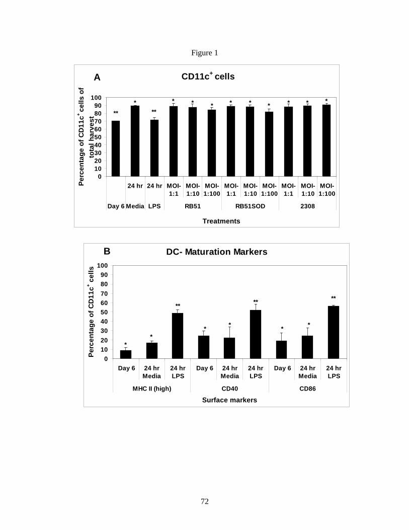
72
Figure 1
DC- Maturation Markers
0102030405060708090
100
Day 6 24 hrMedia
24 hrLPS
Day 6 24 hrMedia
24 hrLPS
Day 6 24 hrMedia
24 hrLPS
MHC II (high) CD40 CD86
Surface markers
Perc
enta
ge o
f CD
11c+ c
ells
**
**
* *
**
* *
**
B
CD11c+ cells
0102030405060708090
100
24 hr 24 hr MOI-1:1
MOI-1:10
MOI-1:100
MOI-1:1
MOI-1:10
MOI-1:100
MOI-1:1
MOI-1:10
MOI-1:100
Day 6 Media LPS RB51 RB51SOD 2308
Treatments
Perc
enta
ge o
f CD
11c+
cells
of
tota
l har
vest
****
A
* * * * * * * * * *
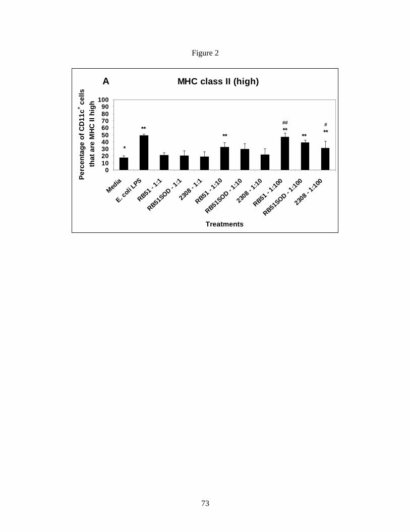
73
Figure 2
A MHC class II (high)
0102030405060708090
100
Media
E. coli L
PS
RB51 - 1:1
RB51SOD - 1:1
2308 -
1:1
RB51 - 1:10
RB51SOD - 1:10
2308 -
1:10
RB51 - 1:100
RB51SOD - 1:100
2308 -
1:100
Treatments
Perc
enta
ge o
f CD
11c+ c
ells
th
at a
re M
HC
II h
igh
*
**
##**
#****
**
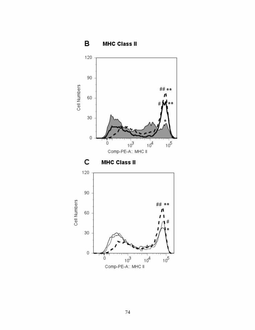
74

75
Figure 3

76
C CD40/CD86
0102030405060708090
100
Media
E. coli L
PS
RB51 - 1
:1
RB51SOD - 1
:1
2308
- 1:1
RB51 - 1
:10
RB51SOD - 1
:10
2308
- 1:10
RB51 - 1
:100
RB51SOD - 1
:100
2308
- 1:10
0
Treatments
Perc
enta
ge o
f CD
11c+ c
ells
th
at a
re C
D40
+ /CD
86+
*
**
**** **# #
##
**#
77
Figure 4
A TNF - α
0
300
600
900
1200
1500
Media
E. coli
LPS
RB 51 1:
1
RB 51 1:
10
RB 51 1:
100
RB51SOD 1:
1
RB51SOD 1:
10
RB51SOD 1:
100
2308
1:1
2308
1:10
2308
1:10
0
Treatments
Con
cent
ratio
n (p
g/m
l)
**
* * * * * * * * * *
B IL-12p70
0
300
600
900
1200
1500
Media
E. coli
LPS
RB 51 1:
1
RB 51 1:
10
RB 51 1:
100
RB51SOD 1:
1
RB51SOD 1:
10
RB51SOD 1:
100
2308
1:1
2308
1:10
2308
1:10
0
Treatments
Con
cent
ratio
n (p
g/m
l)
**
** ** * *

78
Chapter 3
Heat killed and gamma-irradiated Brucella strain RB51 stimulate enhanced dendritic cell
activation but not functioncompared to virulent smooth strain 2308
Running title: Heat killed and irradiated strain RB51stimulate BMDCs.
Naveen Surendrana, Elizabeth M. Hiltboldc, Bettina Heida, Nammalwar Sriranganathanb, Stephen
M. Boyleb, Kurt L. Zimmermanb, Sharon G. Witonskya*.
(Submitted to FEMS immunology and Medical Microbiology)
Keywords: BRUCELLA ABORTUS; DENDRITIC CELL; INNATE IMMUNITY
Abstract:
Brucella spp. are Gram-negative, facultative intracellular bacterial pathogens that cause
abortion in livestock and undulant fever in humans worldwide. B. abortus strain 2308 is a
pathogenic strain that affects cattle and humans. Currently, there are no efficacious human
vaccines available. However, B. abortus strain RB51 is a live attenuated rough vaccine against
bovine brucellosis which is approved by the USDA. Live strain RB51 induces protection via
CD4+ and CD8+ T- cell mediated immunity. To generate an optimal T-cell response, strong
innate immune responses by dendritic cells (DCs) are crucial. Because of safety concerns, using
live vaccine strain RB51 in humans is limited. Therefore in this study, we analyzed the
differential ability of same doses of live, heat-killed (HK) and gamma-irradiated (IR) strain
RB51 in inducing DC activation and function. Smooth strain 2308, live strain RB51 and LPS
were used as controls. Studies using mouse bone marrow derived DCs revealed that, irrespective
of viability, strain RB51 induced higher DC activation than smooth strain 2308. Live strain
79
RB51 induced significantly (p≤0.05) higher DC maturation than HK and IR strains, and only
live strain RB51 infected DCs (at MOI 1:100) induced significant (p≤0.05) TNF -α and IL-12
secretion.
1. Introduction
B. abortus is a Gram-negative, facultative intracellular bacterium that causes abortion in
cattle and undulant fever in humans (Corbel, 2006). Brucellosis, the disease caused by Brucella
spp., is one of the five most prevalent human bacterial zoonoses in the world with more than half
a million human cases reported annually (Pappas et al., 2006). Brucella species are easy to
aerosolize and can be genetically modified to create antibiotic resistant strains. Therefore, they
are ideal agents for development as bioterror weapons (Pappas et al., 2006). Consequently, the
Center for Disease Control and Prevention (CDC) categorizes them as Class B pathogens. There
are no human vaccines available to date. If untreated this disease is devastating in humans and
animals.
B. abortus strain 2308 is a phenotypically smooth strain possessing a surface exposed O-
side chain of lipopolysaccharide (LPS); this is an immunodominant antigen referred to as O-
antigen (Schurig et al., 1991). As with most intracellular bacterial infections, protection against
Brucella involves both a CD4+ T-helper-1 (Th1) and CD8+ cytotoxic T-cell-1 (Tc1) response (He
et al., 2001). B. abortus strain RB51 is a live attenuated stable rough phenotypic mutant derived
from virulent strain 2308. Strain RB51 lacks the O-side chain in its LPS (Schurig et al., 1991).
Live vaccine strain RB51 protects animals by inducing a cell mediated CD4 Th1 and CD8+ Tc1
gamma interferon (IFN-γ) response (He et al., 2001). Despite the knowledge that strain RB51
stimulates protective cell mediated immunity (CMI), there is limited information regarding how

80
B. abortus strains induce innate immune responses which result in protective CMI. To develop a
human vaccine, additional knowledge is needed on how strain RB51 stimulates the innate
response.
Dendritic cells (DCs) are the sentinel cells of the innate immune system and their
interaction with naïve T-cells following antigen capture determines the specificity and
polarization of T-cell mediated immunity (Banchereau and Steinman, 1998). In addition, DCs
are highly susceptible to Brucella infection making them a valuable model for assessing Brucella
mediated immune responses (Billard et al., 2005). In our previous study (Surendran et al., under
review), we demonstrated that rough strain RB51 induced significantly higher DC maturation
and function compared to smooth virulent strain 2308. This enhanced DC activation and function
caused by live vaccine strain RB51 could be the critical initial defining point for a successful T-
cell mediated adaptive immune response. Because safety concerns of live vaccines limit their use
in people, the efficacy of safer heat killed (HK) or irradiated (IR) vaccines should be considered
(Plotkin, 2005).
HK B. abortus is an established CD4 Th1 promoting stimulus. It stimulates cytotoxic
CD8 T-lymphocytes even in the absence of CD4 T-cell help (Finkelman et al., 1988; Street et al.,
1990). By comparison, IR strain RB51 induced CD4 Th1 type responses and if used at one log
higher dose than live strain RB51, it protected against virulent B. abortus challenge in a mouse
model (Sanakkayala et al., 2005). With this study, we wanted to determine whether HK and IR
strain RB51 stimulated comparable innate responses to live vaccine strain RB51 for exploring
their use as a vaccine in people and animals. To assess innate immunity we examined the ability
of HK and IR B. abortus rough strain RB51 and smooth strain 2308 to stimulate murine bone
marrow derived DC (BMDC) activation and function based on cell surface expression of
81
costimulatory molecule and cytokine production. This study assessed simultaneously, for the
first time, the differential ability of live, HK and IR rough and smooth strains of B. abortus at
same doses to stimulate DC activation and function.
2. Materials and Methods
2. 1. Mice: Female 6-8 weeks old BALB/c mice were obtained from Charles River Laboratories
Inc., Wilmington, MA. Mice were used under animal care protocols approved by Institutional
Animal Care and Use Committee at Virginia Tech.
2. 2. Dendritic cell preparation: Bone marrow-derived DCs (BMDCs) were generated, as
previously described (Inaba et al., 1992). Briefly, tibias and fibulas of 7-8 week old BALB/c
mice were incised and bone marrow (BM) cells removed. Following red blood cell lysis and
filtration, the cells were resuspended and plated in RPMI 1640 complete media with 10% non
heat-inactivated fetal bovine serum and 20ng/ml rGM-CSF (Invitrogen, Carlsbad, CA). The cells
were incubated at 37°C in 5% CO2. Fresh media containing rGM-CSF was added at days 2, 4
and 5 and harvested on day 6. The cells harvested on day 6 were typically 70% CD11c+ and
displayed low levels of MHC class II, CD40 and CD86 expression, consistent with immature
DCs. Flow cytometry was performed to confirm DC activation status (Inaba et al., 1992).
2. 3. Brucella strains: Stock cultures of live attenuated rough Brucella abortus vaccine strain
RB51 and virulent smooth strain 2308 from our culture collection (Schurig et al., 1991;
Vemulapalli et al., 2000) were stored at -80°C. An aliquot each of strain RB51 and strain 2308
were subjected to gamma irradiation using a 60Co source irradiator with a radiation output of
2,200 Rads/minute (Model 109-68R by J.L. Shepherd and associates, San Fernando, CA) for 3
hours (396 kilorads of gamma radiation). Another aliquot of strain RB51 and strain 2308 were
82
subjected to heat killing by incubating in an 80°C water bath for 60 minutes. Irradiated and heat
killed bacterial preparations were confirmed to be nonviable by plating aliquots on TSA plates
and confirming lack of growth following 4 days of incubation. All experiments with Brucella
were performed in our CDC approved (C2003 1120-0016) Biosafety Level (BSL)-3 facility.
2. 4. Infection experiments: On day 6, DCs were harvested and plated at 5 X 10⁵ cells/well in
24 well plates and stimulated with live, IR or HK strain RB51 or strain 2308 at 1:10
(DC:Brucella) or 1:100 CFUs/well (i.e. 5 X 10⁶ or 5 X 10⁷ CFU equivalents/well of irradiated or
heat killed B. abortus). Stimulation was enhanced by a short spin at 1300 rpm (400 x g) for 5
minutes at room temperature. The stimulated cells were incubated for 4 hours at 37°C in 5%
CO2. Then cells were washed with media containing gentamicin (Sigma, St. Louis, MO)
30μg/ml. The stimulated cells were incubated for an additional 20 hours in complete media with
10ng/ml rGM-CSF and 30μg/ml gentamicin. Control samples were maintained by incubating
cells with media (negative control) or Escherichia coli LPS 0111:B4 (Sigma) (positive control)
(100ng/ml) following the same protocol (Sanakkayala et al., 2005).
Viability and infection controls: To quantitate and assess viability, at each time point and with
each treatment, Trypan blue was used to differentiate viable and dead cells. Total live and dead
BMDC numbers were determined.
2. 5. Staining and flow cytometry: The cells were harvested 24 hours following infection, and
they were stained with the following monoclonal antibodies at 0.1 - 0.2 μg per million cells for
FACS analysis: PE-Texas red conjugated anti-CD11c, Biotin-conjugated anti-CD40,
Streptavidin Tri-color conjugate, PE-conjugated anti-CD86 were all acquired from Caltag
(Invitrogen), and PE-conjugated anti I-A/I-E, acquired from BD Pharmingen, San Jose, CA.
Cells were washed and analyzed by BD FACSAria™ flow cytometer (Sanakkayala et al., 2005).
83
2. 6. Cytokine analysis: For cytokine measurement, culture supernatants from Brucella infected
BMDCs were collected after 24 hours of incubation and stored at -80°C. TNF-α, IL-12p70 and IL-
4 cytokine levels were subsequently measured using indirect sandwich ELISAs (BD
Pharmingen) (Sanakkayala et al., 2005).
2. 7. Statistical analysis: As the data had a Gaussian distribution, the effect of treatment on
expression of various DC maturation and activation markers was tested using a mixed model
ANOVA with treatment as a fixed effect and day as a blocking factor (Tukey procedure for
multiple comparisons). After a logarithmic (to base e) transformation, TNF-α data was also
analyzed using the above mentioned procedure. For IL-12p70, the treatments were compared
using the exact Kruskal-Wallis test. The main p-value for this test which applies to the overall
dataset for the effect of variable treatments (including samples from all different MOIs per
treatment) was > 0.05 (0.0889). By this method, as different MOIs are analyzed together, there is
no consideration if only certain MOIs potentially have significant effect. As the pattern of IL-
12p70 secretion between different treatments was similar to TNF-α we used the Dunn’s procedure
for two-way comparisons as a post hoc test. Significance was set at p ≤ 0.05. All analyses were
performed using the SAS system (Cary, NC).
3. Results
3. 1. Day 6 harvested BMDCs show an immature phenotype: CD11c+ expression on the
harvested cells was determined to calculate the yield and percentage of BMDCs following 6 days
of culture. BM cells were gated based on size and granularity and almost 70% of the total gated
cells expressed CD11c+ on day 6. CD11c+ BMDCs expressed an immature phenotype based on
surface expression of characteristic maturation markers MHC class II, CD40 and CD86 (Fig.

84
1A). Following 24 hour incubation with different treatments, the percentage of CD11c+ cells
within the DC gate increased to 81-90% of total gated cells (p<0.05) except for LPS treatment
(71.65±2.74%). In addition, >99% of all such CD11c+ cells were positive for expression of
CD11b characteristic of myeloid origin of DCs (data not shown). Following LPS overnight
treatment, BMDCs treated had a mature BMDC phenotype based on MHC class II high, CD40
and CD86 expression (p<0.05).
3. 2. HK or IR B. abortus rough vaccine strain RB51 induces greater DC maturation than
smooth strain 2308: To evaluate how HK or IR Brucella affected DC maturation, immature
BMDCs were stimulated with either HK or IR rough vaccine strain RB51 or smooth pathogenic
strain 2308 at 1:10 (DC: Brucella) or 1:100 CFU equivalents. Additional controls included media
only and LPS treated BMDCs as well as live strain RB51 and 2308-infected (at MOI 1:10 or
1:100) BMDCs. Immature BMDCs treated overnight with media alone retained their immature
phenotype with reduced surface expression of MHC class II and CD40, CD86 costimulatory
markers compared to LPS (Fig. 1A).
3. 3. BMDC – MHC class II high expression: Immature BMDCs stimulated with HK strain
RB51 (HKRB51) at both 1:10 (p=0.0542) (not shown) and 1:100 (p=0.0018) CFU equivalents
showed significant up-regulation of MHC class II high expression compared to media control
(Fig. 1B). In addition, at corresponding doses of 1:10 and 1:100 HKRB51 had higher mean (not
statistically significant) MHC class II high expression than HK strain 2308 (HK2308) stimulated
BMDCs (Fig. 1B). Furthermore, both HKRB51 and HK2308 stimulated DCs showed a non-
significant dose related increase in MHC class II high expression at 1:100 compared to 1:10.
However, live strain RB51 infected BMDCs had greater MHC class II high expression than
HKRB51 (not significant) and HK2308 (p≤0.05) at corresponding doses (Fig. 1B).

85
Irradiated (IR) strain RB51 (IRRB51) induced a relatively higher but not significant
MHC class II high expression than IR strain 2308 (IR2308) stimulated BMDCs at corresponding
doses. At 1:100, IRRB51 induced significantly (p≤0.05) higher MHC class II high expression
than media (Fig. 1B). Moreover, IRRB51 induced mean DC-MHC class II high expression level
was lower (not significant) than that induced by HKRB51 at respective doses (Fig. 1B). At both
MOIs, live strain RB51 induced higher MHC class II high expression on BMDCs compared to
IRRB51 with significant differences (p≤0.05) at MOI 1:100 (Fig. 1B). Live strain RB51 at 1:100
also induced significantly higher (p<0.05) MHC class II high expression than live strain 2308 at
same dose (Fig 1B).
3. 4. BMDC – Costimulatory marker expression: The expression levels of costimulatory
molecules CD40 and CD86 (independent and co-expression) were also analyzed to assess the
effect of live vs. HK or IR Brucella on DC maturation. Fig. 1C shows CD40 expression on live,
HK and IR Brucella infected BMDCs. Only live, but not HK or IR, strain RB51 infected
BMDCs at MOI 1:100 induced significantly higher CD40 expression than media control
(p≤0.05). By comparison, HKRB51 infected BMDCs had a dose related higher mean CD40
expression compared to HK2308 infected BMDCs at corresponding doses but it was not
statistically significant. At 1:100, HKRB51 induced DC - CD40 expression reached closer to
significance (p ≤=0.06) compared to media control.
In comparing CD40 and CD86 expression, results were similar. At 1:100, HKRB51 and
IRRB51induced greater CD86 expression (p≤0.05) vs. media only (Fig. 1D). HKRB51 induced
non significantly higher DC - CD86 expression than HK2308 at both doses respectively. By
contrast, at both 1:10 and 1:100 both live Brucella strains (RB51 and 2308) induced significantly
(p≤0.05) higher CD86 expression on infected DCs compared to media. In addition, live strain
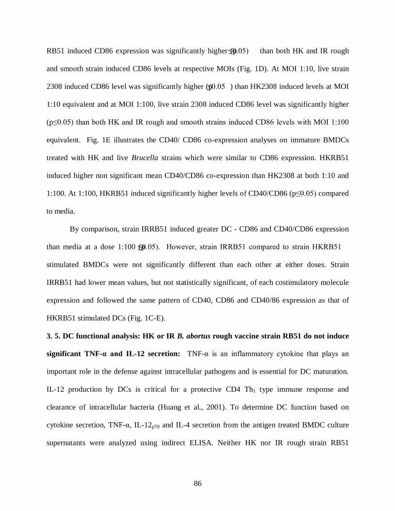
86
RB51 induced CD86 expression was significantly higher (p≤0.05) than both HK and IR rough
and smooth strain induced CD86 levels at respective MOIs (Fig. 1D). At MOI 1:10, live strain
2308 induced CD86 level was significantly higher (p≤0.05 ) than HK2308 induced levels at MOI
1:10 equivalent and at MOI 1:100, live strain 2308 induced CD86 level was significantly higher
(p≤0.05) than both HK and IR rough and smooth strains induced CD86 levels with MOI 1:100
equivalent. Fig. 1E illustrates the CD40/ CD86 co-expression analyses on immature BMDCs
treated with HK and live Brucella strains which were similar to CD86 expression. HKRB51
induced higher non significant mean CD40/CD86 co-expression than HK2308 at both 1:10 and
1:100. At 1:100, HKRB51 induced significantly higher levels of CD40/CD86 (p≤0.05) compared
to media.
By comparison, strain IRRB51 induced greater DC - CD86 and CD40/CD86 expression
than media at a dose 1:100 (p≤0.05). However, strain IRRB51 compared to strain HKRB51
stimulated BMDCs were not significantly different than each other at either doses. Strain
IRRB51 had lower mean values, but not statistically significant, of each costimulatory molecule
expression and followed the same pattern of CD40, CD86 and CD40/86 expression as that of
HKRB51 stimulated DCs (Fig. 1C-E).
3. 5. DC functional analysis: HK or IR B. abortus rough vaccine strain RB51 do not induce
significant TNF-α and IL-12 secretion: TNF-α is an inflammatory cytokine that plays an
important role in the defense against intracellular pathogens and is essential for DC maturation.
IL-12 production by DCs is critical for a protective CD4 Th1 type immune response and
clearance of intracellular bacteria (Huang et al., 2001). To determine DC function based on
cytokine secretion, TNF-α, IL-12p70 and IL-4 secretion from the antigen treated BMDC culture
supernatants were analyzed using indirect ELISA. Neither HK nor IR rough strain RB51

87
produced significant amounts of TNF-α or IL-12 at both doses compared to media control (Fig.
2A & 2B). Only live strain RB51 at a MOI 1:100 induced BMDCs to secrete a significantly
higher amount of both TNF-α and IL-12 (p≤0.05). Irrespective of viability or dose, strain 2308
did not induce significant levels of TNF-α or IL-12 from infected BMDCs. (Fig. 2A & 2B).
None of the strains induced detectable levels of IL-4 cytokine (data not shown).
4. Discussion
We have recently submitted another manuscript (Surendran et al., Veterinary
Microbiology, 2010; accepted) in which we determined that vaccine strain RB51 upregulated DC
activation and function using our in vitro BMDC model. In that study, we determined that the
differences in DC mediated function were not due to differences in viability of Brucella and/or
BMDCs.DC viability and Brucella numbers were analyzed at 1, 4 and 24 hrs. These data
showed that at 4 hrs there were relatively similar levels of Brucella: BMDCs. Data were from 1
of 3 replicates and the counts denoted number of intracellular Brucella per 100 cells. For the
1:100 MOI: at 1 hr, Brucella: BMDCs for strain RB51 were 35,254 and strain 2308, 4,535. For 4
hrs, Brucella: BMDCs for strain RB51 was 6,330 and strain 2308, 19,420; At 24 hrs, Brucella:
BMDCs for strain RB51 was 124; strain 2308, 2,125. These data substantiated that our model
permitted both rough and smooth Brucella strains to infect and stimulate BMDCs. Thus,
increased activation associated with increased numbers of rough strains appeared unlikely. The
results reflected effects of strain differences on BMDC function.
Collectively, both data from the other submitted manuscript and these data presented here
showed that regardless of viability, rough vaccine strain RB51 induced enhanced DC maturation
compared to smooth virulent strain 2308. Additionally, live strain RB51 induced DC maturation
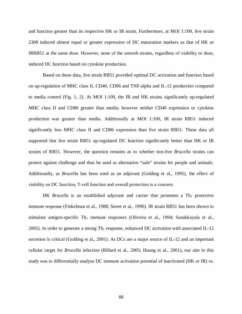
88
and function greater than its respective HK or IR strain. Furthermore, at MOI 1:100, live strain
2308 induced almost equal or greater expression of DC maturation markers as that of HK or
IRRB51 at the same dose. However, none of the smooth strains, regardless of viability or dose,
induced DC function based on cytokine production.
Based on these data, live strain RB51 provided optimal DC activation and function based
on up-regulation of MHC class II, CD40, CD86 and TNF-alpha and IL-12 production compared
to media control (Fig. 1, 2). At MOI 1:100, the IR and HK strains significantly up-regulated
MHC class II and CD86 greater than media; however neither CD40 expression or cytokine
production was greater than media. Additionally at MOI 1:100, IR strain RB51 induced
significantly less MHC class II and CD86 expression than live strain RB51. These data all
supported that live strain RB51 up-regulated DC function significantly better than HK or IR
strains of RB51. However, the question remains as to whether non-live Brucella strains can
protect against challenge and thus be used as alternative “safe” strains for people and animals.
Additionally, as Brucella has been used as an adjuvant (Golding et al., 1995), the effect of
viability on DC function, T-cell function and overall protection is a concern.
HK Brucella is an established adjuvant and carrier that promotes a Th1 protective
immune response (Finkelman et al., 1988; Street et al., 1990). IR strain RB51 has been shown to
stimulate antigen-specific Th1 immune responses (Oliveira et al., 1994; Sanakkayala et al.,
2005). In order to generate a strong Th1 response, enhanced DC activation with associated IL-12
secretion is critical (Golding et al., 2001). As DCs are a major source of IL-12 and an important
cellular target for Brucella infection (Billard et al., 2005; Huang et al., 2001), our aim in this
study was to differentially analyze DC immune activation potential of inactivated (HK or IR) vs.

89
live vaccine and pathogenic strains of B. abortus using the in vitro murine BMDC model. This
would provide additional information on the potential of IR or HK vaccines for human use.
Based on our data, which demonstrated that while HK and IR strain RB51 induced
upregulation of costimulatory molecules but not TNF-alpha or IL-12 production, the question
remains as to whether live vs. HK or IR strains can also upregulate T-cell function and ultimately
protect against challenge. In comparing Brucella, with other live strains of intracellular
organisms such as Listeria monocytogenes (Muraille et al., 2005) and Chlamydia trachomatis
(Rey-Ladino et al., 2005), live strains induced higher levels of DC maturation compared to their
HK or UV-IR forms respectively. Muraille et al. (Muraille et al., 2005) and Takemori et al.
(Tsunetsugu-Yokota et al., 2002) showed that the T-lymphocytes primed by HK Listeria or
Mycobacterium pulsed DCs did not fully differentiate and that only infection with live organisms
induced long term CD8+ T-cell mediated immunity. Additionally, only live Listeria and Bacillus
Calmette-Guierin (BCG) strain of Mycobacterium protected against challenge. (Muraille et al.,
2005)
In comparing our data with results from other laboratories, we found that our data was in
contrast with data presented by Zwerdling et al. (Zwerdling et al., 2008) and Macedo et al.
(Macedo et al., 2008). Their results showed that DC - cytokine secretion was not dependent upon
bacterial viability and HK B. abortus 2308 (at 108 or 109 bacteria/ml) induced DC maturation
and TNF-α and IL-12 secretion in a dose dependent fashion. The probable reasons for this
discrepancy could be the lower DC (5 X 10⁵ cells/ml), HK and IR cell concentrations used in our
study. Our studies with live bacteria do support that live bacteria induce a dose dependent
upregulation of DC costimulatory molecule expression and cytokine production (Surendran et
al., in press; Veterinary Microbiology). In this study, there was a dose dependent response
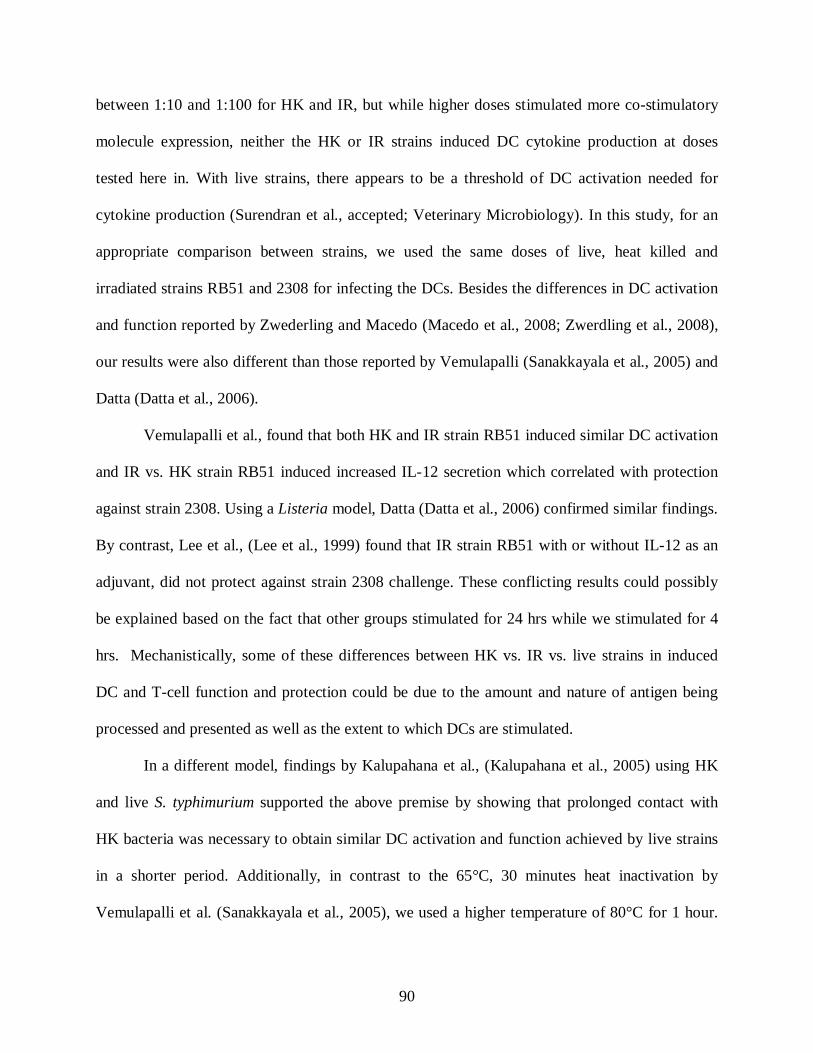
90
between 1:10 and 1:100 for HK and IR, but while higher doses stimulated more co-stimulatory
molecule expression, neither the HK or IR strains induced DC cytokine production at doses
tested here in. With live strains, there appears to be a threshold of DC activation needed for
cytokine production (Surendran et al., accepted; Veterinary Microbiology). In this study, for an
appropriate comparison between strains, we used the same doses of live, heat killed and
irradiated strains RB51 and 2308 for infecting the DCs. Besides the differences in DC activation
and function reported by Zwederling and Macedo (Macedo et al., 2008; Zwerdling et al., 2008),
our results were also different than those reported by Vemulapalli (Sanakkayala et al., 2005) and
Datta (Datta et al., 2006).
Vemulapalli et al., found that both HK and IR strain RB51 induced similar DC activation
and IR vs. HK strain RB51 induced increased IL-12 secretion which correlated with protection
against strain 2308. Using a Listeria model, Datta (Datta et al., 2006) confirmed similar findings.
By contrast, Lee et al., (Lee et al., 1999) found that IR strain RB51 with or without IL-12 as an
adjuvant, did not protect against strain 2308 challenge. These conflicting results could possibly
be explained based on the fact that other groups stimulated for 24 hrs while we stimulated for 4
hrs. Mechanistically, some of these differences between HK vs. IR vs. live strains in induced
DC and T-cell function and protection could be due to the amount and nature of antigen being
processed and presented as well as the extent to which DCs are stimulated.
In a different model, findings by Kalupahana et al., (Kalupahana et al., 2005) using HK
and live S. typhimurium supported the above premise by showing that prolonged contact with
HK bacteria was necessary to obtain similar DC activation and function achieved by live strains
in a shorter period. Additionally, in contrast to the 65°C, 30 minutes heat inactivation by
Vemulapalli et al. (Sanakkayala et al., 2005), we used a higher temperature of 80°C for 1 hour.

91
Theoretically, although not likely, additional heating may have disrupted the Brucella cell
envelopes (Barquero-Calvo et al., 2007) and exposed large amounts of Brucella LPS,
lipoproteins, peptidoglycan, DNA and other molecules recognized by innate immunity.
Additional differences between IR and HK could be due that IR may stimulate a better DC
mediated CD8 response than HK (Datta et al., 2006).
Besides differences in the ability of IR vs. HK to stimulate more CD8 vs. CD4 mediated
immune responses, and the role of IR vs. HK in protection, regulators which influence DC
function and protection are TNF-alpha, IL-12 and IL-10. As previously stated, TNF-alpha
production is critical for maximal IL-12 production and CD4 Th1 response. If either is
decreased, DC mediated T-cell responses and potentially protection could be decreased. Another
mechanism by which protection would be decreased would be through an IL-10 mediated T-
regulatory response which down-regulated IL-12 production by DCs (Huang et al., 2001).
(McGuirk et al., 2002). Correspondingly, HK and/or IR strains may suboptimally stimulate
BMDCs at a given dose which might induce them to become tolerogenic DCs (semimature DCs)
with the inability to produce proinflammatory cytokines (Lutz and Schuler, 2002). As others
have shown that both HK and IR strains of B. abortus induced similar levels of IL-10
(Sanakkayala et al., 2005), we did not determine the ability of HK or IR strains to induce IL-10
secretion from BMDCs. However, it is possible that live vs. HK or IR strains may induce
different levels of IL-10 that could influence DC and T-cell function and protection. Thus, our
findings along with already published studies suggest multiple mechanisms for differences
between live vs. IR vs. HK strains induced DC function, T-cell function and protection.
Additional studies are warranted to further investigate these mechanisms as well as their impact
on protection.
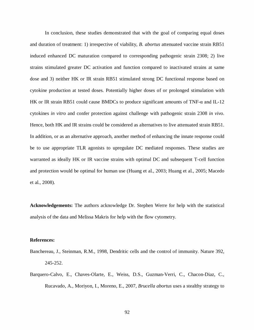
92
In conclusion, these studies demonstrated that with the goal of comparing equal doses
and duration of treatment: 1) irrespective of viability, B. abortus attenuated vaccine strain RB51
induced enhanced DC maturation compared to corresponding pathogenic strain 2308; 2) live
strains stimulated greater DC activation and function compared to inactivated strains at same
dose and 3) neither HK or IR strain RB51 stimulated strong DC functional response based on
cytokine production at tested doses. Potentially higher doses of or prolonged stimulation with
HK or IR strain RB51 could cause BMDCs to produce significant amounts of TNF-α and IL-12
cytokines in vitro and confer protection against challenge with pathogenic strain 2308 in vivo.
Hence, both HK and IR strains could be considered as alternatives to live attenuated strain RB51.
In addition, or as an alternative approach, another method of enhancing the innate response could
be to use appropriate TLR agonists to upregulate DC mediated responses. These studies are
warranted as ideally HK or IR vaccine strains with optimal DC and subsequent T-cell function
and protection would be optimal for human use (Huang et al., 2003; Huang et al., 2005; Macedo
et al., 2008).
Acknowledgements: The authors acknowledge Dr. Stephen Werre for help with the statistical
analysis of the data and Melissa Makris for help with the flow cytometry.
References:
Banchereau, J., Steinman, R.M., 1998, Dendritic cells and the control of immunity. Nature 392,
245-252.
Barquero-Calvo, E., Chaves-Olarte, E., Weiss, D.S., Guzman-Verri, C., Chacon-Diaz, C.,
Rucavado, A., Moriyon, I., Moreno, E., 2007, Brucella abortus uses a stealthy strategy to

93
avoid activation of the innate immune system during the onset of infection. PLoS ONE 2,
e631.
Billard, E., Cazevieille, C., Dornand, J., Gross, A., 2005, High susceptibility of human dendritic
cells to invasion by the intracellular pathogens Brucella suis, B. abortus, and B.
melitensis. Infect Immun 73, 8418-8424.
Billard, E., Dornand, J., Gross, A., 2007, Brucella suis prevents human dendritic cell maturation
and antigen presentation through regulation of tumor necrosis factor alpha secretion.
Infect Immun 75, 4980-4989.
Corbel, M.J. 2006. Brucellosis in humans and animals (World Health Organization).
Datta, S.K., Okamoto, S., Hayashi, T., Shin, S.S., Mihajlov, I., Fermin, A., Guiney, D.G., Fierer,
J., Raz, E., 2006, Vaccination with irradiated Listeria induces protective T cell immunity.
Immunity 25, 143-152.
Finkelman, F.D., Katona, I.M., Mosmann, T.R., Coffman, R.L., 1988, IFN-gamma regulates the
isotypes of Ig secreted during in vivo humoral immune responses. J Immunol 140, 1022-
1027.
Golding, B., Inman, J., Highet, P., Blackburn, R., Manischewitz, J., Blyveis, N., Angus, R.D.,
Golding, H., 1995, Brucella abortus conjugated with a gp120 or V3 loop peptide derived
from human immunodeficiency virus (HIV) type 1 induces neutralizing anti-HIV
antibodies, and the V3-B. abortus conjugate is effective even after CD4+ T-cell
depletion. J Virol 69, 3299-3307.
Golding, B., Scott, D.E., Scharf, O., Huang, L.Y., Zaitseva, M., Lapham, C., Eller, N., Golding,
H., 2001, Immunity and protection against Brucella abortus. Microbes Infect 3, 43-48.

94
He, Y., Vemulapalli, R., Zeytun, A., Schurig, G.G., 2001, Induction of specific cytotoxic
lymphocytes in mice vaccinated with Brucella abortus RB51. Infect Immun 69, 5502-
5508.
Huang, L.Y., Aliberti, J., Leifer, C.A., Segal, D.M., Sher, A., Golenbock, D.T., Golding, B.,
2003, Heat-killed Brucella abortus induces TNF and IL-12p40 by distinct MyD88-
dependent pathways: TNF, unlike IL-12p40 secretion, is Toll-like receptor 2 dependent. J
Immunol 171, 1441-1446.
Huang, L.Y., Ishii, K.J., Akira, S., Aliberti, J., Golding, B., 2005, Th1-like cytokine induction by
heat-killed Brucella abortus is dependent on triggering of TLR9. J Immunol 175, 3964-
3970.
Huang, L.Y., Reis e Sousa, C., Itoh, Y., Inman, J., Scott, D.E., 2001, IL-12 induction by a TH1-
inducing adjuvant in vivo: dendritic cell subsets and regulation by IL-10. J Immunol 167,
1423-1430.
Inaba, K., Inaba, M., Romani, N., Aya, H., Deguchi, M., Ikehara, S., Muramatsu, S., Steinman,
R.M., 1992, Generation of large numbers of dendritic cells from mouse bone marrow
cultures supplemented with granulocyte/macrophage colony-stimulating factor. J Exp
Med 176, 1693-1702.
Kalupahana, R.S., Mastroeni, P., Maskell, D., Blacklaws, B.A., 2005, Activation of murine
dendritic cells and macrophages induced by Salmonella enterica serovar Typhimurium.
Immunology 115, 462-472.
Lee, I.K., Olsen, S.C., Kehrli, M., Bolin, C.A., 1999, The adjuvant effect of a single dose of
interleukin-12 on murine immune responses to live or killed Brucella abortus strain
RB51. Can J Vet Res 63, 284-287.
95
Lutz, M.B., Schuler, G., 2002, Immature, semi-mature and fully mature dendritic cells: which
signals induce tolerance or immunity? Trends Immunol 23, 445-449.
Macedo, G.C., Magnani, D.M., Carvalho, N.B., Bruna-Romero, O., Gazzinelli, R.T., Oliveira,
S.C., 2008, Central role of MyD88-dependent dendritic cell maturation and
proinflammatory cytokine production to control Brucella abortus infection. J Immunol
180, 1080-1087.
McGuirk, P., McCann, C., Mills, K.H., 2002, Pathogen-specific T regulatory 1 cells induced in
the respiratory tract by a bacterial molecule that stimulates interleukin 10 production by
dendritic cells: a novel strategy for evasion of protective T helper type 1 responses by
Bordetella pertussis. J Exp Med 195, 221-231.
Muraille, E., Giannino, R., Guirnalda, P., Leiner, I., Jung, S., Pamer, E.G., Lauvau, G., 2005,
Distinct in vivo dendritic cell activation by live versus killed Listeria monocytogenes. Eur
J Immunol 35, 1463-1471.
Oliveira, S.C., Zhu, Y., Splitter, G.A., 1994, Recombinant L7/L12 ribosomal protein and
gamma-irradiated Brucella abortus induce a T-helper 1 subset response from murine
CD4+ T cells. Immunology 83, 659-664.
Pappas, G., Panagopoulou, P., Christou, L., Akritidis, N., 2006, Brucella as a biological weapon.
Cell Mol Life Sci 63, 2229-2236.
Plotkin, S.A., 2005, Vaccines: past, present and future. Nat Med 11, S5-11.
Rey-Ladino, J., Koochesfahani, K.M., Zaharik, M.L., Shen, C., Brunham, R.C., 2005, A live and
inactivated Chlamydia trachomatis mouse pneumonitis strain induces the maturation of
dendritic cells that are phenotypically and immunologically distinct. Infect Immun 73,
1568-1577.
96
Sanakkayala, N., Sokolovska, A., Gulani, J., Hogenesch, H., Sriranganathan, N., Boyle, S.M.,
Schurig, G.G., Vemulapalli, R., 2005, Induction of antigen-specific Th1-type immune
responses by gamma-irradiated recombinant Brucella abortus RB51. Clin Diagn Lab
Immunol 12, 1429-1436.
Schurig, G.G., Roop, R.M., 2nd, Bagchi, T., Boyle, S., Buhrman, D., Sriranganathan, N., 1991,
Biological properties of RB51; a stable rough strain of Brucella abortus. Vet Microbiol
28, 171-188.
Street, N.E., Schumacher, J.H., Fong, T.A., Bass, H., Fiorentino, D.F., Leverah, J.A., Mosmann,
T.R., 1990, Heterogeneity of mouse helper T cells. Evidence from bulk cultures and
limiting dilution cloning for precursors of Th1 and Th2 cells. J Immunol 144, 1629-1639.
Tsunetsugu-Yokota, Y., Tamura, H., Tachibana, M., Ogata, K., Honda, M., Takemori, T., 2002,
Selective expansion of perforin-positive CD8+ T cells by immature dendritic cells
infected with live Bacillus Calmette-Guerin mycobacteria. J Leukoc Biol 72, 115-124.
Vemulapalli, R., He, Y., Cravero, S., Sriranganathan, N., Boyle, S.M., Schurig, G.G., 2000,
Overexpression of protective antigen as a novel approach to enhance vaccine efficacy of
Brucella abortus strain RB51. Infect Immun 68, 3286-3289.
Zhan, Y., Cheers, C., 1998, Control of IL-12 and IFN-gamma production in response to live or
dead bacteria by TNF and other factors. J Immunol 161, 1447-1453.
Zwerdling, A., Delpino, M.V., Barrionuevo, P., Cassataro, J., Pasquevich, K.A., Garcia
Samartino, C., Fossati, C.A., Giambartolomei, G.H., 2008, Brucella lipoproteins mimic
dendritic cell maturation induced by Brucella abortus. Microbes Infect 10, 1346-1354.
Figure legends:

97
Fig. 1A: Day 6 harvested BMDCs show an immature phenotype. Bone marrow cells isolated
from BALB/c mice were cultured for 6 days in 10% RPMI medium with rGM-CSF (20ng/ml).
Cells were harvested on day 6 and analyzed by flow cytometry for CD11c+ marker expression
using fluorescent labeled CD11c+ antibody. The figure compares the percentages of CD11c+ DCs
expressing the surface maturation markers MHC class II, CD40 and CD86 harvested on day 6 to
that treated overnight with (negative control) media alone or (positive control) E. coli LPS (100
ng/ml). Two asterisks (**) denotes statistically significant data at p ≤ 0.05 compared to data
represented by an asterisk (*). Data represents means ± standard deviations of 3 independent
experiments.
Fig. 1B-1E: Heat killed or irradiated B. abortus rough vaccine strain RB51 induced greater
DC maturation than corresponding smooth strain 2308. Immature BMDCs were infected
with live, HK or IR B. abortus rough vaccine strain RB51 or corresponding smooth strain 2308
at the given doses (DC:Brucella) 1:10 (not shown) or 1:100. At 24 hours post-incubation, the
cells were analyzed for the DC surface expression of MHC class II, CD40 and CD86. Figures
1B-1E represent comparisons of the percentage of CD11c+ cells (BMDCs) expressing MHC
class II high, CD40, CD86 and coexpression of CD40+/CD86+ respectively across different
treatment groups. Media and E. coli LPS are the negative and positive controls respectively.
Double characters **, ##, aa represent statistically significant change at p ≤ 0.05 with the
corresponding single characters *, # and a respectively. Data represents means ± standard
deviations of 3 independent experiments.
Fig. 2: Heat killed or irradiated B. abortus rough vaccine strain RB51 do not induce
significant TNF – α and IL-12 secretion. To assess DC function, TNF-α (A) and IL-12p70 (B)
levels from the culture supernatants of live, HK or IR B. abortus strain RB51 or 2308 infected
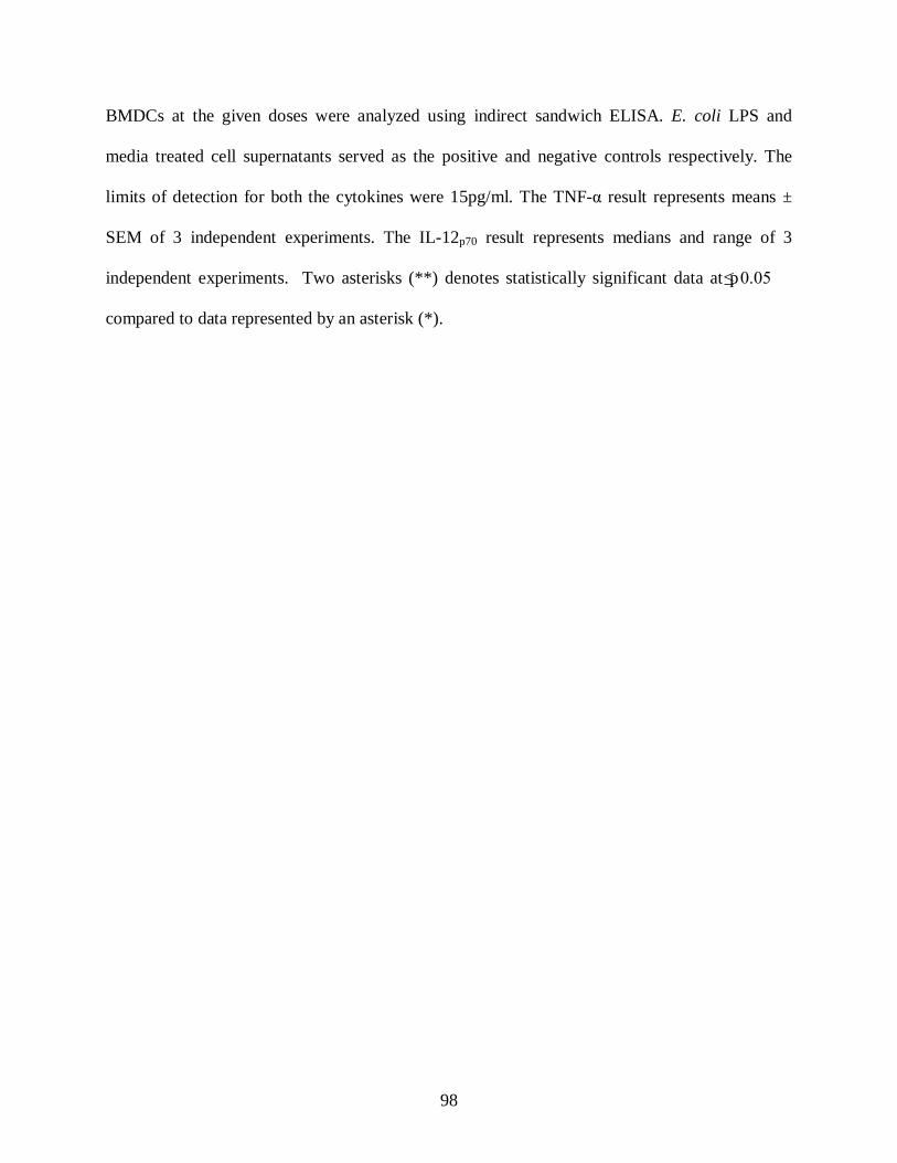
98
BMDCs at the given doses were analyzed using indirect sandwich ELISA. E. coli LPS and
media treated cell supernatants served as the positive and negative controls respectively. The
limits of detection for both the cytokines were 15pg/ml. The TNF-α result represents means ±
SEM of 3 independent experiments. The IL-12p70 result represents medians and range of 3
independent experiments. Two asterisks (**) denotes statistically significant data at p ≤ 0.05
compared to data represented by an asterisk (*).
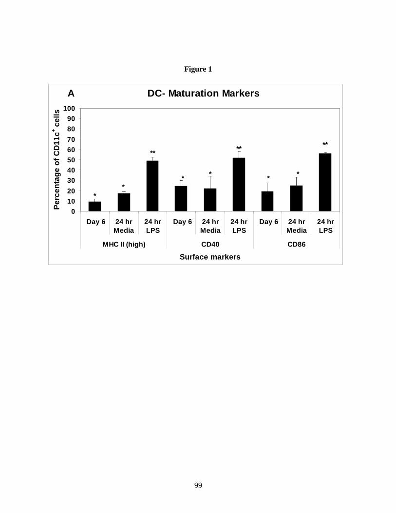
99
Figure 1
A DC- Maturation Markers
0102030405060708090
100
Day 6 24 hrMedia
24 hrLPS
Day 6 24 hrMedia
24 hrLPS
Day 6 24 hrMedia
24 hrLPS
MHC II (high) CD40 CD86
Surface markers
Perc
enta
ge o
f CD
11c+ c
ells
**
**
* *
**
* *
**
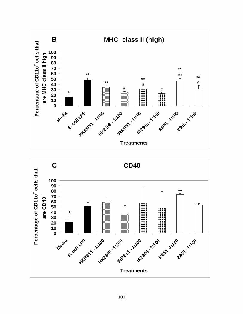
100
B MHC class II (high)
0102030405060708090
100
Med
ia
E. coli
LPS
HKRB51 - 1
:100
HK2308
- 1:10
0
IRRB51
- 1:10
0
IR23
08 - 1
:100
RB51 -1
:100
2308
- 1:10
0
Treatments
Perc
enta
ge o
f CD
11c+ c
ells
that
ar
e M
HC
cla
ss II
hig
h
**
*
**
**
**#
#**
#
##
#
C CD40
0102030405060708090
100
Med
ia
E. coli
LPS
HKRB51 - 1
:100
HK2308
- 1:10
0
IRRB51
- 1:10
0
IR23
08 - 1
:100
RB51 -1
:100
2308
- 1:10
0
Treatments
Perc
enta
ge o
f CD
11c+ c
ells
that
ar
e C
D40
+
*
**
101
D CD86
0102030405060708090
100
Med
ia
E. coli
LPS
HKRB51 - 1
:100
HK2308
- 1:10
0
IRRB51
- 1:10
0
IR23
08 - 1
:100
RB51 -1
:100
2308
- 1:10
0
Treatments
Perc
enta
ge o
f CD
11c+
cells
that
ar
e C
D86
+
*
**
aa
aa
a
a
**** **
**
a#
##
#
##
E CD40+/CD86+
0102030405060708090
100
Med
ia
E. coli
LPS
HKRB51 - 1
:100
HK2308
- 1:10
0
IRRB51
- 1:10
0
IR23
08 - 1
:100
RB51 -1
:100
2308
- 1:10
0
Treatments
Perc
enta
ge o
f CD
11c+
cells
that
ar
e C
D40
+ /CD
86+
*
****
** **
**aa
aa
a
a
a
a#
###

102
Figure 2
A TNF - alpha
0
300
600
900
1200
1500
Media
E. coli
LPS
HKRB51 - 1
:10
HK2308
- 1:10
IRRB51
- 1:10
IR23
08 - 1
:10
RB51 - 1
:10
2308
- 1:10
HKRB51 - 1
:100
HK2308
- 1:10
0
IRRB51
- 1:10
0
IR23
08 - 1
:100
RB51 - 1
:100
2308
- 1:10
0
Treatments
Con
cent
ratio
n (p
g/m
l)
**
**** ** * * * * * * *
B IL-12p70
0
300
600
900
1200
1500
Media
E. coli
LPS
HKRB51 - 1
:10
HK2308
- 1:10
IRRB51
- 1:10
IR23
08 - 1
:10
RB51 - 1
:10
2308
- 1:10
HKRB51 - 1
:100
HK2308
- 1:10
0
IRRB51
- 1:10
0
IR23
08 - 1
:100
RB51 - 1
:100
2308
- 1:10
0
Treatments
Con
cent
ratio
n (p
g/m
l)
**
* * * * **
* *
103
Chapter 4
The ability of Brucella abortus rough vaccine and smooth pathogenic strains to elicit innate
immunity in a murine respiratory model.
Running title: B. abortus RB51 induces strong innate immunity in lung
Naveen Surendran1, Kurt Zimmerman2, Mohamed N. Seleem2, Nammalwar Sriranganathan2,
Stephen M. Boyle2, Elizabeth M. Hiltbold3, Heather Lawler1, Bettina Heid1, Sharon G.
Witonsky1*.
(To be submitted to Vaccine)
Abstract
B. abortus strain RB51 is a live attenuated rough strain approved as the official vaccine
against bovine brucellosis in the United States and many other countries. Another B. abortus
rough vaccine candidate RB51SOD, which overexpresses its own Cu-Zn superoxide dismutase,
has shown to provide better protection upon IP vaccination against pathogenic B. abortus strain
2308 in murine model of brucellosis. However, there are no approved vaccines against human
brucellosis and inhalation of aerosolized Brucella organisms is one route of human infection.
Currently, limited information is available on how Brucella stimulates pulmonary immunity
upon aerosol infection. In this study, we assessed the ability of intranasally delivered rough
vaccine strains RB51, RB51SOD and smooth pathogenic strain 2308 to induce innate response
characterized by pulmonary dendritic cell (DC) activation and function in vivo. We also
evaluated the histopathological changes associated with intranasal inoculation of vaccine as well
as pathogenic strains. Our results show that rough strain RB51 has superior ability (p≤0.05) in
104
stimulating DC activation and function based on surface expression of MHC class II, CD40 and
IFN-γ production compared to rough strain RB51SOD and smooth pathogenic strain 2308. In
addition, rough strain RB51 induced more proinflammatory histopathological changes in lung
compared to the other two Brucella strains.
Introduction
Brucellosis is a worldwide zoonotic disease (4) caused by Brucella spp., Gram-negative
facultative intracellular bacteria. Brucellosis has a huge economic impact on the animal industry
since infection results in abortions, still-births and infertility in livestock. Brucellosis can be
transmitted from animals to humans as animal tissues are the source of the pathogen. Around the
globe, more than 500,000 human infections are reported annually (20). Clinical syndromes in
humans include chronic fatigue, undulant fever, reproductive disorders and general malaise (4).
Brucella spp. are a major potential bio-terror threat, as they are zoonotic, highly infectious,
readily aerosolized, and Brucella can be genetically manipulated to create antibiotic resistant
strains. The Centers for Disease Control and Prevention (CDC) classify Brucella spp. as category
B agents. Inhalation of infected aerosols is implicated as a route for human exposure both
through natural as well as intentional exposure (10, 14). Currently, there are no approved human
vaccines available.
Murine brucellosis is widely accepted as an established model for studying the host
immune response to experimental Brucella infection (i.e., vaccination and challenge) (15, 23,
29). In spite of the importance of aerosol exposure as a route of infection and Brucella spp. use
as a potential bioterror threat, very few studies have focused on a respiratory route of infection
for vaccine efficacy studies (9, 19). Ficht et al., (9) and Olsen et al., (19) failed to show clearance
105
from lung following intraperitoneal (IP) vaccination and aerosol challenge with B. melitensis
and/or B. abortus respectively. Part of the reason for lack of protection may be due to the IP
vaccination route. Based on past experience, it is expected that mucosal vaccination would
enhance a protective immune response against aerosol challenge (17, 21). In order to further
understand the protective immune responses against Brucella, more basic background
information is needed. Most Brucella studies have predominantly focused on vaccinating via IP,
subcutaneous (SC), intravenous (IV) or intramuscular (IM) routes with challenge occurring
primarily by IP exposure (8, 23). However, few studies have discerned novel information on
innate or adaptive protective immune responses particularly against respiratory challenge. While
models have established chronic infection, they were not successful in showing respiratory
protection (1, 9, 12, 13, 28). Therefore, to develop an efficacious vaccination regime, assessment
of the innate immune response following vaccination is critical. As B. abortus strain RB51 has
been successfully developed for use in animals (24) and is a vector for the expression of multiple
homologous and heterologous antigens (30), we investigated the innate response in mice to
vaccine and pathogenic strains of B. abortus.
The three B. abortus strains used for this study are pathogenic strain 2308, and vaccine
strains RB51 and RB51SOD. B. abortus strain 2308, with the O-side chain of lipopolysaccharide
(LPS), is one of the smooth, pathogenic, zoonotic Brucella species affecting cattle and humans
(15). B. abortus strain RB51, which lacks the O-side chain of LPS, is the rough, live attenuated
vaccine strain approved for use in cattle by the United States Department of Agriculture (USDA)
for cattle (23). The other B. abortus rough strain RB51SOD, which overexpresses its own
copper-zinc superoxide dismutase (SOD), has been shown to be more efficacious than strain
RB51 against strain 2308 challenge in murine IP vaccination and challenge models (29). To our

106
knowledge, no studies have been published which characterize the in vivo innate immune
response including the associated histopathological changes to IN inoculation of either B.
abortus pathogenic strain 2308 or rough vaccine strains RB51 or RB51SOD. This information
will enhance our knowledge on differences between the immune responses associated with IN
inoculation of vaccine vs. pathogenic strains. With this knowledge, we may have the ability to
both develop an efficacious human brucellosis vaccine against aerosol infection as well as
provide information on how B. abortus pathogenic strain 2308 limits the immune response in the
lungs.
In these studies, we assessed in vivo the ability of IN delivered rough vaccine strains
RB51 and RB51SOD vs. pathogenic strain 2308 to induce an innate response. In our previous in
vitro study, we demonstrated that rough vaccine strain RB51 elicited enhanced BMDC
maturation and function compared to strain RB51SOD and pathogenic strain 2308 (Surendran et
al., under review). Here we evaluated the differential ability of B. abortus rough vaccine strains
RB51, RB51SOD and smooth pathogenic strain 2308 to elicit pulmonary DC activation and
function in vivo. We also assessed the vaccine and virulent strain induced histopathological
changes in lung, liver and spleen at day 3, 5, 7 and 14 PI. We hypothesized that live rough
vaccine strains, as seen in vitro, would stimulate increased DC activation and function based on
upregulation of costimulatory marker expression and cytokine production. Additionally we
expected rough strains would induce more proinflammatory histopathological changes compared
to smooth pathogenic strain 2308.

107
Materials and Methods
Mice: Female 6-8 weeks old BALB/c mice were obtained from Charles River Laboratories Inc.,
Wilmington, MA. Mice were used under animal care protocols approved by Institutional Animal
Care and Use Committee at Virginia Tech.
Bacterial strains, plasmids and oligonucleotides: Live attenuated rough B. abortus strains
RB51, RB51SOD and virulent smooth strain 2308 used for clearance study were from our stock
culture collection (23, 29). Brucella strains were routinely grown at 37°C in tryptic soy broth
(TSB) or on tryptic soy agar (TSA) (Difco). Chloramphenicol (Cm) was used at a final
concentration of 22µg/ml for growing strain RB51SOD. Primers used in this study are listed in
Table 1.
Recombinant DNA methods: Recombinant DNA methods (DNA ligations, restriction
endonuclease digestions, and agarose gel electrophoresis) were performed according to standard
techniques (22). The polymerase chain reaction (PCR) was performed using Platinum PCR
SuperMix High Fidelity (Invitrogen) and a Gradient Mastercycler® (Eppendorf).
Oligonucleotides were purchased from Sigma-Genosys (Sigma-Aldrich). Restriction and
modification enzymes were purchased from Promega. QIAprep Spin Miniprep Kit from
QIAGEN was used for plasmid extractions and QIAGEN PCR cleanup kit was used for
restriction enzymes removal and DNA gel extraction.
Vectors construction: Brucella strains expressing GFP and HA peptides were generated to
study innate immune response as well as CD4 and CD8 antigen specific response to IN
inoculated bacterial strains. Briefly, Trc promoter (25) with downstream short peptides
(IYSTVASSL-PKY) MHC class I and (VKQNTLKLAT) MHC class II (5, 11) was constructed
108
and amplified in two steps using primers (T-F and T-R1) in the first step. The PCR amplicon was
gel purified and used as a template for the second PCR cycle using primers (T-F and T-R2).
The amplified promoter fusion (Trc::MHC) was purified and cloned into BamHI and SalI
restriction sites of promoterless pNS vector (27) to form the pNSMHC construct. A promoterless
Green Fluorescence Protein gene (GFP) was excised from pGFPuv vector (BD Biosciences
Clontech) and cloned in frame downstream of the promoter in the multiple cloning site area of
pNSMHC to form (pNSMHC/GFP). The Brucella sodC gene with its own promoter was
amplified from B. suis using primers Sod-F and Sod-R (Table 1). sodC gene was cloned into
pNSMHC/GFP vector to form pNSMHC/GFP+SOD. The plasmids were sequenced to confirm
the correct sequence. Transformation of B. abortus strains 2308 and RB51 was done by
electroporation with a Gene Pulser (BTX) set at 2.4 KV, 25 µF and 200 as described
previously (26). Recombinant RB51, RB51SOD and 2308 strains expressing GFP were detected
under UV light. SOD expression was confirmed by SDS-PAGE and Western blot. The strain that
harbors pNSMHC/GFP was named RB51pNSMHCGFP, while the strain that harbors plasmid
pNSMHC/GFP+SOD was named RB51pNSMHCGFPSOD. Strain 2308 that harbors
pNSMHCGFP was named as 2308pNSMHCGFP (Table 2). These strains will be designated as
strains RB51, RB51SOD and 2308 respectively in this manuscript.
All experiments with Brucella were performed in our CDC approved (C2003 1120-0016)
Biosafety Level (BSL) -3 Infectious Disease Unit (IDU) facility.
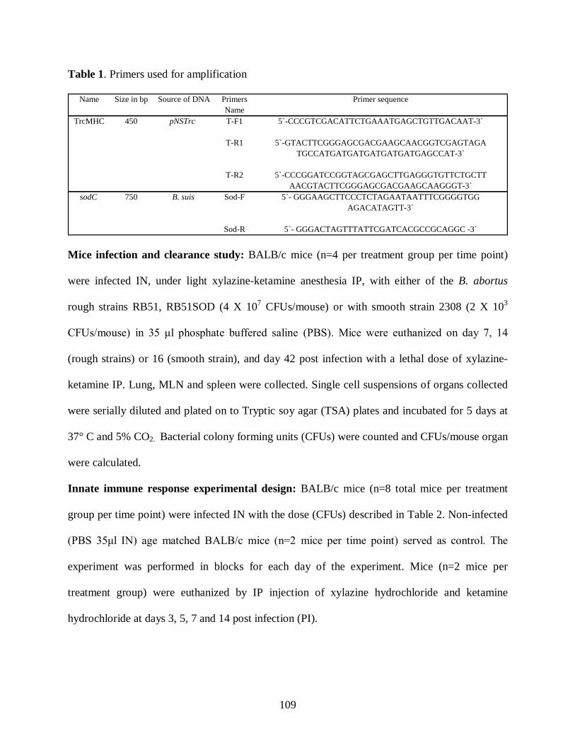
109
Table 1. Primers used for amplification
PrimersName
TrcMHC 450 pNSTrc T-F1 5`-CCCGTCGACATTCTGAAATGAGCTGTTGACAAT-3`
T-R1 5`-GTACTTCGGGAGCGACGAAGCAACGGTCGAGTAGATGCCATGATGATGATGATGATGAGCCAT-3`
T-R2 5`-CCCGGATCCGGTAGCGAGCTTGAGGGTGTTCTGCTTAACGTACTTCGGGAGCGACGAAGCAAGGGT-3`
sodC 750 B. suis Sod-F 5`- GGGAAGCTTCCCTCTAGAATAATTTCGGGGTGGAGACATAGTT-3`
Sod-R 5`- GGGACTAGTTTATTCGATCACGCCGCAGGC -3`
Name Size in bp Source of DNA Primer sequence
Mice infection and clearance study: BALB/c mice (n=4 per treatment group per time point)
were infected IN, under light xylazine-ketamine anesthesia IP, with either of the B. abortus
rough strains RB51, RB51SOD (4 X 10⁷ CFUs/mouse) or with smooth strain 2308 (2 X 10³
CFUs/mouse) in 35 μl phosphate buffered saline (PBS). Mice were euthanized on day 7, 14
(rough strains) or 16 (smooth strain), and day 42 post infection with a lethal dose of xylazine-
ketamine IP. Lung, MLN and spleen were collected. Single cell suspensions of organs collected
were serially diluted and plated on to Tryptic soy agar (TSA) plates and incubated for 5 days at
37° C and 5% CO2. Bacterial colony forming units (CFUs) were counted and CFUs/mouse organ
were calculated.
Innate immune response experimental design: BALB/c mice (n=8 total mice per treatment
group per time point) were infected IN with the dose (CFUs) described in Table 2. Non-infected
(PBS 35μl IN) age matched BALB/c mice (n=2 mice per time point) served as control. The
experiment was performed in blocks for each day of the experiment. Mice (n=2 mice per
treatment group) were euthanized by IP injection of xylazine hydrochloride and ketamine
hydrochloride at days 3, 5, 7 and 14 post infection (PI).
110
Table 2. Dosage and route of administration of Brucella strains for innate experiment.
Collection and preparation of samples: Broncho-alveolar lavage (BAL), mediastinal lymph
node (MLN), spleens, lung and liver were collected at the time of euthanasia. BAL was spun at
250 x g for 5 minutes to isolate the cells and the supernatant was frozen for cytokine analysis.
The BAL samples from same treatment group were pooled at each time point. The spleens were
excised and divided equally for flow cytometry, and histology. The entire MLN was used for
flow cytometric analysis. Briefly, spleen and MLN were dissociated with sterile metallic screens
and a 3 ml syringe plunger. Cells were washed and enumerated using a hemocytometer. Cells
were resuspended at 5 X 10⁶ cells/ml in saline for flow cytometry.
Tissue processing and staining: Portions of the lung, spleen, and liver were collected for
histological examination. Sections were fixed in formalin and embedded in paraffin (31). A
blinded histopathologic review of lung from all IN infected mice was performed by a board
certified anatomic and clinical pathologist (KZ). SW reviewed all spleen and liver samples
followed by joint review (SW and KZ) of these tissues.
Tissue reaction scores: Based on characterization of pulmonary changes, lungs were
characterized based on pneumonia, vascular change, septa thickening, as well as changes to the
pleura, large airway and alveoli. For all parameters, severity was graded as 0-5 (0-1 no change,
1-2 minimal, 2-3 mild, 3-4 moderate, 4-5 severe, >5 marked or extreme). In addition to severity
for all parameters, pneumonia was characterized based on severity, pattern, distribution, percent
Brucella strains Route of inoculation
Dose (CFUs/Mouse) in 35 µl PBS
Experimental
RB51pNSMHCGFP RB51pNSMHCGFPSOD 2308pNSMHCGFP
IN IN IN
4 X 10⁹
4 X 10⁹ 2 X 10⁵
Control PBS IN 35 µl PBS
111
involvement and type of inflammation. For vascular change, the presence and type of vessel wall
changes were noted. For septa thickening, the infiltrative cell type was noted. Alveolar changes
were characterized based on debris/protein, type 2 hyperplasia, grade of inflammation (0-5) and
type of inflammation. Large airway changes were assessed based on hyperplasia, necrosis, and
severity (0-5) of inflammation, peri-lymphoid hyperplasia, peri-edema, and type of inflammatory
cells present. Pleura were graded on inflammation (0-5), fibrosis (0-5), and inflammatory cell
type. Any additional comments were noted. Averages of percentage of interstitial pneumonia
based on KZ assessment were determined, and are reported. Averages of severity, percentage
involvement of pneumonia and other changes are reported (Table 3).
Staining and flow cytometry: The cells were stained with approximately 0.5µg of the
appropriate stain per 0.5 x 10⁶ cells using the following monoclonal antibodies for FACS
analysis: FITC-conjugated anti-CD11c, PE-conjugated anti I-A/I-E (BD Pharmingen), TR-
conjugated anti-CD11c, PE-conjugated anti-CD40 and Biotin-conjugated anti-CD80 (Caltag).
The samples were washed and stained with biotinylated antibody if required. Cells were washed
and analyzed by EPICS XL Flow cytometer (Coulter, Hileah, FL).
Cytokine analysis: For cytokine measurement, BAL supernatants from Brucella infected mice
stored at -80°C were used. IFN- γ, TNF-α, IL-12p70 (bioactive form of IL-12) and IL-4 cytokine
levels were measured using indirect sandwich ELISAs (BD Pharmingen).
Statistical analysis: Kruskal-Wallis non-parametric test was used to analyze the significance of
the DC activation marker expression. Statistical significance of cytokine production in BAL was
analyzed by mixed model ANOVA followed by Dunnett’s procedure for multiple comparisons.
Statistical significance of histopathological lung changes was tested using a general linear model
of ANOVA accessing effects of treatment, day and interaction of treatment-day on severity and
112
percentage of involvement data. Tukey 95% confidence interval pairwise comparisons were used
to test for differences in severity and involvement among all levels of treatment. All statistical
analysis was done using SAS system (NC, USA) and Minitab software (PA, USA).
Results
B. abortus rough vaccine strains cleared more quickly than wild type strain 2308 from IN
infected BALB/c mice: IN doses were chosen relative to already established vaccine strain
RB51, RB51SOD (4 X 10⁸ CFUs/mouse, IP) and challenge strain 2308 (2 X 10⁴ CFUs/mouse,
IP) doses. To assess clearance and/or development of chronic infection, BALB/c mice were
infected IN with 4 X 10⁷ CFUs of either of the rough strains (RB51 or RB51SOD) or with 2 X
103 CFUs of pathogenic strain 2308. Clearance was assessed at day 7, 14 and 42 PI from lungs,
MLN and spleen for rough vaccine strains (Fig. 1 A). Brucella strains RB51 and RB51SOD had
marked clearance in the lung with approximately 104 decreases in titers by day 14 PI. In the
MLN and spleen, titers were approximately 103 and 102 respectively by day 7. Titers persisted at
day 14 PI with spleen and MLN at 103 before clearance at day 42. Clearance was complete from
all organs by day 42 PI. By comparison, the bacterial load progressively increased for smooth
strain 2308 reaching a plateau by day 16 PI (Fig. 1B). Mice remained chronically infected with
increased CFUs in spleen and MLN at day 116 PI, whereas clearance was complete in lung by
day 116 PI. For strain 2308, live bacteria were not cultured from the MLN until day 16 time
point although the titers in spleen were > 80 times the MLN titers. Splenic colonization for
strain 2308 was almost 6 times greater than lung titers by day 16. By day 42, splenic colonization
for strain 2308 remained higher compared to lung and MLN colonization, which were similar.
As of day 42, mice infected with smooth strain 2308 were chronically infected in lung, spleen

113
and MLN. By comparison, by day 7, mice infected IN with higher doses of rough strains had live
bacteria that were cultured from MLN with greater numbers than those recovered from spleen.
With rough strains, titers were similar in all organs by day 14 and completely cleared by day 42
(Fig. 1A). These data suggest differences in bacterial kinetics for systemic spread for rough and
smooth strains in vivo.
Rough strain RB51 significantly upregulated CD11c+/MHC class II high and CD40
expression: The ability of rough GFP expressing strains RB51, RB51SOD and smooth GFP
expressing strain 2308 vs. saline control to induce DC activation was evaluated by assessing
MHC class II, CD40 and CD80 expression on CD11c+ cells (DCs) from BAL, MLN and spleen
at day 3, 5, 7 and 14 PI. In BAL (Fig. 2A), of the total gated granulocytes, strain RB51 infected
mice induced significantly (p≤0.05) higher percentage of CD11c + cells expressing MHC class II
high than PBS inoculated mice at all post infection time periods (day 3, 5, 7 & 14) tested. The
CD11c+/MHC class II high expression induced by strain RB51 was also significantly (p≤0.05)
higher than smooth strain 2308 induced expression at day 5, 7 and day 14 PI. RB51 also induced
significantly (p≤0.05) higher MHC class II high expression greate r than strain RB51SOD at day
5 and day 7. At day 14 PI, strain RB51SOD induced population of CD11c+/MHC class II high
cells were significantly higher (p≤0.05) than induced by strain 2308 infected mice. On average
90-98% of the gated CD11c+ cells in BAL, from all the different treatment groups at all post
infection time points were positive for CD11b expression. This suggested that they were DCs of
myeloid origin (data not shown).
Similarly, strain RB51 infected mice had significantly higher (p≤0.05) CD11c +/MHC
class II high expressing cells compared to PBS mice in MLN at day 3 and 7 (p≤0.05) and 14
(p=0.052) (Fig. 2B). Strain RB51SOD induced CD11c+/MHC class II high expression was only
114
significantly (p≤0.05) higher than PBS control at day 7 PI. In the splee n, there were no
significant changes in CD11c+/MHC class II high cells between any treatments at any time
points (data not shown). Smooth strain 2308 did not induce significant percentages in
CD11c+/MHC class II high expression in BAL or MLN compared to PBS control. Assessing DC
activation in BAL, MLN and spleen, we determined that at day 7 PI strain RB51 stimulated
statistically significant (p≤0.05) increased expression of CD11c +/CD40+ cells in BAL compared
to PBS and strain 2308 treated mice (data not shown). Total cellularity was not assessed. No
other significant findings were detected.
Strain RB51 infected mice induced increased IFN-γ secretion in BAL: To assess potential
innate function, IFN-γ and IL-4 cytokine levels in the BAL supernatants were measured from
infected and control mice at day 3, 5, 7 and 14 PI. RB51 infected mice IN (Fig. 3) had higher
IFN-γ production in BAL at both day 5 (non significant) and 7 PI (significant, p≤0.05) compared
to PBS control. Neither rough strain RB51SOD nor smooth strain 2308 infected mice induced
significantly increased IFN-γ production compared to PBS control at any time points (Fig. 3).
Both RB51 and RB51SOD had significantly decreased IFN-γ production compared to PBS
control at day 3. None of the Brucella strains or PBS control mice induced detectable levels of
IL-4 in BAL supernatants from IN infected mice at any of the time points tested (data not
shown).
Altogether, these data demonstrated that rough vaccine strain RB51 had superior ability
in vivo to induce DC activation and function compared to rough strain RB51SOD and smooth
pathogenic strain 2308.
Strain RB51 infected mice developed interstitial pneumonia with vasculitis: The most
significant histopathologic changes were noted in the lungs with much less significant changes in
115
the spleen and liver. In the lung only strain RB51 induced significant (p=0.0003)
histopathological changes in terms of severity of pneumonia compared to PBS control (Figure 4).
Strain RB51 effect on severity was not influenced by day of treatment or by interaction of day
and treatment type. Strain RB51 induced severity of pneumonia was significantly higher than
both strain RB51SOD (p=0.027) and strain 2308 (p=0.015) induced pneumonia (Figure 4).
However, no other treatment differences were seen for PBS, strain RB51SOD, and strain 2308
for severity or for percentage of lung involvement between any of the groups. Table 3 illustrates
average pulmonary changes based on severity and percentage involvement of pneumonia as well
as severity of vascular change, septa thickening and debri/protein. Overall, strain RB51 induced
significantly higher severity of pneumonia than PBS, strain RB51SOD and strain 2308 at all time
points. At all days, strain RB51 induced significantly higher average severity but non-significant
increases in percentage of pneumonia, severity of vascular change with lymphocytic perivascular
changes, septal thickening and debris/protein compared to both strains RB51SOD and strain
2308, with the following exceptions at day 14. At day 14, strain RB51SOD induced minimally
greater percentage involvement of pneumonia and debri/protein than strain RB51. In addition,
only strain RB51 infected mice, except for 1 strain RB51SOD infected mouse had lymphocytic
perivascular infiltrate. This was most noticeable at day 3 and 5, but also present at days 7 and 14
PI.
Histopathologic review of livers and spleens was performed by SW. In the liver at day 3
PI changes were very minimal in all Brucella infected mice compared to saline controls. At day
5 PI, there was a mild increase in small multi-focal lymphocytic infiltrates within the hepatic
parenchyma in both strain RB51 and strain RB51SOD infected mice. By day 7 PI, infiltrates
were still present, but contained increased numbers of macrophages. Limited changes were
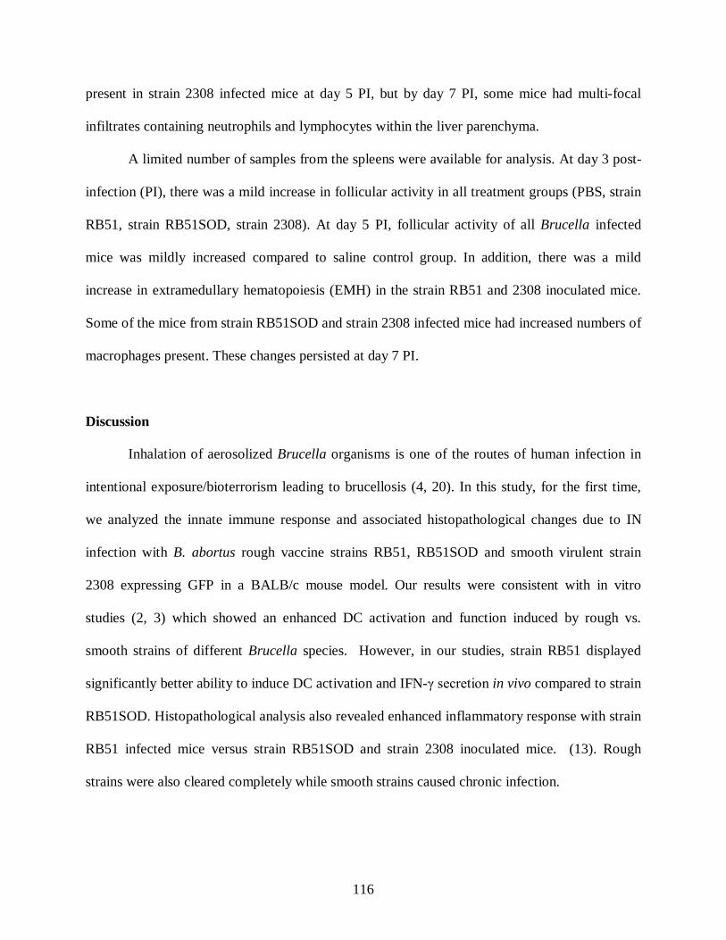
116
present in strain 2308 infected mice at day 5 PI, but by day 7 PI, some mice had multi-focal
infiltrates containing neutrophils and lymphocytes within the liver parenchyma.
A limited number of samples from the spleens were available for analysis. At day 3 post-
infection (PI), there was a mild increase in follicular activity in all treatment groups (PBS, strain
RB51, strain RB51SOD, strain 2308). At day 5 PI, follicular activity of all Brucella infected
mice was mildly increased compared to saline control group. In addition, there was a mild
increase in extramedullary hematopoiesis (EMH) in the strain RB51 and 2308 inoculated mice.
Some of the mice from strain RB51SOD and strain 2308 infected mice had increased numbers of
macrophages present. These changes persisted at day 7 PI.
Discussion
Inhalation of aerosolized Brucella organisms is one of the routes of human infection in
intentional exposure/bioterrorism leading to brucellosis (4, 20). In this study, for the first time,
we analyzed the innate immune response and associated histopathological changes due to IN
infection with B. abortus rough vaccine strains RB51, RB51SOD and smooth virulent strain
2308 expressing GFP in a BALB/c mouse model. Our results were consistent with in vitro
studies (2, 3) which showed an enhanced DC activation and function induced by rough vs.
smooth strains of different Brucella species. However, in our studies, strain RB51 displayed
significantly better ability to induce DC activation and IFN-γ secretion in vivo compared to strain
RB51SOD. Histopathological analysis also revealed enhanced inflammatory response with strain
RB51 infected mice versus strain RB51SOD and strain 2308 inoculated mice. (13). Rough
strains were also cleared completely while smooth strains caused chronic infection.

117
Overall these data support the potential use of strain RB51 as a vaccine strain to protect
against respiratory challenge. These data also raise questions on the subdued immune response
induced by strain RB51SOD and the mechanisms of immune subversion by strain 2308. In this
study, systemic clearance of rough and smooth strains following intranasal infection was
determined. We used these data to select doses and time points, and to identify differences
between vaccine strains, which were cleared and pathogenic strains which resulted in chronic
infection. Further challenge experiments would be necessary to confirm vaccine induced
protection. In comparing dissemination of rough vs. smooth strains, for rough strains, clearance
from the lung (by DCs and other cells as well as killing of strain RB51) was associated with
dissemination to spleen and lymph node by day 14 PI. Clearance was complete from all organs
by day 42 PI. With smooth strains, replication likely occurred in lungs as titers in the lungs
increased between day 7 and 16 PI. Comparing dissemination between smooth and rough strains,
dissemination systemically appeared to be delayed in smooth strains in that titers in spleen and
MLN were less than lung at day 7 PI. One possible explanation for these differences is based on
possible epithelial cell replication (1, 9, 19, 28). Ferrero et al. (6), showed that smooth strains
exhibited marked intracellular replication ability in human lung epithelial cell lines. Our data
supports this possible explanation for allowing smooth Brucella strains to replicate in the lungs,
possibly both within epithelial cells as well as DCs. This replication could allow smooth strain
2308 to have delayed clearance from the lung compared to rough strains. Additionally if the
immune response to strain 2308 is delayed compared to rough strains, it is possible that strain
2308 resided in the lung and replicated in DCs and other cells without rapid systemic activation
and spread to other organs, as occurs with rough strains.
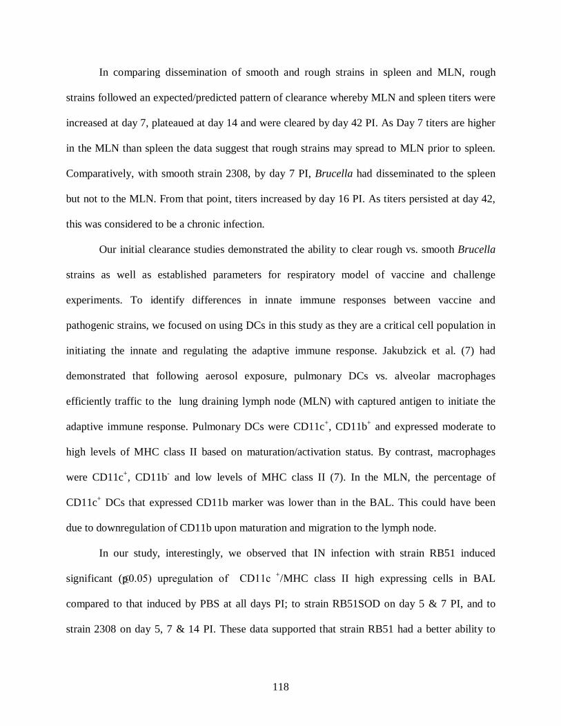
118
In comparing dissemination of smooth and rough strains in spleen and MLN, rough
strains followed an expected/predicted pattern of clearance whereby MLN and spleen titers were
increased at day 7, plateaued at day 14 and were cleared by day 42 PI. As Day 7 titers are higher
in the MLN than spleen the data suggest that rough strains may spread to MLN prior to spleen.
Comparatively, with smooth strain 2308, by day 7 PI, Brucella had disseminated to the spleen
but not to the MLN. From that point, titers increased by day 16 PI. As titers persisted at day 42,
this was considered to be a chronic infection.
Our initial clearance studies demonstrated the ability to clear rough vs. smooth Brucella
strains as well as established parameters for respiratory model of vaccine and challenge
experiments. To identify differences in innate immune responses between vaccine and
pathogenic strains, we focused on using DCs in this study as they are a critical cell population in
initiating the innate and regulating the adaptive immune response. Jakubzick et al. (7) had
demonstrated that following aerosol exposure, pulmonary DCs vs. alveolar macrophages
efficiently traffic to the lung draining lymph node (MLN) with captured antigen to initiate the
adaptive immune response. Pulmonary DCs were CD11c+, CD11b+ and expressed moderate to
high levels of MHC class II based on maturation/activation status. By contrast, macrophages
were CD11c+, CD11b- and low levels of MHC class II (7). In the MLN, the percentage of
CD11c+ DCs that expressed CD11b marker was lower than in the BAL. This could have been
due to downregulation of CD11b upon maturation and migration to the lymph node.
In our study, interestingly, we observed that IN infection with strain RB51 induced
significant (p≤0.05) upregulation of CD11c +/MHC class II high expressing cells in BAL
compared to that induced by PBS at all days PI; to strain RB51SOD on day 5 & 7 PI, and to
strain 2308 on day 5, 7 & 14 PI. These data supported that strain RB51 had a better ability to
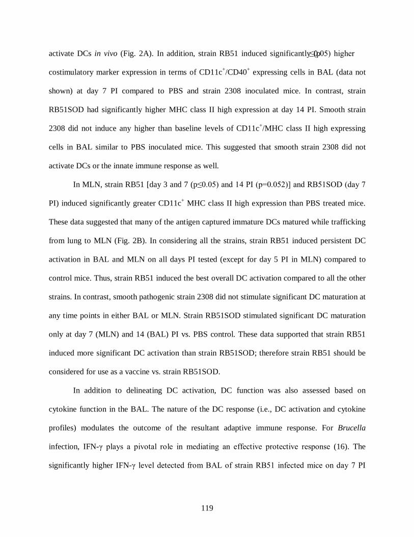
119
activate DCs in vivo (Fig. 2A). In addition, strain RB51 induced significantly (p≤0.05) higher
costimulatory marker expression in terms of CD11c+/CD40+ expressing cells in BAL (data not
shown) at day 7 PI compared to PBS and strain 2308 inoculated mice. In contrast, strain
RB51SOD had significantly higher MHC class II high expression at day 14 PI. Smooth strain
2308 did not induce any higher than baseline levels of CD11c+/MHC class II high expressing
cells in BAL similar to PBS inoculated mice. This suggested that smooth strain 2308 did not
activate DCs or the innate immune response as well.
In MLN, strain RB51 [day 3 and 7 (p≤0.05) and 14 PI (p=0.052)] and RB51SOD (day 7
PI) induced significantly greater CD11c+ MHC class II high expression than PBS treated mice.
These data suggested that many of the antigen captured immature DCs matured while trafficking
from lung to MLN (Fig. 2B). In considering all the strains, strain RB51 induced persistent DC
activation in BAL and MLN on all days PI tested (except for day 5 PI in MLN) compared to
control mice. Thus, strain RB51 induced the best overall DC activation compared to all the other
strains. In contrast, smooth pathogenic strain 2308 did not stimulate significant DC maturation at
any time points in either BAL or MLN. Strain RB51SOD stimulated significant DC maturation
only at day 7 (MLN) and 14 (BAL) PI vs. PBS control. These data supported that strain RB51
induced more significant DC activation than strain RB51SOD; therefore strain RB51 should be
considered for use as a vaccine vs. strain RB51SOD.
In addition to delineating DC activation, DC function was also assessed based on
cytokine function in the BAL. The nature of the DC response (i.e., DC activation and cytokine
profiles) modulates the outcome of the resultant adaptive immune response. For Brucella
infection, IFN-γ plays a pivotal role in mediating an effective protective response (16). The
significantly higher IFN-γ level detected from BAL of strain RB51 infected mice on day 7 PI
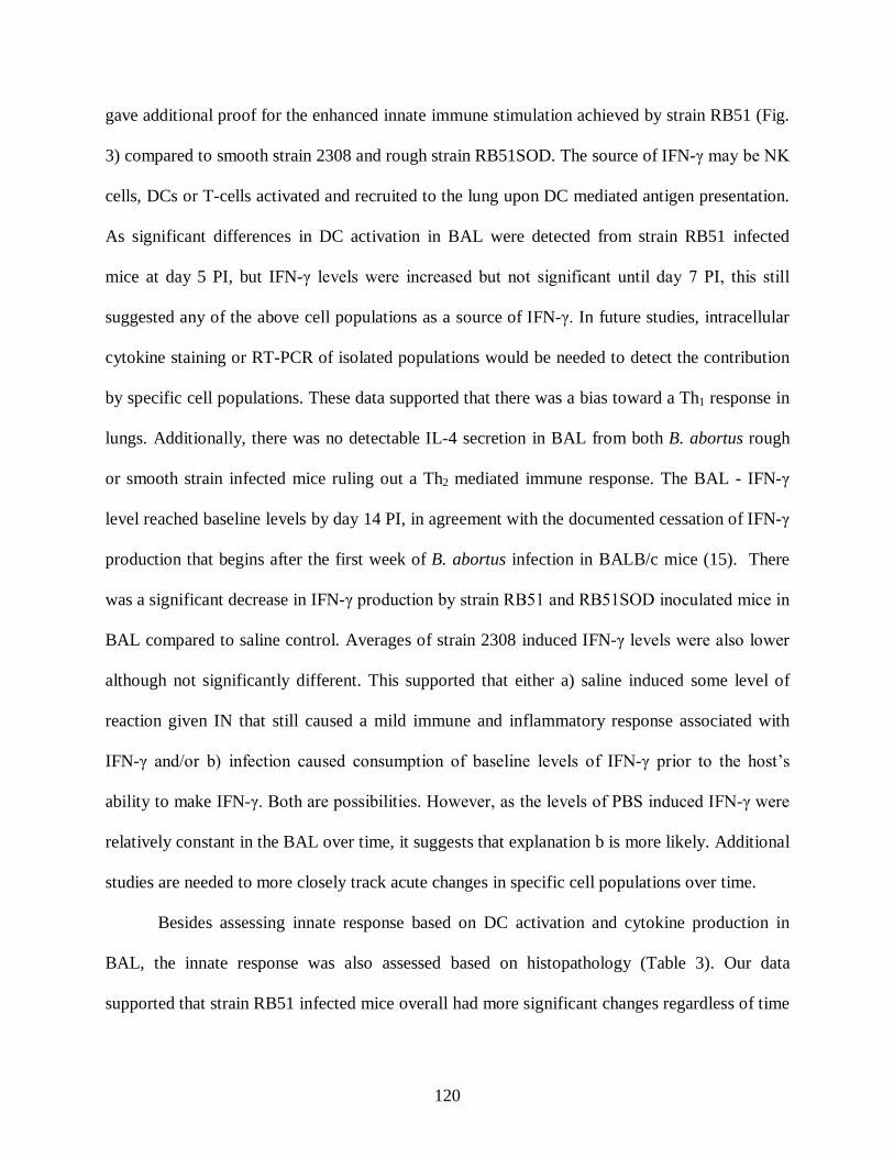
120
gave additional proof for the enhanced innate immune stimulation achieved by strain RB51 (Fig.
3) compared to smooth strain 2308 and rough strain RB51SOD. The source of IFN-γ may be NK
cells, DCs or T-cells activated and recruited to the lung upon DC mediated antigen presentation.
As significant differences in DC activation in BAL were detected from strain RB51 infected
mice at day 5 PI, but IFN-γ levels were increased but not significant until day 7 PI, this still
suggested any of the above cell populations as a source of IFN-γ. In future studies, intracellular
cytokine staining or RT-PCR of isolated populations would be needed to detect the contribution
by specific cell populations. These data supported that there was a bias toward a Th1 response in
lungs. Additionally, there was no detectable IL-4 secretion in BAL from both B. abortus rough
or smooth strain infected mice ruling out a Th2 mediated immune response. The BAL - IFN-γ
level reached baseline levels by day 14 PI, in agreement with the documented cessation of IFN-γ
production that begins after the first week of B. abortus infection in BALB/c mice (15). There
was a significant decrease in IFN-γ production by strain RB51 and RB51SOD inoculated mice in
BAL compared to saline control. Averages of strain 2308 induced IFN-γ levels were also lower
although not significantly different. This supported that either a) saline induced some level of
reaction given IN that still caused a mild immune and inflammatory response associated with
IFN-γ and/or b) infection caused consumption of baseline levels of IFN-γ prior to the host’s
ability to make IFN-γ. Both are possibilities. However, as the levels of PBS induced IFN-γ were
relatively constant in the BAL over time, it suggests that explanation b is more likely. Additional
studies are needed to more closely track acute changes in specific cell populations over time.
Besides assessing innate response based on DC activation and cytokine production in
BAL, the innate response was also assessed based on histopathology (Table 3). Our data
supported that strain RB51 infected mice overall had more significant changes regardless of time

121
as strain RB51 infected mice induced higher severity of pneumonia compared to all the other
treatment groups. Strain RB51 infected mice overall had significantly greater scores in severity
of pneumonia, but nonsignificant increase in percentage of multifocal lymphocytic histiocytic
pneumonia with increased severity of vascular change including fibrinoid necrosis and
perivascular infiltrate. Only strain RB51 infected mice, except for 1 strain RB51SOD infected
mouse, had lymphocytic perivascular infiltrate compared to the other treatment groups.
Additionally, overall strain RB51 infected mice had greater, but non significant, severity of
septal thickening due to macrophages and lymphocytic infiltrate along with increased severity of
debri and protein in the airways. By comparison, strain RB51SOD infected mice had less severe
changes than strain RB51. Finally, strain 2308 infected mice had the least severe average
changes in pneumonia and percentage involvement overall as well as other parameters compared
to the other strains.
Minimal changes were present in the liver and spleen from the infected mice.
Interestingly, whereas only strain RB51 infected mice had lymphocytic perivascular infiltrate in
the lung, none of the strain 2308 infected mice had perivascular infiltrate. Based on the
parameters assessed, strain RB51 had more severe changes for all parameters vs. strain
RB51SOD and strain 2308. The difference in the perivascular infiltrate of strain RB51 was
striking. The mechanism for it is not known. Possible explanations include differences in
endothelial cell activation with or without direct infection of cells, and/or differences in DC
activation, cytokine production by DCs, macrophages, NK cells; any of these changes could
cause differences in cell recruitment. Thus, the mechanism of infiltrate and its corresponding role
in the innate immune response and associated protection warrants further investigation.

122
Overall, these data supported that strain RB51 enhanced DC activation, cytokine
production and inflammatory responses in vivo compared to strains RB51SOD and 2308. While
both rough strains were cleared following IN infection and strain 2308 was not, these data also
supported that some rough strains may enhance innate immune function and protect better than
pathogenic smooth strains that cause persistent infection. There are likely different factors
affecting clearance vs. ability to stimulate protective immunity. In this study as well as others
(Surendran et al., accepted; Veterinary Microbiology; Sriranganathan and Boyle, personal
communication), strain RB51SOD did not stimulate innate response as well as strain RB51. In
vitro studies (Surendran et al., accepted; Veterinary Microbiology) showed that strain RB51 had
greater DC activation and function in BMDC mouse model compared to strain RB51SOD.
Possible explanations for the decreased response to RB51SOD, which are being investigated,
include: whether over-expressed SOD was functional; if it could decrease the inflammation and
subsequent immune response and/or whether SOD biases the DC mediated T-cell response to a
reduced Th1 response compared to strain RB51. Preliminary studies by others (Sriranaganathan
and Boyle, personal communication) support the latter. Although mouse studies supported that
strain RB51SOD was more protective (29), Olsen et al. (18) demonstrated in bison that strain
RB51 showed greater protection compared to strain RB51SOD. There are no other published
studies assessing innate response to strain RB51 vs. RB51SOD. These studies are the first to
assess innate response to strain RB51 and RB51SOD both in vitro and in vivo. As part of these
differences in function, the mechanism for the perivascular infiltrate and its role in the innate
response of strain RB51 warrants additional investigation. The results have a significant impact
on what vaccines may be used in the future both for Brucella as well as using Brucella as a
platform for multivalent vaccines.

123
Acknowledgements: The authors acknowledge Dr. Ramanathan Kasimanickyam and Dr.
Stephen Werre for help with the statistical analysis of the data and Ms. Joan Kalnitsky for help
with flow cytometry.
References
1. Bhattacharjee, A. K., L. Van de Verg, M. J. Izadjoo, L. Yuan, T. L. Hadfield, W. D.
Zollinger, and D. L. Hoover. 2002. Protection of mice against brucellosis by intranasal
immunization with Brucella melitensis lipopolysaccharide as a noncovalent complex with
Neisseria meningitidis group B outer membrane protein. Infect Immun 70:3324-3329.
2. Billard, E., J. Dornand, and A. Gross. 2007. Brucella suis prevents human dendritic
cell maturation and antigen presentation through regulation of tumor necrosis factor alpha
secretion. Infect Immun 75:4980-4989.
3. Billard, E., J. Dornand, and A. Gross. 2007. Interaction of Brucella suis and Brucella
abortus rough strains with human dendritic cells. Infect Immun 75:5916-5923.
4. Corbel, M. J. (ed.). 2006. Brucellosis in humans and animals. World Health
Organization.
5. Cunliffe, S. L., J. R. Wyer, J. K. Sutton, M. Lucas, G. Harcourt, P. Klenerman, A. J.
McMichael, and A. D. Kelleher. 2002. Optimization of peptide linker length in
production of MHC class II/peptide tetrameric complexes increases yield and stability,
and allows identification of antigen-specific CD4+T cells in peripheral blood
mononuclear cells. Eur J Immunol 32:3366-3375.
124
6. Ferrero, M. C., C. A. Fossati, and P. C. Baldi. 2009. Smooth Brucella strains invade
and replicate in human lung epithelial cells without inducing cell death. Microbes Infect
11:476-483.
7. Jakubzick, C., F. Tacke, J. Llodra, N. van Rooijen, and G. J. Randolph. 2006.
Modulation of dendritic cell trafficking to and from the airways. J Immunol 176:3578-
3584.
8. Jimenez de Bagues, M. P., P. H. Elzer, S. M. Jones, J. M. Blasco, F. M. Enright, G.
G. Schurig, and A. J. Winter. 1994. Vaccination with Brucella abortus rough mutant
RB51 protects BALB/c mice against virulent strains of Brucella abortus, Brucella
melitensis, and Brucella ovis. Infect Immun 62:4990-4996.
9. Kahl-McDonagh, M. M., A. M. Arenas-Gamboa, and T. A. Ficht. 2007. Aerosol
infection of BALB/c mice with Brucella melitensis and Brucella abortus and protective
efficacy against aerosol challenge. Infect Immun 75:4923-4932.
10. Kaufmann, A. F., M. D. Fox, J. M. Boyce, D. C. Anderson, M. E. Potter, W. J.
Martone, and C. M. Patton. 1980. Airborne spread of brucellosis. Ann N Y Acad Sci
353:105-114.
11. Kreuwel, H. T., J. A. Biggs, I. M. Pilip, E. G. Pamer, D. Lo, and L. A. Sherman.
2001. Defective CD8+ T cell peripheral tolerance in nonobese diabetic mice. J Immunol
167:1112-1117.
12. Mense, M. G., R. H. Borschel, C. L. Wilhelmsen, M. L. Pitt, and D. L. Hoover. 2004.
Pathologic changes associated with brucellosis experimentally induced by aerosol
exposure in rhesus macaques (Macaca mulatta). Am J Vet Res 65:644-652.

125
13. Mense, M. G., L. L. Van De Verg, A. K. Bhattacharjee, J. L. Garrett, J. A. Hart, L.
E. Lindler, T. L. Hadfield, and D. L. Hoover. 2001. Bacteriologic and histologic
features in mice after intranasal inoculation of Brucella melitensis. Am J Vet Res 62:398-
405.
14. Miller, C. D., J. R. Songer, and J. F. Sullivan. 1987. A twenty-five year review of
laboratory-acquired human infections at the National Animal Disease Center. Am Ind
Hyg Assoc J 48:271-275.
15. Murphy, E. A., M. Parent, J. Sathiyaseelan, X. Jiang, and C. L. Baldwin. 2001.
Immune control of Brucella abortus 2308 infections in BALB/c mice. FEMS Immunol
Med Microbiol 32:85-88.
16. Murphy, E. A., J. Sathiyaseelan, M. A. Parent, B. Zou, and C. L. Baldwin. 2001.
Interferon-gamma is crucial for surviving a Brucella abortus infection in both resistant
C57BL/6 and susceptible BALB/c mice. Immunology 103:511-518.
17. Neutra, M. R., and P. A. Kozlowski. 2006. Mucosal vaccines: the promise and the
challenge. Nat Rev Immunol 6:148-158.
18. Olsen, S. C., S. M. Boyle, G. G. Schurig, and N. N. Sriranganathan. 2009. Immune
responses and protection against experimental challenge after vaccination of bison with
Brucella abortus strain RB51 or RB51 overexpressing superoxide dismutase and
glycosyltransferase genes. Clin Vaccine Immunol 16:535-540.
19. Olsen, S. C., W. R. Waters, and W. S. Stoffregen. 2007. An aerosolized Brucella spp.
challenge model for laboratory animals. Zoonoses Public Health 54:281-285.
20. Pappas, G., P. Panagopoulou, L. Christou, and N. Akritidis. 2006. Brucella as a
biological weapon. Cell Mol Life Sci 63:2229-2236.
126
21. Pilette, C., Y. Ouadrhiri, V. Godding, J. P. Vaerman, and Y. Sibille. 2001. Lung
mucosal immunity: immunoglobulin-A revisited. Eur Respir J 18:571-588.
22. Sambrook J, F. E. a. M. T. 1989 Molecular cloning. A Laboratory Manual., vol. 2nd ed.
Cold Spring Harbor Laboratory Press, New York.
23. Schurig, G. G., R. M. Roop, 2nd, T. Bagchi, S. Boyle, D. Buhrman, and N.
Sriranganathan. 1991. Biological properties of RB51; a stable rough strain of Brucella
abortus. Vet Microbiol 28:171-188.
24. Schurig, G. G., N. Sriranganathan, and M. J. Corbel. 2002. Brucellosis vaccines: past,
present and future. Vet Microbiol 90:479-496.
25. Seleem, M., M. Ali, M. W. Abd Al-Azeem, S. M. Boyle, and N. Sriranganathan.
2007. High-level heterologous gene expression in Ochrobactrum anthropi using an A-
rich UP element. Appl Microbiol Biotechnol 73:1123-1127.
26. Seleem, M., Rajasekaran P, Ali M, Boyle SM & Sriranganathan N 2008. Simple
method for transformation of Ochrobactrum anthropi. World Journal of Microbiology
and Biotechnology 24:2111-2114.
27. Seleem, M. N., R. Vemulapalli, S. M. Boyle, G. G. Schurig, and N. Sriranganathan.
2004. Improved expression vector for Brucella species. Biotechniques 37:740, 742, 744.
28. Smither, S. J., S. D. Perkins, C. Davies, A. J. Stagg, M. Nelson, and H. S. Atkins.
2009. Development and characterization of mouse models of infection with aerosolized
Brucella melitensis and Brucella suis. Clin Vaccine Immunol 16:779-783.
29. Vemulapalli, R., Y. He, S. Cravero, N. Sriranganathan, S. M. Boyle, and G. G.
Schurig. 2000. Overexpression of protective antigen as a novel approach to enhance
vaccine efficacy of Brucella abortus strain RB51. Infect Immun 68:3286-3289.
127
30. Vemulapalli, R., Y. He, N. Sriranganathan, S. M. Boyle, and G. G. Schurig. 2002.
Brucella abortus RB51: enhancing vaccine efficacy and developing multivalent vaccines.
Vet Microbiol 90:521-532.
31. Witonsky, S. G., R. M. Gogal, Jr., R. B. Duncan, and D. S. Lindsay. 2003.
Immunopathologic effects associated with Sarcocystis neurona-infected interferon-
gamma knockout mice. J Parasitol 89:932-940.
Figure legends
Figure 1. Bacterial clearance from BALB/c mice following IN infection with B. abortus rough
and smooth strains. BALB/c mice (n=4) were infected IN with strains RB51, RB51SOD (4 X 10⁷
CFUs/mouse) and 2308 (2 X 10³ CFUs/mouse). Mice were euthanized and the number of
bacteria recovered from lung, mediastinal lymph node (MLN) and spleen were counted on day 7,
14 and 42 post infection for rough strains (A) and on day 7, 16 and 42 post infection for strain
2308 (B). Results represent mean values ± standard deviation.
Figure 2. In vivo dendritic cell (DC) maturation in response to IN infection with B. abortus
rough and smooth strains. On day 3, 5, 7 and 14 following IN infection with B. abortus rough
(RB51, RB51SOD; 4 X 10⁹ CFUs/mouse) and smooth strains (2308; 2 X 10⁵ CFUs/mouse)
expressing GFP, broncho-alveolar lavage (BAL) and mediastinal lymph node (MLN) were
collected and stained with anti-mouse CD11c and MHC class II (I-A/I-E) antibodies. PBS
inoculated mice served as control. (A) Percentage of gated cells in BAL expressing
CD11c+/MHC class II high markers from different treatment groups at day 3, 5, 7 and 14 post
infection. Data represent results pooled from BAL samples of 2 mice per time point per
treatment group from 4 independent experiments. The graph shows median ± 75th percentile. (B)

128
Percentage of gated cells in MLN expressing CD11c+/MHC class II high markers from different
treatment groups at day 3, 5, 7 and 14 post infection. Data represent results from MLN samples
of 2 mice per time point per treatment group from 4 independent experiments. The graph shows
median ± 75th percentile. Double characters ** and ## represent statistically significant change at
p ≤ 0.05 with the corresponding single character representation (*, #). For MLN – CD11c+/MHC
class II high expression at day 14, p values for RB51, RB51SOD and 2308 in comparison to PBS
control mice were 0.052, 0.061 and 0.093 respectively.
Figure 3. IFN-γ secretion in BAL following IN infection with B. abortus rough and smooth
strains. On day 3, 5, 7 and 14 following IN infection with B. abortus rough (RB51, RB51SOD; 4
X 109 CFUs/mouse) and smooth strains (2308; 2 X 10⁵ CFUs/mouse) expressing GFP, broncho-
alveolar lavage (BAL) was collected and IFN-γ level assessed by indirect sandwich ELISA. PBS
inoculated mice served as control. Data represent results from pooled BAL samples of 2 mice per
time point per treatment group from 4 independent experiments. The graph shows mean values ±
standard deviation. Two asterisks (**) denote statistically significant data at p ≤ 0.05 compared
to data represented by an asterisk (*)
Figure 4. Variation in pneumonia severity score following IN infection with B. abortus rough
and smooth strains. Strain RB51 induced significantly more severe pneumonia compared to PBS,
RB51SOD and 2308 treatments. Line represents mean severity score bracketed by 95%
confidence interval. Two asterisks (**) denote statistically significant data at p ≤ 0.05 compared
to data represented by an asterisk (*)
Table 3. Histopathological changes in lung tissue after intranasal inoculation with saline or
rough and smooth strains of B. abortus. Based on histopathological pulmonary changes at day 3,
5, 7 or 14 post infection with saline, rough or smooth strains of B. abortus; lungs were
129
characterized based on pneumonia, vascular change, septa thickening, as well as changes to the
pleura, large airway and alveoli. For all parameters, average (± standard deviation) severity was
graded as 0-5 (0-1 no change, 1-2 minimal, 2-3 mild, 3-4 moderate, 4-5 severe, >5 marked or
extreme).
Figure 5. Histopathology of lungs from BALB/c mice following intranasal infection with B.
abortus strains compared to saline control. BALB/c mice were infected with saline or respective
rough RB51, RB51SOD and smooth strain 2308. At days 3, 5, 7 and 14 post-infection, samples
were collected for clearance, flow cytometry and histopathology. Samples from day 5 lung at
400 x magnification are shown here. (A) PBS control with minimal changes. (B) Strain RB51
infected mouse depicting mild lymphocytic perivascular infiltrate with fibrinoid necrosis of cell
wall along with multifocal lymphocytic and histiocytic interstitial pneumonia. (C) Strain
RB51SOD infected mouse with mild endothelial hypertrophy/activation and mild but decreased
perivascular infiltrate and multifocal lymphocytic and histiocytic interstitial pneumonia. (D)
Strain 2308 infected mouse with mild perivascular infiltrate and multifocal lymphocytic and
histiocytic interstitial pneumonia.
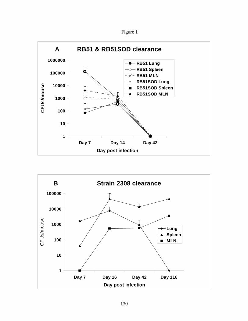
130
Figure 1
A RB51 & RB51SOD clearance
1
10
100
1000
10000
100000
1000000
Day 7 Day 14 Day 42
Day post infection
CFU
s/m
ouse
RB51 LungRB51 SpleenRB51 MLNRB51SOD LungRB51SOD SpleenRB51SOD MLN
B Strain 2308 clearance
1
10
100
1000
10000
100000
Day 7 Day 16 Day 42 Day 116
Day post infection
CFU
s/m
ouse
LungSpleenMLN

131
Figure 2
A BAL - CD11c+/MHC class II high
0102030405060708090
100
D3 D5 D7 D14
Day post infection
Perc
enta
ge (%
) of g
ated
ce
lls
PBS RB51 RB51SOD 2308
*
**
*
**
*
**
*
**
* * *
** #
##
B MLN - CD11c+/MHC class II high
0102030405060708090
100
D3 D5 D7 D14
Day post infection
Perc
enta
ge (%
) of g
ated
ce
lls
PBS RB51 RB51SOD 2308
**
*
** **
*
*
**

132
Figure 3
BAL: IFN-gamma
050
100150200250300350400450
D3 D5 D7 D14
Day post infection
Con
cent
ratio
n (p
g/m
l)
PBS RB51 RB51SOD 2308
**
*
*
** **
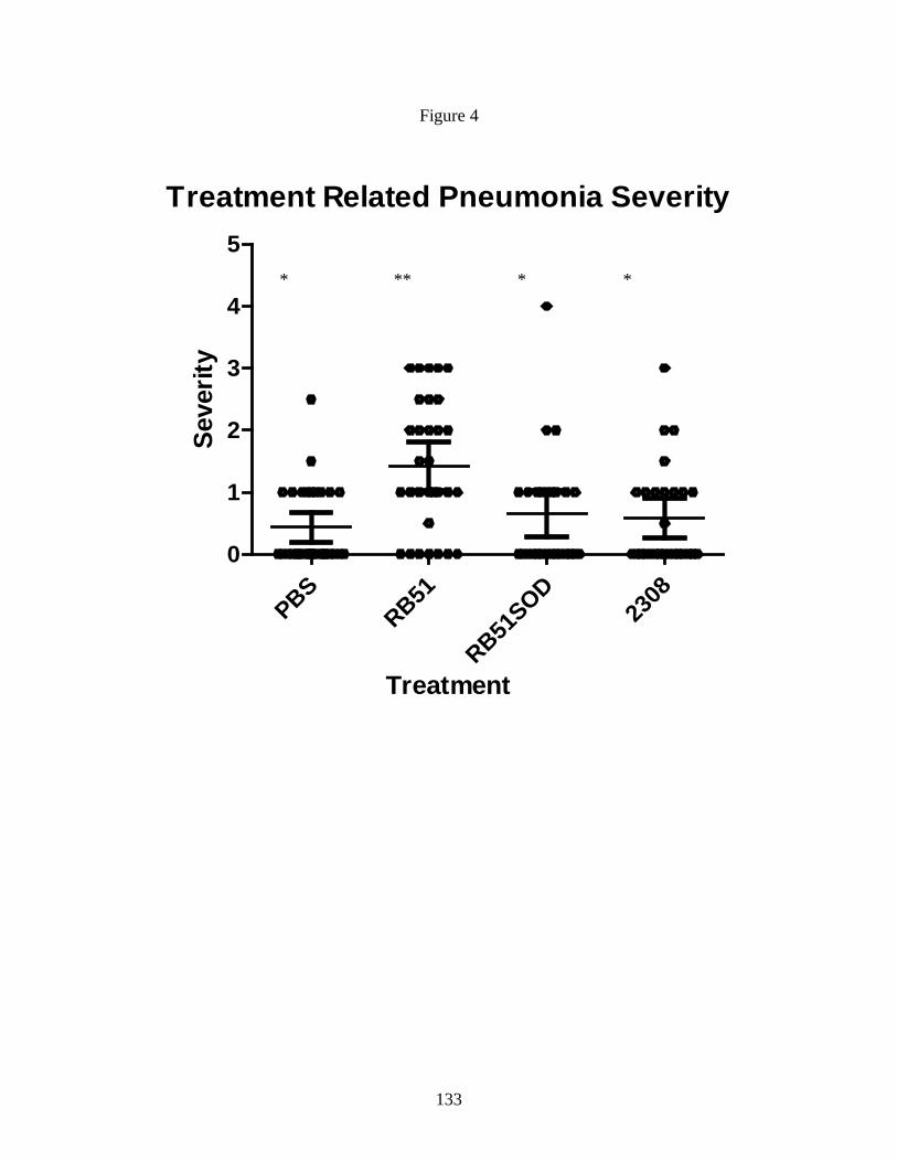
133
Figure 4
Treatment Related Pneumonia Severity
PBSRB51
RB51SOD
2308
0
1
2
3
4
5
Treatment
Seve
rity
** * * *

134
Table 3
Day post infection
Treatment
Vascular change Septa thickening
Debris/protein
Severity
PBS 0.55 (0.72) 5 (6.6) 0.33 (0.70) 0.22 (0.44) 0.33 (0.5)RB51 1.5 (1.19) 17.25 (20.54) 0.57 (1.0) 1.0 (1.41) 0.875 (1.24)
3 RB51SOD 0.875 (1.45) 12.5 (27.64) 0.375 (0.74) 0.22 (0.35) 0.57 (0.75)2308 0.56 (0.72) 7.87 (11.41) 0.142 (0.37) 0.428 (0.78) 0.285 (0.75)PBS 0.375 (0.51) 8.1 (12.51) 0.125 (0.35) 0.25 (0.46) 0.5 (0.53)
RB51 1.68 (1.27) 24.3 (24.99) 0.5 (0.75) 1.5 (1.41) 1.125 (0.83)5 RB51SOD 0.57 (0.53) 12.14 (17.76) 0 (0) 0.57 (0.53) 0.285 (0.48)
2308 0.625 (1.0) 20 (29.76) 0 (0) 0.5 (0.75) 0.375 (0.74)PBS 0.285 (0.48) 2.857 (4.8) 0 (0) 0.285 (0.48) 0.142 (0.37)
RB51 1.07 (0.93) 15.71 (16.93) 0.142 (0.37) 0.857 (0.89) 0.714 (0.75)7 RB51SOD 0.375 (0.51) 9.3 (20.77) 0 (0) 0.375 (0.51) 0.375 (0.51)
2308 0.44 (0.52) 8.33 (13.22) 0 (0) 0.375 (0.51) 0.125 (0.35)PBS 0.857 (0.94) 16.42 (23.22) 0.285 (0.75) 0.57 (1.13) 0.285 (0.75)
RB51 1.375 (0.91) 19.75 (14.70) 0.57 (0.97) 1.28 (0.75) 0.57 (0.53)14 RB51SOD 0.75 (0.95) 20 (33.66) 0 (0) 0.75 (0.95) 0.57 (0.5)
2308 0.9 (0.89) 16 (25.34) 0 (0) 1 (1.0) 0.33 (0.57)
Pulmonary Changes
Pneumonia
Percentage (%) involvement

135
Figure 5
A. PBS
B. RB51

136
C. RB51SOD
D. Strain 2308
137
Chapter 5
Role of TLRs in Brucella abortus mediated murine dendritic cell activation in vitro and
clearance of pulmonary infection in vivo.
Running title: Interaction of B. abortus with TLR KO BMDC in vitro and TLR KO mice in vivo
Naveen Surendran*, Elizabeth M. Hiltbold†, Bettina Heid*, Shizuo Akira‡, Theodore J.
Standiford§, Nammalwar Sriranganathan¶, Stephen M. Boyle¶, Kurt L. Zimmerman¶, Melissa R.
Makris¶, Sharon G. Witonsky*1. (To be submitted to Journal of Immunology)
Abstract
Brucellosis is worldwide zoonoses affecting 500,000 people annually with no approved
human vaccines available. Live attenuated B. abortus vaccine strain RB51 protects cattle through
CD4 and CD8 T-cell mediated responses. However, limited information is known regarding how
Brucella stimulates innate immunity. While Brucella is known to bind TLR2, TLR4 and TLR9,
studies report conflicting results as to level of importance of each TLR. Furthermore, the most
critical toll like receptor(s) (TLRs) for vaccine strain RB51 mediated DC activation is not
known. Additionally, the most critical TLR for clearance is not known in response to
aerosol/respiratory infection of Brucella. Therefore, in this study, we assessed the differential
ability of strain RB51 and strain 2308 to stimulate murine bone marrow derived dendritic cell
(BMDCs) activation and function from TLR2, TLR4 or TLR9 knockout and control BALB/c
mice. Complementary in vivo experiments were also conducted to differentially assess the role of
TLRs on clearance following intranasal infection of pathogenic strain 2308. We determined that
strain RB51 induced significant (p≤0.05) DC activation compared to strain 2308 that was

138
independent of a specific TLR. However, strain RB51 induced TNF – α production was TLR2
and TLR9 dependent and IL-12 production was TLR2 and TLR4 dependent. TLR4 KO mice had
significantly (p≤0.05) delayed pulmonary clearance of strain RB51 in vivo at day 14 post
infection. Nevertheless, clearance of strain 2308 was not significantly different among organs
between control and KO mice.
Introduction
Brucellosis is worldwide zoonoses affecting 500,000 people annually (1, 2). The
causative organism is a Gram-negative, facultative intracellular bacterium from the genus
Brucella classified under α-2 Proteobacteria (3). B. melitensis, B. suis and B. abortus are the
most virulent species in order of pathogenicity (4). Of these, B. abortus causes infertility and
abortion in cattle and undulant fever in humans (4). Brucella can be easily aerosolized,
genetically modified to make antibiotic resistant strains and has the potential to be used as a bio-
terror weapon (2). The Center for Disease Control and Prevention (CDC) categorizes it as a class
B pathogen. There are no approved human vaccines available.
B. abortus strain RB51 is an efficacious live attenuated rough vaccine approved by
United States Department of Agriculture (USDA) against bovine brucellosis (5). Strain RB51,
which lacks the O-side chain lipopolysaccharide (LPS) is a mutant of pathogenic B. abortus
strain 2308 (5). Protection afforded by strain RB51 is mediated through IFN-γ secreting CD4 T-
helper1 (Th1) and cytotoxic CD8 T - cells (Tc1) (6). In order to stimulate a robust adaptive
response, a solid innate response is needed. However, the means by which B. abortus induces
this response have not been fully elucidated.
139
The initial host defense to any bacterial infection involves innate immune cells, including
neutrophils, natural killer (NK) cells, macrophages and dendritic cells (7). Barquero-Calvo et al.
demonstrated that neutrophils do not have a significant role in control of B. abortus (8).
Although a good source of IFN-gamma, NK cells do not contribute to antigen presentation
whereas both macrophages and dendritic cells (DCs) are efficient antigen presenting cells
(APCs) (7). As DCs compared to macrophages are 100 times more efficient in antigen capture;
have the ability to migrate from peripheral tissue to draining lymph node with the antigen to
present the processed antigen to naïve T-cells; produce higher IL-12; have higher rates of
Brucella infection; and are a critical link between innate and adaptive immunity, DCs are an
ideal model for studying Brucella mediated innate immune response (7, 9, 10).
DC activation is initiated by Toll-like receptors (TLRs) recognizing pathogen associated
molecular patterns (PAMPs) (11). Subsequently, TLRs transduce signals via common adaptor
molecules to activate DCs to produce proinflammatory cytokines (12). There are contradictory
data on the most crucial TLRs in recognition of B. abortus by DCs. Weiss et al. (13)
demonstrated that Brucella signals through TLR2, TLR4, and MyD88. As the latter is most
critical for clearance, this suggests a role for TLR9, which also signals through MyD88.
Subsequently, Oliveira et al.(14) and Huang et al. (15) suggested a prominent role for TLR9 in
DC mediated IL-12 production and Brucella clearance. Zwerdling et al. (16) also suggests that
Brucella signals through TLR2 and TLR4. Altogether, published literature suggests that B.
abortus signals through multiple TLRs, including TLR2 (outer membrane proteins), TLR4
(lipopolysaccharide), and TLR9 (CpG DNA). Downstream MyD88 is implicated as the most
critical adapter protein involved in signaling, which leads to NF-κB activation, DC function and
ultimately Brucella clearance in vivo (17, 18). Despite these published TLR studies, there is a

140
dearth of information on whether a differential ability of TLRs for activating DCs exists between
rough and smooth Brucella strains. If present, innate activation of strain RB51 could be
enhanced by using TLR agonists as adjuvants. To answer this question, we infected TLR2,
TLR4, TLR9 KO BALB/c mice derived bone marrow DCs (BMDCs) along with wild type
control BMDCs to analyze differences in activation and function between B. abortus rough strain
RB51 and smooth strain 2308 infected BMDCs.
Additionally, assessing Brucella clearance from TLR KO mice following respiratory
exposure will also provide critical information both on whether vaccine strains can be
upregulated through TLR agonists as well as whether respiratory clearance of B. abortus strain
2308 is altered compared to other routes of challenge. With either natural or intentional
(bioterror) aerosol exposure, Brucella is directly delivered to the pulmonary airway and airway
epithelium. Currently, there are no published studies which have addressed the role of TLRs in
the clearance of rough or smooth B. abortus strains from intranasally infected mice (19).
Therefore, to address the role of TLRs in B. abortus clearance in a pulmonary infection, we
infected BALB/c as well as TLR2, TLR4, TLR9 KO mice with strain RB51 and 2308 to assess
TLR dependent clearance. Our overall aim for this study was to determine the role of TLR2,
TLR4 and TLR9 in the differential activation of DCs upon infection with B. abortus rough and
smooth strains in vitro and in the clearance of Brucella challenge in vivo.
Materials and Methods
Mice: Female 4-6 weeks old BALB/c mice were obtained from Charles River Laboratories Inc.,
Wilmington, MA. TLR4 and TLR9 knockout (KO) mice breeding pairs in BALB/c background
were obtained from S. Akira (Department of Host Defense, Research Institute for Microbial
141
Diseases, Osaka University, Japan). TLR2 KO mice breeding pairs in BALB/c background were
obtained through Dr. Akira from Dr. Standiford (University of Michigan, Ann Arbor, MI).
Female, 6-8 week old control and KO mice were used for all the experiments under animal care
protocols approved by the Institutional Animal Care and Use Committee at Virginia Tech.
Generation of Bone marrow derived DCs: Bone marrow-derived DCs (BMDCs) were
generated, as previously described (20). Briefly, tibias and fibulas of 7-8 weeks old BALB/c
control and KO mice were incised and bone marrow (BM) cells removed. Following red blood
cell lysis and filtration, the cells were resuspended and plated in RPMI 1640 complete media
with 10% non heat-inactivated fetal bovine serum and 20ng/ml rGM-CSF (Invitrogen, Carlsbad,
CA). The cells were incubated at 37°C in 5% C02. Fresh media containing rGM-CSF was added
at days 2, 4 and 5 and harvested on day 6. The cells harvested on day 6 were typically 70%
CD11c+ and displayed low levels of MHC class II, CD40 and CD86, consistent with immature
DCs. Flow cytometry was performed to confirm immature DC status.
Brucella strains: Live attenuated rough B. abortus strain RB51 and virulent smooth strain 2308
were used from our stock culture collection (5). All experiments with Brucella were performed
in our CDC approved (C2003 1120-0016) Biosafety Level (BSL)-3 facility.
DC Infection experiments: On day 6, control and TLR2, TLR4 and TLR9 KO DCs were
harvested and plated at 5 X 10⁵ cells/well in separate 24 well plates and infected with strain
RB51 or strain 2308 at each of the three multiplicities of infection (MOI) 1:1 (DC:Brucella),
1:10 and 1:100. Infection was enhanced by a short spin at 1300 rpm (400 x g) for 5 minutes at
room temperature. The infected cells were incubated for 4 hours at 37°C in 5% C02. The
infection was terminated by washing the cells with gentamicin (Sigma, St. Louis, MO) at
30μg/ml. The cells were then incubated for an additional 20 hours in complete media with
142
10ng/ml rGM-CSF and 30μg/ml gentamicin. Control samples were maintained by incubating
cells with media (negative control) or Escherichia coli LPS 0111:B4 (Sigma) (positive control)
(100ng/ml) following the exact same procedure for infection. In replicate experiments, at each
time point for each treatment, an aliquot of cells was collected to determine bacteria cell
numbers. Cells were washed to remove non-intracellular bacteria, and total BMDCs were
counted. BMDCs were then lysed by treating with 1 ml/well of 0.1% Triton x-100 in sterile
distilled water for 10 minutes, mixed well and 10-fold serial dilutions were plated onto TSA
plates. Intracellular bacteria were counted and number of bacteria per cell was determined.
Viability and infection controls: To quantitate and assess viability of control and KO BMDCs at
24 hour post infection with each treatment, Trypan blue was used to differentiate viable and dead
cells. Total live and dead BMDC cell numbers were determined.
Staining and flow cytometry: The cells were harvested 24 hours following infection, and they
were stained with the following monoclonal antibodies at 0.1 - 0.2 μg per million cells for FACS
analysis: APC conjugated anti-CD11c, pacific blue-conjugated anti-CD86 (Biolegend, San
Diego, CA), Biotin-conjugated anti-CD40, Streptavidin Tri-color conjugate, were all acquired
from Caltag (Invitrogen), and PE-conjugated anti I-A/I-E, acquired from BD Pharmingen, San
Jose, CA. Cells were washed and analyzed by BD FACSAria™ flow cytometer.
Cytokine analysis: For cytokine measurement, culture supernatants from Brucella infected
BMDCs were collected after 24 hours of incubation and stored at -80°C. TNF-α, IL-12p70
(bioactive form of IL-12) IFN-γ and IL-4 cytokine levels were subsequently measured using
indirect sandwich ELISAs (BD Pharmingen).
Mice infection and clearance study: Age matched female control and KO BALB/c mice were
infected intranasally (IN), under light xylazine-ketamine anesthesia IP, with either B. abortus
143
rough strains RB51 (4 X 10⁸ CFUs/mouse) or with smooth strain 2308 (2 X 10⁴ CFUs/mouse) in
25 μl phosphate buffered saline (PBS). Mice were euthanized on day 14 and day 42 post
infection (PI) with a lethal dose of xylazine-ketamine IP. Day 42 post infection time point was
not performed for TLR2 KO mice. At the time of euthanasia, lung, MLN and spleen were
collected. Single cell suspensions of organs collected were serially diluted and plated on to
Tryptic soy agar (TSA) plates and incubated for 5 days at 37° C and 5% CO2 (21). Bacterial
colony forming units (CFUs) were counted and CFUs/mouse were calculated.
Statistical analysis: The effect of TLRs and treatments on expression of various DC maturation
and activation markers was tested using mixed model ANOVA followed by Tukey’s procedure
for multiple comparisons with date of experiment as a blocking factor. Statistical significance of
cytokine production in BMDC supernatant and effect of TLRs on in vivo clearance of Brucella
strains was assessed by exact Kruskal-Wallis test followed by Dunn’s procedure for multiple
comparisons. Significance was set at p ≤ 0.05. All analyses were performed using the SAS
system (Cary, NC, USA).
Results
E. coli LPS downregulates CD11c surface expression on bone marrow derived DCs
through TLR4. To determine that the differences in DC mediated activation and function were
not due to differences in viability of Brucella and/or BMDCs, DC viability and Brucella
numbers were analyzed. Our data showed that at 4hrs there were relatively similar levels of
Brucella: BMDCs (data not shown). After overnight incubation, the percentage yield of CD11c+
cells from uninfected media control and strain 2308 infected cells ranged from 81-92 % and 79-
91 % (all 3 MOIs of 2308 from control and KO mice) respectively. The percentage yield of

144
CD11c+ cells was significantly (p≤0.05) lower for E. coli LPS treated control as well as KO mice
derived cells (57–81%) compared to uninfected media treated cells possibly due to activation
induced downregulation and a marginal nonsignificant increase in cell death (Figure 1). LPS
induced CD11c expression was also significantly lower (p≤0.05) than strain 2308 (all MOIs) in
wild type mice and strain RB51 (at MOI 1:1, 1:10 in wild type mice and also for TLR2 KO at
1:100) induced CD11c expression except for TLR4 KO mice. However, E. coli LPS induced
CD11c expression was significantly (p≤0.05) higher for TLR4 KO (81%) mice compared to
control, TLR2 and TLR9 KO mice suggesting that the LPS induced CD11c downregulation is
predominantly mediated through TLR4 (Table I). At MOI 1:1 and 1:10, strain RB51 induced
CD11c expression (80-88%) of control and KO cells were similar to media (data not shown)
whereas at MOI 1:100, strain RB51 induced CD11c expression was significantly (p≤0.05) lower
than media treated cells except for TLR9 KO cells, showing a dose response (Figure 1). For
BALB/c control, the level of RB51 (MOI 1:100) induced CD11c expression was even
significantly lower than strain 2308 (MOI 1:100) induced expression (Figure 1). Nevertheless, at
MOI 1:100, strain RB51 induced CD11c expression was not significantly different between
TLR4 KO and other mouse cells.
Rough strain RB51 up-regulated MHC class II expression in control and TLR KO
BMDCs. Immature BMDCs from control and KO mice were treated with all 3 MOIs of strain
RB51 or strain 2308 to analyze the role of TLRs and the effect of infection on DC maturation.
As expected, control and KO BMDCs except TLR4 KO cells upregulated significant (p≤0.05)
MHC class II expression upon stimulation with E. coli LPS compared to respective media treated
BMDCs (Figure 2A). LPS mediated MHC class II expression was significantly (p≤0.05) higher
with control, TLR2 KO and TLR9 KO BMDCs compared to TLR4 KO BMDCs (Table I). At

145
MOI 1:100, strain RB51 induced significantly higher MHC class II expression of TLR2
(p=0.0556), TLR4 (p≤0.05) and TLR9 (p≤0.05) KO BMDCs compared to respective BMDCs
treated with media (Figure 2A) (Figure 3). Only TLR4 KO BMDCs expressed significantly
greater (p≤0.05) MHC class II up -regulation with strain RB51 at 1:10 (data not shown)
compared to media and strain RB51 1:100 compared to LPS (Figure 2A). Strain 2308 did not
induce significant up-regulation of MHC class II compared to media at any of the doses tested
with the exception of TLR4 KO BMDCs at MOI 1:100 (Figure 2A) (Figure 3).
In comparing control and KO mice BMDCs for their maturation response based on MHC
class II expression with respect to treatments with strain RB51 or strain 2308, strain RB51 at
MOI 1:100 induced significantly (p≤0.05) higher MHC class II expression in TLR 9 KO BMDCs
compared to TLR2 KO BMDCs and at MOI 1:1 and 1:10 compared to TLR4 KO BMDCs (strain
RB51 induced MHC class II is more TLR2 and 4 dependent than TLR9 dependent).
Additionally, strain 2308 induced MHC class II expression was significantly (p≤0.05) higher
with TLR9 KO BMDCs compared to TLR4 KO BMDCs at MOI 1:10 (Table I). (Strain 2308
induced MHC class II expression is more TLR4 than TLR9 dependent).
Rough strain RB51 induced significantly higher costimulatory marker CD40 and CD86
expression compared to smooth strain 2308 in control and TLR KO BMDCs. Control as
well as TLR KO BMDCs infected with strain RB51 induced significantly higher (p≤0.05) CD40,
CD86 expression and CD40+/CD86+ co-expression at both 1:10 and 1:100 MOIs compared to
media (Figure 2B – 2D). At MOI 1:100, strain RB51 induced CD40, CD40+/CD86+ (also at MOI
1:10 for TLR4 KO), and CD86 expression levels were significantly (p≤0.05) higher than LPS
induced upregulation of the same markers from respective BMDCs. Additionally, BMDCs from
each type of mice induced statistically significant (p≤ 0.05) up-regulation of all co-stimulatory

146
markers (except TLR4 KO CD40) when infected with strain RB51 at MOI 1:100 compared to
strain 2308 infected (MOI 1:100) BMDCs from the same type of mice (Figure 2B – 2D).
CD40+/CD86+ co-expression was significantly higher for strain RB51 infected BMDCs even at
MOI 1:10 compared to strain 2308 infected mice at MOI 1:10 (except for TLR2 KO BMDCs).
By comparison, strain 2308 induced significantly higher CD40 and CD86 (also at MOI
1:10 for CD86 expression with TLR4 KO) expression in all BMDCs compared to media treated
cells, only at MOI 1:100 (Figure 2B-C) (Figure 3). However, CD40+/CD86+ co-expression was
significantly higher for strain 2308 infected BMDCs (except TLR2 KO at MOI 1:10) at both
MOIs 1:10 and 1:100 compared to media (Figure 3). Additionally at MOI 1:100, TLR4 KO
BMDCs infected with strain 2308 induced significantly higher (p≤0.05) CD40, CD86 and
CD40+/CD86+ expression than E. coli LPS treated TLR4 KO BMDCs (Figure 2B – 2D) (Figure
3).
While comparing the level of expression of co-stimulatory markers on BMDCs from
control and TLR KO mice treated with different treatments, we found that CD40, CD86 and
CD40+/CD86+ co-expression on TLR4 KO BMDCs were significantly (p≤0.05) decreased
compared to control and TLR2, TLR9 KO BMDCs (Table I) (Figure 3). Additional significant
differences with LPS are also listed in Table 1. Interestingly at MOI 1:100 both strains RB51 and
2308 induced CD86 and CD40+/CD86+ co-expression was significantly (p≤0.05) decreased for
TLR2 KO mice compared to BALB/c control, TLR9 and TLR4 KO BMDCs (except for strain
2308 stimulated TLR4 KO BMDC - CD86 expression) (Table I) (Figure 3).
Rough strain RB51 induced DC – TNF-α secretion is TLR2 and partly TLR9 dependent.
TNF-α is an essential cytokine released by DCs undergoing maturation. Results from our
previous work (Surendran et al., under review) have established that while smooth strain 2308
147
inhibited TNF-α secretion by interfering with DC maturation and function, rough strain RB51 at
MOI 1:100 induced significantly higher (p≤0.05) TNF -α secretion compared to media control. In
this study, we further investigated the role of TLR2, TLR4 and TLR9 in Brucella induced DC –
TNF-α secretion. Figure 4A shows that both BALB/c control and TLR4 KO BMDCs infected
with strain RB51 at MOI 1:100 secreted similar levels of TNF-α which was significantly
(p≤0.05) higher than respective media, E. coli LPS controls as well as TLR2 KO BMDCs. Strain
RB51 1:100 infected TLR9KO BMDCs secreted significantly greater (p≤0.05) TNF-α compared
to respective media and LPS controls; however, the amount secreted was only one-fifth of the
BALB/c control and TLR4 KO cells. Furthermore, TNF-α induced by TLR9 KO BMDCs was
not significantly higher than that induced by TLR2 KO BMDCs.
Rough strain RB51 induced DC - IL-12p70 secretion is TLR2 and TLR4 dependent. IL-12 is
a key cytokine produced mainly by DCs upon pathogen recognition and subsequent maturation.
IL-12 helps to establish a Th1 response. We previously demonstrated (Surendran et al., under
review) that rough strain RB51 induced significantly (p≤0.05) higher IL -12 secretion from
BMDCs compared to smooth strain 2308. Figure 4B illustrates that both BALB/c control and
TLR9 KO BMDCs secreted similar levels of IL-12p70 in response to strain RB51 (MOI 1:100)
stimulation which was significantly (p≤0.05) higher than respective media and E. coli LPS
controls as well as TLR2 and TLR4 KO BMDCs. Irrespective of the type of mice BMDCs, strain
2308 did not induce significant production of IL-12p70 at any of the MOIs tested (Figure 4B).
Pulmonary clearance of rough strain RB51 is TLR4 dependent in vivo. To determine the
role of TLRs in the clearance of rough and smooth Brucella strains from IN infected animals,
TLR2, TLR4 and TLR9 KO mice were infected IN either with rough strain RB51 (4 X 10⁸
CFUs/ mouse) or with smooth strain 2308 (2 X 10⁴ CFUs/mouse). Bacterial clearance from lung,
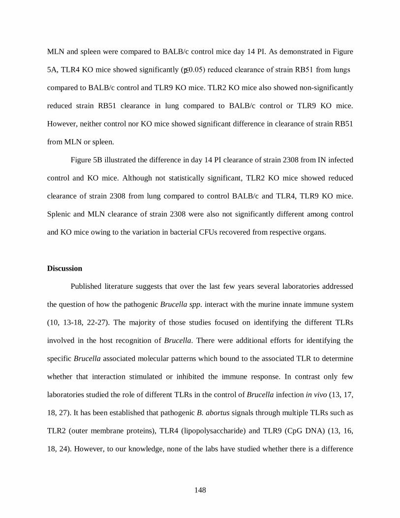
148
MLN and spleen were compared to BALB/c control mice day 14 PI. As demonstrated in Figure
5A, TLR4 KO mice showed significantly (p≤0.05) reduced clearance of strain RB51 from lungs
compared to BALB/c control and TLR9 KO mice. TLR2 KO mice also showed non-significantly
reduced strain RB51 clearance in lung compared to BALB/c control or TLR9 KO mice.
However, neither control nor KO mice showed significant difference in clearance of strain RB51
from MLN or spleen.
Figure 5B illustrated the difference in day 14 PI clearance of strain 2308 from IN infected
control and KO mice. Although not statistically significant, TLR2 KO mice showed reduced
clearance of strain 2308 from lung compared to control BALB/c and TLR4, TLR9 KO mice.
Splenic and MLN clearance of strain 2308 were also not significantly different among control
and KO mice owing to the variation in bacterial CFUs recovered from respective organs.
Discussion
Published literature suggests that over the last few years several laboratories addressed
the question of how the pathogenic Brucella spp. interact with the murine innate immune system
(10, 13-18, 22-27). The majority of those studies focused on identifying the different TLRs
involved in the host recognition of Brucella. There were additional efforts for identifying the
specific Brucella associated molecular patterns which bound to the associated TLR to determine
whether that interaction stimulated or inhibited the immune response. In contrast only few
laboratories studied the role of different TLRs in the control of Brucella infection in vivo (13, 17,
18, 27). It has been established that pathogenic B. abortus signals through multiple TLRs such as
TLR2 (outer membrane proteins), TLR4 (lipopolysaccharide) and TLR9 (CpG DNA) (13, 16,
18, 24). However, to our knowledge, none of the labs have studied whether there is a difference

149
in TLR preference between live attenuated rough vaccine strains and pathogenic smooth strain in
stimulating or subverting the DC mediated innate immune activation and function. Additionally,
despite the fact that brucellosis can be transmitted by accidental/intentional aerosol exposure and
that vaccination strategies did not protect in the lung (28, 29), no published studies focused on
the role by specific TLRs in the pulmonary clearance of either vaccine or pathogenic strains.
Answering such questions is critical for improving the efficacy of existing vaccines and for
developing a successful vaccine strategy against aerosol/intranasal Brucella infection.
Previous work (Surendran et al., under review) showed that live vaccine strain RB51
(MOI 1:100) significantly enhanced BMDC activation and function compared to smooth
pathogenic strain 2308 based on costimulatory marker expression and cytokine production.
While reaffirming the previous results, these data showed that strain RB51 induced DC
activation (based on MHC class II, CD40 and CD86 expression) was not mediated only through
one specific TLR or that signaling occur through all the different TLRs (TLR2, 4 and 9) to
initiate DC activation, possibly because they all signal through MyD88. However, based on the
exhibited TLR dependence in cytokine production, lack of specific TLR dependency for DC
activation may be explained as DC activation requires only minimal stimulation through the
TLRs potentially because they all commonly signal through MyD88. In just assessing DC
activation, as has been demonstrated by others, this supports the involvement of the common
downstream adapter molecule, MyD88 (13, 17, 18). Although not significantly higher than strain
2308 mediated DC activation, strain RB51 induced significant CD40 and CD86 expression on
BMDCs even at MOI 1:10, irrespective of TLR KO status, compared to media control. However,
only at the highest MOI of 1:100, was pathogenic strain 2308 able to induce significant
upregulation of costimulatory molecules CD40 and CD86 on BMDCs compared to media
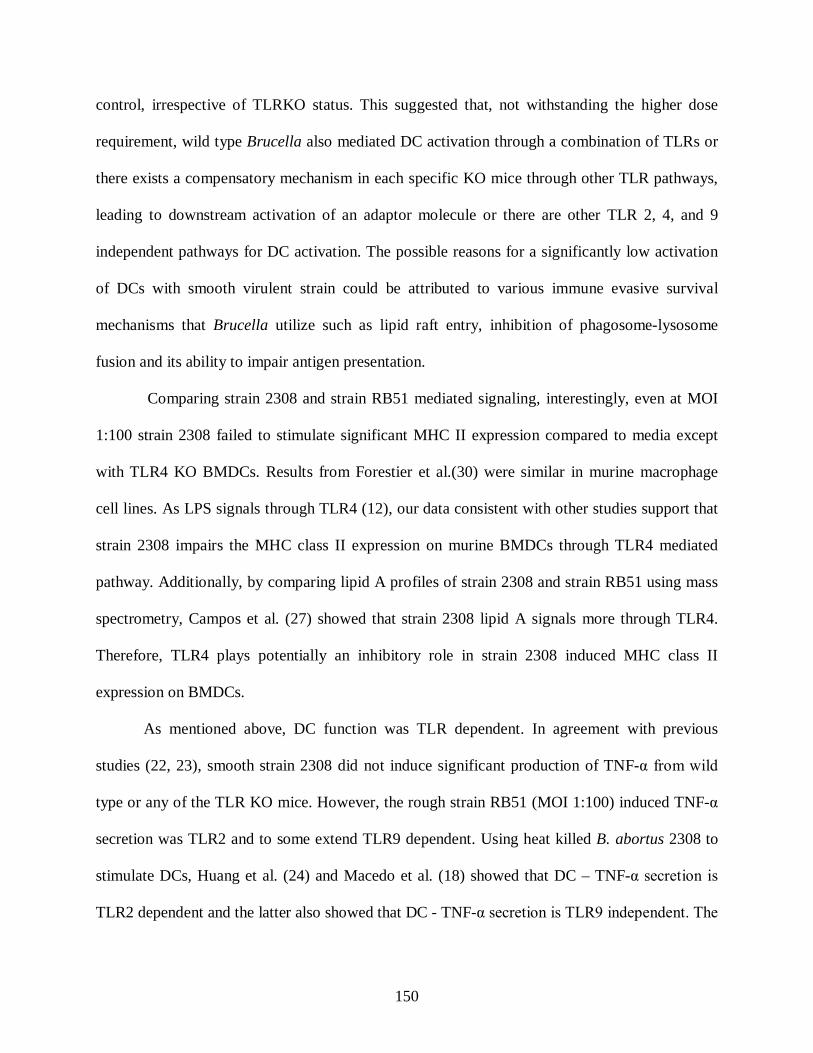
150
control, irrespective of TLRKO status. This suggested that, not withstanding the higher dose
requirement, wild type Brucella also mediated DC activation through a combination of TLRs or
there exists a compensatory mechanism in each specific KO mice through other TLR pathways,
leading to downstream activation of an adaptor molecule or there are other TLR 2, 4, and 9
independent pathways for DC activation. The possible reasons for a significantly low activation
of DCs with smooth virulent strain could be attributed to various immune evasive survival
mechanisms that Brucella utilize such as lipid raft entry, inhibition of phagosome-lysosome
fusion and its ability to impair antigen presentation.
Comparing strain 2308 and strain RB51 mediated signaling, interestingly, even at MOI
1:100 strain 2308 failed to stimulate significant MHC II expression compared to media except
with TLR4 KO BMDCs. Results from Forestier et al.(30) were similar in murine macrophage
cell lines. As LPS signals through TLR4 (12), our data consistent with other studies support that
strain 2308 impairs the MHC class II expression on murine BMDCs through TLR4 mediated
pathway. Additionally, by comparing lipid A profiles of strain 2308 and strain RB51 using mass
spectrometry, Campos et al. (27) showed that strain 2308 lipid A signals more through TLR4.
Therefore, TLR4 plays potentially an inhibitory role in strain 2308 induced MHC class II
expression on BMDCs.
As mentioned above, DC function was TLR dependent. In agreement with previous
studies (22, 23), smooth strain 2308 did not induce significant production of TNF-α from wild
type or any of the TLR KO mice. However, the rough strain RB51 (MOI 1:100) induced TNF-α
secretion was TLR2 and to some extend TLR9 dependent. Using heat killed B. abortus 2308 to
stimulate DCs, Huang et al. (24) and Macedo et al. (18) showed that DC – TNF-α secretion is
TLR2 dependent and the latter also showed that DC - TNF-α secretion is TLR9 independent. The
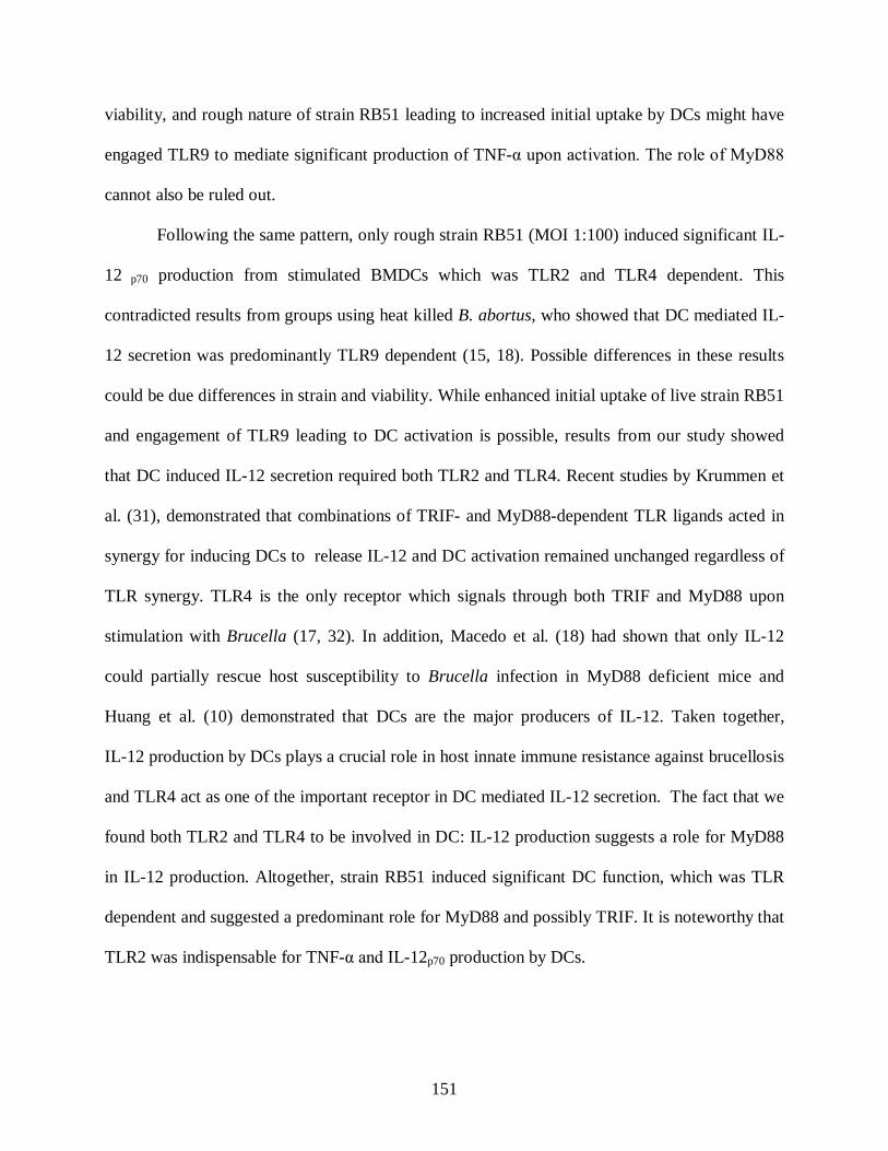
151
viability, and rough nature of strain RB51 leading to increased initial uptake by DCs might have
engaged TLR9 to mediate significant production of TNF-α upon activation. The role of MyD88
cannot also be ruled out.
Following the same pattern, only rough strain RB51 (MOI 1:100) induced significant IL-
12 p70 production from stimulated BMDCs which was TLR2 and TLR4 dependent. This
contradicted results from groups using heat killed B. abortus, who showed that DC mediated IL-
12 secretion was predominantly TLR9 dependent (15, 18). Possible differences in these results
could be due differences in strain and viability. While enhanced initial uptake of live strain RB51
and engagement of TLR9 leading to DC activation is possible, results from our study showed
that DC induced IL-12 secretion required both TLR2 and TLR4. Recent studies by Krummen et
al. (31), demonstrated that combinations of TRIF- and MyD88-dependent TLR ligands acted in
synergy for inducing DCs to release IL-12 and DC activation remained unchanged regardless of
TLR synergy. TLR4 is the only receptor which signals through both TRIF and MyD88 upon
stimulation with Brucella (17, 32). In addition, Macedo et al. (18) had shown that only IL-12
could partially rescue host susceptibility to Brucella infection in MyD88 deficient mice and
Huang et al. (10) demonstrated that DCs are the major producers of IL-12. Taken together,
IL-12 production by DCs plays a crucial role in host innate immune resistance against brucellosis
and TLR4 act as one of the important receptor in DC mediated IL-12 secretion. The fact that we
found both TLR2 and TLR4 to be involved in DC: IL-12 production suggests a role for MyD88
in IL-12 production. Altogether, strain RB51 induced significant DC function, which was TLR
dependent and suggested a predominant role for MyD88 and possibly TRIF. It is noteworthy that
TLR2 was indispensable for TNF-α and IL-12p70 production by DCs.
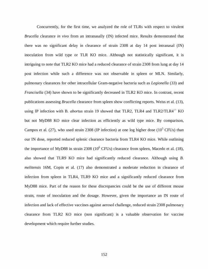
152
Concurrently, for the first time, we analyzed the role of TLRs with respect to virulent
Brucella clearance in vivo from an intranasally (IN) infected mice. Results demonstrated that
there was no significant delay in clearance of strain 2308 at day 14 post intranasal (IN)
inoculation from wild type or TLR KO mice. Although not statistically significant, it is
intriguing to note that TLR2 KO mice had a reduced clearance of strain 2308 from lung at day 14
post infection while such a difference was not observable in spleen or MLN. Similarly,
pulmonary clearances for other intracellular Gram-negative bacteria such as Legionella (33) and
Francisella (34) have shown to be significantly decreased in TLR2 KO mice. In contrast, recent
publications assessing Brucella clearance from spleen show conflicting reports. Weiss et al. (13),
using IP infection with B. abortus strain 19 showed that TLR2, TLR4 and TLR2/TLR4-/- KO
but not MyD88 KO mice clear infection as efficiently as wild type mice. By comparison,
Campos et al. (27), who used strain 2308 (IP infection) at one log higher dose (105 CFUs) than
our IN dose, reported reduced splenic clearance bacteria from TLR4 KO mice. While outlining
the importance of MyD88 in strain 2308 (106 CFUs) clearance from spleen, Macedo et al. (18),
also showed that TLR9 KO mice had significantly reduced clearance. Although using B.
melitensis 16M, Copin et al. (17) also demonstrated a moderate reduction in clearance of
infection from spleen in TLR4, TLR9 KO mice and a significantly reduced clearance from
MyD88 mice. Part of the reason for these discrepancies could be the use of different mouse
strain, route of inoculation and the dosage. However, given the importance an IN route of
infection and lack of effective vaccines against aerosol challenge, reduced strain 2308 pulmonary
clearance from TLR2 KO mice (non significant) is a valuable observation for vaccine
development which require further studies.
153
Conversely, pulmonary clearance of rough strain RB51 was significantly delayed in
TLR4 KO mice compared to wild type, TLR2 and TLR9 KO mice. No statistically significant
difference was observed in spleen and MLN. Previous studies from our lab (Surendran et al.,
under review) analyzing the in vivo pulmonary innate immune responses to IN inoculated strain
RB51 has revealed that the vaccine strain induce enhanced DC response compared to virulent
strain 2308. Although we can only speculate at this time, considering these data on the role of
TLR 4 in strain RB51 infected DC IL-12 secretion, it is probable that in the TLR4 KO mice, the
impaired IL-12 secretion and subsequent subdued immune response could be the explanation for
the observed reduction in pulmonary clearance of strain RB51. Given the information, in future
studies TLR agonists can be assessed along with vaccine strain RB51 for their ability to enhance
protection against intranasal Brucella challenge.
In conclusion, strain RB51 is capable of inducing significant innate immune response
characterized by BMDC maturation and function compared to pathogenic strain 2308. Strain
RB51 induced DC maturation is not dependent on an individual TLR, yet DC function
characterized by TNF-α secretion is TLR2 and in part TLR9 dependent and IL-12 secretion is
TLR2 as well as TLR4 dependent. Virulent strain 2308 stimulated significant DC activation only
at the highest MOI irrespective of TLR KO status but without inducing pro inflammatory or Th1
cytokine secretion. The study also gives novel insights into the TLR dependent clearance of B.
abortus pathogenic strain 2308 as well as vaccine strain RB51 clearance from IN infected mice.
Results from this study will give new direction to develop vaccine strategies against IN Brucella
infection. Future studies should address the potential role of downstream adaptor molecule
MyD88 with respect to vaccine strain induced DC activation and pulmonary clearance of rough
and smooth bacteria.

154
Acknowledgements: The authors acknowledge Dr. Stephen Werre for help with the
statistical analysis of the data.
References
1. Acha, P., Szyfres, B. , ed. 2001. Zoonoses and communicable diseases common to man
and animal. . Pan American Health Organization, WHO.
2. Pappas, G., P. Panagopoulou, L. Christou, and N. Akritidis. 2006. Brucella as a
biological weapon. Cell Mol Life Sci 63:2229-2236.
3. Nene, V., Kole, Chittaranjan., ed. 2009. Genome Mapping and Genomics in Animal-
Associated Microbes. Springer-Verlag, Berlin Heidelberg.
4. Corbel, M. J., ed. 2006. Brucellosis in humans and animals. World Health Organization.
5. Schurig, G. G., R. M. Roop, 2nd, T. Bagchi, S. Boyle, D. Buhrman, and N.
Sriranganathan. 1991. Biological properties of RB51; a stable rough strain of Brucella
abortus. Vet Microbiol 28:171-188.
6. He, Y., R. Vemulapalli, A. Zeytun, and G. G. Schurig. 2001. Induction of specific
cytotoxic lymphocytes in mice vaccinated with Brucella abortus RB51. Infect Immun
69:5502-5508.
7. Tizard, I. R., ed. 2008. Veterinary Immunology: An Introduction. Elsevier.
8. Barquero-Calvo, E., E. Chaves-Olarte, D. S. Weiss, C. Guzman-Verri, C. Chacon-Diaz,
A. Rucavado, I. Moriyon, and E. Moreno. 2007. Brucella abortus uses a stealthy strategy
to avoid activation of the innate immune system during the onset of infection. PLoS ONE
2:e631.
155
9. Billard, E., C. Cazevieille, J. Dornand, and A. Gross. 2005. High susceptibility of human
dendritic cells to invasion by the intracellular pathogens Brucella suis, B. abortus, and B.
melitensis. Infect Immun 73:8418-8424.
10. Huang, L. Y., C. Reis e Sousa, Y. Itoh, J. Inman, and D. E. Scott. 2001. IL-12 induction
by a TH1-inducing adjuvant in vivo: dendritic cell subsets and regulation by IL-10. J
Immunol 167:1423-1430.
11. Janeway, C. A., Jr., and R. Medzhitov. 2002. Innate immune recognition. Annu Rev
Immunol 20:197-216.
12. Banchereau, J., and R. M. Steinman. 1998. Dendritic cells and the control of immunity.
Nature 392:245-252.
13. Weiss, D. S., K. Takeda, S. Akira, A. Zychlinsky, and E. Moreno. 2005. MyD88, but not
toll-like receptors 4 and 2, is required for efficient clearance of Brucella abortus. Infect
Immun 73:5137-5143.
14. Oliveira, S. C., F. S. de Oliveira, G. C. Macedo, L. A. de Almeida, and N. B. Carvalho.
2008. The role of innate immune receptors in the control of Brucella abortus infection:
toll-like receptors and beyond. Microbes Infect 10:1005-1009.
15. Huang, L. Y., K. J. Ishii, S. Akira, J. Aliberti, and B. Golding. 2005. Th1-like cytokine
induction by heat-killed Brucella abortus is dependent on triggering of TLR9. J Immunol
175:3964-3970.
16. Zwerdling, A., M. V. Delpino, P. Barrionuevo, J. Cassataro, K. A. Pasquevich, C. Garcia
Samartino, C. A. Fossati, and G. H. Giambartolomei. 2008. Brucella lipoproteins mimic
dendritic cell maturation induced by Brucella abortus. Microbes Infect 10:1346-1354.

156
17. Copin, R., P. De Baetselier, Y. Carlier, J. J. Letesson, and E. Muraille. 2007. MyD88-
dependent activation of B220-CD11b+LY-6C+ dendritic cells during Brucella melitensis
infection. J Immunol 178:5182-5191.
18. Macedo, G. C., D. M. Magnani, N. B. Carvalho, O. Bruna-Romero, R. T. Gazzinelli, and
S. C. Oliveira. 2008. Central role of MyD88-dependent dendritic cell maturation and
proinflammatory cytokine production to control Brucella abortus infection. J Immunol
180:1080-1087.
19. Neutra, M. R., and P. A. Kozlowski. 2006. Mucosal vaccines: the promise and the
challenge. Nat Rev Immunol 6:148-158.
20. Inaba, K., M. Inaba, N. Romani, H. Aya, M. Deguchi, S. Ikehara, S. Muramatsu, and R.
M. Steinman. 1992. Generation of large numbers of dendritic cells from mouse bone
marrow cultures supplemented with granulocyte/macrophage colony-stimulating factor. J
Exp Med 176:1693-1702.
21. Vemulapalli, R., Y. He, S. Cravero, N. Sriranganathan, S. M. Boyle, and G. G. Schurig.
2000. Overexpression of protective antigen as a novel approach to enhance vaccine
efficacy of Brucella abortus strain RB51. Infect Immun 68:3286-3289.
22. Billard, E., J. Dornand, and A. Gross. 2007. Brucella suis prevents human dendritic cell
maturation and antigen presentation through regulation of tumor necrosis factor alpha
secretion. Infect Immun 75:4980-4989.
23. Billard, E., J. Dornand, and A. Gross. 2007. Interaction of Brucella suis and Brucella
abortus rough strains with human dendritic cells. Infect Immun 75:5916-5923.
24. Huang, L. Y., J. Aliberti, C. A. Leifer, D. M. Segal, A. Sher, D. T. Golenbock, and B.
Golding. 2003. Heat-killed Brucella abortus induces TNF and IL-12p40 by distinct

157
MyD88-dependent pathways: TNF, unlike IL-12p40 secretion, is Toll-like receptor 2
dependent. J Immunol 171:1441-1446.
25. Pei, J., J. E. Turse, and T. A. Ficht. 2008. Evidence of Brucella abortus OPS dictating
uptake and restricting NF-kappaB activation in murine macrophages. Microbes Infect
10:582-590.
26. Salcedo, S. P., M. I. Marchesini, H. Lelouard, E. Fugier, G. Jolly, S. Balor, A. Muller, N.
Lapaque, O. Demaria, L. Alexopoulou, D. J. Comerci, R. A. Ugalde, P. Pierre, and J. P.
Gorvel. 2008. Brucella control of dendritic cell maturation is dependent on the TIR-
containing protein Btp1. PLoS Pathog 4:e21.
27. Campos, M. A., G. M. Rosinha, I. C. Almeida, X. S. Salgueiro, B. W. Jarvis, G. A.
Splitter, N. Qureshi, O. Bruna-Romero, R. T. Gazzinelli, and S. C. Oliveira. 2004. Role
of Toll-like receptor 4 in induction of cell-mediated immunity and resistance to Brucella
abortus infection in mice. Infect Immun 72:176-186.
28. Kahl-McDonagh, M. M., A. M. Arenas-Gamboa, and T. A. Ficht. 2007. Aerosol
infection of BALB/c mice with Brucella melitensis and Brucella abortus and protective
efficacy against aerosol challenge. Infect Immun 75:4923-4932.
29. Olsen, S. C., W. R. Waters, and W. S. Stoffregen. 2007. An aerosolized Brucella spp.
challenge model for laboratory animals. Zoonoses Public Health 54:281-285.
30. Forestier, C., F. Deleuil, N. Lapaque, E. Moreno, and J. P. Gorvel. 2000. Brucella
abortus lipopolysaccharide in murine peritoneal macrophages acts as a down-regulator of
T cell activation. J Immunol 165:5202-5210.
158
31. Krummen, M., S. Balkow, L. Shen, S. Heinz, C. Loquai, H. C. Probst, and S. Grabbe.
Release of IL-12 by dendritic cells activated by TLR ligation is dependent on MyD88
signaling, whereas TRIF signaling is indispensable for TLR synergy. J Leukoc Biol.
32. O'Neill, L. A. 2006. How Toll-like receptors signal: what we know and what we don't
know. Curr Opin Immunol 18:3-9.
33. Archer, K. A., and C. R. Roy. 2006. MyD88-dependent responses involving toll-like
receptor 2 are important for protection and clearance of Legionella pneumophila in a
mouse model of Legionnaires' disease. Infect Immun 74:3325-3333.
34. Malik, M., C. S. Bakshi, B. Sahay, A. Shah, S. A. Lotz, and T. J. Sellati. 2006. Toll-like
receptor 2 is required for control of pulmonary infection with Francisella tularensis.
Infect Immun 74:3657-3662.
Figure legends
Figure 1: E. coli LPS downregulated CD11c expression on bone marrow derived cells. Bone
marrow cells isolated from BALB/c control and TLR KO mice were cultured for 6 days in 10%
RPMI medium with rGM-CSF (20ng/ml). The cells were harvested on day 6 and treated for 4
hours with either E. coli LPS (100 ng/ml) or B.abortus rough or smooth strains (DC:Brucella;
1:1, 1:10, 1:100). Cells were incubated for an additional 20 hours in gentamicin containing
media and analyzed for CD11c+ marker expression using fluorescent labeled CD11c+ antibody.
Cells treated with media served as the control. Asterisks (**) denotes statistically significant data
at p ≤ 0.05 compared to corresponding media control (*). (##) denotes statistically significant
data at p ≤ 0.05 compared to corresponding E. coli LPS (#). Alphabets ‘aa’ represents
159
statistically significant data at p ≤ 0.05 compared to corresponding strain 2308 stimulated DCs at
MOI 1:100 ‘a’. Data represents means ± standard deviations of 3 independent experiments.
Figure 2: Rough strain RB51 up-regulated MHC class II and co-stimulatory marker
expression in control and TLR KO BMDCs. Immature BMDCs from control and KO mice
were infected with either B. abortus rough vaccine strain RB51 or with smooth strain 2308 at
MOIs (DC:Brucella) 1:1, 1:10 and 1:100. At 24 hours post infection, BMDCs were analyzed for
MHC class II, CD40, CD86 expression and CD40+/CD86+ co-expression. Media and E. coli
LPS were the negative and positive controls respectively. A: Comparison of the percentage of
CD11c+ cells (BMDCs) expressing MHC class II high on its surface across different treatment
groups. B: Comparison of the percentage of CD40 expression on CD11c+ cells (BMDCs) across
different treatment groups. C: Comparison of the percentage of CD86 expression on CD11c+
cells (BMDCs) across different treatment groups. D: Comparison of the percentage of
CD40+/CD86+ coexpression on CD11c+ cells (BMDCs) across different treatment groups.
Asterisks (**) denotes statistically significant data at p ≤ 0.05 compared to corresponding media
control (*). (##) denotes statistically significant data at p ≤ 0.05 compared to corresponding
E.coli LPS(#). Alphabets ‘aa’ represents statistically significant data at p ≤ 0.05 compared to
corresponding strain 2308 stimulated DCs at MOI 1:100 ‘a’. Alphabets ‘bb’ represents
statistically significant data at p ≤ 0.05 compared to corresponding strain 2308 stimulated DCs at
MOI 1:10 ‘b’. Data represents means ± standard deviations of 3 independent experiments.
Figure 3: Rough strain RB51 activated BMDCs irrespective of TLR KO status. Immature
BMDCs from control and KO mice were infected with either B. abortus rough vaccine strain
RB51 or with smooth strain 2308 at MOI (DC:Brucella) 1:100. Histograms of single
representative experiment (of 3) from each type of BMDCs for total MHC class II expression,

160
CD40 expression and CD86 expression. Filled grey histogram: BALB/c media control;
continuous thick line: BALB/c; continuous thin line: TLR2 KO; broken thick line: TLR4 KO;
dotted line: TLR9 KO.
Figure 4: Rough strain RB51 induced DC – TNF-α and IL-12 secretion was TLR
dependent. To assess DC function, TNF-α (A) and IL-12p70 (B) levels in 24 hour culture
supernatants of B. abortus strain RB51 or 2308 infected BMDCs at various MOIs were analyzed
using indirect sandwich ELISA. E. coli LPS and media treated cell supernatants served as the
positive and negative controls respectively. The limits of detection for both the cytokines were
15pg/ml. The TNF-α and IL-12p70 results represent medians and 75th percentile of 3 independent
experiments. Asterisks (**) denotes statistically significant data at p ≤ 0.05 compared to
corresponding media control (*). (##) denotes statistically significant data at p ≤ 0.05 compared
to corresponding E.coli LPS(#). Alphabets ‘cc’ represents statistically significant data at p ≤ 0.05
compared to alphabet ‘c’.
Figure 5: Pulmonary clearance of rough strain RB51 is TLR4 dependent. Bacterial
clearance from TLR 2, TLR4, TLR9 KO and BALB/c control mice following IN infection with
A: B. abortus rough strain RB51 and B: smooth strain 2308. Mice were infected IN either with
strain RB51 (4 X 10⁸ CFUs/mouse) or with strain 2308 (2 X 10⁴ CFUs/mouse) and the number
of bacteria recovered from lung, mediastinal lymph node (MLN) and spleen were counted on day
14 post infection (PI). Asterisks (**) denotes statistically significant data at p ≤ 0.05 compared to
corresponding media control (*). Results represent median ± 75th percentile.

161
Figure 1
CD11c
0102030405060708090
100B
ALB
/c
TLR
2KO
TLR
4KO
TLR
9KO
BA
LB/c
TLR
2KO
TLR
4KO
TLR
9KO
BA
LB/c
TLR
2KO
TLR
4KO
TLR
9KO
BA
LB/c
TLR
2KO
TLR
4KO
TLR
9KO
Media E. coli LPS RB51 1:100 2308 1:100
Treatments/Mice
Perc
enta
ge o
f gat
ed c
ells
that
are
C
D11
c+
**
**
****
**** **
##
a
#
**
* *## ##
## ##aa
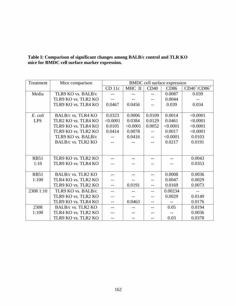
162
Table I: Comparison of significant changes among BALB/c control and TLR KO mice for BMDC cell surface marker expression.
Treatment Mice comparison
BMDC cell surface expression CD 11c MHC II CD40 CD86 CD40+/CD86+
Media TLR9 KO vs. BALB/c TLR9 KO vs. TLR2 KO TLR9 KO vs. TLR4 KO
-- --
0.0467
-- --
0.0456
-- -- --
0.0087 0.0044 0.039
0.039 --
0.034
E. coli LPS
BALB/c vs. TLR4 KO TLR2 KO vs. TLR4 KO TLR9 KO vs. TLR4 KO TLR9 KO vs. TLR2 KO TLR9 KO vs. BALB/c BALB/c vs. TLR2 KO
0.0323 <0.0001 0.0105 0.0414
-- --
0.0006 0.0384
<0.0001 0.0078 0.0416
--
0.0109 0.0129 0.0052
-- -- --
0.0014 0.0461
<0.0001 0.0017
<0.0001 0.0217
<0.0001 <0.0001 <0.0001 <0.0001 0.0103 0.0191
RB51 1:10
TLR9 KO vs. TLR2 KO TLR9 KO vs. TLR4 KO
-- --
-- --
-- --
-- --
0.0043 0.0353
RB51 1:100
BALB/c vs. TLR2 KO TLR4 KO vs. TLR2 KO TLR9 KO vs. TLR2 KO
-- -- --
-- --
0.0191
-- -- --
0.0008 0.0047 0.0169
0.0036 0.0029 0.0073
2308 1:10
TLR9 KO vs. BALB/c TLR9 KO vs. TLR2 KO TLR9 KO vs. TLR4 KO
-- -- --
-- --
0.0463
-- -- --
0.00234 0.0029
--
-- 0.0149 0.0176
2308 1:100
BALB/c vs. TLR2 KO TLR4 KO vs. TLR2 KO TLR9 KO vs. TLR2 KO
-- -- --
-- -- --
-- -- --
0.05 --
0.03
0.0194 0.0036 0.0378
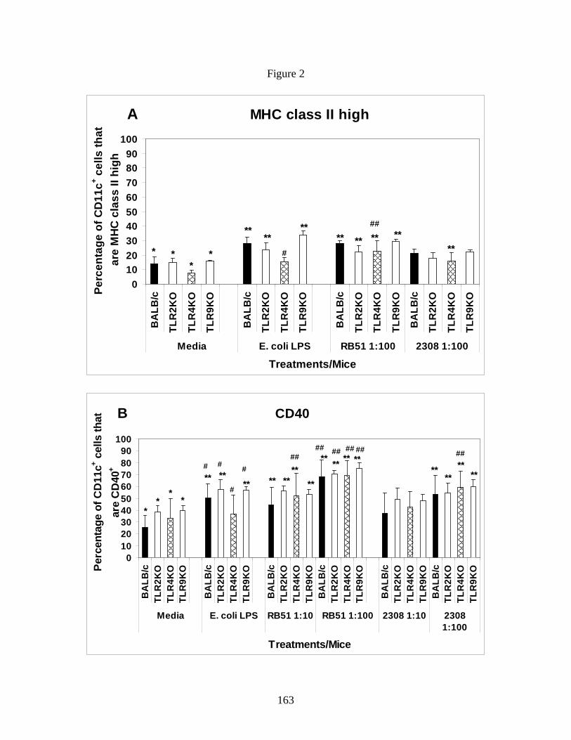
163
Figure 2
A MHC class II high
0102030405060708090
100B
ALB
/c
TLR
2KO
TLR
4KO
TLR
9KO
BA
LB/c
TLR
2KO
TLR
4KO
TLR
9KO
BA
LB/c
TLR
2KO
TLR
4KO
TLR
9KO
BA
LB/c
TLR
2KO
TLR
4KO
TLR
9KO
Media E. coli LPS RB51 1:100 2308 1:100
Treatments/Mice
Perc
enta
ge o
f CD
11c+ c
ells
that
ar
e M
HC
cla
ss II
hig
h
****
**** ** ** **
**
##
* **
* #
B CD40
0102030405060708090
100
BA
LB/c
TLR
2KO
TLR
4KO
TLR
9KO
BA
LB/c
TLR
2KO
TLR
4KO
TLR
9KO
BA
LB/c
TLR
2KO
TLR
4KO
TLR
9KO
BA
LB/c
TLR
2KO
TLR
4KO
TLR
9KO
BA
LB/c
TLR
2KO
TLR
4KO
TLR
9KO
BA
LB/c
TLR
2KO
TLR
4KO
TLR
9KO
Media E. coli LPS RB51 1:10 RB51 1:100 2308 1:10 23081:100
Treatments/Mice
Perc
enta
ge o
f CD
11c+ c
ells
that
ar
e C
D40
+
#### ## ## ##
** ****
##
#
# # # **** **
**
**
** ****
**
** ** ** ** **
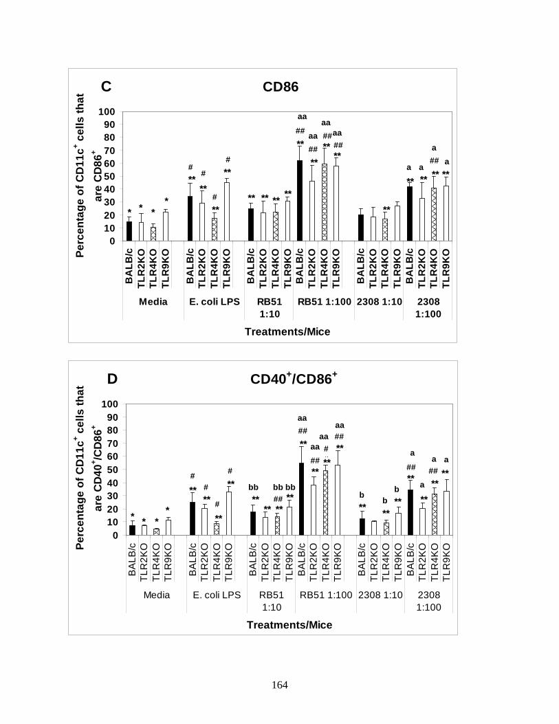
164
C CD86
0102030405060708090
100B
ALB
/cTL
R2K
OTL
R4K
OTL
R9K
O
BA
LB/c
TLR
2KO
TLR
4KO
TLR
9KO
BA
LB/c
TLR
2KO
TLR
4KO
TLR
9KO
BA
LB/c
TLR
2KO
TLR
4KO
TLR
9KO
BA
LB/c
TLR
2KO
TLR
4KO
TLR
9KO
BA
LB/c
TLR
2KO
TLR
4KO
TLR
9KO
Media E. coli LPS RB511:10
RB51 1:100 2308 1:10 23081:100
Treatments/Mice
Perc
enta
ge o
f CD
11c+ c
ells
that
ar
e C
D86
+
****
**
**
** **** ****
**
****
**
** ** ** **
##
####
##
aa
aaaa
aa
##
* * **
##
#
#a a
aa
D CD40+/CD86+
0102030405060708090
100
BA
LB/c
TLR
2KO
TLR
4KO
TLR
9KO
BA
LB/c
TLR
2KO
TLR
4KO
TLR
9KO
BA
LB/c
TLR
2KO
TLR
4KO
TLR
9KO
BA
LB/c
TLR
2KO
TLR
4KO
TLR
9KO
BA
LB/c
TLR
2KO
TLR
4KO
TLR
9KO
BA
LB/c
TLR
2KO
TLR
4KO
TLR
9KO
Media E. coli LPS RB511:10
RB51 1:100 2308 1:10 23081:100
Treatments/Mice
Perc
enta
ge o
f CD
11c+ c
ells
that
ar
e C
D40
+ /CD
86+
****
**
****
** ****
** ****##
aa
aaaa
**
****
**
**
****
**##
##
####
##
bb bb bb
##
aa
* * **
##
#
#
a
a
a a
b bb

165
Figure 3

166
Figure 4
A TNF-alpha
0100200300400500600700800900
1000B
ALB
/c
TLR
2KO
TLR
4KO
TLR
9KO
BA
LB/c
TLR
2KO
TLR
4KO
TLR
9KO
BA
LB/c
TLR
2KO
TLR
4KO
TLR
9KO
BA
LB/c
TLR
2KO
TLR
4KO
TLR
9KO
Media LPS RB51 1:100 2308 1:100
Treatments
Con
cent
ratio
n (p
g/m
l)
* * *
** **
**# # #
## ##
##
cc cc
c
B IL-12
050
100150200250300350400
BA
LB/c
TLR
2KO
TLR
4KO
TLR
9KO
BA
LB/c
TLR
2KO
TLR
4KO
TLR
9KO
BA
LB/c
TLR
2KO
TLR
4KO
TLR
9KO
BA
LB/c
TLR
2KO
TLR
4KO
TLR
9KO
Media LPS RB51 1:100 2308 1:100
Treatments
Con
cent
ratio
n (p
g/m
l)
* *
****
cccc
c c# #
####
167
Figure 5
A Day 14: strain RB51 clearance
10
100
1000
10000
100000B
ALB
/c
TLR
2KO
TLR
4KO
TLR
9KO
BA
LB/c
TLR
2KO
TLR
4KO
TLR
9KO
BA
LB/c
TLR
2KO
TLR
4KO
TLR
9KO
Lungs MLN Spleen
Organs/Mice
Med
ian
CFU
s/m
ice
* *
**
B Day 14: strain 2308 clearance
10
100
1000
10000
100000
BA
LB/c
TLR
2KO
TLR
4KO
TLR
9KO
BA
LB/c
TLR
2KO
TLR
4KO
TLR
9KO
BA
LB/c
TLR
2KO
TLR
4KO
TLR
9KO
Lungs MLN Spleen
Organs/Mice
Med
ian
CFU
s/m
ice

168
Chapter 6
Efficacy of vaccination strategies against intranasal challenge with Brucella abortus in
BALB/c model.
Running title: Intranasal vaccine against B. abortus respiratory infection.
Naveen Surendran1, Nammalwar Sriranganathan2, Heather Lawler1, Stephen M. Boyle2,
Elizabeth M. Hiltbold3, Bettina Heid1, Kurt Zimmerman2, Sharon G. Witonsky1*. (To be
submitted to Infection and Immunity)
Abstract
Brucellosis is a zoonotic disease with affecting 500,000 people worldwide annually.
Inhalation of infected aerosol is one of the major routes of disease transmission in humans.
Currently there are no licensed human vaccines available. B. abortus strain RB51 is a USDA
approved live attenuated vaccine against cattle brucellosis. B. abortus strain RB51SOD has been
shown to be more efficacious compared to strain RB51 against B. abortus pathogenic strain 2308
in mouse models. However, there is lack of information on the ability of these vaccine strains to
protect against intranasal challenge. In this study, with the long term goal to develop protective
vaccine for animals and people against respiratory challenge of Brucella spp., we tested a
number of different vaccination strategies in mice against intranasal infection with strain 2308.
Our strategies included strains RB51 or RB51SOD, evaluating the efficacy of different
vaccination routes, doses, boosting, and various prime-boost strategies against strain 2308
challenge. Despite using multiple protocols to enhance mucosal and systemic protection, our
results revealed that neither vaccine strains provided respiratory protection against intranasal
169
pathogenic Brucella infection. Further studies will address host-pathogen interaction at the lung
microenvironment and elucidate immune mechanisms that will enhance protection to aerosol
infection.
Introduction
Brucella is the most common zoonotic agent worldwide (1). Although B. melitensis is
the most important zoonotic agent among Brucella species, most human cases are caused by B.
abortus (4). B. melitensis, B. suis and B. abortus are the most infectious of the genus in the order
of their pathogenicity (4). Incidence of human disease correlates closely both with prevalence of
infection of livestock animals and risk factors for human exposure to infected animals and/or
related products (8). The highest risk group is abattoir workers, meat inspectors, animal handlers,
veterinarians, and lab workers (16). Clinical syndromes in humans include chronic fatigue,
undulant fever, reproductive disorders and general malaise (4). Around the globe, more than
500,000 human infections are infected annually (13).
Brucella is considered a biological weapon due to its propensity for air borne
transmission and highly infectious nature (13). Brucella is relatively stable in aerosol form and a
small inoculum (10-100 microorganisms) will induce disease (3). The organism is easily
obtained and antibiotic resistant strains can be easily developed (13). Therefore, Center for
Disease Control and Prevention (CDC) included Brucella species as a category B biodefense
agent. At present, safe and protective vaccines licensed for use against human brucellosis are not
commercially available.
To date, vaccination and experimental challenge studies with live Brucella strains have
predominantly focused on intraperitoneal (IP) delivery in mouse models (7). In spite of the
170
importance of aerosol exposure, very few studies have focused on assessing protection against
respiratory challenge (2, 8, 16). Previously, Mense et al. (10) demonstrated that aerosol infection
of BALB/c mice with Brucella spp establishes chronic respiratory and systemic infection of
spleen and liver. In separate studies, Kahl-McDonagh et al. (8) and Olsen et al. (12) described
aerosol infection with B. melitensis 16M and/or B. abortus 2308 respectively. However, IP
vaccination with live attenuated vaccine strains was not protective (8, 12). Part of the lack of
efficacy may be due to the immune response associated with vaccination route. Published
literature shows that mice vaccinated IP protected against IP challenge with Brucella (7), and
mice vaccinated orally with B. abortus strain RB51 protected against oral B. abortus strain 2308
challenge (14). Therefore, protection might be optimum when vaccination and challenge routes
are identical such that associated local and systemic immune responses may be optimal. Hence,
we hypothesized that IN vaccination with live attenuated vaccine would enhance local mucosal
and therefore overall protective immune response against IN Brucella challenge. Additionally, it
is expected that mucosal vaccination would enhance mucosal protective immune response
against aerosol challenge.
No studies have been published assessing the efficacy of IN vaccination with B. abortus
vaccine strains against IN challenge with B. abortus in a mouse model. In this study, we assessed
protection due to IN vaccination with B. abortus live attenuated rough vaccine strains RB51 (15)
and RB51SOD (over expressing Cu-Zn superoxide dismutase) against IN challenge with B.
abortus smooth pathogenic strain 2308 (17). We also assessed the effect of route and prime-
boost vaccination strategies against IN strain 2308 challenge.
171
Materials and Methods
Mice: Female 6-8 weeks old BALB/c mice were obtained from Charles River Laboratories Inc.,
Wilmington, MA. Mice were used under animal care protocols approved by Institutional Animal
Care and Use Committee at Virginia Tech.
Bacterial strains: Live attenuated rough B. abortus strains RB51, RB51SOD and virulent
smooth strain 2308 used were from our stock culture collection. All experiments with Brucella
were performed in our CDC approved (C2003 1120-0016) Biosafety Level (BSL) -3 Infectious
Disease Unit (IDU) facility.
Experimental design: The experimental designs employed to assess different routes and prime
boost protocols is outlined in Table 1.
Table 1. Experimental design of vaccination and challenge studies Groups Vaccine
strain Route of
vaccination Vaccine dose
(CFUs/mouse) Booster dose?
Route of
booster dose
IN - strain 2308 infection dose (CFUs/mouse)
1 RB51 RB51SOD
IN IN
4 X 10⁹ 4 X 10⁹
No No
-- --
2 X 10⁵
2 X 10⁵ 2 RB51
RB51SOD IN IN
4 X 10⁹ 4 X 10⁹
Yes Yes
IN IN
2 X 10⁵
2 X 10⁵ 3 RB51SOD IN 4 X 10⁹ Yes IN 2 X 10⁴ 4
RB51SOD RB51SOD RB51SOD
IN IN IN
1 X 10⁷ 1 X 10⁸
1 X 10⁹
No No No
-- -- --
2 X 10⁴
2 X 10⁴
2 X 10⁴ 5
RB51SOD RB51SOD RB51SOD RB51SOD
IP IM ID SC
4 X 10⁸ 4 X 10⁸ 4 X 10⁸ 4 X 10⁸
No No No No
-- -- -- --
2 X 10⁴
2 X 10⁴
2 X 10⁴
2 X 10⁴ 6
RB51SOD RB51SOD RB51SOD RB51SOD RB51SOD RB51SOD
IP IP IM ID IN IN
4 X 10⁸
4 X 10⁸
4 X 10⁸
4 X 10⁸
4 X 10⁸
4 X 10⁸
Yes Yes Yes Yes Yes Yes
IP IN IN IN IP IM
2 X 10⁴
2 X 10⁴
2 X 10⁴
2 X 10⁴
2 X 10⁴
2 X 10⁴

172
Group 1: To assess vaccine efficacy with single intranasal vaccination and the effect of day,
BALB/c mice (n=5) were immunized intranasally (IN), under xylazine-ketamine anesthesia, with
either B. abortus rough strains RB51 or strain RB51SOD (4 X 10⁹ CFUs/mouse) in 35 μl
phosphate buffered saline (PBS). PBS inoculated mice served as control with all experiments
unless otherwise mentioned. Clearance of vaccine strains was determined prior to challenge. Six
weeks post vaccination, control as well as vaccinated mice were infected IN with smooth strain
2308 (2 X 10⁵ CFUs/mouse). Mice were euthanized at day 14 and day 41 post infection (to
determine if there was a delay in clearance) with a lethal dose of xylazine-ketamine IP. Lung,
mediastinal lymph node (MLN) and spleen were collected. Single cell suspensions of organs
collected were serially diluted and plated on to Tryptic soy agar (TSA) plates and incubated for 5
days at 37° C and 5% CO2. Bacterial colony forming units (CFUs) were counted and
CFUs/mouse were calculated (8, 17).
Group 2: To assess the effect of intranasal boosting and day, mice (n=5 per day per treatment
group) were primed (4 X 10⁹ CFUs/mouse) and boosted (4 X 10⁹ CFUs/mouse) IN 4 weeks
apart. Clearance of vaccine strains was determined prior to boosting and challenge. Mice (n=5)
were challenged 4 weeks post boost with virulent strain 2308 (2 X 10⁵ CFUs/mouse).
CFUs/mouse were calculated at day 14 and day 41 (to determine if there was a delay in
clearance) as before.
Group 3: To determine whether challenge dose was limiting assessment of protection, the
challenge dose was decreased to 2 X 10⁴ CFUs/mouse, that used for IP challenge studies. Strain
RB51SOD was used as it has been demonstrating to be more efficacious than strain RB51 in
mouse models. Mice (n=8 per group) were vaccinated and boosted IN (4 X 10⁹ CFUs/mouse)
173
with strain RB51SOD as in Group 2, challenged IN with strain 2308 (2 X 10⁴ CFUs/mouse) and
day 14 PI CFU/mouse were determined.
Group 4: To identify the optimal vaccine dose, five mice (n=5) per group were given 1 of the
3 doses IN (1 X 10⁷, 1 X 10⁸, or 1 X 10⁹ CFUs/mouse) of strain RB51SOD). Six weeks post
vaccination mice were challenged IN with strain 2308 (2 X 10⁴ CFUs/mouse). Day 14 post
infection clearance was assessed as above.
Group 5: To identify the optimal systemic vaccination route, mice (n=5 per group) were
vaccinated subcutaneously (SC), intraperitoneally (IP), intradermally (ID) or intramuscularly
(IM) with strain RB51SOD (4 X 10⁸ CFUs/mouse) in sterile PBS. Six weeks post vaccination,
mice were challenged IN with strain 2308 (2 X 10⁴ CFUs/mouse) and clearance (CFUs/mouse)
was determined at day 14 post challenge as above.
Group 6: To optimize the prime-boost protocol, mice were vaccinated with strain RB51SOD
(4 X 10⁸ CFUs/mouse) by different prime boost protocols. The study was completed in 2
blocks/experiments. In the 1st block/experiment, mice (n=8 per group) were primed and boosted
(IP/IP and IP/IN) 4 wks apart. In the 2nd block/experiment, mice (n=6 per group) were primed
and boosted (IM/IN, ID/IN, IN/IP, IN/IM) at same time interval. Mice were challenged IN with
strain 2308 (2 X 10⁴ CFUs/mouse) 4 weeks following post boost vaccination. Day 14 clearance
was assessed based on CFUs/mouse to determine protection.
Statistical analysis: Statistical significance of the effect of single IN vaccine, IN vaccine - IN
boost, different vaccine doses and route of systemic vaccination on were done by comparing the
treatments using ANOVA followed by Tukey’s procedure for multiple comparisons. IP vaccine
– IP boost and IP vaccine – IN boost study organs were analyzed using T-test for comparison of
the effect of treatment. For systemic prime – IN boost as well as IN prime – systemic boost
174
studies, treatments were compared using Kruskal-Wallis test followed by Dunn’s procedure for
multiple comparisons. All statistical analysis was done using SAS system (NC, USA).
Results
In Group 1 to assess vaccine efficacy with single intranasal vaccination and the effect of
day, single intranasal vaccination with vaccine strains RB51 and RB51SOD did not protect
against IN challenge with pathogenic strain 2308 at day 14 (Figure 1A) or day 41 (Figure 1B) in
lung, spleen or MLN. Both strain RB51 and strain RB51SOD induced almost a log clearance of
strain 2308 at day 14 PI from spleen but that was not significantly different from PBS control
mice (Figure 1A) (Although the overall p-value from ANOVA for the effect of treatments vs.
organ was 0.08, PBS vs. RB51 comparison in spleen demonstrated a trend toward clearance with
a p value of 0.08 and in lung, PBS vs. RB51SOD had a p value of 0.03).
As vaccinated mice showed almost 1 log clearance in the spleen, in Group 2, the effect
of boosting the mice IN was determined. Figures 2A and 2B illustrate the clearance of B. abortus
pathogenic strain from spleen, lungs and MLN at day 14 and 41 PI respectively. Mice boosted
intranasally did not show significant increase in clearance from any organs tested at both day 14
and day 41 PI compared to control (Figure 2A and 2B) (The overall p value for ANOVA was
0.0643 for the effect boost vs. treatment and among the individual comparisons, PBS vs. RB51
in spleen yielded a p value of 0.013). As there was some non significantly enhanced clearance
from the spleen, we assessed whether the challenge dose was too high to demonstrate vaccine
efficacy (Group 3); the challenge dose was decreased 1 log to 2 X 10⁴ CFUs/mouse.
Nevertheless, intranasal vaccination of strain RB51SOD did not show significant protection in
any organs.

175
Group 4: As the initial vaccine dose, which had been chosen based on that used for
IP vaccination and protection studies did not protect, we sought to identify a more efficacious
vaccine dose. Thus, the effect of vaccine dose on clearance was determined in Group 4
experiments. Figure 3A demonstrated that there was no significant difference in clearance of
strain 2308 between IN doses (107, 108, 109 CFUs/mouse) in any of the organs tested.
As IN vaccination had not proven efficacious, the effect of systemic vaccination
route on challenge was determined (Group 5). In Figure 3B, none of the systemic vaccination
routes induced significant clearance of strain 2308 from the organs tested when compared to PBS
control. Furthermore, clearance of strain 2308 from spleen, lungs or MLN was not significantly
different between any of the different vaccination routes tested.
To determine if prime-boost vaccination strategies would enhance clearance, mice were
vaccinated and challenge as described (Group 6). Similar to homologous IN prime-boost
strategy (Figure 2A-B), homologous IP prime and IP boost strategy using strain RB51SOD (4 X
108 CFUs/mouse) failed to provide significant clearance of strain 2308 (2 X 10⁴ CFUs/mouse)
from mice at day 14 PI compared to control mice (Figure 4A). As it is predicted that the most
effective vaccine strategy which will provide optimal protection against IN infection might
require heterologous prime-boost combinations that involve both systemic and mucosal delivery,
we tested IP vaccine – IN boost strategy with strain RB51SOD to determine protection. No
significant improvement in bacterial clearance from lung or MLN was obtained with this
vaccination strategy although the vaccine RB51SOD induced significant clearance of strain 2308
from spleen (p=0.0146) (Figure 4B). Based on the lack of protection, the experiment was
repeated to include other systemic vaccination routes. As shown in Figure 4C, IM and ID
systemic priming followed by IN boost did not protect mice against IN pathogenic 2308

176
infection in any of organs. However, there was a trend towards significant clearance in spleen
(p=0.086) from IM vaccinated and IN boosted mice. As systemic priming might not prime the
immune system for subsequent mucosal vaccination and as mucosal immunization could
potentially prime the immune system for both systemic and mucosal responses, in a subsequent
study we reversed the heterologous prime-boost strategy for an IN prime followed by systemic
booster vaccination. Different groups of mice primed IN with strain RB51SOD were given a
booster dose of strain RB51SOD either IP or IM before challenging IN with virulent strain 2308.
Nonetheless, the vaccination strategy was not protective against IN strain 2308 challenge as the
bacterial CFUs/mouse recovered from any organs at day 14 PI were not significantly lower than
PBS control group (Figure 4D).
Discussion
Published studies have established that aerosol or IN exposure of mice (or macaques)
with even low (102 - 103) doses of pathogenic B. abortus strain 2308 or B. melitensis 16M caused
chronic pulmonary or systemic infection (8-10). To date, most vaccine efficacy studies against
IN Brucella challenge infection focused on IP vaccination. As this approach was not efficacious,
another strategy is needed (8, 12). Based on other models, mucosal delivery of vaccines should
enhance protection (11). Therefore, we determined whether IN vaccination of B. abortus rough
vaccine strains RB51 and/or RB51SOD was efficacious against IN challenge with smooth
pathogenic strain 2308.
In preliminary studies, we assessed clearance of both vaccine and pathogenic strains to
establish our model. Vaccine doses of IN administered vaccine strains RB51 and RB51SOD
ranged from 107 to 109 CFUs/mouse. As previously shown for vaccines delivered via IP, vaccine
177
strains given IN were completely cleared from lungs, MLN and spleen by 4-6 weeks post
vaccination (data not shown). In contrast, pathogenic strain 2308 administered IN did not clear
until day 116 from lungs; MLN and spleen remained infected even at the lowest inoculum dose
of 2 X 10³ CFUs/mouse (data not shown). Based on some models (10, 12) using 1 log higher
vaccine and challenge doses for IN administered agents, we followed that protocol for the initial
challenge studies. However, IN vaccination followed by IN challenge did not elicit significant
protection either at day 14 or 41 PI in any of the organs tested, compared to the traditional IP
vaccine – IP challenge model (7). The reasons for this lack of protection in this and the
subsequent studies described here, despite IN and/or systemic boosting with the same vaccine
strains could be manifold. Possible explanations for the lack of clearance include insufficient
dose to generate protection, pathogen persistence, and poor ability of vaccine to stimulate CD4
Th1 response in mucosal environment with a CD4 Th2 bias.
As stated, some of the differences could be dose and species dependent clearance.
Whereas decreasing the B. abortus challenge dose did not improve clearance, Smither et al. (16)
demonstrated that decreasing the B. melitensis 16M challenge dose to 4 X 10³ CFUs/mouse did
enhance protection with IN vaccine B. melitensis Rev. 1. In addition, Kahl McDonagh et al. (8)
demonstrated that aerosol B. melitensis infection in mice diminished more rapidly compared to
B. abortus. Thus, the lower challenge dose and species dependent difference in clearance might
have led to improved protection by B. melitensis Rev. 1 against IN B. melitensis challenge. As
smooth vaccine strains such as B. melitensis Rev. 1 enhanced protection against B. melitensis
16M, identifying the mechanisms of induced protection to see if anything may be applied to the
B. abortus model is warranted.

178
Another possible explanation is the inability of the vaccines to stimulate a sufficient CD4
Th1 or Th17 response in a mucosal CD4 Th2 biased environment (5). Interestingly, the vaccine
doses tested did not enhance protection possibly due to the inability to surmount the bias towards
a Th2 response (5). As an additional IN booster dose also failed to enhance clearance, it is
possible that the vaccine does not override a Th2, or T-reg status in the lung.
It was possible that regardless of dose, intranasal vaccination alone did not stimulate
sufficient CD4 Th1 or Th17 muscoal immunity, but possibly with systemic vaccination, a strong
Th1 memory or Th17 response could be elicited. However, in agreement with the IP vaccine –
IN challenge experiment results by Kahl McDonagh et al. (8) and Olsen et al., (12) none of the
systemic vaccination routes (SC, IM, ID or IP) tested using live attenuated B. abortus strain
RB51SOD provided significant protection against B. abortus IN challenge infection. Even IP
prime and boost vaccination did not protect mice against IN challenge. Broadly, possible
explanations for the lack of protection include the vaccine’s inability to stimulate appropriate
innate and/or adaptive immune response in the mucosal or systemic environment. More
specifically, rough vaccine strains may be unable to stimulate appropriate TLR induced
responses to stimulate sufficient DC response, which may or may not be affected by the mucosal
environment. This could affect the ability to elicit a sufficient CD4 Th1 and CD8 Tc1 response
as well as possibly Th17 induced responses.
Ideally, vaccination at a single site would provide protection not only at the relevant
mucosal surface, but also throughout the body (11). However, optimal protection is likely to
require both mucosal and systemic immune effectors, and the most effective mucosal vaccine
strategies might be prime–boost combinations that involve both mucosal and systemic delivery
(11). Our rationale for the IN prime – systemic boost was that the CD4 Th1 response induced by

179
systemic boost could potentially overcome a CD4 Th2 response elicited by initial intranasal
vaccination. We tested both systemic prime – IN boost and IN prime – systemic boost
vaccination strategies and failed to show protection against IN challenge. Additional studies will
focus on the underlying mechanism for the lack of protection.
Besides the inability of the vaccines to induce an apparent protective immune response,
the ability of intracellular pathogens to remain sequestered from the immune response is another
possible reason for lack of protection. In fact, strain 2308 has recently been shown to infect and
replicate within respiratory epithelial cells without inducing cell death or cytotoxicity (6). Thus
this is another possible mechanism by which strain 2308 could persist in the respiratory
epithelium without detection or clearance.
In summary, all of the vaccination strategies tested failed to induce significant protection
in mice against B. abortus strain 2308 IN challenge from spleen (except IP/IN), lungs and MLN.
We tested both the B. abortus rough strains RB51 and RB51SOD as IN vaccines against an IN
strain 2308 challenge infection. We found that strain RB51SOD did not induce significant
improvement in pathogen clearance compared to strain RB51 upon IN vaccination as thought
based on IP vaccine-challenge studies. Results from our studies and currently available literature
suggest that development of a protective vaccine against IN/aerosol challenge requires additional
research focused on the ability of vaccines to elicit specific immunity based on route of
administration, as well as elucidating pulmonary immune responses against IN delivered
pathogens.
180
Acknowledgement: The authors would like to acknowledge Betty Mitchell for her invaluable
help in organ preparation and CFU calculations. The authors would also like to thank Dr.
Ramanathan Kasimanickyam and Dr. Stephen Werre for their help with statistical analysis.
References
1. Acha, P., Szyfres, B. (ed.). 2001. Zoonoses and communicable diseases common to man
and animal. , vol. I, Bacterioses and Mycoses. Pan American Health Organization, WHO.
2. Bhattacharjee, A. K., M. J. Izadjoo, W. D. Zollinger, M. P. Nikolich, and D. L.
Hoover. 2006. Comparison of protective efficacy of subcutaneous versus intranasal
immunization of mice with a Brucella melitensis lipopolysaccharide subunit vaccine.
Infect Immun 74:5820-5825.
3. Bossi, P., A. Tegnell, A. Baka, F. Van Loock, J. Hendriks, A. Werner, H. Maidhof,
and G. Gouvras. 2004. Bichat guidelines for the clinical management of brucellosis and
bioterrorism-related brucellosis. Euro Surveill 9:E15-16.
4. Corbel, M. J. (ed.). 2006. Brucellosis in humans and animals. World Health
Organization.
5. Eisenbarth, S. C., D. A. Piggott, J. W. Huleatt, I. Visintin, C. A. Herrick, and K.
Bottomly. 2002. Lipopolysaccharide-enhanced, toll-like receptor 4-dependent T helper
cell type 2 responses to inhaled antigen. J Exp Med 196:1645-1651.
6. Ferrero, M. C., C. A. Fossati, and P. C. Baldi. 2009. Smooth Brucella strains invade
and replicate in human lung epithelial cells without inducing cell death. Microbes Infect
11:476-483.
181
7. Jimenez de Bagues, M. P., P. H. Elzer, S. M. Jones, J. M. Blasco, F. M. Enright, G.
G. Schurig, and A. J. Winter. 1994. Vaccination with Brucella abortus rough mutant
RB51 protects BALB/c mice against virulent strains of Brucella abortus, Brucella
melitensis, and Brucella ovis. Infect Immun 62:4990-4996.
8. Kahl-McDonagh, M. M., A. M. Arenas-Gamboa, and T. A. Ficht. 2007. Aerosol
infection of BALB/c mice with Brucella melitensis and Brucella abortus and protective
efficacy against aerosol challenge. Infect Immun 75:4923-4932.
9. Mense, M. G., R. H. Borschel, C. L. Wilhelmsen, M. L. Pitt, and D. L. Hoover. 2004.
Pathologic changes associated with brucellosis experimentally induced by aerosol
exposure in rhesus macaques (Macaca mulatta). Am J Vet Res 65:644-652.
10. Mense, M. G., L. L. Van De Verg, A. K. Bhattacharjee, J. L. Garrett, J. A. Hart, L.
E. Lindler, T. L. Hadfield, and D. L. Hoover. 2001. Bacteriologic and histologic
features in mice after intranasal inoculation of Brucella melitensis. Am J Vet Res 62:398-
405.
11. Neutra, M. R., and P. A. Kozlowski. 2006. Mucosal vaccines: the promise and the
challenge. Nat Rev Immunol 6:148-158.
12. Olsen, S. C., W. R. Waters, and W. S. Stoffregen. 2007. An aerosolized Brucella spp.
challenge model for laboratory animals. Zoonoses Public Health 54:281-285.
13. Pappas, G., P. Panagopoulou, L. Christou, and N. Akritidis. 2006. Brucella as a
biological weapon. Cell Mol Life Sci 63:2229-2236.
14. Pasquali, P., A. Rosanna, C. Pistoia, P. Petrucci, and F. Ciuchini. 2003. Brucella
abortus RB51 induces protection in mice orally infected with the virulent strain B.
abortus 2308. Infect Immun 71:2326-2330.

182
15. Schurig, G. G., R. M. Roop, 2nd, T. Bagchi, S. Boyle, D. Buhrman, and N.
Sriranganathan. 1991. Biological properties of RB51; a stable rough strain of Brucella
abortus. Vet Microbiol 28:171-188.
16. Smither, S. J., S. D. Perkins, C. Davies, A. J. Stagg, M. Nelson, and H. S. Atkins.
2009. Development and characterization of mouse models of infection with aerosolized
Brucella melitensis and Brucella suis. Clin Vaccine Immunol 16:779-783.
17. Vemulapalli, R., Y. He, S. Cravero, N. Sriranganathan, S. M. Boyle, and G. G.
Schurig. 2000. Overexpression of protective antigen as a novel approach to enhance
vaccine efficacy of Brucella abortus strain RB51. Infect Immun 68:3286-3289.
Figure legends
Figure 1. IN vaccination – IN challenge study. Mice were vaccinated IN with either B. abortus
strain RB51 (solid black) or strain RB51SOD (checkered) and at 6 weeks post vaccination
challenged IN with B. abortus strain 2308. Day 14 (A) and day 41 (B) post infection (PI)
bacterial clearance was assessed from spleen, lungs and MLN as CFUs/mice. PBS vaccinated
(white) mice served as control. Each column on the graph represents mean CFUs ± standard
deviation.
Figure 2. IN vaccination – IN boost – IN challenge study. Mice were vaccinated IN with either
B. abortus strain RB51 (solid black) or strain RB51SOD (checkered) and at 4 weeks post
vaccination, mice were boosted IN with the same dose of respective vaccine strains. Four weeks
after booster vaccination, mice were challenged IN with B. abortus strain 2308. Day 14 (A) and
day 41 (B) post infection (PI) bacterial clearance was assessed from spleen, lungs and MLN as

183
CFUs/mice. PBS vaccinated (white) mice served as control. Each column on the graph
represents mean CFUs ± standard deviation.
Figure 3. Optimal dose and route of vaccination. A. Mice were vaccinated IN with any of the 3
different doses of B. abortus strain RB51SOD; 1 X 10⁹ (dashed horizontal), 1 X 10⁸ (light
vertical) and 1 X 10⁷ (large grid) CFUs/mouse respectively. After 6 weeks post vaccination, mice
were challenged IN with B. abortus strain 2308 and day 14 post infection bacterial clearance was
assessed from spleen, lungs and MLN. B. Mice were vaccinated with strain RB51SOD through
any of the four different routes of vaccination; subcutaneous (SC – dotted), intradermal (ID –
crossed lines), intraperitoneal (IP – upward diagonal lines), intramuscular (IM – horizontal
brick). After 6 weeks post vaccination, mice were IN challenged with strain 2308 and day 14
post infection CFUs/mice from spleen, lungs and MLN were assessed. PBS vaccinated (white)
mice served as control. Each column on the graph represents mean CFUs ± standard deviation.
Figure 4. Prime-boost vaccination strategy. Mice were vaccinated with strain RB51SOD by
different prime boost protocols. IP prime-IP boost (A) as well as IP prime-IN boost (B)
vaccination strategies were tested against IN challenge infection with strain 2308. Prime and
boost vaccinations were given 4 weeks apart to mice, and virulent strain 2308 challenge was
given IN, 4 weeks post booster vaccination. Day 14 post infection bacterial clearance was
assessed from spleen, lungs and MLN as CFUs/mice. The heterologous prime-boost study was
repeated with additional systemic routes for strain RB51SOD priming other than IP such as ID
and IM followed by IN boosting (C) at 4 weeks post vaccination and IN challenge with strain
2308 at 4 weeks post booster dose. Day 14 post infection bacterial clearance was assessed from
spleen, lungs and MLN; intradermal (ID – crossed lines), intraperitoneal (IP – upward diagonal
lines), intramuscular (IM – horizontal brick), PBS control (white). Similarly additional sets of

184
mice were primed IN and boosted (D) either intraperitoneal (IP – upward diagonal lines) or
intramuscular (IM – horizontal brick) and challenged IN with virulent strain 2308. Day 14 post
infection bacterial clearance was assessed from spleen, lungs and MLN. Each column on the
graphs represents mean CFUs ± standard deviation. ** depicts statistically significant data
compared * at p value < 0.05.
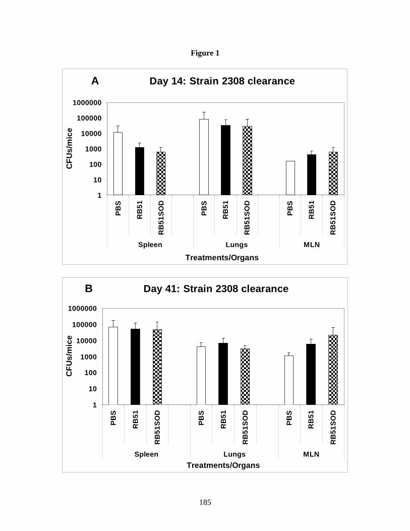
185
Figure 1
A Day 14: Strain 2308 clearance
1
10
100
1000
10000
100000
1000000
PBS
RB
51
RB
51SO
D
PBS
RB
51
RB
51SO
D
PBS
RB
51
RB
51SO
D
Spleen Lungs MLN
Treatments/Organs
CFU
s/m
ice
B Day 41: Strain 2308 clearance
1
10
100
1000
10000
100000
1000000
PBS
RB
51
RB
51SO
D
PBS
RB
51
RB
51SO
D
PBS
RB
51
RB
51SO
D
Spleen Lungs MLNTreatments/Organs
CFU
s/m
ice

186
Figure 2
A Boosted Day 14: Strain 2308 clearance
1
10
100
1000
10000
100000
1000000
PBS
RB
51
RB
51SO
D
PBS
RB
51
RB
51SO
D
PBS
RB
51
RB
51SO
D
Spleen Lungs MLN
Treatments/Organs
CFU
s/m
ice
B Boosted Day 41: Strain 2308 clearance
1
10
100
1000
10000
100000
PBS
RB
51
RB
51SO
D
PBS
RB
51
RB
51SO
D
PBS
RB
51
RB
51SO
D
Spleen Lungs MLN
Treatments/Organs
CFU
s/m
ice
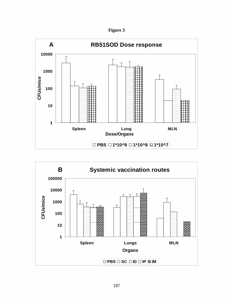
187
Figure 3
A RB51SOD Dose response
1
10
100
1000
10000
Spleen Lung MLNDose/Organs
CFU
s/m
ice
PBS 1*10^9 1*10^8 1*10^7
B Systemic vaccination routes
1
10
100
1000
10000
100000
Spleen Lungs MLN
Organs
CFU
s/m
ice
PBS SC ID IP IM

188
Figure 4
A IPIP - D14 Strain 2308 clearance
1
10
100
1000
10000
100000
PBS
RB
51SO
D
PBS
RB
51SO
D
PBS
RB
51SO
D
Spleen Lungs MLN
Organs/Treatments
CFU
s/m
ouse
B IPIN - D14 strain 2308 clearance
1
10
100
1000
10000
100000
PBS
RB
51SO
D
PBS
RB
51SO
D
PBS
RB
51SO
D
Spleen Lungs MLN
Organs/Treatments
CFU
s/m
ouse
***
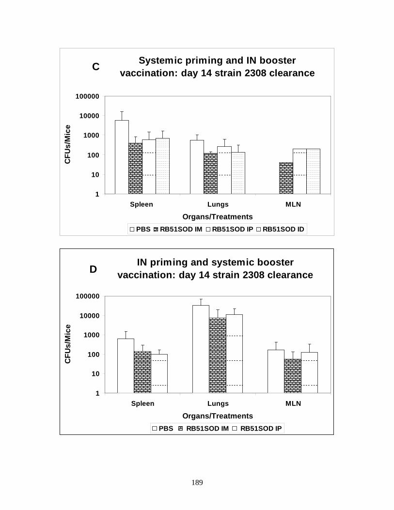
189
Systemic priming and IN booster vaccination: day 14 strain 2308 clearance
1
10
100
1000
10000
100000
Spleen Lungs MLN
Organs/Treatments
CFU
s/M
ice
PBS RB51SOD IM RB51SOD IP RB51SOD ID
C
IN priming and systemic booster vaccination: day 14 strain 2308 clearance
1
10
100
1000
10000
100000
Spleen Lungs MLN
Organs/Treatments
CFU
s/M
ice
PBS RB51SOD IM RB51SOD IP
D

190
Overall Summary and Discussion
“Prevention is better than cure” – As the idiom goes, vaccines have proved to be the
most effective tool in the containment and eradication of many infectious diseases. In 1996,
USDA approved B. abortus vaccine strain RB51 for use against brucellosis in cattle. Later,
another rough vaccine strain RB51SOD was shown to induce better protection against
brucellosis in mouse models. In this dissertation, we compared both the vaccine strains for their
ability to induce innate immune response in a mouse model. Despite the documented ability of
strain RB51SOD to induce better protection than strain RB51, our results showed that vaccine
strain RB51 induced better DC activation and function in vitro based on murine bone marrow
derived dendritic cell activation and function compared to strain RB51SOD and virulent strain
2308. Our in vivo study results, using a respiratory model of infection, corroborated the enhanced
ability of strain RB51 to induce increased pulmonary DC activation and innate immune function
compared to strain RB51SOD and strain 2308. Although our study did not elucidate the
mechanism for this subdued DC response to strain RB51SOD, the reasons could be manifold.
Histopathology results from our in vivo study clearly showed that strain RB51SOD when
inoculated intranasally did not elicit substantial proinflammatory response compared to strain
RB51. Theoretically, over expression of superoxide dismutase (SOD) could be limiting the
inflammatory response and thus the activation of innate immune cells in vitro or in vivo.
Although not the primary objective of this study, the scope of our in vivo study was limited in
that we did not analyze the role played by other innate immune cells such as neutrophils, alveolar
macrophages and natural killer cells in response to intranasally administered vaccines. Moreover,
we did not analyze the type of DC subsets involved in the strain RB51 induced enhanced DC
activation. Also, our in vivo results did not provide conclusive evidence on which cells were

191
predominantly involved in the strain RB51 induced IFN-γ secretion (Th1 response). While
identifying that intranasal inoculation with neither vaccine strains nor smooth strain 2308
induced Th2 mediated IL-4 secretion, we did not analyze the vaccine or pathogen mediated IL-
10, IL-17, IL-6 and/or TGF-β secretion as potential indicators of a T-regulatory or Th17
responses. Furthermore, assessing the level of T-cell activation achieved by strain RB51 induced
activated DCs in the lung environment is critical to improve efficacy of strain RB51 for use as an
IN vaccine. Therefore, future studies will focus on systematically elucidating the pulmonary
immune mechanisms against IN administered vaccine or pathogenic strains.
In spite of the observed differences in stimulating innate immunity, a single dose of either
of the vaccine strains RB51 and RB51SOD given IP, protects mice against IP challenge with
pathogenic strain 2308. Considering the importance of aerosol transmission of Brucella and the
lack of protective vaccines, we tested a variety of vaccination strategies including intranasal
vaccination using either strain RB51 or RB51SOD against intranasal challenge with virulent
strain 2308 in a mouse model.
In contrast to the intraperitoneal (IP) vaccination and challenge models, intranasal (IN)
vaccination as well as the different systemic, homologous and heterologous vaccine strategies
failed to confer protection against IN infection. The probable reasons for this lack of protection
could primarily include the bias towards Th2 response in lung environment, the inability of
vaccine strains to induce strong Th1 response in lung and/or the ability of virulent Brucella to
establish persistent yet undetected infection of respiratory epithelium. Our results suggested that
the vaccination route, dose of IN vaccination or dose of pathogen challenge did not have an
effect on pulmonary pathogen clearance. However, our in vivo TLR KO studies provided
additional information on TLR dependence in DC function and pulmonary clearance.

192
Our in vitro TLR KO studies revealed that the vaccine strain RB51 has specific TLR
requirements in inducing DC activation based on upregulation of costimulatory molecules
(TLR2 dependent) and DC function based on TNF-α (TLR2 and TLR9 dependent) and IL-12
(TLR2 and TLR4 dependent) production. Although TLR2 KO mice induced significantly higher
DC activation compared to media control, the activation levels were significantly lower than
control and TLR9 KO mice. In our in vivo KO clearance studies, the pathogen load was
comparatively higher in TLR2KO mice infected IN with strain 2308 at day 14 post infection
compared to control, TLR4 and TLR9 KO mice. Moreover, pulmonary clearance of strain RB51
was found to be TLR4 dependent. Taken together, TLR2 had a major role in strain RB51
induced DC activation and TNF-α and IL-12 secretion as well as pulmonary clearance of strain
2308. Additionally TLR4 was involved in strain RB51 clearance as well as DC:IL-12 secretion.
Partial TLR9 dependence on strain RB51 induced DC:TNF-α secretion was also noted. While
these data provided some information, questions remained as to whether an individual TLR
agonist can improve protection and/or whether compensatory mechanisms exist in single TLR
KO mice to stimulate innate immune response. Nevertheless, the enhanced DC activation
capability of strain RB51 and TLR dependent pulmonary clearance pattern of IN infected mice
provide additional tools to enhance vaccine efficacy. In future studies, TLR agonists could be
tested to improve the protective ability of strain RB51 against intranasal Brucella challenge.
As safety concerns might limit the use of live vaccine strain RB51 in humans, the use of
heat killed or irradiated strain RB51 should be considered. In our study comparing similar doses
of heat killed or irradiated to live strains (positive control), heat killed and irradiated vaccine
strain RB51 induced less DC activation than live strain 2308. Our data showed that DC
activation achieved by the former treated strains was higher than corresponding smooth strain
193
2308. As similar doses were used, higher doses and an increased infection time might improve
the DC activation and function capacity of inactivated strains. If an optimal dose and TLR
agonist could be identified in subsequent studies, a combination of inactivated vaccine strain
RB51 with the identified TLR agonist could be tested to protect against intranasal Brucella
infection. Altogether, my dissertation research provided important information on vaccine and
pathogen induced innate immune activation which produced valuable insights into the different
ways to improve currently approved animal brucellosis vaccine strain RB51.