university of tikrit college of nursing
TRANSCRIPT

University of Tikrit
College of Nursing
1
Anatomy
Introduction to Anatomy
Anatomical terms :
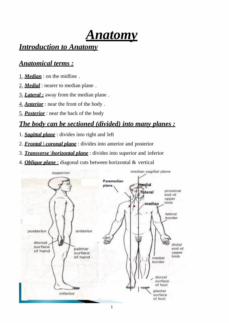
1. Median : on the midline .
2. Medial : nearer to median plane .
3. Lateral : away from the median plane .
4. Anterior : near the front of the body .
5. Posterior : near the back of the body
The body can be sectioned (divided) into many planes :
1. Sagittal plane : divides into right and left
2. Frontal \ coronal plane : divides into anterior and posterior
3. Transverse \horizontal plane : divides into superior and inferior
4. Oblique plane : diagonal cuts between horizontal & vertical

2
Abduction : movement of limb away from the midline of the body .
Adduction : movement of the limb toward the midline of the body .
The body as a whole

3
The skull: This consist of the cranium, which protect the brain, the eyes, and the
mandible.
The thorax: This cavity consists of bony framework supporting various muscles.
Contents of the thorax:
1. lungs. 2. Heart. 3. Trachea 4. esophagus 5. Major blood vessels, aorta, superior & inferior vena cava. 6. Thymus gland 7. Thoracic duct & lymphatic gland.
The abdomen : It is the largest cavity in the body. It is divided into 2 cavity:
-Abdominal cavity -Pelvic cavity.
Abdominal cavity
Stomach, , small and large intestine ,liver, spleen, pancreas, gall bladder,
kidney, ureter abdominal aorta, inferior vena cava
Pelvic cavity Urinary bladder, seminal vesicle, uterus, ovary, sigmoid colon, rectum

4
The skeleton:
- The skeleton is the framework of the body, - Consisting of the bones, cartilages & ligaments which bind together.
Human skeleton Consists of 206 individual bones, is made up of:
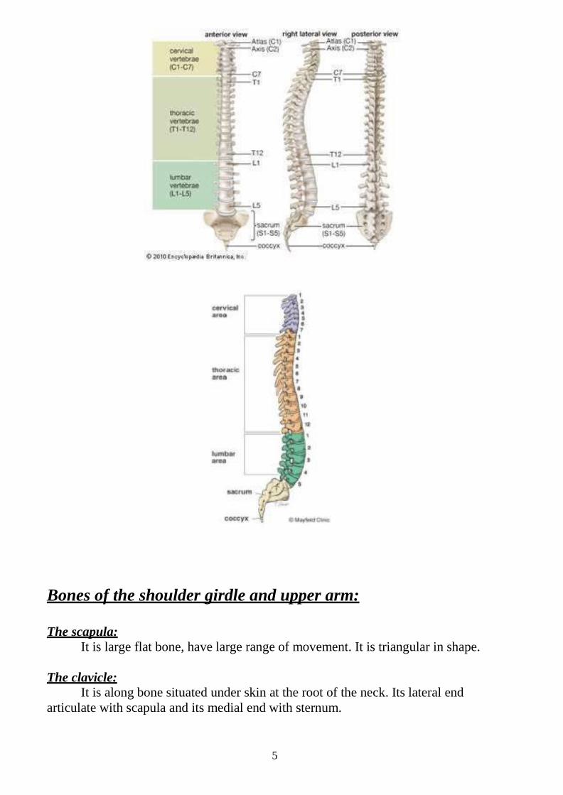
1. The skull, bones of cranium, face & lower jaw. 2. The bones of the trunk, spinal column, ribs & sternum. 3. Bones of limbs, together with shoulder & pelvic girdles. 4. Ribs: are 12 in number. 5. Vertebral column: consist of 33 vertebrae & divided into:
a- cervical = 7. b- thoracic or dorsal = 12. c- lumber = 5. d- sacral = 5. e- coccygeal = 4.
5
Bones of the shoulder girdle and upper arm:
The scapula: It is large flat bone, have large range of movement. It is triangular in shape.
The clavicle:
It is along bone situated under skin at the root of the neck. Its lateral end articulate with scapula and its medial end with sternum.

6
The humerus: It is the long bone of the arm. It consists of shaft and expanded upper and
lower extremities. The slight groove which surrounds the head of humerus called the anatomical neck.
The tapering region where the upper extremity of humerus joint the shaft
called surgical neck. (It is a common site of fracture)..
The humerus may be fractured at almost any level but is commonly fractured at three sites:
1. Surgical neck, with damage the axillary nerve, 2. Mid shaft, with damage to the radial nerve 3. at the lower end, may damage the ulner nerve.

7
Bone of the forearm:
The forearm consist of two long bones when the forearm placed in anatomical position.
The radius on the lateral (outer) side The ulna on the medial (inner) side
The radius: It is the lateral bone of the forearm. It is long bone with a shaft and extended
extremities, . The shaft has sharp medial border facing the ulna and attach to it the interosseous membrane which stretch between the radius and ulna. . At the lateral surface there is the styloid process. On the medial surface, there is articulation with the ulna.
A common fracture occurs about an inch above the lower end of the radius known as Colle's fracture and Smith's fracture

8
The ulna: It is the medial bone of the arm and slightly longer. The upper end of the ulna is expanded. The shaft of the ulna is narrow but expand slightly at its lower end to
form the head of ulna, on the medial side, there is the styloid process. The bones of the forearm are a common site of fracture

9
Bones of the wrist: The carpus or wrist consists of 8 bones arranged in two raws: Proximal raw: scaphoid, lunate, triquetral, pisiform. Distal raw: trapezium, trapezoid, capitate, hamate.

11
Scaphoid fractures are the second commonest group of fractures that are seen following a fall onto an outstretched hand and result in wrist pain, specifically tenderness in the anatomical snuffbox.
The metacarpal bones:
It is bones of the palm. It is long bones each with base, shaft and head. The bases articulate with distal raw of carpal bone and the heads with proximal raw of phalanges.
The phalanges:
11
It is a long bone. The thumb has only two phalanges. The fingers have three phalanges: proximal, middle and distal. The proximal being the longest. The joints between Metacarpals and the phalanges are called metacarpophalangeal joints. Those between phalanges themselves are called the interphalangeal joints.
Bones of pelvic girdle:
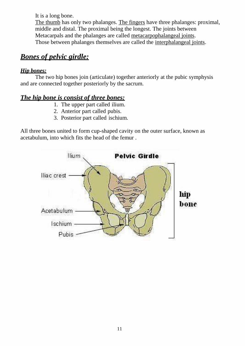
Hip bones: The two hip bones join (articulate) together anteriorly at the pubic symphysis
and are connected together posteriorly by the sacrum.
The hip bone is consist of three bones: 1. The upper part called ilium. 2. Anterior part called pubis. 3. Posterior part called ischium.
All three bones united to form cup-shaped cavity on the outer surface, known as acetabulum, into which fits the head of the femur .

12
Hip joint replacement :
Lower limb bones
The bones of the lower limb: 1. Femur or thigh bone.
This is the longest and strongest bone in the skeleton. 2. The patella ( knee-cap) .
3.The tibia ( shin bone) . It is the second long bone in the body;
4.The fibula .

13
The bones of the foot: The tarsus:
It consists of medial and lateral series of bones: 1. Medial series: consist of talus, navecular and the three cuneiform bones. 2. Lateral series: are composed of calcaneus and cuboid.
The foot has longitudinal arch on medial side; result from the talus is placed on top and above the level of the calcaneus. The transverse arch is marked at level of the base (proximal end of the metatarsus). These arches are very important in walking.

14

15
Knee joint anatomy
16
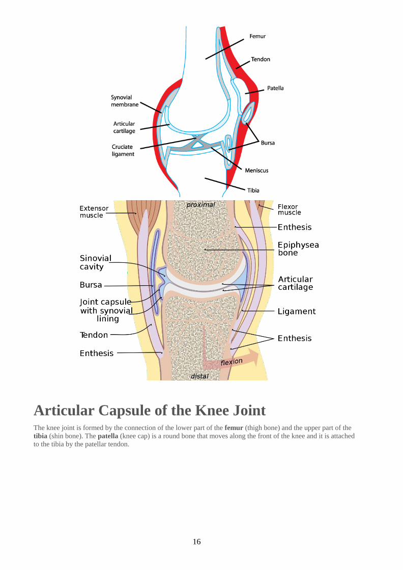
Articular Capsule of the Knee Joint The knee joint is formed by the connection of the lower part of the femur (thigh bone) and the upper part of the tibia (shin bone). The patella (knee cap) is a round bone that moves along the front of the knee and it is attached to the tibia by the patellar tendon.

17
The bones that make up the knee joint are the tibia, femur, and patella.
Articular Capsule: Definition The articular capsule of the knee joint surrounds the knee and consists of two main layers, an outer and inner layer. The outer layer is made up of a tough, fibrous membrane that is made up of ligament tissue.
The inner layer is made up of a synovial membrane, which secretes, a clear, yellowish fluid called synovial fluid.
The articular capsule of the knee also contains bursae, which are fluid filled sacs, and a fat pad located behind the patella.
The articular capsule of the knee also contains bursae, synovial fluid, and a fat pad.
Articular Capsule: Function All of these structures of the articular capsule of the joint (outer layer, inner layer, synovial fluid, bursae, fat pad) all have specific functions in the knee.
Outer Layer: The main function of the fibrous membrane of the outer layers is to provide stability to the joint by holding the bones of the knee (femur, tibia, patella) in their correct positions in the knee.
Inner Layer: the synovial membrane of the inner layer secretes a viscous material called synovial fluid. This synovial fluid functions to lubricate the joint, helping to reduce friction and irritation of the bones, ligaments, and tendons when the knee joint moves.

18
Lecture -2 Dr. Mohammed A.Hayawi
Digestive system
Anatomy of Digestive System
Digestive system consists of:
1. Gastrointestinal Tract (GIT).
2. Accessory organs.
GIT is a digestive tract consists of oral cavity, pharynx, esophagus, stomach, small
intestinal, large intestinal, and anus.

19
ORAL CAVITY
Structures of Oral Cavity are:
1. Lips: It is a mucous membrane protecting the anterior opening of the mouth. It’s also called labia.
2. Lymphoid tissue (Tonsils) :
a. Palatine tonsils are in the oropharynx, at the end of the soft palate.
b. Pharyngeal tonsil (adenoids when enlarge). Often-called adenoids
are located high in the nasopharynx.
c. Lingual tonsil: are at the base of the tongue.

21
3. Hard palate. It is forms the anterior roof of the mouth.
4. Soft palate. It is forms the posterior roof of the mouth.
5. Uvula. Is a fleshy finger like projection of the soft palate which extends downward
from its posterior edge
6. Salivary glands: 3- pairs of salivary glands empty their secretions into the mouth:
The parotid glands (large glands lies anterior to the ear)
The submandibular gland
Small sublingual gland

21
Pharynx: From the mouth, food passes posterior into the oropharynx and laryngopharynx.
The pharynx is subdivided into:
Nasopharynx, is a part of respiratory passageway.
Oropharynx, is posterior to oral cavity.
Laryngopharynx, which continuous with the esophagus.
Alternating contractions of the pharynx’s skeletal muscles propel food through the
pharynx into the esophagus below. This propelling mechanism is called peristalsis.
Esophagus (gullet): It runs from the pharynx through the diaphragm to the stomach, about 25cm (10
inches) long, descends toward thoracic cavity posterior to the trachea, and then enters
the abdominal cavity through the esophageal hiatus, an opening in the diaphragm, to
empties into the stomach. It is essentially a passageway that conducts food to the
stomach.
Esophagus Comprises:
1. Upper esophageal sphincter just below the pharynx and composed from
skeletal muscle fibers, to prevent air from entering esophagus.
2. Middle third of esophagus muscular layers compose from mixture of
skeletal fibers and smooth muscle cells
3. Lower esophageal sphincter (cardiac sphincter) composed from smooth
muscle, which normally remains in state of active contraction to prevent
backflow of materials from the stomach into esophagus. Esophagus innervated
by sympathetic and parasympathetic (esophageal plexus).

22
STOMACH
Stomach is Divided into Four Regions:
1. Cardiac region 3. Body of stomach. 2. Fundus . 4. Pylorus
The pylorus continuous with the small intestine through the pyloric sphincter or
valve.
The stomach is approximately 25cm long, but its diameter depends on how much
food it contains when it’s full. It can hold about 4 liters of food. Stomach is
innervated by sympathetic and parasympathetic nerve fibers (esophageal plexus).

23
Musculature of the Stomach:
the Muscularis mucosa and Muscularis externa of the stomach contain extra layers of
smooth muscle cells in addition to circular and longitudinal layers,
The third layer called oblique layer of smooth layer which strength the stomach wall.
Internally the stomach lining is composed of numerous gastric folds (rugae) these folds are
observed only when the stomach is empty.

24
SMALL INTESTINE * Anatomically , small intestine has three subdivisions:
1. Duodenum: 25 cm.
2. Jejunum:is about 2.5 meters..
3. Ileum: about 3.5 meters, It is joins the large intestine at the iliocecal valve.
*Histologically: Structures of Small Intestinal
1. Plica: the intestinal lining show transverse folds called plica and this is a
permanent feature that does not disappear when the small intestine fills, small
intestine contains roughly 800 plica to increase the surface for absorption .
2. Villi: mucosa of small intestine is project into a series of fingerlike structures
called intestine villi.
Structures of Villi:
a. epithelium ( simple columnar epithelium )
b. capillary network
c. lacteal (lymphatic vessels)
d. nerves

25
3. Payer's patches (aggregated lymphoid nodules):
Lamia propria of ileum contains 20-30 masses of lymphoid tissue (lymphoid nodules)
called payer's patches to protect small intestine from bacteria.
4. Intestinal gland a. Goblet cells.
b. Intestinal glands or crypts of lieberkuhn.
c. Submucosal glands or Brunner's glands

26
LARGE INTESTINE Large Intestine is Divided into Four Structures
Cecum and appendix. is the first part. It contains worm like appendix, a potential trouble spot, since it is usually twisted. It’s an ideal location for
bacteria to accumulate and multiply. Inflammation of the appendix – appendicitis is the usual result.
Colon: divided into: -
1. Ascending colon: travels up the right side of the abdominal cavity and
makes a turn in the right side or hepatic flexure.
2. Transverse colon: travels across the abdominal cavity.
3. Descending colon: turns at the left side to enter the pelvis.
4. Sigmoid colon: S-shaped, the part of colon that enters the pelvis.
The sigmoid colon, rectum and anal canal lie in the pelvis.
Rectum.
Anal canal: The anal canal ends at the anus, which opens to the exterior. The
anal canal has an :
External voluntary skeletal muscle (voluntary sphincter)
Internal involuntary sphincter formed by smooth muscle.
PANCREAS
I. Structures of Pancreas : 1.Head 2.Body 3.Tail
The pancreas is approximately 12–15â•›cm long and 2.5â•›cm thick. It is situated
across the back of the abdomen, behind the stomach. The head of the pancreas is on the right side of the abdomen and it is connected to the duodenum (the first section of the small intestine) through a small tube called the pancreatic duct. The narrow end of the pancreas, called the tail, extends to the left side of the body

27
LIVER
I. Structures of the Liver
The liver is the largest solid organ in the body. In adults, the liver can weigh up to 1.5 kg. It is in the upper-right abdomen, just under the rib cage and below the diaphragm (the thin muscle below the lungs and heart that separates the chest cavity from the abdomen.)
1. Right lobe , 2. left lobe ,3. Caudate lobe ,4. Quadrate lobe

28
Liver and Gall bladder: The liver is the larger gland in the body. It is located under the diaphragm more to the right side of the body. The liver has four lobes and is suspended from the diaphragm and abdominal wall by a delicate mesentery cord, the falciform ligaments.

29
Lecture -3- Dr.Mohammed A.Hayawi
Anatomy Respiratory System
The respiratory system consists of the : Nose , mouth , pharynx , larynx , trachea , bronchi , bronchiole & alveoli .
Nasal Cavity
Nose: It’s the only externally visible part of the respiratory system, during breathing;
the air enters the nose by nostrils.
The nasal cavities are separated into right & left by nasal septum . The sticky mucosa produces by the mucus glands moisten the air and traps
incoming bacteria and other foreign bodies.
The ciliated cells of the nasal mucosa help to control the body temperature.
The posterior nares are situated at the back of the nasal cavity & constitute the entrance to the nasopharynx .
Structures of Nasal Cavity

31
1. Superior, middle, and inferior concha ( عينافترا رعقت بين (.
2. Superior, middle, and inferior turbinate ( وففلم مظع )
3. Soft palate .
4. Nasopharynx.
The nasal cavity is separated inferiorly from the oral cavity by the palate :
1. Hard palate: the palate supported by bone.
2. Soft palate: the unsupported posterior part (not contain bone ).
Cleft palate: it is a genetic defect, characterized by the failure of the bones forming
the palate to fuse medially, results in breathing difficulty.
- The roof of the mouth (palate) is formed between the sixth and ninth weeks of
pregnancy. - The lip forms between the fourth and seventh weeks of
pregnancy

31
Nasal sinuses: The nasal cavity is surrounded by a ring of Para nasal sinuses as the
following: -
1. Frontal sinuses
.sinuses( Sphenoid 2 يينفلساا ديتولا جيبلا(
sinuses Ethmoid ) يوفصملا جيبلا( .3
4. Maxillary sinuses
LARYNX

32
Larynx (Voice Box):
The larynx or voice box, play a role in speech. It is located inferior to the pharynx.
It has special function of voice production It has situated in the midline of the neck between the pharynx above & the
trachea below . It’s formed by eight rigid hyaline cartilages
The largest of cartilage is the thyroid cartilage, which protrudes anteriorly and
is commonly called the Adam’s apple.
The epiglottis protects the superior opening of the larynx. It’s closes the
opening of the larynx during eating and drinking preventing the food and fluids
to enter the larynx.
Trachea :
It is 12 cm long , 2.5 cm in diameter . It divided into two main bronchi.
The trachea consist of number of C – shaped rings of hyaline cartilage. Air entering the trachea from the larynx travels down , its length (10 – 12 cm)
to the level of the fifth thoracic vertebrae. The trachea is lined with a ciliated mucosa.
These cartilages keep the trachea permanently open , so that its wall do not collapse like those of the esophagus .

33
Bronchi : The trachea ends by dividing or bifurcating into two main bronchi ( right & left
) , each bronchus passes to the corresponding lung . From each main bronchus numerous smaller bronchi are given off , like
branches of tree & the smallest bronchial tubes are called bronchioles .
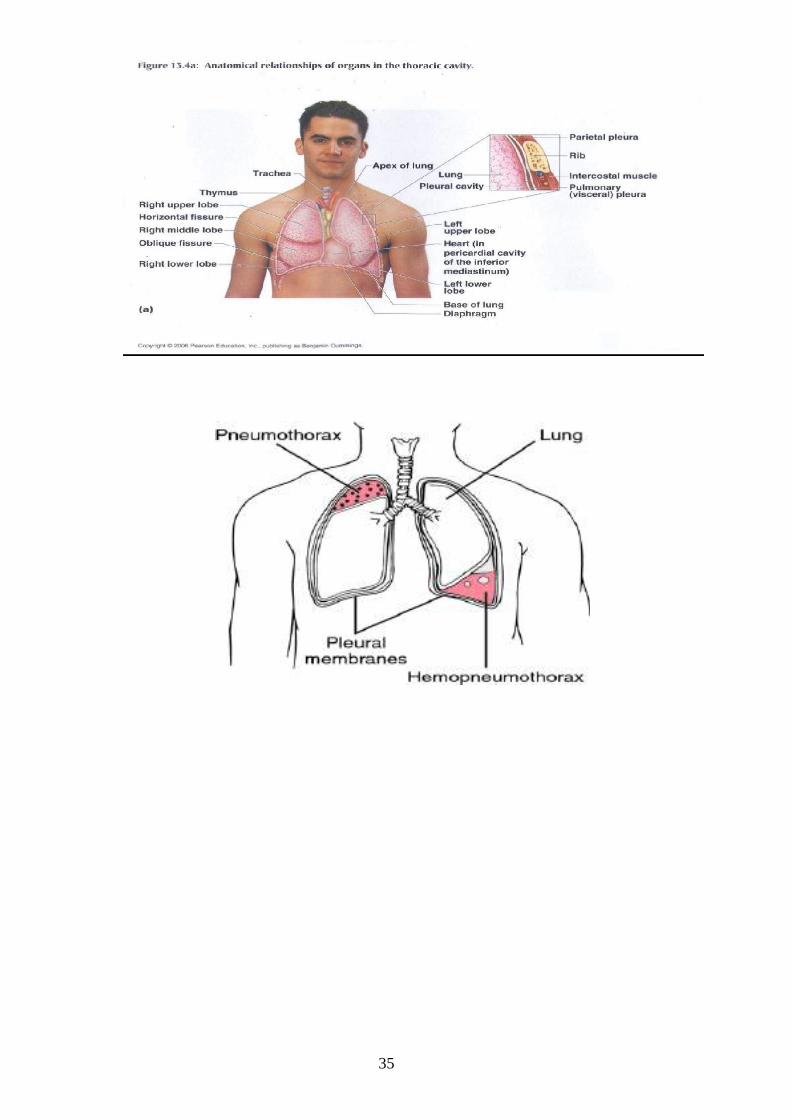
Lungs :
1. Right lung composed from 3- lobes a. Superior lobe. b. Middle lobe. c. Inferior lobe.
2. Left lung is smaller than the right and composed from 2- lobes.

34
a. Superior lobe. b. Inferior lobe
The lungs are pair of conical- shaped organs , each enveloped in a serous
membrane ( pleura ) . The apex of the lung rises into the root of the neck for about one inch above
the clavicle . The base is concave & is related to the upper surface of the diaphragm .
The lungs contain about 3 million alveoli, each alveolus composed of the following
a. Simple Squamous epithelium (type I pneumocytes)
b. Surfactant secreting cells (type II pneumcytes)
c. Macrophages which are responsible for removing debris and microbes from the alveoli
Pleural Membrane:
The covering membranes of the lung consists from:
1. Parietal pleura lines the inner surface of the thoracic cavity.
2. Visceral pleura cover the outer surface of the lungs.
Pleural Cavity:
The thin space between parietal pleura and visceral pleura called pleural
cavity. Both pleura secrete a small amount of pleural fluid .
Pleural fluid gives a moist, slippery coating that provides lubrication, thereby
reducing friction between the parietal and visceral surfaces as you breath.
35

36