university of groningen polymer brush-coatings to prevent ... · polymer brush-coatings to prevent...
TRANSCRIPT

University of Groningen
Polymer brush-coatings to prevent biomaterials associated infectionNejadnik, Mohammad Reza
IMPORTANT NOTE: You are advised to consult the publisher's version (publisher's PDF) if you wish to cite fromit. Please check the document version below.
Document VersionPublisher's PDF, also known as Version of record
Publication date:2009
Link to publication in University of Groningen/UMCG research database
Citation for published version (APA):Nejadnik, M. R. (2009). Polymer brush-coatings to prevent biomaterials associated infection: initial bacterialadhesion and biofilm formation. s.n.
CopyrightOther than for strictly personal use, it is not permitted to download or to forward/distribute the text or part of it without the consent of theauthor(s) and/or copyright holder(s), unless the work is under an open content license (like Creative Commons).
Take-down policyIf you believe that this document breaches copyright please contact us providing details, and we will remove access to the work immediatelyand investigate your claim.
Downloaded from the University of Groningen/UMCG research database (Pure): http://www.rug.nl/research/portal. For technical reasons thenumber of authors shown on this cover page is limited to 10 maximum.
Download date: 26-02-2019

Polymer brush-coatings to prevent
biomaterials associated infection
Initial bacterial adhesion and biofilm formation
Mohammad Reza Nejadnik
“Polymer brush-coatings to prevent biomaterials associated infection, Initial
bacterial adhesion and biofilm formation”
By Mohammad Reza Nejadnik
Printed by Facilitair Bedrijf (GrafiMedia), RUG, Groningen, May 2009
The cover has been designed by Samira Zamani.
Copyright © 2009 by MR Nejadnik
ISBN 978-90-367-3829-3 (printed version)
ISBN 978-90-367-3830-9 (electronic version)
Polymer brush-coatings to prevent biomaterials
associated infection
Initial bacterial adhesion and biofilm formation
Proefschrift
ter verkrijging van het doctoraat in de
Medische Wetenschappen
aan de Rijksuniversiteit Groningen
op gezag van de
Rector Magnificus, dr. F. Zwarts,
in het openbaar te verdedigen op
woensdag 24 juni 2009
om 13.15 uur
door
Mohammad Reza Nejadnik
geboren op 11 november 1978
te Isfahan, Iran

Promotores: Prof.dr.ir. H.J. Busscher
Prof.dr.ir. W. Norde
Prof.dr. H.C. van der Mei
Beoordelingscommissie: Prof.dr. J.E. Degener
Prof.dr. A. Herrmann
Prof.dr.ir. A.J. Schouten

To my dear Samira
Paranimfen: Samira Zamani
Sara Panahkhahi


Contents
Chapter 1 General introduction 1
Chapter 2 Adsorption of pluronic F-127 on surfaces with different
hydrophobicities probed by quartz crystal microbalance
with dissipation
9
Chapter 3 Determination of the shear force at the balance between
bacterial attachment and detachment in weakly adherence
systems, using a flow displacement chamber
27
Chapter 4 Bacterial adhesion and growth on a polymer brush coating 39
Chapter 5 Biofilms on polymer brush-coatings remain susceptible for
antibiotic treatment
59
Chapter 6 Bacterial colonization of polymer brush-coated and
pristine silicone rubber implanted in infected pockets in
mice
73
Chapter 7 General discussion 83
Summary 91
Samenvatting 97
Acknowledgements 103


General introduction
Chapter 1
2
General introduction
Bacterial adhesion and subsequent biofilm formation cause amongst others, infections
related to biomaterial implants, microbially-induced corrosion, and fouling of
membranes and heat exchanger surfaces in food processing systems [1]. The impact in
the medical field is enormous, since the rate of biomaterial associated infection for
initially inserted implants varies from 1 to 30 % with a mortality risk of up to 25%,
depending on the type of medical device [2]. The complex structure of a biofilm on
current biomaterials, containing slime and extracellular polymeric matrix produced by
microbial cells, makes it resistant against antibiotic treatment and the host immune
system. Even high concentrations of antibiotics have been reported to fail in
eradicating mature biofilms [3]. Therefore the fate of an infected biomaterial implant
is generally removal [4,5]. Devices implanted in revision surgery are, however, at a
several-fold greater risk of infection [2] likely due to persistence of bacteria in
adjacent tissue near the implant site [6].
Various coatings and materials like low surface free energy surfaces [7], positively
charged surfaces [8] and polymer brush-coatings [9] have been used to control
bacterial adhesion and biofilm formation. Polymer brush-coatings are currently
considered as the most promising non-fouling coatings, as these coatings reduce the
adhesion of bacteria by orders of magnitude [10].
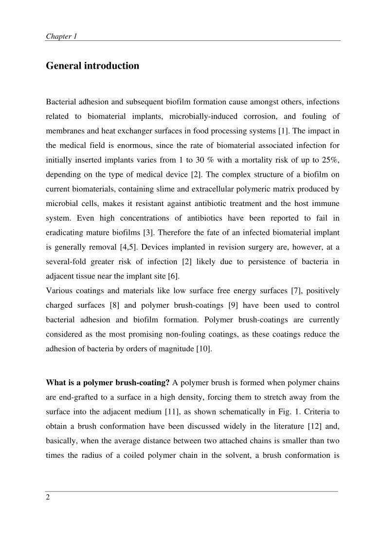
What is a polymer brush-coating? A polymer brush is formed when polymer chains
are end-grafted to a surface in a high density, forcing them to stretch away from the
surface into the adjacent medium [11], as shown schematically in Fig. 1. Criteria to
obtain a brush conformation have been discussed widely in the literature [12] and,
basically, when the average distance between two attached chains is smaller than two
times the radius of a coiled polymer chain in the solvent, a brush conformation is

General introduction
3
achieved. Hydrophilic polymer brush-coatings in an aqueous medium form a highly
hydrated layer at a surface. Compression of such a structure upon bacterial approach
gives rise to an osmotic pressure and decreased mobility (conformational entropy) of
the polymer chains in the brush, which causes repulsion of microorganisms from the
surface. Thus a brush-coating acts as a barrier that prevents microbial adhesion to a
substratum surface [13]. Thickness and density of brush-coatings are critical
parameters in making an efficient non-adhesive brush-coating, as increase in either
two generally enhances the adhesion resistance of the brush [14].
Physisorbed brushes are simply formed, based on the interaction of a part of the
polymer chain with the surface as an anchoring unit. Very often the anchoring block
has a sort of hydrophobic or electrostatic interaction with the substratum surface
[15,16]. Polymer chains can also be attached to a surface by chemical bonds. In order
to achieve that, either a chain is bound to the surface via an activated domain (grafting
to) or a chain is grown from the surface in chemical reactors (grafting from). The latter
method gives a better control over the density and thickness of the brush [17].
Generally chemically-bound brushes are more stable, but require a more complex
fabrication procedure.
Fig. 1. A schematic representation of a polymer brush structure.
Substratum surface
Anchoring sites
Polymer chains

Chapter 1
4
Problem statement and aim of this thesis. Despite the substantial amount of work on
the use of polymer brushes as non-adhesive surfaces in the past decade no
unambiguous judgment can be made yet whether polymer brush-coatings are capable
of preventing implant associated infections. This is mostly due to serious reservations
regarding the following challenges:
a) Stability of brush-coatings
Making a brush-coating that is stable against chemical and mechanical stresses and at
the same time is applicable to biomaterial surfaces has been a challenge for scientists
for years [18,19]. New developments in chemical engineering, however, have made it
possible to grow polymer brushes on virtually every surface with desired properties
[17]. Chemically-bound polymer brushes like the ones made by surface-initiated
polymerizations are generally known to be stable.
b) No prevention but reduction of adhesion
Despite appealing reports about anti-adherence properties of polymer brushes, these
coatings only strongly reduce the number of adhering bacteria and do not completely
prevent their adhesion. How strongly the few bacteria adhere to the brush-coating and
whether they are able to grow into a biofilm, are questions still to be answered.
c) In vivo performance of brush-coatings
In order to validate the success of polymer brush-coatings, in vivo studies are essential
but scarce, which results in lack of important information that is required to develop an
anti-infection strategy based on polymer brush-coatings.
The aim of this thesis is to study the fate of a few bacteria that adhere to a polymer
brush-coating and to evaluate the effects of a brush-coating on bacterial colonization
of biomaterial surfaces in vivo. This will lead to a better understanding of the state of
polymer brush-coatings in the battle against biomaterial associated infection.

General introduction
5
References
1. Costerton JW, Cheng KJ, Geesey GG et al. Bacterial biofilms in nature and disease.
Annual Review of Microbiology 1987, 41:435-64.
2. Darouiche RO. Device-associated infections: A macroproblem that starts with
microadherence. Clinical Infectious Diseases 2001, 33:1567-72.
3. Anwar H, Strap JL, Costerton JW. Establishment of aging biofilms - possible
mechanism of bacterial-resistance to antimicrobial therapy. Antimicrobial Agents and
Chemotherapy 1992, 36:1347-51.
4. Costerton JW, Stewart PS, Greenberg EP. Bacterial biofilms: A common cause of
persistent infections. Science 1999, 284:1318-22.
5. Gristina AG. Biomaterial-centered infection: microbial adhesion versus tissue
integration. Science 1987, 237:1588-95.
6. Broekhuizen CAN, de Boer L, Schipper K et al. Staphylococcus epidermidis is cleared
from biomaterial implants but persists in peri-implant tissue in mice despite
rifampicin/vancomycin treatment. Journal of Biomedical Materials Research Part A
2008, 85A:498-505.
7. Tsibouklis J, Stone M, Thorpe AA et al. Preventing bacterial adhesion onto surfaces:
the low-surface-energy approach. Biomaterials 1999, 20:1229-35.
8. Gottenbos B, Grijpma DW, Van der Mei HC et al. Antimicrobial effects of positively
charged surfaces on adhering Gram-positive and Gram-negative bacteria. Journal of
Antimicrobial Chemotherapy 2001, 48:7-13.
9. Currie EPK, Norde W, Cohen Stuart MA. Tethered polymer chains: surface chemistry
and their impact on colloidal and surface properties. Advances in Colloid and Interface
Science 2003, 100:205-65.
10. Roosjen A, Norde W, Van der Mei HC et al. The use of positively charged or low
surface free energy coatings versus polymer brushes in controlling biofilm formation.
Progress in Colloid and Polymer Science 2006, 132:138-44.
11. Zhao B, Brittain WJ. Polymer brushes: surface-immobilized macromolecules. Progress
in Polymer Science 2000, 25:677-710.
12. Halperin A. Polymer brushes that resist adsorption of model proteins: Design
parameters. Langmuir 1999, 15:2525-33.
13. Leckband D, Sheth S, Halperin A. Grafted poly(ethylene oxide) brushes as nonfouling
surface coatings. Journal of Biomaterial Science Polymer Edition 1999, 10:1125-47.
Chapter 1
6
14. Norde W, Gage D. Interaction of bovine serum albumin and human blood plasma with
PEO-tethered surfaces: influence of PEO chain length, grafting density, and
temperature. Langmuir 2004, 20:4162-67.
15. Brandani P, Stroeve P. Adsorption and desorption of PEO-PPO-PEO triblock
copolymers on a self-assembled hydrophobic surface. Macromolecules 2003, 36:9492-
501.
16. van der Burgh S, Fokkink R, de Keizer A et al. Complex coacervation core micelles as
anti-fouling agents on silica and polystyrene surfaces. Colloids and Surfaces A-
Physicochemical and Engineering Aspects 2004, 242:167-74.
17. Edmonson S, Osborne VL, Huck WTS. Polymer brushes via surface-initiated
polymerizations. Chemical society reviews 2004, 33:14-22.
18. Li JT, Caldwell KD. Plasma protein interaction with pluronic-treated colloids. Colloids
and Surfaces B-Biointerfaces 1996, 7:9-32.
19. Roosjen A, De Vries J, Van der Mei HC et al. Stability and effectiveness against
bacterial adhesion of poly(ethylene oxide) coatings in biological fluids. Journal of
Biomedical Materials Research applied biomaterials 2005, 73:347-54.


Adsorption of pluronic F-127 on
surfaces with different hydrophobicities
probed by quartz crystal microbalance
with dissipation
Reproduced with permission of the American Chemical Society from: M. Reza
Nejadnik, Adam L.J. Olsson, Prashant K. Sharma, Henny C. van der Mei, Willem
Norde, Henk J. Busscher. Langmuir 2009, DOI: 10.1021/la9001169.

Chapter 2
10
Abstract
Tri-block copolymers of polyethylene oxide (PEO) and polypropylene oxide (PPO),
i.e. PEOn-PPOm-PEOn, better known as Pluronic can adsorb to surfaces either in a
pancake or brush-like configuration. The brush-like configuration is advantageous in
numerous applications, since it constitutes a surface, repellent to proteins and
microorganisms. The conformation of an adsorbed Pluronic layer depends on the
hydrophobicity of the substratum surface, but the hydrophobicity threshold above
which a brush-like conformation is adopted is unknown. Therefore, the aim of this
study is to investigate Pluronic F-127 adsorption on surfaces with different
hydrophobicities using a quartz crystal microbalance with dissipation. Adsorption in a
brush-like conformation occurred on surfaces with a water contact angle above 80
degrees, as inferred from the thickness, viscosity and elasticity of the adsorbed layer.
The concentration of Pluronic F-127 in solution affected only the kinetics of
adsorption and not the final layer thickness or conformation of adsorbed Pluronic
molecules.
Adsorption of pluronic
11
Introduction
Tri-block copolymers of polyethylene oxide (PEO) and polypropylene oxide (PPO),
i.e. PEOn-PPOm-PEOn, better known as Pluronic, are used in a variety of industrial and
biomedical applications [1], amongst others to discourage protein adsorption and
bacterial adhesion [2-7]. Treatment of hydrophilic silica surfaces with Pluronic F-108,
reduced adsorption of proteins by 60%, while treatment of hydrophobic surfaces with
Pluronic F-108 completely inhibited protein adsorption [8], possibly due to a different
conformation of Pluronic F-108 on the different sorbent surfaces. Indeed, Schroen et
al. [8] suggested that the conformation of physically adsorbed tri-block copolymers
depends on the hydrophobicity of the sorbent surface: on a hydrophilic surface the two
terminal PEOn blocks attach resulting in a pancake conformation of the adsorbed tri-
block copolymer, whereas on a hydrophobic surface the PPOm block anchors to the
surface leaving the PEOn chains dangling in the adjacent solution. When the adsorbed
number of polymer chains is sufficiently high, a brush conformation is adopted, as has
been demonstrated for adsorption of Pluronic molecules on polystyrene films and on
hydrophobic dimethyl dichlorosilane coated silica [5,7]. Formation of a brush coating
of PEO chains on a surface creates superior non-adhesive properties. A highly
hydrated layer of PEO chains is unfavorably compressed by an approaching particle,
causing a repulsive osmotic force and a reduced mobility of the polymer chains. Both
discourage close contact of the particle with the surface and, consequently, suppress
adhesion. A brush coating of Pluronic F-127 on silicone rubber reduced the adhesion
of bacteria by one order of magnitude and was stable against high shear stresses for
more than 20 h [6,9].
It is crucial to be able to establish the conformation of adsorbed Pluronic molecules on
different surfaces in order to engineer a surface that meets the requirements for a
particular application and to determine the threshold hydrophobicity above which PEO
blocks adopt a brush-like conformation. Unfortunately, most techniques only yield

Chapter 2
12
limited information on the conformation of adsorbed Pluronic. A quartz crystal
microbalance with dissipation (QCM-D) is highly sensitive for studying the adsorption
processes in real time [10], while it gives insight into the characteristics and
conformation of the polymer chains in the adsorbed layer [11,12].
The aim of this study is to determine the threshold hydrophobicity above which the
pancake structure of adsorbed Pluronic F-127 changes into a stable brush-like
conformation, employing a QCM-D probe.
Materials and Methods
Pluronic F-127 (Polyethylene oxide (PEO)- polypropylene oxide (PPO)- polyethylene
oxide block copolymer with an average molecular structure of PEO99PPO65PEO99 and
a molecular weight of 12600), 1-octadecanothiol (HS(CH2)17CH3) and 11-mercapto-1-
undecanol (HSCH2(CH2)9CH2OH) were purchased from Sigma-Aldrich, USA.
Ethanol, ammonia and hydrogen peroxide were of analytical grade and purchased from
Merck, Darmstadt, Germany.
Preparation of crystal surfaces. Quartz crystals coated with gold and titanium were
purchased from Q-Sense AB, Sweden. Prior to use, all crystals were rinsed thoroughly
with ultrapure water and dried under a flow of nitrogen. Cleaned, hydrophobic gold-
coated crystals were made hydrophilic by exposure to UV/ozone treatment for 10 min
followed by immersion into a 3:1:1 mixture of ultrapure water, ammonia solution and
H2O2 maintained at 70-80°C for at least 10 min and a final rinse with water and
UV/ozone treatment for another 10 min. To further vary the hydrophobicity of the
gold-coated crystals, hydrophilic gold-coated crystals were coated with a self
assembled monolayer (SAM) by immersion of the crystals into a 1 mM solution of
HS(CH2)17CH3 (1-octadecanothiol) or HSCH2(CH2)9CH2OH (11-mercapto-1-

Adsorption of pluronic
13
undecanol) dissolved in ethanol for 18 h while shaking moderately to create a
hydrophobic or hydrophilic SAM, respectively.
Contact angle measurements. Prior to adsorption experiments, water contact angles
of 1 µl sessile droplets were measured on all surfaces using a homemade contour
monitor. On each sample, three droplets were placed on different spots and their
average contact angles are reported.
Adsorption experiments. Adsorption of block copolymers to crystals surfaces with
various coatings was investigated using a QCM-D model Q-sense E1 (Q-Sense AB,
Gothenburg, Sweden), which allows monitoring of changes in both resonance
frequency of the quartz crystal and dissipation of its energy as a function of time. The
crystal is excited to oscillate at its fundamental resonance frequency (4.95 MHz) and
its 3rd
, 5th
, 7th
, 9th
, 11th
and 13th
overtones. Addition of mass to the crystal surface is
registered as a decrease in resonance frequency (∆f), whereas the change in dissipation
(∆D) depends on the viscoelastic properties of the adsorbed film and its interaction
with the surrounding. The crystal is fixed in a closed chamber that allows flow of
liquids over the crystal surface, while the signals are recorded.
Measurements were conducted at a flow rate of 0.2 ml min-1
, corresponding with an
approximate shear rate of 24 s-1
at 20°C. After a stable baseline of the QCM-D signal
was obtained while flowing with ultrapure water, polymers were allowed to attach to a
crystal surface from a flowing low (0.0005 g l-1
) or high (0.5 g l-1
) concentration
Pluronic solution for 1 h, followed by rinsing with ultrapure water for another 1 h.
Data modeling. In case of hydrated adsorbed films, calculation of the layer properties
solely based on frequency shift is inaccurate and therefore a viscoelastic model must
be used [13]. We applied the Kelvin-Voigt model to the frequency and dissipation data
of all overtones in order to extract information on the viscoelastic properties and
Chapter 2
14
thickness of the adsorbed layer, using the QCM-D accompanying software package
(Q-Sense, Sweden) [14]. The model consists of a viscous damper (dash-pot) and an
elastic spring connected in parallel. Assuming the density of the adsorbed layer to be
equal to the density of water (1000 kg m-3
) and the viscosity of water to be 0.001 Pa.s
at 20°C, the software program allows calculation of the thickness, viscosity and
elasticity of the adsorbed layer.
Results
Analyses of frequency shifts and dissipation changes revealed that all overtones
exhibit similar trends which only differed in magnitude. Therefore in all figures, we
solely present the behavior of the 3rd
overtone.
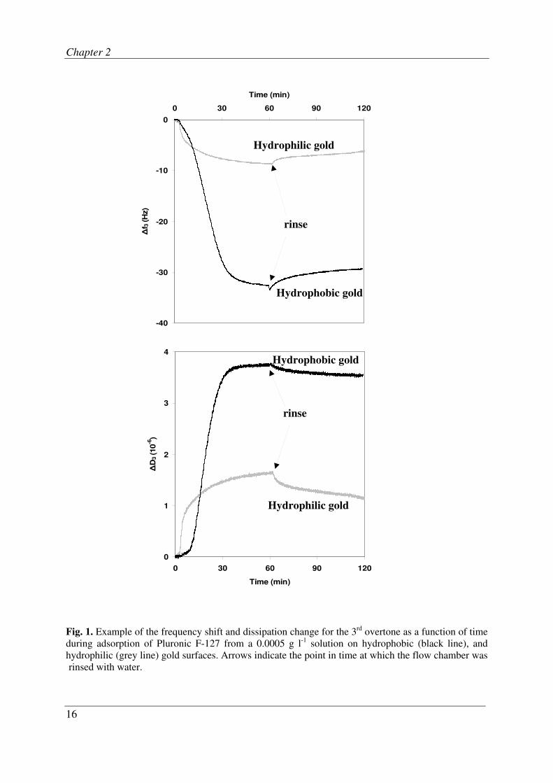
Fig. 1 shows representative QCM-D graphs for adsorbed Pluronic F-127 molecules on
a hydrophilic and hydrophobic gold-coated crystal (see Table 1 for water contact
angles). Both frequency shifts and dissipation changes are larger on the hydrophobic
than on the hydrophilic gold surface. The larger frequency shift indicates a greater
adsorbed mass at the hydrophobic surface. Note that Pluronic adsorption proceeds at a
more or less constant rate until saturation is reached, which is typical for polymer
adsorption [15]. Rinsing with water causes a small decrease in frequency shift and
dissipation change.
Fig. 2 shows the dissipation change versus the frequency shifts during Pluronic
adsorption to the different surfaces. On the hydrophilic gold surface, the hydrophilic
SAM- and titanium-coated surfaces, the dissipation change increases linearly with
increasing frequency shift and these curves are overlapping to a major extent. On
hydrophobic surfaces, however, two distinct transitions are observed and the curves
level off toward higher frequency shifts.
Fig. 3 illustrates the effect of Pluronic concentration on its adsorption to a hydrophobic
gold surface. A higher concentration of Pluronic clearly causes faster and more

Adsorption of pluronic
15
extensive frequency shifts and dissipation changes than a lower concentration. Yet, the
amount adsorbed in steady-state is not influenced by the Pluronic concentration, as
both frequency shift and dissipation change reach the same value after rinsing with
water.
Table 1. Water contact angles of substratum surfaces, together with the viscoelastic properties of
adsorbed layer of Pluronic F-127 on substrata with different hydrophobicities. Contact angles
represent averages ± standard deviations for six measurements over two separately prepared surfaces.
The viscosities and elasticities represent averages ± ranges for two separate measurements over two
separately prepared surfaces.
Substratum Water contact angle
(degrees)
Viscosity
(10-3
Pa.s)
Elasticity
(105 Pa)
Hydrophobic SAM 100 ± 2 2.6 ± 0.0 4.0 ± 0.0
Hydrophobic gold 86 ± 2 2.2 ± 0.0 2.8 ± 0.2
Titanium oxide 61 ± 1 1.2 ± 0.1 0.7 ± 0.2
Hydrophilic SAM 27 ± 1 1.4 ± 0.1 1.3 ± 0.2
Hydrophilic gold 16 ± 6 1.6 ± 0.2 1.1 ± 0.6
The viscosity and elasticity of the adsorbed layer on various surfaces after rinsing and
calculated from the Kelvin-Voigt model, are shown in Table 1. The viscosity of the
adsorbed layer is above 2.2 × 10⁻³
Pa.s on hydrophobic surfaces, whereas the viscosity
does not exceed 1.6 × 10⁻³
Pa.s on hydrophilic ones. The elasticity of the adsorbed
Pluronic layer is also hydrophobicity dependent, exceeding 2.8 × 10⁵ Pa on
hydrophobic surfaces and decreasing toward more hydrophilic surfaces.
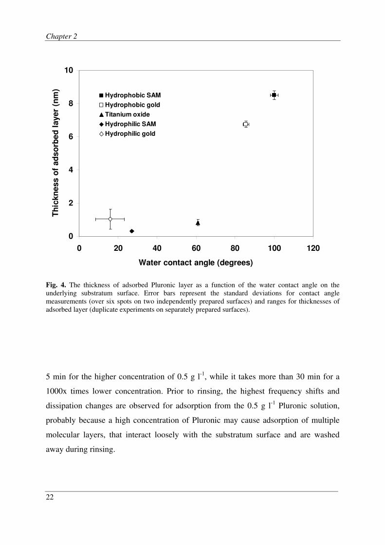
Fig. 4 shows the thickness of the adsorbed layer versus the water contact angle of the
substratum surfaces. The thickness remains below 2 nm until the substratum
hydrophobicity exceeds 80 degrees, above which the adsorbed layer strongly increases
to almost 9 nm.
Chapter 2
16
Fig. 1. Example of the frequency shift and dissipation change for the 3rd
overtone as a function of time
during adsorption of Pluronic F-127 from a 0.0005 g l-1
solution on hydrophobic (black line), and
hydrophilic (grey line) gold surfaces. Arrows indicate the point in time at which the flow chamber was
rinsed with water.
-40
-30
-20
-10
0
0 30 60 90 120
Time (min)
∆f 3
(H
z)
rinse
Hydrophilic gold
Hydrophobic gold
0
1
2
3
4
0 30 60 90 120
Time (min)
∆D
3 (10
-6)
rinse
Hydrophobic gold
Hydrophilic gold
Adsorption of pluronic
17
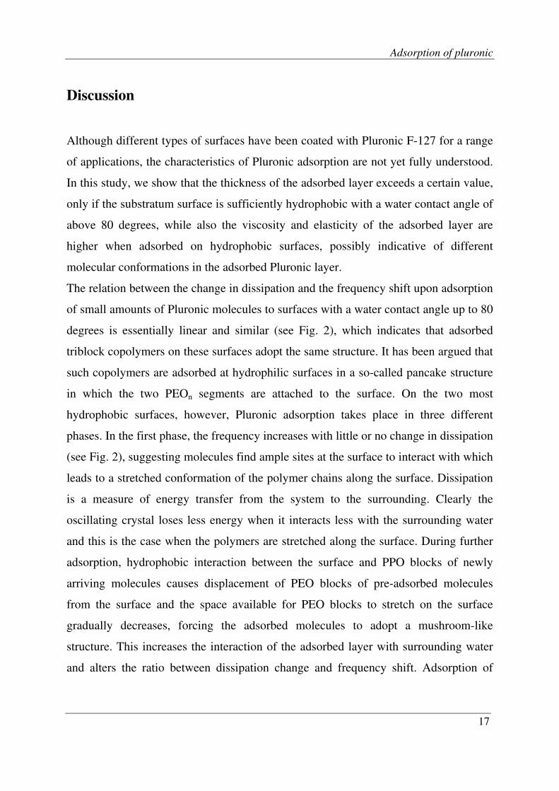
Discussion
Although different types of surfaces have been coated with Pluronic F-127 for a range
of applications, the characteristics of Pluronic adsorption are not yet fully understood.
In this study, we show that the thickness of the adsorbed layer exceeds a certain value,
only if the substratum surface is sufficiently hydrophobic with a water contact angle of
above 80 degrees, while also the viscosity and elasticity of the adsorbed layer are
higher when adsorbed on hydrophobic surfaces, possibly indicative of different
molecular conformations in the adsorbed Pluronic layer.
The relation between the change in dissipation and the frequency shift upon adsorption
of small amounts of Pluronic molecules to surfaces with a water contact angle up to 80
degrees is essentially linear and similar (see Fig. 2), which indicates that adsorbed
triblock copolymers on these surfaces adopt the same structure. It has been argued that
such copolymers are adsorbed at hydrophilic surfaces in a so-called pancake structure
in which the two PEOn segments are attached to the surface. On the two most
hydrophobic surfaces, however, Pluronic adsorption takes place in three different
phases. In the first phase, the frequency increases with little or no change in dissipation
(see Fig. 2), suggesting molecules find ample sites at the surface to interact with which
leads to a stretched conformation of the polymer chains along the surface. Dissipation
is a measure of energy transfer from the system to the surrounding. Clearly the
oscillating crystal loses less energy when it interacts less with the surrounding water
and this is the case when the polymers are stretched along the surface. During further
adsorption, hydrophobic interaction between the surface and PPO blocks of newly
arriving molecules causes displacement of PEO blocks of pre-adsorbed molecules
from the surface and the space available for PEO blocks to stretch on the surface
gradually decreases, forcing the adsorbed molecules to adopt a mushroom-like
structure. This increases the interaction of the adsorbed layer with surrounding water
and alters the ratio between dissipation change and frequency shift. Adsorption of

Chapter 2
18
more PPO blocks leads to increased packing density of the PEO chains till they are
forced to stretch away from the surface and form a brush-like conformation. Further
adsorption increases the frequency shift, with little effect on the dissipation change, as
also observed for a chemically grown brush [16]. At its steady state, the adsorbed layer
on a hydrophobic surface is less dissipative (smaller -∆D/∆f), i.e. more rigid,
compared to the soft one on a hydrophilic surface.
It has been shown that for chemically grafted brushes an increase in the initiator
density leads to an increase in the elasticity of the brush layer [17]. Accordingly,
Lubarsky et al [18] reported that the elasticity of adsorbed protein layers decreases by
increasing the level of hydration of the proteins (decreasing the packing density of
polymeric chains in the layer). This indicates that for a given system the elasticity of
an adsorbed layer is related to the packing density of the polymers in that layer.
Therefore our observation of the higher elasticity of the adsorbed layer on hydrophobic
surfaces compared to hydrophilic ones (see Table 1) supports the suggestion that the
packing density of polymer chains is higher in the adsorbed layer on a hydrophobic
surface. This, in turn, supports that on hydrophobic surfaces the adsorbed triblock
copolymers are in a brush-like conformation. Moreover, the viscosities measured for
adsorbed layers on hydrophobic surfaces are in good agreement with values found by
Fu et al. [17] for chemically grafted brushes. This is understandable because higher
volume density of PEO chains in the brush-like layer suggests a more hydrogel-like
structure with a higher viscosity.
The suggestion that adsorbed Pluronic molecules adopt a brush-like conformation on
hydrophobic surfaces can be evaluated by theoretical calculations. The condition for
having a polymer brush-coating is [19]:
1/D < RFl2 (1)
where D is the grafting density of polymer chains to the surface and RFl, the Flory
radius of a chain in a good solvent (RFl ≈ a × N3/5
, where a is the monomer size and N

Adsorption of pluronic
19
is the degree of polymerization). The Flory version of the Alexander model for a brush
relates the thickness (Tc) and density of a brush as:
Tc ≈ a × N × (a2 × D)
1/3 (2)
Fig. 2. Examples of frequency shifts versus dissipation changes for the 3rd overtone during adsorption
of Pluronic F-127 on different substrates.
Using Eq. (1), the minimum value for D is calculated to be approximately 0.033 nm-2
(taking the size of a PEO monomer 0.35 nm [20]), which corresponds to a layer
thickness of 5.5 nm, as can be calculated using Eq. (2). Thus a layer thickness well
above 5.5 nm would reflect a brush conformation. A thickness above 5.5 is measured
only for the hydrophobic gold and SAM surfaces (see Fig. 4). This is in line with our
interpretation of experimental observations that on the two most hydrophobic surfaces
0
1
2
3
4
-50 -40 -30 -20 -10 0
∆f3 (Hz)
∆D
3 (
10
-6)
Hydrophobic SAM
Hydrophobic gold
Titanium oxide
Hydrophilic SAM
Hydrophilic gold
Time
1
2
3
4
5
1
2
3
4
5
pancake mushroom
brush
Chapter 2
20
a brush-like conformation is adopted. Based on our QCM data, the threshold water
contact angle beyond which Pluronic F-127 adopts a brush-like conformation is
around 80 degrees.
However, it has to be realized that the density of the brush obtained by adsorption of
triblock copolymers is low as compared to the density that could be reached by
chemical grafting. This is because further increase in loss of conformational entropy of
the PEO chains with increasing packing density will stop further adsorption of
Pluronic from the solution.
At this stage it must be noted that experiments were also done on silicone dioxide-
coated crystals, with a water contact angle of 49 ± 3 degrees, yielding a layer thickness
less than 2 nm, as expected for a relatively hydrophilic surface, but with a viscosity
(2.4 ± 0.1 × 10⁻³
Pa.s) and elasticity (3.4 ± 0.5 × 10⁵ Pa) that are both higher than
expected for hydrophilic surfaces. It has been shown that acidic oxides such as silicon
dioxide adsorb PEO chains from the solution via an acid-base reaction [21]. Silicone
dioxide-coated surfaces are also known to yield unexpected interactions with vesicles.
For instance, using a QCM-D, Reimhult et al. [22] indicated that adsorbed vesicles are
more flattened on the silicone dioxide-coated surface than on titanium-coated or
hydrophilic gold coated crystals. Similarly, a possibly strong attraction of Pluronic
molecules toward the silicone dioxide-coated surface leads to an increase in density of
polymer chains in the film, at the expense of the film thickness, which in turn may
give rise to an elevated elasticity of the adsorbed layer.
The frequency shifts and dissipation changes after adsorption of Pluronic F-127 on
surfaces (see Fig. 1) decrease only slightly upon rinsing with water, suggesting that its
adsorption is irreversible. The high affinity of Pluronic for hydrophobic surfaces is
furthermore suggested by the observation that the steady-state adsorption is
independent of the concentration of Pluronic in solution over a wide concentration
range, i.e. 0.0005- 0.5 g l-1
(see Fig. 3). The concentration, however, considerably
influences the kinetics of adsorption, and adsorption reaches its maximum in less than

Adsorption of pluronic
21
Fig. 3. Example of the frequency shift and dissipation change for the 3rd overtone as a function of
time during Pluronic F-127 adsorption from a low (0.0005 g l-1, grey line) and high (0.5 g l-1, black
line) concentration solution on a hydrophobic gold surface.
0
1
2
3
4
5
6
0 30 60 90 120
Time (min)
∆D
3 (10
-6)
0.0005 g l-1
0.5 g l-1
-50
-40
-30
-20
-10
0
0 30 60 90 120
Time (min)
∆f 3
(H
z)
0.5 g l-1
0.0005 g l-1
Chapter 2
22
Fig. 4. The thickness of adsorbed Pluronic layer as a function of the water contact angle on the
underlying substratum surface. Error bars represent the standard deviations for contact angle
measurements (over six spots on two independently prepared surfaces) and ranges for thicknesses of
adsorbed layer (duplicate experiments on separately prepared surfaces).
5 min for the higher concentration of 0.5 g l-1
, while it takes more than 30 min for a
1000x times lower concentration. Prior to rinsing, the highest frequency shifts and
dissipation changes are observed for adsorption from the 0.5 g l-1
Pluronic solution,
probably because a high concentration of Pluronic may cause adsorption of multiple
molecular layers, that interact loosely with the substratum surface and are washed
away during rinsing.
0
2
4
6
8
10
0 20 40 60 80 100 120
Water contact angle (degrees)
Th
ickn
ess o
f ad
so
rbed
layer
(nm
)
Hydrophobic SAM
Hydrophobic gold
Titanium oxide
Hydrophilic SAM
Hydrophilic gold

Adsorption of pluronic
23
Conclusions
The thickness of an adsorbed Pluronic layer and the conformation of the molecules in
the layer strongly depend on the hydrophobicity of the underlying surface and not on
the Pluronic concentration in the adjacent phase. Adsorption of Pluronic F-127 on
hydrophobic surfaces with a water contact angle above 80 degrees yields a brush-like
conformation with a thickness between 6 and 9 nm. The viscosity and elasticity of an
adsorbed Pluronic layer in a brush-like conformation are higher than those in a
pancake conformation.
Acknowledgement
We would like to thank ZON-MW for grant 91107008 enabling the purchase of the
quartz crystal microbalance Qsense-E1 & E4.

Chapter 2
24
References
1. Alexandridis P, Hatton TA. Poly(ethylene oxide)-poly(propylene oxide)-poly(ethylene
oxide) block copolymer surfactants in aqueous solutions and at interfaces:
thermodynamics, structure, dynamics, and modeling. Colloids and Surfaces A-
Physicochemical and Engineering Aspects 1995, 96:1-46.
2. Amiji M, Park K. Prevention of protein adsorption and platelet adhesion on surfaces by
PEO/PPO/PEO triblock copolymers. Biomaterials 1992, 13:682-92.
3. Bridgett MJ, Davies MC, Denyer SP. Control of staophylococcal adhesion to
polystyrene surfaces by polymer surface modification with surfactants. Biomaterials
1992, 13:411-16.
4. Freij-Larsson C, Nylander T, Jannasch P et al. Adsorption behaviour of amphiphilic
polymers at hydrophobic surfaces: effects on protein adsorption. Biomaterials 1996,
17:2199-207.
5. Marsh LH, Coke M, Dettmar PW et al. Adsorbed poly(ethylene oxide)-poly(propylene
oxide) copolymers on synthetic surfaces: Spectroscopy and microscopy of polymer
structures and effects on adhesion of skin-borne bacteria. Journal of Biomedical
Materials Research Part A 2002, 61:641-52.
6. Nejadnik MR, Van der Mei HC, Norde W et al. Bacterial adhesion and growth on a
polymer brush-coating. Biomaterials 2008, 29:4117-21.
7. Norde W, Gage D. Interaction of bovine serum albumin and human blood plasma with
PEO-ththered surfaces: influence of PEO chain length, grafting density, and
temperature. Langmuir 2004, 20:4162-67.
8. Schroen CGPH, Cohen Stuart MA, Maarschalk KV et al. Influence of preadsorbed
block-copolymers on protein adsorption, surface properties, layer thickness and surface
coverage. Langmuir 1995, 11:3068-74.
9. Nejadnik MR, Van der Mei HC, Busscher HJ et al. Determination of the shear force at
the balance between bacterial attachment and detachment in weak-adherence systems,
using a flow displacement chamber. Applied Environmental Microbiology 2008,
74:916-19.
10. Marx KA. Quartz crystal microbalance: A useful tool for studying thin polymer films
and complex biomolecular systems at the solution-surface interface.
Biomacromolecules 2003, 4:1099-120.
11. Keller CA, Kasemo B. Surface specific kinetics of lipid vesicle adsorption measured

Adsorption of pluronic
25
with a quartz crystal microbalance. Biophysical Journal 1998, 75:1397-402.
12. Liu GM, Cheng H, Yan LF et al. Study of the kinetics of the pancake-to-brush
transition of poly(N-isopropylacrylamide) chains. Journal of physical chemistry B
2005, 109:22603-07.
13. Kwon KD, Green H, Bjoorn P et al. Model bacterial extracellular polysaccharide
adsorption onto silica and alumina: quartz crystal microbalance with dissipation
monitoring of dextran adsorption. Environ.Sci.Technol. 2006, 40:7739-44.
14. Voinova MV, Jonson M, Kasemo B. 'Missing mass' effect in biosensor's QCM
applications. Biosensors & Bioelectronics 2002, 17:835-41.
15. Norde, W. Colloids and interfaces in life sciences, CRC Press: 2003.
16. Moya SE, Brown AA, Azzaroni O et al. Following polymer brush growth using the
quartz crystal microbalance technique. Macromolecular Rapid Communications 2005,
26:1117-21.
17. Fu L, Chen XN, He JN et al. Study viscoelasticity of ultrathin poly(oligo(ethylene
glycol) methacrylate) brushes by a quartz crystal microbalance with dissipation.
Langmuir 2008, 24:6100-06.
18. Lubarsky GV, Davidson MR, Bradley RH. Hydration-dehydration of adsorbed protein
films studied by AFM and QCM-D. Biosensors & Bioelectronics 2007, 22:1275-81.
19. Halperin A. Polymer brushes that resist adsorption of model proteins: Design
parameters. Langmuir 1999, 15:2525-33.
20. Efremova NV, Sheth SR, Leckband DE. Protein-induced changes in poly(ethylene
glycol) brushes: Molecular weight and temperature dependence. Langmuir 2001,
17:7628-36.
21. Mathur S, Moudgil BM. Adsorption mechanism(s) of poly(ethylene oxide) on oxide
surfaces. Journal of colloid and interface science 1997, 196:92-98.
22. Reimhult E, Hook F, Kasemo B. Vesicle adsorption on SiO2 and TiO2: Dependence on
vesicle size. Journal of Chemical Physics 2002, 117:7401-04.


Determination of the shear force at the
balance between bacterial attachment
and detachment in weak-adherence
systems, using a flow displacement
chamber
Reproduced with permission of the American Society for Microbiology from: M. Reza
Nejadnik, Henny C. van der Mei, Henk J. Busscher, Willem Norde. Applied and
Environmental Microbiology 2008, 74:916-919.
Chapter 3
28
Abstract
We introduce a procedure to determine shear forces at the balance between bacterial
attachment and detachment under flow, that can be applied to determine adhesion
forces in weakly adhering systems, such as polymer brush-coatings, which are
currently in the center of attention for the control of bacterial adhesion and biofilm
formation.
Determination of the shear force
29
Introduction
Flow displacement systems, like the parallel plate flow chamber (PPFC), provide a
powerful tool to study adhesion of colloidal particles, including bacteria, to surfaces
under different hydrodynamic conditions [1]. Experimental observables, i.e. the
number of adhering bacteria and their distribution on the surface, are used to derive
attachment and detachment characteristics. A usual way to obtain qualitative
information on the strength of the bacterium-surface bond in the PPFC is to simply
pass an air-bubble through the chamber and analyze the number of bacteria remaining
on the surface: the force exerted by the air-bubble on an adhering micron-sized particle
is around 10-7
N [1]. Therefore, this method is too insensitive to be used in systems
with weak bacterium-surface interaction forces, such as polymer brush-coatings.
One of the big advantages of PPFC is the adjustability of the shear rate and shear stress
at the surface. These quantities are related through the equation: τ =F/A= ησ where τ is
the shear stress, F the force, A the area on which the force is exerted, η the absolute
viscosity and σ the shear rate. The wall shear rate is related to the flow rate Q [2]
according to σ =3Q/2b2w with b, the half depth, and w, the width of the chamber. The
force on a single adhering bacterium can then be approximated as the product of wall
shear stress times the bacterial surface area exposed to the shear.
Sufficiently high shear stresses cause adhering bacteria to slide and roll over a surface,
which may lead to detachment. In order to characterize attachment and detachment of
bacteria with respect to wall shear, notions as “shear to prevent adhesion” and “shear
to remove adhered bacteria” have been used [1,3-7]. Shear stresses in the range of 12
to 54 Pa have been reported for the removal of different bacterial strains from regular
surfaces and usually a lower shear stress is required to prevent adhesion [8]. These
characteristic shear stresses however, suffer from some ambiguity because the strength
of adhesion can depend on the history of contact between a bacterium and a
substratum surface, i.e. its residence time and the shear stress applied during adhesion

Chapter 3
30
[9,10]. Moreover, the shear to detach adhered bacteria can not be obtained for a wide
range of adhesion forces within the laminar flow regime.
Bacterial adhesion is the first step in the development of a biofilm and represents the
onset of biomaterials implant-related infection, microbially induced corrosion, and
fouling of membranes and heat exchanger surfaces in food processing systems [11].
Much attention has been directed towards the development of antifouling surfaces
[12]. Polymer brush-coatings are currently considered as the most promising non-
fouling coatings, as they weaken the attractive interaction forces between adhering
bacteria and the underlying substratum [13,14]. Here, for the first time, we present a
method that yields quantitative data on bacteria-surface affinity over a wide range of
interaction forces, including the weak interaction forces as existing on non-fouling
surfaces.
Materials and Methods
Staphylococcus epidermidis HBH276, Staphylococcus aureus ATCC12600 and
Pseudomonas aeruginosa #3 were pre-cultured in 10-ml tryptone soya broth at 37°C
for 24 h from blood agar plates. These pre-cultures were used to inoculate second
cultures of 200-ml for 16 h. Subsequently, bacteria were harvested and washed twice
with demineralized water. To break up bacterial aggregates, bacteria were sonicated
for 3 times 10 s at 30 W. Finally, bacteria were suspended in phosphate-buffered saline
solution (PBS: 10 mM potassium phosphate, 150 mM NaCl, pH 6.8) to a concentration
of 3×10⁸ per ml for all experiments.
Implant grade silicone rubber sheets (Medin, Groningen, The Netherlands) were rinsed
with ethanol and demineralized water, sonicated in 2% RBS35 detergent and rinsed
thoroughly with demineralized water, washed in methanol and rinsed with
demineralized water again to remove oil contaminations and fingerprints. The silicone
rubber was fixed in the bottom plate of the PPFC and exposed to a solution of 0.5 g l-1
Determination of the shear force
31
Pluronic F-127 (PEO99PPO65PEO99, Sigma-Aldrich, USA) in PBS for 20 min. Non-
attached polymer chains were removed from the surface by washing with PBS. This
has been proven to result in a brush layer of PEO-chains on hydrophobic substrata [15-
17].
Our flow chamber and image analysis system have been previously described [1]. The
setup gives the possibility of having a steady flow as well as being able to quickly
switch from low to high flow rates. We used a fluctuating flow protocol to study the
attachment and detachment of different bacterial strains to pristine and PEO brush-
coated silicone rubber. The protocol begins with flowing a bacterial suspension for 30
min at a wall shear stress of 0.005 Pa. Thereafter, the shear stress is instantly adjusted
to either 0.005, 0.3, 0.7, 1.5 or 4.7 Pa, which is maintained for another 30 min. After
each separate increase, the shear stress is reset to 0.005 Pa again. This cycle is repeated
twice (Fig. 1a).
Results and Discussion
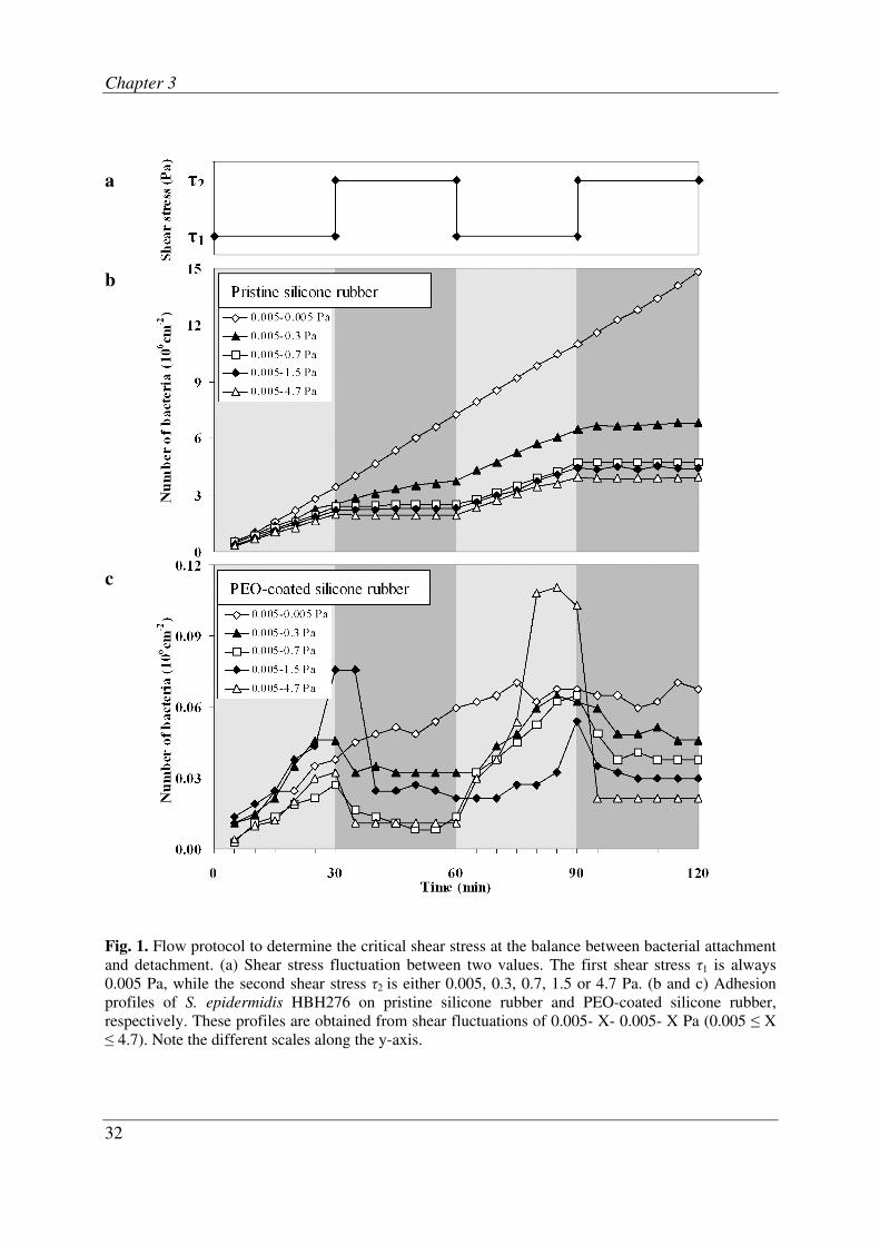
During the first 30 min at a shear stress of 0.005 Pa, bacteria attach to the surface.
Increasing the flow rate implies a higher supply rate of bacteria to the surface, but also
an increased shear stress acting on adhering bacteria. A relatively small increase leads
to additional attachment of bacteria. Increasing the shear above a certain threshold will
result in a net decrease in the number of adhering bacteria due to a dominant
contribution of shear-induced detachment. Here, we refer to the shear stress at which
additional attachment and detachment balance each other as the “critical shear stress”.
Attachment and detachment of S. epidermidis HBH276 on pristine and PEO-coated
silicone rubber under fluctuating shear are presented in Fig. 1. The deposition rate
(slope of the curve in a time frame) of S. epidermidis HBH276 on pristine silicone
rubber is suppressed by increasing the shear stress τ due to the high wall shear
Chapter 3
32
a
b
c
Fig. 1. Flow protocol to determine the critical shear stress at the balance between bacterial attachment
and detachment. (a) Shear stress fluctuation between two values. The first shear stress τ1 is always
0.005 Pa, while the second shear stress τ2 is either 0.005, 0.3, 0.7, 1.5 or 4.7 Pa. (b and c) Adhesion
profiles of S. epidermidis HBH276 on pristine silicone rubber and PEO-coated silicone rubber,
respectively. These profiles are obtained from shear fluctuations of 0.005- X- 0.005- X Pa (0.005 ≤ X
≤ 4.7). Note the different scales along the y-axis.
Determination of the shear force
33
preventing newly arriving bacteria from attaching to the surface. Attachment and
detachment events on PEO-coated surfaces are very different and the deposition rate at
low shear is one to two orders of magnitude smaller than for pristine silicone rubber.
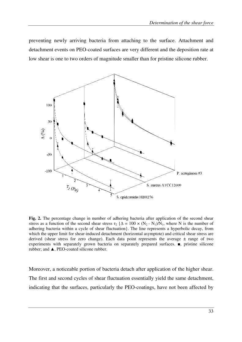
Fig. 2. The percentage change in number of adhering bacteria after application of the second shear
stress as a function of the second shear stress τ2 {∆ = 100 × (N2 - N1)/N1, where N is the number of
adhering bacteria within a cycle of shear fluctuation}. The line represents a hyperbolic decay, from
which the upper limit for shear-induced detachment (horizontal asymptote) and critical shear stress are
derived (shear stress for zero change). Each data point represents the average ± range of two
experiments with separately grown bacteria on separately prepared surfaces. ■, pristine silicone
rubber; and ▲, PEO-coated silicone rubber.
Moreover, a noticeable portion of bacteria detach after application of the higher shear.
The first and second cycles of shear fluctuation essentially yield the same detachment,
indicating that the surfaces, particularly the PEO-coatings, have not been affected by

Chapter 3
34
the high shear. At this stage, it is important to realize that all experiments are carried
out in the initial phase of adhesion, i.e. where the numbers of adhering bacteria
increase linearly with time (see also Fig. 1). Therewith all changes in attachment and
subsequent detachment are due to fluctuations in shear and not to saturation of the
surface. From Fig. 1 it can be seen that S. epidermidis HBH276 is much more loosely
bound to the PEO-coating than to pristine silicone rubber surface.
In order to determine the above defined critical shear stress, we first calculate the net
effect of increasing the shear and present the percentage change in number of adhering
bacteria after application of the second shear stress as a function of the second shear
stress (Fig. 2). This allows us to determine the critical shear stress and an upper-limit
for shear-induced detachment. This change in the number of adhering bacteria upon
increasing the shear shows a hyperbolic decay, as can be seen in Fig. 2. Zero change in
the number of adhering bacteria, defining the critical shear stress, occurs at 2.7 ± 1.1
Pa for S. epidermidis HBH276 on pristine and at only 0.2 ± 0.1 Pa on PEO-coated
silicone rubber, corresponding with critical forces of 2.1 ± 0.9 pN and 0.1 ± 0.1 pN,
respectively on a single bacterium (assuming a staphylococcal radius of 0.5 µm and of
0.6 µm for P. aeruginosa, which is the radius of a sphere with equal volume as this
rod-shaped organism). The upper limit of shear-induced detachment amounts 10% for
S. epidermidis HBH276 on pristine silicone rubber, while over 90% of all adhering
staphylococci can be detached from PEO brush-coated silicone rubber.
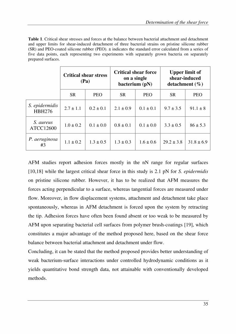
Table 1 compares the critical shear stress and upper limit of shear-induced detachment
for the three different bacterial strains. Clearly, different bacterial strains adhere to
surfaces with different strength. The adhesion strength of staphylococci is greatly
decreased by the presence of a PEO-coating, while the adhesion strength of P.
aeruginosa #3 is hardly affected. This is in line with other findings, showing that PEO
brush-coatings are not effective against hydrophobic P. aeruginosa strains [13].
Determination of the shear force
35
Table 1. Critical shear stresses and forces at the balance between bacterial attachment and detachment
and upper limits for shear-induced detachment of three bacterial strains on pristine silicone rubber
(SR) and PEO-coated silicone rubber (PEO). ± indicates the standard error calculated from a series of
five data points, each representing two experiments with separately grown bacteria on separately
prepared surfaces.
Critical shear stress
(Pa)
Critical shear force
on a single
bacterium (pN)
Upper limit of
shear-induced
detachment (%)
SR PEO SR PEO SR PEO
S. epidermidis
HBH276 2.7 ± 1.1 0.2 ± 0.1 2.1 ± 0.9 0.1 ± 0.1 9.7 ± 3.5 91.1 ± 8
S. aureus
ATCC12600 1.0 ± 0.2 0.1 ± 0.0 0.8 ± 0.1 0.1 ± 0.0 3.3 ± 0.5 86 ± 5.3
P. aeruginosa
#3 1.1 ± 0.2 1.3 ± 0.5 1.3 ± 0.3 1.6 ± 0.6 29.2 ± 3.8 31.8 ± 6.9
AFM studies report adhesion forces mostly in the nN range for regular surfaces
[10,18] while the largest critical shear force in this study is 2.1 pN for S. epidermidis
on pristine silicone rubber. However, it has to be realized that AFM measures the
forces acting perpendicular to a surface, whereas tangential forces are measured under
flow. Moreover, in flow displacement systems, attachment and detachment take place
spontaneously, whereas in AFM detachment is forced upon the system by retracting
the tip. Adhesion forces have often been found absent or too weak to be measured by
AFM upon separating bacterial cell surfaces from polymer brush-coatings [19], which
constitutes a major advantage of the method proposed here, based on the shear force
balance between bacterial attachment and detachment under flow.
Concluding, it can be stated that the method proposed provides better understanding of
weak bacterium-surface interactions under controlled hydrodynamic conditions as it
yields quantitative bond strength data, not attainable with conventionally developed
methods.
Chapter 3
36
References
1. Busscher HJ, Van der Mei HC. Microbial adhesion in flow displacement systems.
Clinical Microbiology Reviews 2006, 19:127-41.
2. Hoek EMV, Elimelech M. Cake-enhanced concentration polarization: a new fouling
mechanism for salt-rejecting membranes. Environ.Sci.Technol. 2003, 37:5581-88.
3. Anamelechi CC, Truskey GA, Reichert WM. Mylar (TM) and Teflon-AF (TM) as cell
culture substrates for studying endothelial cell adhesion. Biomaterials 2005, 26:6887-
96.
4. Guillemot G, Veca-Medina G, Martin-Yken H et al. Shear-flow induced detachment of
Saccharomyces cerevisiae from stainless steel: influence of yeast and solid surface
properties. Colloids and Surfaces B-Biointerfaces 2006, 49:126-35.
5. Gutierrez E, Groisman A. Quantitative measurements of the strength of adhesion of
human neutrophils to a substratum in a microfluidic device. Analytical Chemistry
2007, 79:2249-58.
6. Kurtis MS, Schmidt TA, Bugbee WD et al. Integrin-mediated adhesion of human
articular chondrocytes to cartilage. Arthritis and Rheumatism 2003, 48:110-18.
7. Lorthois S, Schmitz P, Angles-Cano E. Experimental study of fibrin/fibrin-specific
molecular interactions using a sphere/plane adhesion model. Journal of colloid and
interface science 2001, 241:52-62.
8. Rutter, P. R.; Vincent, B. Physiological models in microbiology, Bazin M.J.; Prosser
J.I., Eds.; 1988; Chapter 10.
9. Marshall BT, Sarangapani KK, Lou JH et al. Force history dependence of receptor-
ligand dissociation. Biophysical Journal 2005, 88:1458-66.
10. Xu LC, Vadillo-Rodriguez V, Logan BE. Residence time, loading force, pH, and ionic
strength affect adhesion forces between colloids and biopolymer-coated surfaces.
Langmuir 2005, 21:7491-500.
11. Costerton JW, Stewart PS, Greenberg EP. Bacterial biofilms: A common cause of
persistent infections. Science 1999, 284:1318-22.
12. Nuzzo RG. Biomaterials stable antifouling surfaces. Nature Materials 2003, 2:207-08.
13. Roosjen A, Busscher HJ, Norde W et al. Bacterial factors influencing adhesion of
Pseudomonas aeruginosa strains to a poly(ethylene oxide) brush. Microbiology 2006,
152:2673-82.
Determination of the shear force
37
14. Zhao B, Brittain WJ. Polymer brushes: surface-immobilized macromolecules. Progress
in Polymer Science 2000, 25:677-710.
15. Brandani P, Stroeve P. Adsorption and desorption of PEO-PPO-PEO triblock
copolymers on a self-assembled hydrophobic surface. Macromolecules 2003, 36:9492-
501.
16. Mclean SC, Lioe H, Meagher L et al. Atomic force microscopy study of the interaction
between adsorbed poly(ethylene oxide) layers: effects of surface modification and
approach velocity. Langmuir 2005, 21:2199-208.
17. Norde W, Gage D. Interaction of bovine serum albumin and human blood plasma with
PEO-tethered surfaces: influence of PEO chain length, grafting density, and
temperature. Langmuir 2004, 20:4162-67.
18. Hinterdorfer P, Dufrene YF. Detection and localization of single molecular recognition
events using atomic force microscopy. Nature Methods 2006, 3:347-55.
19. Razatos A, Ong YL, Boulay F et al. Force measurements between bacteria and
poly(ethylene glycol)-coated surfaces. Langmuir 2000, 16:9155-58.


Bacterial adhesion and growth on a
polymer brush coating
Reproduced with permission of Elsevier from: M. Reza Nejadnik, Henny C. van der Mei,
Willem Norde, Henk J. Busscher. Biomaterials 2008, 29:4117-4121.

Chapter 4
40
Abstract
Biomaterials-related infections pose serious problems in implant surgery, despite the
development of non-adhesive coatings. Non-adhesive coatings, like polymer brush-
coatings, have so far only been investigated with respect to preventing initial bacterial
adhesion, but never with respect to effects on kinetics of bacterial growth. Here, we
compare adhesion and 20h growth of three bacterial strains (Staphylococcus aureus,
Staphylococcus epidermidis and Pseudomonas aeruginosa) on pristine and brush-
coated silicone rubber in a parallel plate flow chamber. Brush-coatings were made
using a triblock copolymer of polyethylene-oxide (PEO) and polypropylene-oxide
(PPO). Brush-coatings prevented adhesion of staphylococci to below 5 × 105 cm
-2 after
30 min, which is a 10-fold reduction compared to pristine silicone rubber. Biofilms
grew on both brush-coated and pristine silicone rubber, while the viability of biofilms
on brush-coatings was higher than on pristine silicone rubber. However, biofilms on
brush-coatings developed more slowly and detached almost fully by high fluid shear.
Brush-coating remained non-adhesive after S. epidermidis biofilm formation and
subsequent removal whereas a part of its functionality was lost after removal of S.
aureus biofilms. Adhesion, growth and detachment of P. aeruginosa were not
significantly different on brush-coatings as compared with pristine silicone rubber,
although here too the viability of biofilms on brush-coatings was higher. We conclude
that polymer brush-coatings strongly reduce initial adhesion of staphylococci and
delay their biofilm growth. In addition, biofilms on brush-coatings are more viable and
easily removed by the application of fluid shear.
Bacterial adhesion and growth
41
Introduction
Biomaterials-related infection is one of the main causes of implant failure in an era in
which the number of patients requiring biomaterials implant surgery is steadily
increasing [1-3]. Treatment of infected implants frequently includes long-term
antibiotic use, often with implant removal as a final result. Recently, even scaffolds
used in cartilage repair appeared prone to infection at a rate similar to other
orthopaedic implants [4].
Biomaterials-related infection starts with the initial adhesion of infectious organisms
that subsequently grow to form a biofilm. Bacterial adhesion to surfaces is influenced
by physicochemical properties of the surface [5] and several attempts have been made
to develop non-adhesive coatings [6], such as polymer brush-coatings, in order to
prevent bacterial adhesion and subsequent infection [7,8]. Polymer brushes are end-
tethered polymer chains which are forced to stretch away from a surface into the
adjacent solution due to a high density of chains per unit surface area [9]. Polyethylene
oxide (PEO) brush-coatings have been used to prevent protein adsorption as well as
bacterial adhesion [10-13]. The brush-coating forms a highly hydrated layer of PEO
chains that is compressed upon bacterial approach, leading to a repulsive osmotic force
and to a reduced mobility of the polymer chains. This creates a steric barrier which
discourages close contact and suppresses adhesion [7]. Most types of brush coatings
show significant reductions in microbial adhesion exceeding 90% [10,14,15], while
bacteria also adhere more weakly [16]. Despite such impressive results it remains an
open question whether the few bacteria adhering on a brush-coating are capable of
growing into a mature biofilm.
Pluronic F-127 (polyethylene oxide- polypropylene oxide - polyethylene oxide tri-
block copolymer) can be adsorbed to hydrophobic substrata, such as silicone rubber, to
form low-density polymer brush-coatings [11,12,14,17,18]. These polymer brush-
coatings resist protein adsorption, reduce initial bacterial adhesion and moreover are
Chapter 4
42
stable under fluid shear [16] and in physiological fluids [19]. In this study, we
establish the fate of the few bacteria that adhere on a polymer brush-coating and
determine whether they are able to grow into a biofilm and if so what the effect of
brush-coating is on viability and adhesion strength of the biofilm. To this end, we
compare initial adhesion and subsequent 20 h growth of three bacterial strains
(Staphylococcus aureus ATCC 12600, Staphylococcus epidermidis HBH 276 and
Pseudomonas aeruginosa #3) on pristine and polymer brush-coated silicone rubber in
a parallel plate flow chamber. Furthermore we assess the non-adhesive functionality of
the brush-coating after biofilm growth and removal.
Materials and Methods
Polyethylene oxide (PEO) coating of silicone rubber. Implant grade silicone rubber
sheets (thickness 0.5 mm, water contact angle 112 ± 1°) were obtained from Medin,
Groningen, The Netherlands. Silicone rubber sheets were rinsed with ethanol (Merck,
Darmstadt, Germany) and demineralised water and subsequently sonicated for three
min in 2% RBS 35 detergent (Omnilabo International BV, Breda, The Netherlands)
and rinsed thoroughly with demineralised water, washed in methanol (Merck,
Darmstadt, Germany) and rinsed with demineralised water. A silicone rubber sheet
was fixed in the bottom plate of a parallel plate flow chamber and exposed to a
solution of 0.5 g/l Pluronic F-127 (PEO99PPO65PEO99, molecular weight 12600;
Sigma-Aldrich, USA) in phosphate buffered saline solution (PBS: 10 mM potassium
phosphate, 150 mM NaCl, pH 6.8) for 20 min at room temperature. Non-attached
polymer was removed from the chamber by flow with an excess amount of PBS. NaCl,
K2HPO4 and KH2PO4 were of analytical grade, as purchased from Merck.
Bacterial strains and culturing. Three bacterial strains (Staphylococcus epidermidis
HBH 276, Staphylococcus aureus ATCC 12600 and Pseudomonas aeruginosa #3)

Bacterial adhesion and growth
43
were used in this study. All strains were first grown aerobically overnight at 37°C on
blood agar plates from frozen stocks. These plates were kept at 4°C and never longer
than two weeks. Several colonies were used to make a pre-culture in 10 ml tryptone
soya broth (TSB, OXOID, Basingstoke, England). This pre-culture was incubated at
37°C for 24 h and used to inoculate a second culture of 200 ml which was incubated
for 16 h. The culture was harvested by centrifugation for 5 min at 5000 × g and
washed twice with demineralised water. To break up bacterial aggregates, bacteria
were sonicated intermittently while cooling in an ice/water bath for three times 10 s at
30 W (Vibra Cell model 375; Sonics and Materials, Danbury, CT, USA). These
procedures were found not to cause cell lysis in any of the three strains. Finally,
bacteria were suspended in 200 ml of PBS to a concentration of 3 × 10⁸ per ml for all
experiments.
Parallel plate flow chamber and image analysis system. The parallel plate flow
chamber (175 × 17 × 0.75 mm3) and image analysis system have been previously
described [20]. The flow chamber by design allows laminar flows corresponding with
shear stresses up to 15 Pa, as can be calculated from the volumetric flow rate Q
according to
τ = η σ = η 3Q/2b2w (1)
in which τ is the shear stress, η the absolute viscosity, σ the shear rate and b and w
represent the flow chambers half depth and width, respectively [20]. Images were
taken from pristine silicone rubber and silicone rubber coated with a PEO brush,
affixed to the polymethyl-methacrylate bottom plate of the chamber. The top plate was
made of glass. Bacterial adhesion and detachment were monitored using a Fire wire
CCD camera, mounted on a phase contrast microscope equipped with a 40× ultra long
working distance objective. The camera was coupled to a PC proprietary image
analysis software. Each live image (1392 × 1040 pixels with 8 bit resolution) was
obtained from summation of 15 consecutive images (time interval 0.25 s) in order to

Chapter 4
44
enhance the signal to noise ratio and to eliminate moving organisms from the analysis.
Before each experiment, all tubes and the flow chamber were filled with PBS, while
care was taken to remove air bubbles from the system. Flasks, containing bacterial
suspension and buffer, were positioned at different height with respect to the chamber
to ensure that all fluids circulate through the chamber at the desired rate by hydrostatic
pressure immediately after starting the flow. Different flow rates were achieved by
adjusting the hydrostatic pressure, while maintaining constant flow by recirculation
using a roller pump.
Initial bacterial adhesion and biofilm growth. First, bacteria were allowed to adhere
at room temperature to the substratum surface during 25 min under a shear stress of
0.005 Pa. Subsequently, flow was switched from bacterial suspension to 10% TSB
medium for 5 min to flush out unattached bacteria from the chamber, after which the
temperature was raised to 37 ºC and shear stress was decreased to 0.002 Pa for 20 h.
From the images taken during initial bacterial adhesion and subsequent growth, initial
bacterial deposition rates (j0), numbers of adhering bacteria after 30 min and the
percentage surface coverage by the biofilm were determined. All experiments were
done in fourfold, with separately grown bacteria.
Shear induced detachment of biofilm. After biofilm growth, shear stress was first
increased to 0.01 Pa for 30 min, while temperature was set again at room temperature,
after which shear stress was increased to 0.05 Pa and 4 Pa, each for 10 min. Biofilm
detachment was assessed at the end of each shear stress period as a percentage with
respect to the surface coverage after 20 h of growth.
After biofilm detachment, bacterial adhesion experiments were done in order to assess
whether the brush-coating was still functional using fresh bacterial suspensions. These
experiments were done on brush-coatings immediately after biofilm removal and after
subsequent re-exposure to the Pluronic solution, applied to potentially restore the

Bacterial adhesion and growth
45
brush-coating in case the original brush would have been damaged by removal of the
biofilm.
Biofilm viability. In a separate set of experiments, 20 h biofilms were removed from
the substratum surfaces using a sterile cotton swab and suspended in 1 ml
demineralised water. Subsequently, 15 µl of the suspension was transferred onto a
glass slide and stained for 20 min in the dark with a live and dead stain (BacLightTM
,
Molecular Probes Europe BV) and the percentage of live and dead bacteria was
evaluated using fluorescence microscopy (Leica, Wetzlar, Germany).
X-ray photoelectron spectroscopy (XPS). Elemental surface compositions of pristine
and brush-coated silicone rubber prior to initial bacterial adhesion and after biofilm
detachment as well as after re-exposure to the Pluronic solution were determined using
XPS (Surface Science Instruments, Mountain View, CA, USA). Elemental surface
compositions were expressed in percentages of carbon, oxygen, silicon and nitrogen.
Two separate measurements were taken on different spots for two separately prepared
pristine and brush-coated silicone rubber surfaces.
Statistical analysis. The non-parametric Mann-Whitney test (SPSS 12.0.1 for
Windows package) was employed for comparing two independent groups of data.
Differences were considered statistically significant for p-values less than 0.05.
Results
Initial adhesion of S. aureus and S. epidermidis after 30 min of flow was reduced from
5.2 ± 1.6 x 10⁶ and 4.1 ± 1.7 x 10
6 cm
-2 on pristine silicone rubber to 0.4 ± 0.2 x 10
6
and 0.1 ± 0.1 x 106 cm
-2 on brush-coatings, respectively. Initial adhesion of P.

Chapter 4
46
aeruginosa was not significantly different on brush-coatings than on pristine silicone
rubber and amounted approximately 2 x 106 cm
-2.
Fig. 1 presents the surface coverage by biofilms after subsequent growth. Both
staphylococcal strains show a lag-time of about 3 h on pristine silicone rubber after
which biofilms started to grow exponentially. 100% surface coverage was reached for
both strains after approximately 8 h of growth. Lag-times on brush-coatings amounted
approximately 8 h and after 20 h surface coverage was only 71 ± 9 % and 60 ± 33 %
for S. aureus and S. epidermidis, respectively. Despite the rather large standard
deviations, the differences between surface coverage on brush-coated and pristine
silicone rubber are significant for both strains at all points in time. The kinetics of
bacterial growth for P. aeruginosa was totally different and surface coverage remained
confined to less than 24 %, regardless of the absence or presence of a brush-coating.
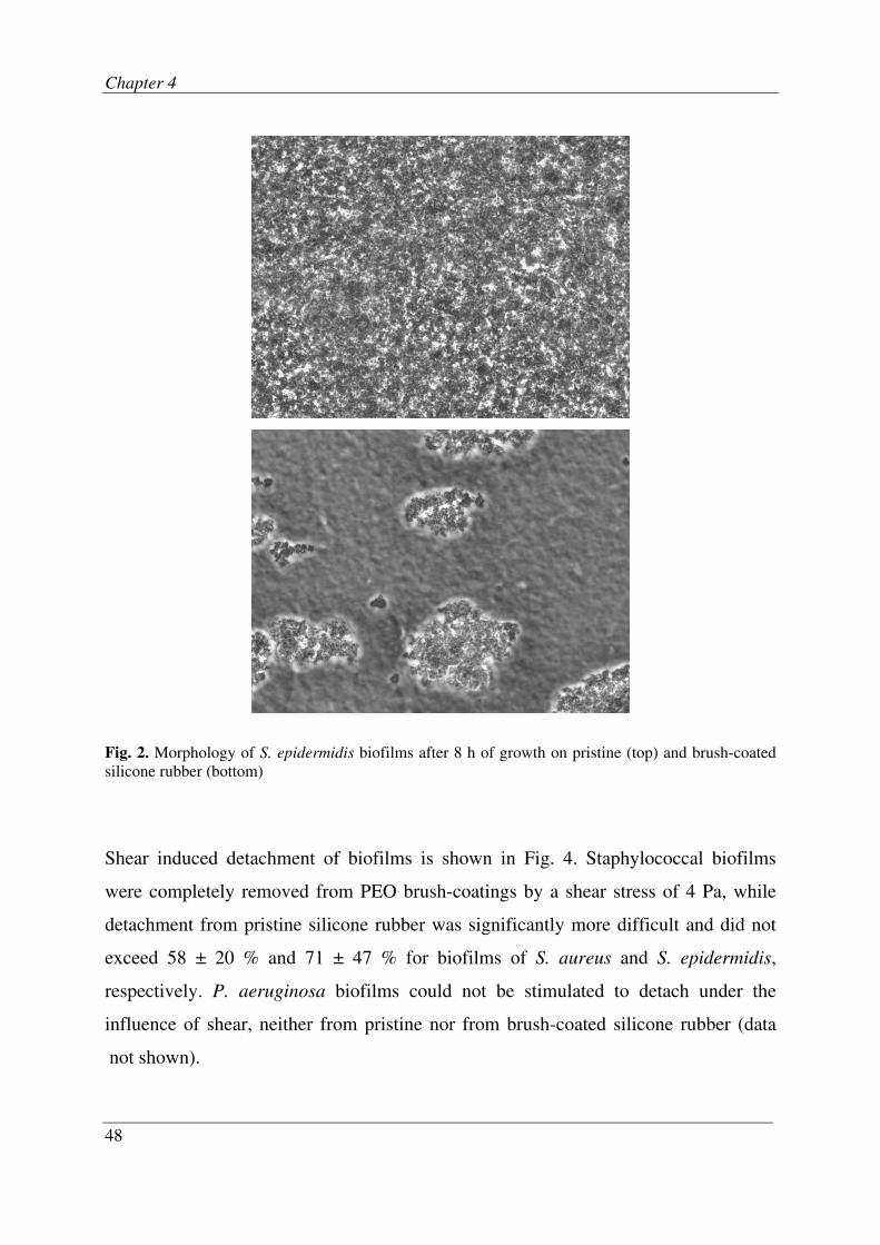
The morphology of S. epidermidis biofilms after 8 h of growth is shown in Fig. 2.
Biofilm covers the pristine silicone rubber surface rather homogeneously whereas
separate clusters were formed at brush-coated surface. A similar pattern was observed
for S. aureus biofilms. The most interesting feature of biofilms formed on brush-
coatings is their increased viability as compared with their viability on pristine silicone
rubber, as can be seen in Fig. 3. Note that this observation is not only true for both
staphylococcal strains but also for P. aeruginosa. Whereas biofilms on pristine
silicone rubber were on average less than 50% alive, the percentage viability of
biofilms on brush-coatings was 81 ± 5 %, 99 ± 0 % and 95 ± 4 % for S. aureus, S.
epidermidis and P. aeruginosa, respectively.

Bacterial adhesion and growth
47
Fig. 1. Surface coverage as a function of time after initial adhesion during bacterial growth on pristine
(∆) and brush-coated (■) silicone rubber for the three bacterial strains involved in this study. Error bars
denote the SD over four separate experiments with different bacterial cultures and silicone rubber
sheets with and without a brush-coating.
S. aureus ATCC 12600
0
20
40
60
80
100
0 2 4 6 8 10 12 14 16 18 20
Time (hours)
Su
rfa
ce c
ov
era
ge (
%)
S. epidermidis HBH 276
0
20
40
60
80
100
0 2 4 6 8 10 12 14 16 18 20
Time (hours)
Su
rfa
ce c
ov
era
ge (
%)
P. aeruginosa #3
0
20
40
60
80
100
0 2 4 6 8 10 12 14 16 18 20
Time (hours)
Su
rfa
ce c
ov
era
ge (
%)
Chapter 4
48
Fig. 2. Morphology of S. epidermidis biofilms after 8 h of growth on pristine (top) and brush-coated
silicone rubber (bottom)
Shear induced detachment of biofilms is shown in Fig. 4. Staphylococcal biofilms
were completely removed from PEO brush-coatings by a shear stress of 4 Pa, while
detachment from pristine silicone rubber was significantly more difficult and did not
exceed 58 ± 20 % and 71 ± 47 % for biofilms of S. aureus and S. epidermidis,
respectively. P. aeruginosa biofilms could not be stimulated to detach under the
influence of shear, neither from pristine nor from brush-coated silicone rubber (data
not shown).

Bacterial adhesion and growth
49
S. aureus ATCC 12600
S. epidermidis HBH
276
P. aeruginosa #3
Silicone
rubber
Brush Silicone
rubber
Brush Silicone
rubber
Brush
Liv
e■
Dead■
Fig. 3. Biofilm viability for the three strains involved in this study on pristine and brush-coated
silicone rubber after 20 h of growth. Data include a total number of over 1000 bacteria taken from 5
images, yielding an average standard deviation of 4 %.
Fig. 4. Shear induced detachment of staphylococcal biofilms at different shear stresses. Error bars
denote the SD over four separate experiments with different bacterial cultures and silicone rubber
sheets with and without a brush-coating.
0
20
40
60
80
100
120
0.01 0.05 4 0.01 0.05 4
Shear stress (Pa)
Deta
ch
men
t %
Silicone rubber PEO-brush coating
S. aureus S. epidermidis
Chapter 4
50
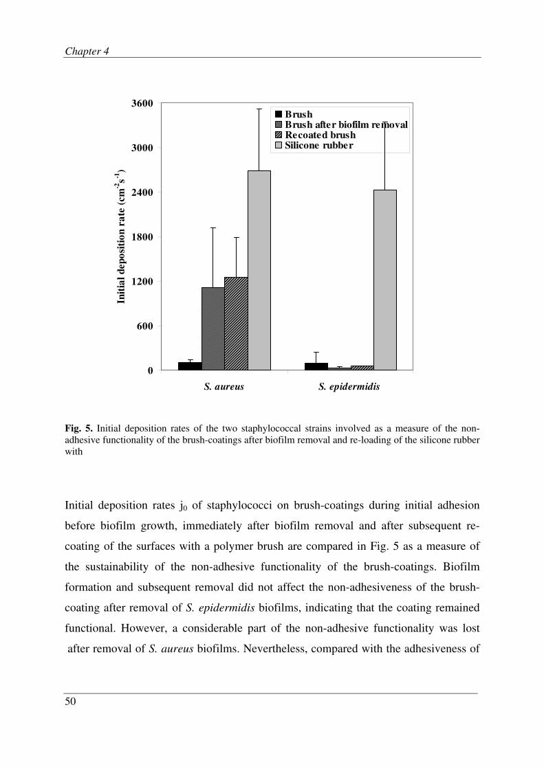
Fig. 5. Initial deposition rates of the two staphylococcal strains involved as a measure of the non-
adhesive functionality of the brush-coatings after biofilm removal and re-loading of the silicone rubber
with
Initial deposition rates j0 of staphylococci on brush-coatings during initial adhesion
before biofilm growth, immediately after biofilm removal and after subsequent re-
coating of the surfaces with a polymer brush are compared in Fig. 5 as a measure of
the sustainability of the non-adhesive functionality of the brush-coatings. Biofilm
formation and subsequent removal did not affect the non-adhesiveness of the brush-
coating after removal of S. epidermidis biofilms, indicating that the coating remained
functional. However, a considerable part of the non-adhesive functionality was lost
after removal of S. aureus biofilms. Nevertheless, compared with the adhesiveness of
0
600
1200
1800
2400
3000
3600
S. aureus S. epidermidis
Init
ial
dep
osi
tio
n r
ate
(cm
-2s
-1)
BrushBrush after biofilm removalRecoated brushSilicone rubber

Bacterial adhesion and growth
51
pristine silicone rubber, brush-coatings after biofilm removal were still effective
against these two staphylococcal strains. The elemental surface composition of brush-
coatings showed minor but insignificant changes in amounts of carbon, oxygen and
silicon (see Table 1). Interestingly, nitrogen residues were left on the brush-coatings
after biofilm removal, probably indicative of nitrogen containing components from
slime or proteinaceous bacterial footprints. A higher amount of nitrogen was found
after removal of a S. aureus biofilm than after removal of a S. epidermidis biofilm,
which might explain the loss of non-adhesive functionality of the brush-coating after
removal of an S. aureus biofilm.
Discussion
Although non-adhesiveness of polymer brush-coatings has been demonstrated before,
hitherto the fate of the “few” bacteria that manage to adhere to a brush-coating has not
received much attention. Therewith this study is the first to show that biofilms develop
more slowly and attach more weakly to polymer brush-coatings than to a pristine
surface.
PEO99-PPO65-PEO99 tri-block copolymers were physically adsorbed to hydrophobic
silicone rubber sheets. Schroen et al. [11] showed that the conformation of physically
adsorbed PEOn-PPOm-PEOn tri-block copolymers depends on the hydrophobicity of
the sorbent surface: at a hydrophilic surface the two terminal PEOn blocks attach
resulting in a pancake conformation for the adsorbed tri-block copolymer, whereas at a
hydrophobic surface the PPOm block anchors at the surface leaving the PEOn chains
dangling in the adjacent solution. At high enough adsorbed amounts, this yields a
brush conformation of the adsorbed layer, as has been demonstrated for adsorption of
Pluronic F-127 on polystyrene films [14] and on hydrophobic dimethyl dichlorosilane
(DDS)-coated silica [12]. Considering the hydrophobicity of silicone rubber as
compared with polystyrene or DDS-coatings, it is safe to assume that on silicone
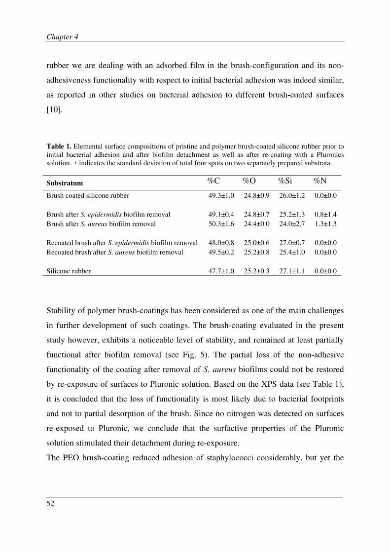
Chapter 4
52
rubber we are dealing with an adsorbed film in the brush-configuration and its non-
adhesiveness functionality with respect to initial bacterial adhesion was indeed similar,
as reported in other studies on bacterial adhesion to different brush-coated surfaces
[10].
Table 1. Elemental surface compositions of pristine and polymer brush-coated silicone rubber prior to
initial bacterial adhesion and after biofilm detachment as well as after re-coating with a Pluronics
solution. ± indicates the standard deviation of total four spots on two separately prepared substrata.
Substratum %C %O %Si %N
Brush coated silicone rubber 49.3±1.0 24.8±0.9 26.0±1.2 0.0±0.0
Brush after S. epidermidis biofilm removal 49.1±0.4 24.8±0.7 25.2±1.3 0.8±1.4
Brush after S. aureus biofilm removal 50.3±1.6 24.4±0.0 24.0±2.7 1.3±1.3
Recoated brush after S. epidermidis biofilm removal 48.0±0.8 25.0±0.6 27.0±0.7 0.0±0.0
Recoated brush after S. aureus biofilm removal 49.5±0.2 25.2±0.8 25.4±1.0 0.0±0.0
Silicone rubber 47.7±1.0 25.2±0.3 27.1±1.1 0.0±0.0
Stability of polymer brush-coatings has been considered as one of the main challenges
in further development of such coatings. The brush-coating evaluated in the present
study however, exhibits a noticeable level of stability, and remained at least partially
functional after biofilm removal (see Fig. 5). The partial loss of the non-adhesive
functionality of the coating after removal of S. aureus biofilms could not be restored
by re-exposure of surfaces to Pluronic solution. Based on the XPS data (see Table 1),
it is concluded that the loss of functionality is most likely due to bacterial footprints
and not to partial desorption of the brush. Since no nitrogen was detected on surfaces
re-exposed to Pluronic, we conclude that the surfactive properties of the Pluronic
solution stimulated their detachment during re-exposure.
The PEO brush-coating reduced adhesion of staphylococci considerably, but yet the
Bacterial adhesion and growth
53
few staphylococci that initially adhered to the brush-coating managed to form a
biofilm, as indicated in Fig. 1, although this occurred more slowly than on pristine
silicone rubber. Detachment of adhering biofilm bacteria happened more frequently
from the brush-coating than from pristine silicone rubber which resulted in the
formation of small spherical microcolonies, whereas on pristine silicone rubber the
biofilm was more contiguous (see Fig. 2). Accordingly, it has been shown that PEO
coatings decreased the surface coverage and cluster group size of Candida albicans
biofilm compared to the pristine surfaces [21]. Previously [16], we have demonstrated
that the interaction force between adhering staphylococci and a brush-coated surface is
one to two orders of magnitude lower compared to pristine silicone rubber. Clearly,
these weaker interactions forces continue to exist on polymer brush-coatings after
bacterial growth, and during growth occasional detachment is probably responsible for
the development of scattered microcolonies rather than of a contiguous biofilm.
The in situ observation of biofilm growth in the parallel plate flow chamber as applied
in our study clearly offers some advantages over methodologies where biofilm
formation is assessed after removal of substrata out of an aqueous phase. Removal of
adhering bacteria and biofilms from an aqueous phase generally encompasses passages
of the substratum through an air-liquid interface [20], which is accompanied by
detachment forces that are huge in comparison with the interaction forces between a
brush-coating and adhering organisms. This explains why the few other studies on
biofilm growth on brush-coatings did not reveal biofilm formation by S. epidermidis
and P. aeruginosa [22] nor by Listeria monocytogenes [23]. Note that information
from the flow chamber is only two-dimensional. However, we believe this is the best
in situ result we can reach since the manipulations required for a three-dimensional
observation would cause uncontrolled detachment of the biofilm due to weak biofilm-
substratum interaction.
The slower formation of biofilms on brush-coatings and their relatively easy
detachment is clinically relevant, as it gives prophylactically administered antibiotics

Chapter 4
54
after implant surgery a better chance to kill possible bacteria that have adhered to a
brush-coated implant surface during surgery prior to the formation of a more mature,
and thus more resistant biofilm. However, formation of small microcolonies on brush-
coatings rather than a thick contiguous film as on pristine silicone rubber due to non-
adhesive nature of brush-coating also implies that nutrients can better penetrate the
biofilm. This explains why biofilms on brush-coatings are more viable than on pristine
silicone rubber, where organisms in the deeper layers of the biofilms are devoid of
adequate nutrient supply.
Conclusions
Brush-coatings made of a triblock copolymer of polyethylene-oxide (PEO) and
polypropylene-oxide (PPO) prevented adhesion of staphylococci to below 5 × 105 cm
-2
after 30 min. Yet, biofilms grew on both brush-coated and pristine silicone rubber,
while the viability of biofilms on brush-coatings was higher than on pristine silicone
rubber. However, staphylococcal biofilms developed more slowly on brush-coatings
and when exposed to high fluid shear, they almost fully detached from a brush-coating
in contrast to detachment from pristine silicone rubber. Adhesion and growth of P.
aeruginosa were not significantly affected by the presence of the brush on silicone
rubber, although here too the viability of biofilms on brush-coatings was higher. The
slow growth of staphylococci, on polymer brush-coatings, may allow more time for
treatment with antibiotics before a mature, more resistant biofilm can develop.
Acknowledgements
The authors acknowledge Joop de Vries for his help in performing the XPS analyses.

Bacterial adhesion and growth
55
References
1. Gristina AG. Biomaterial-centered infection: microbial adhesion versus tissue
integration. Science 1987, 237:1588-95.
2. Campoccia D, Montanaro L, Arciola CR. The significance of infection related to
orthopedic devices and issues of antibiotic resistance. Biomaterials 2006, 27:2331-39.
3. Widmer AF. New developments in diagnosis and treatment of infection in orthopedic
implants. Clinical Infectious Diseases 2001, 33:S94-S106.
4. Kuijer R, Jansen EJP, Emans PJ et al. Assessing infection risk in implanted tissue-
engineered devices. Biomaterials 2007, 28:5148-54.
5. Bos R, Van der Mei HC, Busscher HJ. Physico-chemistry of initial microbial adhesive
interactions - its mechanisms and methods for study. FEMS Microbiology Reviews
1999, 23:179-230.
6. Nuzzo RG. Biomaterials stable antifouling surfaces. Nature Materials 2003, 2:207-08.
7. Leckband D, Sheth S, Halperin A. Grafted poly(ethylene oxide) brushes as nonfouling
surface coatings. Journal of Biomaterial Science Polymer Edition 1999, 10:1125-47.
8. Senaratne W, Andruzzi L, Ober CK. Self-assembled monolayers and polymer brushes
in biotechnology: current applications and future perspectives. Biomacromolecules
2005, 6:2427-48.
9. Zhao B, Brittain WJ. Polymer brushes: surface-immobilized macromolecules. Progress
in Polymer Science 2000, 25:677-710.
10. Roosjen A, Kaper HJ, Van der Mei HC et al. Inhibition of adhesion of yeasts and
bacteria by poly(ethylene oxide) brushes on glass in a parallel plate flow chamber.
Microbiology 2003, 149:3239-46.
11. Schroen CGPH, Cohen Stuart MA, Maarschalk KV et al. Influence of preadsorbed
block-copolymers on protein adsorption, surface properties, layer thickness and surface
coverage. Langmuir 1995, 11:3068-74.
12. Norde W, Gage D. Interaction of bovine serum albumin and human blood plasma with
PEO-tethered surfaces: influence of PEO chain length, grafting density, and
temperature. Langmuir 2004, 20:4162-67.
13. Malmsten M, Emeto K, Van Alstine JM. Effect of chain density on inhibition of protein
adsorption by poly(ethylene glycol) based coatings. Journal of colloid and interface
science 1998, 202:507-17.

Chapter 4
56
14. Marsh LH, Coke M, Dettmar PW et al. Adsorbed poly(ethylene oxide)-poly(propylene
oxide) copolymers on synthetic surfaces: Spectroscopy and microscopy of polymer
structures and effects on adhesion of skin-borne bacteria. Journal of Biomedical
Materials Research Part A 2002, 61:641-52.
15. Cringus-Fundeanu I, Luijten J, Van der Mei HC et al. Synthesis and characterization of
surface-grafted polyacrylamide brushes and their inhibition of microbial adhesion.
Langmuir 2007, 23:5120-26.
16. Nejadnik MR, Van der Mei HC, Busscher HJ et al. Determination of the shear force at
the balance between bacterial attachment and detachment in weak-adherence systems,
using a flow displacement chamber. Applied Environmental Microbiology 2008,
74:916-19.
17. Mclean SC, Lioe H, Meagher L et al. Atomic force microscopy study of the interaction
between adsorbed poly(ethylene oxide) layers: effects of surface modification and
approach velocity. Langmuir 2005, 21:2199-208.
18. Brandani P, Stroeve P. Adsorption and desorption of PEO-PPO-PEO triblock
copolymers on a self-assembled hydrophobic surface. Macromolecules 2003, 36:9492-
501.
19. Liu VA, Jastromb WE, Bhatia SN. Engineering protein and cell adhesivity using PEO-
terminated triblock polymers. Journal of Biomedical Materials Research 2001, 60:126-
34.
20. Busscher HJ, Van der Mei HC. Microbial adhesion in flow displacement systems.
Clinical Microbiology Reviews 2006, 19:127-41.
21. Wesenberg-ward KE, Tyler BJ, Sears JT. Adhesion and biofilm formation of Candida
albicans on native and pluronic-coated polystyrene. Biofilms 2005, 2:63-71.
22. Cheng G, Zhang Z, Chen SF et al. Inhibition of bacterial adhesion and biofilm
formation on zwitterionic surfaces. Biomaterials 2007, 28:4192-99.
23. Dong BY, Jiang HQ, Manolache S et al. Plasma-mediated grafting of poly(ethylene
glycol) on polyamide and polyester surfaces and evaluation of antifouling ability of
modified substrates. Langmuir 2007, 23:7306-13.


Biofilms on polymer brush-coatings
remain susceptible for antibiotic
treatment
M. Reza Nejadnik, Henk J. Busscher, Henny C. van der Mei, Willem Norde
Chapter 5
60
Abstract
Biomaterial associated infection (BAI) is the most important cause of implant failure.
BAI begins with adhesion of bacteria to the biomaterial surface and their subsequent
growth into a biofilm. Polymer brush-coatings are known for their superior anti-
adhesive properties. Yet a polymer brush-coating does not completely prevent
bacterial adhesion and the few adhering bacteria on a brush-coated surface have shown
the ability to grow into a biofilm. However, these biofilms develop slowly and form
scattered colonies which are penetrable to nutrients. Biofilm formation on brush-
coatings may also be more susceptible to antibiotics. Therefore, we here investigate
the growth of Staphylococcus aureus ATCC 12600 biofilms, a common cause of BAI,
on pristine and polymer brush-coated silicone rubber in the absence and presence of
three concentrations of gentamicin (0.5, 5 and 50 µg ml-1
). Biofilms grew on silicone
rubber and maintained their viability in the presence of gentamicin, regardless of its
concentrations. On polymer brush-coatings, however, the presence of gentamicin in
the growth medium suppressed biofilm formation and decreased the number of viable
bacteria. Biofilm growth on brush-coatings was almost completely prevented in the
presence of 50 µg ml-1
gentamicin. We conclude that, contrary to biofilms on
biomaterial surfaces in the absence of a polymer brush-coating, biofilms on polymer
brush-coatings remain susceptible to antibiotics, which prevents biofilm formation.

Susceptibility of biofilms on polymer brush to antibiotic
61
Introduction
Biomaterial associated infection (BAI) remains the number one cause of prosthetic
implant failure, despite the development of various state-of-the-art strategies to control
BAI after implantation. The occurrence of BAI for primary implants varies from 1 to
30% with an associated mortality of up to 25%, depending on the type of implant [1].
Common treatment procedures for patients suffering from BAI include long-term
application of high doses of antibiotics, but frequently the fate of an infected
biomaterial is removal [2]. Microbial adhesion is considered to be the onset of BAI,
after which the adhering organisms grow to form a biofilm. In a biofilm, micro-
organisms are embedded in a complex extracellular polymeric matrix and are resistant
against antibiotic treatment and the host immune system [3].
Surface modifications can significantly reduce microbial adhesion to biomaterial
surfaces [4]. Polymer brush-coatings are currently the most promising non-adhesive
coatings as they reduce the adhesion of various bacterial strains and yeasts by orders of
magnitude [5]. A polymer brush is formed when hydrophilic polymer chains are end-
grafted to a surface in a high density, forcing the polymer chains to stretch away from
the surface into the adjacent medium [6]. Compression of such a structure upon
microbial approach gives rise to an osmotic pressure and decreased mobility
(conformational entropy) of the polymer chains in the brush, which causes repulsion of
the approaching organisms.
Polymer brush-coatings, however, do not reduce microbial adhesion to zero and in
case of staphylococci the few bacteria adhering to a polymer brush have been
demonstrated to be able to form a biofilm [7], albeit at a reduced rate when compared
with uncoated substrata. In addition, we observed alteration of the biofilm structure
from a contiguous layer on pristine silicone rubber to scattered microcolonies on a
polymer brush-coating. Furthermore, the biofilm on the brush-coated surface appeared
to be more viable, probably due to better penetration of nutrients into the
Chapter 5
62
microcolonies. We therefore speculate that antimicrobial agents may also be more
effective towards the bacteria in biofilms formed on a polymer brush.
Hence, the aim of this study is to compare the influence of antibiotic treatment on
biofilm formation on silicone rubber with and without a polymer brush-coating. To
this end, we grew biofilms of Staphylococcus aureus ATCC 12600, a common cause
of BAI, on pristine and polymer brush-coated silicone rubber in media containing
different concentrations of gentamicin, a commonly used antibiotic against
staphylococci, and monitor the development of the biofilms in a parallel plate flow
chamber (PPFC).
Materials and Methods
Preparation of silicone rubber surfaces and brush-coatings. Implant grade silicone
rubber sheets (thickness 0.5 mm, Medin, Groningen, The Netherlands) were rinsed
with ethanol (Merck, Darmstadt, Germany) and demineralised water. Subsequently,
sheets were sonicated for three min in 2% RBS 35 detergent (Omnilabo International
BV, Breda, The Netherlands) and rinsed thoroughly with demineralised water, washed
in methanol (Merck) and rinsed with demineralised water again. A silicone rubber
sheet was fixed in the bottom plate of a PPFC, after which the entire chamber was
sterilized by rinsing with 70% ethanol and sterile demineralised water. Subsequently,
the silicone rubber was exposed to a filter-sterilized solution of 0.5 g l-1
Pluronic F-127
(a copolymer of polyethylene oxide (PEO) and polypropylene oxide (PPO) with
structure PEO99PPO65PEO99, molecular weight 12600; Sigma-Aldrich, USA) in
phosphate buffered saline (PBS: 10 mM potassium phosphate, 150 mM NaCl, pH 6.8)
for 20 min at room temperature. Non-attached polymer was removed from the
chamber by flow with an excess amount of PBS. NaCl, K2HPO4 and KH2PO4 were of
analytical grade, as purchased from Merck.

Susceptibility of biofilms on polymer brush to antibiotic
63
Culturing and harvesting of bacterial cells. S. aureus ATCC 12600 was first grown
aerobically overnight at 37°C on blood agar plates from frozen stocks. These plates
were kept at 4°C, never longer than two weeks. One colony was used to make a pre-
culture in 10 ml tryptone soya broth (TSB, OXOID, Basingstoke, England). This pre-
culture was incubated at 37°C for 24 h and used to inoculate a second culture of 200
ml which was incubated for 16 h. The culture was harvested by centrifugation for 5
min at 5000 × g and washed twice with sterile demineralised water. To break up
bacterial aggregates, bacteria were sonicated intermittently while cooling in an
ice/water bath for three times 10 s at 30 W (Vibra Cell model 375; Sonics and
Materials, Danbury, CT, USA). This procedure was found not to cause cell lysis.
Finally, staphylococci were resuspended in 200 ml sterile PBS to a concentration of 3
× 108 per ml. Minimal inhibitory and minimum bactericidal concentrations (MIC and
MBC) of the strain against gentamicin were determined in liquid medium
supplemented with different concentrations of gentamicin, yielding a MIC and MBC
of 20 µg ml-1
and 40 µg ml-1
, respectively.
Biofilm growth in the absence and presence of gentamicin. The parallel plate flow
chamber (PPFC, 175 × 17 × 0.75 mm3) and image analysis system have been
previously described [8]. Images were taken from pristine silicone rubber and silicone
rubber coated with a PEO brush, affixed to the polymethyl-methacrylate bottom plate
of the chamber. The top plate was made of glass. Staphylococcal adhesion and growth
were monitored using a Fire wire CCD camera, mounted on a phase contrast
microscope equipped with a 40× ultra long working distance objective. The camera
was coupled to a PC with proprietary image analysis software. Each image (1392 ×
1040 pixels with 8 bit resolution) was obtained from summation of 15 consecutive
images (time interval 0.25 s) in order to enhance the signal to noise ratio and to
eliminate moving organisms from the analysis. Before each experiment, all tubes and
the flow chamber were sterilized and filled with PBS, while care was taken to remove
Chapter 5
64
air bubbles from the system. Flasks, containing bacterial suspension and buffer, were
positioned at the same height with respect to the chamber to ensure that all fluids
circulate through the chamber at the desired rate immediately after starting the flow.
Staphylococci were allowed to adhere at room temperature to the substratum surface
during 25 min under a flow-induced shear stress of 0.005 Pa. Subsequently, flow was
switched from bacterial suspension to 10% TSB medium for 5 min to flush out
unattached bacteria from the chamber, after which the temperature was raised to 37 ºC,
shear stress was decreased to 0.002 Pa, and bacteria were allowed to grow for 3.5 h.
After initial adhesion and growth, flow was switched to 10% TSB medium containing
three different concentrations (0.5, 5 and 50 µg ml-1
) of gentamicin sulphate (Sigma-
Aldrich, USA) for 16 h at 37 ºC, i.e. antibiotic concentrations well below or above the
MIC (20 µg ml-1
) and MBC (40 µg ml-1
) values found.
From the images taken during initial bacterial adhesion and subsequent growth,
numbers of adhering bacteria and the percentage surface coverage by biofilm were
determined. All experiments were done in threefold, with separately grown bacteria.
Bacterial viability assay. At the end of each flow experiment, 20 h old biofilms were
removed from the substratum surfaces using a sterile cotton swab and suspended in 0.5
ml demineralised water. Subsequently, 10 µl of the suspension was transferred onto a
glass slide and stained for 20 min in the dark with a live/dead stain (BacLightTM
,
Molecular Probes Europe BV). Using fluorescence microscopy (Leica, Wetzlar,
Germany), the percentages of live and dead bacteria were evaluated, from which the
percentage live bacteria in the biofilms was determined.

Susceptibility of biofilms on polymer brush to antibiotic
65
Fig. 1. Surface coverage by staphylococcal (S. aureus ATCC 12600) biofilm as a function of time
during adhesion and growth on pristine (top) and polymer brush-coated (bottom) silicone rubber. After
adhesion (first 30 min) and initial growth (210 min), biofilms were allowed to grow in medium
containing different concentrations of gentamicin. Error bars represent the standard deviations over
three separate experiments.
0
20
40
60
80
100
0 5 10 15 20
Su
rfa
ce
co
ve
rag
e (
%)
No antibiotic
0.5 µg/ml gentamicin
5 µg/ml gentamicin
50 µg/ml gentamicin
0
20
40
60
80
100
0 5 10 15 20
Time (h)
Su
rfa
ce
co
ve
rag
e (
%)

Chapter 5
66
Results
Fig. 1 shows the surface coverage by staphylococcal biofilms on pristine and polymer
brush-coated silicone rubber during growth in the absence and presence of gentamicin
(control data in the absence of antibiotic were taken from Nejadnik et al., 2008b).
Inclusion of gentamicin in the growth medium only slightly reduced the rate of biofilm
formation on pristine silicone rubber and in the presence of antibiotic in the growth
medium, the silicone rubber surface was completely covered with biofilm in less than
12 h. Biofilm growth on polymer brush-coatings was not affected by the lowest
gentamicin concentration (0.5 µg ml-1
), but higher concentrations of gentamicin, i.e. 5
and 50 µg ml-1
, suppressed the biofilm formation by 41 and 95% respectively.
The percentage surface coverage by biofilm including the fraction of viable bacteria in
the biofilms is presented in Fig. 2. The fraction of viable bacteria in biofilms on
pristine silicone rubber amounts approximately 40% and is hardly influenced by the
absence or presence of gentamicin. The fraction of viable bacteria in biofilms on
polymer brush-coatings is much higher than in biofilms on pristine silicone rubber
(81% versus 40%, see Nejadnik et al., 2008b). Antibiotic presence during growth
however, strongly decreases the fraction of viable bacteria in biofilms on the brushes,
opposite to the observations on uncoated silicone rubber.
The percentage reduction in viable bacteria in the biofilms on silicone rubber and
brush-coated silicone rubber is presented in Table 1, as calculated with respect to the
number of viable bacteria in biofilms grown in the absence of gentamicin. It can be
seen from Table 1 that antibiotic treatment does not cause a reduction in the number of
viable bacteria on silicone rubber, but on polymer brush-coatings the reduction in the
number of viable bacteria increased with increasing gentamicin concentration during
growth.

Susceptibility of biofilms on polymer brush to antibiotic
67
Fig. 2. The percentage surface coverage by biofilm and the fraction of viable bacteria (dashed part) in
these biofilms on pristine (SR) and polymer brush-coated (Brush) silicone rubber after growth in the
absence or presence of different concentrations of gentamicin. Error bars represent the standard
deviations calculated based on three separate experiments.
Table 1. Percentage reduction in number of viable bacteria on silicone rubber and polymer brush-
coated silicone rubber during growth in the presence of various concentrations of gentamicin. All
percentages are calculated with respect to the number of viable bacteria on the surfaces in the absence
of any antibiotics. ± represents the standard deviation of the reduction, calculated based on the
standard deviations of values obtained from triplicate experiments in the presence and absence of
antibiotic.
0.5 µg ml-1
5 µg ml-1
50 µg ml-1
Silicone rubber 0 0 0
Brush 35 ± 24 65 ± 39 96 ± 27

Chapter 5
68
Discussion
Bacteria in biofilms on silicone rubber and other biomaterial surfaces are protected
against antibiotic treatment. This study is the first to demonstrate that staphylococcal
biofilms on a polymer brush-coating remain susceptible to antibiotics, in addition to
the known reduction in biofilm formation on a polymer brush-coating. Both the strong
reduction in biofilm formation on polymer brush-coatings and the susceptibility of
these biofilms to antibiotics make polymer brush-coatings ideal for application on
biomaterials implants.
The mechanism of resistance of bacteria in biofilms against antibiotics is not fully
understood. There is evidence that a biofilm-specific phenotype is formed resulting in
activation of sub-microbial components which make bacteria resistant [9]. Changes in
gene regulation occur within minutes after bacterial attachment to a solid surface [10],
suggesting that adhering bacteria may sense a solid surface leading to a signalling
cascade that causes genes to be up- or down-regulated [11]. Possibly, all or at least
some of the altered organisms are more resistant to antimicrobial agents [9], allowing a
biofilm to form. We here hypothesize that on a highly hydrated polymer brush-coating,
allowing only weak interaction between the biofilm and the underlying surface [12],
organisms do not sense that they are actually on a surface and continue to behave like
planktonic organisms.
Alternatively, there are reports that relate the reduced susceptibility of biofilm to
antibiotics, to a reduced accessibility of the antibiotic to the micro-organisms [9].
Although the extracellular polymeric matrix of the biofilm is not impenetrable for
antimicrobial agents, it may delay the antibiotics to reach the cells in the biofilm,
which is in line with the current finding that it takes longer for bacteria adhering to a
polymer brush-coating to form a thick, dense biofilm. In vivo this may be
advantageous, because antimicrobial mechanisms in the body triggered by the immune
system will have a longer time to act against single adhering bacteria on a polymer
Susceptibility of biofilms on polymer brush to antibiotic
69
brush-coating prior to the formation of a contiguous biofilm. Furthermore, our
previous finding that the biofilm on polymer brush-coating is more viable than on
pristine silicone rubber suggests better penetration of nutrients and may, likewise,
support the idea that penetration is a determining factor in the increased susceptibility
to antibiotics. Irrespective of the underlying mechanism, however, our current
findings have enormous clinical implications, as they show a pathway toward
biomaterial implant coatings that allow antibiotic treatment to prevent biofilm
formation and therewith to decrease the risk of BAI.
Conclusions
Biofilm growth of staphylococci adhering on silicone rubber is not affected by the
presence of gentamicin in the medium, even if the gentamicin concentration is above
MBC, but staphylococcal biofilms on a polymer brush-coating remain susceptible to
antibiotics during growth and, moreover, growth is strongly retarded. Presence of
gentamicin in a concentration above MBC during growth of adhering staphylococci on
a polymer brush-coating, prevented biofilm formation. Polymer brush-coatings
therefore are promising for biomaterial implant coating, especially since they allow
treatment of biofilms by antibiotics, contrary to biofilms on other biomaterial surfaces.

Chapter 5
70
Reference
1. Darouiche RO. Device-associated infections: A macroproblem that starts with
microadherence. Clinical Infectious Diseases 2001, 33:1567-72.
2. Darouiche RO. Current concepts - Treatment of infections associated with surgical
implants. New England Journal of Medicine 2004, 350:1422-29.
3. Costerton JW, Stewart PS, Greenberg EP. Bacterial biofilms: A common cause of
persistent infections. Science 1999, 284:1318-22.
4. Cheng G, Zhang Z, Chen SF et al. Inhibition of bacterial adhesion and biofilm
formation on zwitterionic surfaces. Biomaterials 2007, 28:4192-99.
5. Roosjen A, Norde W, Van der Mei HC et al. The use of positively charged or low
surface free energy coatings versus polymer brushes in controlling biofilm formation.
Progress in Colloid and Polymer Science 2006, 132:138-44.
6. Zhao B, Brittain WJ. Polymer brushes: surface-immobilized macromolecules. Progress
in Polymer Science 2000, 25:677-710.
7. Nejadnik MR, Van der Mei HC, Norde W et al. Bacterial adhesion and growth on a
polymer brush-coating. Biomaterials 2008, 29:4117-21.
8. Busscher HJ, Van der Mei HC. Microbial adhesion in flow displacement systems.
Clinical Microbiology Reviews 2006, 19:127-41.
9. Mah TFC, O'Toole GA. Mechanisms of biofilm resistance to antimicrobial agents.
Trends in Microbiology 2001, 9:34-39.
10. Davies DG, Geesey GG. Regulation of the alginate biosynthesis gene Algc in
Pseudomonas aeruginosa during biofilm development in continuous-culture. Applied
and environmental microbiology 1995, 61:860-67.
11. Jefferson KK. What drives bacteria to produce a biofilm? Fems Microbiology Letters
2004, 236:163-73.
12. Nejadnik MR, Van der Mei HC, Busscher HJ et al. Determination of the shear force at
the balance between bacterial attachment and detachment in weak-adherence systems,
using a flow displacement chamber. Applied Environmental Microbiology 2008,
74:916-19.


Bacterial colonization of polymer brush-
coated and pristine silicone rubber
implanted in infected pockets in mice
Reproduced with permission of Oxford University Press from: M. Reza Nejadnik,
Anton F. Engelsman, Isabel C. Saldarriaga Fernandez, Henk J. Busscher, Willem
Norde, Henny C. van der Mei. Journal of Antimicrobial Chemotherapy 2008, 62:1323-
1325.

Chapter 6
74
Abstract
Curing biomaterial-associated infection (BAI) frequently includes antibiotic treatment,
implant removal and re-implantation. However, revision implants are at a greater risk
of infection as they may attract bacteria from their infected surrounding. Polymer
brush-coatings attract low numbers of bacteria, but the virtue of polymer brush-
coatings in vivo has seldom been investigated. Here we determine the possible benefits
of polymer brush-coated versus pristine silicone rubber in revision surgery, using a
murine model. BAI was induced in 26 mice by subcutaneous implantation of silicone
rubber disks with a biofilm of Staphylococcus aureus Xen29. During development of
BAI, half of the mice received treatment of rifampicin/vancomycin. After 5 days, the
infected disks were removed from all mice, and either a polymer brush-coated or
pristine silicone rubber disk was re-implanted. Revision disks were explanted after 5
days and the number of colony-forming units (cfu’s) cultured from the disks and
surrounding tissue was determined. None of the polymer brush-coated disks after
antibiotic treatment appeared colonized by staphylococci, whereas 83% of the pristine
silicone rubber disks were re-infected. Polymer brush-coated disks also showed
reduced colonization rates in the absence of antibiotic treatment as compared with
pristine silicone rubber disks. Tissue surrounding the disks was culture positive in all
cases. We conclude that Polymer brush-coatings are less prone to re-infection than
pristine silicone rubber when used in revision surgery, i.e. when implanted in a
subcutaneous pocket infected by a staphylococcal BAI. Antibiotic pre-treatment
during the development of BAI hardly had any effect in preventing colonization of
pristine silicone rubber.
Bacterial colonization
75
Introduction
Along with the rapid expansion in use of biomaterial implants for restoration of
function after trauma, oncological surgery or wear, the number of patients suffering
from biomaterials-associated infection (BAI) increases continuously [1]. BAI results
from the colonization of a biomaterials implant surface by bacteria excreting slime and
the subsequent formation of an antibiotic resistant complex, called “biofilm”. BAI is
often initially treated with antibiotics, but most frequently with little success, and
ultimately revision surgery is needed in which the primary infected implant is removed
and a revision implant is inserted [2-4]. The risk of infection for the revision implant
however, is several-folds higher than for the primary implant [1], due to bacterial
persistence in surrounding tissue, even after antibiotic treatment [5].
Polymer brush-coatings are generally considered to attract very few bacteria and may
therefore be extremely useful for application in revision surgery after BAI.
Physisorbed polyethylene oxide (PEO) brush-coatings can reduce the number of
adhering bacteria by one to two orders of magnitude while, moreover, bacteria adhere
weakly to polymer brush-coatings [6]. Physisorbed PEO brush-coatings can be applied
on different hydrophobic biomaterials, including silicone rubber, and are stable under
flow induced shear stresses [6] and in physiological fluids [7].
In this study, the effectiveness of polymer brush-coated silicone rubber in preventing
infection after BAI were compared with the effectiveness of pristine silicone rubber in
revision surgery, using a murine model. Prevention of BAI was evaluated for polymer
brush-coated and pristine silicone rubber in the absence and presence of antibiotic
treatment prior to revision surgery.

Chapter 6
76
Materials and Methods
All materials were of analytical grade and purchased from Merck, Darmstadt,
Germany unless otherwise stated. Implant-grade silicone rubber sheets (thickness 0.5
mm, Medin, Groningen, The Netherlands) were cut into circular disks with radius of 4
mm. Disks were rinsed with ethanol and demineralised water and subsequently
sonicated for 3 min in 2% RBS 35 detergent (Omnilabo International BV, Breda, The
Netherlands) and rinsed thoroughly with demineralised water again, placed in 70%
ethanol for 5 min and rinsed with sterile demineralised water. In order to apply a
polymer brush-coating, cleaned silicone rubber disks were exposed to a sterile solution
of 0.5 g/L Pluronic F-127 (Sigma-Aldrich, St. Louis, MO, USA) in phosphate buffered
saline (10 mM potassium phosphate, 150 mM NaCl, pH 6.8). The disks were kept in
the solution overnight prior to the implantation.
Staphylococcus aureus Xen29 [8] was first grown aerobically overnight at 37°C on a
blood agar plate from a frozen stock. Several colonies were used to make pre-cultures
in 10 mL tryptone soya broth (OXOID, Basingstoke, England) which were incubated
at 37°C for 24 h. Pristine silicone rubber disks were incubated, for 72 h at 37°C on a
rotary shaker at 60 rpm with 10 mL TSB enriched with 4% NaCl, inoculated with 100
µL of the pre-culture.
In order to induce BAI in mice, colonized silicone rubber disks were implanted in
subcutaneous pockets, prepared in the left flanks of female Balb/c OlaHsd mice
(Harlan Netherlands BV, Horst, The Netherlands), as approved by the Animals
Experiments Committee at the University Medical Center Groningen. Prior to
implantation, the left flanks of the mice were shaved and cleaned with 70% ethanol.
Anaesthesia was induced with 3.5% Isoflurane/O2 gas mixture (Zeneca, Zoetermeer,
The Netherlands) and maintained at 1.5% during the implantation procedure.
Buprenorfine (0.03 mg/kg) was administered subcutaneously 30 min before surgery as
an analgesic. The disks were left in situ for 5 days, during which half of the mice

Bacterial colonization
77
received daily intra-peritoneal injections of 0.5 mL antibiotic solution of 2 mg/mL
vancomycin (Abbott BV, Hoofddorp, The Netherlands) and 1 mg/mL rifampicin
(Rifadin, Aventis, Hoevelaken, The Netherlands) in 0.9% NaCl while the other half
received injections of 0.5 mL 0.9% NaCl solution [9]. After 5 days, the infected disks
were removed under similar conditions as described above and either a sterile pristine
or polymer brush-coated silicone rubber disk was implanted. After 5 days, animals
were sacrificed, all disks were explanted and transferred to the laboratory in Eppendorf
tubes containing reduced transport fluid (NaCl 0.9 g/L (NH4)2SO4 0.9 g/L, KH2PO4
0.45 g/L, Mg2SO4 0.19g/L, K2HPO4 0.45 g/L, Na2EDTA 0.37 g/L, L-cysteine HCl 0.2
g/L, pH 6.8). In addition, tissue samples were collected from the implant site and from
a muscle away of the implant site for analysis of bacterial presence and antibiotic
content.
Staphylococci adhering to the disks were detached into suspension by intermittent
sonication for three times 10 s at 30 W (Vibra Cell model 375; Sonics and Materials,
Danbury, CT, USA). 100 µL of this suspension was spread on a blood agar plate and
the number of colonies was determined after incubation for 24 h at 37°C.
Bacterial presence in tissues surrounding an implanted disk were determined after
homogenization by sonication, and subsequent culturing on blood agar plates and
enumeration for cfu’s as described above. Tissue samples, including muscle
homogenate, were further analyzed for the potential presence of bactericidal antibiotic
concentrations, by putting 20 µL of homogenate on Mueller-Hinton agar plates
(OXOID, Basingstoke, England) inoculated with fresh S. aureus Xen29. Growth
inhibition was inspected by eye after incubating the plates for 24 h at 37°C.

Chapter 6
78
Results and Discussion
This study is the first to address the colonization of a non-adhesive polymer brush-
coating in an infected pocket in vivo, mimicking revision surgery after BAI.
None (0/7) of the polymer brush-coated silicone rubber disks implanted in the
antibiotic treated group showed signs of colonization by S. aureus, whereas 83% (5/6)
of pristine silicone rubber disks were culture positive (Fig. 1a). Similarly, in the non-
antibiotic treated group, bacteria were found on 43% (3/7) of the polymer brush-coated
disks, while 83% (5/6) of the pristine silicone rubber disks were infected. Moreover,
culture positive polymer brush-coated surfaces were generally colonized by fewer
bacteria than pristine silicone rubber disks. Bacteria were found in all tissue samples
surrounding disks, albeit that the average number of cfu’s in the antibiotic treated
group was significantly lower than in the absence of antibiotic treatment (Fig. 1b). No
significant difference was seen in bacterial persistence in tissues surrounding the
pristine and the polymer brush-coated silicone rubber disks. None of the tissue
homogenates caused any inhibition of bacterial growth on culture plates, indicating
that all tissues were completely devoid of bactericidal levels of antibiotics. Tissue
homogenate of a mouse sacrificed after antibiotic administration caused an inhibition
zone with diameter of 14 mm on culture plates (positive control), while a negative
control yielded zero inhibition zones.
The colonization rate of pristine silicone rubber disks after revision is high, which is in
line with clinical studies reporting a high rate of infection in revision surgery after BAI
of a primary implant [1]. The infection rate of primary penile prostheses with silicone
rubber tubes, for instance, was only 0.5%, compared to 6.6% in patients undergoing
revision surgery [10]. The colonization rate of pristine silicone rubber disks was
independent of whether BAI was pre-treated with antibiotics or not. This illustrates the
limitation of treating BAI with antibiotics and confirms clinical experiences that the
fate of an infected implant is generally removal [2-4]. Broekhuizen et al. [9] also

Bacterial colonization
79
suggested that in general the negative outcomes of revision surgery are due to bacterial
persistence in tissues surrounding an infected implant. However, the current study
shows that treatment of BAI with antibiotics prior to revision surgery does help to
reduce infection rates in revision surgery in case of polymer brush-coatings. Likely,
antibiotic treatment reduces the number of bacteria in adjacent tissue and the few
bacteria remaining in the tissue are not able to colonize a newly inserted polymer
brush-coated surface.
The reduced colonization rates of polymer brush-coated disks are most likely
attributable to the non-adhesiveness of the coating and not due to differential effects of
antibiotic remnants on pristine and polymer brush-coated silicone rubber, as none of
the tissue homogenates contained bactericidal levels of antibiotics. However, it can not
be completely ruled out that sub-bactericidal concentrations of antibiotics were more
effective on staphylococci loosely adhering on polymer brush-coated disks than on
bacteria more firmly adhering to pristine silicone rubber.
In summary, this is the first time that it has been demonstrated that polymer brush-
coatings may assist in preventing infection of implant surfaces after revision surgery,
by reduction of the number of bacteria adhering to a re-implanted biomaterial surface.

Chapter 6
80
Fig. 1. Total number of colony-forming units (cfu) detected on pristine (SR) and polymer brush-coated
(BRUSH) silicone rubber disks (a) explanted from mice as well as from surrounding tissue (b). Disks
were originally implanted in pockets created by a BAI in animals that had or had not received
antibiotic treatment prior to re-implantation.
0
1-10
10-100
100-1000
>1000
BRUSH BRUSH SR SR
Antibiotic treated No antibiotic treatment
c
f
u
a
0
1-10
10-100
100-1000
>1000
BRUSH BRUSH SR SR
Antibiotic treated No antibiotic treatment
c
f
u
b

Bacterial colonization
81
References
1. Darouiche RO. Device-associated infections: A macroproblem that starts with
microadherence. Clinical Infectious Diseases 2001, 33:1567-72.
2. Costerton JW, Stewart PS, Greenberg EP. Bacterial biofilms: A common cause of
persistent infections. Science 1999, 284:1318-22.
3. Darouiche RO. Current concepts - Treatment of infections associated with surgical
implants. New England Journal of Medicine 2004, 350:1422-29.
4. Gristina AG. Biomaterial-centered infection: microbial adhesion versus tissue
integration. Science 1987, 237:1588-95.
5. Broekhuizen CAN, de Boer L, Schipper K et al. Peri-implant tissue is an important
niche for Staphylococcus epidermidis in experimental biomaterial-associated infection
in mice. Infection and Immunity 2007, 75:1129-36.
6. Nejadnik MR, Van der Mei HC, Busscher HJ et al. Determination of the shear force at
the balance between bacterial attachment and detachment in weak-adherence systems,
using a flow displacement chamber. Applied Environmental Microbiology 2008,
74:916-19.
7. Liu VA, Jastromb WE, Bhatia SN. Engineering protein and cell adhesivity using PEO-
terminated triblock polymers. Journal of Biomedical Materials Research 2001, 60:126-
34.
8. Kadurugamuwa JL, Sin L, Albert E et al. Direct continuous method for monitoring
biofilm infection in a mouse model. Infection and Immunity 2003, 71:882-90.
9. Broekhuizen CAN, de Boer L, Schipper K et al. Staphylococcus epidermidis is cleared
from biomaterial implants but persists in peri-implant tissue in mice despite
rifampicin/vancomycin treatment. Journal of Biomedical Materials Research Part A
2008, 85A:498-505.
10. Quesada ET, Light JK. The Ams 700 Inflatable penile prosthesis - long-term experience
with the controlled expansion cylinders. Journal of Urology 1993, 149:46-48.


General discussion
Chapter 7
84
General discussion
Bacterial adhesion to biomaterial implant surfaces and subsequent growth into a
biofilm may lead to biomaterial associated infection (BAI), the number one cause of
implant failure. Various anti-adhesive coatings have been developed to inhibit
bacterial adhesion to biomaterial surfaces, among which polymer brushes have shown
to be the most promising. Using a physisorbed brush-coating of polyethylene oxide,
we showed, for various staphylococcal strains, that not only less bacteria adhere to a
brush-coated surface, but also the bacteria that do adhere interact less strongly with the
substratum surface. The few bacteria adhering to a polymer brush were, however, able
to form a biofilm, albeit at a reduced rate when compared with uncoated substrata. In
addition, we observed that the biofilm structure altered from a contiguous layer on
pristine silicone rubber to scattered microcolonies on a polymer brush-coating. The
bacteria in the microcolonies are relatively weakly attached to the brush; this could
cause them to behave more planktonic-like than the bacteria in the biofilm on the
pristine surface. Thus, the viability of the bacteria on the brush is considerably higher
probably because of a better access to nutrients. Accordingly we observed that on a
brush, biofilm formation was more susceptible to antibiotic treatment and the presence
of gentamicin in the growth medium suppressed biofilm formation and decreased the
number of viable bacteria significantly. The in vivo study revealed that brush-coatings
considerably reduced the colonization of disks implanted in infected pockets in mice.
Stability of brush coatings. The instability of physisorbed polymer brushes against
mechanical stresses and against complex biological fluids is of great concern [1-3].
These concerns are justified because there is no covalent bond between the polymer
chain and the substratum surface. Despite the fact the brush-coating used in this study
is physisorbed, it showed to be stable against high flow induced shear stresses and for
at least 20 h in contact with bacterial suspensions. Another advantage of this brush-

General discussion
85
coating is the simplicity of the coating process, which can be done very quickly and
without any particular equipment. The brush coating can be applied by dipping a
surface in an aqueous solution of Pluronic F-127 (a triblock copolymer of polyethylene
oxide (PEO) and poly propylene oxide (PPO) with the structure of PEO99-PPO65-
PEO99). This brush-coating is, however, only applicable on hydrophobic surfaces with
a water contact angle above 80 degrees. Nevertheless, this brush is an ideal model for
research purposes where the experimental samples are handled with care and the
conditions of experiment, e.g. the composition of the adjacent environment, are well-
designed. In addition this type of coating might be a successful candidate for
application on medical devices, as demonstrated here for secondary implants after
revision surgery, where the bacterial challenge is relatively mild and short-lasting. For
short-lasting challenges, its instability after a number of days might even be
considered advantageous in view of subsequent tissue integration of an implant. New
developments in chemical engineering like advancements with surface initiated
polymerizations, however, have made it possible to grow polymer brushes on virtually
every surface [4] with desired properties for various industrial and biomedical
applications.
Bacterial adhesion strength versus biofilm persistence on the surface. A polymer
brush-coating creates a highly hydrated layer which weakens the resultant interaction
between bacteria and the substratum surface and facilitates easy removal of adhering
bacteria as was observed by applying flow induced shear stresses in a parallel plate
flow chamber. Easier detachment of a staphylococcal biofilm from a brush-coated than
from a pristine surface (see Fig. 1) shows that the weaker interaction of staphylococci
with the brush layer continues to exist after growth. This confirms the previous finding
[5] that initially adhering bacteria determine the interaction force between the biofilm
and the underlying substratum. Furthermore a smaller shear stress was needed to
balance bacterial attachment and detachment for S. aureus than for S. epidermidis

Chapter 7
86
strain, while applying very high shear stresses more S. epidermidis strains were
detached from a brush-coated or pristine silicone rubber than S. aureus (Table 1).
Interestingly, biofilm detachment experiments (Fig. 1) show a similar trend: at lower
shear stresses a bigger fraction of S. aureus biofilm was detached from the pristine and
brush-coated surfaces while at highest shear stress biofilm detachment was more for S.
epidermidis.
Table 1. Critical shear stresses at the balance between bacterial attachment and detachment and upper
limits for shear-induced detachment of two bacterial strains on pristine silicone rubber (SR) and PEO-
coated silicone rubber (PEO).
Critical shear stress
(Pa)
Upper limit of shear-
induced detachment (%)
SR PEO SR PEO
S. epidermidis HBH276 2.7 ± 1.1 0.2 ± 0.1 9.7 ± 3.5 91.1 ± 8
S. aureus ATCC12600 1.0 ± 0.2 0.1 ± 0.0 3.3 ± 0.5 86 ± 5.3
Brush-coatings and antibiotic treatment against BAI. Our observations revealed
that gentamicin can prevent staphylococci on brush-coated silicone rubber to grow into
a biofilm, whereas in the same concentration it hardly affected biofilm development on
pristine silicone rubber. This interesting result was obtained only when two strategies,
i.e. coating of the surface with a brush-coating and antibiotic treatment, were used
together against development of a biofilm. Our research is among the first ones that
addresses this combined use of different strategies against BAI.
Indeed, in vivo evaluations proved the functionality of polymer-brushes against
colonization by bacteria present in an infected pocket in mice. Our model mimicked
revision implantation after removal of an infected primary implant. It has to be
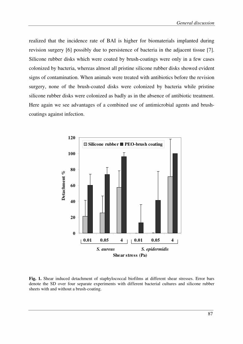
General discussion
87
realized that the incidence rate of BAI is higher for biomaterials implanted during
revision surgery [6] possibly due to persistence of bacteria in the adjacent tissue [7].
Silicone rubber disks which were coated by brush-coatings were only in a few cases
colonized by bacteria, whereas almost all pristine silicone rubber disks showed evident
signs of contamination. When animals were treated with antibiotics before the revision
surgery, none of the brush-coated disks were colonized by bacteria while pristine
silicone rubber disks were colonized as badly as in the absence of antibiotic treatment.
Here again we see advantages of a combined use of antimicrobial agents and brush-
coatings against infection.
Fig. 1. Shear induced detachment of staphylococcal biofilms at different shear stresses. Error bars
denote the SD over four separate experiments with different bacterial cultures and silicone rubber
sheets with and without a brush-coating.
0
20
40
60
80
100
120
0.01 0.05 4 0.01 0.05 4
Shear stress (Pa)
Deta
ch
men
t %
Silicone rubber PEO-brush coating
S. aureus S. epidermidis

Chapter 7
88
Conclusions
Our results suggest that polymer brush-coatings make biofilms more vulnerable
against detachment and eradication by antimicrobial agents. In addition, development
of biofilms may be stopped by the combined use of a brush-coating and antibiotic
treatment. Successful in vivo application of the brush-coating for secondary implants
allows us to state that polymer brushes may be excellent candidates for modifying
biomaterial surfaces in order to reduce the risk of BAI.
Future Research
There is evidence that the efficiency of drugs can be enhanced by localizing it into the
tissue of interest while reducing the drawbacks of systemic drug administration [8].
Since inclusion of antibiotics in the growth medium prevented biofilm formation on a
brush coating, we speculate that integration of antimicrobial agents in a brush may
help to stop biofilm formation. To this end, polymer brush-coatings can be designed to
release antimicrobial agents or can be functionalised with groups that have bactericidal
effects.
Experiments should be conducted to evaluate the damages induced by antibiotic
treatment to a mature biofilm, e.g. 24 h old biofilm, on a brush-coated surface. Various
methods have been developed for biofilm eradication. Efficiency of these methods on
biofilms formed on brush-coatings has to be assessed.
Extensive in vivo evaluations are recommended in order to clarify the performance of
a brush on a primary implant and also for a variety of biomaterials that are exposed to
different conditions in the body.

General discussion
89
References
1. Lee JH, Ju YM, Lee WK et al. Platelet adhesion onto segmented polyurethane surfaces
modified by peo and sulfonated peo-containing block copolymer additives. Journal of
Biomedical Materials Research 1998, 40:314-23.
2. Li JT, Caldwell KD. Plasma protein interaction with pluronic-treated colloids. Colloids
and Surfaces B-Biointerfaces 1996, 7:9-32.
3. Kingshott P, Wei J, Bagge-Ravn D et al. Covalent attachment of poly(ethylene glycol) to
surfaces, critical for reducing bacterial adhesion. Langmuir 2003, 19:6912-21.
4. Edmonson S, Osborne VL, Huck WTS. Polymer brushes via surface-initiated
polymerizations. Chemical society reviews 2004, 33:14-22.
5. Busscher HJ, Bos R, Van der Mei HC. Initial microbial adhesion is a determinant for the
strength of biofilm adhesion. FEMS Microbiol.Lett. 1995, 128:229-34.
6. Darouiche RO. Device-associated infections: A macroproblem that starts with
microadherence. Clinical Infectious Diseases 2001, 33:1567-72.
7. Broekhuizen CAN, de Boer L, Schipper K et al. Peri-implant tissue is an important niche
for Staphylococcus epidermidis in experimental biomaterial-associated infection in mice.
Infection and Immunity 2007, 75:1129-36.
8. Allen TM, Cullis PR. Drug delivery systems: Entering the mainstream. Science 2004,
303:1818-22.


Summary

Summary
92
Summary
Biomaterials associated infection (BAI) is one of the main causes of implant failure in
an era in which the number of patients requiring biomaterials implant surgery is
steadily increasing. Treatment of infected implants frequently includes long-term
antibiotic use, however, often without success, so that finally the implant has to be
removed. Devices implanted in revision surgery are even at a greater risk of infection
likely due to the persistence of bacteria in adjacent tissue near the implant site. BAI
begins with the initial adhesion of infectious organisms that subsequently grow to form
a biofilm. Bacterial adhesion to surfaces is influenced by physicochemical properties
of the surface and several attempts have been made to develop non-adhesive coatings,
such as polymer brush-coatings, in order to prevent bacterial adhesion and subsequent
infection. Brush-coatings are currently the most promising non-adhesive coatings that
yield significant reductions, exceeding 90%, in microbial adhesion. Despite such
impressive results no unambiguous judgment can be made yet whether polymer brush-
coatings are capable of preventing implant associated infections.
Chapter 1 reviews the main challenges that have to be dealt with before a polymer
brush-coating can be practically used against BAI. The main problem is that the fate of
the few bacteria adhering to a brush-coating is not known. Furthermore reports on in
vivo evaluation of polymer brush-coatings are inadequate for drawing a conclusion.
Therefore the main aim of this thesis is to investigate the fate of the few bacteria that
adhere to a polymer brush-coating and to evaluate the effects of a brush-coating on
bacterial colonization of biomaterial surfaces in vivo.
Tri-block copolymers of polyethylene oxide (PEO) and polypropylene oxide (PPO),
i.e. PEOn-PPOm-PEOn, better known as Pluronic, can adsorb to surfaces either in a
pancake or a brush-like conformation. The brush-like conformation is advantageous in
numerous applications, since it constitutes a surface, repellent to indwelling particles,

Summary
93
such as proteins and microorganisms. The conformation of an adsorbed Pluronic layer
depends on the hydrophobicity of the substratum surface, but the hydrophobicity
threshold above which a brush-like conformation is adopted is unknown. In chapter 2
the conformation of Pluronic F-127 (PEO99-PPO65-PEO99) adsorbed on surfaces with
different hydrophobicities was investigated using a quartz crystal microbalance with
dissipation. Adsorption in a brush-like conformation occurred on surfaces with a water
contact angle above 80°, as inferred from the thickness, viscosity and elasticity of the
adsorbed layer.
We used Pluronic F-127 to make a PEO brush-coating on medical-grade silicone
rubber which has a water contact angle of 112°. This coating was very easily made by
a dip-coating process and was used in our experiments for studying the adhesion of
three bacterial strains: Staphylococcus aureus ATCC 12600, Staphylococcus
epidermidis HBH 276 and Pseudomonas aeruginosa #3.
In order to have a measure of the interaction force between adhering bacteria and the
substratum surface we developed a procedure which determines shear forces at the
balance between bacterial attachment and detachment under flow. This protocol can be
applied to determine adhesion forces in weakly adhering systems, e.g. polymer brush-
coatings, not attainable with conventionally developed methods as shown in chapter
3. The adhesion strength of staphylococci was greatly decreased by the presence of a
PEO-coating, while the adhesion strength of P. aeruginosa #3 was hardly affected.
Polymer brush-coatings, have so far only been investigated with respect to reducing
initial bacterial adhesion, but never with respect to effects on kinetics of bacterial
growth. In chapter 4, we compared adhesion and 20 h growth of three bacterial strains
on pristine and brush-coated silicone rubber in a parallel plate flow chamber. Brush-
coatings prevented adhesion of staphylococci to below 5 × 105 cm
-2 after 30 min,
which is a 10-fold reduction compared to pristine silicone rubber. Biofilms grew on
both brush-coated and pristine silicone rubber, while the viability of biofilms on brush-
coatings was higher than on pristine silicone rubber. However, biofilms on brush-

Summary
94
coatings developed more slowly and detached almost fully by high fluid shear. In
addition, we observed that the biofilm structure had altered from a contiguous layer on
pristine silicone rubber to scattered microcolonies on a polymer brush-coating. Brush-
coatings remained non-adhesive after S. epidermidis biofilm formation and subsequent
removal. Adhesion, growth and detachment of P. aeruginosa were not significantly
different on brush-coatings as compared with pristine silicone rubber, although here
too the viability of biofilms on brush-coatings was higher.
The delay in the development of biofilms on brush-coatings and the formation of
scattered colonies which, compared to a contiguous biofilm may be better penetrable
to nutrients, made us speculate that the biofilm formation on a brush-coating is more
susceptible to antibiotics. In chapter 5 we have investigated the growth of S. aureus
biofilms, a common cause of BAI, on pristine and polymer brush-coated silicone
rubber in the absence and presence of three concentrations of gentamicin (0.5, 5 and
50 µg ml-1
). Biofilms grew on silicone rubber and maintained their viability in the
presence of gentamicin, regardless of its concentrations. On polymer brush-coatings,
however, the presence of gentamicin in the growth medium suppressed biofilm
formation and decreased the number of viable bacteria. Biofilm growth on brush-
coatings was almost completely prevented in the presence of 50 µg ml-1
gentamicin.
We concluded that biofilms on polymer brush-coatings remain susceptible to
antibiotics, which prevents biofilm formation.
Performance of brush-coatings against bacterial adhesion and growth is superb
according to our in vitro studies, but the virtue of these polymer brush-coatings in vivo
had to be investigated. The possible benefit of polymer brush-coated versus pristine
silicone rubber in revision surgery was determined in chapter 6, using a murine
model. BAI was induced in 26 mice by subcutaneous implantation of silicone rubber
disks with a biofilm of Staphylococcus aureus Xen29. During development of BAI,
half of the mice received treatment with rifampicin/vancomycin. After 5 days, the
infected disks were removed from all mice, and either a polymer brush-coated or
Summary
95
pristine silicone rubber disk was re-implanted. Revision disks were explanted after 5
days and the number of cfu’s cultured from the disks and surrounding tissue was
determined. None of the polymer brush-coated disks after antibiotic treatment
appeared colonized by staphylococci, whereas 83% of the pristine silicone rubber
disks were re-infected. Polymer brush-coated disks also showed reduced colonization
rates in the absence of antibiotic treatment as compared with pristine silicone rubber
disks. Tissue surrounding the disks was culture positive in all cases. Concluding, we
state that polymer brush-coatings are less prone to re-infection than pristine silicone
rubber when used in revision surgery, i.e. when implanted in a subcutaneous pocket
infected by a staphylococcal BAI. Antibiotic pre-treatment during the development of
BAI hardly had any effect in preventing colonization of pristine silicone rubber while
it seemed to increase the effectiveness of brush-coating.
In chapter 7, various subjects from our findings in chapters 2 to 7 are discussed.
Finally the main conclusions of the project are presented and some suggestions are
given for future research.


Samenvatting

Samenvatting
98
Samenvatting
Biomateriaal geassocieerde infecties vormen één van de belangrijkste oorzaken voor
het falen van implantaten, en dat in een tijdperk waarin het aantal patiënten dat een
biomateriaal implantaat nodig heeft sterk toeneemt. De behandeling van een
biomateriaal infectie bestaat meestal uit langdurig gebruik van antibiotica, echter vaak
zonder succes, zodat uiteindelijk het implantaat weer verwijderd moet worden.
Implantaten die geïmplanteerd zijn bij een hersteloperatie hebben een nog groter risico
van contaminatie, vanwege de reeds aanwezige bacteriën in het aangrenzende weefsel
nabij het implantaat. Biomateriaal geassocieerde infecties beginnen met het hechten
van bacteriën aan het implantaat oppervlak, die daarna groeien en een biofilm vormen.
Bacteriële hechting aan oppervlakken wordt beïnvloed door de fysisch-chemische
eigenschappen van het oppervlak en er zijn langs diverse wegen pogingen gedaan om
een niet-adhesieve coating te ontwikkelingen, zoals polymere borstel-coatings, om te
voorkomen dat bacteriën zich kunnen hechten en dus een infectie veroorzaken.
Borstel-coatings zijn momenteel de meest veelbelovende niet-adhesieve coatings. In
veel gevallen verminderen ze de aanhechting van bacterieën met meer dan 90%.
Ondanks zulke indrukwekkende resultaten kan nog niet eenduidig worden gesteld dat
polymere borstel-coatings in staat zijn om biomateriaal implantaat infecties tegen te
gaan.
Hoofdstuk 1 behandelt de belangrijkste knelpunten die eerst opgelost moeten worden
voordat borstel-coatings in de praktijk gebruikt kunnen worden tegen biomateriaal
geassocieerde infecties. Het belangrijkste punt is dat het gedrag van het kleine aantal
bacteriën dat wel op een borstel-coating hecht niet bekend is. Daarnaast is er niets
bekend over de werking van polymere borstel-coatings in vivo. Daarom is het
hoofddoel van dit proefschrift te onderzoeken wat het effect is van het kleine aantal
bacteriën dat op een polymere borstel-coating hecht en te evalueren hoe een polymere
borstel-coating op een biomateriaal oppervlak zich in vivo gedraagt. Tri-blok
Samenvatting
99
copolymeren van polyethyleen oxide (PEO) en polypropyleen oxide (PPO), d.w.z.
PEOn-PPOm-PEOn, beter bekend als Pluronic, kunnen aan oppervlakken adsorberen in
een pannenkoek- of borstel-conformatie. De borstel-conformatie is voordelig in een
aantal toepassingen: het functioneert als een niet hechtend oppervlak, ten aanzien van
deeltjes, zoals eiwitten en micro-organismen. De conformatie van een geadsorbeerde
Pluronic laag is afhankelijk van de hydrofobiciteit van het substraat oppervlak, maar
de hydrofobiciteits grens waarboven een borstel-conformatie wordt aangenomen is
onbekend. In hoofdstuk 2 werd de configuratie van Pluronic F-127 (PEO99-PPO65-
PEO99) geadsorbeerd aan oppervlakken met verschillende hydrofobiciteiten
onderzocht, gebruikmakend van een kwartskristal-microbalans met dissipatie. Uit
metingen van de viscositeit, elasticiteit en dikte van de geadsorbeerde laag bleek dat
een borstel-conformatie ontstaat op oppervlakken met een waterrandhoek groter dan
80°.
Wij hebben vervolgens Pluronic F-127 gebruikt om een PEO borstel-coating op
medisch siliconenrubber te maken (watercontacthoek 112°). Deze coating was zeer
gemakkelijk te maken met een dip-coating proces en is gebruikt in onze experimenten
voor het bestuderen van de adhesie van drie bacteriestammen: Staphylococcus aureus
ATCC 12600, Staphylococcus epidermidis HBH 276 en Pseudomonas aeruginosa #3.
Om een maat te hebben voor de interactiekrachten tussen hechtende bacteriën en het
onderliggende oppervlak hebben wij een procedure ontwikkeld welke de
afschuifkrachten bepaalt op de grens tussen bacteriële aanhechting en onthechting
onder stroming. Dit protocol kan worden toegepast om interactiekrachten in zwak
hechtende systemen, zoals polymere borstel-coatings, te bepalen, waar dit niet
mogelijk is met conventionele methoden zoals beschreven in hoofdstuk 3. De
interactiekracht van stafylokokken bleek sterk afgenomen in aanwezigheid van een
PEO-coating, terwijl de interactiekracht van P. aeruginosa #3 nauwelijks beïnvloed
werd. Polymere borstel-coatings zijn

Samenvatting
100
tot nu toe alleen maar onderzocht met betrekking tot de vermindering van initiële
aanhechting, maar nooit met betrekking tot de kinetiek van de groei van het kleine
aantal gehechte bacteriën. In hoofdstuk 4, hebben we van drie bacteriestammen de
hechting en groei gedurende 20 uren op siliconenrubber met en zonder een polymere
borstel-coating, in een parallelle plaat-stroomkamer vergeleken. Borstel-coatings
reduceerden de aanhechting van stafylokokken tot minder dan 5 x 105 cm
-2 na 30
minuten, wat een 10-voudige reductie was ten opzichte van siliconenrubber zonder
borstel. Biofilms groeiden op siliconenrubber zowel met als zonder borstel-coating,
terwijl de levensvatbaarheid van biofilms op borstels hoger was dan op
siliconenrubber. Echter, biofilms op borstel-coatings groeiden langzamer en
onthechtten bijna volledig bij hogere stromingsterkten. Daarnaast namen wij waar dat
de structuur van de biofilm was veranderd, t.w. van een continue laag op
siliconenrubber naar verspreide microkoloniën op een polymeer borstel-coating.
Borstel-coatings bleven hun werking behouden nadat een S. epidermidis biofilm was
verwijderd. Aanhechting, groei en verwijdering van P. aeruginosa waren niet
significant anders op siliconenrubber met en zonder borstel, al was ook hier de
levensvatbaarheid van biofilms groter op borstel-coatings.
De vertraging in ontwikkeling van biofilms op borstels en de vorming van verspreide
koloniën welke, in vergelijking met een volledig bedekkende biofilm beter
doordringbaar zijn voor voedingstoffen, deed ons speculeren dat biofilms op borstel-
coatings ook beter toegankelijk blijven voor antibiotica. In hoofdstuk 5 onderzochten
we de groei van S. aureus biofilms, een veel voorkomende veroorzaker van
biomateriaal geassocieerde infecties, op siliconenrubber met en zonder borstel-coating
in de aan- en afwezigheid van drie concentraties gentamicine (0,5, 5 en 50 µg ml-1
).
Biofilms groeiden op het siliconenrubber en behielden hun levensvatbaarheid in
aanwezigheid van gentamicine, ongeacht de concentratie. Op polymere borstel-
coatings, daarentegen, onderdrukte de aanwezigheid van gentamicine in het
groeimedium de vorming van een biofilm en verminderde het aantal levensvatbare

Samenvatting
101
bacteriën. Biofilms groeiden nagenoeg niet meer op borstel-coatings in de
aanwezigheid van 50 µg ml-1
gentamicine. Wij concludeerden dat biofilms op een
polymere borstel-coating inderdaad makkelijker bereikbaar zijn voor antibiotica,
hetgeen biofilm vorming voorkomt.
De goede resultaten op borstel-coatings wat betreft onderdrukking van de groei van
een biofilm in vitro, wekten grote verwachtingen voor wat betreft de in vivo
werkzaamheid, maar dit moest natuurlijk nog worden onderzocht. Het mogelijke in
vivo voordeel van polymeer borstel-coatings versus kaal siliconenrubber voor
hersteloperaties werd onderzocht in hoofdstuk 6. Biomateriaal geassocieerde infecties
werden geïntroduceerd bij 26 muizen door middel van onderhuidse implantatie van
siliconenrubberen schijfjes met een biofilm van Staphylococcus aureus Xen29.
Gedurende drie dagen, werd de helft van de muizen behandeld met
rifampicin/vancomycin. Na 5 dagen werden de geïnfecteerde siliconenrubberen
schijfjes verwijderd bij alle muizen, en vervangen door een siliconenrubberen schijfje
met of zonder polymeer borstel. Deze revisie-schijfjes werden verwijderd na 5 dagen
en het aantal cfu’s op siliconenrubber met en zonder borstel en het omliggende
weefsel bepaald. Geen van de borstel-gecoate schijfjes bleek gekoloniseerd door
stafylokokken na behandeling met antibiotica, terwijl 83% van de siliconenrubber
schijfjes opnieuw geïnfecteerd was. Polymere borstel-coatings toonden ook een
afname van kolonisatie in afwezigheid van antibiotica in vergelijking met
siliconenrubberen schijfjes. Het omringende weefsel was in alle gevallen bacterie-
positief. Concluderend stellen we dat polymeer borstel-coatings minder gevoelig zijn
voor herkolonisatie dan siliconenrubberen in een revisie setting in een onderhuids
model.. In hoofdstuk 7, zijn de verschillende onderdelen van onze bevindingen in
hoofdstuk 2 tot en met 6 besproken. Tenslotte zijn de hoofdconclusies van het project
nogmaals weergegeven, samen met een paar suggesties voor vervolg onderzoek.
I gratefully thank Sara Panahkhahi for her help in translating the summary to Dutch.


Acknowledgements

Acknowledgements
104
Acknowledgements
Sometimes I think words are too short, too simple to carry our deepest feelings. I tend
to believe that genuine feelings are spread effortlessly and anything which must be
said will be understood. All my teachers, family, friends and people who helped me in
the way that puts me here today: I thank you all and, I would like you to know that you
have my deepest respect and gratefulness which is simply beyond expression for me.
I will use this opportunity to review some great memories shaped during my scientific
life which, I realize, is woven into other aspects of my life.
It was a wish of my parents, Forough and Reza, that I become a highly educated
person. They devoted their life to their children so sacrificially that leaves no room for
me to say that I will return it someday.
Samira, my lovely wife, was the one who convinced me that I should do a PhD. She
stood next to me in all joyful and difficult days in the past years. I will not forget that
she gave up a full-time study which meant a lot to her and chose for a low-residency
study so that she could stay with me in Groningen. She has been the kindest partner
one could ever have.
I learnt a lot from all my supervisors in the past four years. I remember the first day I
was in Groningen for interview. My first meeting had been arranged with Henk and he
started the conversation by asking about my trip and reasons for choosing Groningen
etc. He was so friendly that all my nervousness was gone in a minute. Working with
such a professional and at the same time understanding scientist manager was a true
honour for me. Having a smooth PhD job was made possible with helps and advices
from Henny. She always made time if I had a question or needed to discuss something.
I enjoyed working with her in a group to arrange a “labuitje”. Despite the terrible
whether that day, it became a memorable “labuitje” for us and our colleagues. I always
enjoyed discussing subjects with Willem. His enthusiasm and supporting attitude

Acknowledgements
105
towards ideas expressed by a young researcher was spectacular. This was felt even by
people who were not directly working with him. My only regret is that he was not
present in our lab everyday.
Immigration from a country to another requires a lot of work from arranging the
necessary documents to finding your way to live in the new place. It was all very
smooth for me only because of kind helps from Ellen, Ina and Wya. When they said
they would take care of something, I was sure that it would be done in the best
possible way. Kind Ellen and Ina always asked about me and my family’s situation.
Wya was very caring too and I remember that she kindly accompanied me and my
wife in a trip to Den Haag to arrange the residence documents for us in the first month
of our stay in Groningen.
I had very nice officemates in the past four years. I experienced great moments of
sharing fun and frustrations together with Marco, Niels and Mihaela. Marco and I took
climbing courses in Groningen and we enjoyed it very much. In the last year of our
PhD however, we got so busy that we could not continue climbing! Niels and I used to
have very long scientific discussions which sometimes took hours. He had a good
sense of analysis which helped me learn better. Mihaela moved to our room after Niels
left. I wonder if she had a good time as both Marco and I were stressed to finish our
job and might have not been in a good mood.
I enjoyed working with Prashant, Adam, Anton and Isabel in two small projects which
both led to good results for us. I learnt from all of them and I will look forward to a
chance to repeat such collaborations.
I had no bad day in the lab and this was because I had the nicest colleagues around.
They all were goodhearted people whom I remember only from nice moments. Eefje
was my personal coach in the lab when I started my PhD. She indeed was a good
teacher. I will not forget her happy face and her caring character. I had the chance to
learn from Joop, Hans and Betsy in different parts of my research.

Acknowledgements
106
My sisters, Maryam and Mandana, and my only brother Ali supported me kindly
whenever I needed. They have a big share in my good memories. It is the same for my
parents- and my brother-in-law; Homa, Ali and Reza. I am grateful for all the generous
support they provided for me when I was busy with the application and immigration
procedures.
I got to know a few Iranians in Groningen. Hanging out with them gave me a taste of
my homeland which I missed very much. Sara and I were introduced to each other in a
funny way. She became a good friend of mine later on and we have had many good
days together with our other friends.
I enjoyed living in the Netherlands. I discovered several nice things about Dutch
lifestyle. Among those I would like to mention the great cycling culture, the simplicity
in all aspects of life, the respectful culture of adaptation, the openness towards
different opinions and the preservation of historic city architecture. During my stay I
was bothered by nothing other than weather.

