university of groningen lmrr-mediated gene regulation of
TRANSCRIPT

University of Groningen
LmrR-mediated gene regulation of multidrug resistance in Lactococcus lactisAgustiandari, Herfita; Peeters, Eveline; de Wit, Janny G.; Charlier, Daniel; Driessen, Arnold J.M.Published in:Microbiology-Sgm
DOI:10.1099/mic.0.048025-0
IMPORTANT NOTE: You are advised to consult the publisher's version (publisher's PDF) if you wish to cite fromit. Please check the document version below.
Document VersionPublisher's PDF, also known as Version of record
Publication date:2011
Link to publication in University of Groningen/UMCG research database
Citation for published version (APA):Agustiandari, H., Peeters, E., de Wit, J. G., Charlier, D., & Driessen, A. J. M. (2011). LmrR-mediated generegulation of multidrug resistance in Lactococcus lactis. Microbiology-Sgm, 157(5), 1519-1530.https://doi.org/10.1099/mic.0.048025-0
CopyrightOther than for strictly personal use, it is not permitted to download or to forward/distribute the text or part of it without the consent of theauthor(s) and/or copyright holder(s), unless the work is under an open content license (like Creative Commons).
The publication may also be distributed here under the terms of Article 25fa of the Dutch Copyright Act, indicated by the “Taverne” license.More information can be found on the University of Groningen website: https://www.rug.nl/library/open-access/self-archiving-pure/taverne-amendment.
Take-down policyIf you believe that this document breaches copyright please contact us providing details, and we will remove access to the work immediatelyand investigate your claim.
Downloaded from the University of Groningen/UMCG research database (Pure): http://www.rug.nl/research/portal. For technical reasons thenumber of authors shown on this cover page is limited to 10 maximum.
Download date: 20-02-2022

LmrR-mediated gene regulation of multidrugresistance in Lactococcus lactis
Herfita Agustiandari,1 Eveline Peeters,2 Janny G. de Wit,1 Daniel Charlier2
and Arnold J. M. Driessen1
Correspondence
Arnold J. M. Driessen
Received 6 January 2011
Revised 13 February 2011
Accepted 14 February 2011
1Department of Microbiology, Groningen Biomolecular Sciences and Biotechnology Institute andThe Kluyver Centre for the Genomics of Industrial Fermentation, University of Groningen,Nijenborgh 7, 9747 AG Groningen, The Netherlands
2Erfelijkheidsleer en Microbiologie, Vrije Universiteit Brussel (VUB), Pleinlaan 2, B-1050 Brussels,Belgium
Multidrug resistance (MDR) in Lactococcus lactis is due to the expression of the membrane
ATP-binding cassette (ABC) transporter LmrCD. In the absence of drugs, the transcriptional
regulator LmrR prevents expression of the lmrCD operon by binding to its operator site. Through
an autoregulatory mechanism LmrR also suppresses its own expression. Although the lmrR and
lmrCD genes have their own promoters, primer extension analysis showed the presence of a long
transcript spanning the entire lmrR–lmrCD cluster, in addition to various shorter transcripts
harbouring the lmrCD genes only. ‘In-gel’ Cu-phenanthroline footprinting analysis indicated an
extensive interaction between LmrR and the lmrR promoter/operator region. Atomic force
microscopy imaging of the binding of LmrR to the control region of lmrR DNA showed severe
deformations indicative of DNA wrapping and looping, while LmrR binding to a fragment
containing the lmrCD control region induced DNA bending. The results further suggest a drug-
dependent regulation mechanism in which the lmrCD genes are co-transcribed with lmrR as a
polycistronic messenger. This leads to an LmrR-mediated regulation of lmrCD expression that is
exerted from two different locations and by distinct regulatory mechanisms.
INTRODUCTION
In their natural environment, bacteria have to cope withnaturally occurring toxic molecules (plant alkaloids, bilesalts), harmful metabolic end-products, antimicrobialpeptides, and secondary metabolites such as antibiotics. Awidespread mechanism to counteract the inhibitory actionof such molecules is their secretion from the cell bymembrane-bound multidrug resistance (MDR) transpor-ters (Chopra & Roberts, 2001; Neyfakh et al., 1991;Tennent et al., 1985). For instance, the cationic berberinealkaloids produced by many plants are substrates for MDRpumps, such as QacA and NorA of Staphylococcus aureus(Hsieh et al., 1998; Neyfakh et al., 1993; Ng et al., 1994;Schumacher & Brennan, 2003). Soil- or plant-associatedorganisms display the highest abundance of chromoso-mally encoded MDR efflux systems (Paulsen et al., 2000;Saier et al., 1998). MDR transporters are often subject to
regulatory control (Grkovic et al., 2002), as their expressionat a high level may be critical to cells (Eckert & Beck, 1989;Gury et al., 2004). The expression of most MDR trans-porters is either positively or negatively controlled by localregulatory proteins (Eckert & Beck, 1989; Hickman et al.,1990) and/or globally by stress-related regulators. Forexample, the overexpression of the acrAB MDR locus inEscherichia coli is regulated by the global regulators MarA,Rob and SoxS, the local repressor AcrR (Alekshun & Levy,1997; Ma et al., 1996), and the quorum sensor regulatorSdiA (Rahmati et al., 2002).
The Gram-positive bacterium Lactococcus lactis plays amajor role in fermented dairy food production. L. lactisreadily develops an MDR phenotype upon long-termexposure to structurally unrelated compounds such asdaunomycin, Hoechst 33342, ethidium bromide, rhoda-mine 6G and cholate (Bolhuis et al., 1994; Lubelski et al.,2004; Mazurkiewicz et al., 2004). This MDR phenotype isdue to the constitutive expression of the lmrCD genes,which encode a heterodimeric ATP-binding cassette (ABC)MDR transporter that secretes these compounds from thecell (Lubelski et al., 2006). Expression of the lmrCD genes iscontrolled by a local transcriptional regulator termed LmrR(Agustiandari et al., 2008). LmrR acts as a drug-sensitive
Abbreviations: ABC, ATP-binding cassette; AFM, atomic force micro-scopy; EMSA, electrophoretic mobility shift assay; IR, inverted repeat;MDR, multidrug resistance; OP-Cu, Cu-phenanthroline; p/o, promoter/operator; qPCR, quantitative PCR.
A supplementary figure and three supplementary tables are availablewith the online version of this paper.
Microbiology (2011), 157, 1519–1530 DOI 10.1099/mic.0.048025-0
048025 G 2011 SGM Printed in Great Britain 1519

repressor of the expression of the lmrCD genes. Most of thetranscriptional regulators involved in MDR belong to theAraC, MarR, MerR and TetR families of transcriptionalregulators. However, LmrR belongs to PadR, a family ofmostly poorly characterized regulatory proteins that areinvolved in the regulation of detoxification mechanismssuch as phenolic acid metabolism (Gasson et al., 1998;Overhage et al., 1999; Segura et al., 1999). Apart fromLmrR, LadR from Listeria monocytogenes is the onlycharacterized member of the MDR-related PadR regulators(Agustiandari et al., 2008; Huillet et al., 2006). In thisfamily of regulators, the expression of the detoxificationgenes is typically induced by the presence of the toxiccompounds in the medium via a direct interaction with thePadR-like regulator. Indeed, LmrR has been shown to bindseveral of the LmrCD substrates, such as Hoechst 33342,daunomycin and sodium cholate. On the other hand,LmrR does not bind r-coumaric acid and ferulic acid (ourunpublished data), which are the phenolic acid derivativesthat have been shown to bind to PadR (Gury et al., 2004).We have solved the structure of the LmrR dimer in the apoform and in two drug-bound forms, i.e. with Hoechst33342 and daunomycin (Madoori et al., 2009). The dimercontains two N-terminal DNA-binding domains with atypical winged helix–turn–helix (wHTH) motif, while theC-terminal regions form a large flat central pore at thesubunit interface. The latter constitutes the drug-bindingpocket of LmrR, which is symmetrical with equalcontributions from both monomers to the overall struc-ture. On the other hand, the exact induction mechanism ofLmrR will only be determined when the crystal structure ofLmrR bound to DNA is available. However, one possiblemechanism of lmrR upregulation in the cell is via allostericcoupling between the drug- and DNA-binding sites, basedon the comparison of different LmrR structures and thestudy of mutational analysis. The binding of the drugmolecule to LmrR locks the dimer conformation in such away that the DNA recognition helices fail to bind to theirrecognition site on the DNA major grooves (Madoori et al.,2009).
The lmrR gene is located upstream of the lmrCD genes(Agustiandari et al., 2008). In independently isolated drug-resistant strains of L. lactis that are cross-resistant against aseries of drugs, the lmrCD genes are constitutively expressedbecause of the presence of defective forms of LmrR that areno longer able to bind to the promoter/operator (p/o) regionof the lmrCD genes (Lubelski et al., 2006). In these strains,the lmrR gene is also upregulated, suggesting that in wild-type cells, LmrR represses its own expression. Biochemicaldata demonstrate that LmrR indeed binds to its ownpromoter region (Agustiandari et al., 2008). Here, we haveanalysed the interaction between LmrR and the controlregions of the lmrCD and lmrR genes using ‘in-gel’ Cu-phenanthroline (OP-Cu) footprinting analysis and atomicforce microscopy (AFM) imaging. The data suggest distinctmodes of binding of LmrR to the lmrR and lmrCD controlregions, resulting in the formation of different transcripts
that encode the structural genes either with or without thelmrR transcriptional regulator gene. Expression of both lmrRand lmrCD is elevated when cells are grown in the presenceof drugs, suggesting a mechanism in which the regu-lator gene and the functional genes are induced and co-transcribed from a polycistronic messenger.
METHODS
Protein purification. Strep-tagged LmrR protein was overexpressedin L. lactis NZ9000, and purified by strep-tag affinity chromatographyfollowed by chromatography with a heparin column, as describedpreviously (Agustiandari et al., 2008).
Primer extension and RT-PCR analysis. RNA was extracted fromL. lactis MG1363 using TRIzol reagent (Invitrogen). To preventgenomic DNA contamination, RNA samples were treated on-columnwith DNase I using an RNeasy Mini kit (Qiagen). Genomic DNA wasextracted from L. lactis MG1363 using the GenElute BacterialGenomic DNA kit (Sigma-Aldrich). Primer extension analysis wasperformed as described previously (Enoru-Eta et al., 2002) usingAMV Reverse Transcriptase (Roche Applied Science). The 59 end-labelled primers DC620r or DC621r were used for transcription startdetermination of lmrR or lmrC, respectively. Labelling was done using[c-32]P-ATP (GE Healthcare). Reference ladders were generated bychemical sequencing methods (Maxam & Gilbert, 1980). cDNA wasprepared from about 2 mg L. lactis RNA by using Superscript IIReverse Transcriptase (Invitrogen) and 200 ng random primers. Thisreaction was followed by RNase H treatment (Fermentas). Transcriptanalysis was done by PCR with primers Cdprmf/DC621r or DC636f/DC621r, using cDNA as template. Primer sequences are shown inSupplementary Table S1.
Electrophoretic mobility shift assays (EMSAs) and in-gel
OP-Cu footprinting. Labelled DNA fragments were produced byPCR (ReadyMix Taq PCR Reaction Mix, Sigma-Aldrich) using a pairof primers, of which one was 59 end-labelled with [c-32P]ATP (GEHealthcare). For the promoter regions of lmrR and lmrCD, the primerpairs DC634f/DC620r and DC635f/DC621r, respectively, were usedwith L. lactis MG1363 genomic DNA as template. Labelled fragmentswere purified by PAGE. The truncated fragments of the promoterregions of lmrR and lmrCD were prepared similarly using the set ofprimers listed in Supplementary Table S2. EMSAs were performed asdescribed previously (Enoru-Eta et al., 2000). Binding reactions wereperformed in LmrR binding buffer (20 mM Tris, pH 8.0, 1 mMMgCl2, 20 mM KCl, 0.1 mM DTT, 0.4 mM EDTA, 12.5 %, v/v,glycerol) by incubating at 37 uC for 30 min in the presence of 25 mgml21 sonicated herring sperm DNA as a non-specific competitor. KD
values were estimated based on these EMSAs, as the proteinconcentration at which about 50 % of the DNA was bound (expressedin dimer equivalents). In-gel OP-Cu footprinting was performed asdescribed previously (Peeters et al., 2004). Reference ladders weregenerated by chemical sequencing methods (Maxam & Gilbert, 1980).
AFM. For AFM experiments, the DNA fragments were prepared byPCR with ReadyMix Taq PCR Reaction Mix (Sigma-Aldrich). The p/o
region of lmrR was amplified as a 997 bp fragment with the primerpair AFM lmrR pmtr FW/AFM lmrR pmtr RV, and L. lactis MG1363genomic DNA as template. A 1016 bp fragment containing the p/o
region of lmrCD was amplified with the primer pair AFM lmrCDpmtr FW/AFM lmrCD pmtr RV. Following PCR amplification, allfragments were purified by agarose gel electrophoresis using aGenElute Gel Extraction kit (Sigma-Aldrich). A number of trials wereperformed to find the best concentration for both DNA and LmrR,with final concentrations of 1.86 and 0.04 mM for lmrR DNA and LmrR
H. Agustiandari and others
1520 Microbiology 157

protein, respectively; and 0.16 and 0.018 mM for lmrCD DNA and
LmrR protein, respectively. These binding reactions were diluted in
LmrR binding buffer in a total volume of 15 ml. The mixture was then
diluted twofold in adsorption buffer (40 mM HEPES, pH 6.9, 10 mM
NiCl2), and 15 ml of the suspension was deposited on freshly cleaved
mica. This was incubated for 5 min to allow adsorption of the
nucleoprotein complexes. Subsequently, samples were rinsed with
deionized ultrapure water and excess water was blotted off with
absorbent paper. The mica surface was blown dry in a stream of filtered
air. A NanoScope IIIa atomic force microscope (Digital Instruments/
Veeco) was operated in the tapping mode, in air. Images of 5126512
pixels were acquired by using Nanoprobe scanning probe microscopy
(SPM) tips, type RTESP7 (Veeco) with a 115–135 mm cantilever, a
nominal spring constant of 50 N m21 and resonance frequencies in the
range from 244 to 295 kHz. The scan size was 1.5 mm61.5 mm and the
scan rate was 2 Hz. NanoScope 6.11r1 software (Digital Instruments/
Veeco) was used to flatten the images and to make zoomed 3D surface
plots. The contour lengths of DNA molecules or DNA arms of
complexes were measured by manual tracing with ImageJ (Abramoff
et al., 2004). DNA molecules or complexes with overlapping parts or
with visible anomalies were omitted from the analysis.
Quantitative PCR (qPCR). Cultures of L. lactis NZ9000 and
NZ9000(DlmrR) (Agustiandari et al., 2008) were grown overnight
on M17 supplemented with 0.5 % glucose at 30 uC. Cultures were
diluted 1 : 100 to an OD660 of 0.07–0.08 in the same medium with or
without 1 mM Hoechst 33342 (Sigma-Aldrich) or 20 mM daunomycin
(Calbiochem – VWR). These subinhibitory drug concentrations
ensured near-identical growth rates of the different types of cells. Cells
were further grown at 30 uC, and during the early exponential, late
exponential and stationary growth phases, samples of 5 ml were
collected and flash-frozen in liquid nitrogen. Total RNA was isolated
using TRIzol reagent (Zaidi et al., 2008). Residual chromosomal DNA
was removed by using the TURBO DNA-free kit (Ambion, Applied
Biosystems) according to the manufacturer’s instructions. Purified
RNA was quantified by measuring A260 using a NanoDrop ND1000
spectrophotometer. The quality of the RNA preparations was checked
by visualizing the integrity of 16S and 23S rRNA on an agarose gel,
and by verifying the absence of DNA contamination by PCR. The
cDNA molecules were synthesized using an iScript cDNA synthesis kit
(Bio-Rad) as recommended by the manufacturer. Total RNA was
isolated from at least two separately grown replicate cultures.
For the qPCR experiments, the primers were designed to have a length
of 22–23 nt, a G/C content of 45–47 % (see Supplementary Table S3)
and a Tm of about 60–65 uC. The lengths of the primer products ranged
between 200 and 230 bp. qPCR was carried out on a MiniOpticon
Real-Time PCR system (Bio-Rad). After dilution of the cDNA, 4 ml was
added to 21 ml of the PCR mixture (12.5 ml iQ SYBR Green Supermix
and 0.5 ml of each primer at 10 pmol ml21). Thermal cycling conditions
were as follows: initial denaturation at 95 uC for 3 min, followed by 40
cycles of 95 uC for 20 s, 55 uC for 20 s and 72 uC for 30 s. An
additional step starting from 65 to 95 uC was performed to establish a
melting curve. This was used to verify the specificity of the PCR for
each primer set. qPCR measurements were performed in duplicate for
each sample. The tufA gene was used as an internal control and for
normalization of the results (Friedrich & Lenke, 2006).
RESULTS
Mapping of the transcription start sites of lmrCDand lmrR
Primer extension analysis was performed to map thetranscription initiation sites of the lmrCD and lmrR genes
using RNA extracted from L. lactis MG1363 cells (Fig. 1).Transcription of lmrR is initiated at a single G residuelocated 26 nt upstream of the ATG start codon (Figs 1aand 2a). In contrast, lmrCD-specific reverse transcriptionresulted in at least four different cDNA molecules (Figs 1band 2b). Transcripts C and D start at an A residue 55 and61 nt upstream of the ATG start codon of lmrC,respectively. Transcript B starts at a T residue that islocated 100 bp upstream of the ATG start codon of LmrC.A fourth cDNA molecule represents a transcript that islarger than the labelled fragment used for the Maxam–Gilbert sequencing ladder, which was prepared by PCRamplification using primers CDprmf and DC621r (Fig. 1d,Supplementary Table S1). Therefore, this transcript mustalso contain at least part of the ORF of lmrR. To testwhether or not this transcript corresponded to transcript Aas detected by lmrR-specific primer extension, RT-PCRanalysis was performed using primer pairs CDprmf/DC621r and DC636f/DC621r (Fig. 1c, d). These reactionsresulted in amplification, confirming the existence of anmRNA molecule that spans both the lmrR gene and thelmrCD gene. It thus appears that an RNA polymeraseinitiated at the lmrR promoter may proceed till the end oflmrD. Indeed, using the program TransTerm, intrinsicterminators were predicted to occur neither in the lmrRand lmrCD genes nor in the intergenic region betweenlmrR and lmrC, although a terminator was detecteddownstream of lmrD. Putative Shine–Dalgarno (SD)sequences for both lmrR and lmrC were detected betweenthe seventh base and the twelfth base upstream of therespective start codons. Regions that showed sequenceconservation with the consensus 235 and 210 promoterelements could be identified slightly upstream of the startof transcripts A and B (Fig. 2a, b). Both promoters showeda putative Pribnow box with a perfect match to theconsensus and a 235 sequence with a good match, the twobeing separated by a linker of ideal length (17 bp).However, in view of the multiple transcripts observed forlmrCD, additional promoter element(s) might be involvedin lmrCD expression, although it cannot be excluded thatthese transcripts may arise by degradation of the longertranscripts.
Identification of the LmrR-binding sites in thecontrol regions of lmrR and lmrCD
Previously, it has been shown that LmrR protects a longstretch of DNA in the control region of its own geneagainst DNase I (Agustiandari et al., 2008). Here, we showthat LmrR forms multiple complexes with p/o lmrR DNA asobserved in an EMSA (Fig. 3a). This result suggests thepresence of multiple binding sites that likely involve severalcopies of LmrR. Three complexes (B1, B2 and B3) showeda slightly different migration velocity, whereas complex B4,which was detected only at the highest LmrR concentrationused (0.8 mM), was strongly retarded in its mobility. Theaverage apparent binding dissociation constant (KD) of theLmrR–p/o lmrR interaction was around 30 nM. There was a
Multidrug resistance in Lactococcus lactis
http://mic.sgmjournals.org 1521

Fig. 1. Primer extension analyses of transcripts showing the transcription start sites of (a) lmrR using primer DC620r and (b)lmrC using primer DC621r. The amounts of total RNA used were 12.5 mg (lane 1), 25 mg (lane 2), 50 mg (lane 3) and 100 mg(lane 4). The main primer extension products are indicated with arrowheads and are designated A to D. A+G and C+Trepresent the corresponding Maxam–Gilbert sequencing ladders. A systematic correction in the alignment of the cDNA productwith the sequencing ladders has been performed to take into account the difference in migration velocity of the cDNA and thereference ladders due to different ends generated by the AMV reverse transcriptase and the chemical modification and cleavagereactions. (c) RT-PCR analysis with cDNA as template with primers CDprmf and DC621r (lane 1); as lane 1, without addition ofreverse transcriptase (negative control) (lane 2); with primers CDprmf and DC621r and with genomic DNA as template (lane 3);with primers DC636f and DC621r and with cDNA as template (lane 4); as lane 4, but without addition of reverse transcriptase(negative control) (lane 5); with primers DC636f and DC621r and with genomic DNA as template (lane 6). (d) Schematicoverview of the transcripts A, B, C and D with respect to the ORFs (indicated with open arrows) and primer products used forqPCR. The locations of the primers used for primer extension and RT-PCR analysis are also indicated.
H. Agustiandari and others
1522 Microbiology 157

rapid transition in the formation of the different com-plexes, which suggested cooperativity in the binding.EMSAs were also performed with truncated DNA frag-ments containing only a part of the lmrR control region orORF (Fig. 3b, c). Interestingly, LmrR was able to bind DNAprobes consisting of the control region alone (Rtrunc1 andRtrunc2), and also a DNA probe consisting mainly of thelmrR gene, starting only 4 bp upstream of the transcriptionstart (Fig. 3c) (Rtrunc3). However, although the Rtrunc2and Rtrunc3 fragments were of nearly identical length, theLmrR–Rtrunc3 complex migrated differently and was lessstable, indicative of a different architecture and/orstoichiometry of this complex. These binding events werespecific, since no complex formation was observed underidentical conditions with a DNA fragment correspondingto the region upstream of the 235-promoter element oflmrC (Supplementary Fig. S1).
To further determine which regions of the DNA arerecognized by LmrR in each of the multiple complexesobserved in the EMSA, in-gel OP-Cu footprinting was
performed with the various complexes (Fig. 3d). A stretchof 6 nt was clearly protected in the fastest-migratingcomplex B1 (Fig. 2a, yellow bar, and Fig. 3d). This initiallyprotected site might be considered as a ‘core’-bindingbinding site from which LmrR binding is nucleated. Inaddition, a difference in the cleavage pattern, between onthe one hand the complexed B1 form and on the otherhand the free I and F forms, could be observed in anapproximately 13 nt long region, immediately downstreamof this stretch (Fig. 2a, yellow dashed bar, and Fig. 3d). Thisstretch, including the 235 box, is part of a region that ismuch more clearly protected in complexes B2 and B3 (Fig.2a, orange and red bars). Footprinting with a DNAfragment with the top strand labelled revealed no clear-cut differences in the protected regions of complexes B2and B3. This might be explained by (i) a slightly differentarchitecture of the complex, (ii) a different stoichiometryof the complex without supplementary DNA contacts, or(iii) the formation of a ‘sandwich-like’ structure containingtwo DNA molecules. The highly retarded complex B4showed extensive protection encompassing about 102 bp,
Fig. 2. Schematic representation of the transcriptional elements on (a) lmrR and (b) lmrCD control region DNA, including thepositions of the ”35 and ”10 regions, the transcription initiation sites, and the translation start codon (bold type). For thepromoter region of lmrCD, the predicted promoter elements are only shown for transcript B. The letters A to D represent the 59
end of the major transcripts observed in primer extension analysis. In addition, the protected areas observed in the footprintingassays of LmrR binding to p/o lmrR (A) and p/o lmrCD (B) are shown. For p/o lmrR, protection zones are indicated for thecomplexes B1 (yellow), B2 (orange), B3 (red) and B4 (purple). For p/o lmrCD, the protection zone is indicated in yellow. Theball-and-stick symbols represent the positions of the hyper-reactivity sites. The identified imperfect palindromes are shown inthe sequences with double arrows. (c) Representation of the imperfect IRs as identified in p/o lmrR, p/o lmrCD and the PadRconsensus IR. Palindromic residues are in bold type, and the conserved PadR motif is boxed.
Multidrug resistance in Lactococcus lactis
http://mic.sgmjournals.org 1523

Fig. 3. Binding of LmrR to the lmrR p/o region. (a) EMSA of the binding of purified LmrR to a 210 bp labelled DNA fragmentcontaining the lmrR p/o region. The LmrR stock concentration was 81.5 mM (dimer) and was further diluted. No LmrR wasadded in lane 1, and LmrR was added at concentrations of 0.01 mM (lane 2), 0.02 mM (lane 3), 0.03 mM (lane 4), 0.1 mM (lane5), 0.2 mM (lane 6) and 0.8 mM (lane 7). The positions of the free DNA (F) and of the different LmrR-bound DNA complexes (B1,B2, B3 and B4) are indicated. These different complexes and the boxes named I (input DNA) and F (free DNA) were excised forin-gel footprinting analysis. (b) EMSA of the binding of purified LmrR to truncated DNA fragments Rtrunc1 (266 bp), Rtrunc2(170 bp) and Rtrunc3 (152 bp), corresponding to the regions of the lmrR operator site indicated in Fig. 2. LmrR was added at afinal concentration of 1.85 mM (dimer). (c) Schematic of the coverage of the p/o lmrR truncated fragments Rtrunc1, Rtrunc2 andRtrunc3 relative to the lmrR promoter elements and ORF. (d) In-gel OP-Cu footprinting of LmrR binding to the p/o region of lmrR
with the bottom strand labelled (left two panels) or with the top strand labelled (right panel). The EMSA that was used for theexperiment with the bottom strand labelled is shown in Fig. 3(a). Next to each autoradiograph, protected regions are indicatedwith a vertical line. Hyper-reactivity sites are also indicated with ball-and-stick symbols. For the far-left panel, the full line
H. Agustiandari and others
1524 Microbiology 157

including the entire promoter and transcription start site(Fig. 2a, purple bar, and Fig. 3d). In this protected region,an imperfect inverted repeat (IR) is apparent (Fig. 2a, c).This IR exhibited one mismatch as compared with thePadR consensus sequence, but had the optimal spacing of8 nt between the palindromic half sites (Fig. 2c) (Huilletet al., 2006). Several hyper-reactivity signals were observedfor complex B4, indicating local DNA deformations(minor groove widening) upon LmrR binding (Figs 2aand 3d).
The binding of LmrR to DNA fragments covering the p/o
region of the lmrCD genes showed a distinctively differentsignature. Previous footprinting results had indicated thatLmrR binds to two different sites on the lmrCD promoter(Agustiandari et al., 2008): site I, comprising the 235 and210 regions, and site II, which harbours an imperfect IRsimilar to the PadR consensus sequence but with a spacingof 10 bp (Fig. 2c) (Huillet et al., 2006). EMSAs wereperformed with shortened probes corresponding to eithersite I or site II (Supplementary Fig. S1). It appeared thatLmrR bound DNA probes containing the 235 and 210regions (site I) more strongly than the probes containingsite II with the palindromic sequence. With the full-lengthlmrCD p/o DNA, a major specific complex was observedupon binding of LmrR (Fig. 4a). The overall bindingaffinity of this interaction appeared to be two- to fourfoldlower than the affinity for p/o lmrR DNA, with an apparentKD of about 150 nM. In-gel OP-Cu footprinting of LmrRbinding to the lmrCD control region showed a singleextended protected region of about 126 bp (Fig. 4b) thatoverlapped all the transcription initiation sites (site I) inthe lmrCD control region and their cognate promoterelements, and the previously identified imperfect IR (site II;Fig. 2b). At the promoter-distal side of the protectedregion, a hyper-reactivity site was observed, again indic-ating LmrR-induced DNA deformations. These resultsdemonstrate different modes of binding of LmrR to thelmrR and lmrCD p/o regions.
AFM of the binding of LmrR to lmrR and lmrCDpromoter DNA
AFM was used to visualize the architecture of LmrRcomplexes formed with the p/o regions of lmrR and lmrCD(Figs 4c and 5). Tapping-mode AFM in air was used forhigh-resolution topographic imaging of the soft protein/DNA sample surfaces without creating destructive fric-tional forces. With the lmrR p/o DNA, 22 unbound 997 bp-long DNA molecules and 41 DNA–LmrR complexes wereanalysed. The contour length of unbound DNA moleculeswas manually traced using ImageJ software, resulting in amean contour length of 313 nm (SD 29 nm; Fig. 5c). This
yielded an axial basepair rise of 0.31 nm bp21, which isslightly lower than the theoretical rise of B-form DNA (i.e.0.34 nm bp21), but in good agreement with other AFMstudies. This difference can be explained by the limitedresolution of the microscope and the smoothing procedurethat rounds sharp bends (Rivetti et al., 1996). Based onDNA persistence length analysis of other DNA moleculesmeasured under similar experimental conditions, it can beassumed that the molecules are able to freely equilibrate onthe surface before capture (Peeters et al., 2006; Minh et al.,2009).
A heterogeneous population of LmrR–lmrR nucleoproteincomplexes was observed, ranging from having apparently asingle site bound, possibly the ‘core’ nucleation site (Fig.5a, images 1–3), to having apparently two sites bound oreven large complex regions. Here, several LmrR moleculesseemed to be involved in the condensation of the bindingsite area (Fig. 4c, image 4). This type of complex mostprobably corresponds to the B4 population observed in theEMSA (Fig. 3a). It is clear that LmrR binding inducessevere DNA deformations, including sharp DNA bending,DNA condensation and possibly even DNA wrappingaround the protein (local constrained toroidal DNAsupercoiling) or DNA looping (Fig. 5a). The contourlength of the naked DNA arms of all complexes wasmeasured without making a distinction between thedifferent types of complexes (depending on the degree ofbinding; Fig. 5b). These measurements resulted in a meanlength of 79 nm (SD 39 nm) for the short DNA arm and191 nm (SD 40 nm) for the long DNA arm. Therefore, thetotal mean visible contour length of the complexes(short+long arms) was 270 nm (SD 52 nm; Fig. 5c). Thisis a difference of 43 nm from that of the mean length of theunbound DNA molecules, and taking a basepair rise of0.31 nm bp21 into account, this corresponds to about139 bp condensed inside the DNA–LmrR complex. Due tothe heterogeneity of the complexes, the distributions arebroad. These observations demonstrate that the mech-anism of binding of LmrR to the lmrR p/o DNA involvesinteractions with multiple LmrR molecules that are likelybound in a cooperative fashion. Furthermore, the extent ofthe condensation is in good agreement with the length ofthe protected region as observed for complex B4 in in-gelOP-Cu footprinting.
AFM experiments with the lmrCD control region DNAresulted in LmrR–DNA complexes with a more homo-geneous architecture than that of the LmrR–p/o lmrRcomplexes (Fig. 4c). The results indicated that LmrRinduces a significant DNA bending. Typically, thecomplexed region had a bilobed structure. The two‘blobs’ present in the AFM images may represent the
corresponds to protection observed in complexes B1 and B2, whereas the dashed line corresponds to additional protectionobserved in complex B2 alone. A+G and C+T represent the Maxam–Gilbert sequencing ladders. Next to the ladder, theposition of the transcription start is shown. A schematic representation of the protected regions is displayed in Fig. 2.
Multidrug resistance in Lactococcus lactis
http://mic.sgmjournals.org 1525

binding of two LmrR dimers to the DNA (Fig. 4c)
(Madoori et al., 2009). Taken together, the AFM results
support the notion that LmrR binds the lmrR and lmrCD
operator regions by different mechanisms, and indicate
higher-order interactions of LmrR with the operator
region of its own gene.
Expression analysis of the lmrCD and lmrR genesin L. lactis
Our analysis indicated the presence of a long transcriptharbouring both the lmrR gene and the lmrCD gene. Toassess the expression levels of lmrR in growing cell cultures,qPCR was employed on RNA extracted from L. lactis cells
Fig. 4. Binding of LmrR to the lmrCD p/o
region. (a) EMSA of the binding of LmrR to aDNA fragment corresponding to the lmrCD p/o
region. No LmrR was added in lane 1, andLmrR was added at concentrations of 0.01 mM(lane 2), 0.02 mM (lane 3), 0.03 mM (lane 4),0.1 mM (lane 5), 0.2 mM (lane 6) and 0.8 mM(lane 7). The positions of input (I), free DNA (F)and bound complexes (B) are indicated. (b) In-gel OP-Cu footprinting analysis of the LmrR–lmrCD promoter region complex that wasexcised from the gel shown in Fig. 3(b). Nextto the autoradiograph, the protected regionsare indicated with a vertical line and the hyper-reactivity sites as a ball-and-stick symbol.A+G and C+T represent the Maxam–Gilbert sequencing ladders. Next to the ladder,the positions of the transcription starts areshown. A schematic representation of theprotected region is displayed in Fig. 2. (c) Aselection of AFM images of LmrR–p/o lmrCD
protein–DNA complexes (indicated by thearrows), as typically observed.
Fig. 5. AFM analysis of the binding of LmrR to the p/o site of lmrR. (a) A selection of AFM images of LmrR–p/o lmrR protein–DNA complexes (indicated by the arrows). (b) Contour length measurements of the long (grey bars) and short (black bars) armsof LmrR complexed with the DNA fragment. (c) Contour lengths of the sum of the long and short arms of the LmrR-complexedDNA fragments (black bars) and of the free DNA fragments (grey bars). The y axes indicate the number of analysed molecules.
H. Agustiandari and others
1526 Microbiology 157

growing on M17 medium with glucose in the absence andpresence of subinhibitory concentrations of the drugsdaunomycin and Hoechst 33342. As a control, L. lactisNZ9000(DlmrR) was used, which expresses the lmrCDgenes constitutively (Agustiandari et al., 2008). Primer setswere designed to monitor the transcript levels of lmrR,lmrC and lmrD individually, and in addition, a set wasdesigned that detected the intergenic region that separatesthe lmrR and lmrCD genes in the long polycistronic lmrR–lmrCD transcript (Fig. 1d). Expression levels were relatedto that of the housekeeping gene tufA, which encodes thetranslation elongation factor Tu (Friedrich & Lenke, 2006).In addition, the secY transcript, encoding the majorsubunit of the preprotein translocase, was monitored.The expression level of these control genes was constantduring exponential growth, but unlike tufA, the expressionof secY dropped when cells entered the stationary phase(Fig. 6). As expected, the lmrC and lmrD genes were highlyexpressed in L. lactis NZ9000(DlmrR) cells during theexponential and stationary growth phases. With L. lactisNZ9000 wild-type cells, lower levels of lmrCD expressionwere observed that dropped dramatically when the cellsentered the stationary phase. A similar response wasobserved with the transcript containing the lmrR geneand the lmrR–lmrC intergenic region, suggesting that thelong transcript is present during the entire exponentialgrowth phase. When cells were exposed to daunomycin(Fig. 6) or Hoechst 33342 (data not shown), expressionlevels of lmrC and lmrD increased. Since Hoechst 33342also caused an increase in tufA expression, the correspond-ing data could not be quantified. Remarkably, exposure tothe drugs also resulted in increased levels of the transcriptharbouring the lmrR gene and the lmrR–lmrC intergenicregion. Summarizing, these data demonstrate that both theregulatory lmrR gene and the structural lmrCD genes areexpressed in exponentially growing wild-type cells and thattheir expression increases upon exposure to toxic drugs.
DISCUSSION
The ABC transporter LmrCD has previously been shown tobe a major determinant of the MDR phenotype in L. lactis(Lubelski et al., 2006). Transcription of lmrCD iscontrolled by LmrR, a local regulatory repressor whosegene is located upstream of lmrCD (Agustiandari et al.,2008). The lmrR and lmrCD genes are transcribed in thesame direction. LmrR has previously been shown tofunction as a drug-controlled negative transcriptionalregulator of the expression of the lmrCD genes. Ourcurrent primer extension analysis now reveals the presenceof three major transcripts of lmrCD, and one longertranscript spanning the lmrR and lmrCD genes. Theoccurrence of multiple transcripts of lmrCD might indicatethe presence of alternative promoters. Alternative promo-ters are quite frequent in bacteria and may be used to copewith changes in the environment, such as alterednutritional requirements that result in changes in theexpression of a particular gene. In most cases, however, one
promoter is responsible for constitutive expression, whilethe others are inducible by different stimuli (Musso et al.,1977) and may even function with another alternativesigma factor. At this stage it is unclear whether the presenceof these multiple transcripts indeed reflects functionaldifferences in the regulation and/or expression mechanism.Possibly, additional global or local regulators are involvedin the regulation of the different promoters. However,qPCR analysis of the expression of lmrR (from the longtranscript) and of the lmrCD genes (likely both from thelong and shorter transcripts) indicated that these genes areexpressed throughout the exponential growth phase, andthat their expression is further elevated when cells areexposed to toxic drugs. For lmrCD, the drug-inducedexpression levels were lower than those observed in thederegulated strain that lacked the lmrR gene, indicatingthat the drug-induced derepression is not maximized insuch cells.
LmrR binds to two regions in the lmrCD operatorsequence. Site I, comprising the 235 and 210 regionleading to initiation at transcription start site B, appears tobe a high-affinity binding site for LmrR. Site II harbours animperfect IR that is similar to the PadR consensus bindingsite, although the two half-sites are separated by 10 insteadof 8 bp (Huillet et al., 2006). EMSA data suggested that thepalindromic sequence on its own is only weakly recognizedby LmrR (Supplementary Fig. S1), and that the binding ofLmrR to the entire control region of the lmrCD genesresults in one dominant species of DNA–protein complex.In-gel footprinting analysis supported the notion that inthis complex, both site I and site II are protected by LmrR.Visualization of these protein–DNA complexes by AFMrevealed a significant DNA bending, with two protein‘blobs’ present on the DNA, wherein each ‘blob’ likelycorresponds to one LmrR dimer. Taken together, theseresults suggest that site I and site II are each bound by anLmrR dimer in a highly cooperative manner, since it wasnot possible to detect a complex with only the higher-affinity site I bound. On the other hand, an EMSA withLmrR binding to the control region of its own generevealed the formation of several distinct complexes. In-gelfootprinting revealed that in this control region, multiplecopies of the LmrR protein bind and that this is asequential event, nucleated by binding to a site justupstream of the 235 box and extending further down-stream, overlapping the promoter and transcriptioninitiation site, and spreading into the lmrR ORF. APadR-like imperfect IR is located in the middle of thislarge protected zone, and might be recognized by LmrR.This binding seems to involve a cooperative mechanism inwhich protein–protein interactions between adjacentlybound LmrR dimers and DNA conformational changesplay an important role. It yields a higher-order multimericLmrR–DNA complex in which the DNA is condensed,looped or even wrapped around the protein, as suggestedby the AFM observations. Earlier observations describingthe cooperative binding of two dimers of the l repressor to
Multidrug resistance in Lactococcus lactis
http://mic.sgmjournals.org 1527

different and adjacent operator sites in the same DNAmolecule have shown that this is mediated by theinteractions between the carboxyl domains of the repressormolecules, which encourage the DNA to twist and benddue to its flexibility (Hochschild & Ptashne, 1986).Moreover, the binding of the repressor to a strong bindingsite will enhance the binding affinity of a weaker site, thuspromoting cooperative binding between repressor mole-cules as described above. The same mechanism may applyfor the binding of LmrR to the two different sites in thelmrCD operator region. Overall, our data suggest that themechanisms by which LmrR binds to the control regions ofthe lmrCD and lmrR genes are different, with binding to p/o
lmrR occurring in a tighter fashion and with a higherbinding affinity.
Based on our new insights, the following two-stepmechanism of lmrCD regulation is envisaged (Fig. 7).Binding of two LmrR dimers to the lmrCD promoterregion will result in a repression of lmrCD expression.Simultaneously, extensive binding of multiple LmrRdimers to the lmrR control region leads to a strong auto-repression and repression of the lmrCD expression fromthe long transcript. When cells are challenged with toxiccompounds, the drugs enter the cell and bind to LmrR. Atfirst, this likely only causes a reduced binding of LmrR tothe lmrCD operator binding sites. Consequently, there is aderepression of lmrCD transcription. At higher drugconcentrations, the repression at the lmrR operator sitemight also be relieved, since this is a higher-affinity bindinginvolving more LmrR dimers that interact tightly with eachother and with the strongly deformed DNA. Thisderepression yields a polycistronic messenger containingthe information for the regulator and for the transporter,resulting in an even higher production of LmrCD.
Therefore, LmrR-mediated regulation of lmrCD expressionis exerted from two different locations and by differentmechanisms. Meanwhile, LmrR is also involved in anautorepression that is modulated by drugs. Only uponrelease of LmrR from the lmrR operator site (at highintracellular drug concentrations) is additional LmrRregulatory protein produced. These additional regulatoryprotein molecules may ensure a fast response to re-represslmrR and lmrCD expression, as most LmrR dimers arealready saturated with the drug effector molecule. Newlysynthesized LmrCD will insert into the membrane andmediate the export of the drugs from the cell. Due to thedecreased cellular drug levels, LmrR will return to its apoform and reassociate first with p/o lmrR and then with thelmrCD operator site and again inhibit expression. This drug-dependent regulatory phenomenon results in a fine-tuneddemand-dependent expression of the LmrCD transporter.
In the previously studied MDR strains, the lmrR geneharbours mutation(s) that lead to the production of non-functional LmrR variants that are unable to repress theexpression of either lmrR or lmrCD (Agustiandari et al.,2008). This causes not only the upregulation of lmrCD butalso increased levels of the lmrR transcript. Strikingly,microarray analysis of all four drug-resistant strains of L.lactis demonstrated that lmrR is significantly and morestrongly (mean 9.4-fold) upregulated than lmrCD (mean6.7-fold) (Lubelski et al., 2006), consistent with the notionthat LmrR binds the lmrR promoter region more stronglythan the lmrCD promoter region. Consequently, expres-sion of lmrR is controlled by a well-tuned and dampedfeedback autoregulatory loop. This tightly controlled lmrRexpression may serve to ensure a highly sensitive drug-sensing regulatory mechanism of lmrCD expression. Highcellular levels of LmrR would render this mechanism less
Fig. 6. qPCR expression analysis of lmrCD,lmrR and the intergenic region that separatesthe lmrR and lmrC genes in L. lactis NZ9000(WT; white bars) cells grown to differentgrowth stages in the absence and presenceof daunomycin (dau; light-grey bars). L. lactis
NZ9000(DlmrR) cells (dark-grey bars) wereincluded as a control. Expression levels wererelated to those of elongation factor Tu (tufA),and normalized for each gene to the expres-sion in the stationary phase of L. lactis
NZ9000 cells in the absence of daunomycin.secY was used as an additional housekeepinggene. The efficiency of amplification reactionswas determined by running a standard curvewith serial dilutions of cDNA. PCR efficiencieswere similar for the various primer sets andwere above 95 %. Growth phases: e, earlyexponential; l, late exponential; s, stationary.
H. Agustiandari and others
1528 Microbiology 157
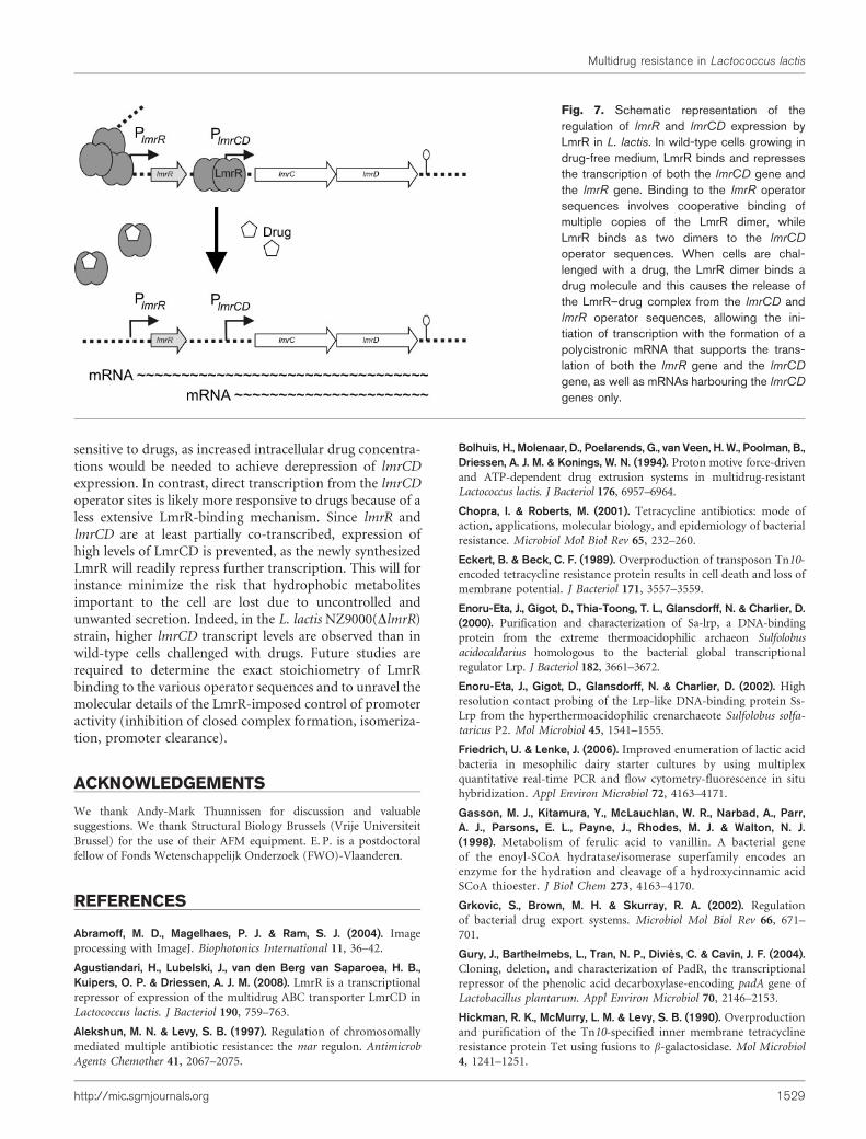
sensitive to drugs, as increased intracellular drug concentra-tions would be needed to achieve derepression of lmrCDexpression. In contrast, direct transcription from the lmrCDoperator sites is likely more responsive to drugs because of aless extensive LmrR-binding mechanism. Since lmrR andlmrCD are at least partially co-transcribed, expression ofhigh levels of LmrCD is prevented, as the newly synthesizedLmrR will readily repress further transcription. This will forinstance minimize the risk that hydrophobic metabolitesimportant to the cell are lost due to uncontrolled andunwanted secretion. Indeed, in the L. lactis NZ9000(DlmrR)strain, higher lmrCD transcript levels are observed than inwild-type cells challenged with drugs. Future studies arerequired to determine the exact stoichiometry of LmrRbinding to the various operator sequences and to unravel themolecular details of the LmrR-imposed control of promoteractivity (inhibition of closed complex formation, isomeriza-tion, promoter clearance).
ACKNOWLEDGEMENTS
We thank Andy-Mark Thunnissen for discussion and valuable
suggestions. We thank Structural Biology Brussels (Vrije Universiteit
Brussel) for the use of their AFM equipment. E. P. is a postdoctoral
fellow of Fonds Wetenschappelijk Onderzoek (FWO)-Vlaanderen.
REFERENCES
Abramoff, M. D., Magelhaes, P. J. & Ram, S. J. (2004). Image
processing with ImageJ. Biophotonics International 11, 36–42.
Agustiandari, H., Lubelski, J., van den Berg van Saparoea, H. B.,Kuipers, O. P. & Driessen, A. J. M. (2008). LmrR is a transcriptional
repressor of expression of the multidrug ABC transporter LmrCD in
Lactococcus lactis. J Bacteriol 190, 759–763.
Alekshun, M. N. & Levy, S. B. (1997). Regulation of chromosomally
mediated multiple antibiotic resistance: the mar regulon. Antimicrob
Agents Chemother 41, 2067–2075.
Bolhuis, H., Molenaar, D., Poelarends, G., van Veen, H. W., Poolman, B.,Driessen, A. J. M. & Konings, W. N. (1994). Proton motive force-driven
and ATP-dependent drug extrusion systems in multidrug-resistant
Lactococcus lactis. J Bacteriol 176, 6957–6964.
Chopra, I. & Roberts, M. (2001). Tetracycline antibiotics: mode of
action, applications, molecular biology, and epidemiology of bacterial
resistance. Microbiol Mol Biol Rev 65, 232–260.
Eckert, B. & Beck, C. F. (1989). Overproduction of transposon Tn10-
encoded tetracycline resistance protein results in cell death and loss of
membrane potential. J Bacteriol 171, 3557–3559.
Enoru-Eta, J., Gigot, D., Thia-Toong, T. L., Glansdorff, N. & Charlier, D.(2000). Purification and characterization of Sa-lrp, a DNA-binding
protein from the extreme thermoacidophilic archaeon Sulfolobus
acidocaldarius homologous to the bacterial global transcriptional
regulator Lrp. J Bacteriol 182, 3661–3672.
Enoru-Eta, J., Gigot, D., Glansdorff, N. & Charlier, D. (2002). High
resolution contact probing of the Lrp-like DNA-binding protein Ss-
Lrp from the hyperthermoacidophilic crenarchaeote Sulfolobus solfa-
taricus P2. Mol Microbiol 45, 1541–1555.
Friedrich, U. & Lenke, J. (2006). Improved enumeration of lactic acid
bacteria in mesophilic dairy starter cultures by using multiplex
quantitative real-time PCR and flow cytometry-fluorescence in situ
hybridization. Appl Environ Microbiol 72, 4163–4171.
Gasson, M. J., Kitamura, Y., McLauchlan, W. R., Narbad, A., Parr,A. J., Parsons, E. L., Payne, J., Rhodes, M. J. & Walton, N. J.(1998). Metabolism of ferulic acid to vanillin. A bacterial gene
of the enoyl-SCoA hydratase/isomerase superfamily encodes an
enzyme for the hydration and cleavage of a hydroxycinnamic acid
SCoA thioester. J Biol Chem 273, 4163–4170.
Grkovic, S., Brown, M. H. & Skurray, R. A. (2002). Regulation
of bacterial drug export systems. Microbiol Mol Biol Rev 66, 671–
701.
Gury, J., Barthelmebs, L., Tran, N. P., Divies, C. & Cavin, J. F. (2004).Cloning, deletion, and characterization of PadR, the transcriptional
repressor of the phenolic acid decarboxylase-encoding padA gene of
Lactobacillus plantarum. Appl Environ Microbiol 70, 2146–2153.
Hickman, R. K., McMurry, L. M. & Levy, S. B. (1990). Overproduction
and purification of the Tn10-specified inner membrane tetracycline
resistance protein Tet using fusions to b-galactosidase. Mol Microbiol
4, 1241–1251.
Fig. 7. Schematic representation of theregulation of lmrR and lmrCD expression byLmrR in L. lactis. In wild-type cells growing indrug-free medium, LmrR binds and repressesthe transcription of both the lmrCD gene andthe lmrR gene. Binding to the lmrR operatorsequences involves cooperative binding ofmultiple copies of the LmrR dimer, whileLmrR binds as two dimers to the lmrCD
operator sequences. When cells are chal-lenged with a drug, the LmrR dimer binds adrug molecule and this causes the release ofthe LmrR–drug complex from the lmrCD andlmrR operator sequences, allowing the ini-tiation of transcription with the formation of apolycistronic mRNA that supports the trans-lation of both the lmrR gene and the lmrCD
gene, as well as mRNAs harbouring the lmrCD
genes only.
Multidrug resistance in Lactococcus lactis
http://mic.sgmjournals.org 1529

Hochschild, A. & Ptashne, M. (1986). Cooperative binding of l
repressors to sites separated by integral turns of the DNA helix. Cell
44, 681–687.
Hsieh, P. C., Siegel, S. A., Rogers, B., Davis, D. & Lewis, K. (1998).Bacteria lacking a multidrug pump: a sensitive tool for drug discovery.Proc Natl Acad Sci U S A 95, 6602–6606.
Huillet, E., Velge, P., Vallaeys, T. & Pardon, P. (2006). LadR, a newPadR-related transcriptional regulator from Listeria monocytogenes,negatively regulates the expression of the multidrug efflux pumpMdrL. FEMS Microbiol Lett 254, 87–94.
Lubelski, J., Mazurkiewicz, P., van Merkerk, R., Konings, W. N. &Driessen, A. J. M. (2004). ydaG and ydbA of Lactococcus lactis encodea heterodimeric ATP-binding cassette-type multidrug transporter.J Biol Chem 279, 34449–34455.
Lubelski, J., de Jong, A., van Merkerk, R., Agustiandari, H., Kuipers,O. P., Kok, J. & Driessen, A. J. M. (2006). LmrCD is a major multidrugresistance transporter in Lactococcus lactis. Mol Microbiol 61, 771–781.
Ma, D., Alberti, M., Lynch, C., Nikaido, H. & Hearst, J. E. (1996). The
local repressor AcrR plays a modulating role in the regulation ofacrAB genes of Escherichia coli by global stress signals. Mol Microbiol19, 101–112.
Madoori, P. K., Agustiandari, H., Driessen, A. J. M. & Thunnissen,A. M. (2009). Structure of the transcriptional regulator LmrR and itsmechanism of multidrug recognition. EMBO J 28, 156–166.
Maxam, A. M. & Gilbert, W. (1980). Sequencing end-labeled DNA withbase-specific chemical cleavages. Methods Enzymol 65, 499–560.
Mazurkiewicz, P., Driessen, A. J. M. & Konings, W. N. (2004).Energetics of wild-type and mutant multidrug resistance secondarytransporter LmrP of Lactococcus lactis. Biochim Biophys Acta 1658,252–261.
Minh, P. N. L., Devroede, N., Massant, J., Maes, D. & Charlier, D.(2009). Insights into the architecture and stoichiometry of Escherichiacoli PepA*DNA complexes involved in transcriptional control andsite-specific DNA recombination by atomic force microscopy. NucleicAcids Res 37, 1463–1476.
Musso, R. E., Di Lauro, R., Adhya, S. & de Crombrugghe, B. (1977).Dual control for transcription of the galactose operon by cyclic AMPand its receptor protein at two interspersed promoters. Cell 12, 847–854.
Neyfakh, A. A., Bidnenko, V. E. & Chen, L. B. (1991). Efflux-mediatedmultidrug resistance in Bacillus subtilis: similarities and dissimilaritieswith the mammalian system. Proc Natl Acad Sci U S A 88, 4781–4785.
Neyfakh, A. A., Borsch, C. M. & Kaatz, G. W. (1993). Fluoroquinoloneresistance protein NorA of Staphylococcus aureus is a multidrug effluxtransporter. Antimicrob Agents Chemother 37, 128–129.
Ng, E. Y., Trucksis, M. & Hooper, D. C. (1994). Quinolone resistancemediated by norA: physiologic characterization and relationship toflqB, a quinolone resistance locus on the Staphylococcus aureuschromosome. Antimicrob Agents Chemother 38, 1345–1355.
Overhage, J., Priefert, H. & Steinbuchel, A. (1999). Biochemical andgenetic analyses of ferulic acid catabolism in Pseudomonas sp. strainHR199. Appl Environ Microbiol 65, 4837–4847.
Paulsen, I. T., Nguyen, L., Sliwinski, M. K., Rabus, R. & Saier, M. H., Jr(2000). Microbial genome analyses: comparative transport capabil-ities in eighteen prokaryotes. J Mol Biol 301, 75–100.
Peeters, E., Thia-Toong, T. L., Gigot, D., Maes, D. & Charlier, D.(2004). Ss-LrpB, a novel Lrp-like regulator of Sulfolobus solfataricusP2, binds cooperatively to three conserved targets in its own controlregion. Mol Microbiol 54, 321–336.
Peeters, E., Willaert, R., Maes, D. & Charlier, D. (2006). Ss-LrpB fromSulfolobus solfataricus condenses about 100 base pairs of its ownoperator DNA into globular nucleoprotein complexes. J Biol Chem281, 11721–11728.
Rahmati, S., Yang, S., Davidson, A. L. & Zechiedrich, E. L. (2002).Control of the AcrAB multidrug efflux pump by quorum-sensingregulator SdiA. Mol Microbiol 43, 677–685.
Rivetti, C., Guthold, M. & Bustamante, C. (1996). Scanning forcemicroscopy of DNA deposited onto mica: equilibration versus kinetictrapping studied by statistical polymer chain analysis. J Mol Biol 264,919–932.
Saier, M. H., Jr, Paulsen, I. T., Sliwinski, M. K., Pao, S. S., Skurray,R. A. & Nikaido, H. (1998). Evolutionary origins of multidrug anddrug-specific efflux pumps in bacteria. FASEB J 12, 265–274.
Schumacher, M. A. & Brennan, R. G. (2003). Deciphering themolecular basis of multidrug recognition: crystal structures of theStaphylococcus aureus multidrug binding transcription regulatorQacR. Res Microbiol 154, 69–77.
Segura, A., Bunz, P. V., D’Argenio, D. A. & Ornston, L. N. (1999).Genetic analysis of a chromosomal region containing vanA and vanB,genes required for conversion of either ferulate or vanillate toprotocatechuate in Acinetobacter. J Bacteriol 181, 3494–3504.
Tennent, J. M., Lyon, B. R., Gillespie, M. T., May, J. W. & Skurray, R. A.(1985). Cloning and expression of Staphylococcus aureus plasmid-mediated quaternary ammonium resistance in Escherichia coli.Antimicrob Agents Chemother 27, 79–83.
Zaidi, A. H., Bakkes, P. J., Lubelski, J., Agustiandari, H., Kuipers, O. P.& Driessen, A. J. M. (2008). The ABC-type multidrug resistancetransporter LmrCD is responsible for an extrusion-based mechanismof bile acid resistance in Lactococcus lactis. J Bacteriol 190, 7357–7366.
Edited by: A. R. Walmsley
H. Agustiandari and others
1530 Microbiology 157

Page 1 of 4
LmrR-mediated gene regulation of multidrug resistance in Lactococcus lactis By: Herfita Agustiandari, Eveline Peeters, Janny G. de Wit, Daniel Charlier and Arnold J. M. Driessen SUPPLEMENTARY TABLES

Page 2 of 4
Supplementary Table S1. Primer sets used for RT-PCR analysis
Primer 5′→3′ sequence
DC620r CTCCTTGTTTTAGGACATTGAGC
DC621r AAGATTGAGAATAAGGCAACCC
DC634f CGGAGATGATTTTTTCTTATCTTATATAG
DC635f CTATTGTAATCTTTAACAGCATTAAC
DC636f ATGGCAGAAATACCAAAAGAATG
CDprmf GTATTACCGACTGACAGAGATTGG

Page 3 of 4
Supplementary Table S2. Primer sets used for extended EMSA analysis
Primer 5′→3′ sequence
Region 1f CAAATAAGAAGAGTGAAGCG
Region 1r GGCAACCCATTTATGCTTCA
Region 2f ACAAATAACGTCGTAAATCG
Region 2r GGCAACCCATTTATGCTTCA
Region 3f ATTGTAATCTTTAACAGCATTAAC
Region 3r GGCAACCCATTTATGCTTCA
Region 4f TTCTCAAAAAATTTATTGAAATTA
Region 4r GGCAACCCATTTATGCTTCA
Region 5f CAAATAAGAAGAGTGAAGCG
Region 5r AAATTTTTTGAGAAGATAAT
Region 6f CAAATAAGAAGAGTGAAGCG
Region 6r GCATTAACAATTAATGCTTGTTTACT
Region 7f CAAATAAGAAGAGTGAAGCG
Region 7r GTTTACCATTTATGAAACTAACTATTG
Region 8f CAAATAAGAAGAGTGAAGCG
Region 8r CGTTGACTTAAACTTTAAAAAG

Page 4 of 4
Supplementary Table S3. Primer sets used for qPCR analysis
Primer 5′→3′ sequence Length GC content (%)
TufAf TGACGAAATCGAACGTGGTCAAG 23 47
TufAr GTCACCAGGCATTACCATTTCAG 23 47
SecYf GCTTGCTATGGCACAATCTATCG 23 47
SecYr ATGGCTGATGGAATACCAGAGAC 23 47
LmrRf ATGTTACGAGCCCAAACCAATG 22 45
LmrRr TCTGTCAGTCGGTAATACTTGC 22 45
LmrR-Cf GTATTACCGACTGACAGAGATTG 23 43
LmrR-Cr GTTTAAGTCAACGATTTACGACG 23 39
LmrCf GCGAAAGACGAAGAACTTTCTGG 23 47
LmrCr ACTGAAACAGTCCCTTCTGTTGG 23 47
LmrDf CGAAAGCTTGCCTGACAAGTATG 23 47
LmrDr CGAATGAAGTTCGTCCAGCAATG 23 47

LmrR-mediated gene regulation of multidrug resistance in Lactococcus lactis By: Herfita Agustiandari, Eveline Peeters, Janny G. de Wit, Daniel Charlier and Arnold J. M. Driessen SUPPLEMENTARY FIGURE LEGEND
Supplementary Fig. S1. EMSA of the binding of LmrR to truncated DNA fragments
corresponding to different parts of the lmrCD control region. The concentration of
wild-type LmrR was kept constant (1.85 µM dimer) in all lanes. The −35 and −10
boxes shown belong to the promoter of transcript B. Double-stranded DNA fragments
(○) were obtained by PCR using the primers indicated in Supplementary Table S2.
The shifted DNA is indicated by ●.

LmrR-mediated gene regulation of multidrug resistance in Lactococcus lactis By: Herfita Agustiandari, Eveline Peeters, Janny G. de Wit, Daniel Charlier and Arnold J. M. Driessen Supplementary Fig. S1