university of groningen flexibility of human walking ... · chapter 2 coordination between arm and...
TRANSCRIPT

University of Groningen
Flexibility of human walkingDonker, Stella Franciscus
IMPORTANT NOTE: You are advised to consult the publisher's version (publisher's PDF) if you wish to cite fromit. Please check the document version below.
Document VersionPublisher's PDF, also known as Version of record
Publication date:2002
Link to publication in University of Groningen/UMCG research database
Citation for published version (APA):Donker, S. F. (2002). Flexibility of human walking: a study on interlimb coordination Groningen: s.n.
CopyrightOther than for strictly personal use, it is not permitted to download or to forward/distribute the text or part of it without the consent of theauthor(s) and/or copyright holder(s), unless the work is under an open content license (like Creative Commons).
Take-down policyIf you believe that this document breaches copyright please contact us providing details, and we will remove access to the work immediatelyand investigate your claim.
Downloaded from the University of Groningen/UMCG research database (Pure): http://www.rug.nl/research/portal. For technical reasons thenumber of authors shown on this cover page is limited to 10 maximum.
Download date: 17-06-2018
Chapter 2
Coordination between arm and leg movements during locomotion
Previous studies examining the effect of walking velocity on the coordination between arm and leg movements have reported the occurrence of 2:1 frequency coordination at low walking velocities and of 1:1 frequency coordination at higher walking velocities. On the one hand, this phenomenon has been interpreted from a biomechanical perspective in terms of the tendency of the arms to move at, or close to, their resonance frequency. On the other hand, it has been interpreted from a dynamical systems perspective in terms of two stable regimes of frequency and phase locking between arm and leg movements, and in terms of velocity-induced phase transitions between these regimes due to loss of stability of the alternate coordination mode. To evaluate these contrasting interpretations, we conducted an experiment to quantify the effect of walking velocity on the stability of the frequency and phase coordination between the individual limb movements. Spectral analyses revealed the presence of 2:1 frequency coordination between arm and leg movements as a consistent feature of the data in only 3 out of 8 participants at walking velocities ranging from 1.0 to 2.0 km/h, in spite of the fact that the eigenfrequencies of the arms were rather similar across participants. The degree of interlimb coupling, as indexed by weighted coherence and variability of relative phase, was lower for the arm movements and for ipsilateral and diagonal combinations of arm and leg movements than for the leg movements. Furthermore, the coupling within all pairs of limb movements was found to increase with walking velocity, whereas no clear signs were observed that the switches from 2:1 to 1:1 frequency coordination and vice versa were preceded by loss of stability. Therefore, neither a purely biomechanical nor a purely dynamical model is optimally suited to explain these results. Instead, an integrative model involving elements of both approaches seems to be required.
Stella Donker, Peter Beek, Robert Wagenaar, & Theo Mulder Journal of Motor Behavior, 33, 86-103, 2001

22 Chapter 2
Introduction Human walking is a complex activity involving a large number of cyclically moving components, that is, legs, pelvis, trunk, arms, and head. Recently, the study of human walking has clearly gone beyond simple analyses of leg movements in terms of the absolute and relative timing of the various phases of Phillipson’s step cycle (cf. Shapiro, Zernicke, Gregor, & Diestel, 1981), indirect coordination measures such as bipedal contact time (Marey, 1874; Rozendal, 1968), and the kinetics of the muscles and joints in the legs (Vaughan, Davis, & O'Connor, 1992). It is now more and more recognized that, in order to understand the dynamics of walking, one cannot restrict the system under study to an isolated pair of walking legs; head, arm, and trunk movements should also be included. A rapidly increasing number of studies testify to this development (e.g., Capozzo, 1982; Clark & Phillips, 1993; Elftman, 1939; Ledebt, Bril, & Wiener-Vacher, 1995; Murray, Sepic, Gardner, & Downs, 1978; Nashner & Forssberg, 1986; Thorstensson, Nilsson, Carlson, & Zomlefer, 1984; Van Emmerik & Wagenaar, 1996a, 1996b; Wagenaar & Beek, 1992; Wagenaar & Van Emmerik, 1994; Warren, Kay, & Yilmaz, 1996).1
In the present article, we focus on a very basic and common property of human walking, namely, the coordination between arm and leg movements. Our specific interest is in the effect of walking velocity on interlimb coordination. This interest was motivated by current theoretical considera-tions and developments, although potential practical implications are recognized. For instance, in the field of applied movement science, walking velocity has been postulated to be a relevant (control) parameter for inducing or promoting desired coordination patterns and avoiding undesired coordination patterns (cf. Van Emmerik, Wagenaar, & Wolters, 1993). Such a theoretically motivated application, however, requires thorough empirical knowledge of the effects of velocity on the dynamics of walking. Movement velocity is an important parameter that plays an essential role in various models of rhythmically coordinated movements. In the literature, the effects of movement velocity on interlimb coordination have been studied from two contrasting theoretical approaches, the biomechanical approach and the dynamical systems approach. We briefly discuss each approach and, in doing so, explain our motivation for conducting the present study.
In the biomechanical approach to movement coordination, an under-standing of the patterns of coordinated movements is sought in terms of the underlying forces and moments that give rise to those patterns. A

Chapter 2 23
commonly applied strategy in studying rhythmic movements from this perspective is to treat the moving limbs as force-driven pendulums and to explain the observed movement patterns in terms of the physical properties of the pendulums and their muscular forcing (cf. Holt, Hamill, & Andres, 1990; Schot & Decker, 1998). An example of such an approach is the study of Webb, Tuttle, and Baksh (1994) on the pendular activity of the upper limbs during slow and normal walking. They started from the observation (cf. Craik, Herman, & Finley, 1976; Webb & Tuttle, 1989) that at customary walking velocities the upper limbs swing in alternation, with each limb swinging forward and backward in phase with the diagonal lower limb, whereas at lower walking velocities the upper limbs swing in phase at a frequency twice as high as the stride frequency of the legs. Those studies advanced the hypothesis that the stride frequency at which “single swinging” changes to “double swinging” occurs at or slightly below the natural pendular frequency of the upper limbs. Webb et al. (1994) explained the occurrence of the observed coordination modes (i.e., single swinging or double swinging) in terms of the biomechanical properties of one of the two components (legs and arms) participating in the coordination. This explanation, however, is somewhat unsatisfactory in that it explains why the arms show a tendency to move at a certain frequency but not why particular, functionally stable forms of coordination between the movements of the arms and the legs are preferred. This restriction may reflect a general limitation of the biomechanical approach to understanding coordinative phenomena. Biomechanical properties play a role in coordinative phenomena, but coordinative structures are functional units of organization that are based foremost on informational rather than on biomechanical principles. As Kelso (1994) pointed out, biomechanical properties may shape the dynamical properties of coordination but they do not determine them. On the other hand, the degree to which the biome-chanical properties of the effector system shape the coordination dynamics may vary from task to task. Evidently, biomechanical principles are likely to play a more prominent role in walking than in the coordination of rhythmic finger movements. Therefore, in studying human locomotion, it will be particularly useful to consider, and if possible to explicitly model, the biomechanical constraints in conjunction with the informational constraints. The expediency of such an approach was aptly argued and demonstrated by Taga (1994). Taga modeled the control of walking as a form of self-organization, in which a rhythmic spatiotemporal pattern is generated through the multilink dynamics of the musculoskeletal system and the nonlinear dynamics of the (central) nervous system, conceived as a

24 Chapter 2
collection of neural oscillators or central pattern generators. Contrary to the biomechanical approach, the main focus in the dynami-
cal systems approach is on the stability properties of spatiotemporal patterns of bodily parts coordinated with each other and the environment (see, e.g., Beek, Peper, & Stegeman, 1995; Kelso, 1995; Schöner & Kelso, 1988). Qualitative changes or “phase transitions” are a special entry point in studying the stability properties of coordination patterns because they are associated with an enormous reduction of information, allowing for the identification of the relevant collective variables or order parameters for describing the coordination patterns over which the transition is defined (Haken, 1977, 1983). The core of the approach as it has evolved to date is formed by a large number of studies on rhythmically coordinated movements, which build on Kelso’s (1981, 1984), by now paradigmatic, experiment on frequency-induced transitions from antiphase to in-phase coordination in rhythmic finger movements and the so-called Haken, Kelso, and Bunz (1985), or HKB, model for this phenomenon. In the HKB model, Haken and his colleagues capitalized on the mathematical theory of nonlinear oscillations by modeling the rhythmic finger movements in terms of nonlinearly coupled nonlinear oscillators. A key feature of this model is the decrease of the interaction or coupling strength between the fingers with increasing frequency, resulting in loss of stability of the antiphase coordination at a particular frequency. The purpose of this kind of nonlinear oscillator models is to find general formal analogies for complex dynamical phenomena that occur quite generally in biological systems displaying oscillatory behavior, no matter their precise material composition or instantiation. Thus, instead of analyzing highly task-dependent (neuro-) physiological mechanisms and biomechanical properties, in the dynamical systems approach, one seeks to identify general, dynamical principles of coordination in the form of frequency and phase attraction, nonequilibrium phase transitions, phase wandering, chaos, and the like.
Thus, in contrast to the biomechanical approach, the thrust of the dynamical systems approach is to understand (rhythmic interlimb) coordination as a form of dynamic pattern formation or self-organization, involving the emergence and annihilation of stable spatiotemporal patterns that can be described mathematically as attractors. According to this perspective, single swinging and double swinging are viewed as coordinative patterns resulting from frequency and phase locking. The switch from single swinging to double swinging in the coordination between arm and leg movements, and vice versa, is viewed as a nonequi-librium phase transition between these two regimes of frequency and phase
Chapter 2 25
locking (Van Emmerik & Wagenaar, 1996b; Wagenaar & Van Emmerik, 1994). According to this interpretation, the behavioral switch in question should be characterized by loss of stability of the frequency- and phase-locking regime prior to the switch and the subsequent entering of a stable frequency- and phase-locking regime after the switch. Particularly, for the switching behavior of interest to constitute a genuine phase transition, one would expect to observe “catastrophe flags” such as hysteresis, that is, the occurrence of jumps at distinct values of the control parameter as a function of the history of the system, and critical fluctuations, that is, enhanced vari-ability resulting from loss of stability of a previously stable mode (cf. Gilmore, 1981).
So far, the stability properties of the coordinative modes between arm and leg movements during walking referred to as single swinging and double swinging have been studied and analyzed in some detail, but conclusive evidence for the occurrence of frequency and phase locking and for the presence or absence of phase transitions has not yet been obtained. Wagenaar and Van Emmerik (1994, 2000) reported evidence for the presence of 2:1 and 1:1 frequency coordination between arm and leg movements during walking at low and customary velocities, respectively, by showing that the frequency ratios in question were close to 2 and 1. The velocity-dependent effects on the stability of the observed coordination patterns was assessed only in terms of the variability of relative phase (which is a valid index of coordinative stability only if relative phase is a point attractor), however, and not in terms of spectral analyses addressing the stability of the frequency coordination. Using variability of relative phase, they found no convincing evidence for the theoretical interpretation that the switches from single swinging to double swinging and vice versa constitute genuine phase transitions in that they are characterized by catastrophe flags such as hysteresis and critical fluctuations (cf. Gilmore, 1981). Although Wagenaar and Beek (1992) reported a hysteresis effect for stride frequency, and Wagenaar and Van Emmerik (2000) observed hints of increased fluctuations in the relative phasing between the arms at intermediate velocities (0.7-0.9 m/s), no hysteresis and critical fluctuations effects have been reported to date for the coordination between arm and leg movements. All in all, the present state of affairs justifies further experimental and numerical analyses of this type of coordination.
Thus, we conducted the present experiment to examine in detail the coordination between arm and leg movements by using a combination of methods to study the observed frequency and phase relations. Our specific aims in the present study were the following:

26 Chapter 2
1. (a) To replicate the basic findings of Craik et al. (1976), Webb et al. (1994), and Wagenaar and Van Emmerik (1994, 2000) regarding the two forms of frequency coordination between arm and leg movements, that is, 2:1 at low walking velocities and 1:1 at customary walking velocities; and (b) to determine whether those frequency relations are instances of frequency locking.
2. To examine the extent to which the observed coordination patterns and switches therein might be related to the eigenfrequencies of the arms.
3. To analyze in detail the relative phasing between arm and leg movements and to examine the possibility of phase locking and its quantitative properties.
4. To establish the effect of walking velocity on the degree of (frequency and phase) coupling between arm and leg movements as compared to other limb combinations.
5. To determine whether qualitative changes in the coordination between arm and leg movements are characterized by hysteresis and critical fluctuations. The findings with regard to these goals allow one to evaluate the relative merits of the current biomechanical (Aims 1 and 2) and dynamical (Aims 1, 3, 4 and 5) accounts of the effect of walking velocity on the coordination between arm and leg movements.
Method Participants Three women and 5 men (mean age = 29 years; range = 24-51 years) participated in the experiment. All participants were naive with regard to our purposes in the experiment. All participants gave their written informed consent and the study was approved by the local Medical Ethics Committee.
Materials The participants walked on a walking belt (Enraf Nonius, model Entred Reha; Bonto BV, Zwolle, The Netherlands) with a computer-controlled velocity (the time to change the velocity by 0.5 km/h was about 1.5 s). Small lightweight triangular frames with a reflective spherical marker (diameter 15 mm) on each corner were attached to the upper arms, forearms, upper legs and lower legs by means of Velcro strips, as shown in Figure 1A. Two additional markers were placed on the heels. The positions of the 26 markers were recorded at a sampling rate of 100 Hz by means of a

Chapter 2 27
three-dimensional (3D) passive registration system called PRIMAS (Furnée, 1989). Four video cameras were placed around the walking belt, two on each side so that at all times the markers were in view of at least two cameras, as was required for the successful 3D reconstruction of their position. The 3D reconstruction error was about 1 mm. Procedure and Design Before each experimental session, participants were acquainted with walking on a walking belt at different velocities. This familiarization process took about 5 minutes. Subsequently, a reference measurement was taken during which the positions of the marker frames in space were recorded for future reference. Participants walked barefooted on the belt and were instructed to walk as naturally as possible. No specific instructions regarding the movements of the arms were given. Each participant took part in a single measurement session, consisting of one large trial. During this trial, walking velocity was gradually increased in steps of 0.5 km/h from 1.0 km/h up to 4.0 km/h and subsequently decreased from 4.0 km/h to 1.0 km/h,
lower arm
Y
upper leg
upper arm
heel
marker frame
lower leg
A.
Z X
B.
Figure 1. (A) Sagittal view of the marker configuration (black circles).Marker frames (each holding three markers) were placed on the upperarms, forearms, upper legs and lower legs. A single marker was attached on each of the heels. (B) The coordinate axes x, y, and z. The sagittal plane was defined by the xy plane. In this study, only the rotational movements (in degrees) of the limbs around the z-axis (i.e., the rotational movements in the sagittal plane, represented by white the arrow aroundthe z-axis in the figure) are discussed.

28 Chapter 2
yielding two observations at the walking velocities 1.0-3.5 km/h and a single observation for 4.0 km/h. About 10 s after changing the velocity of the walking belt, the position data of the markers were recorded for 15 stride cycles. Consequently, the recording time varied from 40 s at the lowest velocity to 30 s at the highest velocity. A session (i.e., recording the entire trial) took about 20 min. At the end of the session, anthropometric measures were taken of the arms of the participant while he or she stood in the anatomical position. Those measures included (a) upper arm length (acromion to epicondylus lateralis), (b) lower arm length (epicondylus lateralis to processus styloideus ulnae), and (c) hand length (processus styloideus ulnae to the tip of the middle finger). In addition, body height and body weight were measured.
Data Collection and Compression Initial processing. From the reference measures, a 3D coordinate system (x-, y-, and z-axis) was defined that coincided with the sagittal, frontal, and transverse plane for each participant as depicted in Figure 1B (cf. Veldpaus, Woltring, & Dortmans, 1988). We calculated and then filtered the rotation (in degrees) of the marker frames around the three axes by means of a lowpass linear phase FIR filter, cut-off frequency 5 Hz (which eliminated the noise from the data while leaving the characteristic frequencies of the movement, which were all lower than 2 Hz, unaffected). The analyses were restricted to the rotational movements of the lower legs and the forearms in the sagittal plane (i.e., rotation around the z-axis).2 To center the time series around the origin of the z-axis, we subtracted the mean rotation of each time series in this coordinate from the position signal at each sample. Thus, the movements of the arms and the legs were expressed as rotational movements (in degrees) around the z-axis.
Spectral analysis. To determine whether different frequency-locked patterns of coordination were present between the arm and leg movements, we analyzed the collected data in three steps. First, we determined the dominant frequency of each limb movement by applying a Fast Fourier Transform (FFT) algorithm to the position data. On the basis of the output of the FFT, we defined the dominant frequency of the limb movement as the frequency at which the largest peak was present in the power spectrum. This peak had to be larger than the mean power plus twice its standard deviation. Second, we calculated the ratio between the dominant frequencies for the arm and leg movements at each side of the body. Similarly, we calculated the ratio between the dominant frequency for the arm movement and the second significant frequency (first super harmonic)

Chapter 2 29
of the leg movement. Third, we established whether the calculated frequency ratios were at or near the integers 1 or 2. Given the accuracy of estimating the dominant frequencies of the individual limb movements, and in view of the theoretical consideration that 1:1 frequency locking is typically more stable than 2:1 frequency locking (e.g., Serrien & Swinnen, 1997), a tolerance region of 10% was accepted for frequency locks around 1 and a tolerance region of 5% for frequency locks around 2 (i.e., 1 ± 0.1 and 2 ± 0.1). (A sharper criterion was used for the less stable frequency mode because the smaller the region of parametric resonance the greater the probability that the measured value in fact belongs to a neighboring region of parametric resonance of higher order, e.g., 2:3 is closer to 1:2 than to 1:1; for details see Treffner & Turvey, 1993).
In addition to the FFT of the time series of the limb movements, we correlated in pairs (i.e., right leg/left leg, left arm/right arm, right arm/right leg, left arm/left leg, right arm/left leg and left arm/right leg) the power spectra of the individual limb movements by using a special kind of cross-spectral analysis, called weighted coherence (Porges & Bohrer, 1980). This analysis was performed to evaluate and compare the shared rhythmicity (i.e., the degree of coupling) between pairs of limb movements and to assess the effect of walking velocity in that regard. First, the coherence, Cxy, between the power spectra of two signals x and y was calculated as follows:
where Pxy(ω) is the cross-spectral density of signal x and y at frequency ω and Pxx(ω) and Pyy(ω) are the power spectral density of signal x and y at frequency ω. In the case of nonhomologous limb pairs, the leg movement was defined as signal x, and in the case of homologous limb pairs the left limb was defined as signal x. The weighted coherence or Cw was then calculated across the frequency band of 0.1 – 2.0 Hz,3 according to the following formula:
.)(
)()(
0.2
1.0
0.2
1.0
∑
∑
=ω
=ω
ω
ω⋅ω=
Pxx
PxxCxyCw
For each participant, the weighted coherence was calculated for all six limb pairs and for each walking velocity.
,)()(
)()(
2
ω⋅ωω
=ωPyyPxx
PxyCxy

30 Chapter 2
Eigenfrequency. The eigenfrequencies of the arms were estimated according to the commonly used pendulum equation for animal locomotion (Holt et al., 1990; Holt, Jeng, Ratcliffe, & Hamill, 1995; Kugler & Turvey, 1987; Wagenaar & Van Emmerik, 2000):
where ω0 = eigenfrequency, π = 3.1416, L = simple pendulum equivalent length, and g = 9.81 m/s2. The simple pendulum equivalent length (L) was calculated from the three segments of the arm (upper arm, lower arm, and hand), according to the following formula: L = I / (M ·D), where I = moment of inertia, M = mass, D = distance from acromion to the center of mass of the arm. Moments of inertia were estimated from anthropomorphic data (Winter, 1990).
Point estimates of relative phase. For the 1:1 frequency coordination,
point estimates of relative phase were calculated using the moments (tlimbx
and tlimby) at which the positive maxima were reached in the angular position data of the limbs:
t t
Figure 2. Example of an experimental trial in which the frequency of the arm movements (upper trace) was twice as high as that of the leg movements (lower trace). For such trials, the relative phase was calculated on the basis of the first maxima of the arm movement subsequent to the maxima of the leg movement. See text for abbreviations.
rota
tion
arou
nd z-
axis
time
Arm movement
Leg movement
limbx(i) limbx(i+1)
t limby(i) t limby(i+1)
,
22
10
gLπ
=ω

Chapter 2 31
,360)(limbx)1(limbx
)(limbx)(limby ⋅−
−=
+ ii
ii
tttt
where limby and limbx were the right and left limbs, respectively, when calculating the point estimate of relative phase between homologous limb movements. In the case of nonhomologous limb pairs, limby and limbx were the arm and leg movements, respectively. In the case of 2:1 frequency coordination, the maximum of the arm movement subsequent to the maximum of the leg movement was used for calculating the relative phase and the second maximum was ignored (see Figure 2 for an example).
We assessed the stability of the coordination between the limbs by calculating the standard deviation of the mean relative phase (SDφ) for all six pairs of limb movements for each participant and for each walking velocity. We calculated means and standard deviations of relative phases by means of directional (or circular) statistics to avoid artifacts caused by phase wrapping (Batchelet, 1981; Burgess-Limerick, Abernethy, & Neal, 1991).
Continuous estimates of relative phase. To examine the continuous aspects of the phase progression of the individual limb movements, we differentiated the angular position time series and projected the angular position time series and the calculated angular velocity time series in the phase plane to obtain the phase angle ϕ(i). After normalization of the angular position and velocity time series to their respective maxima and minima, the phase angles were calculated as follows:
]./)/[(tan 1
)( xdtdxi−=ϕ
We determined discrete values of the continuous estimate of relative phase at the peaks (moment of maximum forward extension of the limb) and valleys (moment of maximal backward extension of the limb) of the time series by subtracting the phase angles of two limb movements, as follows:
,: FSmn mn ϕ−ϕ=θ
where n:m represent the found dominant frequency ratios (1:1 or 2:1). The phase angle of the slower limb, ϕS, was multiplied by n (1 or 2) and the phase angle of the faster limb, ϕF, was multiplied with m (i.e., 1). Hence, we generalized the relative phase to n:m rhythm. In line with the procedure of the point estimate of relative phase, the phase of the arm movement was subtracted from the phase of the leg movement, or, in the case of homologous limb pairs, the phase of the left limb movement was subtracted
φ(i)

32 Chapter 2
from the phase of the right limb movement. Statistical analysis. Using SPSS software, we performed separate
analyses of variance (ANOVAs) with repeated measures on the dependent measures mentioned in the preceding sections with various factorial designs involving the within-subjects factors velocity (6 levels), direction (2 levels [up, i.e., velocity level 1-7, and down, i.e., velocity level 8-13]) and limb pair (6 levels). To evaluate the contrast between the coordination patterns adopted at the lower walking velocities and those adopted at the higher walking velocities, we conducted additional ANOVAs with the factors direction (2 levels) and velocity (2 levels). The factor velocity was reduced to two levels, that is, low walking velocities (the data from 1.0, 1.5 and 2.0 km/h were collapsed) and customary walking velocities (the data from 2.5, 3.0 and 3.5 km/h were collapsed).
Results The results are presented according to the five goals listed in the introduction. First, we analyzed the observed coordination patterns between arm and leg movements in terms of the ratios between their primary frequencies and locking across a band of frequencies (Goal 1). Second, we examined the relation between the eigenfrequencies of the arms and the observed frequency ratios and switches in coordination (Goal 2). Third, we analyzed the relative phasing and possible phase locking between arm and leg movements (Goal 3). Subsequently, the effect of walking velocity on the coupling between arm and leg movements and other limb combinations was examined (Goal 4). Finally, we determined whether the observed switches in the coordination patterns between arm and leg movements were characterized by hysteresis and critical fluctuations (Goal 5). 1. Frequency Coordination In Figure 3 we show, for 1 of the participants, the position time series of ipsilateral pairs of arm and leg movements and the corresponding power spectra at all velocity levels. The power spectra reveal two characteristic properties that were found for all participants: the presence of two significant peaks for the legs (broken line) in the frequency range 0.1-2.0 Hz, and the appearance and disappearance of significant peaks for the arms (solid line) as a function of walking velocity. The dominant peak in the power spectra of the leg movements represents the characteristic frequency

Chapter 2 33
of the leg movement, whereas the second significant peak represents the frequency of knee flexion during the stance phase. In the example shown in
Figure 3. Representative arm and leg movements of 1 of the participants.For each velocity level, the rotational movements in the sagittal plane ofthe arm (solid line) and the ipsilateral leg (broken line) are shown. Foreach limb movement the corresponding power spectra of the frequencyrange 0.1-2.0 Hz are shown. The (first) two peaks in all the power spectraof the leg movements were significant. Significant peaks in the powerspectra of the arm movements are marked with asterisks. Note that the power is plotted on a logarithmic scale.
→→ →→sa
gitt
al r
otat
ion
→→ →→
time(s)→→→→ →→ →→
0.1Hz
→→ →→
2.0Hz
Ve locity Ve locity leve l km/h
1 1.0
2 1.5
3 2.0
4 2.5
5 3.0
6 3.5
7 4 .0
8 3 .5
9 3 .0
10 2.5
11 2.0
12 1.5
13 1.0
Move me nt Power Spe ctra
pow
er
*
*
*
*
*
* *
*
* *
* *
*
*
*
* *
*

34 Chapter 2
Figure 3, only one significant peak is observed in the power spectra of the arm movement at the walking velocities 1.0-2.0 km/h up (i.e., Levels 1-3). This peak coincides with the second (significant) peak in the power spectra of the leg movement. At the walking velocity of 2.5 km/h up (i.e., Level 4), a second peak at approximately 0.6 Hz can be distinguished as becoming the dominant frequency. Now the dominant arm frequency has locked onto the dominant frequency of the leg movement. At the walking velocity of 2.5 km/h down (i.e., Level 10), the peak at the higher frequency (i.e., 1.2 Hz) again becomes the dominant frequency of the arm movement. The arm movements depicted in Figure 3 at 2.5 and 3.0 km/h up and down, and 1.5 km/h down (i.e., Levels 4, 5, 9, 10 and 12) exhibit the frequencies which the arm adopted during low walking velocities but also frequencies which the arm adopted at higher walking velocities. Because, in those particular velocity conditions, both frequencies were present as significant components of the spectrum, 1:1 and 2:1 frequency locking between arm and leg movements may be viewed as competing modes of coordination. This kind of competition existed not only at the level of the coordination as a whole but also between the two sides of the body within a participant. This was evidenced by the fact that in 5 of the 8 participants the coordination between arm and leg movements was asymmetrical in that the frequency ratio of the dominant frequencies of the left ipsilateral limb pair differed from the ratio of the right ipsilateral limb pair (Table 1).
In Table 1, the frequency ratios of the dominant frequencies of ipsilateral limb movements are presented for each participant. One-to-one frequency coordination was observed in all 8 participants at walking velocities ranging from 2.5 to 4 km/h, whereas 2:1 frequency coordination was observed as a consistent feature of the data in 3 out of 8 participants at walking velocities ranging from 1.0 to 2.0 km/h. There were no participants in whom the 2:1 frequency coordination was entirely absent, although in 2 participants it was observed only in one ipsilateral limb pair (i.e., left arm/left leg or right arm/right leg) at the lowest walking velocity of 1.0 km/h. The fact that the ratios never deviated more than one decimal place from the integers 1 and 2 suggests that all limb pairs were frequency locked at all walking velocities. Relatedly, as shown in Table 2, the frequency ratios of the dominant frequency of the arm movement and the first harmonic frequency of the leg movement never deviated more than one decimal place from the integers 1 and 2, except in one case. This indicates that the arm movements were also frequency locked with the frequency of knee flexion at all walking velocities. Note that a 2:1 frequency relation (Table 1) corresponded with a 1:1 frequency relation between the dominant

Chapter 2 35
frequency of the arm movement and the second frequency of the leg movement (knee flexion; Table 2). Likewise, a 1:1 frequency relation (Table 1) corresponded with a 2:1 relation between the dominant frequency of the arm movement and the second frequency of the leg movement (Table 2). Further examination of the ratios indicated that in the case of 2:1 frequency
Walking velocity (km/h)
1.0 1.5 2.0 2.5 3.0 3.5 4.0 3.5 3.0 2.5 2.0 1.5 1.0 Frequency ratios Participant 1
LA/LL 2.1 2.0 2.1 1.0 1.0 1.0 1.0 1.0 1.0 1.0 1.9 1.9 1.9 RA/RL 2.1 2.0 2.1 1.0 1.0 1.0 1.0 1.0 1.0 1.0 1.9 1.9 1.9
Participant 2
LA/LL 2.0 1.0 1.0 1.0 1.0 1.0 1.0 1.0 1.0 1.0 1.0 1.0 2.1 RA/RL 1.0 1.0 1.0 1.0 1.0 1.0 1.0 1.0 1.0 1.0 1.0 1.0 0.9
Participant 3
LA/LL 2.1 2.0 1.9 1.1 1.0 1.0 1.0 1.0 1.0 1.0 2.1 2.1 1.9 RA/RL 2.1 2.0 1.9 1.9 1.0 1.0 1.0 1.0 0.9 1.0 2.1 2.1 1.9
Participant 4
LA/LL 2.0 1.0 1.0 1.0 1.0 1.0 1.0 1.0 1.0 1.0 1.0 1.0 1.0 RA/RL 2.0 0.9 1.0 1.0 1.0 1.0 1.0 1.0 1.0 1.0 1.0 1.0 2.0
Participant 5
LA/LL 1.1 1.0 1.0 1.0 1.0 1.0 1.0 1.0 1.0 1.0 0.9 1.0 2.1 RA/RL 1.1 1.0 1.0 1.0 1.0 1.0 1.0 1.0 1.0 1.0 1.0 1.0 1.0
Participant 6
LA/LL 1.9 2.0 2.1 1.0 1.0 1.0 1.0 1.0 1.0 1.0 1.0 2.0 2.1 RA/RL 2.0 2.0 2.0 1.0 1.0 1.0 1.0 1.0 1.0 1.0 1.0 2.0 2.1
Participant 7
LA/LL 2.1 1.0 1.0 1.0 1.0 1.0 1.0 1.0 1.0 1.0 0.9 1.1 2.0 RA/RL 1.0 1.0 1.0 1.0 1.0 1.0 1.0 1.0 1.0 1.0 1.0 1.0 1.0
Participant 8
LA/LL 2.0 2.1 1.0 1.0 1.0 1.0 1.0 1.0 1.0 1.0 1.0 1.0 1.0 RA/RL 2.0 2.1 0.9 1.0 1.0 0.9 1.0 1.0 1.0 1.0 1.0 1.0 2.1
Table 1 Frequency ratios of the dominant frequencies of the ipsilateral arm and
leg movements as a function of walking velocity
Note. The dominant frequency of the arm movement is divided by the dominant frequency ofthe leg movement for all 8 participants. LA = left arm, RA = right arm, LL = left leg, and RL= right leg.

36 Chapter 2
coordination, the arm frequency was less strongly locked to the dominant frequency of the leg (31 out of 48 ratios deviated from an integer value; see Table 1) than to the frequency of knee flexion (7 out of 48 ratios deviated from an integer value; see Table 2). In contrast, in the case of 1:1 frequency coordination, the arm frequency appeared to be more strongly locked to the dominant frequency of the leg movements (11 out of 160 ratios deviated
Walking velocity (km/h)
1.0 1.5 2.0 2.5 3.0 3.5 4.0 3.5 3.0 2.5 2.0 1.5 1.0 Frequency ratios Participant 1
LA/LL 1.0 1.0 1.0 1.9 2.0 2.1 2.0 1.9 2.0 1.9 1.0 1.0 1.0 RA/RL 1.0 1.0 1.0 2.0 2.0 2.1 2.0 1.9 2.0 1.9 1.0 1.0 1.0
Participant 2
LA/LL 1.1 2.1 2.1 2.1 2.0 2.0 2.1 2.0 1.9 1.9 2.1 2.0 1.0 RA/RL 1.9 2.1 2.1 2.1 2.0 2.0 2.1 2.0 1.9 1.9 2.1 2.0 2.1
Participant 3
LA/LL 1.0 1.0 1.0 2.1 2.0 2.0 2.1 2.0 2.1 2.0 1.0 1.0 0.9 RA/RL 1.0 1.0 1.0 1.0 2.0 2.0 2.1 2.1 2.1 2.0 1.0 1.0 0.9
Participant 4
LA/LL 1.1 1.9 2.1 2.0 2.0 2.0 2.1 2.0 2.0 2.0 2.1 2.0 2.0 RA/RL 1.1 1.9 2.1 2.0 2.0 2.0 2.1 2.0 2.0 2.0 2.1 2.0 1.0
Participant 5
LA/LL 2.1 2.0 2.0 2.0 2.1 2.1 2.0 2.0 2.0 2.1 1.9 1.9 1.0 RA/RL 2.1 2.0 2.0 2.0 2.1 2.1 2.0 2.0 2.0 2.0 1.9 2.1 2.0
Participant 6
LA/LL 1.0 1.0 1.0 2.0 1.9 2.0 1.9 2.1 2.1 2.0 2.0 1.0 1.0 RA/RL 1.0 1.0 1.0 2.0 2.1 2.0 1.9 2.1 1.9 2.0 2.0 1.0 1.0
Participant 7
LA/LL 1.1 2.1 1.9 2.0 2.0 1.9 2.0 2.1 2.0 2.0 2.1 2.1 1.0 RA/RL 1.9 2.1 1.9 2.0 2.0 1.9 2.0 2.1 2.0 2.0 2.1 1.9 2.0
Participant 8
LA/LL 1.0 1.4 1.9 2.0 2.0 2.1 2.1 1.9 1.9 1.9 2.0 2.1 2.1 RA/RL 1.0 1.0 1.9 2.0 2.0 2.1 2.1 1.9 1.9 1.9 2.0 2.1 1.0
Table 2 Frequency ratios of the dominant frequencies of the arm and the second
frequency of the ipsilateral leg movements as a function of walking velocity
Note. The second significant frequency of the leg movement was divided by the dominantfrequency of the arm movement for all 8 participants as function of the walking velocity. LA = left arm, RA = right arm, LL = left leg, and RL = right leg.
Chapter 2 37
from an integer value; see Table 1) than to the frequency of knee flexion (80 out of 160 ratios deviated from an integer value; see Table 2). Note that because the velocities at which a 2:1 frequency relation changed to a 1:1 frequency relation, and vice versa, varied considerably across participants, normalization to walking velocity was not adequate for other analyses in this study.
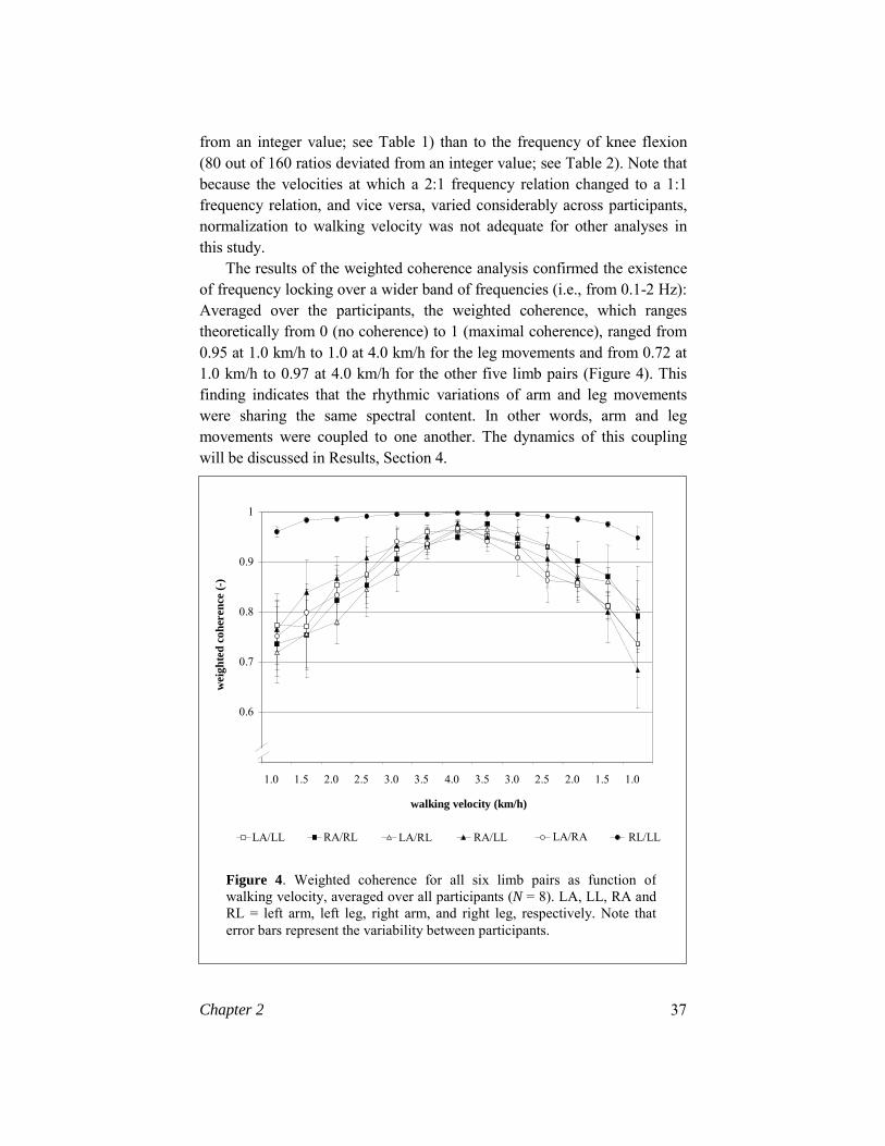
The results of the weighted coherence analysis confirmed the existence of frequency locking over a wider band of frequencies (i.e., from 0.1-2 Hz): Averaged over the participants, the weighted coherence, which ranges theoretically from 0 (no coherence) to 1 (maximal coherence), ranged from 0.95 at 1.0 km/h to 1.0 at 4.0 km/h for the leg movements and from 0.72 at 1.0 km/h to 0.97 at 4.0 km/h for the other five limb pairs (Figure 4). This finding indicates that the rhythmic variations of arm and leg movements were sharing the same spectral content. In other words, arm and leg movements were coupled to one another. The dynamics of this coupling will be discussed in Results, Section 4.
Figure 4. Weighted coherence for all six limb pairs as function ofwalking velocity, averaged over all participants (N = 8). LA, LL, RA and RL = left arm, left leg, right arm, and right leg, respectively. Note that error bars represent the variability between participants.
walking velocity (km/h)
wei
ghte
d co
here
nce
(-)
LA/LL RA/RL LA/RL RA/LL LA/RA RL/LL
0.6
0.7
0.8
0.9
1
1.0 1.5 2.0 2.5 3.0 3.5 4.0 3.5 3.0 2.5 2.0 1.5 1.0

38 Chapter 2
2. Relation Between Coordination and the Eigenfrequencies of the Arms The mean frequency of the arms at which switches from 1:1 to 2:1 coordination were observed was 0.56 Hz, whereas the mean frequencies at which switches occurred in the opposite direction was 0.57 Hz (the range in both cases was 0.45-0.65 Hz). Given that both these values were considerably lower than the estimated eigenfrequencies of the arms, which ranged from 1.06 Hz (Participant 7) to 1.16 Hz (Participant 5), the eigenfrequencies of the arms did not seem to play a decisive role in the observed switches in coordination between arm and leg movements. This result was further amplified by the observation that neither Participant 5 nor Participant 7 showed consistent 2:1 frequency coordination between arm and leg movements, indicating that, to the extent that the eigenfrequency of the arms does play a role in the observed switches in coordination, it is of minor significance. 3. Relative Phase Dynamics The frequency dynamics of the limb movements, as described in the first Results section, were intimately related to their relative phase dynamics. Because of the prevailing task constraints, the mean point estimate of relative phase between the right leg and the left leg (φRL/LL) was 180° (antiphase) at all walking velocities. The point estimate of relative phase between ipsilateral arm and leg movements changed from about 100° in the case of 2:1 frequency coordination to about 190° in the case of 1:1 frequency coordination between arm and leg movements. Given that in the 2:1 frequency coordination the frequency of the arm movements was twice as high as that of the leg movements, yielding two revolutions of 0 to 2π instead of one, this finding indicates that ipsilateral arm and leg movements showed a strong tendency to move in antiphase in both coordination modes (i.e., 2 x 100° and 190°). The point estimate of relative phase between diagonal arm and leg movements changed from about 90° for 2:1 frequency coordination to about 10° for 1:1 frequency coordination, indicating that the relative phase relation between diagonal arm and leg movement changed from a more or less antiphase relation (i.e., 2 x 90°) in the case of 2:1 frequency coordination to a more or less in-phase relation in the case of 1:1 frequency coordination (i.e., 1 x 10°). At low walking velocities, 1:1 frequency relation of the homologous limb pair, left arm/right arm, corresponded with a point estimate of relative phase of about 20° (indicating a more or less in-phase movement). At customary walking velocities, the 1:1 frequency relation corresponded with a point estimate of
Chapter 2 39
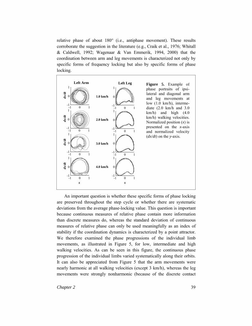
relative phase of about 180° (i.e., antiphase movement). These results corroborate the suggestion in the literature (e.g., Craik et al., 1976; Whitall & Caldwell, 1992; Wagenaar & Van Emmerik, 1994, 2000) that the coordination between arm and leg movements is characterized not only by specific forms of frequency locking but also by specific forms of phase locking.
An important question is whether these specific forms of phase locking are preserved throughout the step cycle or whether there are systematic deviations from the average phase-locking value. This question is important because continuous measures of relative phase contain more information than discrete measures do, whereas the standard deviation of continuous measures of relative phase can only be used meaningfully as an index of stability if the coordination dynamics is characterized by a point attractor. We therefore examined the phase progressions of the individual limb movements, as illustrated in Figure 5, for low, intermediate and high walking velocities. As can be seen in this figure, the continuous phase progression of the individual limbs varied systematically along their orbits. It can also be appreciated from Figure 5 that the arm movements were nearly harmonic at all walking velocities (except 3 km/h), whereas the leg movements were strongly nonharmonic (because of the discrete contact
Figure 5. Example ofphase portraits of ipsi-lateral and diagonal armand leg movements atlow (1.0 km/h), interme-diate (2.0 km/h and 3.0km/h) and high (4.0km/h) walking velocities.Normalized position (x) ispresented on the x-axisand normalized velocity(dx/dt) on the y-axis.
1.0 km/h
2.0 km/h
3.0 km/h
4.0 km/h
x x
-1 0 1 -1
0
1 Left Leg
-1 0 1 -1
0
1
-1 0 1 -1
0
1 -1 0 1
-1
0
1
dx/d
t
Left Arm
dx/d
t dx
/dt
dx/d
t
-1 0 1 -1
0
1
-1 0 1 -1
0
1 -1 0 1
-1
0
1
-1 0 1 -1
0
1

40 Chapter 2
phases with the belt). As a result, the continuous relative phase between arm and leg movements varied systematically within a movement cycle (i.e., from maximal forward extension of a limb to maximal forward extension of the same limb) and over different walking velocities, as is illustrated in Figure 6. These findings suggest that the dynamics of the phasing between the arms and the legs is much more complex than that of a point attractor. Therefore, the variability of continuous relative phase cannot be meaningfully used as an index of the stability of coordination, coupling strength, or both, unless the continuous information in this measure is reduced to specific discrete points (i.e., unless the continuous estimate is turned into a point estimate).
4. Effect of Walking Velocity on Coupling Strength As is apparent from Figure 5, the phase portraits of the individual limb movements varied considerably as a function of walking velocity. As walking velocity increased, the band of orbits decreased, indicating a reduction in the variability between movement cycles. Consequently, the
Figure 6. Example of the progression of the continuous relative phase for diagonal (left arm/right leg [LA/RL]) and ipsilateral (right arm/rightleg) [RA/RL] arm and leg movements. Note that for the sake of clarity, the movement cycles were normalized to 100%. The broken lines represent the mean relative phase (averaged over 8 movement cycles) and the white band represents the variability. (A) RA/RL at 1.0 km/h. (B) LA/RL at 1.0 km/h. (C) RA/RL at 4.0 km/h. (D) LA/RL at 4.0
A. B.
C. D.
0 50 100 -300 -200
-100
0
100
200
rph
(deg
)
0 50 100 -300 -200
-100
0
100
200
rph
(deg
)
0 50 100 -300 -200
-100
0
100
200
rph
(deg
)
0 50 100 -300 -200
-100
0
100
200
rph
(deg
)

Chapter 2 41
relative phase within all pairs of limb movements was more variable at low walking velocities than at customary walking velocities, suggesting that the limb movements were more strongly coupled at higher than at lower walking velocities. Thus, all measures for the coupling between arm and leg movements (i.e., weighted coherence, the variability of the point estimate of relative phase, and the variability of the discrete estimates of continuous relative phase at the peaks and the valleys of the leg movements) indicated that the coupling within all six limb pairs was significantly stronger at customary walking velocities than at low walking velocities (Table 3), as was found previously by Craik et al. (1976) and by Wagenaar and Van Emmerik (1994, 2000). Furthermore, at all velocities the leg movements were significantly stronger coupled than were all other limb pairs, whereas no significant differences in coupling strength were found between ipsilaterally and diagonally paired arm and leg movements (see Table 4 and Figures 4 and 7). The latter results are consistent with those of Wagenaar and Van Emmerik (2000).
F
Variable RA/RL LA/LL RA/LL LA/RL LA/RA RL/LL
Variability of point estimate of relative phase 23.0* 19.1* 25.2* 27.3* 52.6* 27.1*
Variability of continuous relative phase (Peak) 22.3* 50.0* 25.8* 28.3* 135.2* 70.9*
Variability of continuous relative phase (Valley) 32.8* 12.1* 19.5* 25.7* 34.7* 29.6*
Weighted coherence 18.1* 31.0* 21.6* 32.1* 24.7* 27.2*
5. Catastophe Flags: Hysteresis and Critical Fluctuations? The velocity levels at which the coordination pattern shifted from double swinging to single swinging or, conversely, from single swinging to double swinging, varied strongly across participants. Five participants (Participant 3, Participant 4, Participant 5, Participant 6 and Participant 8) showed a hysteresis effect in that the frequency ratios between the ipsilateral arm and leg movements were not always identical at corresponding velocity levels. Only in 2 participants (Participant 6 and Participant 8) were both ipsilateral
Table 3. Analyses of variance with repeated measures for velocity (n=8)
Note. The factor velocity has one degree of freedom, for it compares low walking velocities(1.0 –2.0 km/h) with customary walking velocities (2.5-3.5 km/h). *p < .01.

42 Chapter 2
sides affected by the direction in which the walking velocity was changed; however, in 1 of these participants (Participant 8) the 2:1 frequency coordination was a less consistent feature of the data. Moreover, with regard to the relative phasing within the various pairs of limb movements no systematic hysteresis effects were observed. A repeated measures ANOVA on this variable revealed no significant effects of direction except for the relative phasing between the leg movements F(1, 7) = 6.19, p < .05 for the point estimate of relative phase; F(1,7) = 8.06, p < .05 for the continuous estimate of relative phase, peak; and F(1,7) = 6.77, p < .05 for the continuous estimate of relative phase, valley: The mean value of relative phase was significantly higher with decreasing velocity than with increasing walking velocity.
F
2.5-3.5 km/h 1.0-2.0 km/h
C1 C2 C3 C1 C2 C3
Variability of point estimate of relative phase
34.28** 1.9 1.7 38.7** 3.5 1.8
Variability of continuous relative phase (Peak)
28.2** 3.7 0.6 79.7** 112.4** 6.6*
Variability of continuous relative phase (Valley)
32.9** 9.8* 11.4* 148.2** 19.4** 4.1
Weighted coherence
16.8* 2.9 8.1* 58.5** 0 0.6
Finally, no consistent signs of critical fluctuations in the standard
deviation of the point estimate of relative phase—or in the weighted coherence for that matter—were observed. Averaged across participants, the standard deviation of relative phase was inversely related to walking
Table 4 Analyses of variance with repeated measures for limb pair (N=8)
Note. C1 contrasts limb pair right leg/left leg (RL/LL) with the weighted sum of allother possible limb pairs; C2 contrasts limb pair left arm/right arm (LA/RA) with theweighted sum of ipsilateral (LA/LL and RA/RL) and diagonal (LA/RL and RA/LL)limb pairs; C3 contrasts the weighted sum of ipsilateral with the weighted sum ofdiagonal limb pairs. *p < .05; **p < .01.

Chapter 2 43
velocity, whereas the weighted coherence was positively related to walking velocity (Figures 4 and 7). Although there were considerable individual variations in the patterning of these measures across velocity levels, involving local increases and decreases, none of these patterns showed a marked, explosive increase of variability accompanied by a sharp decrease of the weighted coherence immediately prior to the change in coordination between arm and leg movements. Therefore, we concluded that critical fluctuations were absent.
Discussion
Inspired by recent studies of the role of the upper body in the dynamical organization of walking, we closely examined the effect of walking velocity on the coordination between arm and leg movements. Specifically, we focused on the occurrence of 2:1 frequency coordination at low walking velocities and of 1:1 frequency coordination at customary walking velocities in order to evaluate current theoretical accounts of those
SD o
f poi
nt e
stim
ate
of r
elat
ive
phas
e (d
eg)
0
10
20
30
40
50
60
70
80
1.0 1.5 2.0 2.5 3.0 3.5 4.0 3.5 3.0 2.5 2.0 1.5 1.0
walking velocity (km/h)
LA/LL RA/RL LA/RL RA/LL LA/RA RL/LL
Figure 7. Variability (indicated by SD) of the point estimate of relativephase within al six limb pairs, averaged over participants (N = 8). LA, LL,RA and RL = left arm, left leg, right arm, and right leg, respectively. Notethat error bars represent the variability between participants.

44 Chapter 2
coordination patterns and the corresponding velocity-induced switches between them. In the present section, we discuss our experimental and analytical results according to the five goals listed in the introduction. The sought-after evaluation of current theoretical accounts naturally evolves within the context of this discussion. 1. Observation of 2:1 and 1:1 Frequency Coordination Overall, our experimental results are in agreement with the findings of Craik et al. (1976), Wagenaar and Van Emmerik (1994), and Webb et al. (1994). As in those previous studies, we found that the coordination between arm and leg movements can (sometimes) be characterized as 2:1 frequency coordination at low walking velocities and (always) as 1:1 frequency coordination at customary walking velocities. Comparatively speaking, however, the incidence of the double swinging mode of the arms at low walking velocities was somewhat less in our study than in the previous studies. In our study, 2:1 frequency coordination occurred as a consistent feature of the data in only 3 of the 8 participants, whereas Craik et al. (1976) and Webb et al. (1994) observed this form of coordination in all participants (5 and 27, respectively) and Wagenaar and Van Emmerik (1994, 2000) in nearly all participants. Although this discrepancy in results may seem large, it is important to emphasize that all participants in our study exhibited 2:1 frequency coordination at minimally one velocity level, whereas the issue of individual differences was not addressed in the studies of Craik et al. (1976) and Webb et al. (1994). Nevertheless, the discrepancy in results could have resulted from differences in the methods used. In the study of Craik et al. (1976), participants selected a particular walking velocity in response to verbal commands (e.g., slow, super slow, comfortable), whereas in the present experiment an absolute scaling procedure was used. In principle, it is conceivable that relative velocity scaling would have led to a more consistent occurrence of 2:1 frequency coordination. Having said this, however, it should be noted that the present results suggest that a similar form of relative velocity scaling, that is, scaling with stride frequency, still results in a large variability over participants (see Results, Section 2). In search for the mechanisms underlying a switch from 2:1 to 1:1 frequency coordination, the problem of relative velocity scaling seems well worth pursuing.
That the issue of individual differences should not be underestimated is also apparent from the observation of Wagenaar and Van Emmerik (1994, 2000) that some of their participants were less inclined to exhibit 2:1 frequency coordination; this observation is consistent with our results. Thus,

Chapter 2 45
as long as the distribution of people with and without a tendency to exhibit 2:1 frequency coordination is unknown, it is impossible to assess how atypical our results are (or those of previous studies, for that matter). Another issue to be considered in relation to individual dispositions for adopting certain coordination modes is that of experimental instructions. Verbal commands can be interpreted in many different ways and, consequently, may result in a dichotomy of coordination patterns between participants (Craik et al., 1976; Whitall & Getchell, 1996). Our aim in the present experiment was to determine if participants would voluntarily adopt a different coordination mode when walking at low velocities. We therefore deliberately refrained from giving any instructions or information with regard to their arm movements. In this context, it is interesting to note that some participants were surprised to learn that they had adopted the double-swinging mode. In drawing the comparison with previous studies to a close, it should be highlighted that the present study went beyond those previous studies in consolidating the hypothesis that the observed patterns of frequency coordination may indeed be characterized as instances of frequency locking, both in terms of locking between the dominant frequencies and in terms of locking across a predetermined band of frequencies. 2. The Role of the Eigenfrequencies of the Arms In recognition of the important role of the biomechanical properties of the human musculoskeletal system in the organization of walking (McGeer, 1990; McMahon, 1984; Mochon & McMahon, 1980; Schot & Decker, 1998), several attempts have been made to model walking as a system of mechanically coupled pendulums. Webb et al. (1994) and Wagenaar and Van Emmerik (2000) have attempted to integrate the arm movements in such a pendulum model, allowing for an interpretation of the occurrence of 2:1 and 1:1 frequency coordination in terms of the eigenfrequencies of the arms. Webb et al. provided data in support of this interpretation by showing that 2:1 frequency coordination is adopted consistently at frequencies slightly below the eigenfrequency of the arms. In the present study, however, quite a number of observations were made that run counter to the eigenfrequency hypothesis. The dominant frequencies of the arms near pattern switching were markedly lower than the estimated eigenfrequencies. Furthermore, 2:1 frequency coordination was not a consistent feature of the data in all participants, nor did it occur consistently at the same walking velocity (i.e., at the same dominant frequency of the arms). Whereas the first observation may have been due to imperfections in our (admittedly

46 Chapter 2
crude) estimation of the eigenfrequencies of the arms, the second observation demonstrates that the eigenfrequencies cannot be the sole explanation of the occurrence of 2:1 frequency coordination at low walking velocities. The eigenfrequencies of the arms are likely to play an indispensable role in the observed coordination patterns between arm and leg movements, but do not determine them in a mechanistic fashion. In addition to the eigenfrequencies of the arms, one has to take into consideration a large number of other factors, such as muscle activity, movement-related afferent information, and the mechanical interaction with the environment, in order to account for the coordination dynamics. To highlight this important point, it is useful to discuss some of these factors in more detail.
Craik et al. (1976) indicated that changes in frequency coordination between the arm and leg movements were accompanied by changes in electromyographic activity, suggesting that the arm movements are not a result of gravity only (i.e., are not purely passive). At customary walking velocities, the initiation of the posterior deltoid muscle preceded peak shoulder flexion, whereas at low walking velocities, the initiation of firing followed shoulder flexion. This finding suggests that the shift in frequency coordination is accompanied by a shift in intermuscular coordination. It is important to note in this context that the shift in frequency coordination was accompanied by shifts in the entrainment regimes between the spectral components of the individual limb movements. This finding becomes apparent from Tables 1 and 2 in that, at all walking velocities, the arm movements were locked onto both significant frequencies in the power spectra of the leg movements, suggesting that the second frequency of the leg movements, which emerged as a result of ground contact, affected the arm movements at all walking velocities. This result is consistent with the findings of Thorstensson et al. (1984), who observed double oscillations in the trunk, even at high walking velocities. Another important change in entrainment accompanying the change in coordination was the relative degree to which the arm movements were locked onto the stride frequency (i.e., the dominant frequency of the leg movements) and the frequency of knee flexion (i.e., the second significant frequency of the leg movements). In the case of 1:1 frequency coordination the arm movements were entrained more strongly to the stride frequency than in the case of 2:1 frequency coordination, whereas the reverse was true for the entrainment between the arm movements and the frequency of knee flexion (see Table 2). This finding suggests that the observed changes in frequency coordination are, in fact, functional changes in organization.

Chapter 2 47
3. Relative Phasing between the Limb Movements Extensive analyses of the phasing between arm and leg movements provided support for previous suggestions in the literature (e.g., Craik et al., 1976; Wagenaar & Van Emmerik, 1994, 2000; Whitall & Caldwell, 1992) that the coordination between arm and leg movements is characterized not only by specific forms of frequency locking but also by specific forms of phase locking. Consistent with the literature on bimanual coordination (e.g., Haken et al., 1985; Kelso, 1984), both in-phase and antiphase patterns of coordination were preferred and relatively stable. More interesting, this was the case even when the (dominant) frequencies of arm and leg movements were coordinated as 2:1, suggesting the existence of dynamical remnants of 1:1 frequency coordination within 2:1 frequency coordination, which is consistent with experimental findings on bimanual 2:1 frequency coordination (Sternad, Saltzman, &, Turvey, 1999a, 1999b). Also consistent with this literature is the finding that the 2:1 frequency coordination was less stable than the 1:1 frequency coordination pattern (cf. Serrien & Swinnen, 1997). Contrary to previous findings on interlimb coordination (Jeka, Kelso, & Kiemel, 1993), however, no significant differences were found with regard to the stability of coordination across the different limb pairs. The only significant effect in this regard was that the coupling between the leg movements was stronger than that of all other pairs of limb movements, which did not differ significantly from each other (cf. Wagenaar & Van Emmerik, 2000; Whitall & Caldwell, 1992). The noted discrepancy with the literature on interlimb coordination in nonlocomotory tasks probably reflects the fact that in walking the mechanical properties of the limbs in interaction with those of the environment play an important role, and suggests that insights obtained into an idealized experimental setup may not be so easily transferable to everyday activities.
An important result of our analyses of the continuous relative phase between the arm and leg movements is that one cannot adequately describe the dynamical properties of this variable by assuming the presence of a point attractor because particularly the leg movements deviate strongly from harmonicity. Consequently, the (circular version of the) standard deviation of continuous relative phase cannot be meaningfully used as an index of stability of interlimb coordination during walking. 4. Effect of Walking Velocity on Interlimb Coupling Strength Using a combination of measures (i.e., standard deviation of discrete versions of relative phase and weighted coherence) we found unambigu-ously that the coupling between limb movements in all six limb pairs (i.e.,

48 Chapter 2
homologous, ipsilateral, and diagonal) increased with increasing walking velocity, thus confirming the results of Craik et al. (1976) and Wagenaar and Van Emmerik (1994, 2000). This effect occurred both within and across 2:1 and 1:1 frequency coordination and thus appeared to overrule the differential stability between 2:1 and 1:1 frequency coordination referred to in the preceding discussion. Moreover, it occurred for all pairs of limb movements, regardless of their frequency coordination.
The finding that interlimb coupling strength increases with walking velocity, and thus stride frequency, is at odds with the common finding in the study of rhythmic bimanual coordination that coupling strength is inversely related to movement frequency (Peper, Beek, & Van Wieringen, 1995; Schmidt, Shaw, & Turvey, 1993; Sternad, Turvey, & Schmidt, 1992). In this context, however, it should be noted that in the present study, in which we focused on the occurrence of 2:1 and 1:1 frequency coordination at low walking velocities, we did not examine walking velocities higher than 4.0 km/h. Perhaps a decrease in stability would have been observed at walking velocities close to running (cf. Diedrich & Warren, 1995). Apart from this point concerning the method used, three differences between bimanual coordination and human walking (two of which were already referred to in the preceding) may account for the observed task dependency of the effect of movement frequency on coupling strength. First, the dynamics of interlimb coordination in human walking is strongly affected by mechanical constraints such as forceful contacts with the support surface, which induce nonharmonic oscillations of the leg movements (cf. Figure 5). Second, an important requirement that is likely to affect the stability of interlimb coordination in human walking in a velocity-dependent manner is postural control (Whitall & Clark, 1994; Winter, 1995). Third, it should be remembered that walking is a learned coor-dination pattern, much more so than finger wiggling. From studies on the development of walking, it is known that the alternating phasing of the legs becomes more stable after minimally three months of practice (Whitall & Clark, 1994). In the present study, participants were allowed a limited amount of time to become familiar with walking on a walking belt after which they performed only one long trial. Thus, lack of experience with walking at the very low velocities at which the 2:1 frequency coordination emerges may be an explanation for the observed lower stability of interlimb coordination at these walking velocities. 5. Catastrophe Flags: Hysteresis and Critical Fluctuations? For the observed switches between coordination patterns to qualify as

Chapter 2 49
nonequilibrium phase transitions, they should satisfy certain criteria known as catastrophe flags (Gilmore, 1981). In the present experiment, we tested for two of those flags, namely, hysteresis and critical fluctuations. Essentially, no convincing evidence was found in the experimental data for either. Although there were both inter- and intraindividual variations in the velocity level at which the switch in coordination between arm and leg movements occurred, the variations were independent of the “history of the system”. The directionality of the changes in velocity had a significant effect only on the relative phasing between the leg movements. However, this isolated effect constitutes no evidence for the existence of nonequi-librium phase transitions because the coordination between the leg movements does not change qualitatively when the coordination between arms and legs changes qualitatively. Also with regard to critical fluctua-tions, the result was essentially one of denial. In this case, one might even argue that the observed effect is opposite to what one would expect from the phase transition hypothesis in that the stability of interlimb coordination keeps on increasing with increasing walking velocity, and would thus never lead to a loss of stability. Conversely, when starting in the 1:1 frequency coordination and decreasing walking velocity, the stability of interlimb coordination decreases monotonically across the switch in behavior instead of first losing and then gaining stability. In other words, the observed switches in the coordination between arm and leg movements appear to constitute no nonequilibrium phase transitions of the kind implied by Gilmore (1981) and Haken (1977). It should be recognized, however, that we based this conclusion on a single trial in which walking velocity was increased with increments that may have been too large to enable us to observe hysteresis and critical fluctuations. Coda In sum, the present experimental results cannot be explained satisfactorily in purely biomechanical terms or in purely dynamical terms. In our opinion, our findings emphasize the need for a model that accounts for both the informational (i.e., neural) and musculoskeletal (i.e., biomechanical) constraints on interlimb coordination during walking. After all, human walking is a complex system in which both neural and biomechanical factors play a role, depending on both internal and external (i.e., environmental) constraints. The challenge for future theoretical work on human walking is to develop encompassing models, along the lines formulated by Taga (1994), that enable us to account for velocity-dependent variations in the dynamical stability of the walking pattern in terms of a

50 Chapter 2
fully worked out neuromusculoskeletal system interacting with the environment.
Acknowledgments We thank Bart Nienhuis for his technical assistance in the experimental set-up and for his inspiring suggestions. Furthermore, we are grateful to Ruud Meulenbroek and Piet van Wieringen for their comments on an earlier draft of this article, to Patsy Anderson for her statistical advice, and to Andreas Daffertshofer for his help with the weighted coherence analysis. This research was carried out at the Sint Maartens Kliniek-Research, with financial support from the Foundation for Behavioral Sciences (SGW; grant number 575-23-005), which is funded by the Netherlands Organization for Scientific Research (NWO).
Notes
1. Recently an extensive discussion took place on the newsgroup BIOMCH-L concerning the functions and mechanisms underlying arm swinging in human walking. This discussion nicely illustrated the increasing awareness of the need to incorporate the movements of the upper body and the arms in the study of human walking.
2. In this study, we focused upon the movements of arms and legs,
which may be viewed as multisegmented pendulums. Hence, it was legitimate to restrict our analysis to the movements of the forearms and lower legs; those movements are markedly larger than the movements of the upper arms and upper legs.
3. To check whether this relatively broad frequency band introduced
artifacts, we performed additional analyses of weighted coherence in which we used a smaller frequency band, centered around the (in the case of 2:1 frequency coordination, generalized) dominant frequencies. Those analyses led to similar results as when a relatively broad frequency band was used.

