university of groningen fetal death korteweg, fleurisca joyce · the pathophysiology of late fetal...
TRANSCRIPT

University of Groningen
Fetal deathKorteweg, Fleurisca Joyce
IMPORTANT NOTE: You are advised to consult the publisher's version (publisher's PDF) if you wish to cite fromit. Please check the document version below.
Document VersionPublisher's PDF, also known as Version of record
Publication date:2010
Link to publication in University of Groningen/UMCG research database
Citation for published version (APA):Korteweg, F. J. (2010). Fetal death: classification and diagnostic work-up Groningen: s.n.
CopyrightOther than for strictly personal use, it is not permitted to download or to forward/distribute the text or part of it without the consent of theauthor(s) and/or copyright holder(s), unless the work is under an open content license (like Creative Commons).
Take-down policyIf you believe that this document breaches copyright please contact us providing details, and we will remove access to the work immediatelyand investigate your claim.
Downloaded from the University of Groningen/UMCG research database (Pure): http://www.rug.nl/research/portal. For technical reasons thenumber of authors shown on this cover page is limited to 10 maximum.
Download date: 05-06-2018

Fleurisca J. Korteweg
Jan Jaap H.M. Erwich
Nienke Folkeringa
Albertus Timmer
Nic J.G.M. Veeger
Joke M. Ravisé
Jozien P. Holm
Jan van der Meer
Submitted
8Chapter
New insight into thrombophilic defects in 750 couples with fetal death

ABSTRACT
Context
Inherited thrombophilia has been associated with fetal death while thrombophilic
defects acquired during pregnancy may further contribute. Their relation to cause
of death remains uncertain.
Objective
To determine whether in a cohort of fetal deaths maternal thrombophilic defects,
either acquired or inherited, and inherited paternal defects were more prevalent
than in the normal population. Furthermore, we assessed the association between
these thrombophilic defects and the various fetal death causes.
Design
Multicenter, prospective cohort study from 2002-2006
Setting
50 Dutch secondary and tertiary referral hospitals
Patients
750 couples with singleton ante partum fetal deaths > 20 weeks gestation
Main outcome measures
Antithrombin, protein C, total and free protein S, and von Willebrand factor plasma
levels. Mothers were compared to gestational age-matched healthy pregnant
women and fathers to healthy men. Prevalence of factor V Leiden, prothrombin
G20210A mutation, and lupus anticoagulant were compared to the normal
population. A panel classified death cause.
Results
More women with fetal death had decreased antithrombin (16.8%, p<0.0001) and
protein C (4.0%, p=0.03) and increased von Willebrand factor (15.5%, p<0.0001)
plasma levels than pregnant women (2.5%). However, compared to normal ranges
in the non-pregnant population, we only saw more women with increased von
Willebrand factor (12.4%, p<0.0001). More fathers had decreased free protein
S (6.3%, p<0.0001) and elevated von Willebrand factor (12.1%, p<0.0001) than
healthy men (2.5%). Prevalence of inherited thrombophilias was no higher in
couples with fetal death than in the population. Neither inherited nor acquired
maternal or paternal thrombophilic defects were associated with main cause
of death, but of placental causes abruption and infarction were associated with
acquired maternal defects.
Conclusions
Except for von Willebrand factor and paternal free protein S, acquired and inherited
thrombophilic defects were not associated with fetal death. Acquired maternal
thrombophilic defects were only associated with death causes abruption and
infarction. Routine thrombophilia testing after fetal death is not advised.
Chapter 8
146
INTRODUCTION
About 1 in 200 pregnancies ends in stillbirth due to a range of causes and conditions.1
Maternally inherited thrombophilic defects are inconsistently recognized as risk
factors for pregnancy complications such as preeclampsia, placental abruption,
growth restriction and stillbirth,2,3 while paternal thrombophilic factors can also
be transferred to the fetus and placenta.4 Many studies have addressed the
association between inherited thrombophilic deficiencies and late fetal loss in
families with deficiencies, whereas case-control and cohort studies have studied
women with fetal loss who were tested for inherited thrombophilia after birth. In
reviews a higher risk for fetal loss was observed for antithrombin and protein S
deficiency, factor V Leiden, the prothrombin 20210A mutation, and anticardiolipin
antibodies.5-7
The pathophysiology of late fetal loss associated with inherited thrombophilia was
presumed to be placental thrombosis, either in the maternal or fetal circulation
of the placenta leading to placental infarction and placental insufficiency.8 This
hypothesis has been difficult to prove due to the small numbers involved in
previous studies, while placental insufficiency was often poorly defined and not
related to cause of death.9-12 Pregnancy is a hypercoagulable state due to acquired
thrombophilic defects exposing women to a higher risk of thrombosis.13 In
combination with pre-existing inherited thrombophilic defects, it may increase the
risk of fetal death. Little is known about the contribution of acquired thrombophilic
defects to the hypercoagulable state and fetal death as women in previous studies
were only tested for thrombophilia several weeks after delivery.
The objective of our study was to assess whether in a cohort of women with
intrauterine fetal death (IUFD), thrombophilic defects, either acquired or inherited,
and inherited defects in fathers were more prevalent than in the normal population.
Furthermore, we assessed the association between these thrombophilic defects
and the various IUFD causes within our cohort.
METHODS
In 2002 we initiated a prospective IUFD cohort study in 50 Dutch secondary and
tertiary referral hospitals, serving rural as well as urban populations. Inclusion
criteria were singleton IUFD diagnosed ante partum (heart beat ceased before
labor), after 20 weeks of gestation calculated from the last menstrual period
and confirmed by ultrasonography. Terminations were excluded. The study was
approved by the review boards of all the participating hospitals and written
informed consent was obtained from participants. Data was collected for each
Thrombophilic defects in 750 couples with fetal death
147
IUFD, including medical and obstetric history, maternal and fetal characteristics,
and pregnancy and birth details. Our diagnostic work-up protocol was based on
currently used local Dutch protocols and included: maternal blood tests including
full blood count, chemistry and viral serology; coagulation tests for couples
performed centrally in the laboratory in Groningen; fetal blood tests including viral
serology; microbiological cultures from the mother, fetus and placenta; autopsy;
placental examination; and cytogenetic analysis. Ethnic origin was one of the
studied characteristics of the study population and classified by the investigator
as: Caucasian including Mediterranean groups, African (Negro), Eastern or Other.
Coagulation testsIn women with IUFD, plasma levels of antithrombin (AT), protein C activity (PC),
total and free protein S antigen (TPS, FPS), and von Willebrand factor (VWF)
were measured in samples collected on induction of labor. These plasma levels
were compared to plasma reference values in 110 healthy pregnant women of
comparable gestational age recruited from our obstetrics department, after
informed consent. Healthy pregnant women were tested at four intervals of
gestation: 12-16 weeks, 28-32 weeks, in an early stage of labor (> 34 weeks) and
5-7 weeks postpartum. Women were excluded if they had an individual or family
history of venous thromboembolism, known thrombophilia, complications in past
pregnancies or the present one, or if they used medication. Reference plasma
values were determined for all cases for gestational age periods from 20-27 weeks,
27-34 weeks and >34 weeks (in an early stage of labor). Values at 20-27 weeks
gestation were obtained by linear estimation from values at 12-16 weeks and 28-32
weeks for each individual. Plasma levels of AT, PC, TPS and FPS in women with
IUFD were defined as abnormal if they were below 2.5% of the values in healthy
pregnant women at comparable gestational age, or above 97.5% for VWF.
We also measured plasma levels of AT, PC, TPS, FPS and VWF in the fathers and
we determined normal plasma ranges in 393 healthy men. Abnormal plasma levels
were defined as levels below the lower limit of the normal ranges: AT <74 IU/dL;
PC <64 IU/dL; TPS <67 IU/dL and FPS <65 IU/dL. VWF levels >150 IU/dL were
increased. All these abnormal plasma levels will be referred to as ‘thrombophilic
defects’.
In men and women, factor V Leiden (FVL) and the prothrombin G20210A mutation
(PTG20210A) were determined by polymerase chain reactions. Lupus anticoagulant
was determined as previously described.14 Prevalence of these thrombophilias
was compared to prevalence in the normal population. AT (Chromogenix, Mölndal,
Sweden) and PC activity (Dade Behring, Marburg, Germany) were measured by
chromogenic substrate assays; TPS, FPS and VWF were measured by Enzyme
Linked Immuno Sorbent Assay (DAKO, Glostrup, Denmark).
Chapter 8
148

Autopsy and placental examinationAutopsies and placental examinations (including histology) were performed by
pathologists in the participating hospitals according to guidelines published by
the Royal College of Obstetricians and Gynaecologists and the Royal College
of Pathologists15 and the College of American Pathologists.16,17 Fetal growth
percentiles for birth weight by gestational age at time of diagnosis of IUFD were
calculated according to Kloosterman’s growth charts.18 Small for gestational age
(SGA) was defined as birth weight < 10th percentile.
Adjudication of cause of deathCause of fetal death was classified by a multidisciplinary panel according to the
Tulip classification,19 which covers six main causes: congenital anomaly, placental
pathology, prematurity/immaturity, infection, other (i.e. maternal diseases, fetal
hydrops), or unknown. The cause was classified as unknown if other causes had
been excluded. Risk factors such as smoking and preeclampsia were defined as
contributing to death.
Causes of deaths due to maternal and fetal placental circulation pathology were
placental abruption, a clinical diagnosis supported by placental examination;
significant infarction (the percentage of infarctions in relation to the weight of the
placenta was regarded as sufficient to have caused death) in preterm cases, any
placental infarction, and in term cases, extensive infarction (> 10%) of the placental
area,20 fetal thrombotic vasculopathy (FTV), the presence of avascular villi (at least
one focus of five or more villi), thrombosis in a vessel of the chorionic plate or stem
villus, hemorrhagic endovasculitis, or intramural fibrin in a vessel of the chorionic
plate or stem villus in the absence of umbilical cord blood flow restriction;21,22
and maternal floor infarct (MFI)/massive perivillous fibrin deposition (MPFD),
extensive perivillous fibrin deposition, either predominantly basally located or
diffusely distributed in at least 30% of the parenchyma.23 Other categories of
placental pathology were placental hypoplasia, an absolute, too low placenta
weight (< 10th percentile) and/or a too low placenta/birth weight ratio,24 and other
placental pathology, such as villus immaturity and umbilical cord complications.
StatisticsCategorical variables were expressed as counts and percentages, and continuous
data as means with standard deviation or median and ranges, with exact 95%
confidence intervals (CI) given when appropriate. Differences between groups
for categorical data were evaluated by the Fisher exact test or Chi-square test.
For continuous variables, we used the Student t test or Mann-Whitney U test,
depending on the normality of data. A two-tailed p-value <0.05 was considered
Thrombophilic defects in 750 couples with fetal death
149
to indicate statistical significance. Statistical analyses were performed using SAS
software, version 9.1 (SAS Institute Inc., Cary, NC, USA).
RESULTS
From 2002 to 2006, we enrolled 750 couples. Coagulation tests were performed in
714 (95.2%) women and 664 (88.5%) men. Autopsies were performed in 525 (70.0%)
fetal deaths and placental examinations in 736 (98.1%). The characteristics of men
and women with at least one thrombophilic defect (22.4% and 37.4%, respectively)
and those without such defects are shown in Table 1. A personal history of venous
thromboembolism and known thrombophilia, hypertension-related disease and
anticoagulant thromboprophylaxis during pregnancy were observed more often
in women with a thrombophilic defect. In this subgroup median age at pregnancy,
mean gestational age at delivery (28.4 weeks versus 34.4 weeks) and median birth
weight were lower, while it also contained more SGA babies. Men with at least one
thrombophilic defect were older than men without defects.
Compared to reference values in non-pregnant women, the majority of women
with IUFD and healthy pregnant women had a tendency to higher AT levels, no
difference in PC levels, a tendency to lower TPS, lower FPS and higher VWF levels
(Figure 1). Compared to healthy pregnant women, women with IUFD had lower
levels of AT and higher levels of VWF up to 34 weeks of gestation.
Women with IUFD more often had significantly decreased levels of AT (16.8%)
and PC (4.0%), and increased VWF levels (15.5%) compared to pregnant women
(2.5%, Table 2). Decreased FPS levels were less common (0.9%). However, when
compared to plasma levels in the normal, non-pregnant population, decreased
levels of AT (3.7%, p=0.07) and PC (2.1%, p=0.62) were not observed more often,
in contrast to increased VWF levels (87.6%, p<0.0001) that were still observed
more often in women with IUFD. The prevalence of FVL and PTG20210A in women
with IUFD was comparable to the normal population, whereas lupus anticoagulant
(1.4%) was observed less frequently than expected. Men in the IUFD group more
often had decreased FPS plasma levels (6.3%) and elevated VWF levels (12.1%)
compared to healthy men (2.5%).
Causes of death were placental pathology (64.9%), congenital anomaly (5.3%),
infection (1.9%), other (4.8%), or unknown (23.1%: 15.9% despite thorough
investigation and 7.2% due to insufficient information). Table 3 presents
thrombophilic defects found in women with IUFD (n=750) and their partners in
relation to cause of death. A thrombophilic defect was seen in 39.1% of women
with a placental cause versus 34.1% with a non-placental cause. Overall, in both
men and women, none of the separate thrombophilic defects were associated with
Chapter 8
150

Table 1: Characteristics of the study population
Thrombophilic defect nonen=447
at least 1 n=267
p
Women 62.6% 37.4%
Family history
Venous thromboembolism (1st degree) 4.9 8.5 0.07
Known hereditary thrombophilia 1.5 4.2 0.06
Personal History
Venous thromboembolism 0.9 4.9 0.001
Known hereditary thrombophilia 0 2.0 0.006
Previous IUFD 2.7 3.8 0.50
Recurrent early fetal loss 6.3 5.6 0.87
Age, median (range), years 32 (18-46) 30 (18-46) 0.005
Ethnic origin
Caucasian* 88.4 86.1 0.31
African (Negro) 2.7 5.2
Eastern 4.3 3.7
Other 4.7 4.9
Pregnancy
Nulliparous 49.7 56.9 0.20
Primiparous 21.9 18.4
Multiparous 28.4 24.7
Hypertension-related disease 11.0 25.8 < 0.001
Diabetes-related disease 4.1 3.4 0.84
Smoking 23.3 25.9 0.83
Anticoagulant thromboprophylaxis 1.6 4.5 0.028
Current IUFD
Gestational age, mean (SD) weeks 34.4 (6.3) 28.4 (5.7) <0.001
Birth weight, median (range), grams 2000 (40-4630) 810 (12-4425) <0.001
Small for gestational age† 28.8 43.0 <0.001
Time lap diagnosis and birth, 2 (0-40) 2 (0-23) 0.27
median (range), days
Thrombophilic defect nonen=515
at least 1 n=149
p
Men 77.6% 22.4%
Family history
Venous thromboembolism (1st degree) 4.4 6.9 0.26
Known hereditary thrombophilia 0.9 2.3 0.19
Personal History
Venous thromboembolism 0.4 0.7 0.53
Known hereditary thrombophilia 0.2 0.8 0.40
Age, median (range), years 34 (18-61) 35 (19-60) 0.01
Current IUFD
Gestational age, mean (SD), weeks 31.6 (6.3) 31.7 (6.7) 0.74
Birth weight, median (range), grams 1485 (12-4560) 1395 (51-4630) 0.98
Small for gestational age† 33.5 35.3 0.74
results are given in % unless otherwise indicated, *including Mediterranean groups †according to Kloosterman’s growth charts18 which commence at 25 weeks of gestation
Thrombophilic defects in 750 couples with fetal death
151

Figure 1. Plasma levels of natural anticoagulant proteins and von Willebrand factor in women with intrauterine fetal death (IUFD), healthy pregnant women and male partners of women with IUFD compared to reference values in the normal non-pregnant population (dotted line).
Chapter 8
152

placental versus non-placental causes. Decreased maternal TPS plasma levels
were more often associated with non-placental causes and elevated maternal
VWF levels were associated with “other cause of death”. In addition, when we
considered the 267 women with a thrombophilic defect, 182 (68%) had an IUFD
due to a placental cause (39.1% of the 465 women with a placental cause). This
was comparable to the group of women without thrombophilic defects (283/447,
63%) (p=0.20). Of the 149 men with a thrombophilic defect, 94 (63%) were in
the IUFD group with placental causes (21.7% of 434 men overall with a placental
cause), which is comparable to 340/515 men (66%, p=0.56) without a defect. No
association with placental causes was observed in couples where there was both
a maternal and a paternal thrombophilic defect (32/53; 60% versus 199/317; 63%,
p=0.19) versus couples without a defect.
Analysis of placental causes of death showed that death was due to infarction in
99/182 (54%) and abruption in 30/182 (16%) women with a thrombophilic defect
(Table 4). Compared to all the different placental causes, abruption was more
frequently associated with decreased levels of AT (40.8%, p<0.001), PC (20.4%,
p<0.001), and TPS (10.2%, p<0.001) and increased VWF levels (18.4%, p=0.03);
infarction was associated with decreased AT (26.1%, p<0.001) and elevated VWF
(28.4%, p<0.001) levels and lupus anticoagulant (2.4%, p=0.04); and MFI/MPFD
with elevated VWF (28.6%, p<0.001). Abruption was seen significantly more often
in women with abnormal plasma levels of AT, PC and TPS compared to infarction.
Overall, of placental causes, abruption and infarction were most frequently
observed in women with thrombophilic defects (p<0.0001).
DISCUSSION
Our IUFD cohort study was primarily set up to evaluate valuable diagnostics
to determine cause of fetal death. Here we addressed the contribution of
thrombophilic defects acquired during pregnancy to fetal death, rather than that
of inherited thrombophilia. Thrombophilia testing was therefore performed at
induction of labor and protein levels in women with fetal death were compared
to healthy pregnant women of comparable gestational age. We defined protein
levels as potential risk factors for thrombosis in pregnancy (i.e. as thrombophilic
defects) when they were < 2.5 percentile in healthy pregnant women for AT, PC
and PS, and > 97.5 percentile for VWF. Testing for thrombophilia after fetal death
may be useful in clinical practice if the results can be used to prevent recurrent
fetal loss. Our data provide no support for routine testing of inherited or acquired
thrombophilic defects after fetal death, although acquired defects may play a role
in deaths caused by abruption or infarction.
Thrombophilic defects in 750 couples with fetal death
153
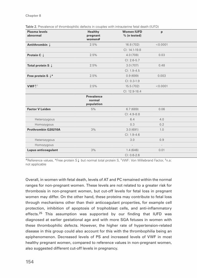
Overall, in women with fetal death, levels of AT and PC remained within the normal
ranges for non-pregnant women. These levels are not related to a greater risk for
thrombosis in non-pregnant women, but cut-off levels for fetal loss in pregnant
women may differ. On the other hand, these proteins may contribute to fetal loss
through mechanisms other than their anticoagulant properties, for example cell
protection, inhibition of apoptosis of trophoblast cells, and anti-inflammatory
effects.25 This assumption was supported by our finding that IUFD was
diagnosed at earlier gestational age and with more SGA fetuses in women with
these thrombophilic defects. However, the higher rate of hypertension-related
disease in this group could also account for this with the thrombophilia being an
epiphenomenon. Decreased levels of PS and increased levels of VWF in most
healthy pregnant women, compared to reference values in non-pregnant women,
also suggested different cut-off levels in pregnancy.
Table 2. Prevalence of thrombophilic defects in couples with intrauterine fetal death (IUFD)
Plasma levelsabnormal
Healthypregnantwomen#
Women IUFD% (n tested)
p Healthymen#
Men IUFD% (n tested)
p
Antithrombin ¯ 2.5% 16.8 (702) <0.0001 2.5% 0.3 (655) <0.0001
CI: 14.1-19.8 CI: 0.04-1.1
Protein C ¯ 2.5% 4.0 (708) 0.03 2.5% 0.6 (661) <0.0001
CI: 2.6-5.7 CI: 0.2-1.5
Total protein S ¯ 2.5% 3.0 (707) 0.48 2.5% 0.5 (659) <0.0001
CI: 1.9-4.5 CI: 0.1-1.3
Free protein S *̄ 2.5% 0.9 (699) 0.003 2.5% 6.3 (656) <0.0001
CI: 0.3-1.9 CI: 4.5-8.4
VWF † 2.5% 15.5 (702) <0.0001 2.5% 12.1 (659) <0.0001
CI: 12.9-18.4 CI: 9.7-14.9
Prevalencenormal
population
Prevalencenormal
population
Factor V Leiden 5% 6.7 (689) 0.06 5% 4.2 (642) 0.41
CI: 4.9-8.8 CI: 2.8-6.1
Heterozygous 6.4 4.0
Homozygous 0.3 0.2
Prothrombin G20210A 3% 3.0 (691) 1.0 3% 0.9 (642) <0.0001
CI: 1.9-4.6 CI: 0.3-2.0
Heterozygous 3.0 0.9
Homozygous - -
Lupus anticoagulant 3% 1.4 (646) 0.01 3% 0 (105) n.a.‡
CI: 0.6-2.6 CI: 0-3.5#Reference values, *Free protein S¯ but normal total protein S, †VWF: Von Willebrand Factor, ‡n.a: not applicable
¯
Chapter 8
154

Significantly increased maternal VWF plasma levels were observed in our IUFD
group compared to healthy pregnant women VWF levels increase during normal
pregnancy,13 but it is unknown when these levels become pathological. VWF
activity was found to be higher in women with early miscarriage than in controls.26
We speculated that our results could be related to non-O blood type27 or an acute
phase response: non-O blood type was indeed associated with higher VWF levels,
whereas C-reactive protein and fibrinogen were not (data not shown).
Abnormal paternal plasma levels of FPS and VWF were observed in the IUFD
group. Others reported no difference in fetal mortality in women with partners
with and without inherited thrombophilia.28 In contrast, a doubled prevalence
(60%) of numerous thrombophilic defects in partners of women with a history of
perinatal mortality versus controls (30%) was reported.29
Table 2. Prevalence of thrombophilic defects in couples with intrauterine fetal death (IUFD)
Plasma levelsabnormal
Healthypregnantwomen#
Women IUFD% (n tested)
p Healthymen#
Men IUFD% (n tested)
p
Antithrombin ¯ 2.5% 16.8 (702) <0.0001 2.5% 0.3 (655) <0.0001
CI: 14.1-19.8 CI: 0.04-1.1
Protein C ¯ 2.5% 4.0 (708) 0.03 2.5% 0.6 (661) <0.0001
CI: 2.6-5.7 CI: 0.2-1.5
Total protein S ¯ 2.5% 3.0 (707) 0.48 2.5% 0.5 (659) <0.0001
CI: 1.9-4.5 CI: 0.1-1.3
Free protein S *̄ 2.5% 0.9 (699) 0.003 2.5% 6.3 (656) <0.0001
CI: 0.3-1.9 CI: 4.5-8.4
VWF † 2.5% 15.5 (702) <0.0001 2.5% 12.1 (659) <0.0001
CI: 12.9-18.4 CI: 9.7-14.9
Prevalencenormal
population
Prevalencenormal
population
Factor V Leiden 5% 6.7 (689) 0.06 5% 4.2 (642) 0.41
CI: 4.9-8.8 CI: 2.8-6.1
Heterozygous 6.4 4.0
Homozygous 0.3 0.2
Prothrombin G20210A 3% 3.0 (691) 1.0 3% 0.9 (642) <0.0001
CI: 1.9-4.6 CI: 0.3-2.0
Heterozygous 3.0 0.9
Homozygous - -
Lupus anticoagulant 3% 1.4 (646) 0.01 3% 0 (105) n.a.‡
CI: 0.6-2.6 CI: 0-3.5#Reference values, *Free protein S¯ but normal total protein S, †VWF: Von Willebrand Factor, ‡n.a: not applicable
Thrombophilic defects in 750 couples with fetal death
155

Overall, FVL, PTG20210A and lupus anticoagulant were not associated with fetal
death. This is in contrast with earlier studies that reported relative risks for late
fetal loss of 2.1-3.3 for maternal FVL, 2.3-3.0 for maternal PTG20210A, and 2.4 for
maternal lupus anticoagulant.6,7,30 Variation in population characteristics could
explain these differences.
Trophoblast invasion of the maternal uterine circulation, spiral artery remodeling
and maintenance of blood fluidity in the intervillous space require a balance
between pro-thrombotic and anti-thrombotic forces. Most deaths in our study
were caused by placental pathology. Overall, none of the maternal and paternal
thrombophilic defects were related to a placental cause, nor for couples with
double thrombophilic defects. Analysis of various placental causes of fetal death
showed that thrombophilic defects were only associated with placental abruption
and infarction. Abruption was associated with decreased levels of AT, PC and
TPS and increased VWF levels, infarction with decreased AT levels, increased
VWF levels, and lupus anticoagulant. Furthermore, both pathologies were
Table 3. Thrombophilic defects in couples with intrauterine fetal death (750) in relation to cause of death
Abnormal plasma levels Placental
causes
Non Placental causes
Congenital Infection Other Unknown Total
487 40 14 36 173 263 p*
Antithrombin ¯ women 18.5 (459) 13.9 (36) 21.4 (14) 18.8 (32) 11.8 (161) 13.6 (243) 0.11
men 0.5 (428) 0 (36) 0 (11) 0 (32) 0 (148) 0 (227) 0.55
Protein C ¯ women 4.1 (462) 0 (38) 21.4 (14) 3.1 (32) 3.1 (162) 3.7 (246) 0.84
men 0.5 (432) 0 (36) 0 (12) 0 (32) 1.3 (149) 0.9 (229) 0.61
Total protein S ¯ women 2.0 (461) 2.6 (38) 7.1 (14) 3.1 (32) 5.6 (162) 4.9 (246) 0.04
men 0.2 (430) 0 (36) 0 (12) 0 (32) 1.3 (149) 0.9 (229) 0.28
Free protein S †̄ women 0.9 (456) 0 (38) 0 (14) 0 (32) 1.3 (159) 0.8 (243) 1.0
men 4.9 (430) 5.9 (34) 8.3 (12) 12.5 (32) 8.8 (148) 8.9 (226) 0.06
VWF ‡ women 17.1 (457) 7.9 (38) 14.3 (14) 34.4 (32) 9.3 (161) 12.7 (245) 0.13
men 12.6 (429) 11.1 (36) 25.0 (12) 9.4 (32) 10.7 (150) 11.3 (230) 0.71
Factor V Leiden women 7.6 (447) 0 (37) 21.4 (14) 3.1 (33) 5.1 (158) 5.0 (242) 0.20
men 4.5 (419) 5.7 (35) 0 (12) 3.1 (32) 2.8 (144) 3.1 (223) 0.41
Prothrombin women 3.4 (448) 0 (37) 7.1 (14) 0 (33) 3.1 (159) 2.5 (243) 0.65
G20210A men 1.0 (419) 0 (37) 0 (12) 0 (32) 1.4 (144) 0.9 (223) 1.0
Lupus women 1.0 (422) 3.0 (33) 7.1 (14) 6.7 (30) 0.7 (147) 2.2 (224) 0.29
anticoagulant men n.a.
Any defect women 39.1 (465) 21.1 (38) 64.3 (14) 51.5 (33) 31.1 (164) 34.1 (249) 0.20
men 21.7 (434) 22.2 (36) 33.3 (12) 25.0 (32) 23.3 (150) 23.9 (230) 0.56
couples 7.4 (432) 5.6 (36) 16.7 (12) 12.5 (32) 8.7 (149) 9.1 (229) 0.19
percentages (n tested) are given, *p-value for comparison of placental versus non-placental causes, †Free protein S¯ but normal total protein S, ‡VWF: Von Willebrand Factor
¯
Chapter 8
156

associated with combined thrombophilic defects and thrombophilic defects in
fathers (decreased FPS, increased VWF levels). Our results suggest that acquired
thrombophilic defects may play a role in deaths caused by abruption or infarction,
which represent one-third of fetal deaths.
Measuring protein levels at the start of induction in women with fetal death might
have influenced our results, since these were compared to healthy pregnant women
at comparable gestational age. Such an effect seems less likely after comparing
the results in the various subgroups. The median time between diagnosis of
IUFD and birth was two days, which makes it unlikely that dead fetus syndrome
played a role.31 Placental abruption might be the cause rather than the result of
associated thrombophilic defects due to disseminated intravascular coagulation
(DIC). Similarly, hypertension-related disease may cause changes in protein levels
as a result of impaired liver function. To address possible confounding factors, we
performed two extra subgroup analyses. Firstly, we excluded women who might
have had DIC due to abruption and, secondly, women with hypertension-related
Table 3. Thrombophilic defects in couples with intrauterine fetal death (750) in relation to cause of death
Abnormal plasma levels Placental
causes
Non Placental causes
Congenital Infection Other Unknown Total
487 40 14 36 173 263 p*
Antithrombin ¯ women 18.5 (459) 13.9 (36) 21.4 (14) 18.8 (32) 11.8 (161) 13.6 (243) 0.11
men 0.5 (428) 0 (36) 0 (11) 0 (32) 0 (148) 0 (227) 0.55
Protein C ¯ women 4.1 (462) 0 (38) 21.4 (14) 3.1 (32) 3.1 (162) 3.7 (246) 0.84
men 0.5 (432) 0 (36) 0 (12) 0 (32) 1.3 (149) 0.9 (229) 0.61
Total protein S ¯ women 2.0 (461) 2.6 (38) 7.1 (14) 3.1 (32) 5.6 (162) 4.9 (246) 0.04
men 0.2 (430) 0 (36) 0 (12) 0 (32) 1.3 (149) 0.9 (229) 0.28
Free protein S †̄ women 0.9 (456) 0 (38) 0 (14) 0 (32) 1.3 (159) 0.8 (243) 1.0
men 4.9 (430) 5.9 (34) 8.3 (12) 12.5 (32) 8.8 (148) 8.9 (226) 0.06
VWF ‡ women 17.1 (457) 7.9 (38) 14.3 (14) 34.4 (32) 9.3 (161) 12.7 (245) 0.13
men 12.6 (429) 11.1 (36) 25.0 (12) 9.4 (32) 10.7 (150) 11.3 (230) 0.71
Factor V Leiden women 7.6 (447) 0 (37) 21.4 (14) 3.1 (33) 5.1 (158) 5.0 (242) 0.20
men 4.5 (419) 5.7 (35) 0 (12) 3.1 (32) 2.8 (144) 3.1 (223) 0.41
Prothrombin women 3.4 (448) 0 (37) 7.1 (14) 0 (33) 3.1 (159) 2.5 (243) 0.65
G20210A men 1.0 (419) 0 (37) 0 (12) 0 (32) 1.4 (144) 0.9 (223) 1.0
Lupus women 1.0 (422) 3.0 (33) 7.1 (14) 6.7 (30) 0.7 (147) 2.2 (224) 0.29
anticoagulant men n.a.
Any defect women 39.1 (465) 21.1 (38) 64.3 (14) 51.5 (33) 31.1 (164) 34.1 (249) 0.20
men 21.7 (434) 22.2 (36) 33.3 (12) 25.0 (32) 23.3 (150) 23.9 (230) 0.56
couples 7.4 (432) 5.6 (36) 16.7 (12) 12.5 (32) 8.7 (149) 9.1 (229) 0.19
percentages (n tested) are given, *p-value for comparison of placental versus non-placental causes, †Free protein S¯ but normal total protein S, ‡VWF: Von Willebrand Factor
Thrombophilic defects in 750 couples with fetal death
157

disease. The results were similar to our overall analyses, indicating limited, if any,
confounding (data not shown).
The need for routine testing of thrombophilic defects after fetal death is not
supported by our results, except in women with a family history of hereditary
thrombophilia or a personal history of venous thromboembolism and IUFD, in
whom testing could help prevent further maternal venous thromboembolisms.5
Testing for abnormal levels of AT, PC, TPS or VWF may yield valuable predictors
for a subgroup at risk for fetal death caused by abruption or infarction. This aspect
should be addressed in future studies.
AcknowledgmentsWe dedicate this manuscript to Jan van der Meer, last author of this manuscript
who recently died unexpectedly. This project was funded by the Netherlands
Organization for Health Research and Development (ZonMw, grant number
2100.0082). We thank the 50 Dutch hospitals for participating in our national IUFD
study.
Table 4. Thrombophilic defects in 487 women with intrauterine fetal death due to placental pathology
Abnormal plasma levels Abruption
52
Infarction
197
FTV*
2
MFI/MPFD†
7
Combinationgroup 1 to 4‡
30
Hypoplasia
90
OtherPlacental
109
p#
Antithrombin ¯ 40.8 (49) 26.1 (184) 0 (2) 14.3 (7) 7.1 (28) 9.3 (86) 5.8 (103) <0.0001
Protein C ¯ 20.4 (49) 3.3 (184) 0 (2) 0 (7) 0 (30) 2.3 (87) 1.0 (103) <0.0001
Total protein S ¯ 10.2 (49) 1.7 (184) 0 (2) 0 (7) 0 (30) 1.2 (87) 0 (102) 0.002
Free protein S ¶̄ 4.1 (49) 0.6 (183) 0 (2) 0 (7) 0 (29) 0 (87) 1.0 (99) 0.31
VWF § 18.4 (49) 28.4 (183) 0 (2) 28.6 (7) 10.7 (28) 12.8 (86) 1.0 (102) <0.0001
Factor V Leiden 4.2 (48) 8.0 (176) 0 (2) 0 (7) 6.7 (28) 3.6 (83) 12.9 (101) 0.28
Prothrombin G20210A 6.3 (48) 3.4 (177) 0 (2) 0 (7) 0 (30) 3.6 (83) 3.0 (101) 0.85
Lupus anticoagulant 0 (48) 2.4 (167) 0 (1) 0 (6) 0 (26) 0 (79) 0 (95) 0.41
Any defect women 61.2 (49) 52.9 (187) 0 (2) 42.9 (7) 23.3 (30) 25.3 (87) 20.4 (103) <0.0001
Any defect couples 15.2 (46) 9.8 (173) 0 (2) 0 (7) 0 (29) 5.1 (79) 4.2 (96) 0.008
percentages (n tested) are given, *FTV: Fetal thrombotic vasculopathy, †MFI: Maternal floor infarct/MPFD: Massive perivillous fibrin deposition ‡combination groups 1 to 4: a combination of one of the following causes of death: abruption, infarction, FTV and MFI/MPFD #p-value indicates differences between all subgroups, ¶Free protein S¯ but normal total protein S, §VWF: Von Willebrand Factor
¯
Chapter 8
158
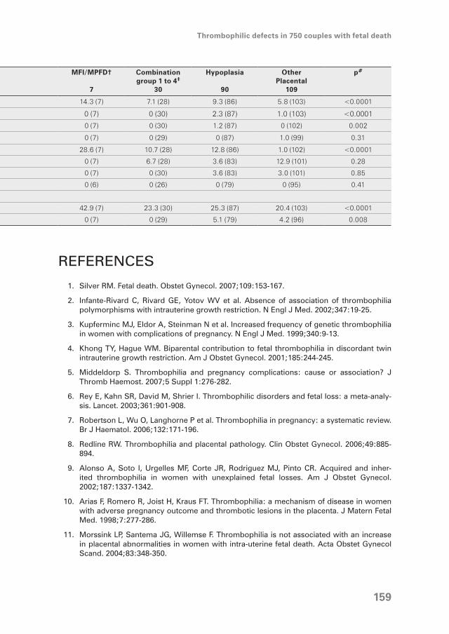
Table 4. Thrombophilic defects in 487 women with intrauterine fetal death due to placental pathology
Abnormal plasma levels Abruption
52
Infarction
197
FTV*
2
MFI/MPFD†
7
Combinationgroup 1 to 4‡
30
Hypoplasia
90
OtherPlacental
109
p#
Antithrombin ¯ 40.8 (49) 26.1 (184) 0 (2) 14.3 (7) 7.1 (28) 9.3 (86) 5.8 (103) <0.0001
Protein C ¯ 20.4 (49) 3.3 (184) 0 (2) 0 (7) 0 (30) 2.3 (87) 1.0 (103) <0.0001
Total protein S ¯ 10.2 (49) 1.7 (184) 0 (2) 0 (7) 0 (30) 1.2 (87) 0 (102) 0.002
Free protein S ¶̄ 4.1 (49) 0.6 (183) 0 (2) 0 (7) 0 (29) 0 (87) 1.0 (99) 0.31
VWF § 18.4 (49) 28.4 (183) 0 (2) 28.6 (7) 10.7 (28) 12.8 (86) 1.0 (102) <0.0001
Factor V Leiden 4.2 (48) 8.0 (176) 0 (2) 0 (7) 6.7 (28) 3.6 (83) 12.9 (101) 0.28
Prothrombin G20210A 6.3 (48) 3.4 (177) 0 (2) 0 (7) 0 (30) 3.6 (83) 3.0 (101) 0.85
Lupus anticoagulant 0 (48) 2.4 (167) 0 (1) 0 (6) 0 (26) 0 (79) 0 (95) 0.41
Any defect women 61.2 (49) 52.9 (187) 0 (2) 42.9 (7) 23.3 (30) 25.3 (87) 20.4 (103) <0.0001
Any defect couples 15.2 (46) 9.8 (173) 0 (2) 0 (7) 0 (29) 5.1 (79) 4.2 (96) 0.008
percentages (n tested) are given, *FTV: Fetal thrombotic vasculopathy, †MFI: Maternal floor infarct/MPFD: Massive perivillous fibrin deposition ‡combination groups 1 to 4: a combination of one of the following causes of death: abruption, infarction, FTV and MFI/MPFD #p-value indicates differences between all subgroups, ¶Free protein S¯ but normal total protein S, §VWF: Von Willebrand Factor REFERENCES
1. Silver RM. Fetal death. Obstet Gynecol. 2007;109:153-167.
2. Infante-Rivard C, Rivard GE, Yotov WV et al. Absence of association of thrombophilia polymorphisms with intrauterine growth restriction. N Engl J Med. 2002;347:19-25.
3. Kupferminc MJ, Eldor A, Steinman N et al. Increased frequency of genetic thrombophilia in women with complications of pregnancy. N Engl J Med. 1999;340:9-13.
4. Khong TY, Hague WM. Biparental contribution to fetal thrombophilia in discordant twin intrauterine growth restriction. Am J Obstet Gynecol. 2001;185:244-245.
5. Middeldorp S. Thrombophilia and pregnancy complications: cause or association? J Thromb Haemost. 2007;5 Suppl 1:276-282.
6. Rey E, Kahn SR, David M, Shrier I. Thrombophilic disorders and fetal loss: a meta-analy-sis. Lancet. 2003;361:901-908.
7. Robertson L, Wu O, Langhorne P et al. Thrombophilia in pregnancy: a systematic review. Br J Haematol. 2006;132:171-196.
8. Redline RW. Thrombophilia and placental pathology. Clin Obstet Gynecol. 2006;49:885-894.
9. Alonso A, Soto I, Urgelles MF, Corte JR, Rodriguez MJ, Pinto CR. Acquired and inher-ited thrombophilia in women with unexplained fetal losses. Am J Obstet Gynecol. 2002;187:1337-1342.
10. Arias F, Romero R, Joist H, Kraus FT. Thrombophilia: a mechanism of disease in women with adverse pregnancy outcome and thrombotic lesions in the placenta. J Matern Fetal Med. 1998;7:277-286.
11. Morssink LP, Santema JG, Willemse F. Thrombophilia is not associated with an increase in placental abnormalities in women with intra-uterine fetal death. Acta Obstet Gynecol Scand. 2004;83:348-350.
Thrombophilic defects in 750 couples with fetal death
159

12. Mousa HA, Alfirevic1 Z. Do placental lesions reflect thrombophilia state in women with adverse pregnancy outcome? Hum Reprod. 2000;15:1830-1833.
13. Stirling Y, Woolf L, North WR, Seghatchian MJ, Meade TW. Haemostasis in normal preg-nancy. Thromb Haemost. 1984;52:176-182.
14. Exner T, Triplett DA, Taberner D, Machin SJ. Guidelines for testing and revised criteria for lupus anticoagulants. SSC Subcommittee for the Standardization of Lupus Anticoagu-lants. Thromb Haemost. 1991;65:320-322.
15. RCOG. Fetal and perinatal pathology. Report of a joint working party. RCOG. 2001. London.
16. Bove KE. Practice guidelines for autopsy pathology: the perinatal and pediatric autop-sy. Autopsy Committee of the College of American Pathologists. Arch Pathol Lab Med. 1997;121:368-376.
17. Langston C, Kaplan C, Macpherson T et al. Practice guideline for examination of the placenta: developed by the Placental Pathology Practice Guideline Development Task Force of the College of American Pathologists. Arch Pathol Lab Med. 1997;121:449-476.
18. Kloosterman GJ. On intrauterine growth. The significance of prenatal care. Int J Gynaecol Obstet. 1970;895-912.
19. Korteweg FJ, Gordijn SJ, Timmer A et al. The Tulip classification of perinatal mortality: introduction and multidisciplinary inter-rater agreement. BJOG. 2006;113:393-401.
20. Fox H. Pathology of the Placenta. second ed. London: Saunders Company; 1997.
21. Kraus FT, Acheen VI. Fetal thrombotic vasculopathy in the placenta: cerebral thrombi and infarcts, coagulopathies, and cerebral palsy. Hum Pathol. 1999;30:759-769.
22. Redline RW, Pappin A. Fetal thrombotic vasculopathy: the clinical significance of exten-sive avascular villi. Hum Pathol. 1995;26:80-85.
23. Katzman PJ, Genest DR. Maternal floor infarction and massive perivillous fibrin deposi-tion: histological definitions, association with intrauterine fetal growth restriction, and risk of recurrence. Pediatr Dev Pathol. 2002;5:159-164.
24. Pinar H, Sung CJ, Oyer CE, Singer DB. Reference values for singleton and twin placental weights. Pediatr Pathol Lab Med. 1996;16:901-907.
25. Isermann B, Sood R, Pawlinski R et al. The thrombomodulin-protein C system is essential for the maintenance of pregnancy. Nat Med. 2003;9:331-337.
26. Marietta M, Facchinetti F, Sgarbi L et al. Elevated plasma levels of factor VIII in women with early recurrent miscarriage. J Thromb Haemost. 2003;1:2536-2539.
27. Kamphuisen PW, Lensen R, Houwing-Duistermaat JJ et al. Heritability of elevated factor VIII antigen levels in factor V Leiden families with thrombophilia. Br J Haematol. 2000;109:519-522.
28. Preston FE, Rosendaal FR, Walker ID et al. Increased fetal loss in women with heritable thrombophilia. Lancet. 1996;348:913-916.
29. de Galan-Roosen AE, Kuijpers JC, Rosendaal FR et al. Maternal and paternal throm-bophilia: risk factors for perinatal mortality. BJOG. 2005;112:306-311.
30. Martinelli I, Taioli E, Cetin I et al. Mutations in coagulation factors in women with unex-plained late fetal loss. N Engl J Med. 2000;343:1015-1018.
31. Lurie S, Feinstein M, Mamet Y. Disseminated intravascular coagulopathy in pregnancy: thorough comprehension of etiology and management reduces obstetricians’ stress. Arch Gynecol Obstet. 2000;263:126-130.
Chapter 8
160