university of dundee reducing stomatal density in barley ... · 2 jon hughes*1, christopher...
TRANSCRIPT

University of Dundee
Reducing stomatal density in barley improves drought tolerance without impacting onyieldHughes, Jonathan; Hepworth, Christopher; Dutton, Christian; Dunn, Jessica A.; Hunt, Lee;Stephens, Jennifer; Waugh, Robbie; Cameron, Duncan D.; Gray, Julie E.Published in:Plant Physiology
DOI:10.1104/pp.16.01844
Publication date:2017
Document VersionAccepted author manuscript
Link to publication in Discovery Research Portal
Citation for published version (APA):Hughes, J., Hepworth, C., Dutton, C., Dunn, J. A., Hunt, L., Stephens, J., ... Gray, J. E. (2017). Reducingstomatal density in barley improves drought tolerance without impacting on yield. Plant Physiology. DOI:10.1104/pp.16.01844
General rightsCopyright and moral rights for the publications made accessible in Discovery Research Portal are retained by the authors and/or othercopyright owners and it is a condition of accessing publications that users recognise and abide by the legal requirements associated withthese rights.
• Users may download and print one copy of any publication from Discovery Research Portal for the purpose of private study or research. • You may not further distribute the material or use it for any profit-making activity or commercial gain. • You may freely distribute the URL identifying the publication in the public portal.
Take down policyIf you believe that this document breaches copyright please contact us providing details, and we will remove access to the work immediatelyand investigate your claim.

Reducing stomatal density in barley improves drought tolerance without impacting on yield. 1
Jon Hughes*1, Christopher Hepworth*2, Chris Dutton1, Jessica A. Dunn1, Lee Hunt1, Jennifer Stephens3, 2
Robbie Waugh3, Duncan D. Cameron2 and Julie E. Gray1 3
Manipulation of a gene involved in the suppression of stomatal development in barley can reduce 4
stomatal density, leading to improved drought tolerance without deleterious effects on yield. 5
J.H. and C.H performed barley physiological and statistical analyses, C.H. and J.H. performed the 6
confocal microscopy; C.D. performed qPCR. J.H. carried out Arabidopsis experiments and J.A.D. 7
contributed to the stomatal analysis. L.H., J.S. and R.W. performed barley gene cloning and 8
transformation. J.E.G., L.H. and R.W. conceived and supervised the project. C.H created the figures; 9
C.H., J.H. and J.E.G. analysed the data and wrote the article with input from the other authors. 10
*Authors contributed equally to this work 11
1Department of Molecular Biology and Biotechnology, University of Sheffield, UK, 12 13 2Department of Animal and Plant Sciences, University of Sheffield, UK 14 15 3 The James Hutton Institute, Invergowrie, Dundee, Scotland 16 17 To whom correspondence should be addressed: E-mail: [email protected] 18
19
Abstract 20
The epidermal patterning factor (EPF) family of secreted signalling peptides regulate the frequency 21
of stomatal development in model dicot and basal land plant species. Here we identify and 22
manipulate the expression of a barley ortholog and demonstrate that when overexpressed HvEPF1 23
limits entry to, and progression through, the stomatal development pathway. Despite substantial 24
reductions in leaf gas exchange, barley plants with significantly reduced stomatal density show no 25
reductions in grain yield. In addition, HvEPF1OE barley lines exhibit significantly enhanced water use 26
efficiency, drought tolerance and soil water conservation properties. Our results demonstrate the 27
potential of manipulating stomatal frequency for the protection and optimisation of cereal crop 28
yields under future drier environments. 29
30
Introduction 31
With the global population set to rise to over 9 billion by 2050 and the predicted instability in global 32
climate patterns, fears over global food security continue to grow (Godfray et al., 2010). Prolonged 33
periods of drought and expanded zones of desertification are expected to become increasingly 34
prevalent as this century progresses (IPCC, 2014). The need to expand agriculture into areas of 35
marginal land, where drought is a severe inhibitor of sustainable agriculture (Fita et al., 2015), 36
continues to increase. 70% of global freshwater is already utilised for irrigation and rain-fed 37
agriculture is now the world’s largest consumer of water (Foley et al., 2011). A potential way to both 38
futureproof against climate change, and to expand crop production onto water-limited marginal 39
lands would be through improvements to crop drought tolerance and water use efficiency (WUE, the 40
ratio of carbon gained to water lost). 41
The vast majority of water is lost from crops via transpiration and reducing this loss provides a 42
potential route towards improving WUE and conserving soil water levels (Hepworth et al., 2015). To 43
this end, much research into the use of anti-transpirants was carried out in 1960’s and 70’s 44
(Davenport et al., 1972). However, although effective in improving water status and increasing fruit 45
size, these chemical solutions were never economically viable on an agricultural scale. 46
The majority of water loss from plants occurs via transpiration through epidermal pores known as 47
stomata, making these cellular structures an attractive target in the battle to prevent water loss. 48
Recently several laboratory studies have demonstrated that it is possible to improve drought 49
tolerance and WUE by reducing the frequency of stomata on leaves; by using genetic manipulation 50
or mutation to reduce stomatal density (SD) improved water use efficiency has been achieved across 51
several model dicot species including Arabidopsis (Yoo et al., 2010; Franks et al., 2015; Hepworth et 52
al., 2015), poplar (Lawson et al., 2014) and tobacco (Yu et al., 2008). In addition, the ectopic 53
expression of a putative transcription factor in maize has led to reduced stomatal density and gas 54
exchange in a monocot (Liu et al., 2015). 55
The manipulation of SD has been facilitated by microscopic studies which characterised the cellular 56
stages of the stomatal lineage, and molecular studies that revealed the developmental mechanisms 57
controlling their progression (Zhao & Sack, 1999; Han & Torii, 2016). The majority of these studies 58
have been carried out using the genetically tractable, model plant species Arabidopsis. During early 59
Arabidopsis leaf development, a subset of epidermal cells known as meristemoid mother cells 60

(MMCs) become primed to enter the stomatal lineage. Each MMC then undergoes an initial 61
asymmetric entry division to produce a meristemoid in addition to a larger daughter cell known as a 62
stomatal lineage ground cell (SLGC). SLGCs either differentiate directly into epidermal pavement 63
cells or undergo further asymmetric divisions to produce secondary meristemoids. Some 64
meristemoids can themselves undergo further asymmetric divisions, each of which reforms a 65
meristemoid and creates an additional SLGC. Each meristemoid eventually differentiates into a 66
guard mother cell, small and rounded in shape, prior to undergoing a symmetric division to form the 67
guard cell pair of the mature stomatal complex. These cell fate transitions and divisions, which 68
ultimately control the number and proportions of stomata and pavement cells in the mature leaf 69
epidermis, are controlled by a sub-group of related basic helix-loop-helix (bHLH) transcription 70
factors; SPCH, MUTE and FAMA (Ohashi-Ito & Bergmann, 2006; MacAlister et al., 2007; Pillitteri & 71
Torii, 2007). SPCH primarily directs expression of genes controlling meristemoid formation including 72
members of the cysteine-rich EPIDERMAL PATTERNING FACTOR (EPF) family of secreted signalling 73
peptides, which in turn activate a pathway that regulates SPCH stability, thus forming a feedback 74
loop that regulates the number of cells entering the stomatal lineage (Adrian et al., 2015; Simmons 75
& Bergmann, 2016). The best characterised negative regulators of stomatal density in this peptide 76
family are EPF1 and EPF2, which are numbered in order of their discovery (Hara et al., 2007; Hara et 77
al., 2009; Hunt & Gray, 2009). Both peptides act extracellularly within the aerial epidermal cell layer 78
to suppress stomatal development through activation of an intracellular MAP kinase signalling 79
pathway (Bergmann et al., 2004; Wang et al., 2007; Lampard et al., 2008). Although their functions 80
somewhat overlap, EPF2 acts earliest in stomatal development to restrict entry of cells into the 81
stomatal lineage, whilst EPF1 acts later to orient subsequent divisions of meristemoid cells and 82
enforce stomatal spacing through the ‘one-cell-spacing’ rule via the inhibition of MUTE expression 83
(Hara et al., 2007; Qi et al., 2017). Manipulation of the expression levels of these peptides in 84
Arabidopsis has led to significant improvements in drought tolerance and WUE in experiments 85
conducted in controlled-environment plant growth rooms (Doheny-Adams et al., 2012; Hepworth et 86
al., 2015). 87
In contrast to the Arabidopsis model system, our knowledge of stomatal development in crops is 88
relatively limited (Raissig et al., 2016). Although the grasses include many of our major global crops, 89
our molecular understanding of their transpirational control mechanisms remains extremely limited. 90
It is known from microscopic observations that grass stomata are formed by a single asymmetric cell 91
division that forms a stomatal precursor cell (a guard mother cell) and an epidermal pavement cell 92
(Stebbins & Jain, 1960). There are no further asymmetric divisions of the stomatal lineage cells 93
analogous to the repeated possible divisions that meristemoids undergo in Arabidopsis (Serna, 94

2011). The mature grass stomatal complex is formed by division of two neighbouring cells that give 95
rise to flanking subsidiary cells, and a symmetric division of the guard mother which produces two 96
dumbbell-shaped guard cells - rather than the characteristically kidney-shaped guard cells of most 97
dicots (Hetherington & Woodward, 2003; Serna, 2011). In contrast to dicots, all grass stomatal 98
development initiates at the leaf base. The patterning of stomata within the leaf epidermis also 99
differs in grasses, with stomata forming in straight files parallel to the leaf vein as opposed to the 100
‘scattered’ distribution seen in Arabidopsis (Stebbins & Khush, 1961; Geisler & Sack, 2002; Serna, 101
2011) 102
Despite these differences in stomatal shape and patterning it appears that the molecular control of 103
stomatal development has similarities across a wide range of plant species. Functional orthologs of 104
genes encoding for bHLH transcription factors involved in Arabidopsis stomatal development have 105
been identified in grasses including; rice, maize (Liu et al., 2009) and brachypodium (Raissig et al., 106
2016) and recently in the early diverging non-vascular mosses (Chater et al., 2016). EPF orthologs are 107
encoded across a range of plant genomes and have recently been shown to effectively regulate moss 108
stomatal patterning (Caine et al., 2016). However, currently it is still not known whether EPFs 109
function in controlling stomatal development in grasses. With the sequencing of the barley genome 110
in 2012 we were able to identify a putative EPF ortholog (HvEPF1, MLOC_67484) that is expressed at 111
low levels during development of aerial tissues (IBSC, 2012). Here we characterise the function of an 112
epidermal patterning factor in grasses. We report the ectopic overexpression of HvEPF1 and the 113
production of transgenic barley lines exhibiting altered stomatal development. Furthermore, our 114
generation of barley lines with reduced SD has provided us with the necessary tools to determine 115
the effect of reduced SD on transpiration, drought tolerance, water use efficiency and yield in a 116
cereal crop. 117
Results 118
11 genes encoding putative EPF-like secreted peptides were identified in the barley genome 119
sequence (IBSC, 2012) (Fig.S1) . MLOC67484 which we refer to here as HvEPF1 encodes a peptide 120
with extensive similarity to Arabidopsis epidermal patterning factors, and contains the 6 conserved 121
cysteine residues (Fig. 1a) that are characteristic of Arabidopsis epidermal patterning factors (Ohki et 122
al., 2011; Lau & Bergmann, 2012). Phylogenetic analysis of the encoded mature peptide sequence 123
indicated that within the Arabidopsis EPF family, HvEPF1 is most closely related to the known 124
inhibitors of stomatal development EPF1 and EPF2 which each contain two additional cysteine 125
residues (Fig. S1). To confirm that this barley peptide gene could function in stomatal regulation, 126
HvEPF1 was ectopically overexpressed in Arabidopsis under the control of the CaMV35S promoter. 127
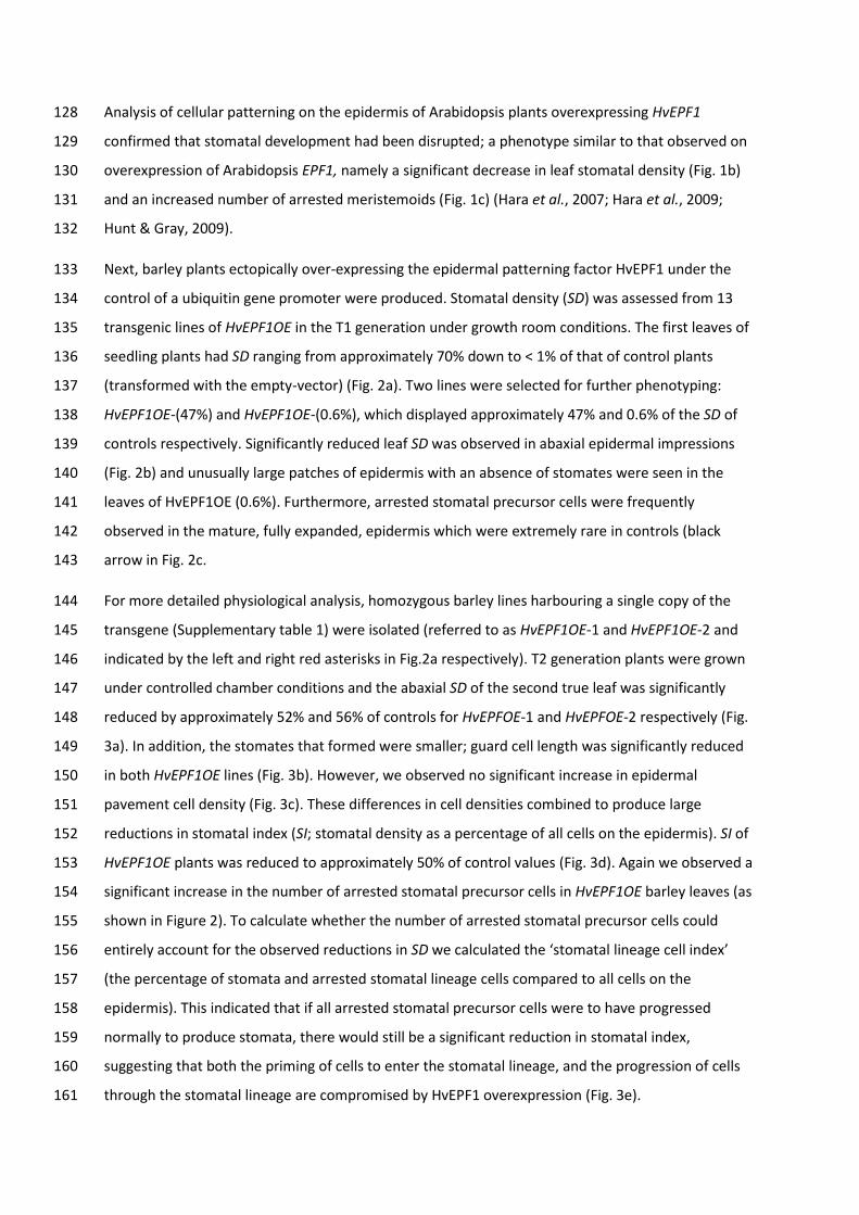
Analysis of cellular patterning on the epidermis of Arabidopsis plants overexpressing HvEPF1 128
confirmed that stomatal development had been disrupted; a phenotype similar to that observed on 129
overexpression of Arabidopsis EPF1, namely a significant decrease in leaf stomatal density (Fig. 1b) 130
and an increased number of arrested meristemoids (Fig. 1c) (Hara et al., 2007; Hara et al., 2009; 131
Hunt & Gray, 2009). 132
Next, barley plants ectopically over-expressing the epidermal patterning factor HvEPF1 under the 133
control of a ubiquitin gene promoter were produced. Stomatal density (SD) was assessed from 13 134
transgenic lines of HvEPF1OE in the T1 generation under growth room conditions. The first leaves of 135
seedling plants had SD ranging from approximately 70% down to < 1% of that of control plants 136
(transformed with the empty-vector) (Fig. 2a). Two lines were selected for further phenotyping: 137
HvEPF1OE-(47%) and HvEPF1OE-(0.6%), which displayed approximately 47% and 0.6% of the SD of 138
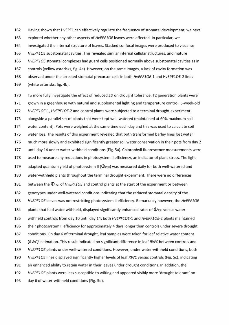
controls respectively. Significantly reduced leaf SD was observed in abaxial epidermal impressions 139
(Fig. 2b) and unusually large patches of epidermis with an absence of stomates were seen in the 140
leaves of HvEPF1OE (0.6%). Furthermore, arrested stomatal precursor cells were frequently 141
observed in the mature, fully expanded, epidermis which were extremely rare in controls (black 142
arrow in Fig. 2c. 143
For more detailed physiological analysis, homozygous barley lines harbouring a single copy of the 144
transgene (Supplementary table 1) were isolated (referred to as HvEPF1OE-1 and HvEPF1OE-2 and 145
indicated by the left and right red asterisks in Fig.2a respectively). T2 generation plants were grown 146
under controlled chamber conditions and the abaxial SD of the second true leaf was significantly 147
reduced by approximately 52% and 56% of controls for HvEPFOE-1 and HvEPFOE-2 respectively (Fig. 148
3a). In addition, the stomates that formed were smaller; guard cell length was significantly reduced 149
in both HvEPF1OE lines (Fig. 3b). However, we observed no significant increase in epidermal 150
pavement cell density (Fig. 3c). These differences in cell densities combined to produce large 151
reductions in stomatal index (SI; stomatal density as a percentage of all cells on the epidermis). SI of 152
HvEPF1OE plants was reduced to approximately 50% of control values (Fig. 3d). Again we observed a 153
significant increase in the number of arrested stomatal precursor cells in HvEPF1OE barley leaves (as 154
shown in Figure 2). To calculate whether the number of arrested stomatal precursor cells could 155
entirely account for the observed reductions in SD we calculated the ‘stomatal lineage cell index’ 156
(the percentage of stomata and arrested stomatal lineage cells compared to all cells on the 157
epidermis). This indicated that if all arrested stomatal precursor cells were to have progressed 158
normally to produce stomata, there would still be a significant reduction in stomatal index, 159
suggesting that both the priming of cells to enter the stomatal lineage, and the progression of cells 160
through the stomatal lineage are compromised by HvEPF1 overexpression (Fig. 3e). 161
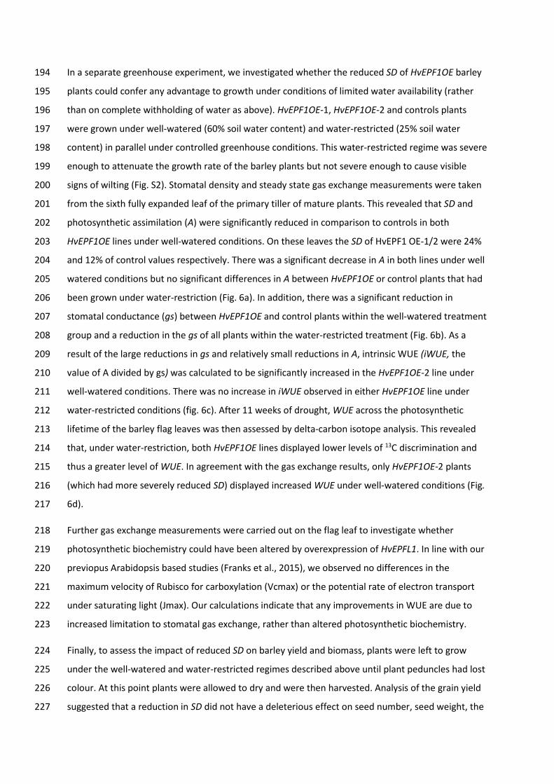
Having shown that HvEPF1 can effectively regulate the frequency of stomatal development, we next 162
explored whether any other aspects of HvEPF1OE leaves were affected. In particular, we 163
investigated the internal structure of leaves. Stacked confocal images were produced to visualise 164
HvEPF1OE substomatal cavities. This revealed similar internal cellular structures, and mature 165
HvEPF1OE stomatal complexes had guard cells positioned normally above substomatal cavities as in 166
controls (yellow asterisks, fig. 4a). However, on the same images, a lack of cavity formation was 167
observed under the arrested stomatal precursor cells in both HvEPF1OE-1 and HvEPF1OE-2 lines 168
(white asterisks, fig. 4b). 169
To more fully investigate the effect of reduced SD on drought tolerance, T2 generation plants were 170
grown in a greenhouse with natural and supplemental lighting and temperature control. 5-week-old 171
HvEPF1OE-1, HvEPF1OE-2 and control plants were subjected to a terminal drought experiment 172
alongside a parallel set of plants that were kept well-watered (maintained at 60% maximum soil 173
water content). Pots were weighed at the same time each day and this was used to calculate soil 174
water loss. The results of this experiment revealed that both transformed barley lines lost water 175
much more slowly and exhibited significantly greater soil water conservation in their pots from day 2 176
until day 14 under water-withheld conditions (Fig. 5a). Chlorophyll fluorescence measurements were 177
used to measure any reductions in photosystem II efficiency, an indicator of plant stress. The light 178
adapted quantum yield of photosystem II (ΦPSII) was measured daily for both well-watered and 179
water-withheld plants throughout the terminal drought experiment. There were no differences 180
between the ΦPSII of HvEPF1OE and control plants at the start of the experiment or between 181
genotypes under well-watered conditions indicating that the reduced stomatal density of the 182
HvEPF1OE leaves was not restricting photosystem II efficiency. Remarkably however, the HvEPF1OE 183
plants that had water withheld, displayed significantly enhanced rates of ΦPSII versus water-184
withheld controls from day 10 until day 14; both HvEPF1OE-1 and HvEPF1OE-2 plants maintained 185
their photosystem II efficiency for approximately 4 days longer than controls under severe drought 186
conditions. On day 6 of terminal drought, leaf samples were taken for leaf relative water content 187
(RWC) estimation. This result indicated no significant difference in leaf RWC between controls and 188
HvEPF1OE plants under well-watered conditions. However, under water-withheld conditions, both 189
HvEPF1OE lines displayed significantly higher levels of leaf RWC versus controls (Fig. 5c), indicating 190
an enhanced ability to retain water in their leaves under drought conditions. In addition, the 191
HvEPF1OE plants were less susceptible to wilting and appeared visibly more ‘drought tolerant’ on 192
day 6 of water-withheld conditions (Fig. 5d). 193
In a separate greenhouse experiment, we investigated whether the reduced SD of HvEPF1OE barley 194
plants could confer any advantage to growth under conditions of limited water availability (rather 195
than on complete withholding of water as above). HvEPF1OE-1, HvEPF1OE-2 and controls plants 196
were grown under well-watered (60% soil water content) and water-restricted (25% soil water 197
content) in parallel under controlled greenhouse conditions. This water-restricted regime was severe 198
enough to attenuate the growth rate of the barley plants but not severe enough to cause visible 199
signs of wilting (Fig. S2). Stomatal density and steady state gas exchange measurements were taken 200
from the sixth fully expanded leaf of the primary tiller of mature plants. This revealed that SD and 201
photosynthetic assimilation (A) were significantly reduced in comparison to controls in both 202
HvEPF1OE lines under well-watered conditions. On these leaves the SD of HvEPF1 OE-1/2 were 24% 203
and 12% of control values respectively. There was a significant decrease in A in both lines under well 204
watered conditions but no significant differences in A between HvEPF1OE or control plants that had 205
been grown under water-restriction (Fig. 6a). In addition, there was a significant reduction in 206
stomatal conductance (gs) between HvEPF1OE and control plants within the well-watered treatment 207
group and a reduction in the gs of all plants within the water-restricted treatment (Fig. 6b). As a 208
result of the large reductions in gs and relatively small reductions in A, intrinsic WUE (iWUE, the 209
value of A divided by gs) was calculated to be significantly increased in the HvEPF1OE-2 line under 210
well-watered conditions. There was no increase in iWUE observed in either HvEPF1OE line under 211
water-restricted conditions (fig. 6c). After 11 weeks of drought, WUE across the photosynthetic 212
lifetime of the barley flag leaves was then assessed by delta-carbon isotope analysis. This revealed 213
that, under water-restriction, both HvEPF1OE lines displayed lower levels of 13C discrimination and 214
thus a greater level of WUE. In agreement with the gas exchange results, only HvEPF1OE-2 plants 215
(which had more severely reduced SD) displayed increased WUE under well-watered conditions (Fig. 216
6d). 217
Further gas exchange measurements were carried out on the flag leaf to investigate whether 218
photosynthetic biochemistry could have been altered by overexpression of HvEPFL1. In line with our 219
previopus Arabidopsis based studies (Franks et al., 2015), we observed no differences in the 220
maximum velocity of Rubisco for carboxylation (Vcmax) or the potential rate of electron transport 221
under saturating light (Jmax). Our calculations indicate that any improvements in WUE are due to 222
increased limitation to stomatal gas exchange, rather than altered photosynthetic biochemistry. 223
Finally, to assess the impact of reduced SD on barley yield and biomass, plants were left to grow 224
under the well-watered and water-restricted regimes described above until plant peduncles had lost 225
colour. At this point plants were allowed to dry and were then harvested. Analysis of the grain yield 226
suggested that a reduction in SD did not have a deleterious effect on seed number, seed weight, the 227

average weight of seed, nor the harvest index (the ratio of above ground biomass to seed weight) 228
under either watering condition (Fig. 7 a-d). In addition, no differences in plant height nor above 229
ground biomass were found between any of the barley lines under either watering regime (Figs. S3, 230
S4). 231
232
Discussion 233
Grasses are an economically important plant group, with the cereal grasses being of critical 234
importance for both food and energy production. Considering future predicted climate scenarios, 235
the creation of drought tolerant cereals is a priority area for both crop improvement and scientific 236
research. 237
The bHLH transcription factors and epidermal patterning factors which were first discovered to be 238
regulators of stomatal development in Arabidopsis have been conserved from basal land plants 239
through to angiosperms including the grasses, and have been suggested as potential targets for crop 240
improvement (Peterson et al., 2010; Ran et al., 2013; Caine et al., 2016; Raissig et al., 2016). Here we 241
report the characterisation of a functional barley EPF ortholog, named HvEPF1, which acts in a 242
similar way to the Arabidopsis EPF1 and EPF2 signalling peptides to limit entry to and progression 243
through the stomatal cell lineage. Our overexpression of the barley HvEPF1 transcript in Arabidopsis 244
led to a significant reduction in SD indicating a level of conservation in peptide function between 245
monocots and dicots. The overexpression of HvEPF1 in barley led to severe reductions in both 246
stomatal formation, and in the entry of epidermal cells into the stomatal lineage, adding weight to 247
this conclusion. 248
The frequent presence of arrested stomatal precursor cells on the epidermis of both Arabidopsis and 249
barley HvEPF1OE plants (Fig. 1c and 2b) suggests that the mode of action of HvEPF1 is most similar 250
to that of Arabidopsis EPF1, which generates a similar epidermal phenotype when overexpressed 251
(Hara et al., 2007; Hara et al., 2009). That is, stomatal precursors enter the developmental lineage 252
but become arrested before the final symmetric cell division and maturation of the stomatal 253
complex. These HvEPF1OE oval-shaped arrested cells appear to halt their development at a 254
meristemoid-like or early guard mother cell stage, prior to transition into mature guard mother cells. 255
Thus, in addition to entry to the stomatal lineage, the transition to a mature guard mother cell that 256
is competent to divide and form a pair of guard cells appears to be regulated by HvEPF1. In 257
Arabidopsis this cellular transition step is under the control of the transcription factor MUTE (Fig. 8) 258
whose activity promotes expression of the receptor-like kinase ERECTA-LIKE1, which in turn 259

mediates EPF1 signalling and the subsequent autocrine inhibition of MUTE (Qi et al., 2017). Barley 260
MUTE may be regulated by HvEPF1 by a similar autocrine pathway and/or by phosphorylation as 261
grass MUTE genes (unlike Arabidopsis MUTE) encode potential MAP kinase phosphorylation sites 262
(Liu et al., 2009). Recent work in the monocot Brachypodium, has revealed MUTE to also be 263
involved in the formation of subsidiary cells (Raissig et al., 2017). In HvEPF1OE plants, stomatal 264
precursors arrest prior to the establishment of subsidiary cells suggesting the overexpression of 265
HvEPF1 may act to inhibit the expression of MUTE. 266
Despite their importance, we know remarkably little about the sequence of events leading to the 267
production of the air-filled spaces that underlie stomata. In conjunction with the stomatal pores, 268
these substomatal cavities facilitate high levels of gas exchange into plant photosynthetic mesophyll 269
cells, and mediate leaf water loss via transpiration. Using confocal microscopy, we could see no 270
evidence for the separation of mesophyll cells below arrested stomatal precursor cells in HvEPF1OE 271
leaves. Our observations begin to throw light on the developmental sequence leading to cavity 272
formation. The arrested stomatal precursor cells in HvEPF1OE do not form substomatal cavities, 273
suggesting that these cavities form following either GMC maturation, like the subsidiary cells of the 274
stomatal complex, or after guard cell pair formation. Alternatively, the formation of a substomatal 275
cavity may be required for guard mother cell maturation. 276
There is much evidence to support a negative correlation between stomatal density and stomatal 277
size across a range of species and Arabidopsis stomatal mutants i.e. those plants with relatively low 278
SD tend to produce larger stomates (Miskin & Rasmusson, 1970; Franks & Beerling, 2009; Doheny-279
Adams et al., 2012). Interestingly, the overexpression of HvEPF1 did not conform to this trend, and 280
led to barley plants with smaller, shorter guard cells. Thus if the EPF signalling pathway directly 281
regulates stomatal size in dicot species (and this remains to be demonstrated), it appears to act in 282
the opposite manner in grass stomatal size determination. 283
Through the ectopic over-expression of HvEPF1 we have created barley transformants with a range 284
of reductions in SD. Although barley plants with substantially reduced numbers of stomata showed 285
some attenuation of photosynthetic rates when well-watered, they exhibited strong drought 286
avoidance and drought tolerance traits when water was withheld. They had lower levels of water 287
loss via transpiration, and they were able to maintain higher levels of soil water content, and 288
delayed the onset of photosynthetic stress responses for several days longer than controls. 289
Remarkably when grown under water-limiting conditions (25% soil pot water content) two barley 290
lines with reductions in SD demonstrated significant improvements in WUE without any deleterious 291
effects on either plant growth or seed yield (biomass, seed weight or seed number). Indeed, it would 292

be interesting to determine whether both WUE and yield may be further optimised in reduced 293
stomatal density lines under less severe watering regimes or through less drastic reductions in SD 294
HvEPF1OE-2 plants (which had the lowest SD in this experiment) also displayed significantly 295
enhanced levels of drought tolerance and WUE under well-watered conditions, without 296
accompanying decreases in either grain yield or plant biomass. The increased iWUE observed in 297
these experiments was a result of a relatively moderate drop in A compared to a larger decrease in 298
gs, suggesting that A was not limited by internal CO2 concentration under the growth conditions of 299
our experiment (Yoo et al., 2009). This may also be a factor in explaining why reductions in SD did 300
not impact on the yield of HvEPF1OE plants. Further explanations include significantly reduced rates 301
of gs and thus water loss in HvEPF1OE plants allowing for more resources to be allocated to the 302
generation of seed and above ground biomass, at the potential cost to root development, as 303
described previously in Arabidopsis EPF over-expressing plants (Hepworth et al., 2016), or increased 304
soil water content leading to improved nutrient uptake and gs under water limitation (Van Vuuren et 305
al., 1997; Hepworth et al., 2015). Thus, although not tested in this study, reducing SD may also 306
enhance resource allocation or nutrient uptake capacity under water-restriction. 307
To conclude, this study describes the function and physiological effect of overexpressing a native 308
epidermal patterning factor in a grass species. The manipulation of HvEPF1 expression levels has 309
improved our understanding of stomatal developmental mechanisms in grasses, and has generated 310
a range of barley plants displaying significantly reduced SD. These barley plants exhibit substantially 311
improved drought tolerance and WUE without reductions in grain yield. This novel discovery adds 312
strength to the proposition that stomatal development represents an attractive target for breeders 313
when attempting to future-proof crops. 314
Materials and Methods 315
Vector Construction 316
HvEPF1 genomic gene was PCR amplified from Hordeum vulgare cultivar Golden Promise DNA using 317
primers in Table S1. The HVEPF1 gene is annotated as MLOC67484 at Ensembl Plants but is 318
incorrectly translated in this prediction. We used FGENESH to generate an alternative translation 319
which includes a putative signal sequence at the N-terminus. The PCR product was recombined 320
pENTR/D/TOP0 then by LR recombination into pCTAPi (Rohila et al., 2004) transformation vector 321
under the control of the CaMV35S promoter, and introduced into Arabidopsis thaliana Col-0 322
background by floral dip (Clough & Bent, 1998). Transformation and expression of the transgene 323
were confirmed by PCR and RT-PCR using the primers in Supplementary Table S2. 324

For barley transformation the HvEPF1 genomic gene was introduced by LR recombination into 325
pBRACT214 gateway vector under the control of the maize ubiquitin promoter, adjacent to a 326
hygromycin resistance gene under the control of a CaMV35S promoter (Fig. S4). Barley 327
transformations were carried out in background Golden Promise using the method described by 328
(Harwood et al., 2009). Plants harbouring just the hygromycin resistance cassette were regenerated 329
alongside to produce ‘empty-vector control’ plants. Potentially transformed plants were 330
regenerated on selective medium and T0 individuals genotyped to confirm gene insertion by PCR. 331
Gene copy number was estimated byIDna Genetics Ltd (www.idnagenetics.com) using a PCR based 332
method HvEPF1 overexpression was confirmed by RT-qPCR of T2 generation plants (Fig. S6). Total 333
RNA was extracted from 10 day old seedlings using Spectrum plant total RNA kit (Sigma, UK) and 334
reverse transcribed using Maxima H Minus Reverse Transcriptase cDNA synthesis kit (Thermo 335
Scientific). RT-qPCR was performed using a Rotor-Gene SYBR® Green PCR kit (Qiagen) with tubulin 336
and GADPH used as housekeeping reference genes, and primers outlined in the supplementary 337
supporting information (Supplementary table 2). Three plants of each transformed line were 338
amplified to confirm overexpression of the HvEPF1 gene. Fold induction values of gene expression 339
were normalised to average 2ΔCt values relative to empty-vector control samples. 340
Plant Growth Conditions 341
For plant growth, seeds were surfaced sterilised in 50% vol/vol ethanol/bleach before being placed 342
onto water saturated filter paper and placed into sealed Petri dishes in the appropriate growth 343
chamber. Arabidopsis plants were grown in a controlled growth chamber (Conviron model 344
MTPS120) at 22°C/16°C, 9 hours light, 150-200 μmol m−2 s−1, 15 hours dark, ambient [CO2] and 60% 345
humidity. Arabidopsis plants were kept well-watered throughout. Barley plants were grown in a 346
MTPS120 growth chamber at 21°C/15°C, 11 hours light at 300µmol.m-2.s1, 13 hours dark, ambient 347
[CO2] and 60% humidity. For plants grown under greenhouse conditions (Fig. 5, Fig. 6), temperature 348
was set at 20°C/16°C, 12 hours light, ambient humidity, and supplementary lighting ensured a 349
minimum of 200 μmol m−2 s−1 at bench level. 350
At 5 days post-germination individual barley seedlings were placed into 13cm diameter pots 351
containing homogenised M3 compost/perlite (4:1) with the addition of Osmocote. For initial 352
phenotyping and leaf developmental characterisation (Fig. 2, Fig. 3, Fig. 4) plants were kept well-353
watered. For the water-restricted experiment, (Fig. 6, Fig. 7) plants were maintained at either 60% 354
(well-watered) or 25% (water-restricted) of soil saturation by the daily weighing of pots. 355
Microscopy and cell counts 356

For both Arabidopsis and barley, stomatal and epidermal cell counts were taken from the abaxial 357
surface of mature, fully expanded leaves or cotyledons. Cell counts were taken from the widest 358
section of the first true leaf avoiding the mid vein. Dental resin (Coltene Whaledent, Switzerland) 359
was applied in the region of maximum leaf width and left to set before removing the leaf and 360
applying clear nail varnish to the resin. Stomatal counts were determined from nail varnish 361
impressions by light microscopy (Olympus BX51). 5 areas per leaf were sampled from 4-8 plants of 362
each genotype and treatment. For epidermal imaging (Fig. 2b-d), mature leaves were excised and 363
the central vein of the leaf cut away. Leaf tissue was then serially dehydrated in ethanol. Samples 364
were then placed into modified Clarke’s solution (4:1 ethanol to glacial acetic acid solution) then 365
cleared in 50% bleach overnight. 366
For epidermal phenotyping, the second fully expanded mature leaf of seedings were excised and a 3-367
5cm strip midway along the proximodistal axis of these leaves were cut out. These leaf samples were 368
then submerged in Clarke’s solution (3:1 ethanol to glacial acetic acid solution). Following 1 hour of 369
vacuum infiltration the samples were left in Clarke’s solution for 24 hours for fixation. Once fixed the 370
samples were transferred into 100% ethanol. Prior to imaging the leaf samples were cleared in 50% 371
bleach solution overnight. The midrib of each sample was then excised and the remaining leaf 372
sections mounted in deionised water on microscope slides for imaging. Samples were viewed by 373
light microscopy (Olympus BX51) using differential interference contrast functionality. For confocal 374
microscopy (Fig. 4a, Fig4b), barley samples were prepared as described (Wuyts et al., 2010) and 375
viewed on a Olympus FV1000 using 20X UPlan S-Apo N.A. 0.75 objective, 543nm laser, 555-655nm 376
emission and Fluorview software . 377
Physiological measurements 378
Throughout the terminal drought experiment the light adapted quantum yield of photosystem II 379
(ΦPSII) was measured daily for both well-watered and water-withheld plants. The most recent fully 380
expanded leaf of the primary tiller was selected for the measurement at day 1 and the same leaf was 381
then monitored throughout the experiment. Readings were taken using a FluorPen FP100 (Photon 382
Systems Instruments) with a saturating pulse of 3000 µmol m-2 s-1. Following the onset of the 383
drought treatment the pots were weighed every day and used to calculate the percentage of initial 384
soil water content remaining. Well-watered controls were maintained at 60% soil water content. 385
Leaf relative water content was determined from excised leaves from well-watered or droughted 386
and their fresh weight measured immediately and leaves were floated on water overnight and 387
weighed to record the hydrated weight. They were oven-dried overnight and weighed to obtain their 388

dry weight; the RWC was calculated using the following formula RWC (%) = (fresh weight−dry 389
weight)/ (hydrated weight−dry weight)*100. 390
A LI-6400 portable photosynthesis system (Licor, Lincoln, NE) was used to carry out infrared gas 391
analysis (IRGA) on the sixth, fully expanded, leaf from the primary tiller whilst still attached to the 392
plant. Relative humidity inside the IRGA chamber was kept at 60%-65% using self-indicating 393
desiccant, flow rate was set at 300 µmol.s-1 and leaf temperature at 20°C. Reference [CO2] was 394
maintained at 500ppm and light intensity at 200µmol.m-2.s1. Plants were allowed to equilibrate for 395
40-45 minutes the IRGA chamber being matched at least every 15 minutes. Once readings were 396
stable measurements were taken every 20 seconds for 5 minutes. For soil water content 397
calculations, the weight of pots containing water saturated (100% water content) or oven dried (0%) 398
compost mix was first determined. Pots were then maintained at either 60% or 25% soil water 399
content by weighing and addition of the appropriate amount of water every two days. 400
401
For carbon isotope discrimination (Fig. 6d), δ13C was assessed from the flag leaf of 5 plants from 402
each of the two watering regimes (well-watered and restricted-watered), as described previously 403
(Hepworth et al., 2015). 404
Once plants had matured and dried down the plants were harvested, with the total number and 405
weight of seeds per plant being recorded and the average seed weight being calculated. All above-406
ground vegetative tissue was dried in an oven at 80oC for two days and then weighed to provide the 407
dry weight. Harvest index (ratio of yield to above-ground biomass) was then calculated. 408
Statistical analysis 409
All comparisons were performed on Graph Pad Prism software. The appropriate post-hoc tests were 410
conducted once significance was confirmed using an ANOVA test and an alpha level of 0.05 or below 411
as significant. 412
Figure legends 413
Figure 1. HvEPF1 shares sequence similarity with Arabidopsis EPF1 and EPF2, and can restrict 414
Arabidopsis stomatal development. (a) Alignment of the putative HvEPF1 mature signalling peptide 415
with members of the Arabidopsis EPF family of signalling peptides. Conserved cysteine residues are 416
highlighted. Amino acid sequences for the mature peptide region were aligned using Multalin and 417
displayed using Boxshade. (b) Overexpression of HvEPF1 under the control of the CaMV35S 418
promoter in Arabidopsis leads to a significant decrease in stomatal density. (c) Epidermal tracings 419

from Arabidopsis cotyledons overexpressing EPF1, EPF2, and HvEPF1 alongside the background 420
control Col-0. Red dots mark location of stomata whilst green dots mark location of arrested 421
meristemoids. N=5 plants, asterisks indicate P<0.05, (Dunnett’s test after one-way ANOVA). Error 422
bars represent SE. 423
Figure 2. Over-expression of HvEPF1 in barley arrests stomatal development and reduces stomatal 424
density. (a) The abaxial stomatal density (SD) of barley plants transformed to ectopically over-425
express HvEPF1 (grey bars) compared to control lines transformed with the empty-vector (black 426
bars). All T1 generation HvEPF1 over-expressing lines demonstrated a significant reduction in SD in 427
comparison to both control lines. Lines chosen for further phenotyping in T2 generations are 428
indicated (red asterisks). (b) Traced abaxial epidermal impressions of T1 generation control, 429
HvEPF1OE-(47%) and HvEPF1OE-(0.6%) lines illustrating the reduction in SD. Red dots denote 430
positions of stomatal complexes. (c) Abaxial epidermal micrographs of HvEPF1OE plants. Black arrow 431
indicates arrested stomatal precursor cell. N=4-8 plants. Asterisks indicated significance to at least 432
P<0.05 versus control lines (Dunnett’s test after one-way ANOVA. (Error bars represent SE. 433
Figure 3. Stomatal characteristics of barley plants overexpressing HvEPF1. (a) Abaxial stomatal 434
densities of HvEPF1 overexpressing T2 barley lines harbouring a single copy of the transgene are 435
significantly decreased. HvEPF1OE-1 (white bars) and HvEPF1OE-2 (grey bars) compared to control 436
lines (black bars). (b) Guard cell length is significantly decreased in both HvEPF1OE lines. (c) 437
Pavement cell density is similar to that of the control in both HvEPF1OE lines. (d) Stomatal index is 438
significantly decreased in both HvEPF1OE lines. (e) Stomatal lineage index (the ratio of stomata and 439
arrested stomatal precursor cells to the total number of epidermal cells) is significantly decreased in 440
both HvEPF1OE lines. N=5 plants, asterisks indicate P<0.05, (Dunnett’s test after one-way ANOVA). 441
Error bars represent SE. 442
Figure 4. Cellular structure of HvEPF1OE stomatal complexes. (a) Representative propidium iodide 443
stained confocal image of a Z-plane below the HvEPF1OE-1 abaxial epidermal surface. Yellow 444
asterisks mark the location of the substomatal cavity under mature guard cells. (b) Higher Z-plane 445
image of the same field of view as (a) to reveal position of stomata. White asterisks mark the 446
location of arrested stomatal precursors and the lack of underlying substomatal cavities in (a). 447
Figure 5. Reducing barley stomatal density enhances drought tolerance though conserving soil and 448
plant water content. (a) 5 week old HvEPF1OE-1 and HvEPF1OE-2 barley plants maintain 449
significantly higher soil water content in comparison to control plants when water is withheld from 450
days 2-14. (b) Both HvEPF1OE-1 and HvEPF1OE-2 lines show significantly higher light adapted 451
quantum yields (ΦPSII) from 10 to 14 days after water was withheld (square symbols; plants from 452

same experiment as (a)). There were no significant differences between ΦPSII of well-watered plants 453
(circular symbols). (c) Relative water content (RWC) of barley leaves from HvEPF1OE lines was 454
significantly higher than controls after 6 days without watering. There were no differences in RWC 455
between well-watered plants. (d) Photograph of representative plants to illustrate enhanced turgor 456
maintenance in HvEPF1OE-1 and HvEPF1OE-2 on day 6 of water-withheld conditions. N=5 plants, 457
asterisk indicates significance to at least P<0.05 (Dunnett’s tests after one-way ANOVA for each 458
watering group). Error bars represent SE. 459
Figure 6. Reducing barley stomatal density lowers stomatal conductance and enhances water use 460
efficiency. (a) Under well-watered conditions a significant decrease in rate of carbon assimilation 461
was observed in both HvEPF1OE lines. Under water-restricted conditions there was no difference in 462
assimilation. (b) Stomatal conductance (gs) was significant decreased in HvEPF1OE lines grown 463
under well-watered conditions in comparison to controls. Under water-restricted conditions there 464
was no difference in gs. (c) Under well-watered conditions, a significant improvement in intrinsic 465
water use efficiency (iWUE) was observed in the HvEPF1OE-2 line when compared to control plants. 466
Under water-restricted conditions there was no difference in iWUE. (d) Carbon isotope 467
discrimination revealed a significant improvement in water use efficiency of the HvEPF1OE-2 barley 468
line under well-watered conditions. Under water-restricted conditions, both HvEPF1OE lines 469
displayed significantly improved water use efficiency in comparison to controls. N=5 plants, asterisk 470
indicates significance to at least P<0.05 (Dunnett’s tests after one-way ANOVA for each watering 471
group). Error bars represent SE. 472
Figure 7. Reducing stomatal density in barley has no deleterious effect on yield. No significant 473
differences in (a) seed number, (b) total weight of seed per plant, (c) average weight of individual 474
seeds, (d) harvest index (the ratio of yield to total shoot biomass) were observed between 475
HvEPF1OE-1, HvEPF1OE-2 and control plants under either watering condition. N=5 plants. Error bars 476
represent SE. 477
Figure 8. HvEPF1 acts to prevent cells entering the stomatal lineage, guard mother cell maturation 478
and substomatal cavity and subsidiary cell formation. Schematic to illustrate the putative mode of 479
action of HvEPF1 in barley stomatal development. Left to right: Undifferentiated epidermal cells at 480
the base of leaves are formed in cellular files. Cells in some files gain the capacity to divide 481
asymmetrically to create small stomatal precursor cells shown here as immature guard mother cells 482
(GMC, green). A developmental step, potentially under the control of the transcription factor MUTE, 483
stimulates guard mother cell maturation (dark green) and division of adjacent epidermal cells to 484
form subsidiary cells (SC, orange). Mature GMCs then divide symmetrically to form pairs of dumbbell 485

shaped guard cells (red). In the underlying mesophyll layer (M, green shaded regions) a substomatal 486
cavity forms during either the mature GMC or guard cell stage, although the exact developmental 487
staging of this is process is unknown. In the HvEPF1 overexpressing plants, HvEPF1 prevents GMC 488
maturation perhaps through the suppression of MUTE activity, resulting in arrested GMCs which are 489
unable to differentiate into mature stomatal complexes complete with subsidiary cells, guard cells 490
and substomatal cavities. Drawn with reference to Brachypodium development in Raissig et al. 2016. 491
492
Supplemental Data 493
Supplemental Figure 1. Phylogenetic tree of predicted Arabidopsis and barley epidermal patterning 494
factor peptide sequences constructed using Multalin. Barley annotations taken from Ensembl Plants 495
apart from HvSto7, which is a putative unannotated EPFL9/Stomagen on Chromosome 7. HvEPF1 496
highlighted in red. 497
Supplemental Figure 2. Growth of barley plants is inhibited by the water-restricted conditions used 498
in this study (25% soil water content) in comparison to growth in well-watered conditions (60% soil 499
water). From left to right: Control plant well-watered, control water-restricted, HvEPF1OE-1 well 500
watered, HvEPF1OE-1 water-restricted, HvEPF1OE-2 well-watered and HvEPF1OE-2 water-restricted. 501
Supplemental Figure 3. Plant heights of controls and HvEPF1OE-1 or HvEPF1OE-2 were not 502
significantly different within either well-watered or water-restricted conditions. Error bars represent 503
SE. 504
Supplemental Figure 4. Above ground biomass of control and HvEPF1OE-1 or HvEPF1OE-2 plant 505
lines were not significantly different under either well-watered or water-restricted conditions. N=5 506
plants. Error bars represent SE. 507
Supplemental Figure 5. Schematic of the gene expression construct inserted into the barley genome 508
to overexpress the HvEPF1 gene 509
Supplemental Figure 6. qPCR results the confirming significant overexpression of HvEPF1 the barley 510
lines detailed in the manuscript. N=5 plants, asterisk indicates significance to at least P<0.05 511
(Dunnett’s tests after one-way ANOVA). Error bars represent SE. 512
Supplemental Table 1. Copy number data for transformed plant lines used in this study. 513
Supplemental Table 2. Primer sequences used for PCR and RT-qPCR detailed in the methods section 514
of the manuscript. 515

516
Acknowledgements 517
We would like to thank Dr. Jennifer Sloan for help with seed harvest, Dr. Heather Walker and 518
Gemma Newsome for assistance with mass-spectrometry. We acknowledge the BBSRC, EPSRC, the 519
Gatsby Charitable Foundation and the Grantham Foundation for funding. 520
521
522
523
524
525
526
References 527
Adrian J, Chang J, Ballenger CE, Bargmann BO, Alassimone J, Davies KA, Lau OS, Matos JL, Hachez 528 C, Lanctot A, et al. 2015. Transcriptome dynamics of the stomatal lineage: birth, 529 amplification, and termination of a self-renewing population. Dev Cell 33(1): 107-118. 530
Caine RS, Chater CC, Kamisugi Y, Cuming AC, Beerling DJ, Gray JE, Fleming AJ. 2016. An ancestral 531 stomatal patterning module revealed in the non-vascular land plant Physcomitrella patens. 532 Development 143(18): 3306-3314. 533
Chater CC, Caine RS, Tomek M, Wallace S, Kamisugi Y, Cuming AC, Lang D, MacAlister CA, Casson S, 534 Bergmann DC, et al. 2016. Origin and function of stomata in the moss Physcomitrella 535 patens. Nature Plants 2: 16179. 536
Clough SJ, Bent AF. 1998. Floral dip: a simplified method for Agrobacterium-mediated 537 transformation of Arabidopsis thaliana. Plant J 16(6): 735-743. 538
Davenport DC, Fisher MA, Hagan RM. 1972. Some Counteractive Effects of Antitranspirants. Plant 539 Physiology 49(5): 722-724. 540
Doheny-Adams T, Hunt L, Franks PJ, Beerling DJ, Gray JE. 2012. Genetic manipulation of stomatal 541 density influences stomatal size, plant growth and tolerance to restricted water supply 542 across a growth carbon dioxide gradient. Philosophical Transactions of the Royal Society of 543 London B: Biological Sciences 367(1588): 547-555. 544
Fita A, Rodríguez-Burruezo A, Boscaiu M, Prohens J, Vicente O. 2015. Breeding and Domesticating 545 Crops Adapted to Drought and Salinity: A New Paradigm for Increasing Food Production. 546 Frontiers in Plant Science 6: 978. 547
Foley JA, Ramankutty N, Brauman KA, Cassidy ES, Gerber JS, Johnston M, Mueller ND, O/'Connell 548 C, Ray DK, West PC, et al. 2011. Solutions for a cultivated planet. Nature 478(7369): 337-549 342. 550
Franks PJ, Beerling DJ. 2009. Maximum leaf conductance driven by CO2 effects on stomatal size and 551 density over geologic time. Proceedings of the National Academy of Sciences 106(25): 10343-552 10347. 553
Franks PJ, W. Doheny-Adams T, Britton-Harper ZJ, Gray JE. 2015. Increasing water-use efficiency 554 directly through genetic manipulation of stomatal density. New Phytologist 207(1): 188-195. 555
Geisler MJ, Sack FD. 2002. Variable timing of developmental progression in the stomatal pathway in 556 Arabidopsis cotyledons. New Phytologist 153(3): 469-476. 557

Godfray HCJ, Beddington JR, Crute IR, Haddad L, Lawrence D, Muir JF, Pretty J, Robinson S, Thomas 558 SM, Toulmin C. 2010. Food Security: The Challenge of Feeding 9 Billion People. Science 559 327(5967): 812-818. 560
Han S-K, Torii KU. 2016. Lineage-specific stem cells, signals and asymmetries during stomatal 561 development. Development 143(8): 1259-1270. 562
Hara K, Kajita R, Torii KU, Bergmann DC, Kakimoto T. 2007. The secretory peptide gene EPF1 563 enforces the stomatal one-cell-spacing rule. Genes & Development 21(14): 1720-1725. 564
Hara K, Yokoo T, Kajita R, Onishi T, Yahata S, Peterson KM, Torii KU, Kakimoto T. 2009. Epidermal 565 cell density is autoregulated via a secretory peptide, EPIDERMAL PATTERNING FACTOR 2 in 566 Arabidopsis leaves. Plant Cell Physiol 50(6): 1019-1031. 567
Harwood WA, Bartlett JG, Alves SC, Perry M, Smedley MA, Leyland N, Snape JW. 2009. Barley 568 transformation using Agrobacterium-mediated techniques. Methods Mol Biol 478: 137-147. 569
Hepworth C, Doheny-Adams T, Hunt L, Cameron DD, Gray JE. 2015. Manipulating stomatal density 570 enhances drought tolerance without deleterious effect on nutrient uptake. New Phytologist 571 208(2): 336-341. 572
Hepworth C, Turner C, Landim MG, Cameron D, Gray JE. 2016. Balancing Water Uptake and Loss 573 through the Coordinated Regulation of Stomatal and Root Development. PLoS One 11(6): 574 e0156930. 575
Hetherington AM, Woodward FI. 2003. The role of stomata in sensing and driving environmental 576 change. Nature 424(6951): 901-908. 577
Hunt L, Gray JE. 2009. The signaling peptide EPF2 controls asymmetric cell divisions during stomatal 578 development. Curr Biol 19(10): 864-869. 579
IBSC 2012. A physical, genetic and functional sequence assembly of the barley genome. International 580 Barley Genome Sequencing Consortium: Nature Publishing Group, a division of Macmillan 581 Publishers Limited. All Rights Reserved. 711-716. 582
IPCC. 2014. IPCC, 2014: Climate Change 2014: Synthesis Report. Contribution of Working Groups I, II 583 and III to the Fifth Assessment Report of the Intergovernmental Panel on Climate Change. 584 151 585
Lau OS, Bergmann DC. 2012. Stomatal development: a plant's perspective on cell polarity, cell fate 586 transitions and intercellular communication. Development 139(20): 3683-3692. 587
Lawson S, Pijut P, Michler C. 2014. The cloning and characterization of a poplar stomatal density 588 gene. Genes & Genomics 36(4): 427-441. 589
Liu T, Ohashi-Ito K, Bergmann DC. 2009. Orthologs of Arabidopsis thaliana stomatal bHLH genes and 590 regulation of stomatal development in grasses. Development 136(13): 2265-2276. 591
Liu Y, Yuan J, Ma H, Song J, Wang L, Weng Q. 2015. Characterization and functional analysis of a B3 592 domain factor from zea mays. Journal of Applied Genetics 56(4): 427-438. 593
MacAlister CA, Ohashi-Ito K, Bergmann DC. 2007. Transcription factor control of asymmetric cell 594 divisions that establish the stomatal lineage. Nature 445(7127): 537-540. 595
Miskin E, Rasmusson DC. 1970. Frrequency and Distribution of Stomata in Barley. Crop Science 596 10(5): 575-578. 597
Ohashi-Ito K, Bergmann DC. 2006. Arabidopsis FAMA controls the final proliferation/differentiation 598 switch during stomatal development. Plant Cell 18(10): 2493-2505. 599
Ohki S, Takeuchi M, Mori M. 2011. The NMR structure of stomagen reveals the basis of stomatal 600 density regulation by plant peptide hormones. Nature Communications 2: 512. 601
Peterson KM, Rychel AL, Torii KU. 2010. Out of the mouths of plants: the molecular basis of the 602 evolution and diversity of stomatal development. Plant Cell 22(2): 296-306. 603
Pillitteri LJ, Torii KU. 2007. Breaking the silence: three bHLH proteins direct cell-fate decisions during 604 stomatal development. BioEssays 29(9): 861-870. 605
Qi X, Han SK, Dang JH, Garrick JM, Ito M, Hofstetter AK, Torii KU. 2017. Autocrine regulation of 606 stomatal differentiation potential by EPF1 and ERECTA-LIKE1 ligand-receptor signaling. Elife 607 6. 608

Raissig MT, Abrash E, Bettadapur A, Vogel JP, Bergmann DC. 2016. Grasses use an alternatively 609 wired bHLH transcription factor network to establish stomatal identity. Proceedings of the 610 National Academy of Sciences 113(29): 8326-8331. 611
Raissig MT, Matos JL, Gil MX, Kornfeld A, Bettadapur A, Abrash E, Allison HR, Badgley G, Vogel JP, 612 Berry JA, et al. 2017. Mobile MUTE specifies subsidiary cells to build physiologically 613 improved grass stomata. Science 355(6330): 1215-1218. 614
Ran J-H, Shen T-T, Liu W-J, Wang X-Q. 2013. Evolution of the bHLH Genes Involved in Stomatal 615 Development: Implications for the Expansion of Developmental Complexity of Stomata in 616 Land Plants. PLoS One 8(11): e78997. 617
Rohila JS, Chen M, Cerny R, Fromm ME. 2004. Improved tandem affinity purification tag and 618 methods for isolation of protein heterocomplexes from plants. Plant J 38(1): 172-181. 619
Serna L. 2011. Stomatal development in Arabidopsis and grasses: differences and commonalities. Int 620 J Dev Biol 55(1): 5-10. 621
Simmons AR, Bergmann DC. 2016. Transcriptional control of cell fate in the stomatal lineage. 622 Current Opinion in Plant Biology 29: 1-8. 623
Stebbins GL, Khush GS. 1961. Variation in the organization of the stomatal complex in the leaf 624 epidermis of monocotyledons and its bearing on their phylogeny. American Journal of 625 Botany: 51-59. 626
Van Vuuren MMI, Robinson D, Fitter AH, Chasalow SD, Williamson L, Raven JA. 1997. Effects of 627 elevated atmospheric CO2 and soil water availability on root biomass, root length, and N, P 628 and K uptake by wheat. New Phytologist 135(3): 455-465. 629
Wuyts N, Palauqui J-C, Conejero G, Verdeil J-L, Granier C, Massonnet C. 2010. High-contrast three-630 dimensional imaging of the Arabidopsis leaf enables the analysis of cell dimensions in the 631 epidermis and mesophyll. Plant Methods 6(1): 1-14. 632
Yoo CY, Pence HE, Hasegawa PM, Mickelbart MV. 2009. Regulation of Transpiration to Improve 633 Crop Water Use. Critical Reviews in Plant Sciences 28(6): 410-431. 634
Yoo CY, Pence HE, Jin JB, Miura K, Gosney MJ, Hasegawa PM, Mickelbart MV. 2010. The 635 Arabidopsis GTL1 Transcription Factor Regulates Water Use Efficiency and Drought 636 Tolerance by Modulating Stomatal Density via Transrepression of SDD1. The Plant Cell Online 637 22(12): 4128-4141. 638
Yu H, Chen X, Hong Y-Y, Wang Y, Xu P, Ke S-D, Liu H-Y, Zhu J-K, Oliver DJ, Xiang C-B. 2008. Activated 639 Expression of an Arabidopsis HD-START Protein Confers Drought Tolerance with Improved 640 Root System and Reduced Stomatal Density. The Plant Cell 20(4): 1134-1151. 641
Zhao L, Sack FD. 1999. Ultrastructure of stomatal development in Arabidopsis (Brassicaceae) leaves. 642 Am J Bot 86(7): 929-939. 643
644