universidad de granada -...
TRANSCRIPT
UNIVERSIDAD DE GRANADA
FACULTAD DE CIENCIAS
Departamento de Química Inorgánica
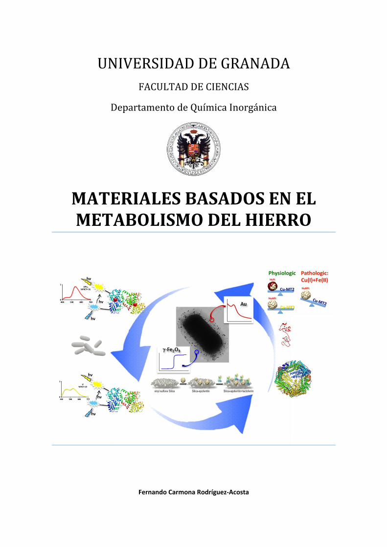
MATERIALES BASADOS EN EL METABOLISMO DEL HIERRO
Fernando Carmona Rodríguez-Acosta

2
Editor: Universidad de Granada. Tesis DoctoralesAutor: Fernando Carmona Rodríguez AcostaISBN: 978-84-9163-197-2URI: http://hdl.handle.net/10481/46358

3
UNIVERSIDAD DE GRANADA
FACULTAD DE CIENCIAS
Departamento de Química Inorgánica
PROGRAMA OFICIAL DE DOCTORADO EN QUÍMICA
TESIS DOCTORAL
MATERIALES BASADOS EN EL
METABOLISMO DEL HIERRO
Fernando Carmona Rodríguez-Acosta
Granada, junio 2014

4

5
MATERIALES BASADOS EN EL METABOLISMO
DEL HIERRO
Memoria de Tesis Doctoral presentada por Fernando Carmona Rodríguez-Acosta
para aspirar al título de Doctor por la Universidad de Granada
Fdo. Fernando Carmona Rodríguez-Acosta
EL DIRECTOR DE LA TESIS DOCTORAL:
José Manuel Domínguez Vera Catedrático del Departamento de Química Inorgánica
Instituto de Biotecnología Universidad de Granada

6

7
El doctorando Fernando Carmona Rodríguez-Acosta y el director de la Tesis José
Manuel Domínguez Vera garantizamos, al firmar esta Tesis Doctoral, que el trabajo
ha sido realizado por el doctorando bajo la dirección del director de la Tesis y hasta
donde nuestro conocimiento alcanza, en la realización del trabajo, se han respetado
los derechos de otros autores a ser citados, cuando se han utilizado sus resultados o
publicaciones.
Granada, junio de 2014
El Doctorando
Fdo.: Fernando Carmona Rodríguez-Acosta
El Director de la Tesis
Fdo.: José Manuel Domínguez Vera
Catedrático del Departamento de Química Inorgánica. Instituto de Biotecnología
Universidad de Granada

8
Esta tesis doctoral se ha llevado a cabo en los laboratorios del departamento
química inorgánica de la Universidad de Granada en el grupo BIONANOMET
gracias al Ministerio de Ciencia e Innovación por la concesión de mi beca FPI y a la
financiación de los proyectos CTQ-2009-09344 y CTQ-2012-32236, así como al
proyecto POSTBIO financiado por BIOSEARCH S. A. y a la Junta de Andalucía
(grupo FQM-368).

9
a Papá, Mamá, Miguel y Rosalía,
las personas que más quiero
en este mundo

10

11
RESUMEN Y ESTRUCTURA DE LA TESIS DOCTORAL

12

13
La presente Tesis Doctoral se enmarca en el estudio de aspectos concretos del
metabolismo de hierro, por una parte con un enfoque más clásico, que pretende
entender mejor la función de determinadas metaloproteínas de hierro y por otra
parte, con un enfoque más innovador, intentando recrear escenarios biológicos
complejos compuestos de varias metaloproteínas donde poder estudiar las
consecuencias patológicas que una disfunción en alguna de ellas podría
desencadenar.
Además, aprovechando el conocimiento generado, hemos llevado a cabo el
desarrollo de nuevos materiales, que podríamos considerar bioinspirados en el
metabolismo del hierro, con el objetivo de que puedan ser conceptualmente útiles
para intervenir en dichos desajustes y actuar a nivel terapéutico en las patologías
que de ello se derivan.
Los resultados experimentales obtenidos y la discusión de los mismos se presentan
en esta Memoria divididos en 6 capítulos.
En el primer capítulo, se revisan los conceptos básicos del tema de investigación
en el que se ha trabajado en la Tesis Doctoral y la motivación del mismo en el
contexto actual. Inicialmente, se exponen las principales características de la
maquinaria química que define el metabolismo y la homeostasis del hierro en los
seres vivos. Seguidamente, se aborda un estudio detallado de dos proteínas
cruciales en el metabolismo del hierro: la ferritina y la lactoferrina, donde se
abordan cuestiones estructurales, funcionales y patológicas de éstas así como su
interacción con otros biomoléculas presentes en su entorno. Posteriormente, se
lleva a cabo una revisión del metabolismo del hierro en bacterias, tanto infecciosas
como beneficiosas, en el marco de la defensa inmune del organismo debida al
hierro. Finalmente, se concluye el capítulo con los objetivos que se han pretendido
alcanzar en esta Tesis Doctoral.
En el segundo capítulo, se ha puesto de manifiesto las discrepancias sobre los
mecanismos de actuación del centro ferroxidasa de la ferritina, haciendo hincapié
en cómo la propia función de la ferritina viene regulada por la composición de su
cápside proteíca, concretamente en la relación entre subunidades H y L, y se ha
pretendido adelantar las implicaciones biológicas que una síntesis “inadecuada”

14
por parte de la célula a la hora de elegir esta combinación de subunidades H y L
para la formación de una ferritina funcional podría tener, especialmente a nivel
cerebral.
En el tercer capítulo, hemos pretendido ir más allá de la controversia existente
entre los diferentes mecanismos de actuación del centro ferroxidasa de la ferritina
y hemos querido poner de manifiesto, por primera vez, que la alta actividad
ferroxidasa y baja capacidad de almacenamiento de hierro de las ferritinas ricas en
subunidades H, tiene implicaciones biológicas en un contexto más amplio que
engloba a otras metaloproteínas presentes en la célula. Este enfoque novedoso se
ha llevado a cabo estudiando la actividad ferroxidasa y la capacidad de
almacenamiento de diferentes ferritinas (con diferente relación H/L) en presencia
de metalotioneínas, proteínas de especial interés a nivel neurológico por
encargarse de la homeóstasis de otros metales, como cobre y cinc.
Este capítulo trata en definitiva abrir un debate sobre las consecuencias de los
diferentes mecanismos de actuación de la ferritina en presencia de otras
metaloproteínas existentes en su entorno como las metalotioneínas, recreando así
un escenario biológico más real y preciso.
En el cuarto capítulo, hemos diseñado una serie de materiales con actividad
antimicrobiana bioinspirados en las proteínas presentes en la leche materna,
donde la ferritina es predominantemente H y existen elevadas concentraciones de
lactoferrina, proteína encargada de captar Fe(III) para privar a los microorganismo
patógenos de este metal esencial para su proliferación.
La existencia de este “tándem” lactoferrina – ferritina H en leche materna es
consistente con los resultados obtenidos en el capítulo anterior y nos inspiró para
llevar a cabo la preparación de materiales formados por ambas proteínas que
combinaran de manera sinérgica las funciones de éstas, es decir, un alto poder
detoxificante de Fe(II), por parte de la ferritina H, y una rápida incorporación del
Fe(III) generado en la lactoferrina, actuando estos materiales como secuestradores
de hierro en cualquiera de sus estados de oxidación, llevando a cabo por tanto una
acción antimicrobiana completa.
15
Es oportuno señalar que el estudio del metabolismo de hierro en bacterias
adquiere una dimensión extraordinaria en el contexto de la búsqueda de nuevos
agentes antibióticos. Puesto que todas las bacterias patógenas requieren hierro
para su proliferación, cualquier vía de inhibir esta captación de hierro conlleva una
acción antibacteriana.
En el quinto capítulo, pretendemos explorar una vía en la que podamos entender
cómo, al mismo tiempo que nuestro organismo priva del hierro a microorganismos
infecciosos, existen bacterias probióticas beneficiosas capaces de captar hierro a
expensas del “host” donde proliferan. En particular hemos demostrado, mediante
una nueva metodología, que la lactoferrina no sólo no tiene acción bacteriostática
frente a estas bacterias beneficiosas, sino que además promueve su proliferación
actuando como donor de hierro, justo al contrario de como ocurre con los
microorganismos patógenos.
Concretamente, hemos podido usar el fenómeno de FRET para demostrar que la
lactoferrina cede hierro a bacterias probióticas y que además lo hace sin ser
internalizada en la bacteria, a diferencia del mecanismo generalizado en el que las
proteínas transportadoras de hierro, como la transferrina, ceden hierro a las
células a través de su internalización por endocitosis.
En el sexto capítulo, y como fruto del estudio del metabolismo de hierro en
bacterias probióticas, hemos podido constatar la presencia una molécula reductora
emitida por estas bacterias capaz de reducir Fe(III) a Fe(II). Este poder de
reducción ha sido aplicado a otros iones metálicos y en particular hemos
demostrado que estas bacterias probióticas son capaces de reducir Au(III) a Au(0)
con la consiguiente formación de nanopartículas de Au que se quedan fijadas en el
biofilm de la bacteria.
Por otra parte, trabajos llevados a cabo en el grupo de investigación en los que he
participado, habían puesto de manifiesto que estas bacterias probióticas son
capaces de incorporar en su superficie nanopartículas de maghemita..

16
Hemos prentendido combinar ambas propiedades de estas bacterias probióticas:
poder de reducción de Au(III) y capacidad de incorporar nanopartíulas de óxido de
hierro en su membrana exterior, para generar bacterias vivas compuestas
simultáneamente por nanopartículas con propiedades ópticas (debido a la
presencia de nanopartículas de Au) y magnéticas (debido a la presencia de
nanopartículas de maghemita). Esto nos ha permitido crear el primer material vivo
con propiedades magneto-ópticas.
Los resultados del trabajo de investigación realizado durante el desarrollo de esta
Tesis Doctoral han dado hasta la actualidad lugar a 5 artíulos, 3 de los cuales han
sido ya publicados. Se incluyen los indicios de calidad de los artículos aceptados:
Carmona, F., Palacios, Ò., Gálvez, N., Cuesta, R., Atrian, S., Capdevila, M., &
Domínguez-Vera, J. M. (2013). Ferritin iron uptake and release in the
presence of metals and metalloproteins: chemical implications in the brain.
Coordination Chemistry Reviews, 257(19), 2752-2764.
INDICIOS DE CALIDAD: Datos del Journal Citation Reports
o Impact Factor: 11,016 o Categorías (incluyendo Nº de revistas y posición revistas):
CHEMISTRY, INORGANIC & NUCLEAR, 44 revistas, posición 1º de 44
o Cuartil: Primer cuartil o Número de citas recibidas: 4
Fernando Carmona, Daniela Mendoza, Scheghajegh Kord, Michela Asperti, Paolo Arosio, Sílvia Atrian, Mercè Capdevila, Jose M. Dominguez-Vera. (2014). Apoferritin and copper-metallothionein: it takes two to tango. Submitted to Chemistry: A European Journal.
ernando armona aniela endo a licia e ía- ernánde , Francisco
Santoyo-Gonzale and os . Domínguez-Vera. (2013). A bioinspired hybrid silica–protein material with antimicrobial activity by iron uptake. Metallomics, 5, 193.

17
INDICIOS DE CALIDAD: Datos del Journal Citation Reports
o Impact Factor: 4,099 o Categorías (incluyendo Nº de revistas y posición revistas):
BIOCHEMISTRY & MOLECULAR BIOLOGY, 290 revistas, posición 79º de 44
o Cuartil: Segundo cuartil o Número de citas recibidas: 1
Carmona, F., Muñoz-Robles, V., Cuesta, R., Gálvez, N., Capdevila, M.,
Maréchal, J. D., & Dominguez-Vera, J. M. (2014). Monitoring lactoferrin iron
levels by fluorescence resonance energy transfer: a combined chemical and
computational study. JBIC Journal of Biological Inorganic Chemistry, 19,
439-447.
INDICIOS DE CALIDAD: Datos del Journal Citation Reports
o Impact Factor: 3,353 o Categorías (incluyendo Nº de revistas y posición revistas):
CHEMISTRY, INORGANIC & NUCLEAR. 44 revistas. Posición 9º de 44 (primer cuartil) BIOCHEMISTRY & MOLECULAR BIOLOGY, 290 revistas, posición 79º de 44 (segundo cuartil)
o Número de citas recibidas: 1
Fernando Carmona, Miguel Martín, Natividad Gálvez, José M. Domínguez-
Vera. (2014). Bioinspired magneto-optical bacteria.
Submitted to Inorganic Chemistry.

18

19
ÍNDICE

20

21
CAPÍTULO 1. INTRODUCCIÓN…………………………………………………….25
1.1. Metabolismo y homeostasis del hierro……………………………....…….…27
1.2. Lactoferrina: una proteína transportadora de hierro “al o
particular”………… ……………………..………………………………………………34
1.3. Ferritina: más allá de un almacén de hierro ….……………………….…...42
1.4. Pinceladas sobre el metabolismo de hierro en bacterias….…..…......49
1.5. Objetivos………………………………………………………..………………………….54
1.6. Biblio rafía…………………………………………………………………………...…..56
CAPÍTULO 2. FERRITIN IRON UPTAKE AND RELEASE IN THE
PRESENCE OF METALS AND METALLOPROTEINS: CHEMICAL
IMPLICATIONS IN THE BRAIN………………………………………………….…63
2.1. Introduction………………………………………………………………………………64
2.2. Ferritin iron uptake……………………………………………………………………70
2.3. Ferritin iron release……………………………………………………...……………76
2.4. Iron and ferritin in the brain……….…………………………………...…………79
2.5. Copper, zinc and related biomolecules in the brain……………………….82
2.6. Interaction of ferritin with metal ions……………………………….…………86
27. Interaction of ferritin with metalloproteins and other
biomolecules………………………………………..………………………….……….…91
2.8. oncludin remarks………………………………………………………...……….…96
2.9. References………………………………………………………………………………….97
CAPÍTULO 3. APOFERRITIN AND COPPER-METALLOTHIONEIN: IT
TAKES TWO TO TANGO………………………………………………..….……….109
3.1. Introduction / Results / Discussion………….…………………….………….109
3.2. Materials and methods………………………………………………….………….117
3.3. References……………………………………………………………………….………119
CAPÍTULO 4. A BIOINSPIRED HYBRID SILICA-PROTEIN MATERIAL
WITH ANTIMICROBIAL ACTIVITY BY IRON UPTAKE…………..……127

22
4.1. Introduction / Results / Discussion….………………………….……………..127
4.2. Materials and methods……………………………………………………………...133
4.3. References…………………………………………………………………………….….134
CAPÍTULO 5. MONITORING LACTOFERRIN IRON LEVELS BY
FLUORESCENCE RESONANCE ENERGY TRANSFER: A COMBINED
CHEMICAL AND COMPUTATIONAL STUDY……………………………….141
5.1. Introduction……………………………………………………………………………..142
5.2. Matherial and methods………………………………………………………….....145
5.3. Results……………………………………………………………………………….…….148
5.4. Discussion………………………………………………………………………………...156
5.5. Conclusions……………………………………………………………………………...159
5.6. References……………………………………………………………………………….160
CAPÍTULO 6. BIOINSPIRED MAGNETO-OPTICAL
BACTERIA…………………………………………………………………….………….167
6.1. Introduction / Results / Discussion…………………………………………..167
6.2. References / Materials and methods………………………………………..175
CAPÍTULO 7. CONCLUSIONES…………………………………………………..181

23
CAPÍTULO 1.

24

25
INTRODUCCIÓN
Salvo raras excepciones, prácticamente todos los organismos estudiados desde el
reino archaea hasta el ser humano son dependientes del hierro para sobrevivir. A
pesar de la ubicua distribución y abundancia de este metal en la biosfera, la vida
debe luchar con dos riesgos paradójicos: tanto la deficiencia como el exceso de
hierro tienen consecuencias fatales. Los mecanismos homeostáticos que regulan la
absorción, transporte, almacén y movilización del hierro celular son por tanto de
una importancia crítica en el origen y desarrollo de la vida, existiendo en
consecuencia una compleja maquinaria bioquímica que, de manera extraordinaria
controla el metabolismo de hierro.
En esta maquinaria, un sinfín de biomoléculas participan en todo un ciclo de
procesos químicos que tienen por finalidad el empleo de este metal para llevar a
cabo una gran variedad de reacciones redox esenciales para los organismos. Y es
que dada la estabilidad termodinámica y el potencial redox de sus dos estados de
oxidación más importantes, Fe(II) y Fe(III), el hierro es un elemento vital en buena
parte debido a su extraordinaria capacidad para actuar como donor y aceptor de
electrones, lo que lo convierte en pieza fundamental en los procesos redox y de
transporte de electrones que tienen lugar en el metabolismo de los seres vivos.
Estos procesos y reacciones, rigen mecanismos vitales como el transporte de
oxígeno, la expresión génica y la fosforilación oxidativa, mecanismo mediante el
cual las células eucariotas sintetizan ATP, principal recurso energético de la célula.
En cerebro, por ejemplo, el hierro es crucial para el desarrollo neuronal y posee un
papel fundamental en el organismo para la funcionalidad de enzimas, síntesis de
grupos hemo y clústeres hierro-azufre así como para el transporte electrónico.1
Durante la última década, numerosos descubrimientos han dado lugar a una
revolución en el entendimiento de eventos moleculares relacionados con el
metabolismo del hierro que han desvelado sorprendentes nuevas funciones de
proteínas de las que poco o nada se conocía anteriormente, así como el
descubrimiento de nuevas proteínas implicadas en la escena del metabolismo del
hierro y su control homeostático. Entre éstas, se incluye a las proteínas
reguladoras de hierro (IRPs 1 y 2), una variedad de ferrireductasas,

26
transportadores de membrana (DMT1 y ferroportina 1), una ferroxidasa
multicobre involucrada en la exportación de hierro de la célula (hefaestina) y
reguladores en el balance mitocondrial de hierro (frataxina y MFT).
Los resultados experimentales actuales, haciendo uso de organismos desde la
levadura hasta mamíferos, han demostrado su poder para elucidar tanto un
mecanismo de hierro normal como sus desórdenes genéticos y sus defectos
moleculares subyacentes.
La absorción del hierro, entendida vagamente hasta hace unos años, se ha
convertido actualmente en un fructífero campo de investigación bien definido.
Recientemente, el ampliamente buscado gen de la hemocromatosis, una
enfermedad hereditaria que afecta al metabolismo del hierro provocando un
acúmulo excesivo e incorrecto de este metal ha sido descubierto, y una activa
investigación se lleva a cabo actualmente para elucidar los mecanismos que
subyacen a este descubrimiento.2
Uno de los más apasionantes y emergentes campos de investigación en el
metabolismo del hierro y sus consecuencias patológicas tiene lugar en el campo de
las enfermedades neurodegenerativas. Recientemente, una sorprendente conexión
entre el metabolismo del hierro y la ataxia de Friedrich ha sido descubierta3 y
actualmente ha sido ampliamente aceptado que el daño oxidativo en neuronas es
una causa primaria de enfermedades degenerativas como la enfermedad de
Alzheimer o la enfermedad de Parkinson.4 Además, como diversos autores han
reportado, el Fe(II) libre es capaz de producir este daño oxidativo en neuronas e
inducir la agregación de algunas proteínas como el péptido beta amiloideo o la alfa
sinucleina, claves en el desarrollo de enfermedades neurodegenerativas como
Alzheimer y Parkinson, respectivamente.5,6
Estos recientes descubrimientos permiten afirmar que el crecimiento en el
entendimiento del metabolismo del hierro y sus implicaciones en la salud ha sido
abrumador, y que los resultados obtenidos durante los últimos años son
probablemente todavía, el prólogo de lo que está por venir.

27
1.1. Metabolismo y homeostasis del hierro
Al hecho ya mencionado de la necesidad de la vida por el hierro, hay que añadir un
conjunto de reacciones que son promovidas por la presencia de Fe(II) libre y que
paradójicamente son fatales para la vida. El Fe(II), y otros iones metálicos en
menor escala como el Cu(I), genera especies altamente oxidantes, conocidas como
especies reactivas del oxígeno (ROS), cuando se encuentra libre (fuera de un
metabolismo controlado). El Fe(II) libre en presencia de oxígeno promueve la
formación de especies oxidantes a través de a través de la conocida reacción de
Fenton7 (Fig. 1), mediante la cual se generan radicales hidroxilo OH·, especies
químicas extremadamente oxidantes, capaces de provocar daño en el DNA,
peroxidación en membranas y alterar la función de diversas proteínas, dando lugar
a un daño celular irreversible, fallo orgánico e incluso eventualmente la
muerte.8,9,10
Figura 1. Reacción de Fenton
Por otra parte, el hierro es un nutriente esencial para microorganismos patógenos,
que requieren este metal para sobrevivir y replicarse. La vida es, en cierta manera,

28
una batalla por el hierro en la que el “host” debe privar de hierro a los huéspedes
indeseados para combatir las infecciones que estos últimos pueden causar. Por
todo ello, tanto un exceso como una deficiencia de hierro pueden llegar a ser
extremadamente dañinos para el organismo, lo que ha obligado a los seres vivos a
desarrollar toda una serie de mecanismos bioquímicos para controlar esta
situación. Dichos mecanismos se encuentran estrictamente regulados en todos los
organismos a nivel molecular a través de una compleja maquinaria química que
comprende una serie de proteínas focalizadas en la absorción, transporte y
almacén del hierro, los tres pilares fundamentales que conforman el metabolismo y
la homeostasis de este metal (Fig. 2).
Figura 2. Metabolismo del hierro

29
El requerimiento de hierro en humanos es a nivel traza. Así, el hombre y la mujer
adultos presentan valores medios de 55 y 45 mg de hierro por kg de peso
respectivamente, que se absorben desde la dieta en torno a cantidades que oscilan
entre 1 y 2 mg diarios.11 Los principales tejidos de almacenamiento de este metal
son el hígado, que contiene el 60% del hierro de depósito, y las células del sistema
retículo endotelial y el tejido muscular, donde se encuentra el 40% restante. No
obstante, este reparto de hierro va cambiando con el paso de los años y la
aparición de ciertas patologías, llegandose incluso a situaciones en las que el
cerebro puede contener más hierro que el propio hígado.
El hierro que es absorbido a nivel intestinal a través de la dieta es captado y
transportado a través del torrente sanguíneo por la serum transferrina, una
glicoproteína perteneciente a la familia de las transferrinas con gran afinidad por
Fe(III).12 Esta familia de proteínas está compuesta, además de por la ya
mencionada serum transferrina encargada de distribuir y suministrar hierro a
todas las células del organismo, por la lactoferrina, presente en fluidos y
secreciones mucosas donde más que desempeñar funciones de transporte como su
“hermana” la transferrina desempeña funciones inmunológicas.
El transporte transferrínico de hierro en sangre conlleva una función implícita de
“buffer” de hierro en suero en tanto que mantiene su concentración baja y
constante, evitando además que éste quede libre y entre a formar parte de ciclos
redox indeseados.
El reconocimiento de la transferrina por parte de la célula tiene lugar a nivel de
membrana, donde es reconocida por un receptor específico denominado receptor
de transferrina (Tfr) e internalizado por endocitosis mediada por vesículas de
clatrina. Una vez en el interior de la célula, el hierro es liberado de la proteína
mediante acidificación por el medio químico del endosoma y expulsado al
citoplasma por un transportador de hierro denominado DMT1 (Fig. 3). Ya en el
citoplasma, es rápidamente incorporado a las diversas enzimas que utilizan este
metal como cofactor, principalmente localizadas en la mitocondria, donde
desempeña funciones de transporte de electrones, síntesis de clústers [Fe-S] y
obtención de energía en proteínas como citocromos y frataxinas.

30
Figura 3. Internalización y distribución del hierro en la célula
El hierro que no es requerido por la célula para fines metabólicos inmediatos no
puede quedar libre y debe ser almacenado en una forma que, por otra parte,
permita su uso cuando la célula lo requiera. Este almacén de hierro es la ferritina,
proteína de almacén de hierro por excelencia en los organismos vivos, capaz de
almacenar miles de átomos de hierro en forma de mineral soluble de Fe(III) que
queda disponible a expensas de las necesidades metabólicas celulares, protegiendo
al mismo tiempo de los efectos dañinos que la presencia de hierro libre puede
originar en la célula. El hierro una vez almacenado en la ferritina no es tóxico.
31
Toda esta maquinaria bioquímica asociada a la homeostasis del hierro intracelular
no está exenta de regulación. Por el contrario, existe un mecanismo sutil que
modula la capacidad de la célula para captar y almacenar hierro en base a la
cantidad intracelular presente del metal a través del control de la síntesis de
ferritina (almacén de hierro) y del receptor de transferrina (transporte de hierro).
Esta regulación en respuesta a la cantidad de hierro intracelular tiene lugar a nivel
post transcripcional por dos proteínas de unión al metal que actúan como factores
de traducción del mRNA: IRP1 y IRP2.13,14 Estas proteínas actúan uniéndose a
secuencias específicas del mRNA, llamadas elementos de respuesta a hierro (IREs),
localizadas entre las secuencias génicas que codifican la síntesis de ferritina y del
receptor de la transferrina, activando o inhibiendo su traducción de manera
opuesta. IRP1 e IRP2 modulan su acción sobre el mRNA en función de la presencia
o ausencia del metal a través de la formación de clústers [Fe-S] en su estructura,
que modulan la acción de estas proteínas en base a una conformacion “abierta”,
carente de clúster [Fe-S] o “cerrada”.15
En presencia de hierro intracelular, IRP1 sufre un cambio conformacional debido a
la formación de clústeres [Fe-S] en su estructura, que impiden su unión al mRNA
que codifica la síntesis de ferritina permitiendo su traducción.16 En otras palabras,
cuando hay un nivel elevado de hierro en la célula, el hierro en sí mismo induce a la
célula a aumentar su capacidad de almacenamiento, sintetizando ferritina. Cuando
por el contrario la cantidad de hierro intracelular es baja, la forma apo de IRP1
adopta una conformación abierta carente de clúster [Fe-S], que se une en el
extremo 5’ de los elementos de respuesta a hierro (IRE) en el mRNA del receptor
de transferrina estabilizándolo y permitiendo su traducción, aumentando en
consecuencia la capacidad de la célula para captar más hierro y aumentando por
ende la síntesis del receptor de transferrina.17 Es importante resaltar que esta
conformación apo de IRP1, al mismo tiempo que promueve la internalización de
hierro transferrínico en la célula inhibe síntesis de ferritina, completando un
elegante entramado de regulación que moviliza conjuntamente a toda una serie de
proteínas en respuesta a las necesidades de la célula (Fig. 4).

32
Fig. 4: Regulación en respuesta a hierro
En esta introducción se ha pretendido mostrar que la homeostasis del hierro en los
organismos vivos se encuentra estrictamente regulada por una serie de proteínas
que actúan de manera cooperativa entre sí y que, en todo momento, el metal se
encuentra presente, o bien unido a proteínas y enzimas o almacenado en la
ferritina. Un pequeño desajuste en esta maquinaria bioquímica podría dar lugar a
alteraciones en el metabolismo del hierro con consecuencias fatales para el
organismo, en tanto que permitiría la presencia de hierro libre tóxico capaz de
causar muerte celular por estrés oxidativo, así como promover el crecimiento de
microorganismos patógenos debido al carácter esencial que este metal supone
para su crecimiento.
En este contexto, la presente Tesis Doctoral se enmarca en el estudio de aspectos
concretos del metabolismo de hierro, por una parte con un enfoque más clásico
que pretende entender mejor la función de metaloproteínas de hierro particulares,
como son la ferritina y la lactoferrina y por otra parte, con un enfoque más
innovador, intentando recrear escenarios biológicos compuestos de varias
metaloproteínas donde poder estudiar cómo la función de una de ellas se ve
afectada y controlada por la presencia de otra, y las consecuencias patológicas que
de ello podrían derivarse.

33
Además, pretendemos aprovechar el conocimiento adquirido en estos estudios en
el desarrollo de materiales que puedan intervenir directamente en el metabolismo
de hierro. Estos materiales, que podemos denominar bioinspirados en el
metabolismo del hierro, están diseñados como fruto del estudio de las
consecuencias que podría desencadenar una disfunción en algunas de las proteínas
que conforman el metabolismo de este metal, y del estudio del papel que adquiere
el hierro libre generado como consecuencia de estas disfunciones desde la óptica
de su interacción con otras proteínas, metales y de su biodisponibilidad para
promover procesos bacterianos infecciosos.

34
1.2. Lactoferrina: una proteína transportadora de hierro “algo
particular”.
La lactoferrina es una glicoproteína de 80 kDa perteneciente a la familia de las
transferrinas. Aislada por primera vez de leche bovina en 1939,18 se encuentra
presente en humanos en secreciones mucosas como lágrimas, fluido uterino y
vaginal, bilis, secreciones nasales, saliva y en calostro y leche materna, donde
presenta concentraciones especialmente elevadas (7 mg/mL y 2 mg/mL,
respectivamente).19,20 También puede encontrarse en el plasma sanguíneo, aunque
a bajas concentraciones ( 1 μ /ml), donde es sintetizada por los neutrófilos.21
A pesar de su considerable parecido con su “hermana” la serum transferrina, con la
que comparte un 60% de homología en su secuencia en humanos,22 no está
totalmente establecido que la lactoferrina actúe realmente como una proteína de
transporte de hierro en sangre. Sin embargo, sí han sido reportadas numerosas
funciones inmunológicas incluyendo actividades antibacterianas, antivirales y
antiparasitarias, entre otras (Fig. 5).23 De hecho, la lactoferrina es considerada
parte del sistema inmune innato debido en gran medida a su extraordinaria
afinidad por hierro, ya que priva a los microorganismos patógenos de un metal
absolutamente esencial para su viabilidad y en consecuencia, impidiendo su
proliferación.

35
Fig. 5: Funciones de la lactoferrina
Los estudios de difracción de rayos X, disponibles solo para las isoformas apo- y
holo- de la lactoferrina, Lf (vacía, 0 Fe/proteína) y LfFe2 (saturada, 2 Fe/proteína),
respectivamente, muestran que la estructura de la lactoferrina consiste en una
cadena polipeptídica de 703 aminoácidos que conforma dos dominios globulares
denominados lóbulos N y C (Fig. 6). El lóbulo N- corresponde a los residuos 1-333,
y el lóbulo C- a los residuos 345-692. Los finales de ambos lóbulos se encuentran
conectados entre sí por una hélice alfa corta. Estos dos lóbulos homólogos N- y C-
están divididos a su vez en dos dominios similares en tamaño conocidos como
subdominios N1 y N2 en el lóbulo N- y subdominios C1 y C2 en el lóbulo C-.

36
Fig. 6: Estructura proteica de la lactoferrina en su forma LfFe2.
La lactoferrina une firmemente (Kf 1022 M) pero de manera reversible, dos iones
Fe(III).24,25 Cada centro de unión de hierro se encuentra en la región interespacial
de los dominios de cada lóbulo. El entorno químico de ambos sitios de
coordinación de Fe(III) es similar, y está constituido por dos oxígenos fenólicos de
dos residuos de tirosina, un imidazol de un residuo de histidina y un caboxilato de
un residuo de aspártico. La esfera de coordinación del Fe(III) se ve completada, en
la forma nativa, con dos átomos de oxígeno de un anión carbonato, estabilizado en
la estructura de la metaloproteína por puentes de hidrógeno a las cadenas
peptídicas que rodean al centro activo (Fig. 7).

37
Fig. 7: Centro activo de la lactoferrina.
La enorme afinidad por Fe(III) de la lactoferrina, sensiblemente superior a la de la
serum transferrina a pesar de su homología estructural, viene determinada en
buena parte por los cambios conformacionales que tienen lugar cuando incorpora
el metal a su estructura. Estos cambios conformacionales se traducen en un
movimiento tipo “bisagra” en torno a los centros activos que secuestran
firmemente el hierro e impiden su transferencia a otra molécula por competición
directa.
La presencia del anión carbonato en la esfera de coordinación de hierro permite un
control de la liberación del metal con el pH (Fig. 8). La pérdida de este ligando
carbonato al ser protonado es, presumiblemente, el primer paso en la
desestabilización de la esfera de coordinación del Fe(III),26 lo que permite una
liberación controlada del ion metálico en un modo que no sería probablemente
posible con un set completo de seis ligandos proteicos. Esta liberación del metal
tiene lugar en el endosoma, una vez internalizada la metaloproteína en la célula,
debido a la consabida bajada de pH que tiene lugar en este tipo de compartimentos
celulares.

38
Fig. 8: Liberación controlada por descenso en el pH del Fe(III) de la lactoferrina.
La incorporación del primer átomo de hierro en la lactoferrina tiene lugar en el
sitio activo del lóbulo C-, originándose entonces un cambio conformacional en la
proteína que activa al lóbulo N-, lo que permite la captación de un segundo
Fe(III).27 Este mecanismo de interacción entre ambos lóbulos no está
completamente establecido y es actualmente objeto de debate, pero es
ampliamente aceptado que la unión del primer átomo del metal da lugar a una
serie de desprotonaciones que originan cambios en la conformación de la proteína
que afectan al sitio de unión en el lóbulo N-, facilitando entonces la captura del
segundo Fe(III).28 En este contexto, cuando dos átomos del metal son incorporados
a la lactoferrina, la conformación del lóbulo C- no cambia significativamente,
mientras que el lóbulo N- se cierra como una bisagra para fijar el metal. Esto da
como resultado diferentes estructuras terciarias: la forma sin hierro,
apolactoferrina (Lf), caracterizada por una conformación abierta del lóbulo N- y
una conformación cerrada del lóbulo C-, mientras que ambos lóbulos se
encuentran cerrados en la forma saturada en hierro, la hololactoferrina (LfFe2),
como se ha demostrado por difracción de rayos X (PDB 1CB6 y 1N76 para LF y
LfFe2, respectivamente).19,29 Este cambio conformacional explica la afinidad de la
lactoferrina por hierro ya que, una vez secuestrados, los átomos de hierro son
inaccesibles a otras moléculas por competición directa (Fig. 9).

39
Fig. 9: Cambios estructurales en lactoferrina en función de su nivel de hierro.
Existen múltiples evidencias que demuestran las diversas funciones inmunológicas
que presenta la lactoferrina: i) su concentración se incrementa marcadamente en
todos los fluidos biológicos durante la mayoría de las reacciones inflamatorias e
infecciones virales, muy especialmente en el nido de la inflamación,30 ii) posee
actividades inmunorreguladoras actuando como señal de infección para
macrófagos y neutrófilos.31 Sin embargo, la mayor parte de la actividad
inmunológica de la lactoferrina viene determinada por su afinidad por hierro. La
evidencia más clara de ello reside en el hecho de que su nivel de saturación de
hierro determina su funcionalidad frente al crecimiento de bacterias infecciosas.
Las tres formas conocidas de lactoferrina, denominadas: i) apolactoferrina (Lf), ii)
la forma mono férrica (LfFe) y iii) la hololactoferrina (LfFe2), difieren en su efecto
sobre el crecimiento de microorganismos patógenos. Así, mientras la Lf inhibe el
crecimiento de un gran número de bacterias infecciosas, LfFe2 muestra una
actividad significativamente menor o nula contra esas mismas bacterias.32,33,34
40
En secreciones mucosas, primera línea de defensa contra microorganismos, la
limitación de hierro es imprescindible para inhibir el crecimiento bacteriano. La
lactoferrina se encuentra presente en estas mucosas enteramente en su forma libre
de hierro (Lf), precisamente para secuestrar todo el hierro posible y privar así de
su disponibilidad para microorganismos infecciosos.35 Su presencia mantiene el
nivel de hierro disponible por debajo del requerido para el desarrollo de un
microorganismo patógeno36 e inhibe además la formación del biofilm bacteriano.37
Esta actividad inmunológica debida a la privación de hierro es meramente
bacteriostática en tanto a que el crecimiento bacteriano solamente es retrasado
por la privación de hierro, y puede ser completamente restaurado de nuevo tras
una suplementación del metal.
A todas estas múltiples funciones de protección de la lactoferrina, hay que añadir
otro efecto beneficioso que confiere sobre el organismo en el marco de su
cooperación con microorganismos beneficiosos para la salud. La lactoferrina se
encuentra presente exclusivamente en mamíferos, lo que sugiere que la aparición
de esta proteína a lo largo de la evolución pudo estar relacionada con la nutrición e
inmuno protección infantil. De hecho, en leche materna la concentración de
lactoferrina puede llegar a alcanzar los 7 mg/mL19,20 donde coexiste, en un
ambiente óptimo para su proliferación, con numerosas especies de bacterias
beneficiosas para el organismo como Lactobacillus y Bifidobacterium, comúnmente
denominadas bacterias probióticas, por el papel clave que ejercen en la nutrición e
inmunoprotección infantil así como en adultos.38, 39, 40
La interacción de la lactoferrina con estas especies de bacterias beneficiosas ha
sido amplio objeto de estudio desde el punto de vista de la incorporación del
hierro por estas bacterias y el papel que la lactoferrina podría desempeñar en la
proliferación y viabilidad de éstas. Numerosos estudios han demostrado que la
lactoferrina es capaz de promover el crecimiento de bacterias beneficiosas
actuando como donor de hierro,41,42 y algunos autores han reportado que en
medios privados del metal, la presencia de hololactoferrina (LfFe2) promueve el
crecimiento de diferentes cepas de bifidobacterias beneficiosas presentes en leche

41
materna, como como B. breve, B.infantis, B.bifidum, B.longum, B. thermophilum y B.
adolescentis.43,44,45 No obstante, el mecanismo de transferencia de hierro
lactoferrínico a estas bacterias no está del todo establecido y aún requiere mucha
investigación para dilucidar el rol que realmente juega la lactoferrina en la
proliferación de estas bacterias beneficiosas para la salud. En este contexto, es
oportuno señalar que la lactoferrina ha sido recientemente aprobada por la FDA
(Food and Drug Agency) como suplemento natural en productos de alimentación y
cosmética para conferirles actividad antimicrobiana.46 Por todo ello, la
investigación sobre la interacción entre lactoferrina y diferentes cepas probióticas
presenta un enorme interés para la industria alimentaria de cara a elaborar
formulaciones que optimicen la doble función de la lactoferrina: capacidad
antimicrobiana y estimulador de proliferación de probióticos.

42
1.3. Ferritina: más allá de un almacén de hierro
Los organismos han desarrollado un sistema para almacenar hierro constituido
por la familia de proteínas de la ferritina. La ferritina es la principal proteína de
almacén del hierro en la mayor parte de los organismos vivos en el reino animal,
vegetal y microbiano. Su localización es básicamente intracelular y su estructura,
altamente conservada, está constituida de una cápside proteica de 450 kDa, la
apoferritina, capaz de albergar en su cavidad una enorme cantidad de Fe(III) en
forma de nanopartícula de ferrihidrita soluble no tóxica, alcanzando
concentraciones alrededor de 1016 veces la concentración de Fe(III) en plasma.
Una vez almacenado, este hierro ferritínico sigue estando disponible para ser
movilizado cuando la célula lo necesite.
La estructura de multitud de ferritinas ha sido determinada mediante difracción de
rayos-X en un amplio rango de organismos y tejidos biológicos, siendo evidente
que se ha conservado una estructura común a través de la evolución. La ferritina
más ampliamente estudiada es la de bazo de caballo, tradicionalmente considerada
como el modelo de ferritina en mamíferos. Ésta consiste en una envoltura proteica
hueca, la apoferritina, con un peso molecular aproximado de 450 kDa compuesta
por 24 subunidades ensambladas en una simetría cúbica que rodea una cavidad de
8 nm de diámetro, capaz de acomodar miles de átomos de hierro como un mineral
de Fe(III) tradicionalmente descrito como ferrihidrita [FeIII10O14(OH)2] (Fig. 10).47
43
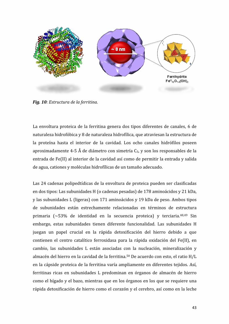
Fig. 10: Estructura de la ferritina.
La envoltura proteica de la ferritina genera dos tipos diferentes de canales, 6 de
naturaleza hidrofóbica y 8 de naturaleza hidrofílica, que atraviesan la estructura de
la proteína hasta el interior de la cavidad. Los ocho canales hidrófilos poseen
aproximadamente 4-5 Å de diámetro con simetría C3, y son los responsables de la
entrada de Fe(II) al interior de la cavidad así como de permitir la entrada y salida
de agua, cationes y moléculas hidrofílicas de un tamaño adecuado.
Las 24 cadenas polipedtídicas de la envoltura de proteica pueden ser clasificadas
en dos tipos: Las subunidades H (o cadenas pesadas) de 178 aminoácidos y 21 kDa,
y las subunidades L (ligeras) con 171 aminoácidos y 19 kDa de peso. Ambos tipos
de subunidades están estrechamente relacionadas en términos de estructura
primaria (~53% de identidad en la secuencia proteica) y terciaria.48,49 Sin
embargo, estas subunidades tienen diferente funcionalidad. Las subunidades H
juegan un papel crucial en la rápida detoxificación del hierro debido a que
contienen el centro catalítico ferroxidasa para la rápida oxidación del Fe(II), en
cambio, las subunidades L están asociadas con la nucleación, mineralización y
almacén del hierro en la cavidad de la ferritina.50 De acuerdo con esto, el ratio H/L
en la cápside proteica de la ferritina varía ampliamente en diferentes tejidos. Así,
ferritinas ricas en subunidades L predominan en órganos de almacén de hierro
como el hígado y el bazo, mientras que en los órganos en los que se requiere una
rápida detoxificación de hierro como el corazón y el cerebro, así como en la leche

44
materna, la ferritina es rica en subunidades H.51,52 En algunos organismos como
bacterias, plantas e invertebrados en general, la cápside proteica se encuentra
exclusivamente compuesta por subunidades H. En vertebrados sin embargo, la
ferritina se presenta compuesta principalmente por una combinación de
subunidades H y L, como es el caso de la ferritina de bazo de caballo, modelo de
ferritina en vertebrados, que contiene aproximadamente un 90% L- y un 10% de
subunidades H (Fig. 11).
Fig. 11: Distribución en tejidos de la ferritina según su ratio H/L
Aunque el núcleo metálico de la ferritina es básicamente ferrihidrita, donde el
hierro presenta un estado de oxidación Fe(III), este núcleo se forma a partir de
Fe(II) y no de Fe(III). Esto conlleva necesariamente a que, en algún momento de su
travesía al interior de la proteína, el Fe(II) sea oxidado a Fe(III), por lo que debe
existir un centro ferroxidasa responsable de esta oxidación. No obstante, el
mecanismo de la reacción de oxidación de Fe(II) a Fe(III) en el centro ferroxidasa
de la ferritina es actualmente objeto de controversia.50,53,54,55 Durante los últimos
años, había sido ampliamente aceptado que el Fe(II) se desplaza a través de los

45
canales hidrofílicos hasta fijarse en el centro ferroxidasa libre de una subunidad H,
donde era catalíticamente oxidado por una reacción con oxígeno molecular hasta
formar un complejo peroxo-di-férrico (DFC). Los DFCs formados migrarían
espontáneamente del centro ferroxidasa, dejándolo completamente vacante y
continuarían su viaje hacia el interior de la cavidad, interaccionando entre ellos
para formar entidades multiméricas de Fe(III), que finalmente se fijarían en los
puntos de nucleación de la cavidad proteica donde iniciarían el crecimiento del
núcleo de hierro.56,57
En contraste con esta propuesta, los recientes resultados obtenidos contradicen
esta naturale a “vacía” del centro ferroxidasa, ampliamente aceptada. El modelo
propuesto por Hagen et al. propone un mecanismo unificado, tanto en archea como
en eucariotas, en el cual dos Fe(II) son oxidados secuencialmente en el centro
ferroxidasa.55 A diferencia con el modelo anterior propuesto, este centro cargado
con Fe(III) es considerado un grupo prostético indefinidamente estable hasta la
llegada de un nuevo átomo de Fe(II), que dispararía un desplazamiento secuencial
de Fe(III) y, en presencia de oxigeno molecular, a la consecuente oxidación
catalítica del Fe(II) entrante a Fe(III) (Fig. 12). El Fe(III) desplazado migraría
entonces a la cavidad de la ferritina donde formaría el núcleo metálico a través de
un mecanismo aún por definir.
Fig. 12: Naturaleza del centro ferroxidasa según los dos modelos planteados.

46
En este contexto, la existencia permanente de una unidad FeIII-OH-FeIII en el centro
ferroxidasa tiene una serie de consecuencias biológicas que sorprendentemente no
han sido consideradas.
No pretendemos en esta tesis aportar datos que avalen una u otra hipótesis.
Pretendemos explorar hechos experimentales, ya reportados en trabajos de la
década de los 80 que no habían sido explotados en términos de implicaciones
biológicas, que van más allá de hipótesis mecanísticas. Como había sido
previamente observado, no todo el Fe(III) formado en el centro ferroxidasa en
ferritinas constituidas básicamente de cadenas H, termina formando parte del
núcleo metálico.52 Este hecho experimental, que hemos podido medir
cuantitativamente en la presente tesis, plantea una serie de cuestiones que no
habían sido consideradas anteriormente y que están directamente relacionadas
con dos hechos: i) en primer lugar, considerar que las ferritinas H no tienen como
única función almacenar hierro sino más bien una función detoxificante de Fe(II),
puesto que parte del Fe(III) que generan no es realmente almacenado y ii) las
implicaciones biológicas que este Fe(III) no almacenado puede tener en el marco
de su posible interacción con otras biomoléculas y metaloproteínas en el escenario
biológico donde es generado.
Los desórdenes en las funciones de la ferritina han sido relacionados con
enfermedades típicamente relacionadas con el hierro, como la hemocromatosis o
la anemia, pero la ferritina está también siendo cada vez más reconocida como una
molécula crucial en algunas patologías neurológicas, como la enfermedad de
Parkinson o Alzheimer.6 Por tanto, conocer en detalle los mecanismos de captación
y almacenamiento de hierro en función de la composición H/L de la cápside
proteica de la ferritina se convierte en un desafío para la comunidad científica, con
vistas a entender la etiología de estos síndromes y finalmente alcanzar el diseño de
nuevos agentes terapéuticos para estas patologías basados en el metabolismo del
hierro.
En este sentido un mal funcionamiento de la ferritina, o lo que es más novedoso,
una inadecuada elección por parte de la célula en la composición de la cápside

47
proteica en cuanto a composición de subunidades H/L, puede dar lugar a la
existencia de Fe(II) o Fe(III) libre disponibles para generar, respectivamente,
radicales hidroxilo u oxidación de otras biomoléculas que den lugar en definitiva a
la aparición de los primeros estadíos de una patología. Estos desarreglos en el
metabolismo del hierro son especialmente importantes en células del sistema
nervioso.58
En particular, en esta tesis nos hemos centrado en cómo el proceso de
almacenamiento de hierro por parte de la ferritina está conjugado con la
naturaleza química de una serie de metaloproteínas involucradas en el
metabolismo de cobre y cinc, como son las metalotioneínas (MTs). Este tipo de
estudio adquiere además especial relevancia desde el punto de vista de la
reactividad Fenton que presentan los iones CuI/II y del papel nocivo que este metal
libre, al igual que el hierro, juega en desórdenes neurodegenerativos como la
enfermedad de Alzheimer, Parkinson, esclerosis y diferentes encefalopatías.59,60
Las metalotioneínas (Fig. 13) son proteínas de bajo peso molecular (6 – 10 kDa)
ricas en residuos de cisteína (33%), cuya función biológica no ha sido del todo
establecida en la actualidad, aunque es bien conocido que juegan un papel clave en
la homeostasis y detoxificación de diferentes metales como cobre y cinc.61,62

48
Fig. 13: Estructura general de las metalotioneínas.
En mamíferos existen diferentes isoformas de metalotioneína (MT1, MT2, MT3 y
MT4) con diferente patrón de expresión en función del tejido en el que se
encuentren. Así, mientras MT1 se encuentra distribuida ubicuamente en el
organismo, las isoformas MT2 y MT3 son predominantemente sintetizadas en
células del sistema nervioso como astrocitos y microglía (MT2) y en neuronas
(MT3).63,64 Esta diferenciación tisular de las metalotioneínas, añadida al hecho de
la existencia de isoformas específicas para células concretas del sistema nervioso,
permite el estudio “a la carta” de sus interacciones en el proceso de
almacenamiento del hierro por las distintas ferritinas presentes en cada una de
estas células nerviosas y las posibles consecuencias en el desarrollo de desórdenes
neurológicos que dichas interacciones pueden originar.

49
1.4. Pinceladas sobre el metabolismo de hierro en bacterias.
Como se ha mencionado a lo largo de esta introducción, el hierro es a la vez un
nutriente esencial para el crecimiento de microorganismos así como un peligroso
metal debido a su capacidad para generar ROS tóxicas.65,66,67 Debido a esto, las
bacterias deben controlar estrictamente la captación y almacén del hierro en su
metabolismo. No es de extrañar por tanto que la respuesta a este estrés oxidativo y
los mecanismos de control que rigen la homeostasis de hierro en bacterias actúen
de manera coordinada (Fig. 14).
Fig. 14: Metabolismo del hierro en Bifidobacterias.
En condiciones aeróbicas y de pH fisiológico, el hierro es un elemento escasamente
disponible para los microorganismos debido a su baja solubilidad y a la
consecuente precipitación de oxi-hidróxidos de Fe(III).68 Por esta razón, los
microorganismos se encuentran generalmente ante una escasez de hierro
biodisponible. Para los patógenos, el problema de la restricción de hierro es más
extremo aún, ya que el “host” limita la disponibilidad de hierro, secuestrándolo a
nivel extracelular con proteínas de la familia de las transferrinas y a nivel

50
intracelular almacenado o no disponible en proteínas como la ferritina,
hemosiderina y frataxina.68,69 Es oportuno señalar que a nivel extracelular, el
medio habitual de proliferación de bacterias, el “host” secuestra hierro a través de
la lactoferrina, que puede llegar a reducir los niveles de hierro libre hasta
concentraciones en torno a 10-18 M, niveles insuficientes para permitir el
crecimiento bacteriano.70,71
El mecanismo de captación de hierro en microorganismos es amplio y depende de
la especie, pudiendo tener lugar a través receptores directos para la entrada de
Fe(II), receptores de grupos hemo-Fe o bien la vía mas habitual, consistente en la
producción de moléculas extracelulares de alta afinidad por Fe(III), denominados
sideróforos.72,73
Los sideróforos son moléculas de bajo peso molecular (<1000 Da) que se
caracterizan por su alta especificidad por Fe(III) (K > 1030).74 Pueden llegar a
encontrarse a en concentraciones extremadamente elevadas (superiores a 200
mg·L-1) en el caso de la aerobactina en E. coli, como respuesta a la restricción de
hierro.75 En la actualidad han sido caracterizados en torno a 500 sideróforos, los
cuales pueden ser clasificados según los grupos funcionales que usan como
ligandos de Fe(III). Entre los más estudiados se encuentran la enterobactina
(triscatecolatos) y la deferoxamina (trishidroxamatos).76
El mecanismo general de captación de hierro mediada por sideróforos en
bacterias Gram-positivas y negativas se muestra en la (Fig. 15). Estos sideróforos
son demasiado grandes para atravesar la membrana y pared bacterianas por
difusión pasiva, por lo que requieren de la presencia de receptores de membrana
específicos para su internalización acoplados a sistemas transportadores ABC-
permeasa. Una vez en el citoplasma, el ferrisideróforo es degradado por una
esterasa o reciclado a través de procesos reductivos que liberan Fe(II) dejando la
molécula intacta para reanudar un nuevo ciclo de captación de hierro.68

51
Fig. 15: Captación de Fe(III) mediada por sideróforo en bacterias.
Puesto que los problemas que las bacterias deben afrontar para adquirir suficiente
hierro de su entorno son especialmente agudos en microorganismos infecciosos
debido a que el “host” limita específicamente la disponibilidad de hierro como
parte de su defensa innata ante patógenos invasivos, una manera común en los
patógenos para adquirir hierro directamente del organismo infectado es
precisamente a través de sistemas de transporte mediado por receptores
específicos para las proteínas de unión a hierro del “host”: los microorganismos
tratan de mimetizar las vías usuales de captación de hierro por parte de las células
del “host”. Estos receptores de membrana liberan el hierro de la lactoferrina y la
transferrina, que posteriormente es internalizado a través de un sistema permeasa
ABC al interior celular. Este proceso tiene lugar en la superficie exterior de la
célula y las formas apo de la proteínas transportadoras de hierro son liberadas
extracelularmente en lugar de ser internalizadas y acumuladas (Fig. 16).

52
Fig. 16: Esquema de la captación de hierro mediada por receptor de transferrina en
bacterias.
No obstante los mecanismos de actuación del “host” frente a las bacterias
dependen de la toxicidad de éstas ya que, para el caso de bacterias beneficiosas
probióticas como Bifidobacterium y Lactobacillus, promueven su proliferación.
Se ún la O/WHO los probióticos se definen como “los microor anismos que
cuando son administrados en cantidades adecuadas, confieren efectos beneficiosos
para la salud del or anismo hu sped”. Entre otros, estos beneficios incluyen una
mayor respuesta inmune, un mejor balance de la microbiota en colon, reducción de
enzimas implicadas en el desarrollo del cáncer, control de retrovirus y reducción
de los niveles de colesterol en sangre, entre otras.77
53
En esa tesis nos hemos centrado en cómo este tipo de bacterias probióticas
proliferan a expensas del “host” de una forma “voluntaria”. En particular, hemos
querido profundizar en cómo la lactoferrina, encargada de privar de hierro a
microorganismos patógenos es, sin embargo, capaz de transportar hierro a
bacterias probióticas haciendo que proliferen en pro del desarrollo adecuado del
organismo hospedador.
En este sentido, la presencia conjunta de lactoferrina y apoferritina H en la leche
materna es un ejemplo de cómo los mamíferos son capaces de diseñar un “tándem”
de proteínas que sirve, tanto para impedir la proliferación de microorganismos
patógenos como para, al mismo tiempo, hacer viable la presencia de bacterias
probióticas beneficiosas para la salud. Una prueba más de la sutil, elegante y
eficiente maquinaria química en pro de un metabolismo de hierro adecuado que
los organismos desarrollan para su supervivencia.

54
1.5. Objetivos
Como se ha puesto de manifiesto en la Introducción, el metabolismo de hierro es
esencial para la vida y cualquier desarreglo conlleva la aparición de patologías
diversas. Pretendemos en esta Tesis, por un lado, descubrir nuevos desajustes en
toda la maquinaria química que define el metabolismo de hierro que den lugar al
desarrollo de patologías y en segundo lugar, que el conocimiento adquirido se
traduzca en la preparación de materiales que puedan ser útil conceptualmente
para intervenir en dichos desajustes y poder actuar a nivel terapéutico.
El objetivo general de esta tesis es en cierta forma indagar en aspectos del
metabolismo de hierro que generen conocimiento y/o inspiren la creación de un
nuevo material que pueda ser aplicado en biomedicina, tanto en terapias variadas
relacionadas con un metabolismo de hierro anómalo, como en diagnóstico de
enfermedades concretas.
De forma más concreta, los aspectos que pretendemos abordar son los siguientes:
1. Tras un exhaustivo análisis bibliográfico pretendemos demostrar que la proteína
ferritina, tradicionalmente considerada como almacén de hierro, no siempre lleva a
cabo esta función, o al menos no “únicamente”. En particular pretendemos
demostrar que la actividad de algunas ferritinas (las denominadas H) están más
relacionadas con llevar a cabo una función de detoxificación de Fe(II) que
propiamente el almacenamiento del metal.
Demostrar que estas ferritinas H tienen muy poca capacidad de almacenamiento
pero una alta actividad ferroxidasa (oxidación de Fe(II) a Fe(III)) explicaría
aspectos nuevos que han pasado desapercibido hasta la actualidad: i) su presencia
en algunos fluidos biológicos acompañada del agente quelatante biológico de
Fe(III) por excelencia, la lactoferrina y ii) la elección que hace cada célula del tipo
de la ferritina que en realidad requiere para llevar a cabo su funcion, como ocurre
en algunas células tan particulares como las neuronas.
Entender el mecanismo de acción de las ferritinas H y su presencia en leche
materna, inspira inmediatamente la preparación de materiales que puedan
mimetizar funcionalmente este fluido biológico, cuya función primordial en el

55
metabolismo de hierro es precisamente la eliminación del Fe(II) tóxico. Estos
materiales podrían ser utilizados como detoxificantes de hierro y por tanto como
antibióticos, puesto que podrían eliminar cualquier forma de hierro libre que
sirviera de alimento a un microorganismo patógeno que pudiera dar lugar a una
infección.
2. Pretendemos establecer marcos químicos amplios en los que podamos estudiar la
interconexión entre diferentes metaloproteínas relacionadas con la homeostasis
de hierro y otros metales, como cobre y zinc. El metabolismo de hierro va ligado al
de otros iones metálicos. La homeóstasis de iones metálicos es un proceso global
que requiere ser estudiado como tal. Para poder abordar la interconexión entre
iones metales a nivel biológico se requiere un enfoque químico nuevo y complejo
dónde poder abordar como la actividad de una metaloproteína se ve afectada por
otra. Pretendemos dar un paso en este sentido y diseñar marcos químicos que
biometicen de una forma más real los procesos de homeóstasis de metales que
ocurren en la célula. En particular pretendemos estudiar como interfiere en el
proceso de almacenamiento de hierro una familia de metaloproteínas involucradas
en el metabolismo de cobre y cinc, como son la metalotioneínas. De los resultados
obtenidos podemos deducir el porqué de la elección de cada célula, en función del
tejido en el que se encuentre, del par ferritina-metalotioneína adecuado para llevar
a cabo sus funciones de forma óptima y lo que es más importante detectar pares
ferritinas-metalotioneínas que podrían generar el inicio de patalogías neuronales.
3. El estudio del metabolismo de hierro en bacterias tiene muchos enfoques.
Aprender aspectos de dicho metabolismo nos puede permitir: i) diseñar una nueva
vía para la aniquilación bacteriana, en relación con la búsqueda de nuevos
antibióticos, ii) entender el porqué de la proliferación bacteriana de algunas
bacterias beneficiosas para la salud, como son las bacterias probióticas y iii) la
preparación de nuevos materiales basados en la actividad bacteriana para
producir nanopartículas metálicas, que eventualmente puedan representar una
nueva vía de nanomateriales con aplicaciones biomédicas.

56
1.6. Bibliografía
1 D. J. Pinero, J. R. Connor, H. R. Lieberman, R. B. Kanarek, C. Prasad (Eds.). Nutri- tional Neuroscience, CRC Press, Taylor & Francis Group, Boca Raton, 2005, p. 235. 2 T. LaVaute, S. Smith, S. Cooperman, K. Iwai, W. Land, E. Meyron-Holtz, S. K. Drake, G. Miller, M. Abu-Asab, M. Tsokos, R. Switzer III, A. Grinberg, P. Love, N. Tresser, T. A. Rouault. Nat. Genet., 2001, 27, 209–214. 3 H. Puccio, D. Simon, M. Cossee, P. Criqui-Filipe, F. Tiziano, J. Melki, C. Hindelang, R. Matyas, P. Rustin, M. Koenig. Nat. Genet., 2001, 27, 81–186. 4 N. B. Cole, D. D. Murphy, J. Lebowitz, L. Di Noto, R.L. Levine, R. L. Nussbaum. J. Biol. Chem., 2005, 280, 9678. 5 Q. Q. Pankhursta, D. Hautotb, N. Khanc, J. Dobson. J. Alzheimers Dis., 2008, 13, 49. 6 D. Berg, G. Beceker, P. Riederer, O. Rieb. Neurotox. Res., 2002, 4, 637. 7 K. Jomova, M. Valko. Curr. Pharm. Des., 2011, 17, 3460-3473. 8 H. Heli, S. Mirtorabi, K. Karimian. Expert Opinion on Therapeutic Patents, 2011, 21, 819. 9 J. A. Imlay, S. M. Chin, S. Linn. Science, 1988, 240, 640–642. 10 S. Jang, J. A. Imlay. J. Biol. Chem., 2007, 282, 929–937. 11 S. Laurent, D. Forge, M. Port, A. Roch, C. Robic, L. Vander Elst, R. N. Muller. Chem. Rev., 2008, 108, 2064–2110. 12 K. Gkouvatsos, G. Papanikolaou, K. Pantopoulos. Biochim. et Biophys. Acta, 2012, 1820, 188–202. 13 R. J. Cherry, A. J. Bjornsen, D. C. Zapien. Langmuir, 1998, 14, 1971. 14 T. Yoshinobu, J. Suzuki, H. Kurooka, W. C. Moon, H. Iwasaki. Electrochim. Acta, 2003, 48, 3131. 15 K. Iwai, S. K. Drake, N. B. Wehr, A. M. Weissman, T. LaVaute, N. Minato, R. D. Klausner, R. L. Levine, T. A. Rouault. Proc. Natl. Acad. Sci. U.S.A., 1998, 95, 4924. 16 J. Zahringer, B. S. Baliga, H. N. Munro. Proc. Natl. Acad. Sci. U.S..A, 1976, 73, 857.

57
17 M. W. Hentze, L. C. Kühn. Proc. Natl. Acad. Sci. U.S.A., 1996, 93, 8175–82. 18 M. Sorensen, S. P. L. Sorensen. Comptes-rendus des Travaux du Laboratoire Carlsberg, 1939, 23, 55–99. 19 B. F. Anderson, H. M. Baker, G. E. Norris, D. W. Rice, E. N. Baker. J. Mol. Biol., 1989, 209, 711–734. 20 E. N. Baker, P. F. Lindley. J. Inorg. Biochem., 1992, 47, 147–160. 21 R. D. Brown, K. A. Rickard, H. Kronenberg. Pathology, 1983, 15, 27–31. 22 M. H. Metz-Boutigue, J. Jolles, J. Mazurier, F. Schoentgen, D. Legrand D. G. Spik. Eur. J. Biochem., 1984, 145, 659–676. 23 E. N. Baker, H. M. Baker. Biochimie, 2009, 91, 3–10. 24 H. Vogel. J. Biochem. Cell Biol., 2012, 90, 233–244. 25 E. N. Baker, H. M. Baker. Cell Mol. Life Sci., 2005, 62, 2531–2539. 26 R. T. A. MacGillivray, S. A. Moore, J. Chen, B. F. Anderson, H. Baker, Y. Luo Y. Biochemistry, 1998, 37, 7919–7928. 27 R. Pakdaman, M. Petitjean, J. M. El Hage Chahine. Eur. J. Biochem., 1998, 254, 144–153. 28 A. F. Bou, J. M. El Hage Chahine. J. Mol. Biol., 2000, 303, 255–266. 29 J. umar W. Weber S. nchau, S. Yadav, S. B. Singh, K. Saravanan, M. Paramasivam, S. Sharma, P. Kaur, A. Bhushan, A. Srinivasan, C. Betzel, T. P. Singh. Indian J. Biochem. Biophys., 2003, 40, 14–21. 30 H. S. Birgens. Scand. J. Haematol., 1985, 34, 326–331. 31 D. Latorre, F. Berlutti, P. Valenti, S. Gessani, P. Puddu. Biochem. Cell Biol., 2012, 90, 269–278. 32 N. Orsi. Biometals, 2004, 17, 189–196. 33 T. F. Byrd, M. A. Horwitz. J. Clin. Investig., 1991, 88, 351–357. 34 E. Griffiths, L. Duffy, F. Schanbacher, D. Dryja, A. Leavens, R. Neiswander, H. Qiao, D. DiRienzo, P. Ogra. Dig. Dis. Sci., 2003, 48, 1324–1332. 35 Y. Makino, S. Nishimura. J. Chromatogr., 1992, 579, 346–349.

58
36 R. A. Finkelstein, C. V. Sciortino, M. A. McIntosh. Rev. Infect. Dis., 1983, 5, 759–777. 37 P. K. Singh, M. R. Parsek, E. P. Greenberg, M. J. Welsh MJ. Nature, 2002, 417, 552–555. 38 N. G. Hord. Annu. Rev. Nutr., 2008, 28, 215–231. 39 H. Gill, J. Prasad. Advances in experimental medicine and biology, 2008, 606, 423-454. 40 S. Fijan. Int. J. Environ. Res. Public Health, 2014, 11, 4745–4767. 41 C. Liepke, K. Adermann, M. Raida, H. J. Mager, W. G. Forssmann, H. D. Zucht. Eur. J. Biochem., 2002, 269, 712–718. 42 M. Sherman, S. Bennett, F. Y. Hwang, C. Yu. Biometals, 2004, 17, 285–289. 43 R. Miller-Catchpole, E. Kot, G. Haloftis, S. Furmanov, A. Bezkorovainy. Nutr. Res.., 1997, 17, 205–213. 44 M. M. Rahman, W. S. Kim, H. Kumura, K. Shimazaki K. Int. J. Food Sci. Technol., 2010, 45, 453–458. 45 H. Saito, H. Miyakawa, N. Ishibashi, T. Yoshitaka, H. Hayasawa, S. Shimamura. Biosci. Microflora, 1996, 15, 1–7.
46 "Meveol: orphan drug status granted by the FDA for the treatment of cystic
fibrosis". United States Food and Drug Administration. 2009-11-05. Retrieved
2010-01-23. 47 N. D. Chasteen, P. M. Harrison. J. Struct. Biol., 1999, 126, 182. 48 S. Levi, S. J. Yewdall, P. M. Harrison, P. Santambrogio, A. Cozzi, E. Rovida, A. Albertini, P. Arosio. Biochem. J., 1992, 288, 591. 49 P. D. Hempstead, S. J. Yewdall, A. R. Fernie, D. M. Lawson, P. J. Artymiuk, D. W. Rice, G. C. Ford, P. M. Harrison. J. Mol. Biol., 1997, 268, 424. 50 E. C. Theil. Curr. Opin. Struct. Biol., 2011, 15, 304. 51 M. Wagstaff, M. Worwood, A. Jacobs. Biochem. J., 1978, 173, 969. 52 P. Arosio, P. A. Ponzone, R. Ferrero, L. Renoldi, S. Levi. Clin. Chim. acta; Int. J. Clin. Chem., 1986, 161, 201–208. 53 X. Liu, E. C. Theil. Proc. Natl. Acad. Sci. U.S.A., 2004, 101, 8557–8562.

59
54 P. Arosio, R. Ingrassia, P. Cavadini. Biochim. Biophys. Acta, 2009, 1790, 589–599. 55 H. K. Ebrahimi, E. Bill, P. L. Hagedoorn, W. R. Hagen. Nat. Chem. Biol., 2012, 8, 941–948.
56 T. Tosha, H-L. Ng, O. Bhattasali, T. Alber, E. C. Theil. J.Am.Chem.Soc., 2010, 132, 14562. 57 F. Bou-Abdallah, G. C. Papaefthymiou, D. M. Scheswohl, S. D. Stanga, P. Arosio, N. D. Chasteen. Biochem. J., 2002, 364, 57. 58 R. K. Watt, R. J. Hilton, D. M. Graff. Biochim. Biophys. Acta, 2010, 1800, 745. 59 H. Kozlowski, M. Luczkowski, M. Remelli, D. Valensin. Coord. Chem. Rev., 2012, 256, 2129. 60 J. H. Viles. Coord. Chem. Rev., 2012, 256, 2271. 61 M. Capdevila, R. Bofill, Ò. Palacios, S. Atrian. Coord. Chem. Rev., 2012, 256, 46–52. 62 Ò. Palacios, S. Atrian, M. Capdevila. J. Biol. Inorg. Chem., 2011, 16, 991–1009. 63 J. Hidalgo, R. Chung, M. Penkowa, M. Vasak., RSC Publishing, Cambridge, 2009, 5, 279. 64 M. Vasak, G. Meloni. RSC Publishing, Cambridge, 2009, 5, 319. 65 J. A. Imlay, S. M. Chin, S. Linn. Science, 1988, 240, 640–642. 66 S. Jang, J. A. Imlay. J. Biol. Chem., 2007, 282, 929–937. 67 J. A. Imlay, S. Linn. Science, 1988, 240, 1302–1309. 68 S. C. Andrews, A. K. Robinson, F. Rodriguez-Quinones. FEMS Microbiol. Rev., 2003, 27, 215–237. 69 R. A. Finkelstein, C. V. Sciortino, M. A. McIntosh. Clin. Infect. Dis., 1983, 5, 759–777. 70 A. G. Oglesby-Sherrouse, M. L. Vasil. PLoS One, 2010, 5, 9930. 71 A. Nandal, C. C. Huggins, M. R. Woodhall, J. McHugh, F. Rodriguez-Quinones, M. A. Quail, J. R. Guest, S. C. Andrews. Mol. Microbiol., 2010, 75, 637–657. 72 A. Bagg, J. B. Neilands. Microbiol. Rev., 1987, 51, 509–518.

60
73 V. Braun, H. Killmann. Trends Biochem. Sci., 1999, 24, 104–109. 74 C. Jakopitsch, G. Regelsberger, P. G. Furtmuller, F. Ruker, G. A. Peschek, C. Obinger. J. Inorg. Biochem., 2002, 91, 78–86. 75 D. Touati. Arch. Biochem. Biophys., 2000, 373, 1–6. 76 V. Braun, H. Killmann. Trends Biochem. Sci., 1999, 24, 104–109. 77 J. M. Saavedra. Am. J. Clin. Nutr., 2001, 73, 1147S–1151S.

61
CAPÍTULO 2.

62

63
FERRITIN IRON UPTAKE AND RELEASE IN THE PRESENCE OF METALS AND METALLOPROTEINS: CHEMICAL IMPLICATIONS IN THE BRAIN
Fernando Carmonaa, Òscar Palaciosb, Natividad Gálveza, Rafael Cuestac, Sílvia
Atriand, Mercè Capdevilab, José M. Domínguez-Veraa.
a Departamento de Química Inorgánica and Instituto de Biotecnología, Facultad de
Ciencias, Universidad de Granada, E-18071 Granada, Spain
b Departament de Química, Facultat de Ciències, Universitat Autònoma de Barcelona,
Cerdanyola del Vallès, E-08193 Barcelona, Spain
c Departamento de Química, Escuela de Linares, Universidad de Jaén, Jaen, Spain
d Departament de Genètica, Facultat de Biologia and Institut de Biomedicina,
Universitat de Barcelona, Avda. Diagonal 643, E-08028 Barcelona, Spain
Abstract
Living organisms have developed a chemical machinery based on the ferritin
protein for the storage, under a nontoxic form, of the iron that is not required for
immediate metabolic purposes. Whereas free iron causes extensive cell damage,
ferritin iron is not toxic, yet still available for cell requirements. However, iron
storage in ferritin is increasingly being recognized as a crucial process related with
some neurodegenerative disorders and therefore, an understanding of the
management of iron in the brain, especially the processes of iron uptake and
release in ferritin, is compulsory to clarify the role of this metalloprotein in these
neuropathologies.
Although knowledge of iron storage and iron release in ferritin is nowadays still
limited, even less information is currently available about the influence of free
metal ions and other brain metalloproteins in these processes.
In this sense, this review is an excellent opportunity to collect all the information
today available about the influence of metals and metalloproteins in ferritin
loading and unloading events, which until now are dispersed in the literature.
Furthermore, we will focus on the importance of all the above-mentioned
interactions in the brain, since the importance of the correct and safe balance of

64
metals in the brain after their well-known implication in neurodegenerative
processes such as the l heimer’s ( ) Parkinson (P ) and prion protein (PP )
diseases is obvious. In this work, we will not only recall the importance and role of
ferritin in the brain but also the putative influence of the interaction between
ferritin and some metals and/or metalloproteins and other biomolecules on these
neurological dysfunctions. The final part of the review will be devoted to draw
some guidelines to where the future prospects point to on the basis of the existing
information.
1. Introduction
Iron is essential for life since it is required for the active sites of many
metalloproteins that play a key role in crucial biological processes such as oxygen
transport, storage and use of oxygen in many oxidation–reduction reactions as
well as in electron transfer reactions within the cell [1]. Particularly in the brain,
iron is crucial for neuronal development, gene expression, enzyme function,
dopamine, heme and iron–sulfur cluster synthesis as well as electron transport [2].
However, excess iron is highly toxic. Iron(II) promotes the formation of highly
reactive oxygen species (ROS) via distinct pathways [3]. Among these, the most
common one is the Fenton reaction, in which hydroxyl radicals OH• are produced
by the reaction of iron(II) and hydrogen peroxide. ROS are extremely powerful
oxidizing agents capable of causing irreversible cell damage, organ failure and
eventually death [4]. Furthermore, free iron is a key nutrient for pathogenic
microorganisms, which require iron to survive and replicate. Life is, in some way, a
battle for iron and therefore, hosts must deprive undesirable guests of iron in
order to combat the infections they cause. Therefore, both excess and deficiency of
iron are harmful, and organisms have been forced to develop mechanisms to
manage this situation. To do so, they store the iron that is not required for
immediate metabolic needs in a non-toxic form. This stored iron is neither
available for producing damaging radicals nor for allowing the viability of
pathogen microorganisms. Once required, iron can be recovered from the store to
participate in cell metabolism.
Ferritin is the primary iron storing protein in most living organisms throughout

65
the animal, plant and microbial kingdoms. The structure of many ferritins isolated
from a wide range of organisms and biological tissues have been determined, and
it becomes evident that a common structure has been conserved throughout
evolution. The most extensively studied ferritin is that of horse spleen,
traditionally considered as the model of mammalian ferritin. It consists of a hollow
protein shell (Mr about 450 kDa) composed of 24 subunits arranged in cubic
symmetry, that surrounds an aqueous cavity of 8 nm in diameter, capable o
accommodating thousands of iron atoms as an iron(III) mineral, traditionally
described as ferrihydrite [FeIII10O14(OH)2] [5]. Ferrihydrite is consistent with a
single hexagonal phase (P63 mc; a = 5.95 Å , c = 9.06 Å ). In its ideal form, this
structure contains 20% tetrahedrally and 80% octahedrally coordinated iron(III)
[6].
However, it is important to note that the nature of the ferritin core is not
completely accepted. In fact, recent studies have pointed out that a polyphasic
model, including ferrihydrite and magnetite (or other iron(II)-containing phases)
would describe in a more realistic manner the ferritin core, especially once the
protein has undergone some chemical disorder [7]. In any case, ferrihydrite is
certainly a labile mineral and it is therefore the ideal choice to allow an adequate
turn over of iron from the ferritin store to the cell.
Ferritin is remarkably stable to temperature and pH changes, as demonstrated by
its stability up to 70 ºC and over extreme pH values of 3–10. At pH < 3 the 24
subunits dissociate but reversibly reassemble at pH > 3 [8]. The 24-polypeptide
chains of the apoferritin shell can be classified into two types: the H (or heavy)
subunits of 178 amino acids and 21 kDa, and the L (or light) subunits with 171
amino acids and 19 kDa, Fig. 1. The two types of ferritin subunits are closely
related both in terms of primary (53% protein sequence identity) and tertiary
structure [9,10]. However they have different functionality. Thus, whereas the H
subunit plays a key role in the rapid detoxification of iron, since it contains a
catalytic ferroxidase center for rapid iron(II) oxidation, the L subunit is associated
with iron nucleation, mineralization and long-term iron storage in the ferritin
cavity [11]. In agreement with this, the H/L ratio in a ferritin shell varies widely in
different tissues. L-subunit-rich ferritins predominate in iron storage organs such
as the liver and spleen, while organs that require iron detoxification properties
such as the heart and brain contain H-rich ferritins [12]

66
Fig. 1. Horse spleen ferritin structure: a 24-subunit oligomer with a combination of
heavy (H) and light (L) polypeptide subunits that form a spherical hollow molecule.
It is interesting to note that ferritins in bacteria, plants and invertebrates are
exclusively made by H-like subunits, i.e. they are pure H-like ferritins. Ferritins
with a combination of H and L subunits are found only in vertebrates, and they
have thoroughly been characterized in mammals. As an example, the traditionally
considered model of mammalian ferritin, horse spleen ferritin, usually contains
90% L- and 10% H- subunits.
The multimeric construction of the ferritin shell allows the generation of different
types of channels leading to the polymer cavity. Eight hydrophilic channels of 4–5
Å in diameter and with C3 symmetry (Fig. 1) allow the transfer of water, metal
cations and hydrophilic molecules of the appropriate size from the external
solution to the cavity or vice versa.
Synthesis of the ferritin H and L monomers is mainly regulated at translation level
in response to labile iron concentrations by two mRNA-binding proteins: IRP1 and
IRP2 (iron-regulator protein). When cytoplasmic iron is high, IRP1 forms a Fe-S
protein that acts as the aconitase enzyme. When iron is low, IRP1 adopts an open
conformation, devoid of the Fe-S cluster that binds to the 5’ iron responsive
67
elements (IRE) of the ferritin mRNA, this repressing its translation [13].
Conversely, IRP2 presence in the cell is regulated by degradation, quickly induced
by iron overload [14]. These systems function in close coordination with an
antithesis control of the transferrin receptor (TfR) synthesis, because the union of
IRP1 to the 3’ loops of TfR mRNA in low iron conditions stabilizes this messenger,
increasing the synthesis rate of TfR, and therefore provoking an enhanced
transferrin uptake. However, besides this smart control mechanism exerted by
iron levels, at least ferritin H synthesis is also transcriptionally regulated by
classical promoter elements and transcription factors that act in inflammatory
scenarios [15], which would account for the H-rich ferritin shells usually
associated with this kind of processes.
Disorders in ferritin functions have been related with typical iron-related diseases,
such as hemochromatosis or anemia, but ferritin is also increasingly being
recognized as a crucial molecule in some neurological pathologies, as Parkinson
(PD) or Alzheimer’s ( ) diseases [16]. Therefore, knowing the ferritin iron
uptake, storage, and release mechanisms in detail becomes a challenge for the
scientific community in order to understand the etiology of these syndromes and
eventually to approach the design of new therapeutic agents based on iron
metabolism.
The ferritin core forms from iron(II), and not iron(III). As it will be discussed in the
next sections, iron(II) is oxidized and stored mainly as an iron(III) mineral.
Likewise, when the cell requires iron from ferritin this is removed as iron(II). If the
ferritin protein is purely viewed as an iron store, there are two processes that have
to operate properly: a fast and efficient iron storage, and a controlled release of
iron. In a biological scenario, when ferritin loses its iron storing ability, free
iron(II) remains in the cytoplasm. Equally, when ferritin releases iron in an
uncontrolled manner, iron(II) is produced and dumped into the cytoplasm. In
other words, a dysfunction of ferritin always gives rise to the existence of free
iron(II), with independence of the reason of its malfunction. As mentioned above,
free iron(II) promotes ROS that produces oxidative damage. Interestingly,
oxidative damage in neurons is a primary cause of degenerative diseases such as
AD [17]. Furthermore, as recently reviewed by Kozlowski et al. [18], Fernández et
al. [19], Hureau and Dorlet [20] and Viles [21], free iron(II) and other metals might
contribute to some neurological diseases by inducing aggregation of some

68
proteins, as it is the case of ß-amyloid, α-synuclein or the prion protein that
respectively accumulate in patients of AD, PD and prion protein disease (PPD).
The reasons for the malfunctioning of ferritin, either at iron uptake, storage or iron
release, are still not clear, but data are increasingly supporting two lines of
evidence. First, the existence of an excess of a chemical reductant interacting with
ferritin has been postulated [22]. Second, genetic mutations affecting either the
structure of the ferritin polypeptide [23], or the H [24] or L [25] ferritin mRNA IRE
regions this leading to undesirable H/L ratios in the ferritin shells have been
recently reported. Both mentioned pathways of ferritin dysfunctions have been
intensively studied, related to neurological disorders and reviewed [26].
Ferritin can bind or accommodate metal ions other than Fe in several types of
binding sites [27,28]. More importantly, the disruption of the ferritin function in
iron uptake or iron release is influenced by the presence of some metals [29], some
metalloproteins [30,31], and some other types of biomolecule. Zinc(II), for
example, binds to specific ferritin sites and inhibits ferritin iron core formation
[32–39]. Copper(II) also binds tightly to ferritin but has an opposite effect to
zinc(II), since it has a positive catalytic effect on the uptake of iron by ferritin [40].
Otherwise, some metalloproteins such as ceruloplasmin [41] or metallothioneins
[42] are capable to respectively promote the uptake or the removal of iron from
ferritin, the latter giving rise not only to the existence to free toxic iron(II) but also
to putatively damaging free copper(I) or zinc(II) ions that were initially bound to
it. Other biomolecules such as glutathione (GSH), xanthine oxidase and superoxide
dismutase (SOD) have also been reported to interact with ferritin (cf. last
paragraph of Section 7).
However, the study and analysis of the interactions between ferritins and all these
biomolecules and metal ions, as well as their biological significance, has remained
until now dispersed in the literature. In this sense, this review aims at collecting all
the information now available about the influence of metals and biomolecules,
basically metalloproteins, in ferritin iron uptake and iron release. It is our belief
that this will provide a solid basis of knowledge from where to extract
comprehensive conclusions and to build future prospects. In fact the scenario we
try to draw does not center exclusively in iron in ferritin but in the “ferritin + metal

69
or biomolecule” system a landscape much more complex than the former but
probably more realistic in terms of physiological significance. Finally, we will try to
focus on the importance and consequences of all the above-mentioned interactions
in the brain. The uncontrolled release of iron, but also of other metal ions such as
zinc or copper, all of them essential but of well established toxicity when in excess,
has been revealed to have important consequences in the brain, mainly in
neurodegenerative pathologies. Consequently, we are confident that the review
and synthesis of all the current available data on this subject will shed light on the
possibleinterpretation of the underlying mechanisms triggering these diseases as
well as provide clues to succeed in the design of strategies for their amelioration.
Fig. 2. The trip of iron(II) through the hydrophilic channel to reach the ferroxidase
center (top). The two iron coordination sites at the ferroxidase center (bottom).

70
2. Ferritin iron uptake
The process of iron uptake by ferritin has been extensively studied and reviewed
[43]. Our purpose here is to briefly describe the chemistry of iron during its travel
across the ferritin channels to the cavity, allowing the readerto envisage how the
different steps that comprise the global ferritin iron uptake process can be affected
by the presence of metals and metallobiomolecules.
The iron concentration in the cytosol is sensibly higher than that of any other
transition metal. The reductive ambient of cytosol clearly favors the presence of
iron(II) over iron(III), which leads to respective average values of 10−7 and 10−18 M
[44].
Iron(II)-citrate and iron(II)-thiol-containing biomolecules such as cysteine and
glutathione seem to be the major components of the cytosolic iron pool [45]. The
high concentration of glutathione in the cytosol (ranging between 2 and 8 mM)
enables iron(III) reduction at physiologic pH. Iron(II) is then taken up by
apoferritin, which is mainly located in the cytoplasm.
Iron(II) and other metal ions trafficking from the surrounding exterior to the
ferritin cavity has been proposed to occur through the 3-fold channels [46].
Calculations of the electrostatic surface potential of ferritin identified a funnel of
negative charge that would attract the iron(II), and other metal cations, toward the
ferritin hollow [47]. In the most widely accepted model, iron(II) moves through the
hydrophilic channels until observation a ferroxidase center of an H-subunit (Fig. 2)
where it is catalytically oxidized by reaction with cellular oxygen [48] to form a
diferric peroxo complex (DFC) [49,50], where iron atoms occupy two specific
coordination sites (A and B).
Most of the structural information about the ferroxidase center has been inferred
by a combination of kinetics and site-directed mutagenesis [51]. In the
recombinant human H-ferritin, the necessary amino acids include Glu27, Glu62
and His65 at site A, and Glu107, Gln141 and Ala144 at site B. The participation of
some of these amino acids has been confirmed in the X-ray structure of the zinc(II)
complex of the same recombinant ferritin (Fig. 2) [38].

71
Fig. 3. Nucleation sites (top) and schematic representation of the formation of the
first iron clusters. Ferritin core growth (bottom): from the nucleation sites to the
final iron oxide nanoparticle
The structure of the ferroxidase center of ferritin from the hyper-thermophilic
archaeal anaerobe Pyrococcus furiosus under aerobic conditions reported by Hagen
et al. exhibits three iron atoms [52]: an iron site A appears to be the strongest
affinity site since it is found occupied in the isolated protein, while sites B and C are
found only occupied after soaking apobacterionferritin crystals in iron(II)
solutions under aerobic conditions. The iron–iron distances between sites A and B
vary between 2.6 and 3.6 Å in the different subunits.
In another X-ray absorption spectroscopy study on eukaryotic ferritin, Theil et al.
[53] described three different states that were trapped by rapid freeze quench of
iron(II) ferritin reacting with O2 : an initial diiron(II) high-spin state, which is
described with iron being in an average six-coordinate environment with a Fe–Fe
distance of 3.44 Å, and a peroxodiron(III) intermediate with the very short Fe–Fe
distance of 2.53 Å, which finally evolves toward a µ-oxo/hydroxo diiron(III)
species with a Fe–Fe distance of 3.00 Å.
An X-ray structure was reported for Escherichia coli bacterioferritin by Le Brun et
al. [54]. The diiron species obtained by soaking under aerobic conditions, followed
by flash freezing, was proposed to be in the reduced state. The iron coordination
environment is contributed by one His and one Glu residue per iron and two

72
bridging Glu residues. The iron–iron distance was 3.7 Å. Longer soaking times
provided an iron center proposed to be a µ-oxo/hydroxo diiron(III) species, with
an intact coordination sphere and an iron–iron distance of 3.6 Å.
Recently, Bertini et al. reported the first X-ray picture of iron(III) products at the
ferroxidase site in higher eukaryote ferritins [55] by flash freezing crystals of frog
ferritin soaked during two different times in iron(II) solutions under aerobic
conditions. The structure for long soaking times resulted in the observation of
invariant diiron(III) sites with a Fe–Fe distance of about 3.1 Å, suggesting the
presence of a µ-oxo/hydroxo bridge between iron atoms. The electronic
absorption spectra is typical of iron(III) ferritin in solution. The structure for short
soaking times was investigated by using copper(II) instead of iron(II) to avoid
oxidation. Two different copper(II) ions were found at sites A and B of the
ferroxidase center with a Cu–Cu distance of about 4.3 Å. While the coordination
environment of copper at site A was similar to that of iron(III) in the structure for
long soaking times, copper at site B also contains a bridging carboxylate but differs
from that of site A because of the absence of bridging water/hydroxo and the
additional coordination to the protein residue His54. Interestingly, His54 binding
to metal ion had been observed in the cobalt(II) ferritin structure [46]. The authors
hypothesized that the coordination of iron(II) at the ferroxidase center is similar to
that of copper(II) and concluded that oxidation at the ferroxidase center requires
changes in the coordination environment of iron(II) at site B and a parallel
reduction in the metal–metal distance.
The path from the ferroxidase center to the cavity has been analyzed on the basis
of a paramagnetic-NMR study [56]. Theil, Bertini et al. have proposed that the DFCs
continue their travel to the cavity through the channels. In this journey, the DFC
species interact between them to form multimeric iron(III) entities, which
ultimately are fixed at the nucleation sites of the cavity and that initiate the growth
of the iron core (Fig. 3).
In contrast to this model, Hagen et al. assume that core formation starts directly
after formation of the ferroxidase center without occurrence of small clusters en
route to core formation. Studies of this group on the P. furiosus ferritin suggest that
the ferroxidase center, i.e. the invariant metallic motive constituted by iron atoms
bridged by a peroxo group, which are permanently bound to specific amino acids
of the protein, is a stable prosthetic group and that after formation of a stable
73
diiron center, iron(II) is oxidized on its way to the core by the iron(III) in the
ferroxidase center, and electrons are transferred to O2 [57]. EPR [58] and kinetic
studies [59] support this assumption.
On the other hand, studies by Le Brun et al. on the mechanism of iron deposition in
mammalian ferritins suggest an additional pathway based on a reaction on the
mineral surface that would allow simultaneously the iron oxidation and nucleation
steps [60,61]. Thus iron(II) could migrate through the 3-fold channels and be
directly oxidized to iron(III) on the surface of the mineral core.
Likewise, it has been traditionally considered that some oxo anions, in particular
phosphate, increase the rate of iron core formation [62]. Ferritins from different
origins contain phosphate that is associated with the iron core. In bacterial ferritin,
the phosphate/iron ratio can be as high as close to unity [63], whereas a ratio of
approximately 0.1 has been found in mammalian ferritins [64]. Recently, Watt et
al. showed that phosphate inhibited iron loading into L ferritin, due to the lack of
the ferroxidase center, whereas iron loading into H ferritin showed identical iron
loading pattern in the presence or absence of phosphate [65].
In any case, analyzing all the literature data, it appears that although the ferritin
uptake process has been extensively addressed, there is a limited knowledge of the
mechanism of growth of the iron core at the ferritin cavity, and still much needs to
be learned about how the iron core grows at the nucleation sites to yield the final
nanoparticle.

74
Fig. 4. (a) Structure of human heart, horse spleen and recombinant L ferritin. L
subunits are in red and H in cream. (b) Modeled morphology of the iron core and its
projection taking into account only L nucleating subunits. (c) Experimental
transmission electron microscopy HAADF-STEM images every ferritin, exhibiting
expected morphologies after the 24-n model.
In this context, and after a transmission electron microscopy study of horse spleen,
heart and recombinant H and L ferritins, we have recently proposed a new model
for the ferritin iron core growth based on 24-n nucleation sites (where n is the
number of H subunits) [66]. This implies that only L subunits participate in the
mineral growth with a nucleation center per subunit. The iron atoms nucleate at
these 24-n sites, grow and connect to produce the final iron particle. The
subtraction of some nucleation sites to the initial 24 subunits breaks the original
cubic morphology and leads to the capability of reproducing other complex
morphologies found in ferritins containing distinct combinations of H and L
subunits (Fig. 4).
From these studies, it is evident that the iron storing ability of a ferritin is mainly
related to the number of its L subunits. This is in line with previous evidence that
L-chain rich ferritins form iron particles of greater average size, crystallinity and
75
magnetic order.
Bearing in mind the different stages that iron atoms wander from the entry into
the ferritin capsid until reaching its cavity, it is reasonable to consider that the
presence of some other metal cations than those of iron or some biomolecules
could affect and/or alter the global process of ferritin iron uptake. In particular,
three mechanisms could be envisaged: (i) some metal cations blocking the
ferroxidase center, this avoiding the required iron(II) oxidation, (ii) some metal
cations blocking the nucleation centers, this inhibiting the necessary nucleation of
the multimeric iron(III) species to start the growth of the mineral core, and (iii)
some biomolecules promoting the iron(II) oxidation, this facilitating the ferritin
iron reconstitution. Specific literature examples of each of these three possible
mechanisms of ferritin iron uptake alteration will be discussed in detail in the
following sections. What is necessary to emphasize here is that the process of iron
storage can be, and indeed is, affected by parallel metal metabolisms, either
increasing or decreasing the iron uptake of ferritin. Therefore, the iron storage
process must definitively be analyzed in a broader and more realistic scenario
where metal trafficking – not only iron – should be analyzed as a whole. As an
example of this approach, we have recently found out that an excessive proportion
of H subunits in the ferritin capsid results in a significant decrease of the amount of
stored iron with the consequent existence of free iron(III), which is not fixed in the
nucleation sites and that in turn is capable to oxidize some metalloproteins,
ultimately leading to the liberation of other metals such as copper(I) or zinc(II)
[67]. This kind of processes, where different free metal ions are generated and
released by a cascade process starting in a non-appropriate iron storage of ferritin
are especially significant in the brain, where improper iron uptake by ferritin has
been suggested to result in the progression of neurological diseases. Hence,
Dobson and co-workers proposed that AD patients could have a dysfunction in the
brain iron storage that occurs as a consequence of disrupted ferritin H and L chain
synthesis [68].

76
3. Ferritin iron release
A detailed knowledge of the iron release process from ferritin is also crucial to
understand how iron can be made available when it is required by the cell and
because of the possible role of free iron(II) in oxidative stress and in the
progression of neurodegenerative diseases.
At physiological conditions, iron is released by ferritin when the iron concentration
in the cytosol decreases, either because it has been taken for cell metabolism or
because the overexpression of ferroportin, which acts as an iron export pump. The
in vivo mechanism of iron release from ferritin is still undetermined. However it
has already been made patently clear that the ferroxidase centers of the H-
subunits, which are central to the mechanism of ferritin iron uptake as stated in
the previous section, are not involved in iron release. Therefore, the ferritin iron
uptake and the ferritin iron release processes utilize distinct pathways [69].
Four global models have been proposed to account for the ferritin iron release: (i)
the existence of an equilibrium between the iron stored in ferritin and the iron in
cytoplasm, (ii) the ferritin capsid degradation, (iii) the participation of a chaperone
that would dock with ferritin and directly remove iron(III), and (iv) the existence
on an electron donor biomolecule that would dock with ferritin to reduce the
iron(III) of the ferrihydrite mineral and facilitate iron(II) mobilization, which
would be chelated by a chaperone molecule outside the ferritin molecule (Fig. 5).
Although there is a lack of definitive evidence to support the reduction-chelation
model (option iv above), this mechanism is thought to be the one that operates in
vivo since some data seem to support this hypothesis more strongly than others.
On the one hand, the well-known reducing nature of the intracellular environment
provides molecules such as flavins, glutathione and ascorbic acid, which are
capable of reducing the iron from the ferritin core [70]. Furthermore, in vitro
studies show that the most efficient method to remove iron from ferritin is by
reduction-chelation (Fig. 5) [71]. In fact, this is the strategy commonly used in the
laboratory to produce apoferritin from ferritin, emptying the ferritin capsid of iron,
frequently by using thioglycolic acid (TGA) as reducing agent and 2,2’-bipyridyl
(bipy) or ferrozine (fz) as iron(II) chelators.
Direct chelation and mobilization of iron(III) from ferritin by iron biochelators
(option iii above), as transferrin is, has been described to be too slow to take place

77
in a physiologically relevant time scale [72]. Furthermore, ferritin is essentially an
intracellular protein and does not seem reasonable to envisage a mechanism of
iron release from ferritin by a plasmatic protein. Likewise, a genuine large
hexadentate iron chelator such as desferrioxamine, a well known siderophore and
a drug for iron chelatotherapy, extracts iron from ferritin albeit at a slow rate [73],
probably because of the difficulties that this large molecule finds to pass through
the narrow 3-fold ferritin channels. Only by using smaller and, in fact non-
biologically available iron(III) chelators, such as the aceto- and benzo-
hydroxamate molecules, and in the presence of physiological concentrations of
urea (reported as an agent able to open the ferritin pores [74]), complete removal
of iron from ferritin can be achieved in 1 h at pH 7.4 [75].
Fig. 5. Schematic representation of the two main mechanisms proposed for ferritin
iron release. Top: direct chelation (model iii). A molecule (L) directly chelates
iron(III) at the hydrophilic channels. Bottom: reduction-chelation (model iv). A
reductant reduces iron(III) to iron(II), which leaves the ferritin and it is taken up by a
chaperone.

78
A route recently proposed by Bou-Abdallah et al., involving the release of iron from
ferritin iron by triazine chelates, which are capable of rapidly mobilizing iron from
ferritin, must be also considered. This way of iron release is catalyzed by oxygen
and involves reduction of the iron core by the superoxide anion. The reduced iron
diffuses out of the ferritin shell and forms iron(III) complexes with the
concomitant production of superoxide anions [76].
Going deeper into the reduction-chelation mechanism, two general scenarios have
been conceived depending on the size of the reductant. For biomolecules small
enough to traverse the ferritin channels, the reaction would take place in the
ferritin cavity, giving rise to iron(II) and the oxidized biomolecule. This route has
been shown to occur when catechol and 6-hydroxydopamine (6-OHDA) react with
ferritin [77,78]. Interestingly, the interaction of 6-OHDA and ferritin triggers a
cycle of reactions that could sustain PD [79]. In this disease, the neuromelanin in
the substantia nigra contains high iron levels, part of which could produce OH•
radicals via Fenton reaction. These can readily oxidize the neurotransmitter
dopamine to the neurotoxic 6-OHDA, which is a strong reducing agent, and
therefore is, in turn, able to release iron(II) from ferritin [79]. This cycle of events
could well explain the development of PD due to continuous neuron damage.
For biomolecules significantly larger than the ferritin channels, it has been
considered that the reduction of iron(III) to iron(II) could occur by electron
tunneling, without direct physical interaction between the biomolecule and the
ferritin iron core. In this sense, it must be noted that electron transfer proteins are
capable of reducing the iron core of some ferritins, suggesting the existence of both
a docking site and an electron transfer pathway through the protein shell of
ferritin [80]. In some cases, this docking site has been discovered, as it occurs
when ferredoxin specifically binds to the heme group of bacterial ferritin and
opens electron transfer through the protein shell [81]. Likewise, a combination of
computational modeling methods allowed showing how flavin molecules bind to
the ferritin protein surface and transfer electrons across the protein shell reaching
the iron core [82]. We also recently demonstrated that some metallothioneins can
promote iron release from ferritin by pumping electrons through the shell that
reduce iron(III) to iron(II), which diffuses into the intracellular milieu [42].
In any case, either if the reductant biomolecule traverses and contacts the ferritin
core or if an electron tunneling process takes place, both options lead to the

79
mobilization of iron(II), which finally diffuses through the ferritin channels into the
cell cytoplasm, where it is able to participate in free radical-producing processes.
Therefore, when cells require iron from ferritin, more than a reductive
biomolecule is needed because, in order to prevent the formation of ROS from free
iron(II), a metallochaperone or an iron(II) chelator is also necessary to rapidly
bind and sequester the liberated iron(II) [83].
If we do accept that the ferritin iron release takes place in the cell through a
controlled reduction–chelation mechanism, it is also reasonable to consider that it
could be affected and/or altered by the excess, or default, of some biomolecules. In
particular, two possibilities could be envisaged: (i) the excess of the reductant,
which would give rise to an uncontrolled reduction of iron(III), and consequently
to an uncontrolled iron(II) delivery, and (ii) the absence or default of the
chaperone, necessary to capture the released iron(II), and prevent ROS formation.
The implications and consequences of these two possibilities of ferritin iron
release alterations will be extensively discussed in the following sections.
4. Iron and ferritin in the brain
As it has been previously stated, iron is essential for life, and in the brain is
important for neuronal development, gene expression, enzyme function,
dopamine, heme and iron–sulfur cluster synthe- sis as well as in electron transport
[84–86].
More rigorous and definitive data are still needed to know the exact map of the
distribution of iron and ferritin within the brain. The huge amount of data that can
be found in the literature are often contradictory [87]. However, it is now accepted
that the iron amounts in substantia nigra, a small nervous structure of about 500
mg located bilaterally in mesencephalon, which produces the dopamine
neurotransmitter, and hippocampus would be close to 200 ng/mg and 50 ng/mg,
respectively. It is accepted that PD is caused by a progressive degeneration of
nervous cells located in substantia nigra, whereas AD is provoked by the death of
nervous cells in hippocampus. The reason for the degeneration of neurons in
substantia nigra in PD remains unknown at present, although it is noteworthy that
large concentrations of iron, comparable to those in the liver, are found in this
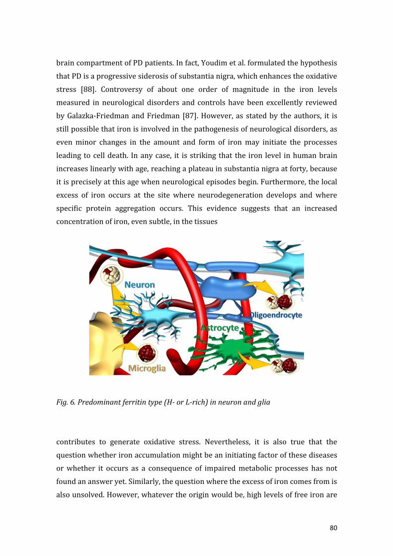
80
brain compartment of PD patients. In fact, Youdim et al. formulated the hypothesis
that PD is a progressive siderosis of substantia nigra, which enhances the oxidative
stress [88]. Controversy of about one order of magnitude in the iron levels
measured in neurological disorders and controls have been excellently reviewed
by Galazka-Friedman and Friedman [87]. However, as stated by the authors, it is
still possible that iron is involved in the pathogenesis of neurological disorders, as
even minor changes in the amount and form of iron may initiate the processes
leading to cell death. In any case, it is striking that the iron level in human brain
increases linearly with age, reaching a plateau in substantia nigra at forty, because
it is precisely at this age when neurological episodes begin. Furthermore, the local
excess of iron occurs at the site where neurodegeneration develops and where
specific protein aggregation occurs. This evidence suggests that an increased
concentration of iron, even subtle, in the tissues
Fig. 6. Predominant ferritin type (H- or L-rich) in neuron and glia
contributes to generate oxidative stress. Nevertheless, it is also true that the
question whether iron accumulation might be an initiating factor of these diseases
or whether it occurs as a consequence of impaired metabolic processes has not
found an answer yet. Similarly, the question where the excess of iron comes from is
also unsolved. However, whatever the origin would be, high levels of free iron are
81
toxic owing to the formation of ROS that finally lead to neuronal death [89]. Also,
the pathologic aggregation of proteins seems to be modulated by iron [90–92].
Both total brain and substantia nigra iron is stored in ferritin [93–95]. Ferritin is
also highly synthesized within the glial compartment, predominantly in
oligodendrocytes, microglia and astrocytes [96] (Fig. 6). Under normal conditions,
most of the iron in the brain is safely stored, and there is no need for a substantial
increase of the total amount of iron(II) to provoke an increase of the amount of
free radicals. Therefore, a minimal ferritin disorder could justify a degeneration of
the nervous cells.
As it occurs in the distribution of H- and L- ferritin content throughout the body,
the H/L ferritin ratio varies in the different regions of brain in agreement with the
requirements of every cell type to detoxify or store iron [97]. H-rich ferritin is
found mainly in neurons whereas L-rich ferritin is more abundant in microglia
[98]. Both, H- and L- ferritin subunits are synthesized in oligodendrocytes and
astrocytes [99] (Fig. 6). Furthermore, the concentrations of H- and L-ferritins in
the substantia nigra increase during life, probably a protective response to the
increase of iron with age. The distribution of types of ferritin within substantia
nigra, especially the presence of H-rich ferritin in neurons and L-rich ferritin in glia
agree with the protection role of H-ferritin against the cytotoxic effect of iron(II)
and the storage capability of L-ferritin.
Hippocampus is a part of the temporal lobe cortex, which plays a crucial role in
memory. Its atrophy with the decline in the number of nervous cells is the starting
point of AD. The cause of this neurodegenerative disorder also remains
unidentified, although it seems that iron mediated oxidative stress may play a role.
Galazka-Friedman [100] reported that the Mössbauer spectroscopic pattern of
hippocampus samples and that of ferritin were highly coincident, as it happened
with that of substantia nigra. The only difference in these spectra was the intensity
of signals, which reflects different iron concentrations in the two brain areas. The
average size of iron cores of ferritin in the hippocampus measured by electron
microscopy was about 3.1 nm. In agreement with previous studies reported by our
group [66], which correlate size and morphology of the ferritin core with shell
composition, this small size should correspond to a high content of H subunits, in
agreement with ELISA studies that revealed a higher average concentration of H
ferritin (150 ± 30 ng/mg) than L ferritin (20 ± 10 ng/mg) on the same type of

82
samples [100].
The complex distribution of ferritin in the brain and the subtle balance in the
appropriate composition of H and L chains show that ferritin can be a key
component in brain function.
5. Copper, zinc and related biomolecules in the brain
Since iron uptake and release by ferritin shells is likely to be influenced, when non
disrupted, by excess of other significant metal ions in brain cells or their
subcellular compartments, it is worth catching a glimpse into the incidence of
Zn(II) and Cu(I)/(II) in mammalian organisms, and precisely, in mammalian brain
cells. Zn(II) is present in the brain at large amounts (10 g/g, ww) [101]. Most of
this zinc accumulates inside cells, where it can reach cytosolic concentrations up to
150 µM, as in neurons [102,103] (Fig. 7), in contrast with the 0.15 µM Zn(II) typical
of central nervous system (CNS) extracellular fluid and serum [102]. Despite these
impressive amounts, free Zn(II) is a minimal percentage of total Zn(II), hardly
reaching levels above 1 nM [104,105]. This implies that, in physiological
conditions, most of the brain Zn(II) is coordinated to some biomolecule, mainly
proteins and other smaller compounds [106–108]. Most of the pool brain Zn is
highly role-specific, being related to neuronal intracellular signaling and
neurotransmission [109]. Noteworthy, the so-called zinc-enriched neurons (ZEN)
accumulate 10–15% of the total brain Zn(II) confined in their presynaptic vesicles
(Fig. 7) [110]. Pathologies that lead to abnormally increased synapsis rate
(ischemia, epilepsy) supposes an excessive postsynaptic uptake of Zn(II) followed
by a massive accumulation of intracellular free Zn(II) that, at the end, directly
provokes neuronal death or takes part in other undesirable
processes by interacting with biomolecules such as ferritin. When Zn(II) reaches
levels that would compromise cell functionality and viability [111,112], it is
translocated into specific cell compartments (mitochondria in neurons), or
sequestered by buffering peptides in the cytosol, mainly metallothioneins (MT)
[113]. MTs are small (6–10 kDa), cysteine rich (33%) metalloproteins that bind a
wide spectrum of heavy metal ions. They are major players in the homeostasis of
physiological Zn(II) and/or Cu(I), as well as in metal detoxification processes. The

83
mammalian MT system includes four highly similar MT isoforms (MT1–MT4, the
latter not present in the brain), which are 60- to 68-amino acid peptides with fully
conserved Cys motif distribution. The tissular expression pattern of the four MTs
shows substantial differentiation. Hence, MT1 and MT2 are ubiquitous, highly
inducible isoforms that respond to a considerable number of factors mainly metal
overload and oxidative stress. In CNS, MT1/2 are predominantly synthesized in
astrocytes and microglia, both in gray and white matter [114], and have been
widely associated with the prevention and healing of CNS inflammation and
injuries, being greatly induced under these conditions. MT3, the brain-specific
isoform, first identified as a growth inhibitory factor (GIF) [115], is constitutive in
neurons (prominently hippocampal neurons), some glial cells and in the
extracellular brain space [116], and it appears mainly involved in neuronal growth
and survival regulation, and, significantly, in processes localized in axon and
dendrite interfaces, as the same synaptic transmission. A recent review of protein
interactions in which MT is involved highlights the role of the different MT
isoforms in the brain Zn(II) secretion and re-circulation events [117]. Although
Zn(II) is a redox-inactive ion, the zinc- thionein/thionein cycle, besides preventing
deleterious free Zn(II) concentrations, provides an indirect pathway of redox
buffering mechanism through the thiol/disulfide equilibrium of their abundant
cysteine residues [118]. Finally, it is worth noting that the tripeptide glutathione
(GSH) has also been shown to thwart the
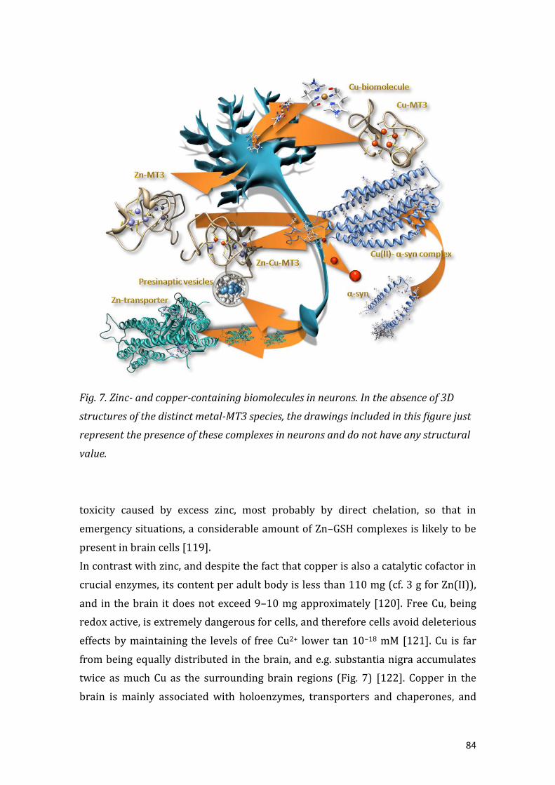
84
Fig. 7. Zinc- and copper-containing biomolecules in neurons. In the absence of 3D
structures of the distinct metal-MT3 species, the drawings included in this figure just
represent the presence of these complexes in neurons and do not have any structural
value.
toxicity caused by excess zinc, most probably by direct chelation, so that in
emergency situations, a considerable amount of Zn–GSH complexes is likely to be
present in brain cells [119].
In contrast with zinc, and despite the fact that copper is also a catalytic cofactor in
crucial enzymes, its content per adult body is less than 110 mg (cf. 3 g for Zn(II)),
and in the brain it does not exceed 9–10 mg approximately [120]. Free Cu, being
redox active, is extremely dangerous for cells, and therefore cells avoid deleterious
effects by maintaining the levels of free Cu2+ lower tan 10−18 mM [121]. Cu is far
from being equally distributed in the brain, and e.g. substantia nigra accumulates
twice as much Cu as the surrounding brain regions (Fig. 7) [122]. Copper in the
brain is mainly associated with holoenzymes, transporters and chaperones, and

85
homeostatic proteins [123]. Cu-containing enzymes are mainly involved in cell
respiration, iron metabolism, ROS defense mechanisms, and significantly, the
synthesis of neurotransmitters (dopamine [124]) and neuron myelinization [122].
Copper is mainly transported in blood bound to ceruloplasmin (CP), although
minor amounts are also coordinated to albumin. In the frame of this review
attention has to be drawn to the membrane anchored CP reported in astrocytes
[125], due to its ferroxidase activity. Brain cells, and especially neurons, avoid
excessive intracellular copper by pumping it out to the cell membranes or
supposedly by sequestering it into MT complexes, although the knowledge on
brain Cu-MT and their behavior is far more limited than that of Zn-MT. It is obvious
that direct deleterious effects of Cu in the brain by ROS generation (Fenton
reaction) derived from its redox nature are effectively avoided by MT coordination.
But the most fascinating aspect of Cu ions in the brain concerns the increased
evidence about their involvement in the onset and progression of
neurodegenerative disorders as AD, PD, amyotrophic lateral sclerosis (ALS), and
transmissible spongiform encephalopathies (TSEs) (for excellent recent reviews,
see [18,21]). All these disorders are triggered by the aggregation of misfolded
monomers and subsequent formation of insoluble deposits that accumulate in the
brain, of the respectively associated proteins: the Aß-peptide α-synuclein, SOD and
the prion proteins. In all these cases, interaction of Zn(II)-MT3 with the
aggregation-prone Cu-loaded peptides seems to preclude their harmful effects.
Precisely, it has been shown that the interaction of Zn7 -MT3 and Cu(II)-Aß-
peptides or Cu(II)-α-synuclein triggers a metal swap reaction which eliminates
Cu(I) from the brain peptides and generates Zn,Cu-MT3 species with four Cu(I)
ions bound to a partially oxidized MT3 domain, and the four Zn(II) ions remaining
in the domain [126,127]. Therefore MT3 in brain metal homeostasis appears far
from acting just as a mere reservoir of metal ions, becoming instead a key
controller of redox reactions and, indirectly, of metal-induced protein damage,
misfolding and aggregation.

86
6. Interaction of ferritin with metal ions
Iron-, zinc- and copper-handling proteins, as well as the metabolism and
homeostasis of these physiological metal ions, are intimately related in all
organisms. As an example, mutations in the genes controlling the multi-copper
ceruloplasmin protein have been associated with iron overload diseases in humans
[128]. People with low levels of ceruloplasmin have been shown to have increased
iron deposits in various tissues [129]. Therefore, it seems reasonable to assume
that in the framework of the interaction between iron, copper and zinc
metabolisms, the ferritin protein could play a central role. This statement could
have sense when taking into consideration the higher iron levels, in most tissues,
with respect to those of copper and zinc, but also because of the
Fig. 8. Structure of the cadmium(II) apoferritin (Protein Data Bank file: 1AEW) [10].
Cadmium(II) concentrates preferentially around the entry of the C3 channels.
huge number of iron atoms confined inside ferritin, a situation that does not occur
in the case of copper or zinc, this multiplying the consequences of any dysfunction
taking place in ferritin owing to the amount of free and toxic iron that can be
released.
The interaction of ferritin with metal ions has been extensively studied in order to
gain a better understanding of the interactions and/or interferences that the later
can cause in ferritin function, either in the iron uptake or iron release processes.

87
Different studies provide evidence that metal cations are bound by ferritin [28]
usually in two separate types of sites of very different binding constants. These
two classes of binding sites are localized either within the cavity or at the external
shell [28]. Interestingly, the largest number of binding sites is for divalent metal
cations, although some of them are functional and others are nonspecific and do
not play any decisive role in the genuine properties of ferritin.
Among the metals studied in the literature, we have considered here those that
have a well-described role on the ferritin function as well as a biological relevance,
especially at brain level, i.e. cadmium(II), zinc(II) and copper(II). Zinc(II) and
cadmium(II) cause inhibition of iron uptake by ferritin [130]. Before any iron fills a
ferritin capsid, all metals compete for the iron binding sites during iron
reconstitution. Once the iron core starts formation, metal ions alter the ferritin
function by a different mechanism, some of them inhibiting, other promoting the
iron core growth.
Cadmium(II) binding is diminished by the presence of a ferritin core, clearly
pointing out that cadmium(II) compete with iron(II) for the same sites inside the
ferritin shell. In fact, when the ferritin cavity is empty, up to 24 Cd(II) can be bound
to its inner surface, one per subunit [28]. However, the mechanism by which
cadmium(II) affects the ferritin iron reconstitution is not only based on the com-
petition for iron binding at the cavity level, but also lies in the fact that
cadmium(II) strongly binds other nonspecific sites, especially at the external shell
surface. There, cadmium(II) has probably the greatest affinity for these sites, and is
able to exclude almost all other metals from binding. It should be noted that
cadmium binds sites localized around the entry of the hydrophilic channels (Fig.
8), and that it may even hinder the entry of other metals, including iron, and
therefore block the ferritin iron reconstitution. Based on the interaction with the
ferritin shell and on the effect that this interaction provokes in the solubility of the
ferritin protein, cadmium(II) is commonly used as a precipitant agent of ferritin
[131] and in fact, most commercial ferritins contain significant amounts of
cadmium coming from the preparation process.
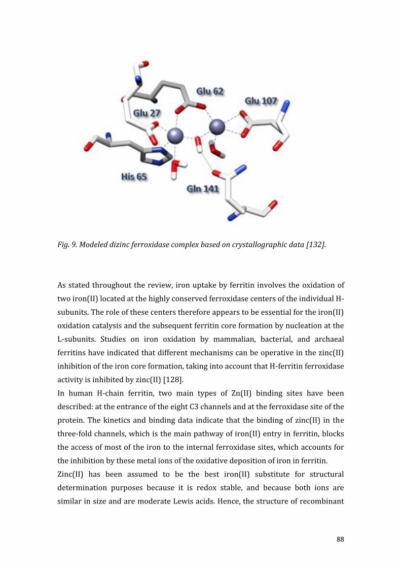
88
Fig. 9. Modeled dizinc ferroxidase complex based on crystallographic data [132].
As stated throughout the review, iron uptake by ferritin involves the oxidation of
two iron(II) located at the highly conserved ferroxidase centers of the individual H-
subunits. The role of these centers therefore appears to be essential for the iron(II)
oxidation catalysis and the subsequent ferritin core formation by nucleation at the
L-subunits. Studies on iron oxidation by mammalian, bacterial, and archaeal
ferritins have indicated that different mechanisms can be operative in the zinc(II)
inhibition of the iron core formation, taking into account that H-ferritin ferroxidase
activity is inhibited by zinc(II) [128].
In human H-chain ferritin, two main types of Zn(II) binding sites have been
described: at the entrance of the eight C3 channels and at the ferroxidase site of the
protein. The kinetics and binding data indicate that the binding of zinc(II) in the
three-fold channels, which is the main pathway of iron(II) entry in ferritin, blocks
the access of most of the iron to the internal ferroxidase sites, which accounts for
the inhibition by these metal ions of the oxidative deposition of iron in ferritin.
Zinc(II) has been assumed to be the best iron(II) substitute for structural
determination purposes because it is redox stable, and because both ions are
similar in size and are moderate Lewis acids. Hence, the structure of recombinant

89
human H dizinc(II) ferroxidase complex has been solved by X-ray crystallography
(Protein Data Bank file 2CEI) [38]. A perspective view of this is displayed in Fig. 9.
The zinc at site A is coordinated to a nitrogen atom from His65 and three oxygen
atoms, two from Glu27, Glu62 and the third one presumed to be from a water
molecule. The zinc at site B is coordinated by both carboxylate oxygen atoms of
Glu107 and Glu62, which bridge the two metal sites. Furthermore, a computational
study identified a hydroxide bridge between sites A and B, and another water
molecule at site B and showed that no other ligand or peripheral molecules are
present in the experimental dizinc ferroxidase center [132]. Finally, and according
to these authors, the modeled structures of the diiron and dizinc complexes are
close, suggesting that the dizinc ferroxidase is an accurate model of the native
diiron complex.
Although the empty ferroxidase site accommodates two zinc(II) ions, the binding
of just one zinc(II) is enough to abolish the iron(II) oxidation capability of the
ferroxidase center [57]. It is interesting to note here that zinc(II) binds relatively
tightly to horse spleen apoferritin, displacing iron(II) under anaerobic conditions
but however, once the ferroxidase center is filled with two iron(III), zinc(II) is no
longer able to bind there, nor does it inhibit the iron core formation. A similar
situation has been described for cadmium(II). Therefore, the existence of a
competition between metals for different apoferritin binding sites takes place but
once the ferritin biomineralization process is triggered neither cadmium or zinc
are able to affect the ferritin machinery.
Interestingly, in the case of the recombinant human H-ferritin, the structure of the
distinct complexes with zinc(II) reveal a surprising similarity between them,
indicating that the dinuclear site in this recombinant ferritin is quite a rigid site for
this metal ion. Surprisingly, this is not the case when iron occupies these same
binding sites. As previously described, coordination of iron at the ferroxidase
center undergoes changes during oxidation with the subsequent iron-iron distance
shortening [55].
The rigidity of the zinc(II) coordination at the ferroxidase site with respect to the
flexibility found for iron is probably a consequence of the non-redox chemistry of
zinc(II), which has a definitive importance in how zinc(II) affects the ferritin iron
uptake. This is more evident when the coordination of copper to the ferroxidase
center and its effect to the ferritin function is analyzed (see below).

90
The lack of ferroxidase activity in ferritin due to the presence of zinc(II) is
extremely relevant in neurons, where the concentration of zinc(II) becomes very
significant (Section 5) and the ferritin is predominantly H-rich (Section 4). A
minimum amount of free zinc(II) could inhibit the appropriate iron(II) oxidation
by ferritin, giving rise to a cascade radical process that would conclude with
neuronal death.
Furthermore, the interaction of zinc(II) with ferritin can be extrapolated to other
neurological scenarios. For instance, AD is complicated by prooxidant
intraneuronal free iron(II) and, interestingly, by zinc(II) overloading within
amyloid plaque. Interestingly, the β-amyloid protein precursor has been
described to exhibit ferroxidase activity due to the existence of what Bush et al.
denominate a H-ferritin-like active site, which is captivatingly also inhibited by
zinc(II) [133]. However, it must be taken into account that the ferroxidase activity
of the AD β-amyloid protein precursor has been recently called into question and
asked for its revaluation [134].
The interaction of copper with ferritin has also been investigated. Copper(II), as in
the case of zinc(II), was shown to bind relatively tightly to horse spleen ferritin.
Once bound, copper(II) has a catalytic effect on the aerobic oxidative uptake of
iron(III) by ferritin, which means that it induces an opposite effect to that of
zinc(II). The difference between the effect of zinc(II) and copper(II) has been
explained by Moore et al. in terms of the capability of copper(II) to take part in
redox reactions [40]. In the absence of any other metal ions, two iron(II) bind at
this site, are oxidized by O2 and form the DFC. Now, returning to the opposite
effects of zinc(II) and copper(II) in horse spleen ferritin, it is plausible to consider
that if these two ions are bound in a way that they occupy just one end of the site,
an incoming iron(II) occupying the other end might be oxidized by copper(II) but
not by zinc(II). The iron(III) produced could then migrate from the site to allow
another iron(II) to take its place alongside the produced copper(I). At this point a
two-electron reduction of O2 would generate copper(II) and iron(III) and, with the
migration of iron(III) away from the site, the cycle could be reinitiated [40].
Unfortunately, only crystal data of ferritin–copper(II) and not copper(I) complex
are yet available to confirm this hypothesis, but it should be highlighted that, as
stated before, both the distances and bond angles found for the zinc(II) complexes
of ferritin point out to an extreme rigidity of the site that would justify a zinc

91
inhibitory role of the ferroxidase activity, but a promoting role for copper.
The loss of the ferroxidase activity of ferritin in the brain becomes a transcendent
problem when pathological iron(II) accumulates and neurodegenerative processes
are triggered. It is quite intriguing to realize that the presence of free, and toxic
iron(II), can be generated either by an excess of zinc that inhibits the ferroxidase
activity of ferritin, or by a deficiency of copper. In fact, when copper lacks, there is
also a malfunction of the multicopper ferroxidase ceruloplasmin protein and a
development of the aceruloplasminemia disease, which leads to glial iron
accumulation and dementia.
7. Interaction of ferritin with metalloproteins and other biomolecules
Two metalloproteins, specifically Cu- or Zn-containing proteins, have been
reported in literature to somehow interact with ferritin: ceruloplasmin, a blue
multicopper oxidase with ferroxidase activity [135], and metallothioneins (see
Section 5).
The oldest studies, carried out before the 2000 decade, called into question the
mechanism of ferritin iron uptake described before in this review, and which is
nowadays universally accepted. Aust and coworkers defended, at the end of the
90s, the docking between ceruloplasmin and ferritin [41]. This research group was
defending that the ferroxidase activity of the H-subunits was leading to iron loaded
ferritins with properties notably differing from those of native ferritins. In parallel,
they reported that the enzymatic load of apoferritin with iron by using intact
human ceruloplasmin rendered iron loaded ferritins with nearly identical
properties to those of native ferritins [136]. Therefore, to accomplish the iron(II)
to iron(III) oxidation by ceruloplasmin, a protein–protein complex formation was
hypothesized, and the existence of a specific recognition site for the interaction of
both proteins and the stimulation of its ferroxidase activity was sug- gested. In fact,
the symptoms of iron overload as well as of excessive deposition of insoluble iron
found in patients diagnosed with aceruloplasminemia (low concentrations of
active ceruloplasmin) or Menkes disease (a disorder that affects copper levels in
the body, leading to copper deficiency) supported the proposed interaction.
Unfortunately, this research line was stopped and no further data giving continuity
to this hypothesis have ever been reported.

92
Much more recently, our groups reported an intriguing interaction between
metallothionein and ferritin. Two proteins apparently designed by nature to
protect cells, and organisms, from free transition metal ions, and to respectively
maintain the homeostatic levels of Zn and Cu, and Fe generated deleterious effects
when observation each other. This serendipitous discovery occurred during the
search for a biomolecule able to capture the released iron(II) from ferritin and thus
capable to prevent ROS formation (Section 3 of this work). For this study, the Zn-
complexes of the mammalian metallothionein isoforms found in the brain, MT1,
MT2 and MT3 (Section 5), and the model horse spleen ferritin where chosen. The
obtained results were astonishing (respectively 55.6, 23.9, and 59.5% of iron
release) and they correlated well with the Zn/Cu-thionein character of the assayed
MTs [137]. Further experiments carried out either with Cu-loaded or Zn,Cu-
heteronuclear complexes of these MTs confirmed parallel behaviors and suggested
that not only iron(II) and zinc(II), but also copper(I) can be liberated to the media
if ferritins and metallothioneins meet in the cell [authors’ unpublished results].
Interestingly, neither binding between the two participant proteins nor a
disruption of the ferritin structure was detected during the interaction. However,
iron and zinc liberation from their respective metalloproteins was reported to be
the consequence of the oxidation of the cysteinic residues of the MTs. Therefore, a
proposal of a tentative redox reaction between the two biomolecules by electron
transfer through the ferritin shell was proposed owing to the size of the Zn-MT
complexes, which impairs them reaching the mineral by traversing the protein
shell through the ferritin channels (Fig. 10). Recent theoretical studies [82] and
experimental observations [138] commented above give nowadays further
support to this proposal.
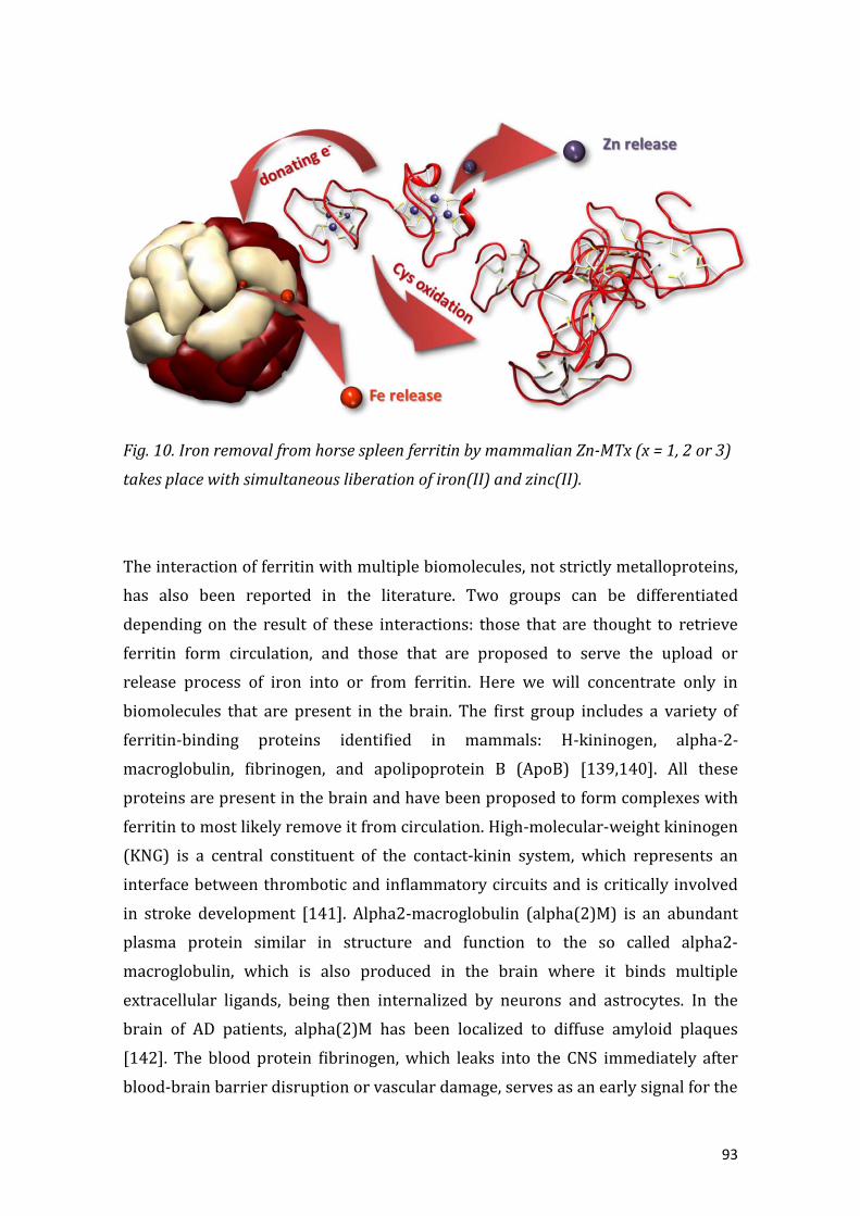
93
Fig. 10. Iron removal from horse spleen ferritin by mammalian Zn-MTx (x = 1, 2 or 3)
takes place with simultaneous liberation of iron(II) and zinc(II).
The interaction of ferritin with multiple biomolecules, not strictly metalloproteins,
has also been reported in the literature. Two groups can be differentiated
depending on the result of these interactions: those that are thought to retrieve
ferritin form circulation, and those that are proposed to serve the upload or
release process of iron into or from ferritin. Here we will concentrate only in
biomolecules that are present in the brain. The first group includes a variety of
ferritin-binding proteins identified in mammals: H-kininogen, alpha-2-
macroglobulin, fibrinogen, and apolipoprotein B (ApoB) [139,140]. All these
proteins are present in the brain and have been proposed to form complexes with
ferritin to most likely remove it from circulation. High-molecular-weight kininogen
(KNG) is a central constituent of the contact-kinin system, which represents an
interface between thrombotic and inflammatory circuits and is critically involved
in stroke development [141]. Alpha2-macroglobulin (alpha(2)M) is an abundant
plasma protein similar in structure and function to the so called alpha2-
macroglobulin, which is also produced in the brain where it binds multiple
extracellular ligands, being then internalized by neurons and astrocytes. In the
brain of AD patients, alpha(2)M has been localized to diffuse amyloid plaques
[142]. The blood protein fibrinogen, which leaks into the CNS immediately after
blood-brain barrier disruption or vascular damage, serves as an early signal for the

94
induction of glial scar formation via the TGF- /Smad signaling pathway [143]. At
least two mechanisms of interaction between these proteins and ferritin have been
proposed [140]: one is direct binding, such as that proposed for H-kininogen, and
fibrinogen; the other is an indirect binding through heme on the surface of the
ferritin molecule, such as that described for the heme-binding protein, ApoB. In
this case, some distinctions should be made: those biomolecules supposed to
contribute to the uptake process and those involved in the release of iron from
ferritin. Some fatty acids appear to achieve the first goal, while, SOD and xanthine
oxidase and cytochrome P-450 seem to fulfill the second purpose. Arachidonic acid
(ARA) is one of the most abundant fatty acids in the brain and its disturbed
metabolism may be associated with neurological disorders such as AD and bipolar
disorder [144]. Interestingly, Bu et al. [145] recently reported a 60 µM affinity
specific binding site of ferritin for ARA. Also, they described that its binding
enhances iron mineralization and decreases iron release at the same time that pro-
tects the fatty acid from oxidation. This binding site was proposed to be located in
the 2-fold intersubunit pocket, allowing the ARA tail to project well into the
ferrihydrite mineralization site on the L-ferritin subunit, while shorter saturated
fatty acids as caprylate were described not to have significant effects on
mineralization.
As stated in previous sections, iron(II) can be released from ferritin by reductants
small enough to reach the mineral core by using the ferritin channels, and this is
the case of O2•−. Interestingly, two distinct research groups reported diverging
results with respect to the capability of O2•− to affect the release of iron(II) from
ferritin and this implied the participation of two other biomolecules: SOD and
xanthine oxidase. Both, the research groups of Biemond et al. [146] and of Thomas
et al. [147] found that iron was mobilized from ferritin by xanthine oxidase.
However, after observation that iron release could be completely inhibited by SOD,
Thomas et al. concluded that O2•− was the active iron-mobilizing species. Similar
conclusions can be drawn from the studies between the interaction of ferritin with
cytochrome P-450 [148] or with the intracellularly ocurring Cu(I)-glutathione
complex Cu(GSH)2 [149]. Oppositely, the observation that SOD had no blocking
effect on the release of iron from ferritin by xanthine oxidase, led to Biemond et al.
to suggest that probably electrons were transferred from the enzyme-substrate
complex to ferritin via an unknown mechanism. Finally, Bolann and Ulvik reported

95
that in hypoxic tissues xanthine oxidase can release iron from ferritin by an O2•−
independent process [150].
8. Concluding remarks
Over millions of years, living organisms have produced ferritin for a crucial
function: iron storage. Ferritin is at the center of iron metabolism, providing
protection by scavenging free iron and storing iron when needed. Dysfunction in
ferritin, either at iron uptake or iron release leads to the existence of free iron(II),
which can be the beginning of a range of neurological pathologies.
The enormous progress made in understanding the mechanism of action of
ferritin, nevertheless, revealed a number of transcendent issues that still remain to
be solved such as: (i) the influence of the H- or L-nature of its subunits in the
formation of the iron core, (ii) the identification of the putative ferritin-binding
biomolecules that can promote iron release or (iii) the role and interference of
metals and metalloproteins in ferritin iron uptake and iron release. This review
has been mainly focused in the last issue. However, much more data collecting
information from realistic scenarios where other free metal ions different than
iron, and other metalloproteins interact with ferritin are still needed. It is our
belief that this review will provide a basis of knowledge from where to extract
comprehensive conclusions and to build future prospects.
We have reviewed different situations in which the interaction of ferritin with a
metal ion or a metalloprotein gives rise to the existence of free toxic metals, iron or
iron plus other metals. For example, it is true that zinc(II) can be found at high
concentrations in neurons but fortunately it is mainly complexed or
compartimentalized. However, any anomalous zinc(II) metabolism provoking the
appearance of free zinc(II) in neuron would cause an inhibition of the ferroxidase
activity of ferritin and the subsequent existence of not stored free iron(II). Then,
the free zinc(II) and iron(II) would result in causing neuron death. Interestingly,
the presence of the multicopper ceruloplasmine could overcome this problem as
this protein promotes iron(II) oxidation and also ferritin reconstitution. This is a
genuine example to show how the metabolism of these three metals is interrelated
and how its study in depth requires a full approach where interactions between
free and protein-containing metals must be considered.
96
In another scenario, an excessive proportion of H-subunits in ferritin can result in
an increase of iron(II) oxidation level but a in significant decrease of the amount of
iron stored in the mineral core of ferritin, with the consequent existence of free
iron(III), which is capable to oxidize some metalloproteins, such as
metallothioneins, ultimately leading to the liberation of metal ions like copper(I) or
zinc(II). Likewise, the iron(III) stored in the mineral core of ferritin can also
oxidize the metal-MT complexes causing the liberation of iron(II) and of the metal
ions bound to metallothioneins.
Although we are aware of the evident difficulties associated with the study of more
integrative systems, it is our conviction that the future goals for the elucidation of
the iron uptake and release processes in ferritin should aim at considering not only
the presence of distinct metals but also that of other metalloproteins, which coexist
with ferritin in the different tissues and cell compartments. It is our belief that only
with this approach, a realistic view of the chemical implications of their
interactions can be achieved. This is especially important in the brain when trying
to shed light into the neuropathologies associated with the dysfunction of
metalloproteins that give rise to an uncontrolled release of metals, a fact that
probably underlines the origin of some of the most dramatic neurological diseases
affecting nowadays the worldwide population.
Acknowledgments
This work was supported by the “Spanish Ministerio de Ciencia e Innovación ”
MICINN and FEDER (grants BIO2012-39682-C02-01 and BIO2012-39682-C02-02
respectively to S.A. and M.C., and CTQ2012-32236 to J.M.D.-V.). Authors from
Barcelona Universities are members of the 2009SGR-1457 “Grup de Recerca de la
Generalitat de atalunya.” uthors from the Universidad de Granada and aen are
members of the FQM-368 “Grupo de Investigacion de la unta de ndalucia.”

97
References
[1] R. Crichton, Inorganic Biochemistry of Iron Metabolism: from Molecular
Mechanisms to Clinical Consequences, John Wiley & Sons, England, 2001.
[2] D.J. Pinero, J.R. Connor, in: H.R. Lieberman, R.B. Kanarek, C. Prasad (Eds.), Nutri-
tional Neuroscience, CRC Press, Taylor & Francis Group, Boca Raton, 2005, p. 235.
[3] K. Jomova, M. Valko, Curr. Pharm. Des. 17 (2011) 3460.
[4] H. Heli, S. Mirtorabi, K. Karimian, Expert Opinion on Therapeutic Patents
21(2011) 819.
[5] N.D. Chasteen, P.M. Harrison, J. Struct. Biol. 126 (1999) 182.
[6] F.M. Michel, L. Ehm, S.M. Antao, P.L. Lee, P.J. Chupas, G. Liu, D.R. Strongin, M.A.A.
Schoonen, B.L. Phillips, J.B. Parise, Science 316 (2007) 1726.
[7] N. Gálvez, B. Fernández, P. Sánchez, R. Cuesta, M. Ceolín, M. Clemente, S.
Trasobares, M. López-Haro, J.J. Calvino, O. Stéphan, J.M. Domínguez-Vera, J. Am.
Chem. Soc. 130 (2008) 8062.
[8] S.P. Martsev, A.P. Vlasov, P. Arosio, Protein Eng. 11 (1998) 377.
[9] S. Levi, S.J. Yewdall, P.M. Harrison, P. Santambrogio, A. Cozzi, E. Rovida, A.
Albertini, P. Arosio, Biochem. J. 288 (1992) 591.
[10] P.D. Hempstead, S.J. Yewdall, A.R. Fernie, D.M. Lawson, P.J. Artymiuk, D.W.
Rice, G.C. Ford, P.M. Harrison, J. Mol. Biol. 268 (1997) 424. [11] E.C. Theil, Curr.
Opin. Struct. Biol. 15 (2011) 304.
[12] M. Wagstaff, M. Worwood, A. Jacobs, Biochem. J. 173 (1978) 969.
[13] J. Zähringer, B.S. Baliga, H.N. Munro, Proc. Natl. Acad. Sci. U.S.A. 73 (1976) 857.
[14] K. Iwai, S.K. Drake, N.B. Wehr, A.M. Weissman, T. LaVaute, N. Minato, R.D.
Klausner, R.L. Levine, T.A. Rouault, Proc. Natl. Acad. Sci. U.S.A. 95 (1998) 4924.
[15] S.V. Torti, E.L. Kwak, S.C. Miller, L.L. Miller, G.M. Ringold, K.B. Myambo, A.P.
Young, F.M. Torti, J. Biol. Chem. 263 (1988) 12638.
[16] D. Berg, G. Beceker, P. Riederer, O. Rieb, Neurotox. Res. 4 (2002) 637.
[17] B. Uttara, A.V. Singh, P. Zamboni, R.T. Mahajan, Curr. Neuropharmacol. 7
(2009) 65.
[18] H. Kozlowski, M. Luczkowski, M. Remelli, D. Valensin, Coord. Chem. Rev. 256
(2012) 2129.
[19] A. Binolfi, L. Quintanar, C.W. Bertoncini, C. Griesinger, C.O. Fernández, Coord.
Chem. Rev. 256 (2012) 2188.

98
[20] C. Hureau, P. Dorlet, Chem. Rev. 256 (2012) 2175. [21] J.H. Viles, Coord. Chem.
Rev. 256 (2012) 2271.
[22] J.M. Domínguez-Vera, B. Fernández, N. Galvez, Future Med. Chem. 2 (2010)
609.
[23] A.R.J. Curtis, C. Fey, C.M. Morris, L.A. Bindoff, P.G. Ince, P.F. Chinnery, A.
Coulthard, M.J. Jackson, A.P. Jackson, D.P. McHale, D. Hay, W.A. Barker, A.F.
Markham, D. Bates, A. Curtis, J. Burn, Nat. Genet. 28 (2001) 350.
[24] J. Kato, K. Fujikawa, M. Kanda, N. Fukuda, K. Sasaki, T. Takayama, M. Kobune,
K. Takada, R. Takimoto, H. Hamada, T. Ikeda, Y. Niitsu, Am. J. Hum. Genet. 69
(2001) 191.
[25] M.E. Martin, S. Fargion, P. Brissot, B. Pellat, C. Beaumont, Blood 91 (1998) 319.
[26] R.K. Watt, R.J. Hilton, D.M. Graff, Biochim. Biophys. Acta 1800 (2010) 745. [27]
J.G. Joshi, A. Zimmerman, Toxicology 48 (1988) 21.
[28] S. Pead, E. Durrant, B. Webb, C. Larsen, D. Heaton, J. Johnson, G.D. Watt, J.
Inorg. Biochem. 59 (1995) 15.
[29] I.G. Macara, T.G. Hoy, P.M. Harrison, Biochem. J. 135 (1973) 785.
[30] A. Mazur, S. Green, A. Saha, A. Carleton, J. Clin. Invest. 37 (1985) 18097. [31]
S.-H. Juan, J.-H. Guo, S.D. Aust, Arch. Biochem. Biophys. 341 (1997) 280.
[32] G.R. Bakker, R.F. Boyer, J. Biol. Chem. 261 (1986) 13182.
[33] N.E. Le Brun, A.M. Keech, M.R. Mauk, A.G. Mauk, S.C. Andrews, A.J. Thomson,
G.R. Moore, FEBS Lett. 397 (1996) 159.
[34] A. Treffry, P.M. Harrison, J. Inorg. Biochem. 21 (1984) 9.
[35] X. Yang, N.E. Le Brun, A.J. Thomson, G.R. Moore, N.D. Chasteen, Biochemistry
39 (2000) 4915.
[36] T.J. Stillman, P.D. Hempstead, P.J. Artymiuk, S.C. Andrews, A.J. Hudson, A.
Treffry, J.R. Guest, P.M. Harrison, J. Mol. Biol. 307 (2001) 587.
[37] F. Bou-Abdallah, P. Arosio, S. Levi, C. Janus-Chandler, N.D. Chasteen, J. Biol.
Inorg. Chem. 8 (2003) 489.
[38] L. Toussaint, L. Bertrand, L. Hue, R.R. Crichton, J.P. Declercq, J. Mol. Biol. 365
(2007) 440.
[39] M.J. Yablonski, E.C. Theil, Biochemistry 31 (1992) 9680.
[40] J. McKnight, N. White, G.R. Moore, J. Chem. Soc. Dalton Trans. (1997) 4043.
[41] C.A. Reilly, M. Sorlie, S.D. Aust, Arch. Biochem. Biophys. 354 (1998) 165, and
references therein.

99
[42] R. Orihuela, B. Fernández, O. Palacios, E. Valero, S. Atrian, R.K. Watt, J.M.
Domínguez-Vera, M. Capdevila, Chem. Commun. 47 (2011) 12155. [43] P.M.
Proulx-Curry, N.D. Chasteen, Coord. Chem. Rev. 144 (1995) 347. [44] R.C. Hider, X.
Kong, Dalton Trans. 42 (2013) 3220.
[45] P.M. Harrison, P. Arosio, Biochim. Biophys. Acta 1275 (1996) 161.
[46] E.C. Theil, R.K. Behera, T. Tosha, Coord. Chem. Rev. 257 (2013) 579, and
references therein.
[47] T. Douglas, D.R. Ripoll, Protein Sci. 7 (1998) 1083. [48] X. Liu, E.C. Theil, Acc.
Chem. Res. 38 (2005) 167.
[49] T. Tosha, H-L. Ng, O. Bhattasali, T. Alber, E.C. Theil, J. Am. Chem. Soc. 132
(2010) 14562.
[50] F. Bou-Abdallah, G.C. Papaefthymiou, D.M. Scheswohl, S.D. Stanga, P. Arosio,
N.D. Chasteen, Biochem. J. 364 (2002) 57.
[51] X. Liu, E.C. Theil, Proc. Natl. Acad. Sci. U.S.A. 101 (2004) 8557.
[52] J. Tatur, W.R. Hagen, P.M. Matias, J. Biol. Inorg. Chem. 12 (2007) 615.
[53] J. Hwang, C. Krebs, B.H. Huynh, D.E. Edmondson, E.C. Theil, J.E. Penner-Hahn,
Science 287 (2000) 122.
[54] A. Crow, T.L. Lawson, A. Lewin, G.R. Moore, N.E. Le Brun, J. Am. Chem. Soc. 131
(2009) 6808.
[55] I. Bertini, D. Lalli, S. Mangani, C. Pozzi, C. Rosa, E.C. Theil, P. Turano, J. Am.
Chem. Soc. 134 (2012) 6169.
[56] P. Turano, D. Lalli, I.C. Felli, E.C. Theil, I. Bertini, Proc. Natl. Acad. Sci. U.S.A. 107
(2010) 545.
[57] K.H. Ebrahimi, P.-L. Hagedoorn, W.R. Hagen, J. Biol. Inorg. Chem. 15 (2010)
1243.
[58] J. Tatur, W.R. Hagen, FEBS Lett. 579 (2005) 4729.
[59] K.H. Ebrahimi, P.L. Hagedoorn, J.A. Jangejon, W.R. Hagen, J. Biol. Inorg. Chem.
14 (2009) 1265.
[60] N.E. Le Brun, A. Crow, M.E.P. Murphy, A.G. Mauk, G.R. Moore, Biochim.
Biophys. Acta 1800 (2010) 732.
[61] A. Lewin, G.R. Moore, N.E. Le Brun, Dalton Trans. (2005) 3597.
[62] J.L. Johnson, M. Cannon, R.K. Watt, R.B. Frankel, G.D. Watt, Biochemistry 38
(1999) 6706.
[63] J.S. Rohrer, Q.T. Islam, G.D. Watt, D.E. Sayes, E.C. Theil, Biochemistry 29 (1990)

100
259.
[64] A. Treffry, P.M. Harrison, Biochem. J. 171 (1978) 313. [65] R.J. Hilton, A. David,
R.K. Watt, Biometals 25 (2012) 259.
[66] J.D. López-Castro, J.J. Delgado, J.A. Perez-Omil, N. Galvez, R. Cuesta, R.K. Watt,
J.M. Domínguez-Vera, Dalton Trans. 41 (2012) 1320.
[67] F. Carmona, O. Palacios, S. Atrian, J.M. Dominguez-Vera, M. Capdevila, under
review.
[68] Q.Q. Pankhursta, D. Hautotb, N. Khanc, J. Dobson, J. Alzheimers Dis. 13 (2008)
49.
[69] S. Yasmin, S.C. Andrews, G.R. Moore, N.E. Le Brun, J. Biol. Chem. 286 (2011)
3473.
[70] S. Sirivech, E. Frieden, S. Osaki, Biochem. J. 143 (1974) 311.
[71] M.S. Joo, G. Tourillon, D.E. Sayers, E.C. Theil, Biol. Met. 3 (1990) 171. [72] D.C.
Harris, Biochemistry 17 (1978) 3071.
[73] J. Johnson, J. Kenealey, R.J. Hilton, D. Brosnahan, R.K. Watt, G.D. Watt, J. Inorg.
Biochem. 105 (2011) 202.
[74] X. Liu, W. Jin, E.C. Theil, Proc. Natl. Acad. Sci. U.S.A. 100 (2003) 3653.
[75] N. Gálvez, B. Ruiz, R. Cuesta, E. Colacio, J.M. Domínguez-Vera, Inorg. Chem. 44
(2005) 2706.
[76] F. Bou-Abdallah, J. McNally, X.X. Liub, A. Melman, Chem. Commun. 47 (2011)
731.
[77] P. Sánchez, N. Gálvez, E. Colacio, E. Min˜ ones J.M. Domínguez-Vera, Dalton
Trans. (2005) 811.
[78] W. Linert, G.N.L. Jameson, J. Inorg. Biochem. 79 (2000) 319.
[79] G.N.L. Jameson, W. Linert, in: G. Poli, E. Cadenas, L. Packer (Eds.), Free Radicals
in Brain Pathophysiology, Marcel-Decker, New York, 2000, p. 247.
[80] G.D. Watt, D. Jacobs, R.B. Frankel, Proc. Natl. Acad. Sci. U.S.A. 85 (1988) 7457.
[81] S.K. Weeratunga, C.E. Gee, S. Lovell, Y. Zeng, C.L. Woodin, M. Rivera,
Biochemistry 48 (2009) 7420.
[82] V. Subramanian, D.G. Evans, J. Phys. Chem. B 116 (2012) 9287.
[83] H. Shi, K.Z. Bencze, T.L. Stemmler, C.C. Philpott, Science 320 (2008) 1207. [84]
D.H. Boldt, Am. J. Med. Sci. 318 (1999) 207.
[85] J. Connor, in: J. Connor (Ed.), Metals and Oxidative Damage in Neurological
Disorders, Plenum Press, New York, 1997, p. 23.
101
[86] J.R. Connor, S.A. Benkovic, Ann. Neurol. 32 (1992) S51.
[87] J. Galazka-Friedman, A. Friedman, Acta Neurobiol. Exp. 57 (1997) 217.
[88] M.B.H. Youdim, D. Ben-Shachar, P. Riederer, Acta Neurol. Scand. 126 (1989)
47.
[89] R.A. Cherny, C.S. Atwood, M.E. Xilinas, D.N. Gray, W.D. Jones, C.A. McLean, K.J.
Barnham, I. Volitakis, F.W. Fraser, Y. Kim, X. Huang, L.E. Goldstein, R.D. Moir, J.T.
Lim, K. Beyreuther, H. Zheng, R.E. Tanzi, C.L. Masters, A.I. Bush, Neuron 30 (2001)
665.
[90] M. Perez, J.M. Valpuesta, E.M. De Garcini, C. Quintana, M. Arrasate, J.L. Lopez-
Carrascosa, A. Rabano, J.G. De Yebenes, J. Avila, Am. J. Pathol. 152 (1998) 1531.
[91] A. Takeda, M. Hashimoto, M. Mallory, M. Sundsumo, L. Hansen, A. Sisk, E.
Masliah, Lab. Invest. 78 (1998) 1169.
[92] M. Hashimoto, E. Masliah, Brain Pathol. 9 (1999) 707.
[93] D.T. Dexter, C.J. Carter, F.R. Wells, F. Javoy-Agid, Y. Agid, A. Lees, P. Jenner, C.D.
Marsden, Brain 114 (1991) 1953.
[94] B. Hallgren, P. Sourander, J. Neurochem. 3 (1958) 41.
[95] J. Sian-Huelsmann, S. Mandel, M.B.H. Youdim, P. Riederer, J. Neurochem. 118
(2011) 939.
[96] J.R. Connor, K.L. Boeshore, S.A. Benkovic, S.L. Menzies, J. Neurosci. Res. 37
(1994) 461.
[97] J.R. Connor, B.S. Snyder, P. Arosio, D.A. Loefer, P. Le Witt, J. Neurochem. 65
(1995) 717.
[98] P. Ponka, C. Beaumont, D. Richardson, Semin. Hematol. 35 (1998) 35.
[99] L. Zecca, M.B. Youdim, P. Riederer, J.R. Connor, R.R. Crichton, Nat. Rev. Neu-
rosci. 5 (2004) 863.
[100] J. Galazka-Friedman, Hyperfine Interact. 182 (2008) 31. [101] C.J.
Frederickson, Int. Rev. Neurobiol. 31 (1989) 145. [102] A. Takeda, Brain Res. 34
(2000) 137.
[103] R.A. Colvin, Am. J. Physiol. Cell Physiol. 282 (2002) 317. [104] C.E. Outten,
T.V. O’Halloran Science 292 (2001) 2488. [105] L. . inney T.V. O’Halloran
Science 300 (2003) 931.
[106] I. Bertini, L. Decaria, A. Rosato, J. Biol. Inorg. Chem. 15 (2010) 1071. [107] L.
Decaria, I. Bertini, R.J.P. Williams, Metallomics 2 (2010) 706. [108] W. Maret, Y. Li,
Chem. Rev. 109 (2009) 4682.

102
[109] A. Takeda, Biometals 14 (2001) 343.
[110] C.J. Frederickson, J.Y. Koh, A.I. Bush, Nat. Rev. Neurosci. 6 (2005) 449. [111]
D.W. Choi, M. Yokoyama, J.K. Koh, Neuroscience 24 (1988) 67.
[112] G.J. Lees, A. Lehmann, M. Sandberg, A. Hamberger, Neurosci. Lett. 120 (1990)
155.
[113] . apdevila R. Bofill O. Palacios S. trian oord. Chem. Rev. 256 (2012)
46.
[114] J. Hidalgo, R. Chung, M. Penkowa, M. Vasak, in: A. Sigel, H. Sigel, R.K.O. Sigel
(Eds.), Metallothioneins and Related Chelators, Metal Ions in Life Sciences, vol. 5,
RSC Publishing, Cambridge, 2009, p. 279.
[115] Y. Uchida, Biol. Signals 3 (1994) 211.
[116] M. Vasak, G. Meloni, in: A. Sigel, H. Sigel, R.K.O. Sigel (Eds.), Metallothioneins
and Related Chelators, Metal Ions in Life Sciences, vol. 5, RSC Publishing,
Cambridge, 2009, p. 319.
[117] S. Atrian, M. Capdevila, Biomolecular Concepts (2013),
http://dx.doi.org/10.1515/bmc-2012-0049.
[118] W. Maret, A. Krezel, Mol. Med. 13 (2007) 371. [119] C.-J. Chen, S.-L. Liao, J.
Neurochem. 85 (2003) 443.
[120] E. Gaggelli, H. Kozlowski, D. Valensin, G. Valensin, Chem. Rev. 106 (2006)
1995.
[121] T.D. Rae, P.J. Schmidt, R.A. Pufahl, V.C. Culotta, T.V. O’Halloran Science 284
(1999) 805.
[122] K.M. Davies, D.J. Hare, V. Cottam, N. Chen, L. Hilgers, G. Halliday, J.F.B. Mercer,
K.L. Double, Metallomics 5 (2013) 43.
[123] L. Decaria, I. Bertini, R.J.P. Williams, Metallomics 3 (2011) 56.
[124] T.C. Steveson, G.D. Ciccotosto, X.M. Ma, G.P. Mueller, R.E. Mains, B.A. Eipper,
Endocrynology 144 (2003) 188.
[125] L.W.J. Klomp, Z.S. Farhangrazi, L.L. Dugan, J.D. Gitlin, J. Clin. Invest. 98 (1996)
207.
[126] G. Meloni, V. Sonois, T. Delaine, L. Guilloreau, A. Gillet, J. Teissie, P. Faller, M.
Vasak, Nat. Chem. Biol. 4 (2008) 366.
[127] G. Meloni, M. Vasak, Free Radic. Biol. Med. 50 (2011) 1471.
[128] K. Yoshida, K. Furihata, S. Takeda, A. Nakamura, K. Yamamoto, H. Morita, S.
Hiyamuta, S. Ikeda, N. Shimizu, N. Yanagisawa, Nat. Genet. 9 (1995)
103
267.
[129] Z.L. Harris, Y. Takahashi, H. Miyajima, M. Serizawa, R.T.A. MacGillivray, J.D.
Gitlin, Proc. Natl. Acad. Sci. U.S.A. 92 (1995) 2539.
[130] N.D. Chasteen, S. Sun, S. Levi, P. Arosio, in: C. Hershko (Ed.), Progress in Iron
Research, Plenum, New York, 1994, p. 23.
[131] P.J. Artymiuk, E.R. Bauminger, P.M. Harrison, D.M. Lawson, I. Nowik, A.
Treffry, S.J. Yewdall, in: R.B. Frankel, R.P. Blakemore (Eds.), Iron Biominerals,
Plenum Press, New York, 1991, p. 269.
[132] D.E. Bacelo, R.C. Binning Jr., Chem. Phys. Lett. 507 (2011) 174.
[133] J.A. Duce, A. Tsatsanis, M.A. Cater, S.A. James, E. Robb, K. Wikhe, S.L. Leong, K.
Perez, T. Johanssen, M.A. Greenough, H.-H. Cho, D. Galatis, R.D. Moir, C.L. Masters, C.
McLean, R.E. Tanzi, R. Cappai, K.J. Barnham, G.D. Ciccotosto, J.T. Rogers, A.I. Bush,
Cell 142 (2010) 857.
[134] K.H. Ebrahimi, P.-L. Hagedoorn, W.R. Hagen, PLoS One 7 (2012) e40287.
[135] N.E. Hellman, J.D. Gitlin, Annu. Rev. Nutr. 22 (2002) 439.
[136] M.E. Van Eden, S.D. Aust, Arch. Biochem. Biophys. 381 (2000) 119, and
enclosed references.
[137] O. Palacios, S. Atrian, M. Capdevila, J. Biol. Inorg. Chem. 16 (2011) 991.
[138] H. Yao, Y. Wang, S. Lovell, R. Kumar, A.M. Ruvinsky, K.P. Battaile, I.A. Vakser,
M. Rivera, J. Am. Chem. Soc. 134 (2012) 13470.
[139] M. Hashimoto, Y. Nambo, T. Kondo, K. Watanabe, K. Orino, J. Equine Sci. 22
(2011) 1, and references therein.
[140] A. Usami, M. Tanaka, Y. Yoshikawa, K. Watanabe, H. Ohtsuka, K. Orino,
Biometals 24 (2011) 1217, and references therein.
[141] F. Langhauser, E. Göb, P. Kraft, C. Geis, J. Schmitt, M. Brede, K. Göbel, X.
Helluy, M. Pham, M. Bendszus, P. Jakob, G. Stoll, S.G. Meuth, B. Nieswandt, K.R.
McCrae, C. Kleinschnitz, Blood 120 (2012) 4082.
[142] D.M. Kovacs, Exp. Gerontol. 35 (2000) 473.
[143] C. Schachtrup, J.K. Ryu, M.J. Helmrick, E. Vagena, D.K. Galanakis, J.L. Degen,
R.U. Margolis, K. Akassoglou, J. Neurosci. 30 (2010) 5843.
[144] S.I. Rapoport, J. Nutr. 138 (2008) 2515.
[145] W. Bu, R. Liu, J.C. Cheung-Lau, I.J. Dmochowski, P.J. Loll, R.G. Eckenhoff,
FASEB J. 26 (2012) 2394.
[146] P. Biemond, H.G. van Eijk, A.J.G. Swaak, J.F. Koster, J. Clin. Invest. 73 (1984)

104
1576.
[147] C.E. Thomas, L.A. Morehouse, S.D. Aust, J. Biol. Chem. 260 (1985) 3275. [148]
S. Puntarulo, A.I. Cederbaum, Biochim. Biophys. Acta 1289 (1996) 238.
[149] M.E. Aliaga, C. Carrasco-Pozo, C. López-Alarcón, C. Olea-Azar, H. Speisky,
Bioorg. Med. Chem. 19 (2011) 534.
[150] B.J. Bolann, R.J. Ulvik, Biochem. J. 243 (1987) 55.

105
RESULTADOS/CONCLUSIONES DEL CAPITULO 2 QUE MOTIVARON EL
CAPITULO 3.
En este artículo previo se ha puesto de manifiesto las discrepancias sobre los
mecanismos de actuación del centro ferroxidasa de la ferritina, haciendo hincapié
en cómo la propia función de la ferritina viene regulada por la composición de su
cápside proteíca, concretamente en la relación entre subunidades H y L, y se ha
pretendido adelantar las implicaciones bioló icas que una síntesis “inadecuada”
por parte de la célula a la hora de elegir esta combinación de subunidades H y L
para la formación de una ferritina funcional podría tener, especialmente a nivel
cerebral.
En el siguiente artículo hemos pretendido ir más allá de la controversia existente
entre los diferentes mecanismos de actuación del centro ferroxidasa de la ferritina
y hemos querido poner de manifiesto, por primera vez, que la alta actividad
ferroxidasa y baja capacidad de almacenamiento de hierro de las ferritinas ricas en
subunidades H, tiene implicaciones biológicas en un contexto más amplio que
engloba a otras metaloproteínas presentes en la célula. Este enfoque novedoso se
ha llevado a cabo estudiando la actividad ferroxidasa y la capacidad de
almacenamiento de diferentes ferritinas (con diferente relación H/L) en presencia
de metalotioneínas, proteínas de especial interés a nivel neurológico por
encargarse de la homeóstasis de otros metales, como cobre y cinc.
Este siguiente capítulo trata en definitiva abrir un debate sobre las consecuencias
de los diferentes mecanismos de actuación de la ferritina en presencia de otras
metaloproteínas existentes en su entorno como las metalotioneínas, recreando así
un escenario biológico más real y preciso.

106

107
CAPÍTULO 3.

108
109
APOFERRITIN AND COPPER-METALLOTHIONEIN: IT TAKES TWO
TO TANGO
Fernando Carmona,[a]
Daniela Mendoza,[a]
Scheghajegh Kord,[b]
Michela Asperti,[c]
Paolo
Arosio,[c]
Sílvia Atrian,[d]
Mercè Capdevila,[e]
Jose M. Dominguez-Vera*[a]
Abstract: The simultaneous measurement of the decrease of available FeII and the increase of
available FeIII allowed the analysis of the ferroxidase activity of two distinct apoferritins. Although
recombinant human apoferritin (HuFtH) rapidly oxidizes FeII to FeIII, this is not properly stored in
the ferritin cavity, as occurs in horse spleen H/L-apoferritin (HsFt). Fe storage in these apoferritins
was also studied in the presence of two Cu-loaded mammalian metallothioneins (MT2 and MT3), a
scenario occurring in different brain cell types. For HuFtH, unstored FeIII triggers the oxidation of
Cu-MT2 with concomitant CuI release. In contrast, for HsFt, there is no reaction with Cu-MT2.
Similarly, Cu-MT3 does not react during either HuFtH or HsFt Fe reconstitution. Significantly, the
combination of ferritin and metallothionein isoforms reported in glia and neuronal cells are
precisely those that avoid a harmful release of FeII and CuI ions.
Iron is an essential element for organisms but it is highly toxic in excess. It is
well known that free FeII promotes the formation of highly reactive oxygen
species capable of causing irreversible cell damage.[1] Organisms have
developed chemical machinery based on the ferritin family of proteins to
manage the availability of vital, but potentially toxic, free FeII.[2-4] Thus, every type
of tissue must contain an appropriate type of ferritin (Ft) that scavenges for and
stores any Fe which is not required for immediate metabolic purposes. Therefore
the Fe is rendered non-toxic and yet still available for when it is required by the
cell. The structures of many Ft proteins isolated from a wide range of organisms
and biological tissues have been determined. The most extensively studied
ferritin is that found in horse spleen, traditionally used as a model for
mammalian Ft. It consists of a hollow protein shell composed of 24 subunits
arranged in cubic symmetry and has an Mr of about 450 kDa. The shell
encapsulates an aqueous cavity of 8 nm in diameter, capable of accommodating
thousands of Fe atoms as an FeIII mineral, often described as ferrihydrite,
[FeIII10O14(OH)2 ]. [5]
There are two different types of monomer included in the 24 subunits which

110
compose the Ft shell: the heavy (H) subunits, 178 amino acids and 21 kDa, and the
light (L) subunits, 171 aminoacids and 19 kDa, and each have different
functionality. Thus, whereas the H monomers play a key role in rapid Fe
detoxification, because they contain a catalytic ferroxidase center for F e I I
oxidation, the L subunits seem to be associated with Fe nucleation,
mineralization and long-term Fe storage in the ferritin cavity.[2] In accordance
with this, the H/L ratio in a ferritin shell varies widely in different organisms and
different tissues, but it is worth noting that Ft proteins with a combination of H
and L subunits are only found in vertebrates. In this subphylum, L-subunit-rich
ferritins predominate in Fe storage organs such as the liver and spleen, whereas
organs that require Fe detoxification, such as the heart and most brain cells,
contain mostly H-rich ferritins. As an example, horse spleen Ft contains about
90% L- and 10% H- subunits. In bacteria, plants and invertebrates, ferritins are
exclusively composed of H-like subunits.
To load apoferritin (apoFt), a Ft molecule that does not contain Fe, requires FeII
and not, FeIII as the substrate. In broad terms, it is accepted that FeII moves
through the hydrophilic channels until it finds the ferroxidase center of an H-
subunit, here it is catalytically oxidized to FeIII by reaction with cellular O2. It
then moves to the cavity where it is finally stored as a ferrihydrite
nanoparticle.[6] The mechanism of the ferroxidase oxidation of FeII to FeIII, as well
as the path of FeIII from the ferroxidase center to the cavity, are currently a point
of controversy.[2,6-10] In any case, if any FeIII remains at the ferroxidase center,
somehow close to the surface of ferritin, some biomolecules could be able to
scavenge it. In fact, Hagen et al. have shown that transferrin, a large protein (80
kDa) that is unable to enter the Ft cavity, scavenges the two FeIII ions of the
ferroxidase center.[10] However, in these experiments transferrin just served as
an FeIII sensor to confirm the nature of the ferroxidase center and the results
have little biological relevance because Ft is basically intracellular, whereas
transferrin is the plasma FeIII transport protein.
Considering this background, and with the aims of, (a) gaining further insight
into the biological consequences of ferroxidase center function, and (b)
correlating the apoferritin composition resulting FeIII unavailable, we decided to
study how FeII is oxidized by two apoFts with different H/L ratios: recombinant
human H-apoFt (HuFtH), a homopolymer of 24 identical H- subunits each with a
111
ferroxidase center, and the commonly studied horse spleen apoFt (HsFt), a
heteropolymer formed of approximately 3 H- and 21 L-subunits (L-subunits lack
the ferroxidase center). The FeII to FeIII oxidation activities of HuFtH and HsFt, and
their capacities to make the formed FeIII unavailable, were studied by following a
very simple protocol. This was based on monitoring the decrease of available
FeII, and the increase of available FeIII, in a batch of 15 fresh samples initially
containing FeII and the respective apoFts in catalytic proportions (approx. 200
FeII/apoFt). The addition, at different reaction times, of an excess of ferrozine (fz)
or apolactoferrin (Lf) serves as an indicator, via measurement of the respective
UV-vis absorbances at 562 nm ([FeII(fz)3]2+) or 464 nm (FeIII-Lf), of the
concentration of available FeII or FeIII at every step of the Ft reconstitution
process. The combination of FeII decrease and FeIII increase availability patterns
provides a dynamic picture of the overall Fe uptake process carried out by Ft.
Hence, monitoring ([FeII(fz)3]2+) concentration, i.e., recording the time
dependence of A562nm allows evaluation of the capability of each apoFt to oxidize
FeII because the faster is the FeII oxidation process, the faster is the decrease of
the UV-vis absorbance at 562 nm. As shown in Figure 1, the free FeII
concentration decreases in the presence of HsFt (L-rich) to reach a plateau
corresponding to a remaining 40% of the initial FeII, while the decrease of free
FeII in the presence of HuFtH (entirely H-subunits) is significantly higher, so that
within a few minutes almost all the initial FeII is no longer available to react
with fz. These results corroborate the idea that the ferroxidase activity of apoFt
increases with its H/L ratio, as activity is clearly higher for HuFtH than for
HsFt.

112
Figure 1. Fe(II) availability in the presence of two different apoFts: Absorbance at
562 nm, i.e. [FeII(fz)3 ]2+ concentration, after addition, at different times, of
ferrozine to the mixtures HsFt + FeII (orange) and HuFtH + FeII (yellow). Green
data correspond to the control (absence of protein). As shown in the control, under
our experimental conditions, FeII remains stable in solution at least 300 minutes.
An analogous batch of experiments was performed but Lf in the presence of
bicarbonate was added instead of fz, (see Experimental). Monitoring the UV-vis
absorbance of the FeIII-Lf complex at 464 nm provides information about the
concentration of available FeIII produced by the Ft reconstitution process. As can
be seen in Figure 2, HsFt does not yield any peaks at this wavelength, suggestive
of the low availability of FeIII formed by this apoFt, whereas HuFtH gives rise to
the development of the UV-vis band at 464 nm typical of FeIII-Lf, and which is
independent of the allowed FeII-HuFtH reaction time. This absorbance correlates
with a permanent concentration of 0.16 mM of free FeIII, which corresponds to
almost 50% of the FeII that was allowed to react with HuFtH.
According to previous reports on this subject, we cannot rule out the possibility of
a small fraction of the initial FeII being stabilized in the ferritin cavity and
therefore unavailable for fz complexation.[11] However, the rapid increase of
A464nm due to the formation of FeIII -Lf in the presence of HuFtH confirms that for
this protein, FeII is not available for fz complexation because a large proportion of

113
the initial FeII is actually oxidized to FeIII rather than stabilized within the protein
as FeII. In either case, the presence or absence of a small fraction of stabilized FeII
within the ferritin cavity is neither the focus of this work nor is it crucial for the
conclusions which wewill draw, our focus is on the presence/absence of
available FeIII during the Ft mineralization process. In this context, our results
clearly demonstrate that once FeII is oxidized by HsFt, the resulting FeIII is in an
inaccessible form, probably because these FeIII ions are stored inside the protein
cavity and Lf is too large to pass through the Ft channels to bind FeIII. However,
although HuFtH oxidizes practically all of the initial FeII, half of the formed FeIII
is available to large biomolecules such as Lf. Furthermore, according to our
results and contrasting them with those reported by Hagen et al.,[10] the amount
of FeIII complexed by Lf during the Fe reconstitution process of HuFtH is almost
double than that corresponding to the number of Fe atoms at the ferroxidase
centers (24 x 2 per protein). Here, the protein is present in catalytic quantities,
and therefore a large excess of FeII has been added (a situation that is probably
closer to the actual in vivo scenario) and so we can conclude that the target of
Lf is probably not only the FeIII ions fixed at the ferroxidase centers but also the
FeIII cations formed at such centers and which are not really stored by HuFtH
within the protein cavity. This protein, therefore, conclusively exhibits high
ferroxidase activity but a low Fe storage capacity.
Figure 2. FeIII availability after oxidation of FeII by two different apoFt proteins:
UV-vis spectra after addition, at different times, of lactoferrin to the mixtures

114
HuFtH + FeII (left) and HsFt + FeII (right). The inset corresponds to the time
dependence of the absorbance at 464 nm, i.e. [FeIII-Lf] concentration.
In contrast, HsFt exhibits low ferroxidase activity but with a high FeIII storage
capacity since we did not observe Fe with Lf, thus indicating that although
relatively little FeIII is formed, it is genuinely stored within the ferritin cavity (i.e., it
is not available for a chelating agent). More interestingly, and in a second stage of
this work, we focused on the biological relevance of our results by investigating
some of the chemical consequences of the observed FeIII availability during
reconstitution of Ft. We tried to go one step further beyond the demonstration that
once FeII has been oxidized at the ferroxidase center of Ft, some FeIII ions can then
be scavenged by an FeIII chelator. If, according to the present results and those of
Hagen et al.,[10] FeIII produced in the Ft ferroxidase center is available for
complexation by a large molecule such as transferrin, it should also be able to act
as an oxidant against metallothioneins (MTs).[12] This possibility takes special
relevance in view of the Fenton reactivity of CuI/II ions and the widely assumed
presence of Cu-containing MTs in brain cells. Bearing this in mind, we studied
the Fe reconstitution of the two different ferritins from our study (HuFtH and
HsFt) in the presence of the Cu-loaded forms of two mammalian MT isoforms
(MT2 and MT3). MTs are small (6 - 10 kDa) and cysteine rich (33%)
metaloproteins. They are naturally found bound to either ZnII and/or CuI but
their biological function is still a matter of debate although they are accepted as
major players in the homeostasis of physiological Zn and Cu.[13,14] We paid
particular attention to MT2 and MT3 because the former is predominantly
synthesized in astrocytes and microglia, where ferritin is mainly found in the L-
rich form, while the latter is a brain-specific isoform constitutively expressed in
neurons, where H-rich ferritins are predominant.[15,16] Therefore, the
coexistence of certain combinations of Ft and MT (i.e., MT2 with L-rich or MT3
with H-rich ferritins) can be considered to be a direct reflection of the situation
encountered in different types of brain cells. In accordance with the Zn-thionein
character of MT2[17] and the partial Cu-thionein character of MT3,
[18,19] they
would constitute mixed Zn,Cu-MT complexes (instead of homonuclear Cu-MT
species) when synthesized in the brain, even in the presence of physiologically
high Cu concentrations. For this reason we used the metal-MT2 and metal-MT3
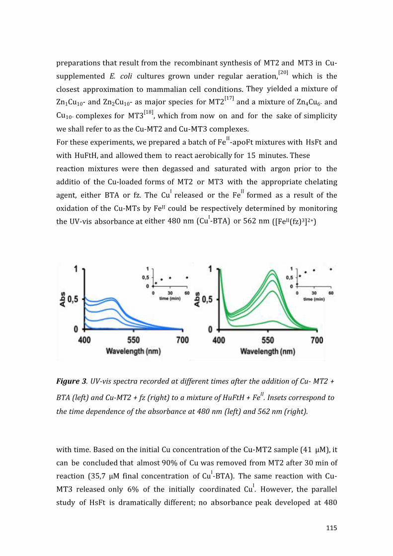
115
preparations that result from the recombinant synthesis of MT2 and MT3 in Cu-
supplemented E. coli cultures grown under regular aeration,[20] which is the
closest approximation to mammalian cell conditions. They yielded a mixture of
Zn1Cu10- and Zn2Cu10- as major species for MT2[17] and a mixture of Zn4Cu6- and
Cu10- complexes for MT3[18]
, which from now on and for the sake of simplicity
we shall refer to as the Cu-MT2 and Cu-MT3 complexes.
For these experiments, we prepared a batch of FeII-apoFt mixtures with HsFt and
with HuFtH, and allowed them to react aerobically for 15 minutes. These
reaction mixtures were then degassed and saturated with argon prior to the
additio of the Cu-loaded forms of MT2 or MT3 with the appropriate chelating
agent, either BTA or fz. The CuI
released or the FeII
formed as a result of the
oxidation of the Cu-MTs by FeII could be respectively determined by monitoring
the UV-vis absorbance at either 480 nm (CuI-BTA) or 562 nm ([FeII(fz)3]2+)
Figure 3. UV-vis spectra recorded at different times after the addition of Cu- MT2 +
BTA (left) and Cu-MT2 + fz (right) to a mixture of HuFtH + FeII. Insets correspond to
the time dependence of the absorbance at 480 nm (left) and 562 nm (right).
with time. Based on the initial Cu concentration of the Cu-MT2 sample (41 µM), it
can be concluded that almost 90% of Cu was removed from MT2 after 30 min of
reaction (35,7 µM final concentration of CuI-BTA). The same reaction with Cu-
MT3 released only 6% of the initially coordinated CuI. However, the parallel
study of HsFt is dramatically different; no absorbance peak developed at 480

116
nm, highlighting the absence of free CuI since, in agreement with the high Fe
storage capacity of HsFt, there is a very low availability of FeIII in the medium
to oxidize either Cu-MT2 or Cu-MT3.
It is therefore reasonable to assume that the FeIII formed after oxidation at the
HuFtH ferroxidase center is the agent responsible for oxidizing Cu-MT2 on the
basis of three different experiments: (i) The mixtures of HuFtH, Cu-MT2/Cu-
MT3 and BTA do not exhibit any significant UV-vis absorption corresponding to
CuI-BTA in the absence of Fe
II. This points out that, in the absence of Fe
II, neither
MT2 nor MT3 are oxidized. (ii) Addition of an FeIII
salt, in the same
concentration as that generated in the FeII + HuFtH experiment, to an anaerobic
Cu-MT2 solution, but not in one containing Cu-MT3, in the presence of BTA
produces CuI-BTA with a Cu
I delivery pattern similar to that of the FeII + HuFtH +
Cu-MT2 experiment (see Experimental).This clearly indicates that FeIII is a
powerful oxidant capable of oxidizing the mammalian MT2 isoform in its Cu
loaded form, while that of MT3 practically resists this oxidizing environment. (iii)
When HuFtH is incubated with FeII and after 15 min of reaction (once the Fe
II
oxidation is almost complete, Figure 1) Cu-MT2 -together with fz- is added to the
reaction mixture, the ([FeII(fz)3]2+) complex forms and its concentration increases
with time (Figure 3). This final experiment evidences that after the reduction of
the FeII concentration to almost zero during the Fe reconstitution process of
HuFtH, reaction upon addition of Cu-MT2 again raises the FeII concentration to
59% of the initial value as a consequence of the oxidation of MT2 by FeIII
.
In summary, in this paper we demonstrate that part of the FeIII
formed at the
ferroxidase centers of HuFtH is available, not only for complexation with an FeIII
chelator, but also to oxidize Cu-loaded MT2, giving rise to the concomitant
release of two toxic metals ions, CuI and Fe
II. The latter reaction does not take
place when the apoferritin is a H/L heteropolymer such as HsFt. In this case, all
the FeIII formed at the ferroxidase center is unavailable for either Lf
complexation or MT oxidation. The chemical scenario we have created is
especially significant in the brain, where incorrect Fe uptake by Ft has been
suggested to result in the progression of neurological diseases. Hence, Dobson
and co-workers proposed that patients with Alzheimer's disease could have a
dysfunction in brain Fe storage due to a disruption in the balance of ferritin H-
and L- subunit synthesis.[21] In this sense, our results point out that in cells where

117
Cu-MT2 and Ft coexist, there is no danger if the ferritin is L-rich, as it occurs in
astrocytes and microglia,[16] and both proteins would preserve their integrity and
functionality. However, if this imbalance in L- and H-subunit synthesis leads to the
synthesis of pure H apoferritins, and therefore to some events analogous to those
we have described here in which the high ferroxidase activity and low Fe
storage capacity of a H-rich Ft would provoke CuI liberation from Cu-MT2, then
the presence of two free and toxic metals such as CuI and FeII, could produce
fatal results for the cell. Interestingly, Cu-MT3 does not react with either HuFtH
or HsFt in the presence of FeII
thus making the Cu-MT3 and Ft pair a non-
deleterious combination in neurons. This is extremely relevant because in neuron
cells Ft is mainly present in H-rich forms,[16]
which would lead to a harmful
interaction should the predominant MT isoform present be that of MT2. What we
wish to underline here is that the process of Fe storage in ferritin affects parallel
metal metabolisms depending on the protein shell itself. Following on from that
we have shown that an excessive proportion of H-subunits in the ferritin shell
results in a significant decrease in the amount of Fe stored, meaning that there is
free FeIII available that is capable of oxidizing Cu-MT2, ultimately leading to the
liberation of toxic FeII and Cu
I. As a final conclusion, it is worth highlighting that
the current results reconcile the simultaneous presence of ferritins and MT in
cells,[12]
provided that the correct combination of isoforms is preserved,
otherwise metal-related neuronal disorders could be triggered if parting from
this ideal combination.
Experimental Section
Materials. Horse spleen apoFt (HsFt) was obtained from Sigma and was
exhaustively dialyzed against Milli-Q water using a Spectra/Por Float-A- Lyzer
with a molecular weight cut off (MWCO) of 300,000 Da. Recombinant human H-Ft
(HuFtH) was prepared as previously described[21,22] and rendered iron free by
dialysis against sodium hydrosulfite (dithionite), Na2S2O4, and complexation with
2,2’-bipyridyl at pH 6.0[23]
, to isolate an apoferritin containing 10 Fe/protein. The
Cu loaded forms of the mammalian metallothionein isoforms MT2 and MT3 were
recombinantly synthesized and characterized (ICP-AES and ESI-MS) as previously
reported.[17,18]
The final preparations were 0.13 x 10⁻⁴ M (MT2) and 0.35 x 10⁻⁴
M (MT3) in 50 mM Tris-HCl, pH 7.5 buffer, and they were deaerated under N2 flux

118
to avoid the presence of O2 during the described reactions. Monoferric lactoferrin
was purchased from Fonterra. Lf was prepared following the same protocol as
reported previously.[24]
Measurement of available FeII. Batches of 15 fresh solutions for each type of
apoferritin, 0.2 µM in 0.15 M HEPES buffer, pH 7.0, and fresh aqueous solutions
of Fe(SO4)2(NH4)·6H2O (final Fe 0.0353 mM) were mixed. 1 mL of an aqueous
ferrozine solution (3-(2-pyridyl)-5,6-diphenyl-1,2,4-triazine-p,p-disulfonic acid
monosodium salt hydrate, Aldrich), final concentration 0.935 mM, was added to
every sample after 1, 2, 3, 4, 5, 8,10, 15, 20, 30, 60, 100, 200 and 300 min and
the UV-visible spectra immediately recorded. Available FeII concentration was
calculated for every sample in the batch from the UV-vis absorbance measured at
562 nm, due to formation of the ([FeII(fz)3]2+) complex (ε = 27,900 M-1cm-1).
Measurement of available FeIII
. A batch of four fresh solutions of each apoferritin,
0.2 µM in 0.15 M HEPES buffer containing 10 mM of NaHCO3, pH 7.0, and fresh
aqueous solutions of Fe(SO4)2(NH4)·6H2O (final Fe 0.353 mM) were mixed. 200
µL of an aqueous solution of Lf (final concentration 200 μM) was added to every
sample after 5, 10, 20 and 60 min and the UV-vis spectra recorded. Available FeIII
concentration was calculated for every sample of the batch from the UV-vis
absorbance measured at 464 nm, due to formation of the [FeIII(Lf)] complex (ε =
2,600 M-1cm-1).
Measurement of CuI during ferritin reconstitution in the presence of Cu- MT. In a
parallel experiment, HuFtH and HsFt (0.2 µM) in 0.15 M HEPES buffer, pH 7.0,
were incubated with aqueous solutions of Fe(SO4)2(NH4)·6H2O (final [Fe] 0.035
mM) at 37 ºC and in aerobic conditions for 15 min. The reaction mixture was then
totally deoxygenated under nitrogen flux and kept under an anaerobic
atmosphere prior to the addition of Cu-MT2 (100 µL, 5.2 µM, [Cu] 41 µM) or Cu-
MT3 (100 µL, 4.7 µM, 40 µM). Then, 10 µL of 0.4 mM BTA
(bathocuproinedisulfonic acid disodium salt) was added to the mixture in order to
detect CuI
by observation of the UV-vis band at 480 nm due to formation of the
[CuI(BTA)2]+ complex (ε = 14,000 M-1cm-1). The same experiments carried out in
the absence of FeII did not produce an absorbance peak at 480 nm for the UV-vis
spectra of [CuI(BTA)2]+.
Measurement of FeIII reduction to Fe
II during HuFtH ferritin reconstitution in the
presence of Cu-MT2. The previous protocol was followed but using ferrozine

119
instead of BTA as a chelating agent for the purposes of measuring the FeII
concentration. We noted that after 15 min of FeII reconstitution by HuFtH, the
FeII concentration had dramatically decreased (Figure 1). Therefore the increase
of FeII concentration in this experiment corresponds to the oxidation of Cu-MT2
by FeIII
with the subsequent formation of FeII, which is ultimately detected and
quantified by its complexation with fz.
Oxidation of Cu-MT by FeIII
. In a parallel experiment, Cu-MT2 (100 µL, 3 µM) and
Cu-MT3 (100 µL, 3 µM) were incubated at 37 ºC in a degassed aqueous solution of
FeIII acetate (0.02 mM) under Ar. BTA or fz was added to detect and quantify the
CuI or Fe
II concentrations.
Acknowledgements
Work supported by the Spanish MINECO and FEDER funds with grants CTQ2012-
32236 to J.M. Domínguez-Vera, BIO2012- 39682-C02-01 to S. Atrian, and BIO2012-
39682-C02-02 to M. Capdevila. The authors from the Barcelona universities are
members of the Grup de Recerca de la Generalitat de Catalunya refs. 214SGR-
00423. F. C. is grateful to the Spanish MINECO for a FPI Fellowship. We thank the
Centres Científics i Tecnològics (CCiT) of the Universitat de Barcelona (ICP-AES)
and the Servei d’ nàlisi Química (S Q) of the Universitat utònoma de Barcelona
(ESI- S) for allocatin instrument time. S. rtime workin at S. trian’s
laboratory, was responsible for the recombinant syntheses of the Cu-MT
preparations.
Keywords: erritin • etallothionein • erroxidase activity • e metabolism
References
[1] a) K. Jomova, M. Valko, Curr. Pharm. Des. 2011, 17, 3460-3473. b) H. Heli, S.
Mirtorabi, K. Karimian, Expert Opinion on Therapeutic Patents 2011,21, 819.
[2] a) E. C. Theil, Curr. Opin. Chem. Biol. 2011, 15, 304-311; b) X. F. Liu, E. C.Theil,
Acc. Chem. Res. 2005, 38, 167-175.
[3] F. Bou-Abdallah, Biochim. Biophys. Acta 2010, 1800, 719-731.
[4] F. Carmona, O. Palacios, N. Gálvez, R. Cuesta, S. Atrian, M. Capdevila, J. M.

120
Domínguez-Vera, Coord. Chem. Rev. 2013, 257, 2752– 2764.
[5] N. D. Chasteen, P. M. Harrison, J. Struct. Biol. 1999, 126, 182-194.
[6] J. M. Bradley, G. R. Moore, N. E. Le Brun, J. Biol. Inorg. Chem. 2014, DOI
10.1007/soo775-014-1136-3 and references therein.
[7] X. Liu, E. C. Theil, Proc. Natl. Acad. Sci. U.S.A. 2004, 101, 8557–8562.
[8] P . Arosio, R. Ingrassia, P. Cavadini, Biochim. Biophys. Acta, 2009, 1790, 589–
599.
[9] F. Bou-Abdallah, G. Zhao, H. R. Mayne, P. Arosio, N. D. Chasteen, J. Am. Chem. Soc.
2005, 127, 3885−3893.
[10] H. K. Ebrahimi, E. Bill, P. L. Hagedoorn, W. R. Hagen, Nat. Chem. Biol. 2012, 8,
941–948.
[11] J. S. Rohrer, R. B. Frankel, G. C. Papaefthymiou, E. C. Theil, Inorg. Chem. 1989,
28, 3393-3395.
[12] R. Orihuela, B. Fernández, Ò. Palacios, E. Valero, S. Atrian, R. K. Watt, J. M.
Domínguez-Vera, M. Capdevila, Chem. Commun. 2011, 47, 12155-12157.
[13] M. Capdevila, R. Bofill, Ò.Palacios, S. Atrian, Coord. Chem. Rev. 2012, 256, 46–
52.
[14] Ò. Palacios, S. Atrian, M. Capdevila, J. Biol. Inorg. Chem. 2011;16, 991–1009.
[15] B. A. Masters, C. J. Quaife, J. C. Erickson, E. J. Kelly, G.J. Froelick, B.P.
Zambrowicz, R.L. Brinster, R.D. Palmiter, J. Neurosci. 1994, 14, 5844-5857.
[16] P. Ponka, C. Beaumont, D. Richardson, Semin. Hematol. 1998, 35, 35.
[17] E. Artells, Ò. Palacios, M. Capdevila, S. Atrian, Metallomics 2013, 5, 1397-1410.
[18] E. Artells, Ò. Palacios, M. Capdevila, S. Atrian, FEBS J 2014, 281, 1659-1678.
[19] G. Meloni, V. Sonois, T. Delaine, L. Guilloreau, A. Gillet, J. Teissié, P. Faller, M.
Vašák Nat Chem Biol. 2008, 4; 366-372.
[20] A. Pagani, L. Villarreal, M. Capdevila, S. Atrian, Mol. Microbiol. 2007, 63, 256-
269.
[21] Q. Q. Pankhursta, D. Hautotb, N. Khanc, J. Dobson, J. Alzheimers Dis. 2008, 13,
49-Pagina final.
[22] P. Santambrogio, A. Cozzi, S. Levi, E. Rovida, F. Magni, A. Albertini, P. Arosio,
Protein Expression Purification 2000, 19, 212–218.
[23] E. R. Bauminger, P. M. Harrison, D. Hechel, I. Nowik, A. Treffry, Biochim.

121
Biophys. Acta, 1991, 1118, 48–58.
[24] F. Carmona, V. Muñoz-Robles, R. Cuesta, N. Gálvez, M. Capdevila, J- D.
Maréchal, J. M. Dominguez-Vera, J. Biol. Inorg. Chem. 2014, 19, 439–447.

122

123
RESULTADOS/CONCLUSIONES DEL CAPITULO 3 QUE MOTIVARON EL
CAPITULO 4.
Los resultados del artículo anterior ponen de manifiesto que las ferritinas H tienen
alta actividad ferroxidasa pero baja capacidad de almacenar hierro. Este hecho
adquiere especial importancia en la leche materna, donde la ferritina es
predominantemente H y existen elevadas concentraciones de lactoferrina, proteína
encargada de captar Fe(III) para privar a los microorganismo patógenos de este
metal esencial y evitar así su proliferación.
En el siguiente artículo, la existencia de este “tándem” lactoferrina – ferritina H en
leche materna nos inspiró para llevar a cabo la preparación de materiales
formados por ambas proteínas que combinaran de manera sinérgica las funciones
de éstas, es decir, un alto poder detoxificante de Fe(II) por parte de la ferritina H y
una rápida incorporación del Fe(III) generado en la lactoferrina, actuando estos
materiales como secuestradores de hierro en cualquiera de sus estados de
oxidación, llevando a cabo por tanto una acción antimicrobiana completa.
La bibliografía contiene ejemplos de materiales y moléculas discretas capaces de
actuar como antimicrobianos, todos ellos basados en la captación de Fe(III). En
este trabajo desarrollamos un material, basado en como funcionan las proteínas
ferritina H y lactoferrina, para secuestrar cualquier forma de hierro y poder llevar
a cabo una acción antimicrobiana más integral.

124

125
CAPÍTULO 4.

126

127
A BIOINSPIRED HYBRID SILICA-PROTEIN MATERIAL WITH
ANTIMICROBIAL ACTIVITY BY IRON UPTAKE
Fernando Carmona,ab Daniela Mendoza,a Alicia Megıa-Fernandez,bc Francisco Santoyo-
Gonzalezbc and Jose M. Domınguez-Vera*ab
Abstract. A silica–protein hybrid material has been prepared by simultaneous covalent deposition
of apoferritin and lactoferrin on functionalized silica. This material exhibits strong antibacterial
activity against E. coli due to its high iron-uptake capacity.
As bacteria are able to infect almost all tissues in the human body, they cause a spectrum
of illnesses ranging from common skin infections to severe and fatal diseases. Indeed,
bacterial infection is one of the leading causes of death in the world. Furthermore,
some bacteria have developed aremarkable ability to resist currently available
antimicrobials, thus high- lighting the need for the development of new antimicrobial
agents. A promising strategy in this respect involves inhibiting their ability to obtain the
nutrients required for growth.1 An effective route to combat bacterial infection is to
create materials that can compete with bacteria for iron, thereby depriving them of
this metal, which they need to proliferate. However, such a route must be more efficient in
terms of iron uptake than siderophores, low-molecular-weight iron chelators that are
produced and exported by bacteria during periods of nutrient deprivation.2 A good
alternative to the creation of new materials for this purpose is to mimic existing and
efficient biological systems. Animals closely regulate iron levels3 and sequester this
nutrient as a mechanism of preventing bacterial proliferation. Two kinds of proteins,
namely transferrins (including lactoferrin), which are glycoproteins that remove
extracellular Fe(III) due to their high Fe(III) affinity,4,5 and ferritin, which stores the iron
that is not required for immediate metabolic purposes, play a crucial role in this
mechanism.6,7
Lactoferrin is considered to be one of the key components of the immune system,
partly due to its high affinity for Fe(III),8,9 and ferritin consists of a spherical
protein shell (apoferritin) surrounding an aqueous cavity that incorporates Fe(II),
which is oxidized during its journey to the cavity to form a Fe(III) mineral core

128
therein.6,7 The apoferritin shell is assembled from 24 polypeptide chains of two
types, namely heavy (H) (21 kDa) and light (L) subunits (19 kDa). It has
traditionally been thought that H-subunits play a key role in Fe(II) oxidation as
they contain catalytic ferroxidase centres, whereas L-subunits are associated with
Fe(III) nucleation.6 However, it is interesting to note that pure L ferritin has not
been found in iron storage organs. All known ferritins always contain at least a
small amount of H subunits (10% in the liver, for example). As such, it seems that
the iron uptake and storage process requires the oxidative activity of at least a
small number of H subunits. Nevertheless, pure or H-rich ferritin is frequently
found in humans, especially in breast milk, the heart or serum, probably due to its
strong ferroxidase activity, which helps to protect against toxic Fe(II).10
In light of these findings, we considered that H-apoferritin and lactoferrin could be
an effective combination for iron uptake. Interestingly, breast milk contains both
lactoferrin and apoferritin, the latter of which is an H-rich or H-pure apoferritin.10
As a result, we supposed that a material capable of confining both H-apoferritin
and lactoferrin could constitute an effective system for Fe(II) uptake as Fe(II)
would be oxidized by apoferritin to Fe(III), some of which would be stored in its
cavity, with the remainder, and any available Fe(III), being captured by lactoferrin.
In this sense, it is interesting to note that most previous iron uptake strategies
based on molecular recognition technologies11 involve solids containing
immobilized molecules with a well-known affinity for e(III) such as
desferrioxamine,12 3-hydro-xypyridin-4-ones,13 or EDTA derivatives.14 Despite the
fact that free Fe(II) is markedly more dangerous than Fe(III) as it catalyses the
production of reactive oxygen species, which are extremely powerful oxidizing
agents that are capable of causing extensive cell damage, no hybrid solids with
iron(II) molecules have been reported.
The aim of this work was therefore to create new materials containing both
apoferritin (especially H pure) and lactoferrin to remove iron from the media, thus
depriving pathological microorganisms of this metal. To achieve this goal, we
decided to use a vinyl sulfone silica functionalized with both proteins in a step-by-
step manner. The resulting hybrid silica–protein material spatially confines both
proteins and mimics breast milk to some extent, thus making it potentially useful

129
as an antimicrobial a ent due to the cooperative effect of the apoferritin–
lactoferrin combination for iron uptake.
The functionalization of silica with vinyl sulfone commences with the silanization
of commercial silica followed by the Michael-type addition of a bis-vinyl sulfone.
This process gives rise to a functionalized silica containing available vinyl sulfone
groups to which different biomolecules can be covalently attached (ESI 1).15
We initially analysed the reactivity of each protein with the vinyl sulfone silica.
Under similar conditions, the two apoferritins studied [horse spleen (HSFt) and H-
recombinant (HFt)] showed a higher reactivity than lactoferrin, therefore we first
carried out the immobilization of a default apoferritin followed, in a second step,
by an excess of lactoferrin (Scheme 1). Both protein immobilizations were
performed by incubating the aforementioned proteins with the vinyl sulfone silica
in phosphate buffer at pH 7.5.‡ After 24 h of incubation with apoferritin, the
resulting solid was exhaustively washed with buffer (1 M NaCl) to remove the non-
covalently immobilized apoferritin. UV-visible spectroscopic analysis of the wash
solution showed practically no signal for both HSFt and HFt, thus confirming that
almost all the apoferritin had bound covalently to the vinyl sulfone and that
negligible amounts of protein were retained by non-covalent interactions (Fig. 1).
Once the silica–apoferritin material had been isolated, it was incubated with an
excess of lactoferrin and, after 24 h, the solid containing both proteins (silica–Ft +
Lf hereafter) was rinsed and dried at room temperature.
scheme 1

130
Fig 1. UV-visible spectra of proteins (H-apoferritin and lactoferrin) before and after incubation with
silica (left) and silica–apoferritin (right). Dashed lines correspond to the spectra of the wash
solution (non-covalent bound protein).
From the difference in the absorbance values of both protein solutions at 280 nm
before and after incubation with vinyl sulfone silica, and after subtracting the small
amount of protein removed upon washing the solid (Fig. 1), values of 33.0 mg (for
apoferritin) and 4.6 mg per gram of vinyl sulfone silica (for lactoferrin) were
obtained. Note that the absorbance at 280 nm for the solution obtained after
washing the silica–HFt + Lf material is minimal. Furthermore, if this solution is
mixed with the supernatant solution and dialyzed using a molecular weight cut-off
of 100 kDa (larger than the molecular weight of lactoferrin), no absorbance at 280
nm is observed at all, thus confirming that apoferritin is not displaced by the
covalent attachment of lactoferrin and that both proteins are definitely fixed to the
silica.
Once isolated, the hybrid silica–Ft + Lf materials were incubated with an Fe(II)
solution (5 mL, 10 mM) and, after 6 h, the resulting solids centrifuged and washed
twice with buffer and 1 M NaCl. The supernatant and wash solutions were mixed
and the iron concentration measured by ICP to quantify the iron removal efficacy
of the silica–Ft + Lf (silica–HSFt + Lf and silica–Ft + Lf). The results are shown in
Table 1 along with values for control samples, functionalized vinyl sulfone silica
and silica containing only one protein, either HSFt or Lf.

131
The best values were obtained with recombinant H-apoferritin and lactoferrin
supported on the functionalized silica, which agrees with the expected existence of
a cooperative effect of both proteins that leads to a very efficient iron uptake. This
effect is lower in the silica–HSFt + Lf material as a result of the lower ferroxidase
activity of horse spleen apoferritin with respect to H-pure apoferritin (silica–HFt +
Lf). Likewise, it is interesting to highlight the low iron removal efficiency of the
silica containing only lactoferrin (silica–Lf), which exhibits a similar yield to the
silica containing no protein (silica). This is probably due to the high affinity of
lactoferrin for Fe(III) but not for Fe(II). However, in the presence of a protein with
ferroxidase activity, such as apoferritin, which oxidizes Fe(II) to Fe(III), lactoferrin
multiplies its effectiveness and ultimately makes the apoferritin–lactoferrin
combination the best option for iron uptake, especially if the apoferritin exhibits a
high ferroxidase activity, as is the case for H-recombinant apoferritin.
In light of these promising results, we decided to check the antimicrobial activity of
the silica–HFt + Lf material against E. coli. The antimicrobial activity of lactoferrin
against E. coli has been reported both in solution16 or immobilized on the surface
of a copolymer.17 However, the antimicrobial activity of our hybrid materials is
based on the cooperative effect of two proteins: first by uptake and oxidation of
Fe(II) (apoferritin) and ultimately the uptake of Fe(III) (apoferritin + lactoferrin).
Our hybrid materials are therefore designed to act as antibacterial agents due to
their ability to sequester Fe(II), the most toxic form of iron.

132
Fig 2. Fluorescence microscopy images of E. coli proliferation obtained using a Live/Dead Bacterial
Viability Kit, where live and dead bacteria exhibit green and red emissions, respectively. Silica–HFt
+ Lf dramatically inhibits bacterial viability.
Bacterial inhibition by silica–HFt + Lf was quantified using the Live/Dead Bacterial
Viability Kit, counting the number of live (green) and dead (red) bacteria by
fluorescence microscopy.‡ The average live/dead ratio (viability v) was used to
quantify the bacterial inhibition effect of the hybrid silica–protein material in
comparison with control experiments. As can be seen from Fig. 2, the silica–HFt +
Lf material dramatically decreases the bacterial viability in MRS + cys (viability v =
0.9), a medium where these bacteria proliferate adequately (viability control v =
60.5).
Interestingly, the silica–Lf material, which only contains lactoferrin and removes
small amounts of iron (Table 1), did not significantly inhibit bacterial viability (v =
54.5). Likewise, in agreement with its higher iron uptake ability (Table 1), silica–
HSFt inhibits more strongly than silica–Lf (v = 24.1) but is markedly less effective
as an antibacterial agent than the silica material containing both proteins (silica–
HFt + Lf, viability v = 0.9).

133
These results demonstrate the antibacterial activity of this novel hybrid silica–HFt
+ Lf material and confirm that the mechanism of antibacterial activity of this
hybrid solid lies in the synergistic action of H apoferritin and lactoferrin for iron
uptake.
Conclusions
The results of this study confirm our initial bio-inspired hypothesis that the
confinement of apoferritin and lactoferrin supported on a material such as silica is
a useful route for iron(II) uptake and therefore for use as an antimicrobial agent. A
high antibacterial activity has been demonstrated against E. coli K-12, especially
when using the silica containing both H-recombinant apoferritin and lactoferrin.
Likewise, the correlation between bacterial inhibition and iron uptake confirms
that the antimicrobial activity of these materials resides in their iron uptake
capacity. Finally, we are convinced that this kind of material may find use as the
active ingredient in creams or topical drugs for the treatment of microbial
infections in cosmetics and personal hygiene.
This work was supported by INE O and E ER (project TQ2009-09344) unta
de ndaluc ıa (project P07-FQM-02899) and BIOSEARCH SA (contract PostBIO).
Notes and references
‡ The vinyl sulfone silica15 (ESI) (100 mg) was incubated with stirring with the
protein (2.5 mL, 0.01875 mM) in phosphate buffer at pH 7.5 at 4 ºC. After 24 h, the
mixture was centrifuged at 10000 rpm for 10 min at 20 ºC. The solid was washed
with buffer containing 1 M NaCl. Both supernatant solutions, from the first
centrifugation and washed solid, were mixed and the protein concentration
measured by the absorbance at 280 nm (ε280HS = 33 900 and ε280HFt = 27 900 M-1cm-1)
in the UV-visible spectrum. From the difference in the absorbance values at 280 nm in
the initial protein solution and the mixture of supernatant solutions (after
centrifugation and after washing), values of 33.0 mg for apoferritin, and 4.6 mg for
apolactoferrin per gram of vinyl sulfone silica were obtained.
Preparation of vinyl sulfone functionalized silica–lactoferrin + apoferritin (silica–HFt
+ Lf and silica–HSFt + Lf): the vinyl sulfone silica (100 mg) was incubated with
stirring with apoferritin (HSFt or HFt) (2.5 mL, 0.01 mM) in phosphate buffer at pH

134
7.5 at 4 ºC. After 24 h, the mixture was centrifuged at 10000 rpm for 10 min at 20 ºC.
The solid was washed with buffer containing 1 M NaCl. The resulting solid (silica–Ft
or silica–HFt) was incubated with stirring with apolactoferrin (2.5 mL, 0.05 mM) in
phosphate buffer at pH 7.5 at 4 ºC. After 24 h, the mixture was centrifuged at 10000
rpm for 10 min at 20 ºC. The solid was washed with buffer containing 1 M NaCl and
dried at room temperature. Iron uptake by the hybrid silica–protein materials: the
silica–protein (500 mg) was incubated with a solution of Fe(NH4)2(SO4)26H2O (5 mL,
10 mM) for 6 h without stirring and then centrifuged at 10000 rpm for 10 min at 20
ºC. The solid was washed with a 1 M NaCl solution. The supernatant solution from the
centrifugation and the washed solution were mixed and the iron concentration
measured by Inductive Coupled Plasma. From the ratio between final and initial iron
concentration, the yield of iron removal was obtained for each sample (Table 1).
Inhibition of E. coli K-12: a bacteria culture (400 mL of 1·107 CFU) in MRS + cys was
subjected to the hybrid material (100 mg) for 16 h. Quantification of bacterial
proliferation was performed by using the Live/Dead Bacterial Viability Kits SYTO9
(green) and propidium iodide (red) (Invitrogen), counting the number of live (green)
and dead (red) bacteria in a batch of three experiments using the Image-Pro Plus 6.0
software. The average live/dead ratio (viability v) was used to quantify the effect of
the hybrid material on bacterial inhibition by comparing with control experiments
where no silica–protein material was present. The presence of silica–HFt + Lf resulted
in a decrease in viability to 0.9 with respect to control experiments where the
viability was 60.5. Values of 24.1 and 54.5 were obtained for silica–HFt and silica–Lf,
respectively.
1 J. J. Bullen, H. J. Rogers, P. B. Spalding and C. G. Ward, FEMS Immunol. Med.
Microbiol., 2005, 43, 325–330.
2 E. D. Weinberg, Metallomics, 2010, 2, 732–740. 3 T. Zheng and E. M. Nolan,
Metallomics, 2012, 4, 866–880.
4 H. J. Vogel, Biochem. Cell Biol., 2012, 90, 233–244.
5 E. N. Baker and H. M. Baker, Cell. Mol. Life Sci., 2005, 62, 2531–2539.
6 E. C. Theil, Curr. Opin. Chem. Biol., 2011, 15, 304–311; E. C. Theil, R. K. Beheraa
and T. Takehiko, Coord. Chem. Rev., 2012, 257, 579–586.
7 N. E. Le Brun, A. Crow, M. E. P. Murphy, A. G. Mauk and G. R. Moore, Biochim.
Biophys. Acta, 2010, 1800, 732–744; A. Lewin, G. R. Moore and N. E. Le Brun,

135
Dalton Trans., 2005, 3597–3610.
8 R. D. Baynes and W. R. Bezwoda, Adv. Exp. Med. Biol., 1994, 357, 133–141.
9 D. Legrand, Biochem. Cell Biol., 2012, 90, 252–268.
10 P. Arosio, A. Ponzone, R. Ferrero, I. Renoldi and S. Levi, Clin. Chim. Acta, 1986,
161, 201–208.
11 H. Hiroshi, I. M. M. Rahman, K. Sanae, M. Teruya and F. Yoshiaki, Chemosphere,
2011, 82, 1161–1167.
12 N. A. A. Rossi, M. Ibrahim, J. K. Jackson, H. M. Burt, S. A. Horte, M. D. Scotta and J.
N. Kizhakkedathua, Bioma- terials, 2009, 30, 638–648.
13 J. Golenser, A. Domb, D. Teomim, A. Tsafack, O. Nisim, P. Ponka, W. Eling and Z. I.
Cabantchik, J. Pharmacol. Exp. Ther., 1997, 281, 1127–1135.
14 F. Tian, E. A. Decker and J. M. Goddard, J. Agric. Food Chem., 2012, 60, 2046–
2052.
15 M. Ortega-MuÑoz, J. Morales-Sanfrutos, A. Megia-Fernandez, F. J. Lopez-
Jaramillo, F. Hernandez-Mateo and F. Santoyo-Gonzalez, J. Mater. Chem., 2010, 20,
7189–7196.
16 Y. Chih-Ching, C.-J. Shen, W.-H. Hsu, Y.-H. Chang, H.-T. Li, H.-L. Chen and C.-M.
Chen, Biometals, 2011, 24, 585–594.
17 A. Oyane, Y. Yokoyama, M. Uchida and A. Ito, Biomaterials, 2006, 27, 3295–
3303.

136

137
RESULTADOS/CONCLUSIONES DEL CAPITULO 4 QUE MOTIVARON EL
CAPITULO 5.
En el artículo siguiente, pretendemos explorar una vía en la que podamos entender
cómo, al mismo tiempo que nuestro organismo priva del hierro a microorganismos
infecciosos, existen bacterias probióticas beneficiosas capaces de captar hierro a
expensas del “host” donde proliferan. En particular hemos demostrado, mediante
una nueva metodología, que la lactoferrina no sólo no tiene acción bacteriostática
frente a estas bacterias beneficiosas sino que además promueve su proliferación
actuando como donor de hierro, justo al contrario de como ocurre con los
microorganismos patógenos.
Concretamente, hemos podido usar el fenómeno de FRET para demostrar que la
lactoferrina cede hierro a bacterias probióticas y que además lo hace sin ser
internalizada en la bacteria, a diferencia del mecanismo generalizado en el que las
proteínas transportadoras de hierro, como la transferrina, ceden hierro a las
células a través de su internalización.

138

139
CAPÍTULO 5.

140

141
MONITORING LACTOFERRIN IRON LEVELS BY FLUORESCENCE
RESONANCE ENERGY TRANSFER: A COMBINED CHEMICAL AND
COMPUTATIONAL STUDY
Fernando Carmona, Vıctor Muno -Robles, Rafael Cuesta, Natividad Ga lvez, Merce, Capdevila,
Jean-Didier Marechal, Jose M. Dominguez-Vera
Abstract.Three forms of lactoferrin (Lf) that differed in their levels of iron
loading (Lf, LfFe, and LfFe2) were simultaneously labeled with the fluorophores
AF350 and AF430. All three resulting fluorescent lactoferrins exhibited
fluorescence resonance energy transfer (FRET), but they all presented different
FRET patterns. Whereas only partial FRET was observed for Lf and LfFe,
practically complete FRET was seen for the holo form (LfFe2). For each form of
metal-loaded lactoferrin, the AF350–AF430 distance varied depending on the
protein conformation, which in turn depended on the level of iron loading. Thus,
the FRET patterns of these lactoferrins were found to correlate with their iron
loading levels. In order to gain greater insight into the number of fluorophores
and the different FRET patterns observed (i.e., their iron levels), a computational
analysis was performed. The results highlighted a number of lysines that have the
greatest influence on the FRET profile. Moreover, despite the lack of an X-ray
structure for any LfFe species, our study also showed that this species presents
modified subdomain organization of the N-lobe, which narrows its iron-binding
site. Complete domain rearrangement occurs during the LfFe to LfFe2 transition.
Finally, as an example of the possible applications of the results of this study, we
made use of the FRET fingerprints of these fluorescent lactoferrins to monitor the
interaction of lactoferrin with a healthy bacterium, namely Bifidobacterium breve.
This latter study demonstrated that lactoferrin supplies iron to this bacterium, and
suggested that this process occurs with no protein internalization.
Keywords: Lactoferrin, Iron metabolism, Protein–ligand docking, FRET, Structural
analysis
142
Introduction
Lactoferrin is a glycoprotein (80 kDa) of the transferrin family with a high
affinity for iron(III) [1, 2]. Lactoferrin possesses various biological functions,
including antibacterial, antiviral, and antiparasitic activities [3]. High lactoferrin
levels are found in colostrum and milk, and this protein is also present in most
mucosal secretions, including uterine fluid, vaginal, and nasal secretions, as well
as in tears [4, 5].
The extraordinary affinity of lactoferrin for iron undoubtedly determines part of
its functionality. Indeed, lactoferrin is considered to form part of the innate
immune system due to its effects on pathogen growth. Iron is essential for life and
is a key nutrient for pathogenic microorganisms, which require this metal to
survive and replicate. Life can to some extent be considered a battle for iron, so
hosts must deprive undesirable guests of iron in order to combat the infections
they cause. As a result, iron uptake by lactoferrin prevents the development of
pathogenic microorganisms which cannot then access this metal. This is why blood
lactoferrin levels increase markedly during infection. Indeed, lactoferrin
concentrations increase in all biological fluids during most inflammatory
reactions and viral infections, with the highest levels being detected at the nidus
of the inflammation [6].
The clearest evidence that the immune function of lactoferrin is related to its iron
affinity is the fact that whereas apolactoferrin (lactoferrin without iron) inhibits
the growth of a large number of species of bacteria (such as Escherichia coli, some
Pseudomonas, Yersinias, Listeria, Streptococcus, and Staphylococcus),
hololactoferrin (iron-saturated lactoferrin) exhibits significantly lower inhibitory
activities towards such bacteria [7–9]. The iron loading level of lactoferrin
therefore partially determines its functionality.
It should be noted that lactoferrin also has some effects on the immune system
that have nothing to do with iron affinity. Thus, lactoferrin exerts antiviral effects
by binding directly to DNA (or RNA) and by preventing the entry of viruses into
cells, thereby stopping infections at an early stage [10–13].
Lactoferrin can also serve as an iron donor, supporting the growth of certain
beneficial bacteria [14, 15]. This occurs in breast milk, where lactoferrin,
Lactobacilli, and Bifidobacteria coexist in an environment that is optimized for

143
proliferation.
X-ray diffraction studies, which are only available for the apo and holo forms of
lactoferrin, have shown that it consists of one polypeptide chain containing 703
amino acids, which forms two homologous globular domains named the N- and C-
lobes. The N-lobe corresponds to amino acid residues 1–333 and the C-lobe to
residues 345–692, and the ends of these domains are connected by a short a-
helix (Fig. 1). These two homologous lobes are further subdivided into two similarly
sized domains known as domain 1 and domain 2 (domains N1 and N2 in the N-lobe
and domains C1 and C2 in the C-lobe), and the two iron-binding sites are located
within the interdomain clefts of each lobe. In human lactoferrin, when two iron
atoms are incorporated into the apoprotein, the C-lobe conformation does not
change significantly whereas the N-lobe closes like a hinge to grip the metal. This
results in different tertiary structures: iron-free apolactoferrin is characterized by
an open conformation of the N-lobe and a closed conformation of the C-lobe,
whereas both lobes are closed in the iron-rich hololactoferrin, as shown by X-ray.
Fig. 1 X-ray structures of Lf and LfFe2. Arrows indicate the conformational movement of the N-lobe
when coordinating iron. The iron-binding sites are magnified to highlight the chemical environment of
the iron at each site
The chemical environments of the two iron(III) coordination sites are similar and

144
consist of two phenolate oxygens from two tyrosine residues, one imidazole from
a histidine residue, one carboxylate from an aspartate residue, and a synergistic
adjacent bicarbonate or carbonate, which is stabilized by hydrogen bonding to the
peptide chains that surround the active site. Despite this similarity, the two
coordination sites of iron(III) can be distinguished kinetically and
spectroscopically [18]. The three known forms of lactoferrin, namely
apolactoferrin (Lf), the monoferric form (LfFe), and hololactoferrin (LfFe2) differ
in their iron loading levels, which determine the functionality of the protein.
The lysine residues on the external surface of lactoferrin can be used to
covalently couple molecules [19–22] or clusters [23], permitting new
functionality to be added to the protein. This option has been exploited for
different applications, including increasing cell internalization levels, oral
administration, rheology modulation, and for monitoring toxic non-transferrin
bound iron. We use this functionalization to determine the iron loading level of
the protein. The idea of incorporating a fluorescent moiety into lactoferrin is of
considerable and widespread interest, especially in the field of biomedicine, as
adequate functionalization could lead to fluorescence resonance energy transfer
(FRET), an effect/process that is commonly used in sensors in biological
research [24–26].
In the work described in the present paper, a pair of Alexa Fluor® (AF)
fluorophores, AF350 and AF430, were chemically bound to Lf, LfFe, and LfFe2. As
the emission band of the AF350 donor overlaps with the excitation band of the
AF430 acceptor, simultaneous labeling of these lactoferrin species with both
fluorophores (AF350 and AF430) offers the opportunity to achieve FRET, which is
strongly dependent on the distance between the AF350 donor and the AF430
acceptor. We show in this work that the FRET yield, which is the ratio of the
emission intensities for the acceptor and donor, differs for Lf, LfFe, and LfFe2
because the conformational changes that the protein undergoes during iron
incorporation lead to variations in the donor–acceptor distance. As a result, the
FRET pattern can be used as an indicator of the tertiary structure of lactoferrin,
thus providing information on its iron load. Finally, we made use of this
correlation between the FRET pattern and iron loading level to determine the iron
loading state of lactoferrin during its interaction with probiotic bacteria, a
process which ends with the transfer of iron from the lactoferrin to the
145
bacteria.
Moreover, we performed a computational study to rationalize this dependency of
the FRET pattern on the iron loading level. The reason that this study is
interesting is that the evolution of the FRET pattern with the tertiary structure of
the protein can be used to probe how, upon the uptake of the first iron(III) ion, the
other lobe of the protein is prompted to take up the second iron(III) ion. No defin-
itive answer is available to this question at present.
Materials and methods
Monoferric lactoferrin (LfFe) was purchased from Fonterra (Auckland, New
Zealand; ref. no: 81665295/100). The apo and holo forms were prepared from this
monoferric lactoferrin.
Preparation of apolactoferrin (Lf)
Lyophilized human milk LfFe (20 mg/mL) was totally desaturated of iron
following successive dialysis steps against 0.1 M citric acid/citrate buffer (pH
3.50) using a high retention seamless cellulose tubing membrane with a molecular
weight cutoff (MWCO) of 12,000 Da. The citric acid/citrate buffer was changed
several times until no characteristic iron lactoferrin absorbance at 464 nm (ε464 =
2,500 M-1) was observed. The resulting colorless pale apolactoferrin solution was
then dialyzed against phosphate buffer pH 6.80 and subsequently
chromatographed (Sephadex G-25, GE Healthcare, Little Chalfont, UK). The
concentration of apolactoferrin was measured by UV/vis spectroscopy based on
the absorbance at 280 nm (ε280 = 92,300 M-1).
Preparation of diferric lactoferrin (LfFe2)
Based on a previously published procedure [27], 20 mg of lyophilized human milk
LFe were dissolved in 1 mL of TRIS–Cl buffer pH 6.80 containing 5 mM sodium
bicarbonate. This solution was then titrated with Fe citrate at 37 ºC under
continuous stirring until the appearance of a characteristic dark reddish solution.
Fe citrate was freshly prepared every day following a previously reported pro-
tocol [28].
Iron contents per lactoferrin were calculated directly by UV/vis spectroscopy from
the relationship between the absorbance at 464 nm (ε464= 2,500 M-1) and that at

146
280 nm (ε280 = 92,300 M-1). The iron/protein values obtained were 0 (Lf), 1.09
(LfFe), and 1.97 (LfFe2). These values were obtained from triplicate preparations,
with less than 8 % variation from the mean observed. The same lactoferrins
were used for all subsequent experiments. This means that the iron content of
every lactoferrin remained constant throughout the experiment.
Preparation of fluorescent lactoferrins
Lf, LfFe, or LfFe2 (2 mL, 20 mg/mL) was incubated in a buffered PBS solution at
pH 8 with an excess of (a) AF350, (b) AF430, and finally (c) a mixture of the
AF350 and AF430 fluorophore succinimidyl ester derivatives. The two former
assays allowed the molar ratio required for the third reaction to be determined.
Specifically, an AF350:AF430:protein molar ratio of 30:15:1 was used to prepare
the samples. The labeling reactions were performed at room temperature under
continuous stirring for 24 h. The resulting solutions were then exhaustively dia-
lyzed at room temperature for 3 days against several changes of Milli-Q water
(Millipore, Billerica, MA, USA) using a high retention seamless cellulose tubing
membrane with a MWCO of 12,000 Da, and size exclusion chromatography
(Sephadex G-25) was then performed to remove the unbound fluorophores.
Protein-containing fractions were isolated. For apolactoferrin samples labeled
with both fluorophores, the concentration of AF430 was directly calculated from
the absorbance values of the UV–visible spectra at 430 nm (ε430 = 16,000 M-1)
because the absorbances of apolactoferrin and AF350 are negligible at this
wavelength. The concentration of AF350 was obtained from the absorbance at
350 (ε350 = 19,000 M-1) by subtracting the absorbance of AF430 at this
wavelength. The concentration of lactoferrin was obtained directly from the
absorbance at 280 nm (ε280 = 92,300 M-1) because the absorbances of both
fluorophores at this wavelength can be considered negligible. A similar procedure
was performed to determine the number of fluorophores for LfFe and LfFe2, with
the UV–visible spectrum of the starting lactoferrin subtracted from each labeled
lactoferrin spectrum beforehand. The average number of fluorophores per
protein was determined after performing the reaction in triplicate. Values were
very consistent (with less than 5 % variation from the mean) and were rounded
to the nearest integer (Table 1). FRET yields from the three samples were also
calculated, and standard deviations from the average were less than 3%.

147
All UV–visible spectra were recorded with a Thermo Scientific (Waltham, MA,
USA) Spectronic Unicam UV300 spectrophotometer, and all fluorescence spectra
were recorded with a Varian (Palo Alto, CA, USA) Cary Eclipse fluorescence
spectrophotometer.
Incubation of AF-labeled lactoferrins with Bifidobacterium breve
Human milk B. breve (10 mg/mL; Biosearch S.A., Granada, Spain; CECT7263) was
incubated with LfFe2-AFs (4.86 x 10⁻⁶ M) under anaerobic conditions at 37 ºC in
Hank’s solution for 1, 2, 3, and 6 h. Bacterial suspensions were then centrifuged at
4,000 rpm and the supernatant solutions were analyzed using a Varian Cary
Eclipse fluorescence spectrophotometer. No fluorescence was observed from the
bacterial pellets. The iron concentrations of the supernatant solutions were
measured by ICP, with values of >10-9 M obtained. Likewise, the protein
concentrations in the supernatant solutions remained constant (in the range 4-5
x 10-6 M, as measured by UV/vis spectroscopy based on the absorbance at 280 nm;
ε280 = 92,300 M-1).
Docking study
Protein–ligand dockings were performed using the program GOLD 5.1. A harmonic
restraint of 5.5 kJ/mol was added between the Ne of the corresponding lysine
(Lys301 or Lys296) and the C of the carboxylate group of the fluorophore. In the
simulations where Lys301 or Lys296 were flexible, the Dunbrack rotamer library
was used. The cavity was defined as a 20-A sphere around Tyr82.

148
Structural statistics
The two scripts used in this work were written in Python and ported to the UCSF
Chimera environment. Both can be obtained for free from the authors.
Results
Following a known procedure, apolactoferrin (Lf) and hololactoferrin (LfFe2)
were prepared from the commercial monoferric form (LfFe). These three forms of
lactoferrin with different iron loadings were treated with an excess of each AF
fluorophore (or a mixture of both AF fluorophores) for 24 h and then
exhaustively dialyzed until no fluorescence was detected in the dialysis reservoir.
After isolating the protein-containing fractions, measurement of the UV–vis
spectra allowed the final concentrations of each lactoferrin sample and the AF430
and AF350 fluorophores to be calculated. These results yielded the average
number of fluorophores per lactoferrin, as shown in Table 1.
Our observations of the reactions of Lf, LfFe, and LfFe2 with AF350 and AF430
allowed us to establish that the number of reaction sites does not depend on the
type of fluorophore used, and that they should be reacted with the proteins to
determine the stoichiometric ratio (see ‘‘Materials and methods’’). The
fluorescence spectra of the solely AF350- and AF430-labeled lactoferrins showed
emission bands centered at 445 and 540 nm upon excitation at 327 and 425 nm,
respectively. As expected, the fluorescence properties of AF350 and AF430 were
not significantly modified after covalent coupling to lactoferrin.
AF350 and AF430 were chosen for use in this work due to the fact that the
emission band of AF350 overlaps with the excitation band of AF430. Therefore,
the simultaneous labeling of lactoferrin with both fluorophores (AF350 and
AF430) is a good option for achieving FRET. The lactoferrins (Lf, LfFe, and LFe2)
were finally incubated with an excess of a mixture of AF350 and AF430, using the
same procedure as employed for the single AFs. The number of AF430 and AF350
moieties in each labeled sample was calculated from the absorbances at 430 and
350 nm in the UV–visible spectra (Fig. 2; see ‘‘Materials and methods’’).
As shown in Table 1, incubation of the lactoferrins (Lf, LfFe, and LFe2) with an
excess of a mixture of AF350 and AF430 yielded similar labeling results. The
number of AF350 and AF430 molecules when the lactoferrins were labeled with a
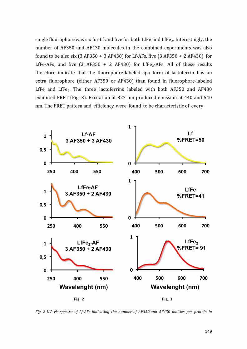
149
single fluorophore was six for Lf and five for both LfFe and LfFe2. Interestingly, the
number of AF350 and AF430 molecules in the combined experiments was also
found to be also six (3 AF350 + 3 AF430) for Lf-AFs, five (3 AF350 + 2 AF430) for
LfFe-AFs, and five (3 AF350 + 2 AF430) for LfFe2-AFs. All of these results
therefore indicate that the fluorophore-labeled apo form of lactoferrin has an
extra fluorophore (either AF350 or AF430) than found in fluorophore-labeled
LfFe and LfFe2. The three lactoferrins labeled with both AF350 and AF430
exhibited FRET (Fig. 3). Excitation at 327 nm produced emission at 440 and 540
nm. The FRET pattern and efficiency were found to be characteristic of every
Fig. 2 Fig. 3
Fig. 2 UV–vis spectra of Lf-AFs indicating the number of AF350 and AF430 moities per protein in

150
the combined AF350+AF430 labeling experiment. Concentrations of AF430, AF350, and protein
were calculated from the absorbances at 430, 350, and 280 nm (see ‘‘Materials and methods’’)
Fig. 3 FRET fluorescence emission spectra of the Lf- AF350+AF430, LfFe-AF350+AF430 and LfFe2-
AF350+AF430 samples
fluorescent lactoferrin. In all cases, the emission at lower energy can be attributed
to the AF430 acceptor and that at higher energy to the emission of the AF350
donor. In order to evaluate these different efficiencies, the %FRET value of each
lactoferrin was calculated using the following equation, which normalizes the
intensity of the emission of AF430 (IAF430) with respect to the intensity of the
emission from both fluorophores (IAF350 + IAF430):
The calculated %FRET yield values obtained from the data shown in Fig. 3 imply
that the efficiency of the energy transfer between both fluorophores varies.
Whereas complete energy transfer from the excited AF350 to AF430 does not
occur in Lf-AFs (%FRET = 50) or in LfFe-AFs (%FRET = 41), practically complete
energy transfer occurs in LfFe2-AFs (%FRET = 91). Consequently, the FRET pattern
represents a ‘‘fin erprint’’ of the particular fluorescent lactoferrin that produces it
and is related to the conformational changes that occur to the protein upon the
incorporation of iron.
Once these results had been analyzed, we aimed to correlate the energy transfer
efficiency with the distance between the donor (AF350) and the acceptor (AF430),
which obviously should be different in the three iron-loaded forms of Lf.
In order to gain greater insight into the differences observed in the number of
fluorophores and the FRET patterns observed among the distinct iron-loaded
states of lactoferrin, a computational analysis was performed. Calculations were
first employed to ascertain the location of the binding site of the extra AF430 that
is present in Lf-3AF350+3AF430 as compared to LfFe- and LfFe2-

151
3AF350+2AF430. Second, computation was used to identify the region (or regions)
of the protein that influences the FRET efficiency.
First, a comparative analysis of the solvent-accessible surface (SAS) for the 46
lysines in the X-ray structures of Lf and LfFe2 was performed (see SI1 in the
Electronic supplementary material, ESM). As has been reported previously,
succinimidyl ester derivatives of AFs are excellent dyes for labeling proteins
through reactions with the available lysines [29]. To identify the location of the
additional AF430 in the apo-Lf form, we hypothesized that the sixth lysine (i.e., the
one to which the additional AF430 binds) should show a dramatic decrease in
solvent accessibility when the apo form converts to the holo form of lactoferrin (in
other words, upon changing from Lf-AFs to between the donor (AF350) and the
acceptor (AF430), which obviously should be different in the three iron-loaded
forms of Lf.
In order to gain greater insight into the differences observed in the number of
fluorophores and the FRET patterns observed among the distinct iron-loaded
states of lactoferrin, a computational analysis was performed. Calculations were
first employed to ascertain the location of the binding site of the extra AF430 that
is present in Lf-3AF350+3AF430 as compared to LfFe- and LfFe2-3AF350+2AF430.
Second, computation was used to identify the region (or regions) of the protein
that influences the FRET efficiency.
First, a comparative analysis of the solvent-accessible surface (SAS) for the 46
lysines in the X-ray structures of Lf and LfFe2 was performed (see SI1 in the
Electronic supplementary material, ESM). As has been reported previously,
succinimidyl ester derivatives of AFs are excellent dyes for labeling proteins
through reactions with the available lysines [29]. To identify the location of the
additional AF430 in the apo-Lf form, we hypothesized that the sixth lysine (i.e., the
one to which the additional AF430 binds) should show a dramatic decrease in
solvent accessibility when the apo form converts to the holo form of lactoferrin (in
other words, upon changing from Lf-AFs to

152
Fig. 4 Locations of the four lysines whose solvent accessible surface (SAS) values decrease the most
upon converting from the Lf to the LfFe2 form. The backbones are represented by ribbons. Regions
colored green correspond to the C2 and N2 subdomains, and those colored silver correspond to the
C1 and N1 subdomains. The lysines of interest are represented as blue spheres at the Ne position and
are labeled.
LfFe2-AFs). A program written in Python in house and implemented in the UCSF
Chimera environment [30] was designed for this analysis. Such an analysis could
not be done of the monoferric species since no X-ray structure of LfFe was
available.
Although most of the lysines were found to be highly exposed to the solvent, four
residues (Lys282, Lys285, Lys296, and Lys301) presented a noticeable decrease
in SAS during the conversion from Lf to LFe2. All of these residues are found in the
N-lobe, and more specifically in the N1 region of the protein: Lys282 and Lys285
are located at one of the external faces of the subdomain and Lys296 and Lys301
at the entrance and core of the iron-binding site, respectively (Fig. 4).
Significantly, Lys282, Lys285, and Lys296 exhibit large SAS values, thereby
suggesting that interaction with a fluorophore should be independent of the
protein conformation. In contrast, Lys301 exhibited a large SAS value in Lf but a
value of almost zero in LFe2, meaning that this residue changes from being

153
highly exposed in the apo form (Lf) to deeply buried in the holo one (LfFe2).
To better ascertain whether Lys301 is the Lys to which AF430 binds, and to gain
a better understanding of its molecular features, protein–ligand dockings of the
fluorophore at this site were performed using the available crystal structures of Lf
and LfFe2. As stated in the ‘‘Materials and methods’’ section, these calculations
were undertaken using the program GOLD and the Chemscore scoring functions
[30]. A restraint was added between Ne of the lysine and the carbon atom of the
carbonyl group of the fluorophore during the run in order to achieve geometries
consistent with pre-reaction AF430 binding states.
Fig. 5 Lowest-energy solutions for the conformation of AF430 that are consistent with its binding to
Lys301 in LfFe2 (left panel) and Lf (right panel). The fluorophores are represented as balls and sticks
and the protein residues as sticks. The surfaces are also depicted in order to highlight the residues
(red) that show the most steric hindrance with the fluorophore (blue) in LfFe2
predicted binding affinities for AF430 binding at Lys301 range from 18.8 to -22.1
scoring units on going from Lf to LfFe2, respectively. For calculations performed
with the Chemscore function, these values imply a predicted binding affinity that
could reach the micromolar range for the former but that binding is impossible
for the latter [31].
These findings were further confirmed by repeating the calculations with AF430

154
on LfFe2, but adding greater flexibility to the Lys301 residue during docking (Fig.
5). Although the predicted affinity of the best binding mode increased at 13.75 U,
the binding affinity was still low and was substantially different to that obtained
for Lf. Moreover, for the conjugation to occur, Lys301 must escape from a
buried situation in LfFe2 (where it interacts strongly with a network of polar
interactions with the carboxylic residues E216 and D297) to a conformation that
is highly exposed to the solvent. Marked steric hindrance is observed for the
environments of the iron-binding sites in the resulting geometry. These results
confirm that conjugation of the dye at this position is only possible in the apo
form of Lf.
Additional calculations were performed to identify the region of the protein that
could be responsible for the change in FRET efficiency upon shifting from the
apo-Lf to the holo-LfFe2 form. These calculations, which considered the variations
in the distances between all lysine residues during the transition from Lf to LfFe2,
were performed using an additional program written in house in Python and
implemented in the UCSF Chimera package [32]. The script provided a 46 x 46
matrix of the entire set of Lys–Lys distances. The comparative analysis of the X-ray
structures of Lf and LfFe2 showed that most of the 46 Lys residues have inter-
residue distances that only rarely vary by >3 A on average when the iron loading
of the protein changes, thus remaining within the Fo rster distance range (Fig. 6).
However, eight Lys residues, namely Lys99, 100, 113, 197, 200, 237, 241, and
243, showed a different pattern, with variations of between 5 and 10 A . Also,
Lys180 showed variations that were slightly larger than 4 A .

155
Fig. 6 Bubble chart showing the average variations in inter-residue distances upon transitioning from
Lf to LfFe2. The size of each circle is proportional to the magnitude of the variation. Lys residue
labels are given on the abscissa
Fig. 7 Locations of the nine lysines whose average variations in inter-residue distance upon
transitioning from Lf to LfFe2 exceed 4 A . The backbones are represented as ribbons. Regions
colored green correspond to the C2 and N2 subdomains, and those colored silver correspond to the
C1 and N1 subdomains. Putative lysines are represented as green spheres at the Ne position and are
labeled

156
Importantly, these eight residues were all found to be located in the N2 subregion
of the N-lobe. This subregion makes the greatest contribution to the
conformational transition from Lf to LfFe2 (Fig. 7). Lys301, which was previously
identified as the binding site for the ‘‘extra’’ 430, is located at the interface
between the N1 and N2 subdomains. Both of these results suggest that, even upon
shifting to the LfFe form of the metalloprotein, a conformational change occurs to
the macromolecule consisting of a subdomain rearrangement of the N-lobe.
Using our improved understanding of the relationship between the FRET profile
and iron loading, the possibility of using fluorophore labeling to monitor iron
loading in lactoferrin-dependent processes was investigated. In particular, we
monitored the process by which lactoferrin can supply iron to a healthy
bacterium, in this case B. breve. The bacteria were incubated with LfFe2-AFs for
different durations (1–6 h) and the FRET patterns of the supernatant solutions
obtained after bacterial centrifugation were analyzed. After 6 h, the resulting
supernatant solution exhibited a FRET pattern typical of the iron-free Lf-AFs form
(Fig. 8). A negligible amount of iron was detected in this solution (concentration
below 10-9 M), while the concentration of the protein (Lf) remained constant.
This finding suggests that all of the iron initially bound to lactoferrin is released
and incorporated into the bacteria after this time. Likewise, no fluorescence was
observed from the centrifuged bacteria. Interestingly, we observed that the
FRET pattern does not change gradually but sharply, in two steps: from LfFe2 to
LfFe, and then from LfFe to Lf (SI2).
Discussion
The present study of the FRET signals obtained when functionalizing lactoferrin
with two distinct fluorophores, AF430 and AF350, provides not only novel
molecular information on the iron-loading state of the protein but also on the
mechanisms underlying these iron loading/unloading processes. Furthermore, the
results illustrate how this methodology can be useful when performing in vivo
and in vitro experiments involving Lf iron release or loading.
Our data show that both the change in the number of fluorophores that can be
bound to lactoferrin and the variations in their FRET patterns as a function of the
iron loading level of the protein are strongly dependent on the protein
conformation. This means that the FRET responses can be used to analyze the

157
iron-dependent conformational
Fig. 8 FRET fluorescence emission spectra for LfFe2-AFs before (left) and after (right) incubation
with B. breve for 6 h
change of lactoferrin when it converts from its apo form to its holo (diferric) form,
and can shed light on previously unanswered questions about this process. More
precisely, the FRET responses can be used to probe how, following the uptake of
the first iron(III) ion, the other lobe of the protein is prompted to bind the second
ion. To the best of our knowledge, none of the information currently available
provides a definitive answer to this question.
The results of our computational study, based on an SAS analysis of the Lys
residues in Lf and LfFe2, clearly indicate that Lys301 is the only residue that is
able to selectively bind AF430 in Lf (but not in LFe2). This result is supported by
those obtained from simulated protein–ligand dockings that explicitly modeled
the binding of the fluorophore to the selected amino acid and showed that AF430
can easily access a catalytically consistent orientation for binding at Lys301 in Lf
but not in LfFe nor LfFe2 (Fig. 5). Since Lys301 is located at the iron-binding site
of the N-lobe and is located at the interface between the N1 and N2 subdomains, it
appears that some type of conformational change occurs in this domain as a
consequence of the binding of the first iron to the protein. Additional calculations
on the available X-ray structures also allowed us to demonstrate that the
differences in the FRET patterns obtained for Lf, LfFe, and LfFe2 are likely to be
related to the fluorophores attached to the Lys residues located at the N2 lobe.
This suggests that the transition between Lf and LfFe2 is likely to involve a major
contribution from an interdomain change in the N-lobe.

158
At this point, it is interesting to note that uptake of the first iron(III) by lactoferrin
has been reported to occur at the C-site, at which point the N-site becomes
capable of acquiring a second iron(III) ion [33]. However, the mechanism by
which the N-lobe is activated once this first iron(III) ion has been bound to the C-
lobe is not well known and the source of debate. To date, it is believed that the
binding of the first iron(III) is followed by a series of proton dissociations that
probably trigger changes in the conformation of the protein, thereby affecting
the N-site and allowing the capture of the second iron(III) [18].
On the one hand, our results suggest that the capture of the first iron(III) ion
triggers a partial but significant enough interdomain change at the N-lobe.
Lys301, which is located at the interface between the N1 and N2 subdomains,
binds an AF430 in Lf but not in LfFe2. The steric hindrance of Lys301 is associated
to a certain degree with the closing motion that takes place between the N1 and
N2 subdomains upon the shift from the Lf to the LfFe form of the protein, as
shown by further SAS analysis.
Therefore, our results show that the binding of the first metal to Lf involves a
reorganization of the N2 domain with respect to the N1 one. However, the FRET
efficiency remains almost unaffected (50 vs 41 %) by the Lf to LfFe transition,
leading us to suspect that the conformational change is relatively limited.
The capture of the second iron(III) by the N-lobe results in a significant
conformational change of the protein as a whole. A drastic increase in FRET is
observed upon transitioning from LfFe (41 %) to LfFe2 (91 %). This suggests that
a marked change in the N2/N1 interface occurs with the binding of the second
iron. In the absence of an X-ray structure for human LfFe, the results of our joint
computational–experimental study suggest that the structure of LfFe presents an
intermediate conformation of the N-lobe that is mainly characterized by
intersubdomain reorganization. This observation is consistent with the recently
released structure of an intermediate conformation of di-iron and di-bismuth
human transferrin, where the main structural changes were observed at the N2
domain and at the interface between both subdomains [34]. Our interpretations
are also consistent with those of other authors, who have envisaged the possibility
that cooperativity between the C- and N-lobes occurs via dynamic event
modifications of the interlobe chain, rather than the lobes themselves [18, 33].
In addition to the novel information obtained on the molecular mechanism of iron

159
loading by lactoferrin, the present study illustrates how the FRET profile of lacto-
ferrin can be used as a tool to monitor some specific processes of iron metabolism
in which this protein plays a relevant role. In particular, lactoferrin may support
the growth of some nonpathological but beneficial bacteria [14, 15]. Indeed,
lactoferrin coexists with several lactic acid bacteria, such as Bifidobacterium and
Lactobacilli, in breast milk. These Gram-positive anaerobic bacteria proliferate in
the human intestinal tract and are classified as probiotic bacteria because of their
health benefits [35–37]. Our results confirm that, as for other healthy bacteria,
human iron-saturated lactoferrin acts as an iron donor for B. breve. The evolution
of the FRET pattern of the supernatant solution after the incubation of LfFe2 with
the bacteria (Fig. 8) points to complete iron transfer from LfFe2 to the bacteria.
Moreover, the fact that the protein concentration in the extrabacterial medium
does not significantly change suggests that lactoferrin does not require protein
internalization and that, unlike other transferrins, it is not involved in Fe(III)
transport by receptor-mediated endocytosis. This kind of iron trafficking has only
been observed in certain Gram-negative bacteria [38].
Conclusions
Fluorescent lactoferrins with different iron loading levels were prepared by
labeling the proteins with the Alexa Fluor dyes AF350 and AF430. All of the
fluorescent lactoferrin samples showed FRET, but interestingly with distinctive
patterns. This indicates that energy transfer from the donor to the acceptor
fluorophore varies with the iron loading level of lactoferrin. Since the iron loading
level of lactoferrin determines its tertiary structure, the FRET pattern of the
sample correlates with its iron content. Moreover, the results of our joint
experimental-computational study provide an additional piece of evidence relating
to the sub- domain dynamics of the N-lobe upon iron binding. They are
consistent with a cooperative transfer mechanism between the C- and N-lobes of
lactoferrin that involves interdomain conformational changes during the
transition from the Lf to the LfFe form, although the closed con- formation found
in LfFe2 is not achieved during this transition.
In a further achievement, we used this FRET to iron level correlation for
lactoferrin as a tool to monitor the interaction between human lactoferrin and
Bifidobacteria. Specifically, we used this methodology to demonstrate that

160
lactoferrin supplies iron to these healthy bacteria and, furthermore, that this
process takes place without iron(III) trafficking by receptor mediated endocytosis.
Acknowledgments This work was supported by MINECO and FEDER (projects
CTQ2012-32236, CTQ2011-23336, and BIO2012- 39682-C02-02) and BIOSEARCH
SA. F.C. and V.M.R. are grateful to the Spanish MINECO for FPI fellowships
References
1. Vogel H (2012) J Biochem Cell Biol 90:233–244
2. Baker EN, Baker HM (2005) Cell Mol Life Sci 62:2531–2539
3. Baker EN, Baker HM (2009) Biochimie 91:3–10
4. Anderson BF, Baker HM, Norris GE, Rice DW, Baker EN (1989) J Mol Biol
209:711–734
5. Baker EN, Lindley PF (1992) J Inorg Biochem 47:147–160
6. Birgens HS (1985) Scand J Haematol 34:326–331
7. Orsi N (2004) Biometals 17:189–196
8. Byrd TF, Horwitz MA (1991) J Clin Investig 88:351–357
9. Griffiths E, Duffy L, Schanbacher F, Dryja D, Leavens A, Neiswander R, Qiao H,
DiRienzo D, Ogra P (2003) Dig Dis Sci 48:1324–1332
10. Arnold RR, Cole MF (1977) Science 197:263–265
11. Valenti P, Antonini G (2005) Cell Mol Life Sci 62:2576–2587
12. Berkhout B, Floris R, Recio I, Visser S (2004) Biometals 17:291–294
13. Kanyshkova TG, Semenov DV, Buneva VN, Nevinsky GA (1999) FEBS Lett
451:235–237
14. Liepke C, Adermann K, Raida M, Magert H-J, Forssmann W-G, Zucht H-D
(2002) Eur J Biochem 269:712–718
15. Sherman M, Bennett S, Hwang FY, Yu C (2004) Biometals 17:285–289
16. Kumar J, Weber W, Mu nchau S, Yadav S, Singh SB, Saravanan K, Paramasivam
M, Sharma S, Kaur P, Bhushan A, Srinivasan A, Betzel C, Singh TP (2003) Indian J
Biochem Biophys 40:14–21
17. Chug TDY, Raymond KH (1993) J Am Chem Soc 115:6765–6768
18. Bou AF, El Hage Chahine JM (2000) J Mol Biol 303:255–266

161
19. Moosmann A, Blath J, Lindner R, Mu ller E, Bo ttinger H (2011) Bioconjug
Chem. 22:1545–1558
20. Breuer W, Cabantchik ZI (2001) Anal Biochem 15:194–202
21. Nojima Y, Suzuki Y, Iguchi K, Shiga T, Iwata A, Fujimoto T, Yoshida K, Shimizu
H, Takeuchi T, Sato A (2008) Bioconjug Chem 19:2253–2259
22. Zalipsky S (1995) Bioconjug Chem 6:150–165
23. Xavier PL, Chaudhari K, Verma PK, Pal SK, Pradeep T (2010) Nanoscale
2:2769–2776
24. Myc A, Majoros IJ, Thomas TP, Baker JR Jr (2007) Biomacro- molecules 8:13–
18
25. Wang YA, Li JJ, Chen H, Peng X (2002) J Am Chem Soc 124:2293–2298
26. Green M, Howman E (2005) Chem Commun 121–123
27. Bates GW, Billups C, Saltman P (1967) J Biol Chem 242:2816–2821
28. Faller B, Nick H (1994) J Am Chem Soc 116:3860–3865
29. Fernandez B, Galvez N, Sanchez P, Cuesta R, Bermejo R, Dom ınguez-Vera JM
(2008) J Biol Inorg Chem 13:349–355
30. Pettersen EF, Goddard TD, Huang CC, Couch GS, Greenblatt DM, Meng EC,
Ferrin TE (2004) J Comput Chem 25:1605–1612
31. Marechal JD, Yu J, Brown S, Kapelioukh I, Rankin E, Wolf C, Roberts G, Paine
M, Sutcliffe M (2006) Drug Metab Dispos 34:534–538
32. Verdonk ML, Cole JC, Hartshorn MJ, Murray CW, Taylor RD (2003) Proteins
52:609–623
33. Pakdaman R, Petitjean M, El Hage Chahine JM (1998) Eur J Biochem 254:144
153
34. Yang N, Zhang H, Wang M, Hao Q, Suna H (2012) Sci Rep 2:999
35. Macfarlane S, Macfarlane GT, Cummings JH (2006) Aliment Pharmacol Ther
24:701–714
36. Shah NP (2000) J Dairy Sci 83:894–907
37. Kaur IP, Chopra K, Saini A (2002) Eur J Pharm Sci 15:1–9
38. Heymann JJ, Weaver KD, Mietzner TA, Crumbliss AL (2007) J Am Chem Soc
129:9704–9712

162

163
RESULTADOS/CONCLUSIONES DEL CAPITULO 5 QUE MOTIVARON EL
CAPITULO 6.
Como fruto del estudio del metabolismo de hierro en bacterias probióticas, hemos
podido constatar la presencia de una o varias moléculas reductoras emitidas por
estas bacterias capaces de reducir Fe(III) a Fe(II). Este poder de reducción ha sido
aplicado a otros iones metálicos y en particular hemos demostrado que estas
bacterias probióticas son capaces de reducir Au(III) a Au(0) con la consiguiente
formación de nanopartículas de Au que se quedan fijadas en el biofilm de la
bacteria.
Por otra parte, trabajos llevados a cabo en el grupo de investigación en los que he
participado, han puesto de manifiesto que estas bacterias probióticas son capaces
de incorporar en su superficie nanopartículas de maghemita.
Hemos prentendido combinar ambas propiedades de estas bacterias probióticas:
poder de reducción de Au(III) y capacidad de incorporar nanopartíulas de óxido de
hierro en su membrana exterior, para generar bacterias vivas que contengan
simultáneamente nanopartículas con propiedades ópticas (debido a la presencia
de nanopartículas de Au) y magnéticas (debido a la presencia de nanopartículas de
maghemita). Esto nos ha permitido crear el primer material vivo con propiedades
magneto-ópticas.

164

165
CAPÍTULO 6.

166

167
BIOINSPIRED MAGNETO-OPTICAL BACTERIA
Fernando Carmona,a Miguel Martín,a Natividad Gálvez,a,* Jose M. Dominguez-Vera,a,*
KEYWORDS: Magnetic Nanoparticles, Gold Nanoparticles, Bioinspired Materials.
“Two-in-one” magneto-optical bacteria have been produced using the probiotic Lactobacillus
fermentum® for the first time. We took advantage of two features of bacteria to synthesize this novel
and bifunctional nanostructure: their metal-reducing properties, to produce gold nanoparticles,
and their capacity to incorporate maghemite nanoparticles at their external surface. The magneto-
optical bacteria survive the process and behave as magnets at room temperature.
Tremendous interest in the possibility of using bifunctional gold-magnetite
nanomaterials for biomedical and electronic applications has led to increased
research into the synthesis of such materials.1-8 Synthetic methods usually
involve the use of toxic chemicals, high temperatures and pressures, and result in
particles that become unstable or aggregate upon interaction with biological
media. An alternative approach to traditional synthetic chemistry is the
biosynthesis of nanomaterials which employs natural organisms that reduce metal
ions into stable nanoparticles.9-16 Moreover, it should be borne in mind that
there is ever increasing pressure to develop green, eco-friendly and
economically-viable synthetic routes to nanomaterials. This has resulted in
researchers turning towards biological organisms for inspiration.
Microorganisms, such as bacteria and fungi, can be successfully used for large
scale production of small particles at an extracellular level.9-16 Biosynthesized
nanoparticles usually exhibit enhanced stability and afford better control over
morphology. Furthermore, bio-based fabrication has been shown to be
reproducible and includes the possibility of synthesizing hydrophilic
nanoparticles.17
Despite these advantages, the use of microorganisms as potential nanoparticle
biofactories is a relatively new area of research. Most of the known examples
deal with the synthesis of microbial mediated zero-valent metal nanoparticles,
especially gold. Gold nanoparticles are in fact formed by a variety of metal-
reducing microorganisms. Although the mechanism has not yet been fully
elucidated, it is roughly assumed that the biofilm would capture Au(III) ions on its

168
external surface. The Au(III) ions are then thought to be reduced by biomolecules
secreted by the bacteria producing Au atoms that would aggregate at specific
sites to form nanoparticles.9
On the other hand, other bacteria are capable of adsorbing, at extracellular
level, amorphous magnetite nanoparticles by a biologically induced process, of
which the mechanism is still unknown. Similarly, we have recently reported that
the direct adhesion of magnetic nanoparticles onto the biofilm of some bacteria is
also a viable route for producing novel magnetic bacteria.22
Inspired by the existence of both metal-reducing microorganisms capable of
producing extracellular gold nanoparticles from the metal cations and
microorganisms that can capture iron oxide nanoparticles at an extracellular level,
we have designed a new route that incorporates both processes. In this work we
describe the preparation of a new type of ‘‘two-in-one’’ magneto-optical bacteria
based on maghemite and gold nanoparticles.
The presence of both magnetic and gold nanoparticles in a single nanostructure is
a powerful way to combine the properties of two of the most interesting metallic
nano-building blocks. On the one hand, magnetic nanoparticles are of paramount
importance in biomedicine as diagnostic tools in magnetic resonance imaging, as
mediators for hyperthermic cancer treatment, and as drug-delivery vehicles.23-26
On the other hand, gold nanoparticles are being employed in biomedicine because
of their unique optical, electrical and photothermal properties.27-30 In this context,
in recent years, a wide range of gold-magnetic nanoparticles have been developed
by a variety of physical, chemical and biological methods, most of them, leading to
core-shell Au-magnetite nanoparticles.31-35
Here we show that bacteria such as Lactobacillus fermentum®, known to have a
positive effect on the maintenance of human health since they constitute an
important part of natural microbiota, can reduce Au(III) ions to produce discrete
extracellular gold nanoparticles and then, in a second step, are able to
incorporate maghemite nanoparticles, also at the external surface, therefore
producing bifunctional magneto-optical bacteria. As far as we know, this is the
first example of a microorganism simultaneously containing optical gold and
magnetic maghemite nanoparticles.
Furthermore, toxicity assessments showed that neither the gold nor the
maghemite particles were especially toxic or inhibitory to these bacteria. Thus,

169
using this hybrid natural-synthetic approach, we succeeded in obtaining living
bacteria that behave as magnets at room temperature and exhibit optical
properties. We found that when an aqueous Au(III) solution was added to a
culture of Lactobacillus fermentum®, the reaction mixture turned from pale
yellow to red within 30 min, indicating the formation of gold nanoparticles
(Figure 1). The UV–visible absorption spectrum recorded from the gold- loaded
bacteria exhibited a surface plasmon band at 520 nm, which is characteristic of Au
nanoparticles, that was not observed for the supernatant after bacteria
centrifugation.
Figure 1. UV-Vis spectra of just gold and gold+maghemite- labelled Lactobacillus fermentum®.
To gain further insight into the role of Lactobacillus fermentum® in gold
nucleation, we chemically reduced Au(III) in the absence of the bacteria, but in
the presence of the extracellular reductant solution generated by the cultivated
bacteria. For this purpose, Lactobacillus fermentum® were cultivated, centrifuged
and the supernatant solution isolated. Interestingly, when an aqueous Au(III)
solution was added to the supernatant extracted from the Lactobacillus
fermentum® culture, the reaction mixture changed from pale yellow to produce
different colors within 1 h and finally formed a black precipitate, indicating the
formation of gold aggregates. Based on these observations, it can be deduced
that gold particles are likely produced with the aid of bacterial extracellular
reducing agents. In parallel, they also suggest the existence of a chemical site of
gold nucleation on the external bacterial surface. This would explain the

170
stability of the gold nanoparticles once incorporated onto the bacteria and also
the absence of any size (or color) evolution with time. Purification and
physicochemical characterization of the biomolecules involved in the microbial
synthesis of gold nanoparticles need to be investigated. Further analytical and
proteomic studies are currently being conducted in order to attain a thorough
understanding of the mechanism and nature of the reducing agent(s) and the
nucleation site.
Having isolated the gold nanoparticle-loaded bacteria, they can serve as
precursors for the incorporation of maghemite nanoparticles, thus incorporating
magnetic properties to thereby obtain the first magneto-optical microorganisms.
A liquid culture of gold- Lactobacillus fermentum® was incubated with an acidic
solution of maghemite nanoparticles.36 The resulting bacteria, labeled with gold
and maghemite nanoparticles, were collected by centrifugation then dispersed in
water to form a reddish-brown solution. This solution was examined by UV-Vis
spectroscopy and Transmission Electron Microscopy (TEM). As shown in Figure
1, the surface plasmon resonance remains at the same wavelength, which confirms
that gold nanoparticles remain intact after the incorporation of the maghemite
ones. In fact, the supernatant liquid after the final isolation of the magneto-optical
bacteria did not contain any gold.

171
Figure 2. a) TEM micrograph of a thin epoxy resin section showing the presence of particles at
the external surface of the gold+maghemite-labeled bacteria. b) An area of (a) at higher
magnification, showing the different contrast of gold and iron-containing nanoparticles. c)
HAADF-STEM micro- graph of a single bacterium. d) EDX compositional maps of iron (green)
and gold (red) collected over the whole HAADF- STEM image in (c).
Large accumulations of nanoparticles on the external bacterial surface were
revealed by TEM (Figures 2ª and 2b). Gold and maghemite nanoparticles are
attached to each bacterium, as demonstrated by high-angle annular dark field
scanning transmission electron microscopy (HAADF-STEM) (Figure 2c) and
energy-dispersive X-ray spectroscopy (EDX) (Figure 2d). However, maghemite
nanoparticles (in green, Figure 2d) tend to form aggregates while gold
nanoparticles are well dispersed throughout the bacteria surface. The intensity of
the HAADF-STEM images depends primarily on the atomic number (Z) and
thickness of the specimen. A typical HAADF image of the gold+maghemite-labeled
bacteria (Figure 2c) shows clear evidence of a different contrast between gold and
iron-containing nanoparticles, which serves to distinguish between the two kinds

172
of particles. Every bacterium contains less-bright particles, corresponding to
iron oxide structures with a lower Z, and brighter particles, corresponding to
the gold nano-blocks. The presence of both gold and maghemite nanoparticles on
each bacterium was unequivocally confirmed when inspected by EDX Au and Fe
mapping, as can be seen in Figure 2d. Both gold and ma- ghemite particles had
relatively homogeneous size distributions and were approximately spherical. The
gold nanoparticles produced were in the size range of 3 - 18 nm, with an average
of 7 ± 2 nm whereas the maghemite particles were centered around 10 nm.
Figures 3a and 3b show typical HREM images of agglomerates of maghemite and
gold nanoparticles, respectively, surrounding the bacterial wall. Under high
resolution HREM the lattice fringes of both nanoparticles can be observed, thus
confirming their crystalline nature. Measured d-spacing and electron diffraction
patterns were indexed according to the maghemite and gold structures (Figures 3c
and 3d, respectively).
Figure 3. a) HREM micrograph of maghemite nanoparticles. b) HREM micrograph of a single gold
nanoparticle. c) Electron diffraction pattern of maghemite particles of (a) with labeled reflexions.
d). Electron diffraction pattern of the gold particle of (b) with labeled reflexions.

173
Figure 4 provides clear evidence that the magneto-optical gold+maghemite-
Lactobacillus fermentum® bacteria have ferromagnetic properties at room
temperature as they transfer across a liquid medium placed in the magnetic field of
an external magnet. Note that the area of the liquid medium furthest from the
magnetic field source is almost colorless, this underlines the fact that both
maghemite and gold nanoparticles are collectively associated in the same bacterial
platform. Magnetic studies of lyophilized gold+maghemite-Lactobacillus
fermentum® samples were performed using a superconducting quantum
interference device (SQUID). Hysteresis loop with coercivity (10 Oe) is observed at
300 K, indicating a permanent magnetism even at room temperature. The
magnetization vs. H curve at room temperature showed a sharp increase, reaching
saturation at low fields (Figure 4). These results are indicative of a collective ferro-
magnetic phase at room temperature. This behavior is also apparent from the
temperature dependence of the field-cooled (FC) and zero-field-cooled (ZFC)
magnetization. The data presented a maximum in the ZFC curve at 130 K, which is
generally ascribed to the average blocking temperature of the magnetic moment.
Magnetization decreases slightly with increasing temperature but nonetheless
shows permanent magnetization at room temperature.
Figure 4. Field-cooled (FC) and zero-field-cooled (ZFC) curves of lyophilized gold+maghemite-
Lactobacillus fermentum® powder. Inset: hysteresis curves at 300 K of lyophilized
gold+maghemite-Lactobacillus fermentum® powder. Photo: Application of a magnetic field to an
aqueous dispersion of gold+maghemite-Lactobacillus fermentum® produced attrac- tion of the
magneto-optical bacteria
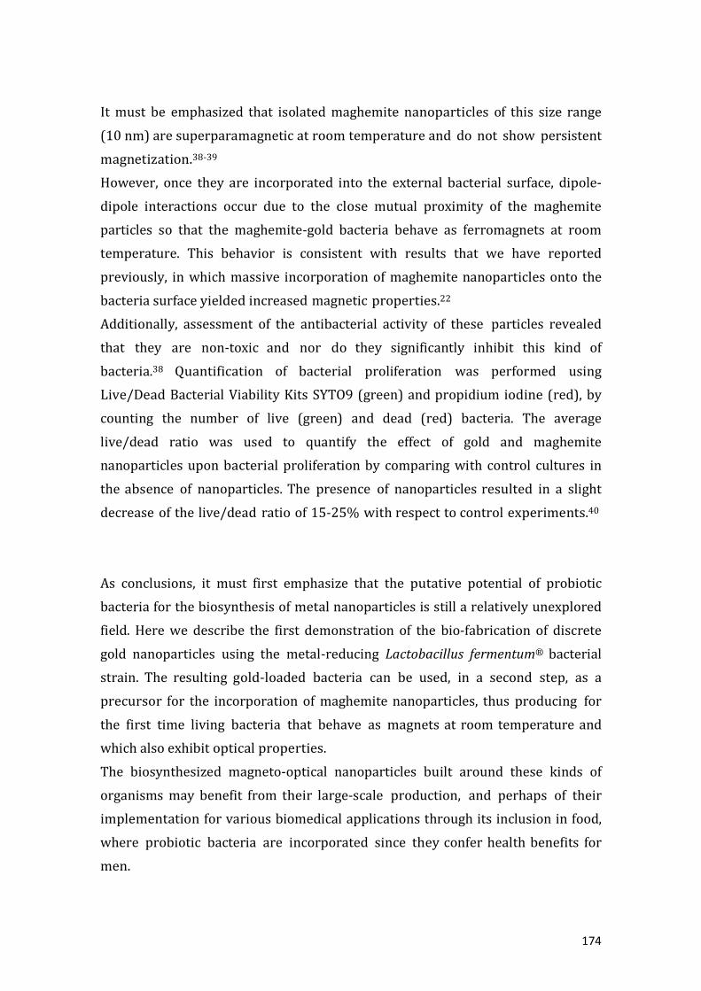
174
It must be emphasized that isolated maghemite nanoparticles of this size range
(10 nm) are superparamagnetic at room temperature and do not show persistent
magnetization.38-39
However, once they are incorporated into the external bacterial surface, dipole-
dipole interactions occur due to the close mutual proximity of the maghemite
particles so that the maghemite-gold bacteria behave as ferromagnets at room
temperature. This behavior is consistent with results that we have reported
previously, in which massive incorporation of maghemite nanoparticles onto the
bacteria surface yielded increased magnetic properties.22
Additionally, assessment of the antibacterial activity of these particles revealed
that they are non-toxic and nor do they significantly inhibit this kind of
bacteria.38 Quantification of bacterial proliferation was performed using
Live/Dead Bacterial Viability Kits SYTO9 (green) and propidium iodine (red), by
counting the number of live (green) and dead (red) bacteria. The average
live/dead ratio was used to quantify the effect of gold and maghemite
nanoparticles upon bacterial proliferation by comparing with control cultures in
the absence of nanoparticles. The presence of nanoparticles resulted in a slight
decrease of the live/dead ratio of 15-25% with respect to control experiments.40
As conclusions, it must first emphasize that the putative potential of probiotic
bacteria for the biosynthesis of metal nanoparticles is still a relatively unexplored
field. Here we describe the first demonstration of the bio-fabrication of discrete
gold nanoparticles using the metal-reducing Lactobacillus fermentum® bacterial
strain. The resulting gold-loaded bacteria can be used, in a second step, as a
precursor for the incorporation of maghemite nanoparticles, thus producing for
the first time living bacteria that behave as magnets at room temperature and
which also exhibit optical properties.
The biosynthesized magneto-optical nanoparticles built around these kinds of
organisms may benefit from their large-scale production, and perhaps of their
implementation for various biomedical applications through its inclusion in food,
where probiotic bacteria are incorporated since they confer health benefits for
men.

175
Notes and references
(1) Quaresma, P.; Osorio, I.; Doria, G.; Carvalho, P. A.; Pereira, A.; Langer, J.; Araujo,
J. P.; Pastoriza-Santos, I.; Liz-Marzan, L. M.; Fran- co, R. RSC Advances. 2014, 4,
3659-3667.
(2) Umut, E.; Pineider, F.; Arosio, P.; Sangregorio, C.; Corti, M.; Tabak, F.; Lascialfari,
A.; Ghigna, P. J. Mag. and Mag. Mater. 2012, 324, 2373-2379.
(3) Sheng, Y.; Xue, J. J. of Colloid and Interf. Sci. 2012, 374, 96-101. (4)
Bhattacharya, D.; Chakraborty, S. P.; Pramanik, A.; Baksi, A.; Roy, S.; Maiti, T. K.;
Ghosh, S. K.; Pramanik, P. J. Mater. Chem. 2011, 21, 17273.
(5) Yu, H.; Chen, M.; Rice, P. M.; Wang, S. X.; White, R. L.; Sun, S. Nano Lett. 2005, 5,
379.
(6) Jain, P. K.; El-Sayed, I. H.; El-Sayed, M. A. Nanotoday. 2007, 2, 18.
(7) Salgueiriño-Maceira,V.; Correa-Duarte, M. A.; Lopez- Quintela, M. A.; Rivas, J. J.
of Nanosci. and Nanotech. 2009, 9, 3684- 3688.
(8) Salgueiriño-Maceira, V.; Correa-Duarte, M. A.; Lopez-Quintela,M.A.;Rivas, J.
Sensor Letters. 2007, 5, 113-117.
(9) Suresh, A. K.; Pelletier, D. A.; Wang, W.; Broich, M. L.; Moon, J. W.; Gu, B.; Allison,
D. P.; Joy, D. C.; Phelps, T. J.; Doktycz, M. J. Acta Biomaterialia. 2011, 7, 2148–2152.
(10) Bhambure, R.; Bul, M.; Shaligram, N.; Kamat, M.; Singhal, R. Chem. Eng. Technol.
2009, 32,1036–41.
(11) Mukherjee, P.; Senapati, S.; Mandal, D.; Ahmad, A.; Khan, M. I.; Kumar, R.;
Sastry, M. Chembiochem. 2002, 3, 461–463.
(12) Mukherjee, P.; Ahmad, A.; Mandal, D.; Senapati, S.; Sainkar, S. R.; Khan, M. I.;
Ramani, R.; Parischa, R.; Ajayakumar, P. V.; Alam, M.; Sastry, M.; Kumar, R. Angew.
Chem. Int. Ed. 2001, 40, 3585–3588.
(13) Ahmad, A.; Senapati, S.; Khan, M. I.; Kumar, R.; Sastry, M. Langmuir. 2003, 19,
3550–3553.
(14) Das, S. K.; Das, A. R.; Guha, A. K. Langmuir. 2009, 25, 8192– 8109.
(15) Ramanathan, R.; Field, M. R.; O'Mullane, A. P.; Smooker, P. M.; Bhargava, S. K.;
Bansal, V. Nanoscale. 2013, 5, 2300-2306.
(16) Inbakandana, D.; Venkatesan, R.; Khan, S. A. Colloids and Surfaces B:
Biointerfaces. 2010, 81, 634–639.

176
(17) Bansal, V.; Bharde, A.; Ramanathan, R.; Bhargava, S. K. Adv. In Colloid and
Interf. Sci. 2012, 179-182, 150-168.
(18) Roh, Y.; Gao, H.; Vali, H.; Kennedy, D. W.; Yang, Z. K.; Gao, W.; Dohnalkova, A. C.;
Stapleton, R. D.; Moon, J. W.; Phelps, T. J.; Fredrickson, J. K.; Zhou, J. Appl. Environ.
Microbiol. 2006, 72, 3236–3244.
(19) Zachara, J. M.; Kukkadapu, R. K.; Fredrickson, J. K.; Gorby, Y. A.; Smith, S. C.
Geomicrobiol. J. 2002, 19, 179–207.
(20) Zhang, C. L.; Liu, S.; Phelps, T. J.; Cole, D. R.; Horita, J.; Forti- er, S. M.; Elless, M.;
Valley, J. W. Geochim. Cosmochim. Acta. 1997, 61, 4621–4632.
(21) Perez-Gonzalez, T.; Jimenez-Lopez, C.; Neal, A. L.; Rull-Perez, F.; Rodriguez-
Navarro, A.; Fernandez-Vivas, A.; Iañez-Pareja, E.; Geochim. Et Cosmochim. Acta.
2010, 74, 967–979.
(22) Martín, M.; Carmona, F.; Cuesta, R.; Rondón, D.; Gálvez, N.; Domínguez-Vera, J.
M. Adv. Funct. Mater. 2014, DOI: 10.1002/adfm. 201303754.
(23) Cheon, J.; Lee, J. H. Acc. Chem. Res. 2008, 41, 1630-1640. (24) Dobson, J. Nat.
Nanotech. 2008, 3, 139–143.
(25) Mornet, S.; Vasseur, S.; Grasset, F.; Duguet, E. J. Mater. Chem. 2004 14, 2161–
2175.
(26) Laurent, S.; Forge, D.; Port, M.; Roch, A.; Robic, C.; Elst, L. V. Chem. Rev. 2008,
108, 2064-2110.
(27) Daniel, M. C.; Astruc, D. Chem. Rev. 2004, 104, 293.
(28) Pastoriza-Santos, I.; Liz-Marzan, L. M. Meth. Mol. Biol. 2013, 1025, 75-93.
(29) Lim, J. K.; Majetich, S. A. Nano Today. 2013, 8, 98-113.
(30) Dreaden, E. C.; Alkilany, A. M.; Huang, X.; Murphy, C. J.; El- Sayed, M. A. Chem.
Soc. Rev. 2012, 41, 2740-2779.
(31) Wang, L.; Park, H. Y.; Lim, S. I.; Schadt, M. J.; Mott, D.; Luo, J.; Wang, X.; Zhong, C.
J. J. Mater. Chem. 2008, 18, 2629–2635.
(32) Salgueiriño-Maceira, V.; Correa-Duarte, M. A.; Farle, M.; Lopez-Quintela, A.;
Sieradzki, K.; Diaz, R. Chem. Mater. 2006, 18, 2701.
(33) Correa-Duarte, M. A.; Salgueiriño-Maceira, V.; Rodriguez-Gonzalez, B.; Liz-
Marzan, L. M.; Kosiorek, A.; Kandulski, W.; Giersig, M. Adv. Mater. 2005, 17, 2014.
(34) Liu, H. L.; Wub, J. H.; Min, J. H.; Kim, Y. K. J. of Alloys and Compounds. 2012,
537, 60–64.

177
(35) Spasova, M.; Salgueirino-Maceira, V.; Schlachter, A.; Hilgen- dorff, M.; Giersig,
M.; Liz-Marzan, L. M.; Farle, M. J. Mater. Chem. 2005, 15, 2095-2098.
(36) Liquid cultures of Lactobacillus fermentum® were grown in MRS broth
(Panreac, 413785) at 37 ºC with orbital agitation for 24 h until early stationary
phase. Bacteria were then collected via centrifugation and resuspended in Hank’s
Balanced Salt Solution to a final concentration of 1 mg/mL (3.5x10⁸ CFU/mL). An
Au(III) aqueous solution (Gold tetrachloride, Sigma-Aldrich, 99%), was dissolved
in Type 1 Milli-Q ultrapure water and then added to the bacterial sus- pension to
give a final concentration of 1 mM. The reaction mixture was kept at 37 ºC and
under continuous stirring for 30 min. until the appearance of a characteristic red-
wine colored solution, indicating the formation of gold nanoparticles. The resulting
gold-biomass was centrifuged at 100 rpm for 30 min. The supernatant exhibited no
band in the visible region. The quantity of gold measured by inducively coupled
plasma mass spectrometry was zero. Control reactions in the absence of
Lactobacillus fermentum®, where the culture su- pernatants of Hank's Balanced
Salt Solution and MRS broth both exhibited no color change or absorbance at 520
nm, clearly indicated that the presence of bacteria in the biosynthesis of gold
nanoparticles is a prerequisite. An acidic solution (pH 2) of maghemite
nanoparticles (1 μL, at 0.95 - 1 M of iron), prepared as previously reported,39 was
added to the gold-loaded Lactobacillus fermentum® and maintained at 37 ºC under
orbital agitation for 30 min. The reaction mixture was washed with several
centrifugation cycles (100 rpm, 5 ºC, 30 minutes), to remove any unbound
maghemite nanoparticles, and the final bacterial pellet resuspended in a 50 mM
sodium citrate, PBS, Milli-Q water 1:1:4 mixture at pH 5. The resulting
gold+maghemite-Lactobacillus fermentum® suspension remained stable at room
temperature.
(37) The sample of gold+maghemite- Lactobacillus fermentum® was embedded in
an epoxy resin. Fixation was achieved by adding 2.5% glutaraldehyde in a 0.1 M
sodium cacodylate buffer solution at 4 ºC for 4 h. The sample was washed three
times in 0.1 M sodium cacodylate buffer for 15 minutes. Then, drying cicles with
ethanol and propylene oxide were applied. Finally, the sample was embedded in an
epoxy resin and left overnight at 4 ºC. After ultramicro-cutting, samples were
observed with a FEI TITAN G2 microscope.

178
(38) Valero, E.; Tambalo, S.; Marzola, P.; Ortega-Muñoz, M.; Lopez-Jaramillo, J.;
Santoyo, F.; Lopez, J. D.; Delgado, J. J.; Calvino, J. J.; Cuesta, R.; Dominguez-Vera, J. M.;
Gálvez, N. J. Am. Chem. Soc. 2011, 133, 4889.
(39) Park, J.; Lee, E.; Hwang, N. M.; Kang, M.; Kim, S. C.; Hwang, Y.; Park, J. G.; Noh,
H-J.; Kim, J-Y.; Park, J-H.; Hyeon, T. Angew. Chem. Int. Ed. 2005, 44, 2872.
(40) Quantification of bacteria proliferation was performed by using the Live/Dead
Bacterial Viability Kits SYTO9 (green) and Propidium Iodine (red) (Invitrogen),
counting the number of live (green) and dead (red) bacteria in a batch of three
experiments by the software Image-Pro Plus 6.0. The average live/dead ratio was
used to quantify the bacteria proliferation by comparing with control experiments
where no nanoparticles were present. The presence of maghemite and gold
nanoparticles resulted in a decrease of the live/dead ratio of 15 – 25 % for three
independent experiments carried out in Hank’s Balanced Salt Solution ( i ure SI).

179
CONCLUSIONES

180
181
1. La ferritina H, formada exclusivamente por subunidades H en su cápside
proteica, tiene una gran actividad ferroxidasa para oxidar de forma catalítica Fe(II)
a Fe(III) pero muy poca capacidad de almacenar Fe(III). Mas allá de entrar en
discusiones mecanísticas sobre el proceso de reconstitución de hierro, nuestra
investigación se ha centrado en el destino que sigue el Fe(III) generado por la
ferritina H, y hemos demostrado que el Fe(III) que no es almacenado queda en una
forma biodisponible en el organismo, no ya sólo para un agente quelatante
sintético de Fe(III), sino también para biomoléculas de gran tamaño como
proteínas. En este marco, hemos demostrado que este Fe(III) generado por la
ferritina H es capaz de actuar como oxidante frente a otras metaloproteínas
presentes en la célula. Concretamente hemos concluido que la cantidad de Fe(III)
no almacenada es capaz de oxidar a una isoforma de metalotioneína, la MT2, con la
que coexiste en determinadas células del cerebro, y que como producto de esta
reacción se generan iones metálicos libres y tóxicos como son el Fe(II) y el Cu(I).
2. Por otra parte, hemos puesto de manifiesto que no todas las isoformas de
metalotioneínas (MT) reaccionan de manera similar con el Fe(III) generado en el
proceso de reconstitución de ferritina H. Concretamente, la MT3 no reacciona con
dicho Fe(III). Este resultado es de extrema importancia porque “curiosamente” la
ferritina H se encuentra en células como neuronas donde la isoforma de la
metalotioneína es MT3. En vista de estos resultados, hemos extendido este estudio
a otros pares ferritina – MT, y hemos demostrado que para el caso de ferritina
mixta H/L, no se produce Fe(III) disponible para oxidar a ningún tipo de
metalotioneína (MT), incluida la isoforma más reactiva MT2. De nuevo
“curiosamente” T2 sólo coincide fisioló icamente con ferritinas de tipo H/L.
Este complejo entramado de interacciones entre metaloproteínas viene a poner de
manifiesto que la elección del par ferritina-metalotioneína debe ser el apropiado
en cada célula: MT3 con ferritinas H pero no MT2, puesto que la presencia de
ferritina H y MT2 genera espontáneamente Fe(II) y Cu(I), extremadamente
dañinos para la propia célula. Este resultado deja entrever que cualquier
disfunción genética que de lugar a la síntesis de una ferritina H en lugar de una H/L

182
en neurona sería fatal para la célula y podría representar el primer estadío del
desarrollo de una patología neurológica.
3. La alta actividad ferroxidasa y baja capacidad de almacenamiento de la
ferritina H permite entender por qué ferritina H y lactoferrina coexisten en la leche
materna. Nuestros resultados ponen de manifiesto que la existencia de este
tándem proteico es una estrategia eficaz para llevar a cabo la detoxificación de
Fe(II) y la captación de Fe(III) en el organismo en pro de llevar a cabo una
actividad antimicrobiana eficaz.
Inspirados en la existencia de este tándem ferritina H + lactoferrina en leche
materna, hemos diseñado y sintetizado un material que contiene ambas proteínas
inmovilizadas y que funciona extraordinariamente bien como captador de hierro
en todas sus formas: el Fe(II) libre es oxidado por la ferritina H mientras que el
Fe(III) generado es captado por la lactoferrina. Este efecto sinérgico de ambas
proteínas nos ha permitido diseñar materiales con potencial actividad
antimicrobiana que representan un nuevo concepto de material destinado a una
eficaz defensa contra el desarrollo de microorganismos patógenos al privarle de
este metal esencial.
4. Hemos logrado estudiar y monitorizar los cambios conformacionales que
tienen lugar en la lactoferrina durante el proceso de incorporación y liberación de
Fe(III) en su estructura a través de espectroscopía de fluorescencia, mediante
arcaje con dos fluoróforos AF350 y AF430, que presentan fenómeno de FRET con
un rendimiento dependiente de la conformación de la proteína y por ende de su
contenido en hierro. Además, hemos hecho uso de la correlación rendimiento del
FRET-contenido en hierro de la lactoferrina para demostra de forma elegante que
esta proteína proporciona hierro a las bacterias probióticas beneficiosas para la
salud con las que coexiste en leche materna y que, además, este proceso tiene lugar
sin la necesidad de internalización de la proteína por parte de la bacteria.
5. Hemos podido demostrar, que al igual que otras bacterias reportadas en la
literatura, las bacterias probióticas también emiten una (o varias) moléculas

183
reductoras capaces de reducir metales u otras especies químicas. Concretamente,
hemos puesto de manifiesto que estas bacterias son capaces de reducir Au(III) a
Au(0) y además controlar el tamaño y deposición del Au(0) como nanopartícula en
el biofilm bacteriano. Esto nos ha permitido preparar, aprovechando
conocimientos previos desarrollados en el grupo, bacterias probióticas que
contienen simultáneamente nanopartículas de Au (con propiedades ópticas) y
nanopartículas de maghemita (con propiedades magnéticas). La bacteria final se
comporta como un imán a temperatura ambiente y absorbe en el espectro visible
debido a la banda SPR.
Estas bacterias siguen vivas tras la incorporación de las nanopartículas de Au y
maghemita. Hasta donde llegan nuestros conocimientos, es el primer ejemplo de
un organismo vivo que presente propiedades magnéticas y ópticas. Esto abre una
nueva vía de utilizar bacterias probióticas con propiedades únicas para
aplicaciones biomédicas.