unit iv: plant anatomy (structural organisation) chapter ......present in collenchyma.based on...
TRANSCRIPT

The learner will be able to,
• Study major types of plant cells and
their function.
• Differentiate the various types of
cells.
• Study the relationship between the
distribution of tissues in the various
parts of plants.
• Describes the ground tissue system
[cortex and pith] and vascular
systems
• Interpret cross sections and
longitudinal sections of dicot and
monocot root, stem and leaf.
• Compare the internal organization
of dicot root and monocot root.
Tissue and Tissue System
Chapter
9
Chapter Outline
9.1 Meristematic tissue9.2 Permanent tissues9.3 The tissue system9.4 Epidermal tissue system9.5 Fundamental tissue system9.6 Vascular tissue system9.7 Comparision of primary structure
Nehemiah Grew
Father of Plant
Anatomy
1641–1712
Katherine Esau (1898–1997)
A legendary Role model for women in science. She was a scintillating Botany teacher and pioneering researcher for six decades. Her classic book Anatomy
of Seed Plants is the best literature in Plant Anatomy. In recognition of her distinguished service to science, she was awarded National
Medal of Science
(1989) by USA.
Unit IV: Plant Anatomy (Structural Organisation)
Learning Objectives
This chapter introduces the internal structure of higher Plants. The study of internal structure and organisation of plant is called plant Anatomy (Gk: Ana =
as under; temnein = to cut). Plants have cells as the basic unit. The cells are organised into tissues. The tissues in turn are organised into organs. The different organs in a plant have different internal structures. It is studied by means of dissection and microscopic examination.
1
2
Milestones in Anatomy
• 1837 Hartig: Coined the term Sieve
tubes
• 1839 Schleiden: Coined the term Collenchyma
• 1857 Hofmeister: Proposed Apical cell
theory
• 1858 Nageli. C: Coined the term Xylem
and Phloem, Meristem and supporter of Apical cell theory
• 1865 Mettenius: Coined the term Sclerenchyma
• 1868 Hanstein: Proposed Histogen
theory
• 1885 Tschirch: Coined the term Sclereids Named Four types of Sclereids (Brachy, Macro, Osteo & Astro) in 1889
• 1914 Haberlandt: Coined the term xylem as Hadrome and Phloem as Leptome and Classification of
meristem.
• 1924 Schmidt A: Proposed Tunica –
Corpus theory
• 1926 Schűepp: Mass, rib, & plate
meristem
• 1946 Bloch: Discovered the Trichosclereids
• 1952 Popham: Explained the organization of Shoot apex of
Angiosperms
• 1955 Duchaigne: Discovered the Annular collenchyma
• 1961 Clowes: Proposed Quiescent
centre concept
• 1963 Sanio: Coined the term Tracheids
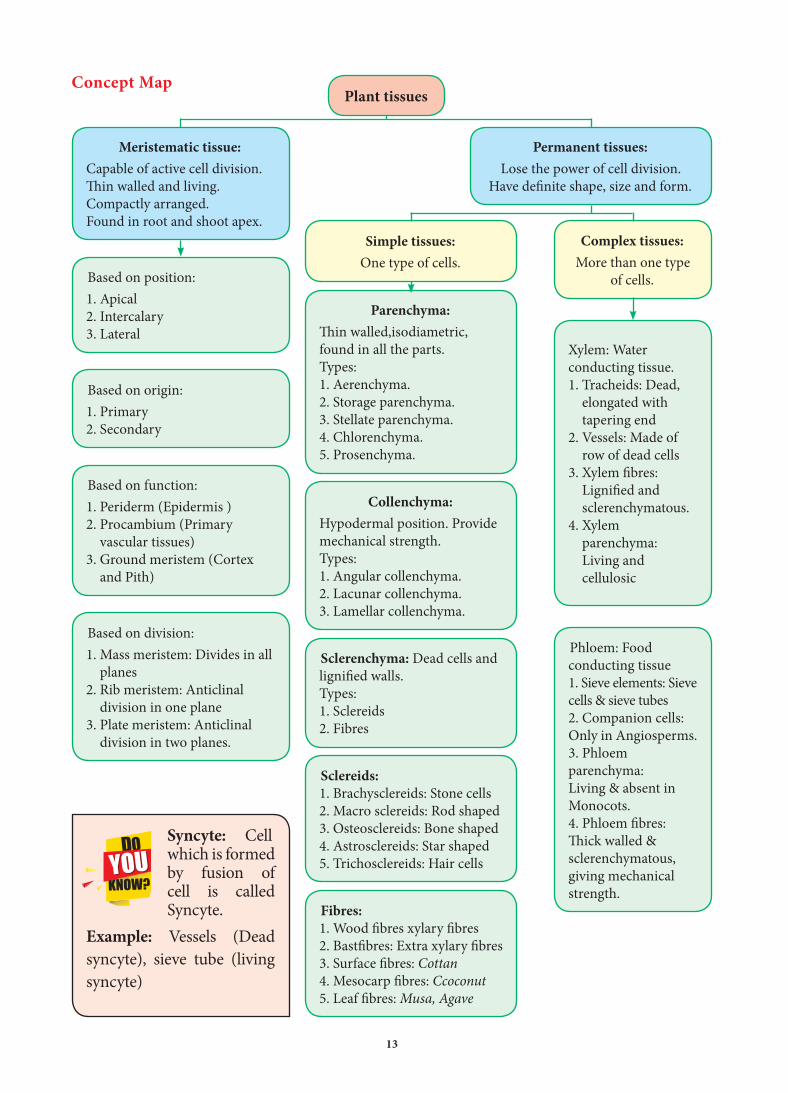
The Tissues
A Tissue is a group of cells that are alike in origin, structure and function. The study
of tissue is called Histology. A plant is made up of different types of tissues.
There are two principal groups:1. Meristematic tissues2. Permanent tissues
9.1 Meristematic Tissue
9.1.1 Characteristics and classification
The characters of meristematic tissues:
(Gr. Meristos-Divisible)The term meristem is coined by
C. Nageli 1858.• The meristematic cells are isodiametric
and they may be, oval, spherical or polygonal in shape.
• They have generally dense cytoplasm with prominent nucleus.
• Generally the vacuoles in them are either small or absent.
• Their cell wall is thin, elastic and essentially made up of cellulose.
• These are most actively dividing cells.
• Meristematic cells are self-perpetuating.
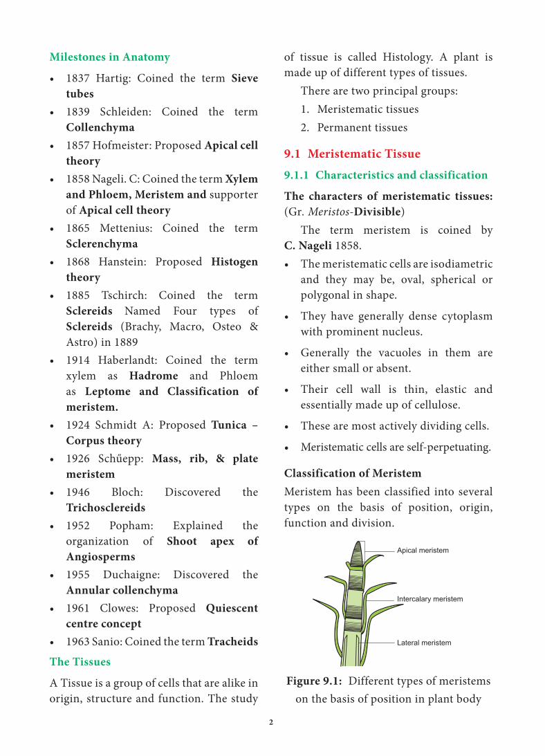
Classification of Meristem
Meristem has been classified into several types on the basis of position, origin, function and division.
Apical meristem
Intercalary meristem
Lateral meristem
Figure 9.1: Different types of meristems on the basis of position in plant body

3
Shoot Apical Meristem
Apical Cell Theory
Apical cell theory is proposed by Hofmeister (1852) and supported by Nageli (1859). A single apical cell is the structural and functional unit.
Theories of Meristem Organization and
Function
Many anatomists illustrated the root and shoot apical meristems on the basis of number and arrangement and accordingly proposed the following theories – An extract of which are discussed below.
Figure 9.2: Shoot apical meristem a) Apical cell theory, b) Histogen theory, c) Shoot Tunica corpus theory
HistogenPleromePeriblemDermatogen
Corpus
Leaf primordia
Tunica
Apical cell
Leaf primodium
Classification of Meristem
Position Origin Function Plane of division
Lateral meristem
Occurs along the longitudinal axis of stem and root. It is responsible for secondary tissues and thickening of stem and root. Example: vascular
cambium and cork cambium.
Apical meristem
Present in apices of root and shoot. It is responsible for increase in the length of the plant, it is called as
primary growth.
Intercalary meristem
Occurs between the mature tissues. It is
responsible for elongation of internodes.
Primary
Meristem
It is derived from
embryonic stages and
differentiated into primary permanent
tissues.
Secondary
meristem It is derived during later stage of development of the plant
body. It produces cork cambium and interfascicular
cambium.
Protoderm
It gives rise to epidermal tissue
system and develops into
epidermis,stomata and hairs.
Procambium
It gives rise to primary vascular tissues. Example:
xylem and phloem .
Ground Meristem
It gives rise to all tissues except epidermis and
vascular strands.
Mass meristem
It divides in all planes. Example:
endosperm,young embryo and sporangium
Rib meristem or
File meristem
It divides anticlinally in one plane. Example: development of cortex and pith
Plate meristem
It divides anticlinally in two planes. Example: development of
epidermis
b. c.a.
4
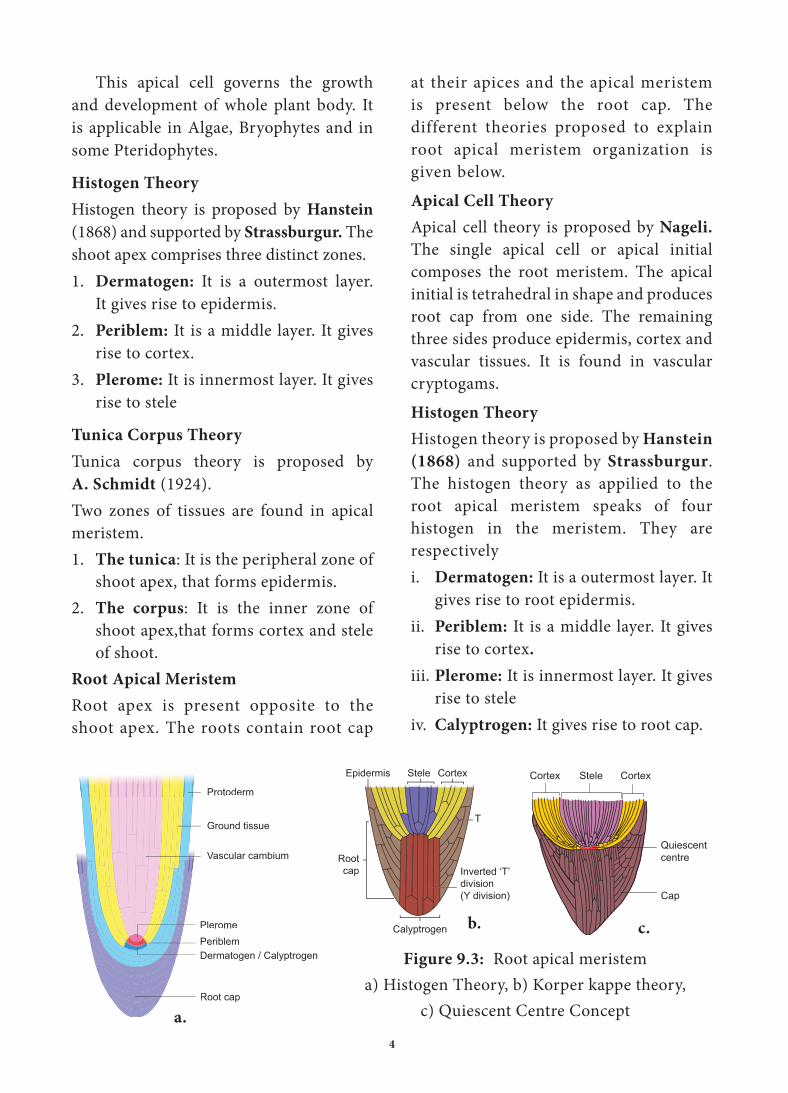
at their apices and the apical meristem is present below the root cap. The different theories proposed to explain root apical meristem organization is given below.Apical Cell Theory
Apical cell theory is proposed by Nageli. The single apical cell or apical initial composes the root meristem. The apical initial is tetrahedral in shape and produces root cap from one side. The remaining three sides produce epidermis, cortex and vascular tissues. It is found in vascular cryptogams.Histogen Theory
Histogen theory is proposed by Hanstein
(1868) and supported by Strassburgur. The histogen theory as appilied to the root apical meristem speaks of four histogen in the meristem. They are respectivelyi. Dermatogen: It is a outermost layer. It
gives rise to root epidermis.ii. Periblem: It is a middle layer. It gives
rise to cortex.
iii. Plerome: It is innermost layer. It gives rise to stele
iv. Calyptrogen: It gives rise to root cap.
This apical cell governs the growth and development of whole plant body. It is applicable in Algae, Bryophytes and in some Pteridophytes.
Histogen Theory
Histogen theory is proposed by Hanstein (1868) and supported by Strassburgur. The shoot apex comprises three distinct zones.1. Dermatogen: It is a outermost layer.
It gives rise to epidermis.2. Periblem: It is a middle layer. It gives
rise to cortex.3. Plerome: It is innermost layer. It gives
rise to stele
Tunica Corpus Theory
Tunica corpus theory is proposed by A. Schmidt (1924).Two zones of tissues are found in apical meristem.1. The tunica: It is the peripheral zone of
shoot apex, that forms epidermis.2. The corpus: It is the inner zone of
shoot apex,that forms cortex and stele of shoot.
Root Apical Meristem
Root apex is present opposite to the shoot apex. The roots contain root cap
Figure 9.3: Root apical meristem a) Histogen Theory, b) Korper kappe theory,
c) Quiescent Centre Concept
Vascular cambium
Ground tissue
Protoderm
PeriblemPlerome
Dermatogen / Calyptrogen
Root cap
Epidermis Stele Cortex
T
Inverted ‘T’division (Y division)
Calyptrogen
Rootcap
Quiescent centre
Cap
CortexSteleCortex
b. c.
a.
5
Parenchyma (Gk: Para-beside;
enehein- to pour)
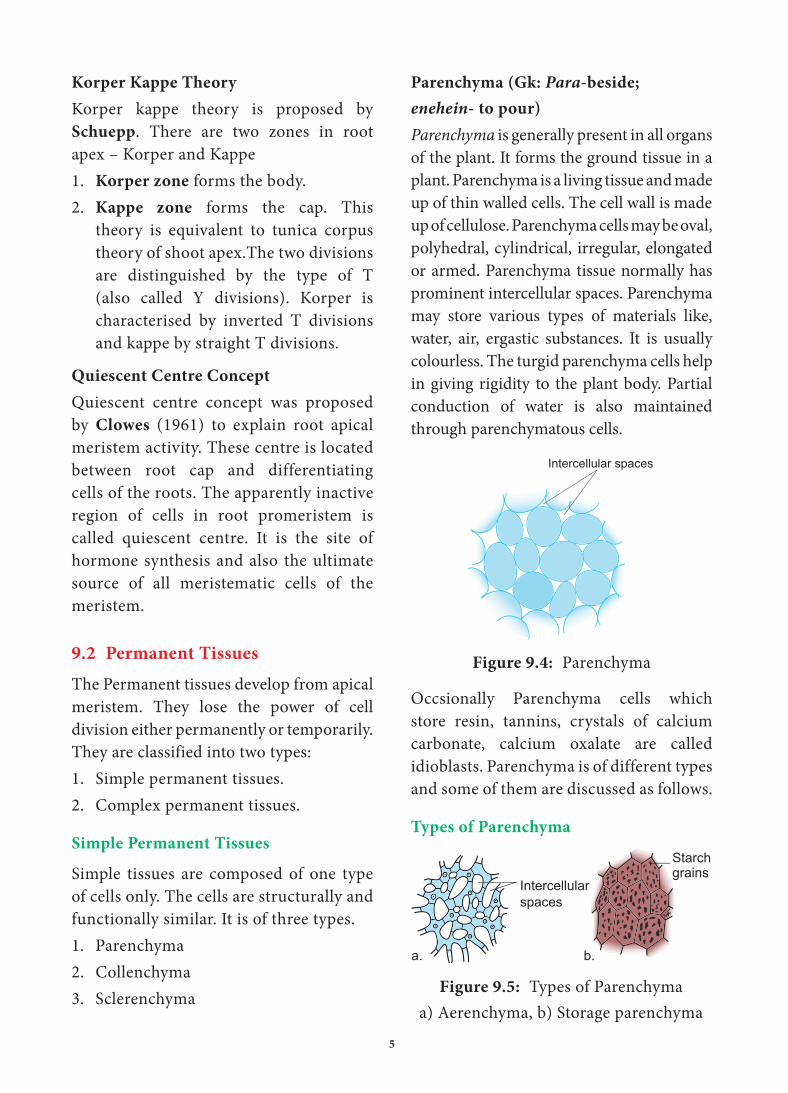
Parenchyma is generally present in all organs of the plant. It forms the ground tissue in a plant. Parenchyma is a living tissue and made up of thin walled cells. The cell wall is made up of cellulose. Parenchyma cells may be oval, polyhedral, cylindrical, irregular, elongated or armed. Parenchyma tissue normally has prominent intercellular spaces. Parenchyma may store various types of materials like, water, air, ergastic substances. It is usually colourless. The turgid parenchyma cells help in giving rigidity to the plant body. Partial conduction of water is also maintained through parenchymatous cells.
Intercellular spaces
Figure 9.4: Parenchyma
Occsionally Parenchyma cells which store resin, tannins, crystals of calcium carbonate, calcium oxalate are called idioblasts. Parenchyma is of different types and some of them are discussed as follows.
Types of Parenchyma
Intercellular spaces
Starch grainsg
a. b.
Figure 9.5: Types of Parenchyma a) Aerenchyma, b) Storage parenchyma
Korper Kappe Theory
Korper kappe theory is proposed by Schuepp. There are two zones in root apex – Korper and Kappe1. Korper zone forms the body.2. Kappe zone forms the cap. This
theory is equivalent to tunica corpus theory of shoot apex.The two divisions are distinguished by the type of T (also called Y divisions). Korper is characterised by inverted T divisions and kappe by straight T divisions.
Quiescent Centre Concept
Quiescent centre concept was proposed by Clowes (1961) to explain root apical meristem activity. These centre is located between root cap and differentiating cells of the roots. The apparently inactive region of cells in root promeristem is called quiescent centre. It is the site of hormone synthesis and also the ultimate source of all meristematic cells of the meristem.
9.2 Permanent Tissues
The Permanent tissues develop from apical meristem. They lose the power of cell division either permanently or temporarily. They are classified into two types:1. Simple permanent tissues.2. Complex permanent tissues.
Simple Permanent Tissues
Simple tissues are composed of one type of cells only. The cells are structurally and functionally similar. It is of three types.1. Parenchyma2. Collenchyma3. Sclerenchyma

6
1. Aerenchyma:
Parenchyma which contains air in its intercellular spaces. It helps in aeration and buoyancy. Example: Nymphae and Hydrilla.
2. Storage Parenchyma:
Parenchyma stores food materials. Example: Root and stem tubers.
5. Prosenchyma:
Parenchyma cells became elongated, pointed and slightly thick walled. It provides mechanical support.
4. Chlorenchyma
Parenchyma cells with chlorophyll. Function is photosynthesis. Example: Mesophyll of leaves.
3. How?.... Stellate
Parenchyma
Star shaped parenchyma. Example: Petioles of Banana
and Canna.
Parenchyma
Intercellular Spaces
Chloroplasts
Palisade Parenchyma
Spongy Parenchyma
Intercellular Space
a. b. c.
Figure 9.5: a) Stellate parenchyma, b) Chlorenchyma, c) Prosenchyma
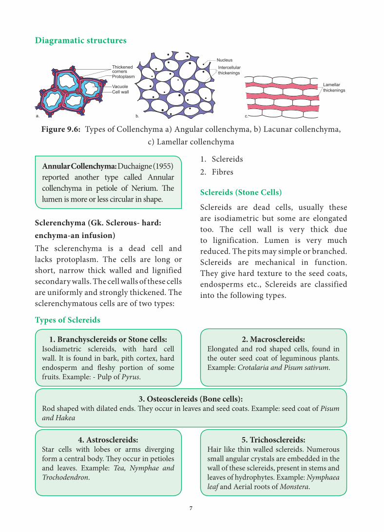
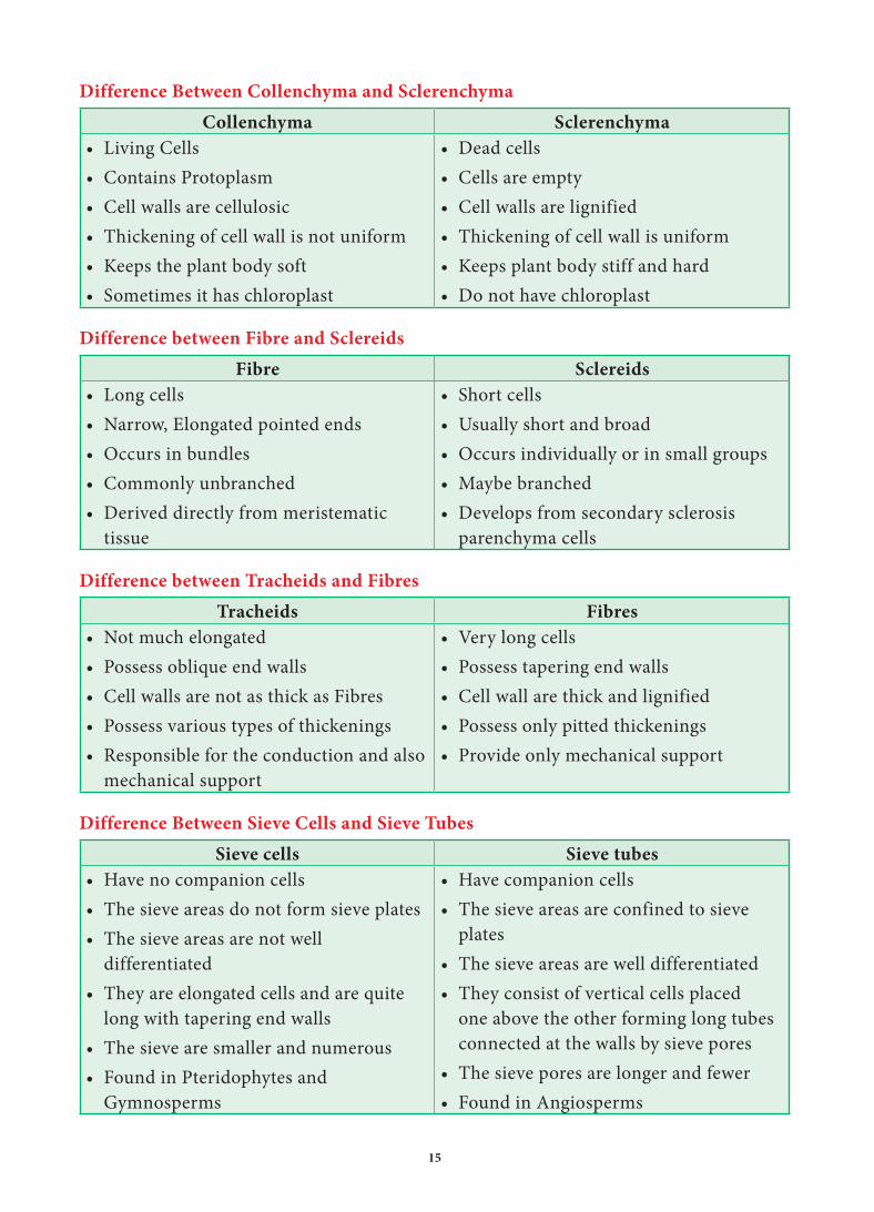
Collenchyma (Gk. Colla-glue;
enchyma – an infusion)
Collenchyma is a simple, living mechanical tissue. Collenchyma generally occurs in hypodermis of dicot stem. It is absent in the roots and also occurs in petioles and pedicels. The cells are elongated and appear polygonal in cross section. The cell wall is unevenly thickened. It contains more of hemicellulose and pectin besides cellulose. It provides mechanical support and elasticity to the growing parts of the plant. Collenchyma consists of narrow cells. It has only a few
small chloroplast or none. Tannin maybe present in collenchyma.Based on pattern of pectinisation of the cell wall, there are three types of collenchyma
Types of Collenchyma
1. Angular collenchymaIt is the most common type of collenchyma with irregular arrangement and thickening at the angles where cells meets.Example:Hypodermis of Datura and
Nicotiana
2. Lacunar collenchymaThe collenchyma cells are irregularly arranged. Cell wall is thickening on the walls bordering intercellular spaces. Example:Hypodermis of Ipomoea
3. Lamellar collenchymaThe collenchyma cells are arranged compactly in layers(rows). The Cell wall is thickening is at tangential walls.These thickening appear as successsive tangential layers. Example:Hypodermis of Helianthus
7
Diagramatic structures
Nucleus
Intercellular thickenings
b.
Thickened corners
VacuoleCell wall
Protoplasm
a.
Lamellar thickenings
c.
Figure 9.6: Types of Collenchyma a) Angular collenchyma, b) Lacunar collenchyma, c) Lamellar collenchyma
Annular Collenchyma: Duchaigne (1955) reported another type called Annular collenchyma in petiole of Nerium. The lumen is more or less circular in shape.
Sclerenchyma (Gk. Sclerous- hard:
enchyma-an infusion)
The sclerenchyma is a dead cell and lacks protoplasm. The cells are long or short, narrow thick walled and lignified secondary walls. The cell walls of these cells are uniformly and strongly thickened. The sclerenchymatous cells are of two types:
1. Sclereids2. Fibres
Sclereids (Stone Cells)
Sclereids are dead cells, usually these are isodiametric but some are elongated too. The cell wall is very thick due to lignification. Lumen is very much reduced. The pits may simple or branched. Sclereids are mechanical in function. They give hard texture to the seed coats, endosperms etc., Sclereids are classified into the following types.
Types of Sclereids
1. Branchysclereids or Stone cells:Isodiametric sclereids, with hard cell wall. It is found in bark, pith cortex, hard endosperm and fleshy portion of some fruits. Example: - Pulp of Pyrus.
3. Osteosclereids (Bone cells):Rod shaped with dilated ends. They occur in leaves and seed coats. Example: seed coat of Pisum
and Hakea
4. Astrosclereids:Star cells with lobes or arms diverging form a central body. They occur in petioles and leaves. Example: Tea, Nymphae and
Trochodendron.
5. Trichosclereids:Hair like thin walled sclereids. Numerous small angular crystals are embedded in the wall of these sclereids, present in stems and leaves of hydrophytes. Example: Nymphaea
leaf and Aerial roots of Monstera.
2. Macrosclereids:Elongated and rod shaped cells, found in the outer seed coat of leguminous plants. Example: Crotalaria and Pisum sativum.
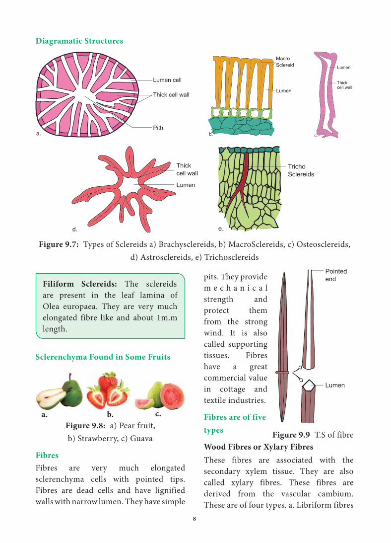
8
Diagramatic Structures
Thick cell wall
Lumen cell
Pitha.
MacroSclereid
Lumen
b.
Lumen
Thickcell wall
c.
Tricho Sclereids
e.
Lumen
Thickcell wall
d.
Figure 9.7: Types of Sclereids a) Brachysclereids, b) MacroSclereids, c) Osteosclereids, d) Astrosclereids, e) Trichosclereids
Filiform Sclereids: The sclereids are present in the leaf lamina of Olea europaea. They are very much elongated fibre like and about 1m.m length.
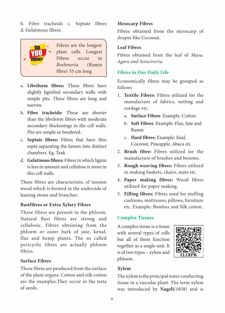
Sclerenchyma Found in Some Fruits
Figure 9.8: a) Pear fruit, b) Strawberry, c) Guava
Fibres
Fibres are very much elongated sclerenchyma cells with pointed tips. Fibres are dead cells and have lignified walls with narrow lumen. They have simple
pits. They provide m e c h a n i c a l strength and protect them from the strong wind. It is also called supporting tissues. Fibres have a great commercial value in cottage and textile industries.
Fibres are of five
types
Wood Fibres or Xylary Fibres
These fibres are associated with the secondary xylem tissue. They are also called xylary fibres. These fibres are derived from the vascular cambium. These are of four types. a. Libriform fibres
Pointed end
Lumen
Figure 9.9 T.S of fibre
b. c.a.
9
b. Fibre tracheids c. Septate fibres d. Gelatinous fibres.
Fibres are the longest plant cells. Longest Fibres occur in Boehmeria (Ramie fibre) 55 cm long
a. Libriform fibres: These fibres have slightly lignified secondary walls with simple pits. These fibres are long and narrow.
b. Fibre tracheids: These are shorter than the libriform fibres with moderate secondary thickenings in the cell walls. Pits are simple or bordered.
c. Septate fibres: Fibres that have thin septa separating the lumen into distinct chambers. Eg. Teak
d. Gelatinous fibres: Fibres in which lignin is less in amount and cellulose is more in this cell walls.
These fibres are characteristic of tension wood which is formed in the underside of leaning stems and branches.
Bastfibres or Extra Xylary Fibres
These fibres are present in the phloem. Natural Bast fibres are strong and cellulosic. Fibres obtaining from the phloem or outer bark of jute, kenaf, flax and hemp plants. The so called pericyclic fibres are actually phloem fibres.
Surface Fibres
These fibres are produced from the surface of the plant organs. Cotton and silk cotton are the examples.They occur in the testa of seeds.
Mesocarp Fibres
Fibres obtained from the mesocarp of drupes like Coconut.
Leaf Fibres
Fibres obtained from the leaf of Musa,
Agave and Sensciveria.
Fibres in Our Daily Life
Economically fibres may be grouped as follows1. Textile Fibres: Fibres utilized for the
manufacture of fabrics, netting and cordage etc.a. Surface Fibres: Example: Cotton.b. Soft Fibres: Example: Flax, Jute and
Ramiec. Hard fibres: Example: Sisal,
Coconut, Pineapple, Abaca etc.
2. Brush fibre: Fibres utilized for the manufacture of brushes and brooms.
3. Rough weaving fibres: Fibres utilized in making baskets, chairs, mats etc.
4. Paper making fibres: Wood fibres utilized for paper making.
5. Filling fibres: Fibres used for stuffing cushions, mattresses, pillows, furniture etc. Example: Bombax and Silk cotton.
Complex Tissues
A complex tissue is a tissue with several types of cells but all of them function together as a single unit. It is of two types – xylem and phloem.
Xylem
The xylem is the principal water conducting tissue in a vascular plant. The term xylem was introduced by Nageli(1858) and is

10
derived from the Gk. Xylos – wood. The xylem which is derived from Procambium is called primary xylem and the xylem which is derived from vascular cambium is called secondary xylem. Early formed primary xylem elements are called protoxylem, whereas the later formed primary xylem elements are called metaxylem.
Protoxylem lies towards the periphery and metaxylem that lies towards the centre is called Exarch. It is common in roots.
Protoxylem lies towards the centre and meta xylem towards the periphery this condition is called Endarch. It is seen in stems.
Protoxylem is located in the centre surrounded by the metaxylem is called Centrarch. In this type only one vascular strand is developed. Example: Selaginella
sp.
Protoxylem is located in the centre surrounded by the metaxylem is called Mesarch.In this type several vascular strands are developed. Example: Ophioglossum sp.
Student Activity
Cell lab: students prepare the slide and identify the different types tissues.
Xylem Consists of Four Types of Cells
1. Tracheids2. Vessels or Trachea3. Xylem Parenchyma4. Xylem Fibres
Xylem is called hadrome phloem
is called leptome. These terms are coined by haberlandt (1914)
Tracheids
Tracheids are dead, lignified and elongated cells with tapering ends. Its lumen is broader than that of fibres. In cross section, the tracheids are polygonal.
There are different types of cell wall thickenings due to the deposition of secondary wall substances. They are annular (ring like), spiral (spring like), scalariform (ladder like) reticulate (net like) and pitted (uniformly thick except at pits). Tracheids are imperforated cells with bordered pits on their side walls. Only through this conduction takes place in Gymnosperms. They are arranged one above the other. Tracheids are chief water conducting elements in Gymnosperms and Pteridophytes. They also offer mechanical support to the plants.
Annular Spiral Reticulate Scalariform Pitted thickening
Figure 9.10: Types of secondary wall thickenings in tracheids and vessels
Vessels or Trachea
Vessels are elongated tube like structure. They are dead cells formed from a row of vessel elements placed end to end. They are perforated at the end walls. Their lumen is wider than Tracheids. Due to the dissolution of entire cell wall, a single pore is formed at the perforation plate. It is called simple perforation plate, Example: Mangifera. If the perforation

11
plate has many pores, it is called multiple
perforation plate. Example Liriodendron.
The secondary wall thickening of vessels are annular, spiral, scalariform, reticulate, or pitted as in tracheids, Vessels are chief water conducting elements in Angiosperms and absent in Pteridophytes and Gymnosperms. In Gnetum of Gymnosperm, vessels occur. The main function is conduction of water, minerals and also offers mechanical strength.
Xylem Fibre
The fibres of sclerenchyma associated with the xylem are known as xylem fibres. Xylem fibres are dead cells and have lignified walls with narrow lumen. They cannot conduct water but being stronger provide mechanical strength. They are present in both primary and secondary xylem. Xylem fibres are also called libriform fibres.
The fibres are abundantly found in many plants. They occur in patches, in continuous bands and sometimes singly among other cells. Between fibres and normal tracheids, there are many transitional forms which are neither typical fibres nor typical tracheids. The transitional types are designated as fibre-tracheids. The pits of fibre-tracheids are smaller than those of vessels and typical tracheids.
Vessels are found in Gymnosperms like Ephedra, Gnetum and Welwitschia
Vesselless angiospermic families Winteraceae, Tetracentraceae and Trochodendracae.
Xylem Parernchyma
The parenchyma cells associated with the xylem are known as xylem parenchyma. These are the only living cells in xylem tissue. The cell wall is thin and made up of cellulose. Parenchyma arranged longitudinally along the long axis is called axial parenchyma. Ray parenchyma is arranged in radial rows. Secondary xylem consists of both axial and ray parenchyma, Parenchyma stores food materials and also helps in conduction of water.
Phloem
Phloem is the food conducting complex tissues of vascular plants. The term phloem was coined by C. Nageli (1858) The Phloem which is derived from procambium is called primary phloem and the phloem which is derived from vascular cambium is called secondary phloem. Early formed primary phloem elements are called protophloem whereas the later formed primary phloem elements are called metaphloem. Protophloem is short lived. It gets crushed by the developing metaphloem.
Phloem Consists of Four Types of Cells
1. Sieve elements2. Companion cells3. Phloem parenchyma4. Phloem fibres
Sieve Elements
Sieve elements are the conducting elements of the phloem. They are of two types, namely sieve cells and sieve tubes.
Sieve Cells
These are primitive type of conducting

12
elements found in Pteridophytes and Gymnosperms. Sieve cells have sieve areas on their lateral walls only. They are not associated with companion cells.
Sieve Tubes
Sieve tubes are long tube like conducting elements in the phloem. These are formed from a series of cells called sieve tube elements. The sieve tube elements are arranged one above the other and form vertical sieve tube. The end wall contains a number of pores and it looks like a sieve. So it is called as sieve plate. The sieve elements show nacreous thickenings on their lateral walls. They may possess simple or compound sieve plates The function of sieve tubes are believed to be controlled by campanion cells.
In mature sieve tube, Nucleus is absent. It contains a lining layer of cytoplasm. A special protein (P. Protein = Phloem Protein) called slime body is seen in it. In mature sieve tubes, the pores in the sieve plate are blocked by a substance called callose (callose plug).The conduction of food material takes place through cytoplasmic strands. Sieve tubes occur only in Angiosperms.
Sieve plate
Sieve tube
Phloemparenchyma
Companian Cells
Figure 9.11: Different types of phloem elements
Companion Cells
The thin walled, elongated, specialized parenchyma cells, which are associated with the sieve elements, are called companion cells. These cells are living and they have cytoplasm and a prominent nucleus. They are connected to the sieve tubes through pits found in the lateral walls. Through these pits cytoplasmic connections are maintained between these elements. These cells are helpful in maintaining the pressure gradient in the sieve tubes. Usually the nuclei of the companion cells serve for the nuclei of sieve tubes as they lack them. The companion cells are present only in Angiosperms and absent in Gymnosperms and Pteridophytes. They assist the sieve tubes in the conduction of food materials.
Phloem Parenchyma
The parenchyma cells associated with the phloem are called phloem parenchyma. These are living cells. They store starch and fats. They also contain resins and tannins in some plants. Primary phloem consists of axial parenchyma and secondary phloem consists of both axial and ray parenchyma. They are present in Pteridophytes,Gymnosperms and Dicots.
Phloem Fibres (or) Bast Fibres
The fibres of sclerenchyma associated with phloem are called phloem fibres or bast fibres. They are narrow, vertically elongated cells with very thick walls and a small lumen. Among the four phloem elements, phloem fibres are the only dead tissue. These are the strengthening as well as supporting cells.
13
Syncyte: Cell which is formed by fusion of cell is called Syncyte.
Example: Vessels (Dead syncyte), sieve tube (living syncyte)
Plant tissues
Meristematic tissue:
Capable of active cell division. Thin walled and living. Compactly arranged. Found in root and shoot apex.
Based on position:1. Apical2. Intercalary3. Lateral
Based on origin:1. Primary2. Secondary
Based on function:1. Periderm (Epidermis )2. Procambium (Primary
vascular tissues)3. Ground meristem (Cortex
and Pith)
Based on division: 1. Mass meristem: Divides in all
planes 2. Rib meristem: Anticlinal
division in one plane 3. Plate meristem: Anticlinal
division in two planes.
Permanent tissues:
Lose the power of cell division. Have definite shape, size and form.
Simple tissues:
One type of cells.
Parenchyma:
Thin walled,isodiametric, found in all the parts.Types:1. Aerenchyma.2. Storage parenchyma.3. Stellate parenchyma.4. Chlorenchyma.5. Prosenchyma.
Collenchyma:
Hypodermal position. Provide mechanical strength.Types:1. Angular collenchyma.2. Lacunar collenchyma.3. Lamellar collenchyma.
Sclerenchyma: Dead cells and lignified walls.Types:1. Sclereids2. Fibres
Sclereids:
1. Brachysclereids: Stone cells2. Macro sclereids: Rod shaped3. Osteosclereids: Bone shaped4. Astrosclereids: Star shaped5. Trichosclereids: Hair cells
Xylem: Water conducting tissue.1. Tracheids: Dead,
elongated with tapering end
2. Vessels: Made of row of dead cells
3. Xylem fibres: Lignified and sclerenchymatous.
4. Xylem parenchyma: Living and cellulosic
Phloem: Food conducting tissue1. Sieve elements: Sieve cells & sieve tubes 2. Companion cells: Only in Angiosperms.3. Phloem parenchyma: Living & absent in Monocots.4. Phloem fibres: Thick walled & sclerenchymatous, giving mechanical strength.
Fibres:
1. Wood fibres xylary fibres 2. Bastfibres: Extra xylary fibres3. Surface fibres: Cottan
4. Mesocarp fibres: Ccoconut 5. Leaf fibres: Musa, Agave
Complex tissues:
More than one type of cells.
Concept Map

14
Table 9.1: Different types of tissues
Distribution Main functions Nature Cell shape Wall materials
Parenchyma Cortex, Pith medullary rays and Packing tissues in vascular system
Packing tissue, support, gaseous exchange, food storage
Living Usually Isodiametric
Mainly Cellulose and Pectinase
Collenchyma Outer region of cortex as in angles of stems, mid-rib of leaves
Mechanical Living Elongated, Polygonal
Mainly Cellulose, Pectin and Hemi-cellulose
Sclerenchyma(a) Fibre
Outer region of cortex, pericycle of stems, vascular bundles
Mechanical Dead Elongated and Polygonal with tapering ends
Mainly Lignin
(b) Sclereids Cortex, Pith, Phloem shells and stones of fruits and seed coats
Mechanical Protection
Dead Roughly Isodiametric with much variation
Mainly lignin
Tracheids and Vessels
Vascular System Translocation of water and mineral salts
Dead Elongated and Tubular
Mainly lignin
Phloem Sieve tubes
Vascular System Translocation of organic solutes
Living Elongated and Tubular
Cellulose, Pectin and Hemicellulose
Companion Cells
Vascular System Work in association with sieve tubes
Living Elongated and narrow
Cellulose, Pectin and Hemicellulose
Difference Between Meristematic Tissue and Permanent Tissue
Meristematic tissue Permanent tissue
• Cells divide repeatedly• Cells are undifferentiated• Cells are small and Isodiametric• Intercellular spaces are absent• Vacuoles are absent• Cell walls are thin• Inorganic inclusions are absent
• Do not divide• Cells are fully differentiated• Cells are variable in shape and size• Intercellular spaces are present• Vacuoles are present• Cell walls maybe thick or thin• Inorganic inclusions are present
15
Difference Between Collenchyma and Sclerenchyma
Collenchyma Sclerenchyma
• Living Cells• Contains Protoplasm• Cell walls are cellulosic• Thickening of cell wall is not uniform• Keeps the plant body soft• Sometimes it has chloroplast
• Dead cells• Cells are empty• Cell walls are lignified• Thickening of cell wall is uniform• Keeps plant body stiff and hard• Do not have chloroplast
Difference between Fibre and Sclereids
Fibre Sclereids
• Long cells• Narrow, Elongated pointed ends• Occurs in bundles• Commonly unbranched• Derived directly from meristematic
tissue
• Short cells• Usually short and broad• Occurs individually or in small groups• Maybe branched• Develops from secondary sclerosis
parenchyma cells
Difference between Tracheids and Fibres
Tracheids Fibres
• Not much elongated• Possess oblique end walls• Cell walls are not as thick as Fibres• Possess various types of thickenings• Responsible for the conduction and also
mechanical support
• Very long cells• Possess tapering end walls• Cell wall are thick and lignified• Possess only pitted thickenings• Provide only mechanical support
Difference Between Sieve Cells and Sieve Tubes
Sieve cells Sieve tubes
• Have no companion cells• The sieve areas do not form sieve plates• The sieve areas are not well
differentiated• They are elongated cells and are quite
long with tapering end walls• The sieve are smaller and numerous• Found in Pteridophytes and
Gymnosperms
• Have companion cells• The sieve areas are confined to sieve
plates• The sieve areas are well differentiated• They consist of vertical cells placed
one above the other forming long tubes connected at the walls by sieve pores
• The sieve pores are longer and fewer• Found in Angiosperms

16
recognized three tissue systems in the plants. They are:
1. Epidermal tissue system (derived from protoderm)
2. Ground tissue system (derived from ground meristem)
3. Vascular tissue system (derived from procambium)
Histology
(Greek. histos – web, logos – science) It is the study of tissues,
their composition, and structure as observed with the help of microscope.
9.3 The Tissue System
Introduction to Tissue System, Types
and Characteristics of tissue System
As you have learnt, the plant cells are organised into tissues, in turn the tissues are organised into organs. Different organs in a plant show differences in their internal structure. This part of chapter deals with the different type of internal structure of various plant organs and its adaptations to diverse environments.
A group of tissues performing a similar function, irrespective of its position in the plant body, is called a tissue
system. In 1875, German Scientist Julius von Sachs
Figure 9.12:
Julius von Sachs
Figure 9.13: Tissue system
17
Stem Epidermis
It is protective in function and forms the outermost layer of the stem. It is a single layer of parenchymatous rectangular cells. The cells are compactly arranged without intercellular cells. The outer walls of epidermal cells have a layer called cuticle. The cuticle checks transpiration. The cuticle is made up of cutin. In many plants it is also mixed wax to form epicuticular wax. Epidermal pores may be present here and there. Epidermal cells are living. Chloroplasts are usually absent except in guard cells of stomata. In many plants a large number of epidermal hairs occur on the epidermis.
Leaf Epidermis
The leaf is generally dorsiventral. It has upper and lower epidermis. The epidermis is usually made up of a single layer of cells that are closely packed. Generally the
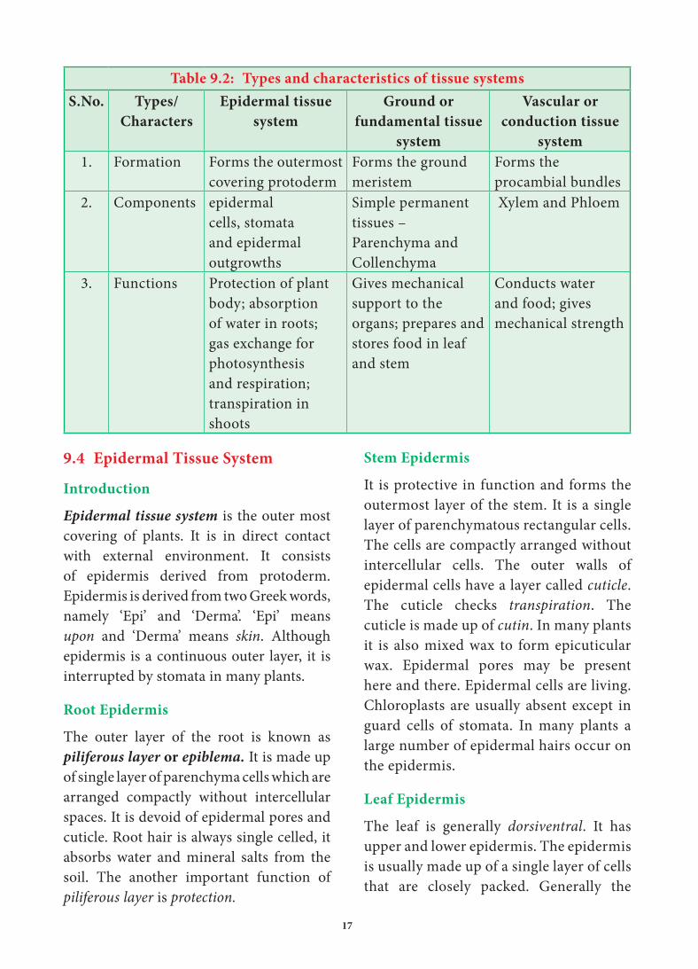
Table 9.2: Types and characteristics of tissue systems
S.No. Types/
Characters
Epidermal tissue
system
Ground or
fundamental tissue
system
Vascular or
conduction tissue
system
1. Formation Forms the outermost covering protoderm
Forms the ground meristem
Forms the procambial bundles
2. Components epidermal cells, stomata and epidermal outgrowths
Simple permanent tissues – Parenchyma and Collenchyma
Xylem and Phloem
3. Functions Protection of plant body; absorption of water in roots; gas exchange for photosynthesis and respiration; transpiration in shoots
Gives mechanical support to the organs; prepares and stores food in leaf and stem
Conducts water and food; gives mechanical strength
9.4 Epidermal Tissue System
Introduction
Epidermal tissue system is the outer most covering of plants. It is in direct contact with external environment. It consists of epidermis derived from protoderm.Epidermis is derived from two Greek words, namely ‘Epi’ and ‘Derma’. ‘Epi’ means upon and ‘Derma’ means skin. Although epidermis is a continuous outer layer, it is interrupted by stomata in many plants.
Root Epidermis
The outer layer of the root is known as piliferous layer or epiblema. It is made up of single layer of parenchyma cells which are arranged compactly without intercellular spaces. It is devoid of epidermal pores and cuticle. Root hair is always single celled, it absorbs water and mineral salts from the soil. The another important function of piliferous layer is protection.
18
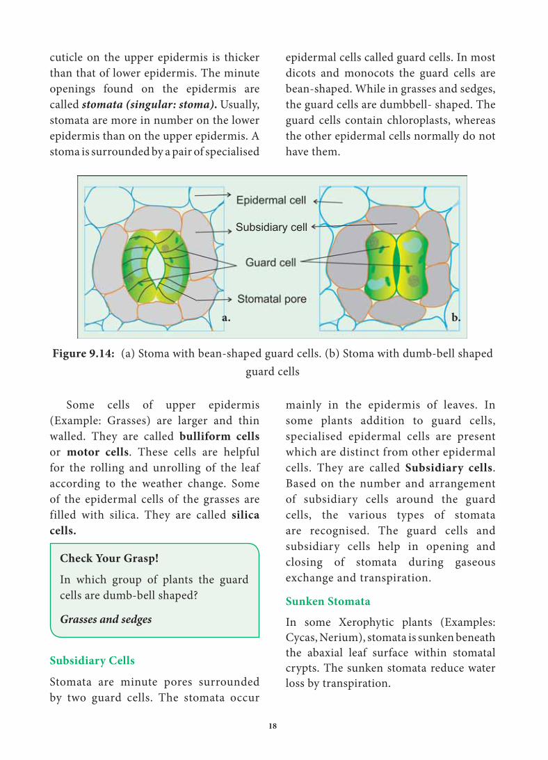
epidermal cells called guard cells. In most dicots and monocots the guard cells are bean-shaped. While in grasses and sedges, the guard cells are dumbbell- shaped. The guard cells contain chloroplasts, whereas the other epidermal cells normally do not have them.
cuticle on the upper epidermis is thicker than that of lower epidermis. The minute openings found on the epidermis are called stomata (singular: stoma). Usually, stomata are more in number on the lower epidermis than on the upper epidermis. A stoma is surrounded by a pair of specialised
Subsidiary cell
Figure 9.14: (a) Stoma with bean-shaped guard cells. (b) Stoma with dumb-bell shaped
guard cells
Some cells of upper epidermis (Example: Grasses) are larger and thin walled. They are called bulliform cells
or motor cells. These cells are helpful for the rolling and unrolling of the leaf according to the weather change. Some of the epidermal cells of the grasses are filled with silica. They are called silica
cells.
Check Your Grasp!
In which group of plants the guard cells are dumb-bell shaped?
Grasses and sedges
Subsidiary Cells
Stomata are minute pores surrounded by two guard cells. The stomata occur
mainly in the epidermis of leaves. In some plants addition to guard cells, specialised epidermal cells are present which are distinct from other epidermal cells. They are called Subsidiary cells. Based on the number and arrangement of subsidiary cells around the guard cells, the various types of stomata are recognised. The guard cells and subsidiary cells help in opening and closing of stomata during gaseous exchange and transpiration.
Sunken Stomata
In some Xerophytic plants (Examples: Cycas, Nerium), stomata is sunken beneath the abaxial leaf surface within stomatal crypts. The sunken stomata reduce water loss by transpiration.
b.a.

20
main zones – cortex, pericycle and pith. It is classified into extrastelar ground tissue (Examples: cortex and endodermis) and intrastelar ground tissue (Examples: pericycle, medullary ray and pith)
Extrastelar Ground Tissue
The ground tissues present outside the stele is called extrastelar ground tissue. (Cortex)
Intrastelar Ground Tissue
The ground tissues present within the stele are called intrastelar ground tissues. (pericycle, medullary rays and pith).
Different Components of Ground
Tissue Systems are as follows
Hypodermis
One or two layers of continuous or discontinuous tissue present below the epidermis, is called hypodermis. It is protective in function.
In dicot stem, hypodermis is generally collenchymatous, whereas in monocot stem, it is generally sclerenchymatous. In many plants collenchyma form the hypodermis.
General Cortex
The Cortex occurs between the epidermis and pericycle. Cortex is a few to many layers in thickness, In most cases, it is made up of parenchymatous tissues. Intercellular spaces may or may not be present.
The cortical cells may contain non living inclusions of starch grains, oil, tannins and crystals.
Sometimes in young stem, chloroplasts develop in peripheral cortical cells, which is called chlorenchyma.
Prickles
Prickles, are one type of epidermal emergences with no vascular supply. They are stiff and sharp in appearance. (Example: Rose).
Functions of Epidermal
Tissue System
1. This system in the shoot checks excessive loss of water due to the presence of cuticle.
2. Epidermis protects the underlying tissues.
3. Stomata is involved in transpiration and gaseous exchange.
4. Trichomes are also helpful in the dispersal of seeds and fruits, and provide protection against animals.
5. Prickles also provide protection against animals and they also check excessive transpiration
6. In some rose plants they also help in climbing.
7. Glandular hairs repel herbivorous animals.
9.5 Fundamental Tissue System
The ground or fundamental tissue system constitutes the main body of the plants. It includes all the tissues except epidermis and vascular tissues. In monocot stem, ground tissue system is a continuous mass of parenchymatous tissue in which vascular bundles are found scattered. Hence ground tissue is not differentiated into cortex, endodermis, pericycle and pith. Generally in dicot stem, ground tissue system is differentiated into three
Figure 9.17:
Prickles

21
In the leaves, the ground tissue consists of chlorenchyma tissues. This region is called mesophyll. In hydrophytes, cortex is Aerenchymatous (with air cavities).
Its general function is storage of food as well as providing mechanical support to organs.
Endodermis
The cells of this layer are barrel shaped and arranged compactly without intercellular spaces.
Endodermis is the innermost cortical layer that separates cortex from the stele. This layer may be a true endodermis as in root or it is an endodermis like layer in stems. This layer is morphologically homologous to the endodermis found in the root.
The cells of endodermis like layer had living cells containing starch grains. Hence it is known as starch sheath. In true root endodermis, radial and inner tangential walls of endodermal cells possess thickenings of lignin, suberin and
some other carbohydrates in the form of strips they are called casparian strips.
The endodermal cells, which are opposite to the protoxylem elements, are thin walled without casparian strips. These cells are called passage cells. Their function is to transport water and dissolved salts from the cortex to the protoxylem.
Water cannot pass through other endodermal cells due to casparian strips. The main function of casparian strips in the endodermal cells is to prevent the re-entry of water into the cortex once water entered the xylem tissue.
The other suberized cells acts as water-tight layer between vascular and non-vascular regions to check the loss of water.
Pericycle
Pericycle is single or few layered parenchymatous found inner to the endodermis. It is the outermost layer of the stele. Rarely thick walled sclerenchymatous. In angiosperms, pericycle gives rise to lateral roots.
Pith or Medulla
The central part of the ground tissue is known as pith or medulla. Generally this is made up of thin walled parenchyma cells with intercellular spaces. The cells in the pith generally stores starch, fatty substances, tannins, phenols, calcium oxalate crystals, etc.
Albuminous Cells: The cytoplasmic nucleated parenchyma, is associated with the sieve cells of Gymnosperms. Albuminous cells in Conifers are analogous to companion cells of Angiosperms. It also called as strasburger cells.
9.6 Vascular Tissue System
This section deals with the vascular tissue system of gymnosperms and angiosperms stems and roots.The vascular tissue system consists of xylem and phloem. The elements of xylem and phloem are always organized in groups. They are called vascular bundles.
The stems of both groups have an eustele while roots are protostele. In eustelic organization, the stele contains usually a ring of vascular bundles separated by interfascicular region or medullary ray
The structural and organizational variation in vascular bundles is shown below.

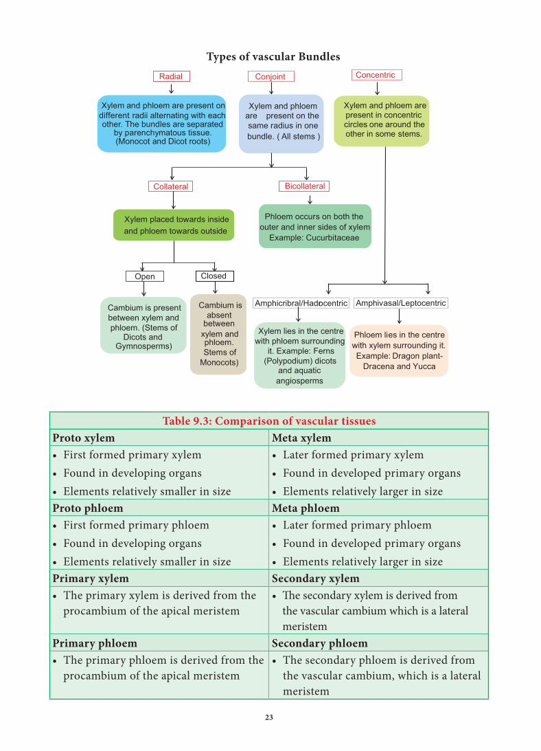
23
Xydifferent
lem and phloem are present onradii alternating with each
other. The bundles are separatedby parenchymatous tissue.(Monocot and Dicot roots)
Xylem and phloemare present on thesame radius in onebundle. ( All stems )
Xylem and phloem arepresent in concentriccircles one around theother in some stems.
Xylem placed towards insideand phloem towards outside
Phloem occurs on both theouter and inner sides of xylem
Example: Cucurbitaceae
Cambium isabsent
betweenxylem andphloem.Stems of
Monocots)
Cambium is presentbetween xylem andphloem. (Stems of
Dicots andGymnosperms)
Xylem lies in the centrewith phloem surrounding
it. Example: Ferns(Polypodium) dicots
and aquaticangiosperms
Phloem lies in the centrewith xylem surrounding it.Example: Dragon plant-
Dracena and Yucca
Amphivasal/Leptocentric
Closed
Amphicribral/Hadrocentric
Open
Collateral Bicollateral
Radial Conjoint Concentric
Table 9.3: Comparison of vascular tissues
Proto xylem Meta xylem
• First formed primary xylem• Found in developing organs• Elements relatively smaller in size
• Later formed primary xylem• Found in developed primary organs• Elements relatively larger in size
Proto phloem Meta phloem
• First formed primary phloem• Found in developing organs• Elements relatively smaller in size
• Later formed primary phloem• Found in developed primary organs• Elements relatively larger in size
Primary xylem Secondary xylem
• The primary xylem is derived from the procambium of the apical meristem
• The secondary xylem is derived from the vascular cambium which is a lateral meristem
Primary phloem Secondary phloem
• The primary phloem is derived from the procambium of the apical meristem
• The secondary phloem is derived from the vascular cambium, which is a lateral meristem
Types of vascular Bundles
24
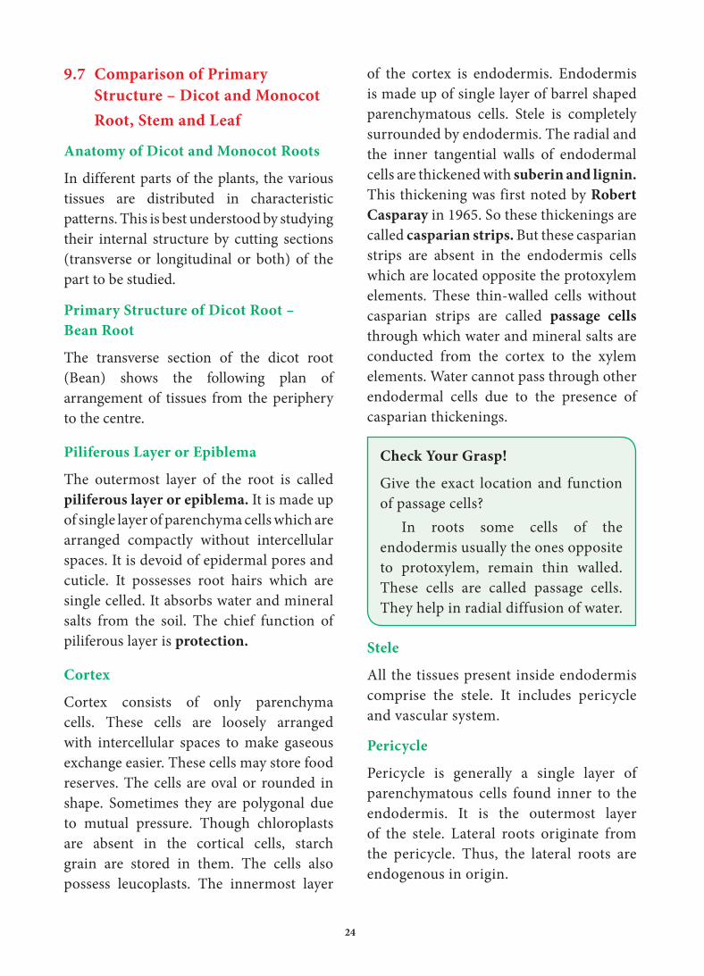
of the cortex is endodermis. Endodermis is made up of single layer of barrel shaped parenchymatous cells. Stele is completely surrounded by endodermis. The radial and the inner tangential walls of endodermal cells are thickened with suberin and lignin.
This thickening was first noted by Robert Casparay in 1965. So these thickenings are called casparian strips. But these casparian strips are absent in the endodermis cells which are located opposite the protoxylem elements. These thin-walled cells without casparian strips are called passage cells
through which water and mineral salts are conducted from the cortex to the xylem elements. Water cannot pass through other endodermal cells due to the presence of casparian thickenings.
Check Your Grasp!
Give the exact location and function of passage cells?
In roots some cells of the endodermis usually the ones opposite to protoxylem, remain thin walled. These cells are called passage cells. They help in radial diffusion of water.
Stele
All the tissues present inside endodermis comprise the stele. It includes pericycle and vascular system.
Pericycle
Pericycle is generally a single layer of parenchymatous cells found inner to the endodermis. It is the outermost layer of the stele. Lateral roots originate from the pericycle. Thus, the lateral roots are endogenous in origin.
9.7 Comparison of Primary
Structure – Dicot and Monocot
Root, Stem and Leaf
Anatomy of Dicot and Monocot Roots
In different parts of the plants, the various tissues are distributed in characteristic patterns. This is best understood by studying their internal structure by cutting sections (transverse or longitudinal or both) of the part to be studied.
Primary Structure of Dicot Root –
Bean Root
The transverse section of the dicot root (Bean) shows the following plan of arrangement of tissues from the periphery to the centre.
Piliferous Layer or Epiblema
The outermost layer of the root is called piliferous layer or epiblema. It is made up of single layer of parenchyma cells which are arranged compactly without intercellular spaces. It is devoid of epidermal pores and cuticle. It possesses root hairs which are single celled. It absorbs water and mineral salts from the soil. The chief function of piliferous layer is protection.
Cortex
Cortex consists of only parenchyma cells. These cells are loosely arranged with intercellular spaces to make gaseous exchange easier. These cells may store food reserves. The cells are oval or rounded in shape. Sometimes they are polygonal due to mutual pressure. Though chloroplasts are absent in the cortical cells, starch grain are stored in them. The cells also possess leucoplasts. The innermost layer


26
Cortex
The cortex is homogenous. i.e. the cortex is made up of only one type of tissue called parenchyma. It consists of many layers of thin-walled parenchyma cells with lot of intercellular spaces. The function of cortical cells is storage. Cortical cells are generally oval or rounded in shape. Chloroplasts are absent in the cortical cells, but they store starch. The cells are living and possess leucoplasts. The inner layer of the cortex is endodermis. It is composed of single layer of barrel shaped parenchymatous cells. This forms a complete ring around the stele. There is a band like structure made of suberin
and lignin present in the radial and inner tangential walls of the endodermal cells. They are called casparian strips named after casparay who first noted the strips. The endodermal cells, which are opposite the protoxylem elements, are thin walled without casparian strips. These cells are called passage cells. Their function is to
transport water and dissolved salts from the cortex to the xylem. Water cannot pass through other endodermal cells due to casparian strips. The main function of casparian strips in the endodermal cells is to prevent the re-entry of water into the cortex once water entered the xylem tissue.
Stele
All the tissues inside the endodermis comprise the stele. This includes pericycle, vascular system and pith.
Pericycle
Pericycle is the outermost layer of the stele and lies inner to the endodermis. It consists of single layer of parenchymatous cells.
Vascular System
Vascular tissues are seen in radial arrangement. The number of protoxylem groups is many. This arrangement of xylem is called polyarch. Xylem is in
Anatomical differences between dicot root and monocot root
S.No. Characters Dicot root Monocot root
1. Pericyle Gives rise to lateral roots, phellogen and a part of vascular cambium.
Gives rise to lateral roots only.
2. Vascular tissue Usually limited number of xylem and phloem strips.
Usually more number of xylem and phloem strips,
3. Conjunctive tissue
Parenchymatous; Its cells are differentiated into vascular cambium.
Mostly sclerenchymatous but sometimes parenchymatous. It is never differentiated in to vascular cambium.
4. Cambium It appears as a secondary meristem at the time of secondary growth.
It is altogether absent.
5. xylem Usually tetrach Usually polyarch

27
exarch condition, the tissue which is present between the xylem and the phloem, is called conjunctive tissue. In maize, the conjunctive tissue is made up of sclerenchymatous tissue.
Pith
The central portion is occupied by a large pith. It consists of thin-walled parenchyma cells with intercellular spaces. These cells are filled with abundant starch grains.
Anatomy of Dicot and Monocot Stems
The transverse section of the dicot stem [sunflower] shows the following plan of arrangement of tissues from the periphery to the centre.
Epidermis
It is protective in function and forms the outermost layer of the stem. It is a single layer of parenchymatous rectangular cells. The cells are compactly arranged without intercellular spaces. The outer walls of epidermal cells have a layer called cuticle. The cuticle checks the transpiration. The cuticle is made up of waxy substance known as cutin. Stomata may be present here and there. Epidermal cells are living. Chloroplasts are usually absent. A large number of multicellular hairs occur on the epidermis.
Cortex
Cortex lies below the epidermis. The cortex is differentiated into three zones. Below the epidermis, there are few layers of collenchyma cells. This zone is called hypodermis. It gives mechanical strength of the Stem. These cells are living and thickened at the corners.
Inner to the hypodermis, a few layers of collenchyma cells are present. This zone is called hypodermis. It gives mechanical strength to the stem. These cells are living and thickened at the corners. Inner to the hypodermis, a few layers of chlorenchyma cells are present with conspicuous intercellular spaces. This region performs photosynthesis. Some resin ducts also occur here. The third zone is made up of parenchyma cells. These cells store food materials. The innermost layer of the cortex is called endodermis. The cells of this layer are barrel shaped and arrange compactly without intercellular spaces. Since starch grains are abundant in these cells, this layer is also known a starch sheath. This layer is morphologically homologous to the endodermis found in the root. In most of the dicot stems, endodermis with casparian strips is not developed.
Check Your Grasp!
Why the endodermis in dicot stem is also referred to as the starch sheath?
The cells of the endodermis are rich in starch grains and thus this layer is also referred to as the starch sheath.
Stele
The central part of the stem inner to the endodermis is known as stele. It consists of pericyle, vascular bundles and pith. In dicot stem, vascular bundles are arranged in a ring around the pith. This type of stele is called eustele.
Pericycle
Pericycle is the layers of cells that occur between the endodermis and vascular bundles. In the stem of sunflower


30
Table 9.4: Anatomical differences between dicot stem and monocot stem
S.No. Characters Dicot Stem Monocot Stem
1. Hypodermis Collenchymatous Sclerenchymatous2. Ground tissue Differentiated into cortex,
endodermis and pericycle and pith
Not differentiated, but it is a continuous mass of parenchyma.
3. Starch Sheath Present Absent4. Medullary rays Present Absent5. Vascular
bundles(a) Collateral and open (a) Collateral and closed(b) Arranged in a ring (b) Scattered in ground tissue(c) Secondary growth occurs (c) Secondary growth usually
does not occur.
Table 9.5: Anatomical differences between root and stem
S.No. Characters Root Stem
1. Epidermis Absence of cuticle and epidermal pores.
Presence of cuticle and epidermal pores.
Presence of unicellular root hairs.
Presence of unicellular and multicellular trichomes
2. Outer Cortical cells
Chlorenchyma absent Chlorenchyma present
3. Endodermis Well defined ill-defined or absent.4. Vascular
bundlesRadial arrangement Conjoint arrangement
5. Xylem Exarch Endarch
Anatomy of a Dicot and Monocot Leaves
Leaves are very important vegetative organs. They are mainly concerned with photosynthesis and transpiration. Like stem and roots, leaves also have the three tissue system – dermal, ground and vascular. The dermal tissue system consists of an upper and lower epidermis. The ground tissue system that lies between the epidermal layers of leaf is known as mesophyll tissue. Often it is differentiated into palisade parenchyma on the adaxial (upper) side and spongy parenchyma on the abaxial (lower) side.
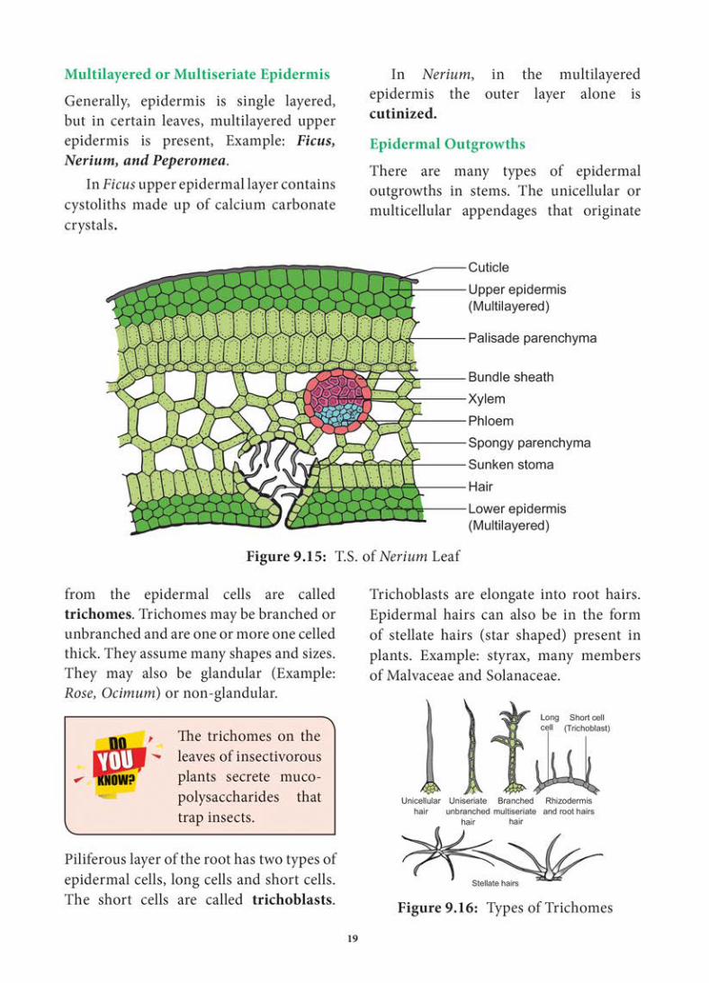
In dorsiventral leaves the mesophyll is differentiated into palisade and spongy parenchyma, the former occurring on the upper side and the later on the lower side Example: Sunflower. In isobilateral leaf palisade is present on both sides of the leaf and inbetween them spongy parenchyma is present. Example: Nerium. In some plants Example: Ficus calcium crystals are present. There are also leaves where spongy tissue alone is present in some epidermal cells Example: Grasses.
The presence of air spaces is a special feature of spongy cells. They facilitate the
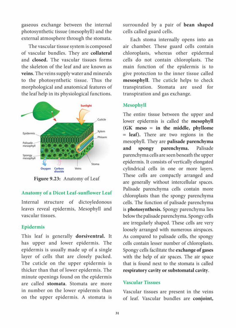

32
Collateral and closed. Xylem is present towards the upper epidermis, while the phloem towards the lower epidermis. Vascular bundles are surrounded by a compact layer of parenchymatous cells called bundle sheath or border
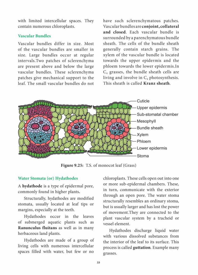
parenchyma.
Xylem consists of metaxylem and protoxylem elements. Protoxylem is present towards the upper epidermis,while the phloem consists of sieve tubes, companion cells and phloem parenchyma. Phloem fibres are absent. Xylem consists of vessels and xylem parenchyma. Tracheids and xylem fibres are absent.
CuticleUpper epidermisPalisade parenchymaProtoxylemMetaxylemSpongy parenchymaPhloemBundle sheathStomaEpidermal hairLower epidermisRespiratory cavity
Figure 9.24: T.S. of Dicot Leaf (Sunflower)
Anatomy of a Monocot Leaf – Grass Leaf
A transverse section of a grass leaf reveals the following internal structures.
Epidermis
The leaf has upper and lower epidermis. They are made up of a single layer of thin walled cells. The outer walls are covered by thick cuticle.
The number of stomata is more or less equal on both the epidermis. The stomata is surrounded by dumb – bell shaped
guard cells. The guard cells-contain chloroplasts, whereas the other epidermal cells do not have them.
Some special cells surround the guard cells. They are distinct from other epidermal cells.
These cells are called subsidiary cells.
Some cells of upper epidermis are large and thin walled. They are called bulliform
cells or motor cells. These cells are helpful for the rolling and unrolling of the leaf according to the weather change.
Some of the epidermal cells of the grass are filled with silica. They are called silica cells.
Mesophyll
The ground tissue that is present between the upper and lower epidermis of the leaf is called mesophyll. Here, the mesophyll is not differentiated into palisade and
spongy parenchyma. All the mesophyll cells are nearly isodiametric and thin walled. These cells are compactly arranged

34
Differences Between Stomata and
Hydathodes
Stomata Hydathodes
Occur in epidermis of leaves, young stems.
Occur at the tip or margin of leaves that are grown in moist shady place.
Stomatal aperture is guarded by two guard cells.
Aperture of hydathodes are surrounded by a ring of cuticularized cells.
The two guard cells are generally surrounded by subsidiary cell.
Subsidiary cells are absent.
Opening and closing of the stomatal aperture is regulated by guard cells.
Hydathode pores remain always open.
These are involved in transpiration and exchange of gases.
These are involved in guttation.
Can mangroove trees
grow in salt water?
These amazing trees and shrubs cope with
salt. Salt water can kill Plants, so mangroves must extract fresh water from the sea water that surrounds them. Many mangrove species survive by filtering out as much as 90 percent of the salt found in seawater as it enters their roots.
Mangrove excrete salt through glands in their leaves.
Halophiles
• Plants that grow in salty environment are called Halophiles.
• Plant growth in saline habitat developed numerous adaptations to salt stress. The secretion of ions by salt glands is the best known mechanism for regulating the salt content of plant shoots.
• Salt glands typically are found in halophytes. (Plants that grow in saline environments)
Figure 9.26: Halophytes
Figure 9.27: Removes excess salts through special salt glands on leaves
35
Summary
A Tissue is a group of cells that are alike in origin, structure and function.There are two principal groups: (1) Meristematic tissues and (2) Permanent tissues. Meristematic tissues comprise of self-perpetuating cells. Meristems are classified into several types on the basis of position, origin, function and activity. Many anatomists illustrated the root and shoot apical meristems on the basis of the type and arrangement and accordingly proposed many theories. The permanent tissues normally develop from apical meristem. They are classified into two types: 1)Simple permanent tissues and 2)Complex permanent tissues. Simple tissues are composed of a single type of cells only. It is of three types: (1) Parenchyma (2) Collenchyma and (3) Sclerenchyma. A complex tissue is a tissue with several types of cells but all of them function together as a single unit. It is of two types – xylem and phloem. Secretory tissues produce different types of chemicals. Some are in the form of enzymes, hormones, rubber, gum etc.
The tissues can be classified on the basis of their function, structure and location into epidermal tissue system, ground tissue system and vascular tissue system. Epidermal tissue system develops as the outermost covering of the entire plant body. It consists of epidermal cells and associated structures. All tissues except epidermis and vascular tissues constitute the ground tissue. The vascular tissue system is formed of vascular bundles.
In the primary structure, the outermost layer of the root is called piliferous layer. Cortex consists of only parenchyma cells. All the tissues present inside endodermis comprise the stele. In dicot (Example: bean)
root, xylem is tetrach. Its phloem patch consists of sieve tubes, companion cells and phloem parenchyma. In monocot (Example: maize) root, xylem is polyarch.
In dicot (Example: sunflower) stem, stele is eustele type and its vascular bundles are wedge shaped, conjoint, collateral, open and endarch. In monocot stem (Example: maize) vascular bundles are scattered and skull shaped, conjoint, collateral, closed and endarch.
In dicot (Example: sunflower) and monocot (Example: grass) leaves vascular bundles are conjoint, collateral and closed.
Hydathodes discharge liquid water with various dissolved substances from the interior of the leaf to its surface. Plants that grow in salty environment are called halophiles. Salt glands typically are found in halophytes.
Evaluation
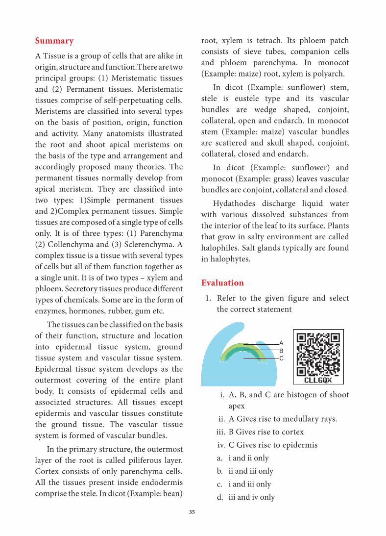
1. Refer to the given figure and select the correct statement
CBA
i. A, B, and C are histogen of shoot apex
ii. A Gives rise to medullary rays.iii. B Gives rise to cortexiv. C Gives rise to epidermisa. i and ii only b. ii and iii only c. i and iii only d. iii and iv only

36
2. Read the following sentences and identify the correctly matched sentences.i. In exarch condition, the
protoxylem lies outside of metaxylem.
ii. In endarch condition, the protoxylem lie towords the centre.
iii. In centarch condition, metaxylem lies in the middle of the protoxylem.
iv. In mesarch condition, protoxylem lies in the middle of the metaxylem.
a. i, ii and iii only b. ii, iii and iv only c. i, ii and iv only d. All of these
3. In Gymnosperms, the activity of sieve tubes are controlled bya. Nearby sieve tube members. b. Phloem parenchyma cellsc. Nucleus of companion cells. d. Nucleus of albuminous cells.
4. When a leaf trace extends from a vascular bundle in a dicot stem, what would be the arrangement of vascular tissues in the veins of the leaf?a. Xylem would be on top and the
phloem on the bottomb. Phloem would be on top and the
xylem on the bottomc. Xylem would encircle the phloemd. Phloem would encircle the xylem
5. Grafting is successful in dicots but not in monocots because the dicots havea. Vascular bundles arranged in a ringb. Cambium for secondary growthc. Vessels with elements arranged end
to endd. Cork cambium
6. Why the cells of sclerenchyma and tracheids become dead?
7. Explain sclereids with their types.8. What are sieve tubes ?explain.9. Distinguish the anatomy of dicot root
from monocot root.10. Distinguish the anatomy of dicot stem
from monocot stem.
37
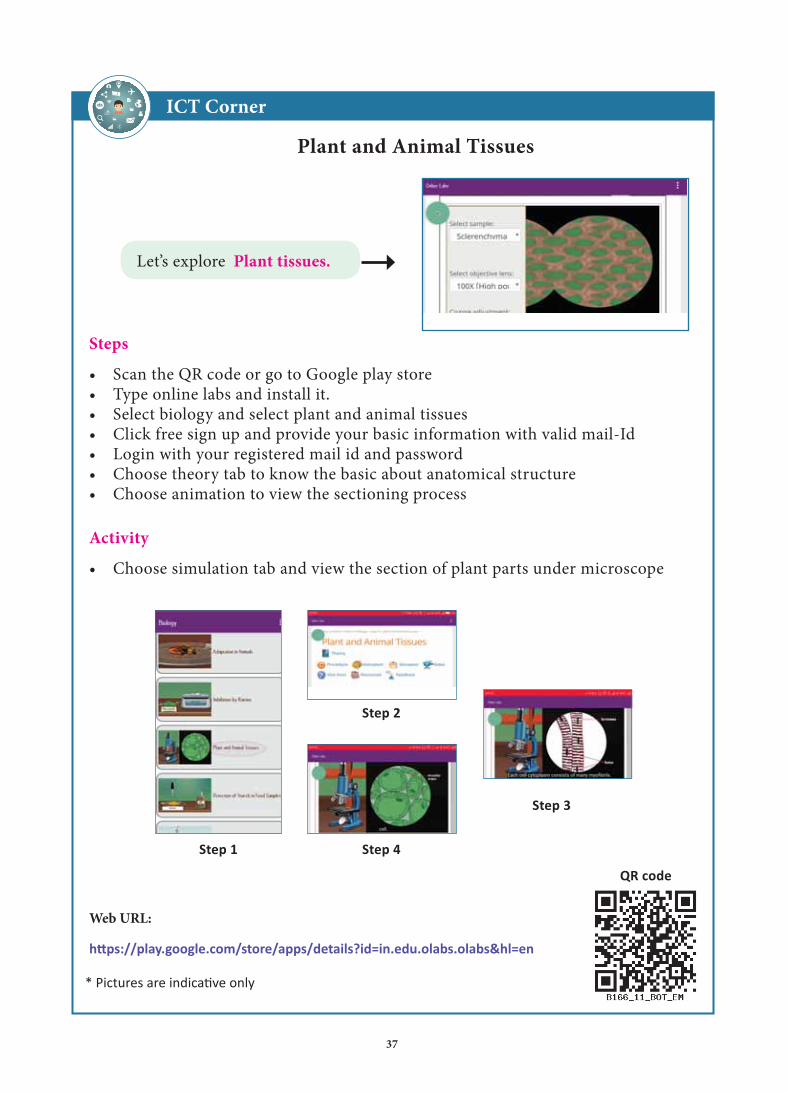
t
Web URL:
ICT Corner
Plant and Animal Tissues
Steps
• Scan the QR code or go to Google play store • Type online labs and install it.• Select biology and select plant and animal tissues• Click free sign up and provide your basic information with valid mail-Id• Login with your registered mail id and password• Choose theory tab to know the basic about anatomical structure • Choose animation to view the sectioning process
Let’s explore Plant tissues.
Activity
• Choose simulation tab and view the section of plant parts under microscope